Embed Size (px)
Citation preview

WATER & SODIUM BALANCE – An Overview
UNIVERSITY OF PNG SCHOOL OF MEDICINE AND HEALTH SCIENCES
DISCIPLINE OF BIOCHEMISTRY & MOLECULAR BIOLOGY PBL MBBS 3 / BMLS 3 / BDS 4 LECTURES
VJ Temple
1

WATER (FLUID) BALANCE (STEADY STATE)
• Amount of daily water intake varies among individuals ;
• Amount of daily water loss varies among individuals;
• Water loss is normally seen as changes in volume of urine production;
• Urine Flow Rate can vary widely in a very short time;
• To maintain water balance:
• Amount of daily water intake must equal amount of daily water loss,
• Disruption of balance may cause:
• Net water gain: Over hydration; or
• Net water loss: Dehydration2

How much fluid (water) is contained in the body?
• Water/Fluid is a major body constituent;
• An average person (Wt 70 kg) contains about 42 liters of Total Body Water (TBW);
• TBW is about 60% of the total body weight;
• TBW is separated into two major compartment;
• Extra-Cellular Fluid Compartment (ECF)
• Intra-Cellular Fluid Compartment (ICF)
• Fig. 1: Water tank model illustration ECF, ICF, TBW
3

Fig. 1: Schematic diagram of water tank model to illustrate body fluid compartments [1]
4

What are the major fluid compartments in the body?
• Major fluid compartments (Fig. 2):
• Intra-Cellular Fluid Compartment (ICF): Volume of Fluid Inside Cells;
• ICF constitute about 66.6% of TBW
• Extra-Cellular Fluid Compartment (ECF): Volume of Fluid Outside Cells;
• ECF constitute about 33.3% of TBW
• ECF is made up of Plasma and Interstitial Fluid
• Plasma is about 25% of ECF
• Interstitial Fluid is about 75% of ECF
5

6

What are the major sources and routes of fluid intake?
• Some major sources of fluid intake:
• Water Drinking;
• Water contained in our various foodstuffs;
• Metabolic water;
7

What are some of the major routes in the body for fluid loss?
• Some major routes of fluid loss: • Urinary loss, Fecal loss • Insensible water loss – such as evaporation from the
Respiratory Tract and Skin Surface (not including sweat which is sensible since it has a purpose)
• Sweat Losses –• At normal room temperature, sweating accounts for
about 25% of heat losses; • In cold environments, H2O losses in sweat decreases;• In warm environments, or with exercise, sweat losses
increases;
• Pathological losses: vascular bleeding, vomiting, and diarrhea;
8

What are some of the consequences of fluid loss?
• Selective loss of fluid from either ICF or ECF compartments gives rise to distinct signs and symptoms:
• Loss of ICF can cause Cellular Dysfunction: resulting in Lethargy, Confusion and Coma;
• Loss of ECF can lead to Circulatory Collapse, Shock, Renal shutdown;
• Loss of TBW produces similar effects as loss of ICF or ECF;
• Signs of (substantial) fluid loss is spread across ICF & ECF;
9
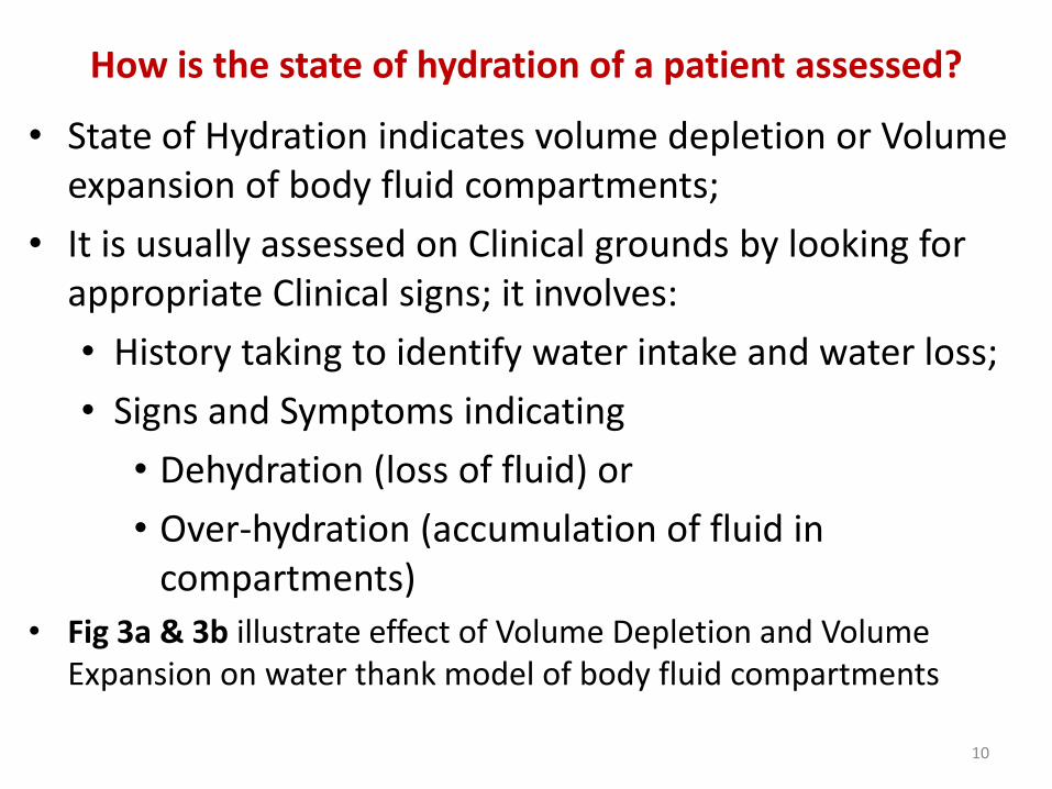
How is the state of hydration of a patient assessed?
• State of Hydration indicates volume depletion or Volume expansion of body fluid compartments;
• It is usually assessed on Clinical grounds by looking for appropriate Clinical signs; it involves:
• History taking to identify water intake and water loss;
• Signs and Symptoms indicating
• Dehydration (loss of fluid) or
• Over-hydration (accumulation of fluid in compartments)
• Fig 3a & 3b illustrate effect of Volume Depletion and Volume Expansion on water thank model of body fluid compartments
10

Fig. 3a: Dehydration: Loss of fluid in ICF & ECF due to increased urinary output; Fig. 3b: Overhydration: Increased fluid intake resulting in increased fluid volume in ICF and ECF [1]
11

How is water balance regulated?
• Water balance is regulated by Arginine Vasopressin (AVP; also called Anti-Diuretic Hormone, ADH);
• AVP is a hormone produced by Posterior Pituitary Gland;
• AVP tightly regulates water excretion by the kidneys;
• Osmolality in ICF is equal to that in ECF;
• Specialized cells in Hypothalamus are involved in maintaining the Osmolality between ICF and ECF;
• When the Hypothalamus detects differences in the Osmolality between ICF and ECF it regulates the secretion of AVP from Posterior Pituitary gland;
12

• Regulation is as follows:
• A rising Osmolality promotes the secretion of AVP,
• A declining Osmolality switches off the secretion of AVP,
• AVP causes water to be retained by the kidneys,
• Fluid deprivation results in stimulation of AVP secretion causing reduction in Urine Flow Rate to as little as 0.5 ml/min in order to conserve body water;
• Within ONE hour after drinking about 2 liters of water, the Urine Flow Rate may rise to about 15 ml/min as AVP secretion is Shut Down;
• By regulating water Excretion or Retention, AVP maintains normal concentrations of Electrolytes within the body (Figure 4)
13

Fig. 4: Regulation of water balance by Arginine Vasopressin (AVP):Increased Osmolality activates production of AVP and reduces urine flow rate;Decreased Osmolality inhibits production of AVP and increases urine flow rate; [1]
14

SODIUM BALANCE
• Amount consumed should equal amount loss per day;
• Total Sodium in the body is made up of:
• Non-Exchangeable Sodium = 25% of Total Sodium;
• Exchangeable sodium = 75% of Total Sodium;
• Non-Exchangeable Sodium is in tissues: Bone and Cartilage and has slow turnover rate;
• Most Exchangeable Sodium is in ECF;
• Exchangeable Sodium circulates in Plasma as Na+;
• Normal range of Sodium in plasma = 135 to 145mmol/L;
• Plasma Conc. Na+ ions does not indicate Sodium balance;
• Plasma Conc. Na+ primarily reflects body water content;
15

Sodium intake and Sodium loss:
Sodium Intake:
• Varies among individuals depending on Habits, Taste, Availability;
• Health individuals: Total body sodium does not change even if intake falls to 5mmol/day or increases to 750mmol/day;
Sodium Loss:
• Loss of sodium varies among individuals;
• Sodium is excreted mainly via the Kidneys;
• Some is lost in sweat (5 mmol/day) and in feces (5 mmol/day);
• GIT is the major route of pathological Sodium loss;
• Diarrhea and vomiting may result in death from Salt and Water Depletion in Pediatric Cases;
16

What factors regulate Sodium excretion?
• Sodium Excretion is regulated by:
• Intrinsic Renal Mechanisms,
• Suppression of Aldosterone Secretion,
• Stimulation of Secretion of Atrial Natriuretic Factor (ANF)
17

What is the role of Aldosterone in regulation of Sodium Balance?
• Aldosterone is a hormone produce in the Adrenal Cortex;
• Aldosterone decreases Urinary Sodium Excretion by Increasing Re-absorption of Na+ in Renal Tubules in exchange for Tubule excretion of K+ and H+
• Aldosterone also decreases loss of Na+ in Sweat Glands and Mucosal Cells of the Colon, but in normal circumstances these effects are minimal;
18

How is secretion of Aldosterone regulated?
• Major stimulus for secretion of Aldosterone:
• Volume of ECF,
• Osmolality of ECF,
• Specialized cells in Juxtaglomerular Apparatus of Kidneys detect decrease in Blood Pressure and secrete Renin,
• Renin converts Angiotensinogen (produced in Liver) to Angiotensin I;
• Angiotensin I is converted to Angiotensin II by Angiotensin Converting Enzyme (ACP);
• Angiotensin II then act on Adrenal Cortex to produce Aldosterone;
• Aldosterone acts on Kidney Tubules causing reabsorption of Na+ in exchange for excretion of K+, and H+ (Fig. 5)
19

Fig. 5: Regulation of Sodium balance by Aldosterone? [1]
20

What is the role of Atrial Natriuretic Factor (ANF) in regulation of Sodium balance?
• Atrial Natriuretic Factor (ANF) is a polypeptide hormone secreted by Cardiocytes in the Right Atrium of the Heart – thus, it is a Cardiac Hormone;
• ANF increases Urinary Sodium excretion: Natriuresis;
• ANF regulates ECF volume,
• ANF regulates concentration of Sodium in plasma,
21

What are the electrolytes in the ECF and ICF?
• Na+ is the Principal Cation in ECF,
• K+ is the Principal Cation in ICF,
• Proteins and Phosphates are the main Anions in ICF,
• Chloride (Cl-) & Bicarbonate (HCO3-) are the main Anions in ECF,
• Na+ are present at highest concentration in ECF and make the largest contribution to total plasma Osmolality,
• Despite the low amount of K+ in ECF, changes in Plasma K+ conc. is very important and may have life threatening consequences;
• Urea and Creatinine are measured with Plasma Electrolytes because they provide an indication of Renal Function,
• Increase in concentrations of Urea and Creatinine usually indicates a decrease in Glomerular Filtration Rate in the kidneys;
22

How are solute and solvent related to solution?
• Solution is made up of Solute and Solvent,
• Concentration of solution is a ratio of two variables:
• Amount of Solute (e.g., Na+ ions) and
• Amount of Solvent (Water),
• Concentration of solution can change when either or both variables change For example:
• Na+ ion concentration of 140mmol/l may becomes 130mmol/l if the amount of Na+ ions in the solution is reduced or the amount of solvent (water) is increased;
23

What is Osmolality (Osmolarity)?
• Osmolality is the concentration of osmotically active particles in a solution,
• particles that cannot cross semi-permeable membrane
• Water moves easily across cell membrane separating ECF from ICF;
• Osmosis is flow of solvent across semi-permeable membrane from low solute concentration to higher solute concentration,
• Osmotic pressure is the driving pressure for water to change the concentration of osmotically active particles,
• Osmotic pressure is the same on both sides of the cell membrane,
• Osmolality of ICF equals Osmolality of the ECF: Isotonic solutions,
• Water moves across cell membrane to maintain Osmolality of ECF & ICF; even if it causes the cells to shrink or expand in volume,
24

How is Osmolality of Serum or Plasma calculated?
• Concentrations of osmotically active solutes are used:
• Simple formula for calculating Osmolality :
Serum Osmolality = 2 x [Serum Sodium ions] = 2[Na+]• (Unit for Osmolality is mmol/kg, or mOsmol/Kg or mOsmol/L;
Unit for Plasma or Serum Sodium ion is mmol/L);
• Simple formula is used when Concentrations of Plasma Urea and Glucose are within the reference ranges;
• If either or both are abnormally high, then concentration of either or both (in mmol/L) must be used in the calculation of Osmolality;
• NB: Normal Osmolality of Serum or Plasma (and other body fluids except urine) is in the range 285 to 295 mmol/kg(285 to 295 mOsmol/L);
25

Example for calculating Osmolality
Normal Conditions (i.e., Plasma or
Serum concentrations of Urea and
Glucose are within normal range)
• ECF Osmolality can be roughly estimated as:
{Where Posm is plasma Osmolality;.
Since intracellular Osmolarity is the
same as extra-cellular Osmolality
under normal conditions, this also
provides an estimate of
intracellular Osmolality}
26

Example for calculation of Osmolality
Clinical Laboratory Measurement:
• Plasma Osmolarity measured in Clinical laboratory also includes contributions from Glucose and Urea;
• Normally the contribution from Glucose and Urea is small
• Under certain Pathological conditions, the concentrations of these substances can be very high;
• Plasma Osmolality measured in clinical laboratory:
P = 2[Na+] + 2[K+] + [Glucose] + [Urea](P = Plasma or Serum Osmolality)
• Glucose and BUN normally contribute about 5mOsm each (about 2% ) of Plasma Osmolarity measured in the clinical lab
27

How is effective Osmole different from ineffective Osmole?
• Ineffective Osmole:
• Urea crosses the semi-permeable cell membranes just as easily as water, therefore it does not contribute to redistribution of water between ECF and ICF;
• Effective Osmoles:
• Glucose, Na+ and Anions associated with Na+ do not cross the semi-permeable membrane;
• They have concentration gradients across the cell membrane and are osmotically active;
• They determine the distribution of water between ECF and ICF;
28

How is Effective Osmole calculated?
Two ways for calculating Effective Osmole:
• Effective Osmole:
P (effective) = 2[Na+] + [Glucose]
• Effective Osmole:
P (effective) = P (measured) – [Urea]
• (P = plasma or serum Osmolality)
29

What is Osmolal Gap and how is it calculated?
OSMOLAL GAP (OG):
• Difference between Measured Osmolality (MO) and Calculated Osmolality (CO)
Osmolal Gap (OG) = MO – CO
• Large positive OG helps to identify presence in serum of osmotically active substances, such as, Ethanol, Methanol, Iso-propanol, Ethylene Glycol and Acetone
• Proper interpretation of OG also requires knowledge of Anion Gap (AG), and blood pH
Anion Gap = [Na+] – {[HCO3-] + [Cl-]}
30

What is HYPONATRAEMIA?
• Hyponatraemia is a significant fall in the concentration of Na+ ions below the reference range for plasma or serum;
• (what reference range is used for Serum Na+ ion in PMGH?)
• “Hypo-Osmolality” is synonymous with Hyponatraemia
because Na+ ion is the major cation in the ECF in
sufficient amount such that a decrease in concentration
would significantly affect the Osmolality;
31

List two possibilities of Hyponatraemia?
• Hyponatraemia due to Fluid Retention:
• More fluid than normal is retained in the body compartments and dilutes the constituents in ECF causing Hyponatraemia; (Fig. 6a)
• Hyponatraemia due to Loss of Na+ ions :
• When loss of Na+ ions exceeds loss of fluid, Hyponatraemia may result, (Fig. 6b)
• Example: Loss of fluid (vomiting or fistulae) that contain Na+ ions are replaced simply by water;
32

Fig. 6a: Fluid retention in ECF & ICF causing HyponatraemiaFig. 6b: Sodium loss resulting in Hyponatraemia [1]
33

• Water tank model in Fig. 6a & 6b emphasizes that Biochemical observation of Hyponatraemia gives no clear explanation about the Volume of the ECF compartment;
• Both laboratory results of these patients indicate Hyponatraemia, with no indication of fluid retention or loss of Sodium;
• Thus, the courses of Hyponatraemia should be made by proper History taking and Clinical Examination of the Patient, not by assessing the laboratory results alone;
34

• Some patients with reduced ECF volume may present with either Reduced, Increased or Normal Plasma Sodium concentration (Figure 7a, 7b, 7c),
• These diagrams clearly indicates that Clinicians MUST always give greater emphasis and attention to History, Signs and Symptoms of the Patients not to the Laboratory results on plasma Sodium alone;
35

Fig. 7a: Reduced ECF with Hyponatraemia (low plasma Sodium conc.);Fig. 7b: Reduced ECF with Hypernatraemia (high plasma Sodium conc.);Fig. 7c: Reduced ECF with Normal plasma Sodium conc.
36

What are some of the causes of Hyponatraemia with fluid retention?
• Decreased water excretion:
• Examples: Nephrotic Syndrome, Renal Failure;
• Increased Water Intake:
• Examples: Inappropriate IV Saline, Compulsive water drinking
37
TAKE NOTE:
• In general if fluid loss is not apparent from the Clinical history of a patient then the reason for the Hyponatraemia is usually WATER RETENTION;
• Hyponatraemia due to water overload without a decrease in total body Sodium is the commonest Biochemical disturbance encountered in clinical practice;
• Further consideration of Hyponatraemia of this type, depends on whether the patient has Oedema:
• Two possible conditions are:
• Oedematous Hyponatraemia
• Non-Oedematous Hyponatraemia
38

OEDEMATOUS HYPONATRAEMIA• Patients with generalized Oedema have an increase in both
Total Body Sodium and Water:
• Some causes of Oedema:
• Heart Failure: • Effective blood volume may be reduced because pumping
action of the heart is unable to maintain a satisfactory circulation of Blood and ECF;
• Hypo-albuminaemia, • Effective blood volume may be reduced because Hypo-
albuminaemia lowers Plasma Oncotic Pressure, which disrupts normal exchange of solutes and fluid in capillary bed resulting in unsatisfactory circulation of Blood and ECF;
• Albumin makes the biggest contribution to plasma Oncotic pressure;
• Oedema occurs if blood albumin concentration falls very low; 39

• In response to reduced effective blood volume, Aldosterone is secreted and causes Sodium retention to allow the ECF volume to expand;
• Reduction in effective blood volume is one of the Non-Osmotic Stimuli for the secretion of AVP (Arginine Vasopressin) and consequently water is retained;
• Hyponatraemia results from the Retention of relatively more water than Sodium in the ECF;
40

41

What are some of the causes of Hypo-albuminaemia?
• Decreased biosynthesis of albumin due to: • Liver disease causing inadequate biosynthesis of
Albumin;
• Loss of albumin exceeds biosynthetic capacity of liver as occurs in Nephrotic syndrome;
• Malnutrition or Mal-absorption;
• Abnormal distribution or dilution:• Over-hydration or if there is increased capillary
permeability as occurs in Septicaemia;
• Abnormal excretion or degradation:• Nephrotic Syndrome, Protein-losing Enteropathies,
Burns, Haemorrhage and Catabolic states;
42

NON-OEDEMATOUS HYPONATRAEMIA
• Patients with Non-Oedematous Hyponatraemia have normal total body Sodium and exhibit the features of Syndrome of Inappropriate Anti-diuresis (SIAD)
• Patients are Hyponatraemic, Normotensive, have normal Glomerular Filtration Rate (GFR) and normal serum Urea and Creatinine concentration;
• Urine Flow Rate is usually less than 1.5 liter/day;
43

• SIAD may occur in conditions such as:
• Infections, e.g. Pneumonia,
• Malignancy, e.g. Carcinoma of Bowel or Lung,
• Trauma, e.g. Abdominal Surgery,
• Drug-induced, e.g. Thiazide Diuretics, Chlorpropamide
• Patients suffering from any of the above may have Non-Osmotic AVP stimulation and, if they are exposed to excessive water loads, in the form of oral drinks or intravenous glucose solutions, they will become Hyponatraemic;
44

HYPONATRAEMIA DUE TO SODIUM LOSS
• Occurs during Pathological Sodium Loss
• May be from GIT or Urine
• Vomiting (severe and protracted as occurs in Pyloric Stenosis)
• Diarrhoea;
• Fistula
45

46

Urinary loss of Sodium may be due to
• Aldosterone deficiency due to failure of the Adrenal Glands (Addison’s disease);
• Drugs that antagonize Aldosterone action;• Initially in such patients:
• Sodium loss is accompanied by Water loss and Serum Sodium ion concentration remains normal;
• As Sodium loss proceeds, the reduction in ECF and blood volume stimulates AVP secretion;
• Non-osmotic control of AVP secretion overrides osmotic control mechanism;
• Increased AVP secretion causes water retention and thus the patient becomes Hyponatraemic;
• Patient becomes Hyponatraemic because a deficit of Isotonic Sodium-containing fluid is replaced only by water, either Orally or Intravenously;
• In all cases patients should be given Oral Rehydration Solution47

SUMMARY
• Water is lost as Urine and as obligatory “Insensible” losses from skin and lungs;
• Na+ ions may be lost in prolonged vomiting, diarrhea and intestinal fistulae,
• AVP regulates Renal water loss and causes changes in Osmolality of body fluid compartments;
• Aldosterone regulates Renal Na+ ion and Na+ content in ECF,
• Changes in Na+ ion content in ECF cause changes in ECF volume because of the combined actions of AVP and Aldosterone,
• Hyponatraemia due to water retention is the commonest biochemical disturbance encountered in clinical practice,
• In some patients non-osmotic regulation of AVP overrides the osmotic regulatory mechanism, resulting in water retention, which is a non-specific feature of illness,
48

• Patients with Hyponatraemia without oedema, but have normal serum urea and creatinine and blood pressure, have water overload,
• Patients with Hyponatraemia and with Oedema are likely to have both water and sodium overload,
• Hyponatraemia may occur in patient with Gastrointestinal or Renal fluid losses, causing Sodium depletion,
• Low Sodium concentration in serum may occur because water retention is stimulated by increased AVP secretion,
• Patients with Hyponatraemia due to Sodium depletion may show clinical signs of fluid loss such as Hypotension, such patients usually do not have Oedema,
49
REFERENCES
1. A. Gwa, RA Cowan, DJ O’Reilly, MJ Stewart, J Shepherd. “An illustrated colour text: Clinical Biochemistry” 2nd Edition, 1999, Churchill Livingstone, London.
2. G Beckett, S. Walker, P. Rae, P. Ashby. “Lecture Notes: Clinical Biochemistry” 7th Edition, 2008, Blackwell Publishing, Australia.
3. VJ. Temple. “Biochemistry 1001: Review and Viva Voce Questions and Answers Approach”, Sterling Publishers Private Ltd 2012, New Delhi.
50


![Sodium Phytate Presentation.pptx [Read-Only]formulatorsampleshop.com/v/reference/Sodium Phytate Presentation.pdfLaurate (Skin Conditioning Agent), Sodium Benzoate (Preservative), Sodium](https://img.pdfslide.us/doc/110x75/5eb52012fb0f3e0d55767ea6/sodium-phytate-read-onlyformulatorsampleshopcomvreferencesodium-phytate-presentationpdf.jpg)