Embed Size (px)
Citation preview

Walker, S., Maskell, N., Barratt, S., & Thompson, J. (2018).Conservative Management in Traumatic Pneumothoraces: AnObservational Study. Chest, 153(4), 946-953.https://doi.org/10.1016/j.chest.2017.10.015
Peer reviewed versionLicense (if available):CC BY-NC-NDLink to published version (if available):10.1016/j.chest.2017.10.015
Link to publication record in Explore Bristol ResearchPDF-document
This is the accepted author manuscript (AAM). The final published version (version of record) is available onlinevia Elsevier at https://doi.org/10.1016/j.chest.2017.10.015 . Please refer to any applicable terms of use of thepublisher.
University of Bristol - Explore Bristol ResearchGeneral rights
This document is made available in accordance with publisher policies. Please cite only thepublished version using the reference above. Full terms of use are available:http://www.bristol.ac.uk/red/research-policy/pure/user-guides/ebr-terms/

Conservative Management in Traumatic Pneumothoraces: An Observational Study. Total Word Count: 2757
Conservative Management in Traumatic Pneumothoraces: An Observational Study.
Running head: Conservative Management in Traumatic Pneumothoraces
Authors: Walker, Steven P1,2 MBChB. Barratt, Shaney L1, 2 BMBS PhD. Thompson, Julian3,4 FRCA MD(Res) FRCA FFICM. Maskell,
Nick A1,2 DM FRCP FCCP.
Affiliations: 1Academic Respiratory Unit, School of Clinical Sciences, University of Bristol, Bristol, United Kingdom; 2North Bristol Lung Centre, Southmead Hospital, Bristol, United Kingdom 3Intensive Care Unit, Southmead Hospital, Bristol, United Kingdom 4Severn Major Trauma Network Correspondence should be addressed to: Julian Thompson at Intensive care unit, Southmead Hospital, Bristol, United Kingdom. Email [email protected]
Summary conflict of interest statements: No conflict of interest for SW, SB, JT & NM. Funding: No financial disclosures.

Abbreviations
Advanced Trauma Life Support (ATLS)
Computerised tomography (CT)
General Anesthetic (GA)
Glasgow Coma Scale (GCS)
Hazard ratio (HR)
Injury severity score (ISS)
Interquartile range (IQR)
Major Trauma Centre (MTC)
Peripheral Oxygen Saturation (SpO2)
Positive pressure ventilation (PPV)
Standard Deviation (SD)
Systolic blood pressure (SBP)
Trauma Audit and Research Network (TARN)
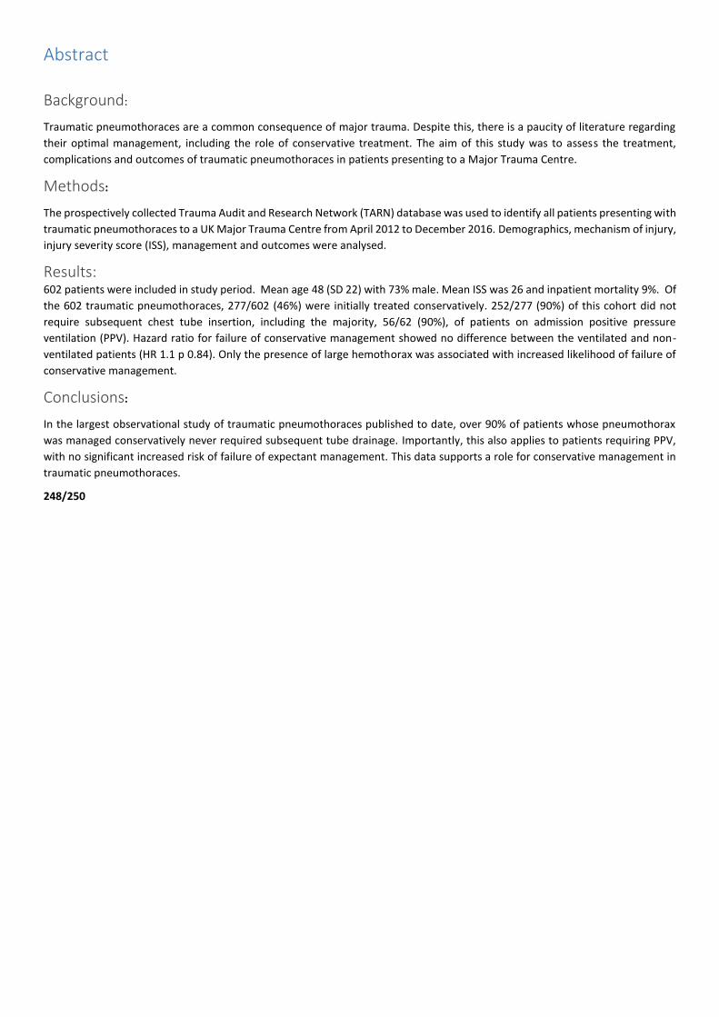
Abstract
Background:
Traumatic pneumothoraces are a common consequence of major trauma. Despite this, there is a paucity of literature regarding
their optimal management, including the role of conservative treatment. The aim of this study was to assess the treatment,
complications and outcomes of traumatic pneumothoraces in patients presenting to a Major Trauma Centre.
Methods:
The prospectively collected Trauma Audit and Research Network (TARN) database was used to identify all patients presenting with
traumatic pneumothoraces to a UK Major Trauma Centre from April 2012 to December 2016. Demographics, mechanism of injury,
injury severity score (ISS), management and outcomes were analysed.
Results: 602 patients were included in study period. Mean age 48 (SD 22) with 73% male. Mean ISS was 26 and inpatient mortality 9%. Of
the 602 traumatic pneumothoraces, 277/602 (46%) were initially treated conservatively. 252/277 (90%) of this cohort did not
require subsequent chest tube insertion, including the majority, 56/62 (90%), of patients on admission positive pressure
ventilation (PPV). Hazard ratio for failure of conservative management showed no difference between the ventilated and non-
ventilated patients (HR 1.1 p 0.84). Only the presence of large hemothorax was associated with increased likelihood of failure of
conservative management.
Conclusions:
In the largest observational study of traumatic pneumothoraces published to date, over 90% of patients whose pneumothorax
was managed conservatively never required subsequent tube drainage. Importantly, this also applies to patients requiring PPV,
with no significant increased risk of failure of expectant management. This data supports a role for conservative management in
traumatic pneumothoraces.
248/250
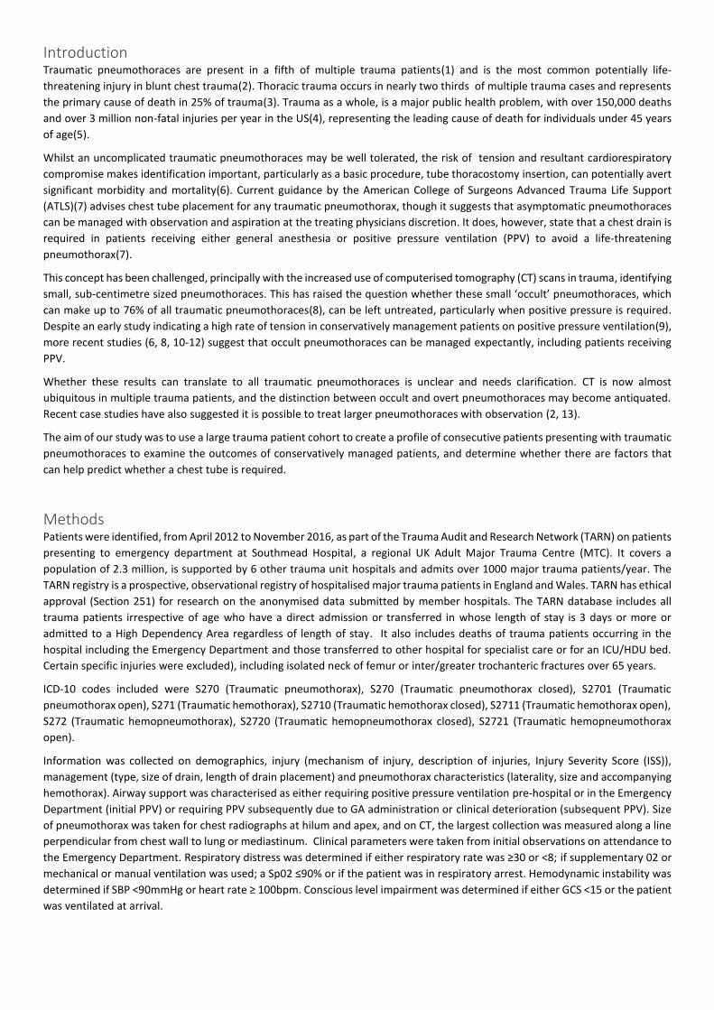
Introduction Traumatic pneumothoraces are present in a fifth of multiple trauma patients(1) and is the most common potentially life-
threatening injury in blunt chest trauma(2). Thoracic trauma occurs in nearly two thirds of multiple trauma cases and represents
the primary cause of death in 25% of trauma(3). Trauma as a whole, is a major public health problem, with over 150,000 deaths
and over 3 million non-fatal injuries per year in the US(4), representing the leading cause of death for individuals under 45 years
of age(5).
Whilst an uncomplicated traumatic pneumothoraces may be well tolerated, the risk of tension and resultant cardiorespiratory
compromise makes identification important, particularly as a basic procedure, tube thoracostomy insertion, can potentially avert
significant morbidity and mortality(6). Current guidance by the American College of Surgeons Advanced Trauma Life Support
(ATLS)(7) advises chest tube placement for any traumatic pneumothorax, though it suggests that asymptomatic pneumothoraces
can be managed with observation and aspiration at the treating physicians discretion. It does, however, state that a chest drain is
required in patients receiving either general anesthesia or positive pressure ventilation (PPV) to avoid a life-threatening
pneumothorax(7).
This concept has been challenged, principally with the increased use of computerised tomography (CT) scans in trauma, identifying
small, sub-centimetre sized pneumothoraces. This has raised the question whether these small ‘occult’ pneumothoraces, which
can make up to 76% of all traumatic pneumothoraces(8), can be left untreated, particularly when positive pressure is required.
Despite an early study indicating a high rate of tension in conservatively management patients on positive pressure ventilation(9),
more recent studies (6, 8, 10-12) suggest that occult pneumothoraces can be managed expectantly, including patients receiving
PPV.
Whether these results can translate to all traumatic pneumothoraces is unclear and needs clarification. CT is now almost
ubiquitous in multiple trauma patients, and the distinction between occult and overt pneumothoraces may become antiquated.
Recent case studies have also suggested it is possible to treat larger pneumothoraces with observation (2, 13).
The aim of our study was to use a large trauma patient cohort to create a profile of consecutive patients presenting with traumatic
pneumothoraces to examine the outcomes of conservatively managed patients, and determine whether there are factors that
can help predict whether a chest tube is required.
Methods Patients were identified, from April 2012 to November 2016, as part of the Trauma Audit and Research Network (TARN) on patients
presenting to emergency department at Southmead Hospital, a regional UK Adult Major Trauma Centre (MTC). It covers a
population of 2.3 million, is supported by 6 other trauma unit hospitals and admits over 1000 major trauma patients/year. The
TARN registry is a prospective, observational registry of hospitalised major trauma patients in England and Wales. TARN has ethical
approval (Section 251) for research on the anonymised data submitted by member hospitals. The TARN database includes all
trauma patients irrespective of age who have a direct admission or transferred in whose length of stay is 3 days or more or
admitted to a High Dependency Area regardless of length of stay. It also includes deaths of trauma patients occurring in the
hospital including the Emergency Department and those transferred to other hospital for specialist care or for an ICU/HDU bed.
Certain specific injuries were excluded), including isolated neck of femur or inter/greater trochanteric fractures over 65 years.
ICD-10 codes included were S270 (Traumatic pneumothorax), S270 (Traumatic pneumothorax closed), S2701 (Traumatic
pneumothorax open), S271 (Traumatic hemothorax), S2710 (Traumatic hemothorax closed), S2711 (Traumatic hemothorax open),
S272 (Traumatic hemopneumothorax), S2720 (Traumatic hemopneumothorax closed), S2721 (Traumatic hemopneumothorax
open).
Information was collected on demographics, injury (mechanism of injury, description of injuries, Injury Severity Score (ISS)),
management (type, size of drain, length of drain placement) and pneumothorax characteristics (laterality, size and accompanying
hemothorax). Airway support was characterised as either requiring positive pressure ventilation pre-hospital or in the Emergency
Department (initial PPV) or requiring PPV subsequently due to GA administration or clinical deterioration (subsequent PPV). Size
of pneumothorax was taken for chest radiographs at hilum and apex, and on CT, the largest collection was measured along a line
perpendicular from chest wall to lung or mediastinum. Clinical parameters were taken from initial observations on attendance to
the Emergency Department. Respiratory distress was determined if either respiratory rate was ≥30 or <8; if supplementary 02 or
mechanical or manual ventilation was used; a Sp02 ≤90% or if the patient was in respiratory arrest. Hemodynamic instability was
determined if SBP <90mmHg or heart rate ≥ 100bpm. Conscious level impairment was determined if either GCS <15 or the patient
was ventilated at arrival.

Statistical analysis Descriptive statistics were used to summarise patient characteristics and clinical data. Means (±SD) were calculated for parametric
data and medians (IQR) were calculated non-parametric data. Several checks for normality, including Kolmogorov-Smirnov,
Shapiro-Wilk, kurtosis and skewness calculations were performed. Continuous parametric variables were analysed using
independent t-test and continuous non-parametric variables were analysed using Mann-Whitney test. Categorical data was
analysed using Chi-squared test. A P-value of <0.05 was considered statistically significant.
Univariate proportional hazard ratios were calculated using Cox regression analysis for factors associated with failure of
conservative treatment (size of pneumothorax, mechanism of injury, ISS, presence of ribs fractures, clinical features (respiratory,
hemodynamic, GCS), presence of hemothorax, bilateral versus unilateral pneumothorax, use of PPV and surgical procedures).
Further multivariable cox regression analysis was performed to determine which factors (age, size of pneumothorax, ISS, presence
of ribs fractures, clinical conditions (respiratory, hemodynamic, GCS), presence of hemothorax, bilateral versus unilateral
pneumothorax and use of PPV) were independently predictive of failure of conservative management. These factors were decided
on in a priori statistical analysis plan. All statistical analysis was performed using IBM SPSS statistics version 23.0 (SPSS Inc. Chicago,
IL)
Results Demographics 3771 trauma patients presenting to this MTC were registered on the TARN database from April 2012 to December 2016. 765
patients were identified using the search criteria. 636 patients with pneumothoraces were identified, with 602 patients (see
Figure 1) included for analysis. Table 1 summaries patient demographics, mechanism of injuries, ISS, pneumothorax
characteristics, management and outcomes for patients managed non-conservatively and conservatively. Table 2 summaries
the characteristics and outcomes for successful and failed observed management.
Traumatic pneumothoraces were present in 636/3771 (17%) of trauma patients during the study period. The mean age was 48
(SD 22), 438/602 (73%) were male, and 330/602 (55%) suffered their injury as a result of a road traffic accident. The mean ISS
score (26) represented very severe injuries; and 189/602 (31%) required immediate invasive ventilation and 56/602 (9%) died
during admission. 325/602 (54%) of patients had an intervention performed pre-hospital or on admission, either with needle
decompression, chest tube insertion or chest surgery, with the remaining 277/602 (46%) of the pneumothoraces were initially
treated conservatively. The patients managed conservatively, had significantly smaller pneumothoraces compared to patient
managed with an immediate intervention (median 5.5mm vs 22mm), with the majority less than 10mm in size (see Figure 2).
Patients who were managed with an immediate intervention also had a higher incidence of respiratory, hemodynamic and
neurological compromise, and a higher proportion of significant hemothorax than those managed conservatively. Both groups,
have comparable ages, ISS score, mortality rate and total LOS.
Of the 277/602 (46%) of patients managed conservatively, 252/277 (90%) did not require subsequent thoracic intervention. This
included the majority, 56/62 (90%), of patients requiring immediate PPV who were treated conservatively. There was no
significant difference in the failure rate between the patients on PPV (6/62, 9.7%) and those not requiring PPV (19/215, 8.8%) in
the conservative arm.
Using univariate analysis, size of pneumothorax, mechanism of injury, presence of rib fractures, clinical condition, surgery and ISS were not significantly associated with failure of conservative management (Table 3). The median size of pneumothorax (5.3 vs 8.2 mm p 0.13) was comparable between groups and did not increase the likelihood of progression requiring chest tube insertion, with HR of 1.61 (p 0.08) and 2.84 (p 0.07) on univariate and multivariable analysis Univariate and multivariable analysis also confirmed that acute PPV does not appear to confer an additional risk of failure of conservative management (HR 1.1 p 0.8 and HR 1.5 p 0.96 respectively). Additionally, requiring subsequent PPV during inpatient stay, either due to clinical deterioration, or for general anesthesia did not represent an increase risk of failure of conservative management. In contrast, the presence of a hemothorax was associated with increased likelihood of failure of expectant management (hazard ratios of 4.08 (p < 0.01)) and confirmed by multivariable cox regression analysis (Table 4).
Of the 25/252 patients who failed observed management, 23 had a large chest drain inserted and 2 went to have thoracic surgery
(rib fixation with hemothorax evacuation). The main indication for chest tube insertion was increasing pneumothorax (19/23) and
enlarging hemopneumothorax (4/23). The mean duration prior to chest tube insertion was 2.96 days (SD 4.03). Requiring
subsequent chest insertion in the conservative arm led to a non-significant increase length of stay (11 vs 10 days p 0.597). The 2
mortalities in this group had severe ISS of 40 (>25 represents severe/critical injuries) with intracranial hemorrhages and it is
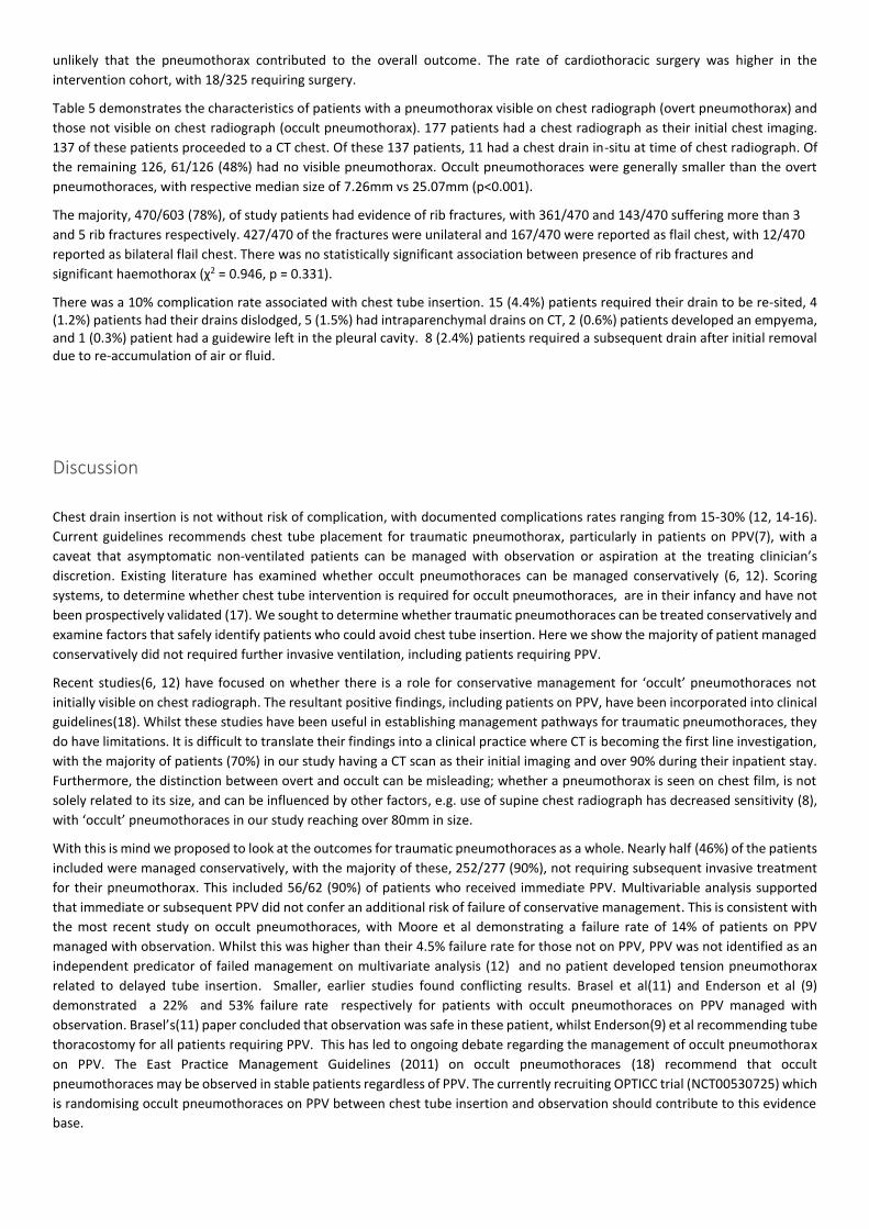
unlikely that the pneumothorax contributed to the overall outcome. The rate of cardiothoracic surgery was higher in the
intervention cohort, with 18/325 requiring surgery.
Table 5 demonstrates the characteristics of patients with a pneumothorax visible on chest radiograph (overt pneumothorax) and
those not visible on chest radiograph (occult pneumothorax). 177 patients had a chest radiograph as their initial chest imaging.
137 of these patients proceeded to a CT chest. Of these 137 patients, 11 had a chest drain in-situ at time of chest radiograph. Of
the remaining 126, 61/126 (48%) had no visible pneumothorax. Occult pneumothoraces were generally smaller than the overt
pneumothoraces, with respective median size of 7.26mm vs 25.07mm (p<0.001).
The majority, 470/603 (78%), of study patients had evidence of rib fractures, with 361/470 and 143/470 suffering more than 3
and 5 rib fractures respectively. 427/470 of the fractures were unilateral and 167/470 were reported as flail chest, with 12/470
reported as bilateral flail chest. There was no statistically significant association between presence of rib fractures and
significant haemothorax (χ2 = 0.946, p = 0.331).
There was a 10% complication rate associated with chest tube insertion. 15 (4.4%) patients required their drain to be re-sited, 4 (1.2%) patients had their drains dislodged, 5 (1.5%) had intraparenchymal drains on CT, 2 (0.6%) patients developed an empyema, and 1 (0.3%) patient had a guidewire left in the pleural cavity. 8 (2.4%) patients required a subsequent drain after initial removal due to re-accumulation of air or fluid.
Discussion
Chest drain insertion is not without risk of complication, with documented complications rates ranging from 15-30% (12, 14-16).
Current guidelines recommends chest tube placement for traumatic pneumothorax, particularly in patients on PPV(7), with a
caveat that asymptomatic non-ventilated patients can be managed with observation or aspiration at the treating clinician’s
discretion. Existing literature has examined whether occult pneumothoraces can be managed conservatively (6, 12). Scoring
systems, to determine whether chest tube intervention is required for occult pneumothoraces, are in their infancy and have not
been prospectively validated (17). We sought to determine whether traumatic pneumothoraces can be treated conservatively and
examine factors that safely identify patients who could avoid chest tube insertion. Here we show the majority of patient managed
conservatively did not required further invasive ventilation, including patients requiring PPV.
Recent studies(6, 12) have focused on whether there is a role for conservative management for ‘occult’ pneumothoraces not
initially visible on chest radiograph. The resultant positive findings, including patients on PPV, have been incorporated into clinical
guidelines(18). Whilst these studies have been useful in establishing management pathways for traumatic pneumothoraces, they
do have limitations. It is difficult to translate their findings into a clinical practice where CT is becoming the first line investigation,
with the majority of patients (70%) in our study having a CT scan as their initial imaging and over 90% during their inpatient stay.
Furthermore, the distinction between overt and occult can be misleading; whether a pneumothorax is seen on chest film, is not
solely related to its size, and can be influenced by other factors, e.g. use of supine chest radiograph has decreased sensitivity (8),
with ‘occult’ pneumothoraces in our study reaching over 80mm in size.
With this is mind we proposed to look at the outcomes for traumatic pneumothoraces as a whole. Nearly half (46%) of the patients
included were managed conservatively, with the majority of these, 252/277 (90%), not requiring subsequent invasive treatment
for their pneumothorax. This included 56/62 (90%) of patients who received immediate PPV. Multivariable analysis supported
that immediate or subsequent PPV did not confer an additional risk of failure of conservative management. This is consistent with
the most recent study on occult pneumothoraces, with Moore et al demonstrating a failure rate of 14% of patients on PPV
managed with observation. Whilst this was higher than their 4.5% failure rate for those not on PPV, PPV was not identified as an
independent predicator of failed management on multivariate analysis (12) and no patient developed tension pneumothorax
related to delayed tube insertion. Smaller, earlier studies found conflicting results. Brasel et al(11) and Enderson et al (9)
demonstrated a 22% and 53% failure rate respectively for patients with occult pneumothoraces on PPV managed with
observation. Brasel’s(11) paper concluded that observation was safe in these patient, whilst Enderson(9) et al recommending tube
thoracostomy for all patients requiring PPV. This has led to ongoing debate regarding the management of occult pneumothorax
on PPV. The East Practice Management Guidelines (2011) on occult pneumothoraces (18) recommend that occult
pneumothoraces may be observed in stable patients regardless of PPV. The currently recruiting OPTICC trial (NCT00530725) which
is randomising occult pneumothoraces on PPV between chest tube insertion and observation should contribute to this evidence
base.

The size of pneumothorax was not a predictor of failed observation in our study on univariate and multivariate analysis, with non-
significant differences in size of pneumothorax between the successful and failed observed groups. Pneumothorax size had
previously been thought to be a predictor of progression(19), with De Moya et al(17) proposing a scoring system using size of
occult pneumothorax and its relationship to the hilum to guide management. However this has not been successfully
validated(12), with Moore et al demonstrating that pneumothorax size was not an independent predicator of failed observation
(12).
The presence of a hemothorax appears to be predictive of failure of conservative management in both overt and occult
pneumothoraces(12). This is consistent with clinical practice. A significant hemothorax is an indication for chest tube insertion, to
evacuate blood from the pleural space and avoid complication such as infection and a fibrothorax and when combined with the
presence of pneumothorax, this provides a strong incentive for intervention.
In this study, patients who do not require prehospital or admission chest procedures generally did not require a chest procedure
later in hospitalization. When this information is combined with previous trials on traumatic pneumothoraces it appears that there
is a subpopulation that can be managed conservatively. Certainly, when there is no significant hemopneumothorax (<2cm in size)
there can be consideration for expectant management. Mechanism of injury, ISS or size of pneumothorax do not appear to
provide a strong indication for intervention. Additionally in our study, clinical condition did not confer an adverse prognosis,
although in other studies respiratory distress has (12). Although the use of ventilation has been controversial, it appears from our
findings and previous studies that pneumothoraces can be managed conservatively with careful observation on PPV with no
increased risk of harm(6, 12).
This is a retrospective observational trial, and as such will be subject to the inherent limitations of such a study. The data was
collected from a single centre, with a low rate of penetrating chest wall injury (5%), which should be considered when generalising
the findings to other centres. Selection bias may have been introduced by physician selection and variation in initial imaging
modality, and the decision to intervene may have affected the conservatively treated cohort characteristics and it is likely that the
high risk unwell patients were underrepresented in the conservatively treated arm. Those treated with an immediate
intervention, despite similar ISS, likely represented a more unwell population, with higher rates of cardiorespiratory compromise,
PPV use, surgical referral rates and higher mortality rates. The length of stay criteria (length of stay is 3 days or more or admitted
to a High Dependency Area regardless of length of stay) is likely to have biased against patients successfully conservatively
managed and not requiring a prolonged hospital admission, suggesting that the overall rate of effective conservative management
is probably greater. Efforts were made to minimise bias, by including large number of consecutive unselected patients into the
analysis and careful documentation and comparison of cohort characteristics.
Conclusion Our study represents the largest observational study on traumatic pneumothoraces to date. It demonstrates that the majority of
conservatively managed patients were successfully managed without requiring a chest drain. This includes the majority of patients
on positive pressure ventilation (PPV), the use of which did not present an increased risk of failure of expectant management. This
study provide support for an observed, expectant approach if the treating physician does not feel an immediate chest drain is
warranted in the patient with a traumatic pneumothorax. Future prospective randomised trials examining the outcomes of a
conservative approach in traumatic pneumothorax, regardless of pneumothorax size or use of PPV would help clarify which
patients are best managed expectantly.
Word count 2902
• Guarantor statement: Dr Steven Walker takes responsibility for the content of the manuscript, including the data and analysis
• Author contributions: SW performed statistical analysis and prepared the manuscript. SW, SB, JT & NAM conceived the design of the study. All authors have read and approved the final manuscript for submission.
• Financial/nonfinancial disclosures No financial disclosures.
References
1. Di Bartolomeo S, Sanson G, Nardi G, Scian F, Michelutto V, Lattuada L. A population-based study on pneumothorax in severely traumatized patients. Journal of Trauma and Acute Care Surgery. 2001;51(4):677-82. 2. Idris BM, Hefny AF. Large pneumothorax in blunt chest trauma: Is a chest drain always necessary in stable patients? A case report. International journal of surgery case reports. 2016;24:88-90. 3. Kshettry VR, Bolman 3rd R. Chest trauma. Assessment, diagnosis, and management. Clinics in chest medicine. 1994;15(1):137. 4. Centers for Disease Control and Prevention NCfHSN. National hospital discharge survey: 2007 summary. National health statistics reports, no. 29. National health statistics reports, no. 29. Atlanta, GA: NCHS; 2010. [ 5. Centers for Disease Control and Prevention NCfIPaC. Web-based Injury Statistics Query and Reporting System (WISQARS) 2007 [cited 2017 Mar 2]. Available from: http://www.cdc.gov/injury/wisqars.
6. Wilson H, Ellsmere J, Tallon J, Kirkpatrick A. Occult pneumothorax in the blunt trauma patient: tube thoracostomy or observation? Injury. 2009;40(9):928-31. 7. Subcommittee A, Tchorz KM, group IAw. Advanced trauma life support (ATLS®): the ninth edition. The Journal of Trauma and Acute Care Surgery. 2013;74(5):1363. 8. Ball CG, Kirkpatrick AW, Feliciano DV. The occult pneumothorax: what have we learned? Canadian Journal of Surgery. 2009;52(5):E173. 9. Enderson BL, Abdalla R, Frame SB, Casey MT, Gould H, Maull KI. Tube thoracostomy for occult pneumothorax: a prospective randomized study of its use. Journal of Trauma and Acute Care Surgery. 1993;35(5):726-30. 10. Plurad D, Green D, Demetriades D, Rhee P. The increasing use of chest computed tomography for trauma: is it being overutilized? Journal of Trauma and Acute Care Surgery. 2007;62(3):631-5. 11. Brasel KJ, Stafford RE, Weigelt JA, Tenquist JE, Borgstrom DC. Treatment of occult pneumothoraces from blunt trauma. Journal of Trauma and Acute Care Surgery. 1999;46(6):987-91. 12. Moore FO, Goslar PW, Coimbra R, Velmahos G, Brown CV, Coopwood Jr TB, et al. Blunt traumatic occult pneumothorax: is observation safe?—results of a prospective, AAST multicenter study. Journal of Trauma and Acute Care Surgery. 2011;70(5):1019-25. 13. Ryan MT, Caputo ND, Lakdawala V, Jara F. Spontaneous resolution of a large traumatic pneumothorax. The American journal of emergency medicine. 2012;30(5):833. e3. 14. Etoch SW, Bar-Natan MF, Miller FB, Richardson JD. Tube thoracostomy: factors related to complications. Archives of surgery. 1995;130(5):521-6. 15. Bailey R. Complications of tube thoracostomy in trauma. Journal of accident & emergency medicine. 2000;17(2):111-4. 16. Ball CG, Lord J, Laupland KB, Gmora S, Mulloy RH, Ng AK, et al. Chest tube complications: how well are we training our residents? Canadian Journal of Surgery. 2007;50(6):450. 17. De Moya MA, Seaver C, Spaniolas K, Inaba K, Nguyen M, Veltman Y, et al. Occult pneumothorax in trauma patients: development of an objective scoring system. Journal of Trauma and Acute Care Surgery. 2007;63(1):13-7. 18. Mowery NT, Gunter OL, Collier BR, Jose'J Jr D, Haut E, Hildreth A, et al. Practice management guidelines for management of hemothorax and occult pneumothorax. Journal of Trauma and Acute Care Surgery. 2011;70(2):510-8. 19. Garramone Jr R, Jacobs L, Sahdev P. An objective method to measure and manage occult pneumothorax. Surgery, gynecology & obstetrics. 1991;173(4):257-61.
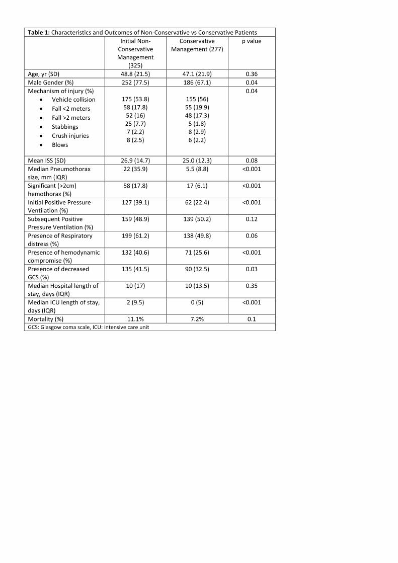
Table 1: Characteristics and Outcomes of Non-Conservative vs Conservative Patients
Initial Non-Conservative Management
(325)
Conservative Management (277)
p value
Age, yr (SD) 48.8 (21.5) 47.1 (21.9) 0.36
Male Gender (%) 252 (77.5) 186 (67.1) 0.04
Mechanism of injury (%)
• Vehicle collision
• Fall <2 meters
• Fall >2 meters
• Stabbings
• Crush injuries
• Blows
175 (53.8) 58 (17.8) 52 (16) 25 (7.7) 7 (2.2) 8 (2.5)
155 (56) 55 (19.9) 48 (17.3)
5 (1.8) 8 (2.9) 6 (2.2)
0.04
Mean ISS (SD) 26.9 (14.7) 25.0 (12.3) 0.08
Median Pneumothorax size, mm (IQR)
22 (35.9) 5.5 (8.8) <0.001
Significant (>2cm) hemothorax (%)
58 (17.8) 17 (6.1) <0.001
Initial Positive Pressure Ventilation (%)
127 (39.1) 62 (22.4) <0.001
Subsequent Positive Pressure Ventilation (%)
159 (48.9)
139 (50.2) 0.12
Presence of Respiratory distress (%)
199 (61.2) 138 (49.8) 0.06
Presence of hemodynamic compromise (%)
132 (40.6) 71 (25.6) <0.001
Presence of decreased GCS (%)
135 (41.5) 90 (32.5) 0.03
Median Hospital length of stay, days (IQR)
10 (17) 10 (13.5) 0.35
Median ICU length of stay, days (IQR)
2 (9.5) 0 (5) <0.001
Mortality (%) 11.1% 7.2% 0.1 GCS: Glasgow coma scale, ICU: intensive care unit
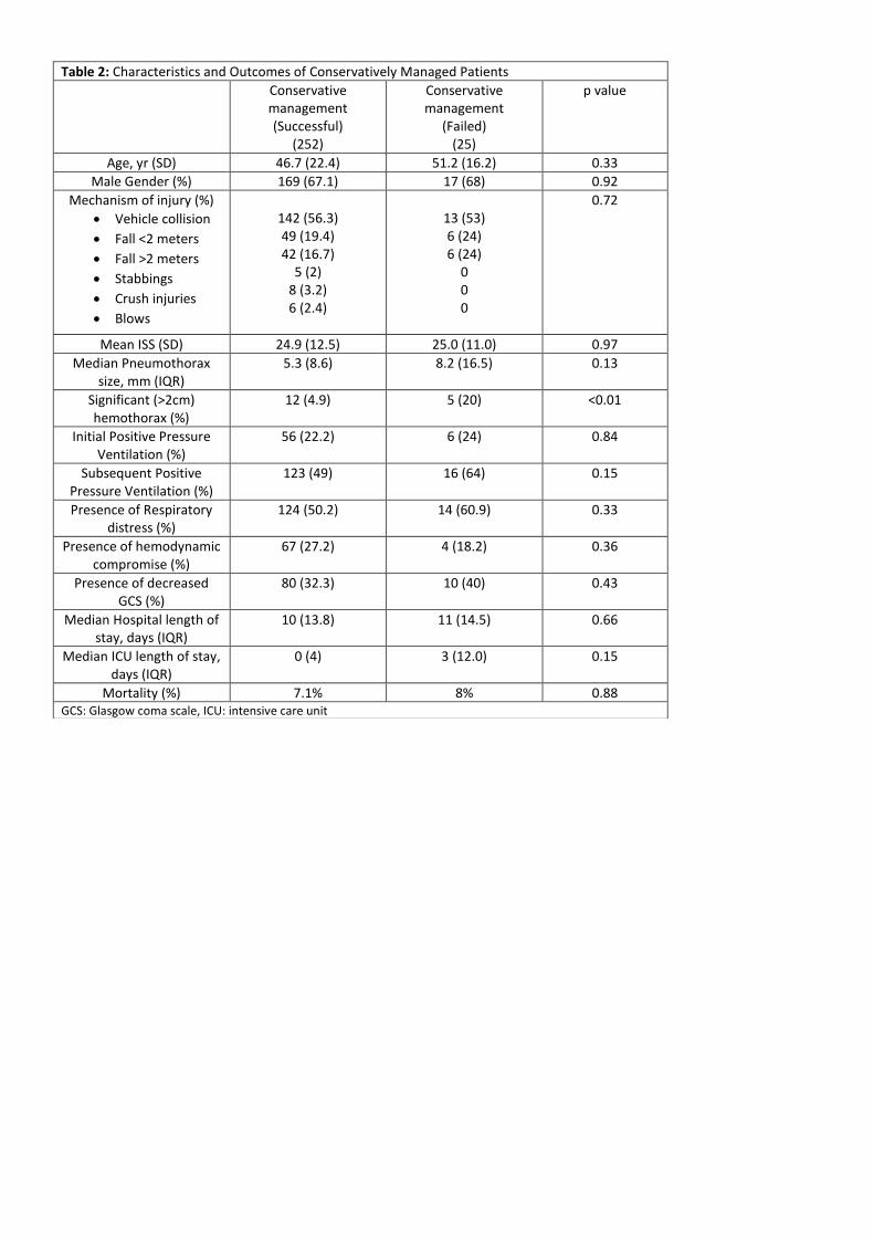
Table 2: Characteristics and Outcomes of Conservatively Managed Patients
Conservative management (Successful)
(252)
Conservative management
(Failed) (25)
p value
Age, yr (SD) 46.7 (22.4) 51.2 (16.2) 0.33
Male Gender (%) 169 (67.1) 17 (68) 0.92
Mechanism of injury (%)
• Vehicle collision
• Fall <2 meters
• Fall >2 meters
• Stabbings
• Crush injuries
• Blows
142 (56.3) 49 (19.4) 42 (16.7)
5 (2) 8 (3.2) 6 (2.4)
13 (53) 6 (24) 6 (24)
0 0 0
0.72
Mean ISS (SD) 24.9 (12.5) 25.0 (11.0) 0.97
Median Pneumothorax size, mm (IQR)
5.3 (8.6) 8.2 (16.5) 0.13
Significant (>2cm) hemothorax (%)
12 (4.9) 5 (20) <0.01
Initial Positive Pressure Ventilation (%)
56 (22.2) 6 (24) 0.84
Subsequent Positive Pressure Ventilation (%)
123 (49)
16 (64) 0.15
Presence of Respiratory distress (%)
124 (50.2) 14 (60.9) 0.33
Presence of hemodynamic compromise (%)
67 (27.2) 4 (18.2) 0.36
Presence of decreased GCS (%)
80 (32.3) 10 (40) 0.43
Median Hospital length of stay, days (IQR)
10 (13.8) 11 (14.5) 0.66
Median ICU length of stay, days (IQR)
0 (4) 3 (12.0) 0.15
Mortality (%) 7.1% 8% 0.88 GCS: Glasgow coma scale, ICU: intensive care unit
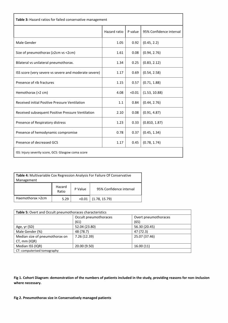
Table 3: Hazard ratios for failed conservative management
Hazard ratio P value 95% Confidence interval
Male Gender 1.05 0.92 (0.45, 2.2)
Size of pneumothorax (≥2cm vs <2cm) 1.61 0.08 (0.94, 2.76)
Bilateral vs unilateral pneumothorax. 1.34 0.25 (0.83, 2.12)
ISS score (very severe vs severe and moderate severe) 1.17 0.69 (0.54, 2.58)
Presence of rib fractures 1.15 0.57 (0.71, 1.88)
Hemothorax (>2 cm) 4.08 <0.01 (1.53, 10.88)
Received initial Positive Pressure Ventilation 1.1 0.84 (0.44, 2.76)
Received subsequent Positive Pressure Ventilation 2.10 0.08 (0.91, 4.87)
Presence of Respiratory distress 1.23 0.33 (0.810, 1.87)
Presence of hemodynamic compromise 0.78 0.37 (0.45, 1.34)
Presence of decreased GCS 1.17 0.45 (0.78, 1.74)
ISS: Injury severity score, GCS: Glasgow coma score
Table 4: Multivariable Cox Regression Analysis For Failure Of Conservative Management
Hazard Ratio
P Value 95% Confidence interval
Haemothorax >2cm 5.29 <0.01 (1.78, 15.79)
Table 5: Overt and Occult pneumothoraces characteristics
Occult pneumothoraces (61)
Overt pneumothoraces (65)
Age, yr (SD) 52.04 (23.80) 56.30 (20.45)
Male Gender (%) 48 (78.7) 47 (72.3)
Median size of pneumothorax on CT, mm (IQR)
7.26 (12.39) 25.07 (37.46)
Median ISS (IQR) 20.00 (9.50) 16.00 (11) CT: computerised tomography
Fig 1. Cohort Diagram: demonstration of the numbers of patients included in the study, providing reasons for non-inclusion
where necessary.
Fig 2. Pneumothorax size in Conservatively managed patients