Embed Size (px)
Citation preview
Waldenström’s macroglobulinemia: Genetic Basis and Therapy.
Steve Treon MD, MA, PhD
Dana Farber Cancer Institute
Harvard Medical School
Boston, Massachusetts, United States
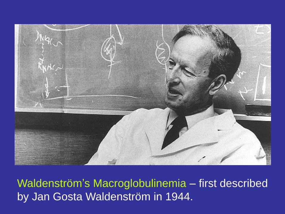
Waldenström’s Macroglobulinemia – first described
by Jan Gosta Waldenström in 1944.
Waldenström’s macroglobulinemia:
Similar but Different to Myeloma.
Genetic Predisposition
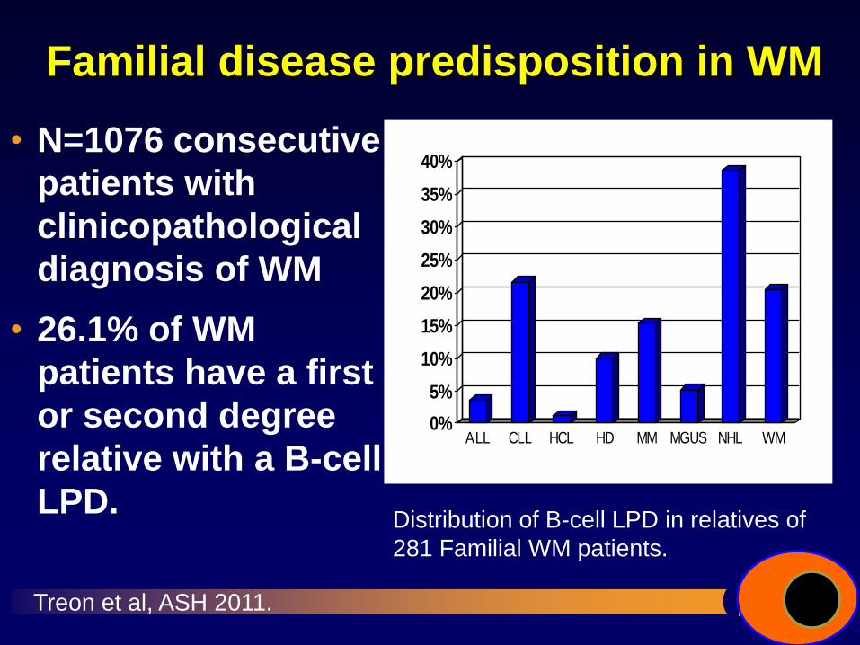
Familial disease predisposition in WM
• N=1076 consecutive
patients with
clinicopathological
diagnosis of WM
• 26.1% of WM
patients have a first
or second degree
relative with a B-cell
LPD.
0%
5%
10%
15%
20%
25%
30%
35%
40%
ALL CLL HCL HD MM MGUS NHL WM
Distribution of B-cell LPD in relatives of
281 Familial WM patients.
Treon et al, ASH 2011.
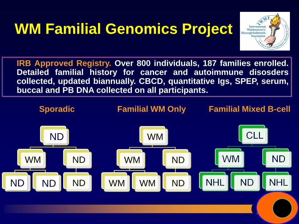
WM Familial Genomics Project
IRB Approved Registry. Over 800 individuals, 187 families enrolled. Detailed familial history for cancer and autoimmune disosders collected, updated biannually. CBCD, quantitative Igs, SPEP, serum, buccal and PB DNA collected on all participants.
WM
WM
WM WM
ND
ND
WM
WM
WM WM
ND
ND
ND
ND ND
Sporadic Familial WM Only Familial Mixed B-cell
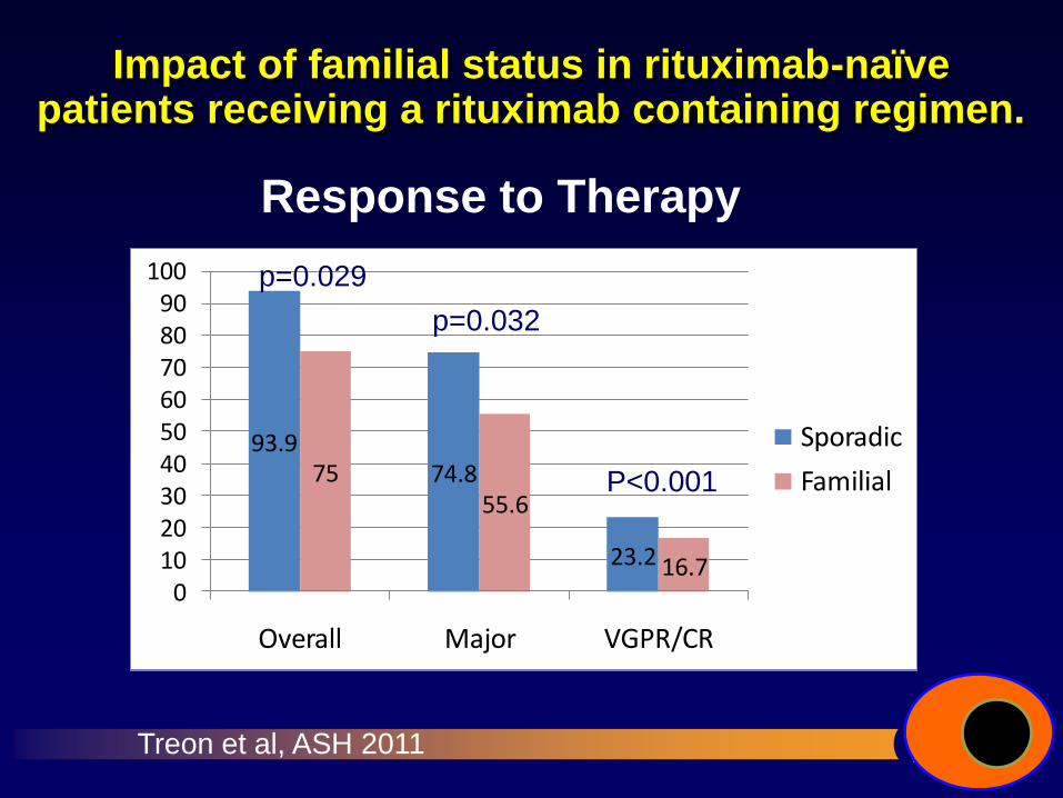
Impact of familial status in rituximab-naïve patients receiving a rituximab containing regimen.
Response to Therapy
93.974.8
23.2
7555.6
16.70
102030405060708090
100
Overall Major VGPR/CR
Sporadic
Familial
p=0.032
P<0.001
p=0.029
Treon et al, ASH 2011
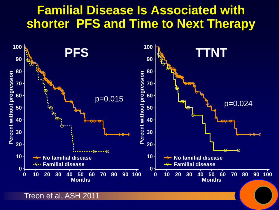
Familial Disease Is Associated with shorter PFS and Time to Next Therapy
0
10
20
30
40
50
60
70
80
90
100
Pe
rce
nt
wit
ho
ut
pro
gre
ss
ion
No familial disease
Familial disease
PFS
0 10 20 30 40 50 60 70 80 90 100 Months
0
10
20
30
40
50
60
70
80
90
100
Pe
rce
nt
wit
ho
ut
pro
gre
ss
ion
No familial disease
Familial disease
TTNT
0 10 20 30 40 50 60 70 80 90 100 Months
Treon et al, ASH 2011
p=0.015 p=0.024
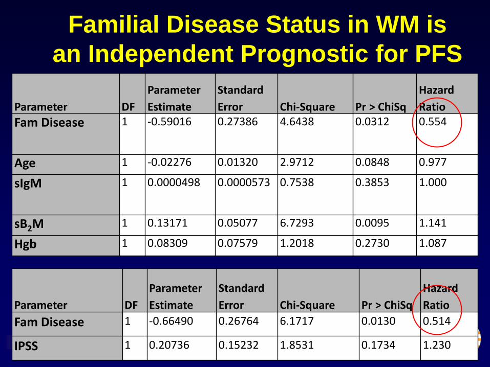
Parameter DF
Parameter
Estimate
Standard
Error Chi-Square Pr > ChiSq
Hazard
Ratio
Fam Disease 1 -0.59016 0.27386 4.6438 0.0312 0.554
Age 1 -0.02276 0.01320 2.9712 0.0848 0.977
sIgM 1 0.0000498 0.0000573 0.7538 0.3853 1.000
sB2M 1 0.13171 0.05077 6.7293 0.0095 1.141
Hgb 1 0.08309 0.07579 1.2018 0.2730 1.087
Parameter DF
Parameter
Estimate
Standard
Error Chi-Square Pr > ChiSq
Hazard
Ratio
Fam Disease 1 -0.66490 0.26764 6.1717 0.0130 0.514
IPSS 1 0.20736 0.15232 1.8531 0.1734 1.230
Familial Disease Status in WM is
an Independent Prognostic for PFS
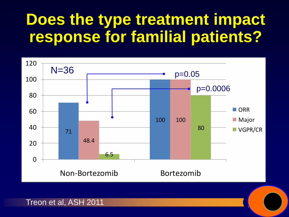
Does the type treatment impact response for familial patients?
71
100
48.4
100
6.5
80
0
20
40
60
80
100
120
Non-Bortezomib Bortezomib
ORR
Major
VGPR/CR
p=0.0006
p=0.05 N=36
Treon et al, ASH 2011
Is there a common
genetic predisposition
with other cancers
in WM patients?
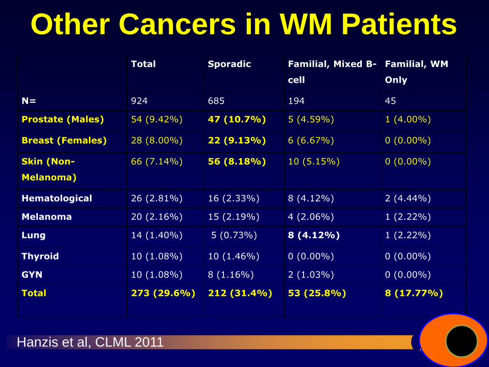
Other Cancers in WM Patients Total Sporadic Familial, Mixed B-
cell
Familial, WM
Only
N= 924 685 194 45
Prostate (Males) 54 (9.42%) 47 (10.7%) 5 (4.59%) 1 (4.00%)
Breast (Females) 28 (8.00%) 22 (9.13%) 6 (6.67%) 0 (0.00%)
Skin (Non-
Melanoma)
66 (7.14%) 56 (8.18%) 10 (5.15%) 0 (0.00%)
Hematological 26 (2.81%) 16 (2.33%) 8 (4.12%) 2 (4.44%)
Melanoma 20 (2.16%) 15 (2.19%) 4 (2.06%) 1 (2.22%)
Lung 14 (1.40%) 5 (0.73%) 8 (4.12%) 1 (2.22%)
Thyroid 10 (1.08%) 10 (1.46%) 0 (0.00%) 0 (0.00%)
GYN 10 (1.08%) 8 (1.16%) 2 (1.03%) 0 (0.00%)
Total 273 (29.6%) 212 (31.4%) 53 (25.8%) 8 (17.77%)
Hanzis et al, CLML 2011
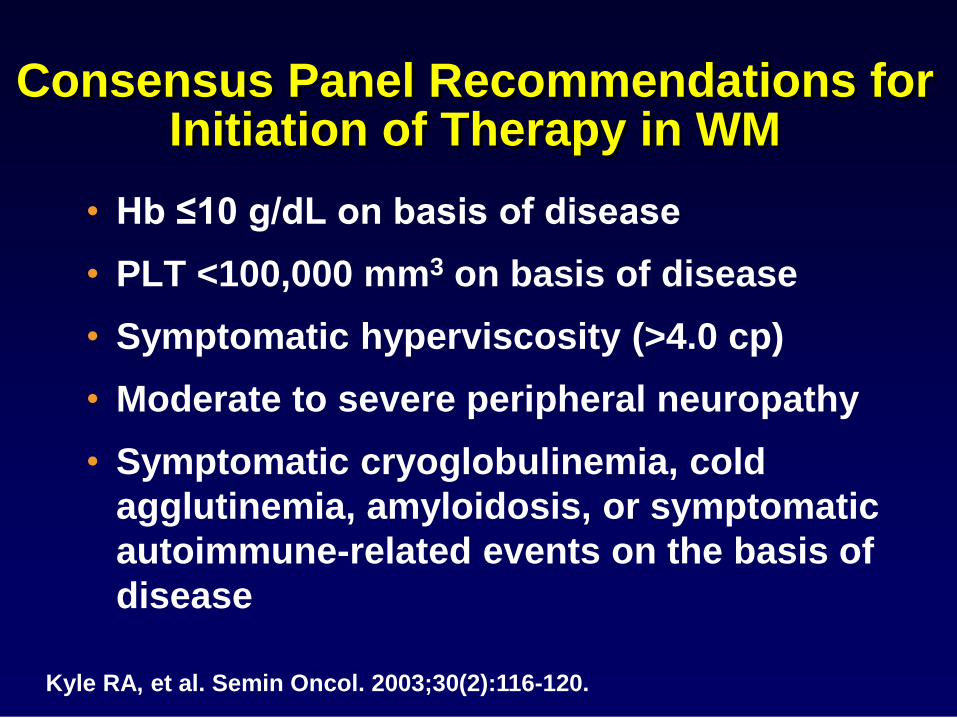
Consensus Panel Recommendations for Initiation of Therapy in WM
• Hb ≤10 g/dL on basis of disease
• PLT <100,000 mm3 on basis of disease
• Symptomatic hyperviscosity (>4.0 cp)
• Moderate to severe peripheral neuropathy
• Symptomatic cryoglobulinemia, cold
agglutinemia, amyloidosis, or symptomatic
autoimmune-related events on the basis of
disease
Kyle RA, et al. Semin Oncol. 2003;30(2):116-120.
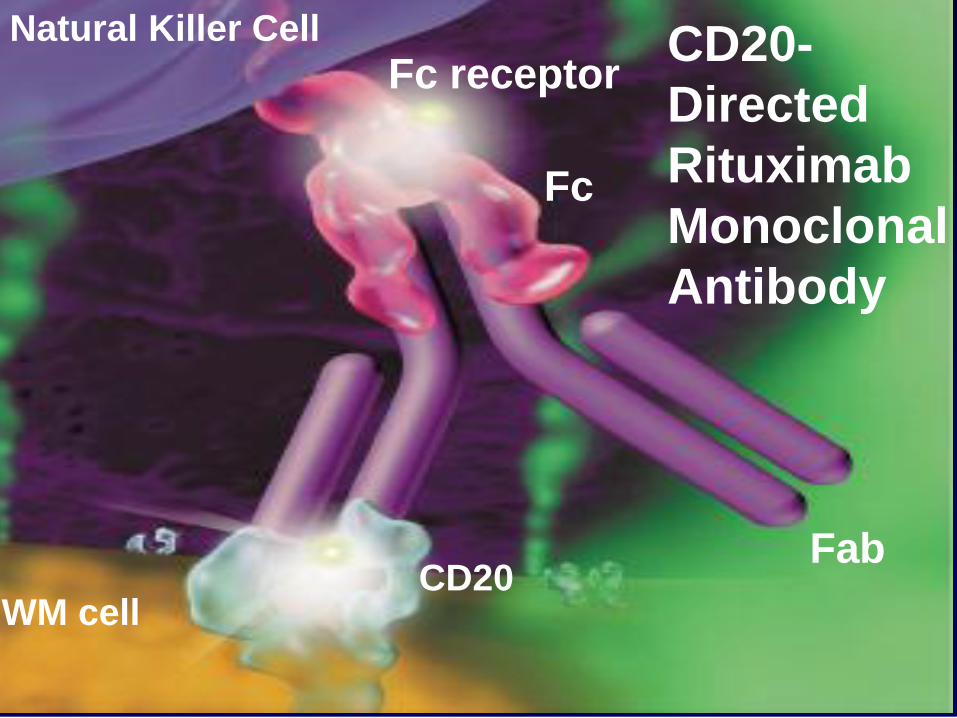
Fab
Fc
Fc receptor Natural Killer Cell
CD20 WM cell
CD20-
Directed
Rituximab
Monoclonal
Antibody
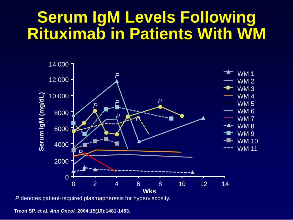
Serum IgM Levels Following Rituximab in Patients With WM
Treon SP, et al. Ann Oncol. 2004;15(10):1481-1483.
P denotes patient-required plasmapheresis for hyperviscosity.
14,000
12,000
0 0 2 4 6 8 10
Wks
Seru
m I
gM
(m
g/d
L)
10,000
8000
6000
4000
2000
12 14
WM 1 WM 2 WM 3 WM 4 WM 5 WM 6 WM 7 WM 8 WM 9 WM 10 WM 11
P
P
P
P P
P
P
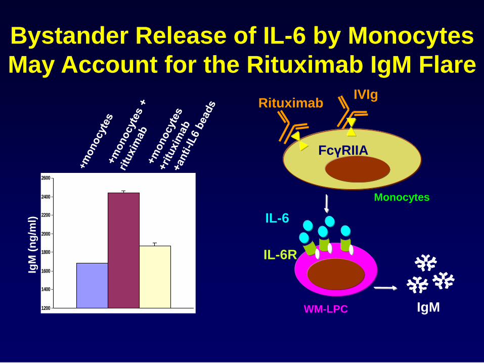
Bystander Release of IL-6 by Monocytes
May Account for the Rituximab IgM Flare
1200
1400
1600
1800
2000
2200
2400
2600
IgM
(n
g/m
l)
IL-6
Monocytes
FcγRIIA
Rituximab
WM-LPC
IL-6R
IgM
IVIg
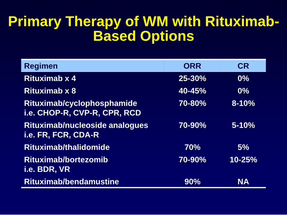
Primary Therapy of WM with Rituximab-Based Options
Regimen ORR CR
Rituximab x 4 25-30% 0%
Rituximab x 8 40-45% 0%
Rituximab/cyclophosphamide
i.e. CHOP-R, CVP-R, CPR, RCD
70-80% 8-10%
Rituximab/nucleoside analogues
i.e. FR, FCR, CDA-R
70-90% 5-10%
Rituximab/thalidomide 70% 5%
Rituximab/bortezomib
i.e. BDR, VR
70-90% 10-25%
Rituximab/bendamustine 90% NA
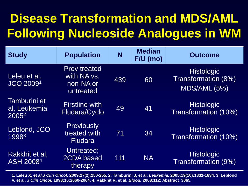
Disease Transformation and MDS/AML
Following Nucleoside Analogues in WM
Study Population N Median
F/U (mo) Outcome
Leleu et al, JCO 20091
Prev treated with NA vs. non-NA or untreated
439 60
Histologic Transformation (8%)
MDS/AML (5%)
Tamburini et al, Leukemia 20052
Firstline with Fludara/Cyclo
49 41 Histologic
Transformation (10%)
Leblond, JCO 19983
Previously treated with
Fludara 71 34
Histologic Transformation (10%)
Rakkhit et al, ASH 20084
Untreated; 2CDA based
therapy 111 NA
Histologic Transformation (9%)
1. Leleu X, et al.J Clin Oncol. 2009;27(2):250-255. 2. Tamburini J, et al. Leukemia. 2005;19(10):1831-1834. 3. Leblond
V, et al. J Clin Oncol. 1998;16:2060-2064. 4. Rakkhit R, et al. Blood. 2008;112: Abstract 3065.
Advances in the Biology of Waldenstrom’s Macroglobulinemia
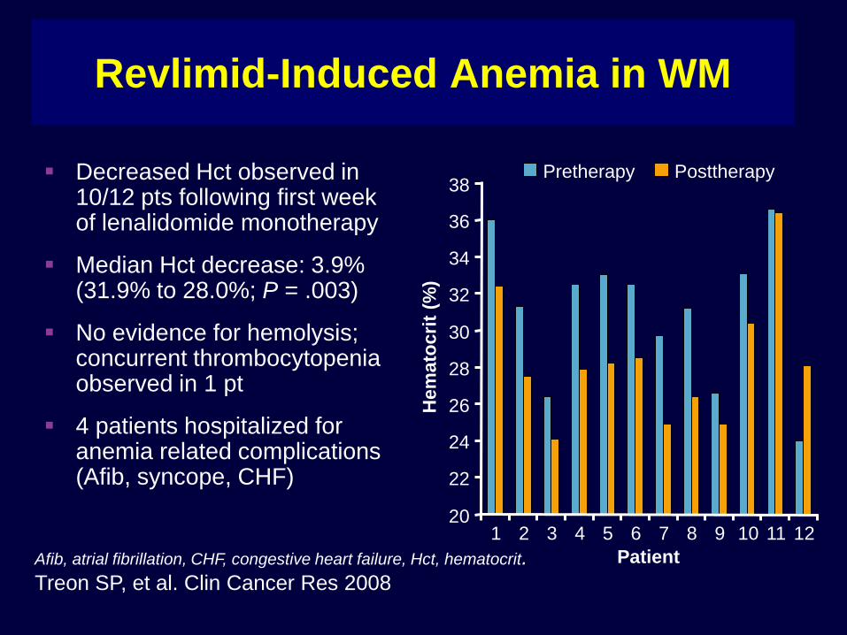
Revlimid-Induced Anemia in WM
Decreased Hct observed in 10/12 pts following first week of lenalidomide monotherapy
Median Hct decrease: 3.9% (31.9% to 28.0%; P = .003)
No evidence for hemolysis; concurrent thrombocytopenia observed in 1 pt
4 patients hospitalized for anemia related complications (Afib, syncope, CHF)
Afib, atrial fibrillation, CHF, congestive heart failure, Hct, hematocrit.
Treon SP, et al. Clin Cancer Res 2008
20
22
24
26
28
30
32
34
36
38
1 2 3 4 5 6 7 8 9 10 11 12
Pretherapy Posttherapy
Hem
ato
cri
t (%
)
Patient
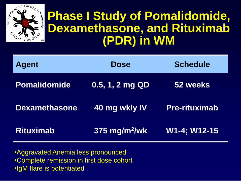
Phase I Study of Pomalidomide, Dexamethasone, and Rituximab
(PDR) in WM
Agent Dose Schedule
Pomalidomide 0.5, 1, 2 mg QD 52 weeks
Dexamethasone 40 mg wkly IV Pre-rituximab
Rituximab 375 mg/m2/wk W1-4; W12-15
•Aggravated Anemia less pronounced
•Complete remission in first dose cohort
•IgM flare is potentiated
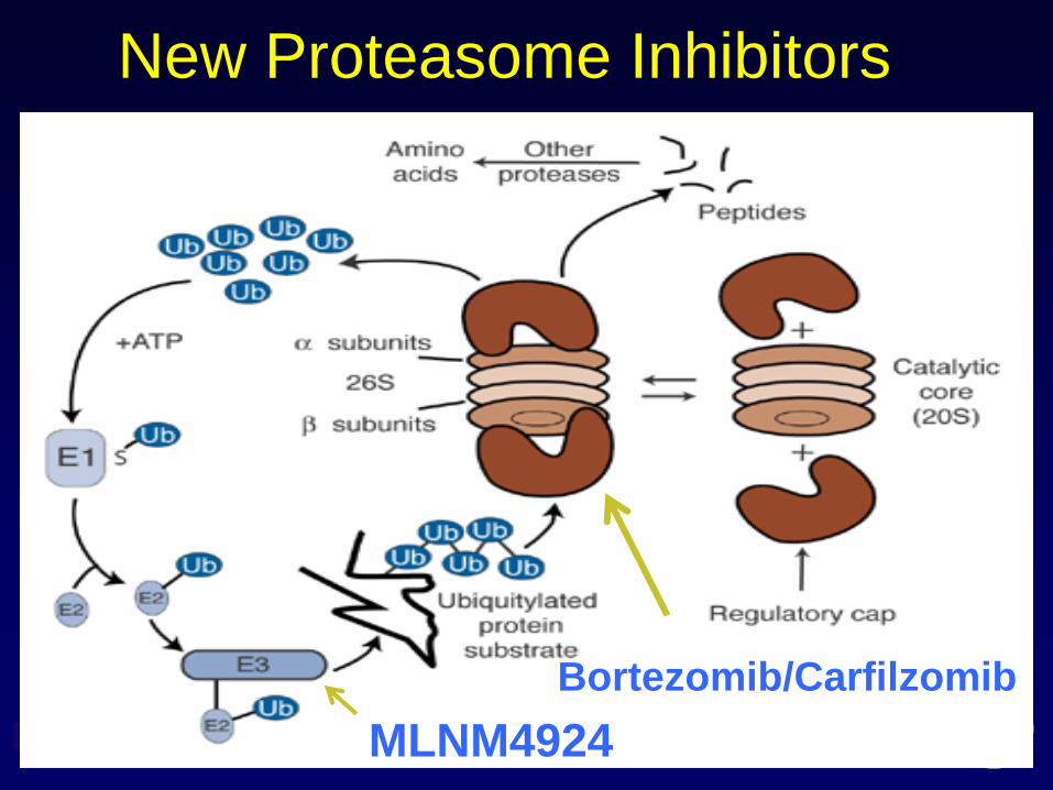
New Proteasome Inhibitors
Bortezomib/Carfilzomib
MLNM4924
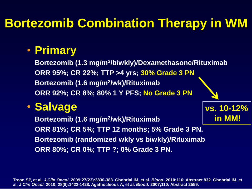
Bortezomib Combination Therapy in WM
• Primary Bortezomib (1.3 mg/m2/biwkly)/Dexamethasone/Rituximab
ORR 95%; CR 22%; TTP >4 yrs; 30% Grade 3 PN
Bortezomib (1.6 mg/m2/wk)/Rituximab
ORR 92%; CR 8%; 80% 1 Y PFS; No Grade 3 PN
• Salvage Bortezomib (1.6 mg/m2/wk)/Rituximab
ORR 81%; CR 5%; TTP 12 months; 5% Grade 3 PN.
Bortezomib (randomized wkly vs biwkly)/Rituximab
ORR 80%; CR 0%; TTP ?; 0% Grade 3 PN.
Treon SP, et al. J Clin Oncol. 2009;27(23):3830-383. Ghobrial IM, et al. Blood. 2010;116: Abstract 832. Ghobrial IM, et al. J Clin Oncol. 2010; 28(8):1422-1428. Agathocleous A, et al. Blood. 2007;110: Abstract 2559.
vs. 10-12%
in MM!
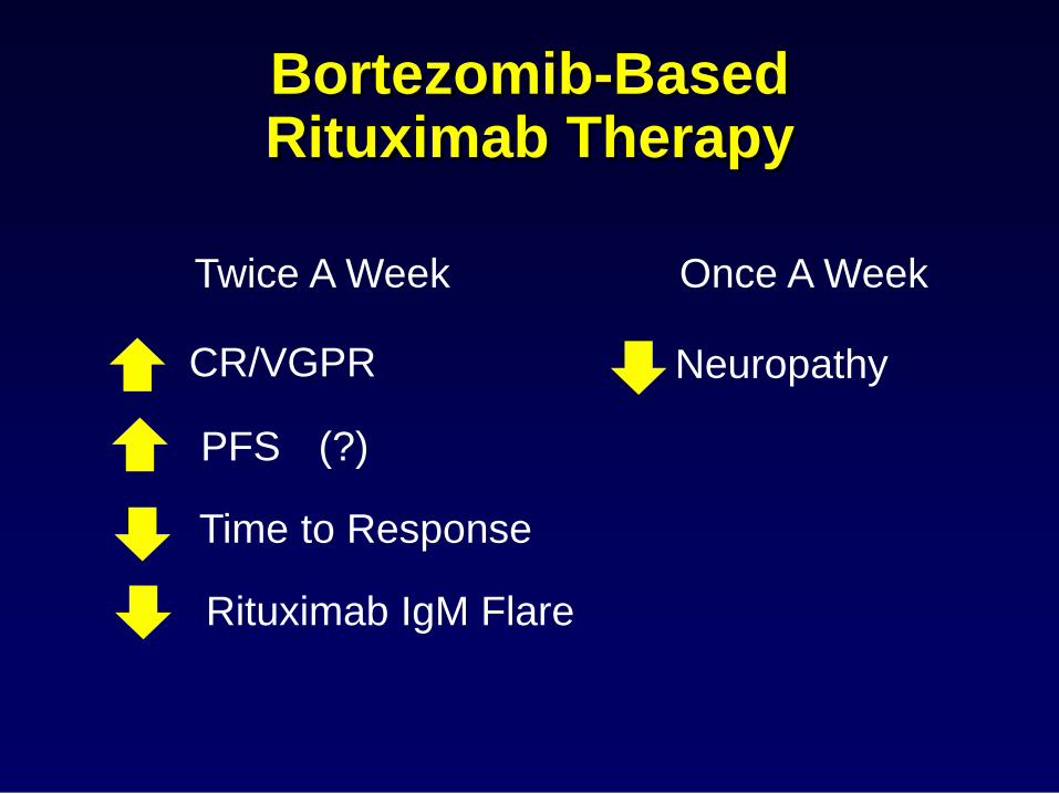
Bortezomib-Based Rituximab Therapy
Twice A Week Once A Week
CR/VGPR
PFS (?)
Time to Response
Rituximab IgM Flare
Neuropathy
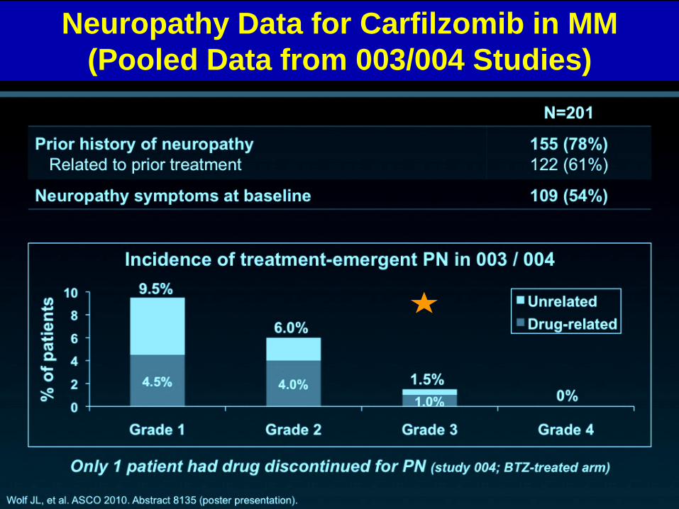
Neuropathy Data for Carfilzomib in MM
(Pooled Data from 003/004 Studies)
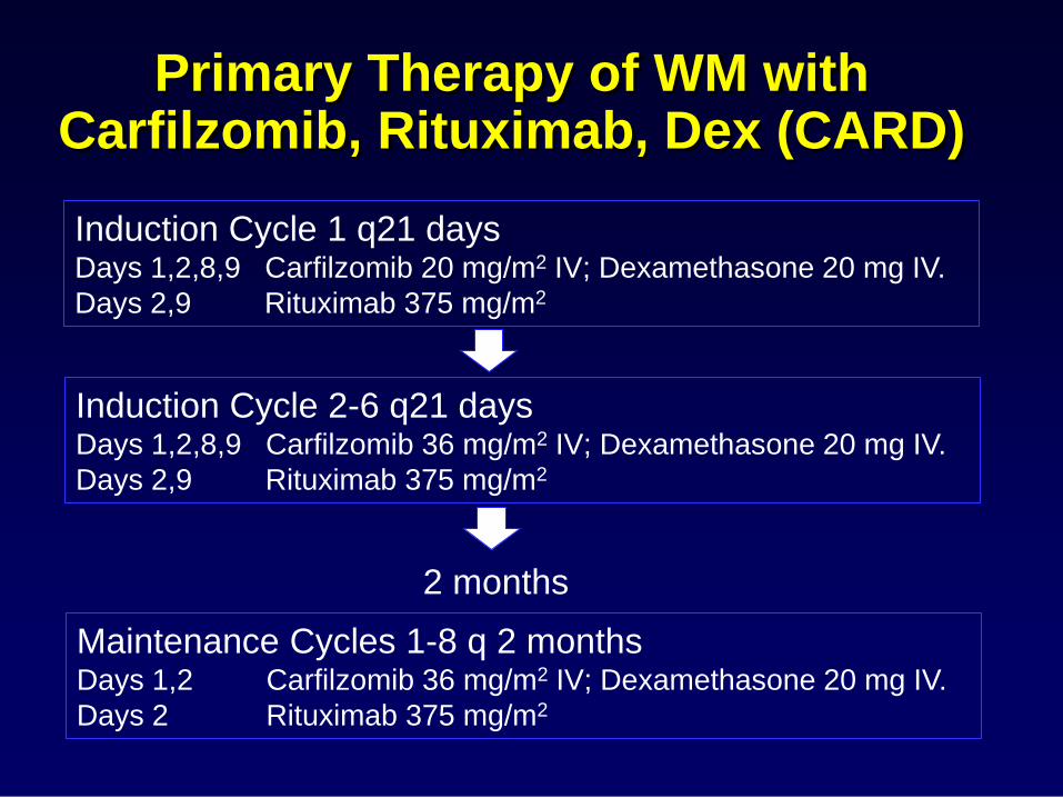
Primary Therapy of WM with Carfilzomib, Rituximab, Dex (CARD)
Induction Cycle 1 q21 days Days 1,2,8,9 Carfilzomib 20 mg/m2 IV; Dexamethasone 20 mg IV.
Days 2,9 Rituximab 375 mg/m2
Induction Cycle 2-6 q21 days Days 1,2,8,9 Carfilzomib 36 mg/m2 IV; Dexamethasone 20 mg IV.
Days 2,9 Rituximab 375 mg/m2
Maintenance Cycles 1-8 q 2 months Days 1,2 Carfilzomib 36 mg/m2 IV; Dexamethasone 20 mg IV.
Days 2 Rituximab 375 mg/m2
2 months
N
N
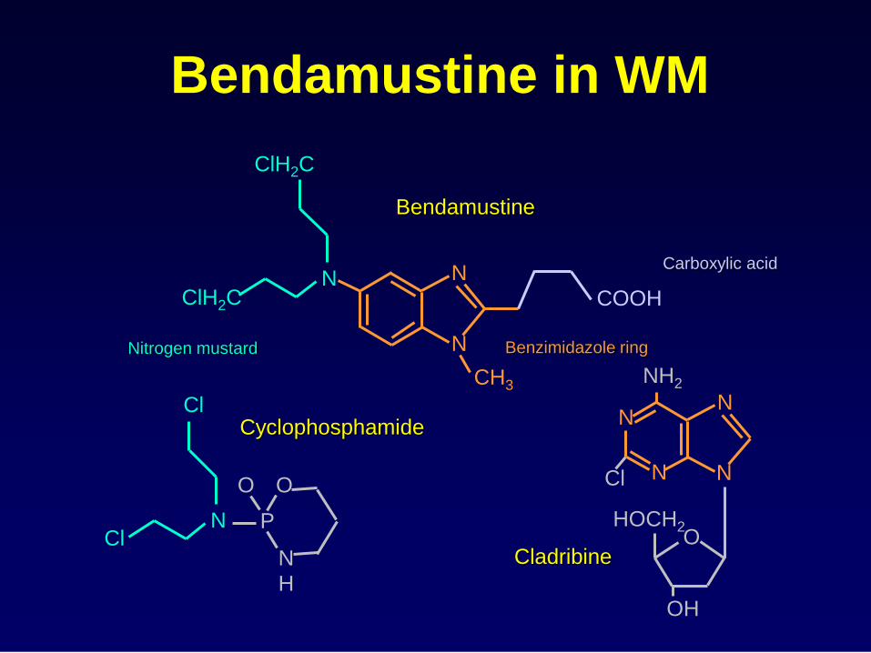
CH3
COOH
N ClH2C
ClH2C
Bendamustine
N
N
N
N
NH2
Cl
O
OH
HOCH2
Cladribine
Benzimidazole ring
N Cl
Cl
N
P
O O
H
Cyclophosphamide
Nitrogen mustard
Carboxylic acid
Bendamustine in WM
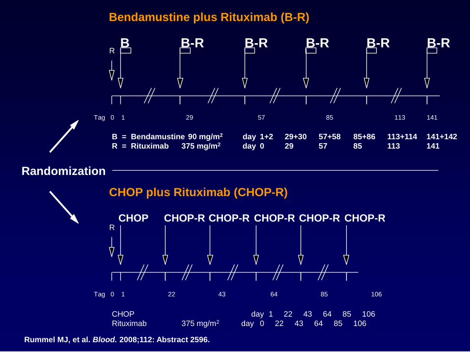
CHOP plus Rituximab (CHOP-R)
Randomization
Bendamustine plus Rituximab (B-R)
B = Bendamustine 90 mg/m2 day 1+2 29+30 57+58 85+86 113+114 141+142
R = Rituximab 375 mg/m2 day 0 29 57 85 113 141
0 1 29 57 85 113 141
R B B-R B-R B-R
Tag
B-R B-R
0 1 22 43 64 85 106
R CHOP CHOP-R CHOP-R CHOP-R
Tag
CHOP-R CHOP-R
CHOP day 1 22 43 64 85 106
Rituximab 375 mg/m2 day 0 22 43 64 85 106
Rummel MJ, et al. Blood. 2008;112: Abstract 2596.
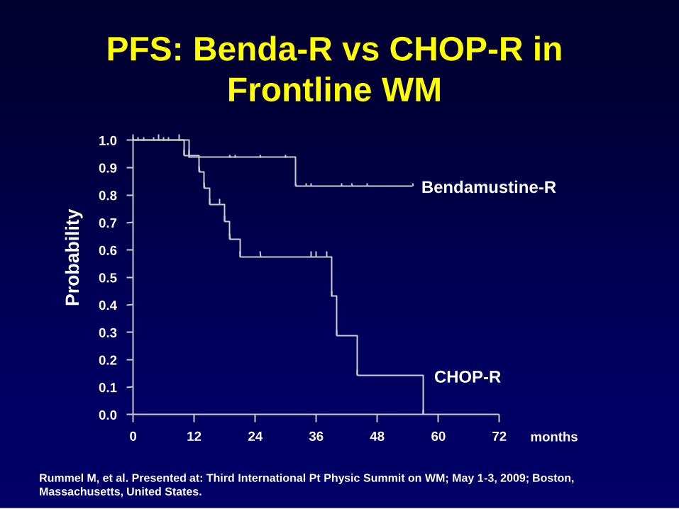
PFS: Benda-R vs CHOP-R in
Frontline WM
0 12 24 36 48 60 72
0.0
0.1
0.2
0.3
0.4
0.5
0.6
0.7
0.8
0.9
1.0
months
Pro
ba
bilit
y
Bendamustine-R
CHOP-R
Rummel M, et al. Presented at: Third International Pt Physic Summit on WM; May 1-3, 2009; Boston,
Massachusetts, United States.
Advances in the Biology of Waldenstrom’s Macroglobulinemia
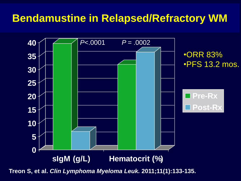
Bendamustine in Relapsed/Refractory WM
0
5
10
15
20
25
30
35
40
sIgM (g/L) Hematocrit (%)
Pre-Rx
Post-Rx
P<.0001 P = .0002
Treon S, et al. Clin Lymphoma Myeloma Leuk. 2011;11(1):133-135.
•ORR 83%
•PFS 13.2 mos.
To Maintain or Not to Maintain?
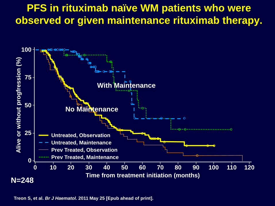
Untreated, Observation
Untreated, Maintenance
Prev Treated, Observation
Prev Treated, Maintenance
N=248
PFS in rituximab naïve WM patients who were
observed or given maintenance rituximab therapy.
100
75
50
25
0 0 10 20 30 40 50 60 70 80 90 100 110 120
Time from treatment initiation (months)
Ali
ve o
r w
ith
ou
t p
rog
fressio
n (
%)
With Maintenance
No Maintenance
Treon S, et al. Br J Haematol. 2011 May 25 [Epub ahead of print].
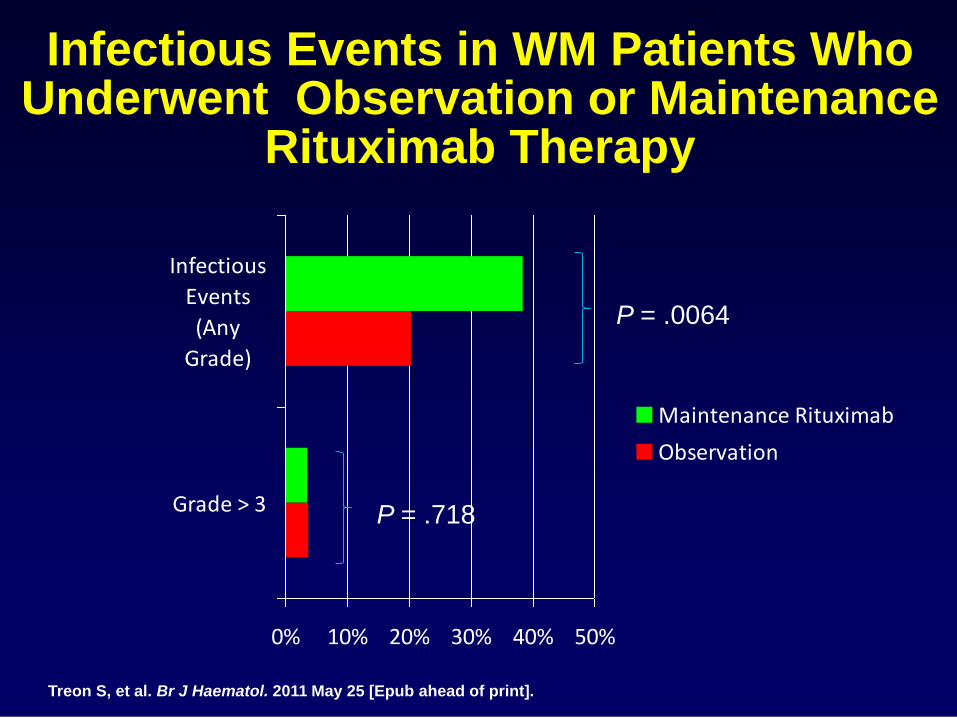
Infectious Events in WM Patients Who Underwent Observation or Maintenance
Rituximab Therapy
0% 10% 20% 30% 40% 50%
Grade > 3
Infectious
Events
(Any
Grade)
Maintenance Rituximab
Observation
P = .0064
P = .718
Treon S, et al. Br J Haematol. 2011 May 25 [Epub ahead of print].
Other Options for relapsed/refractory patients.
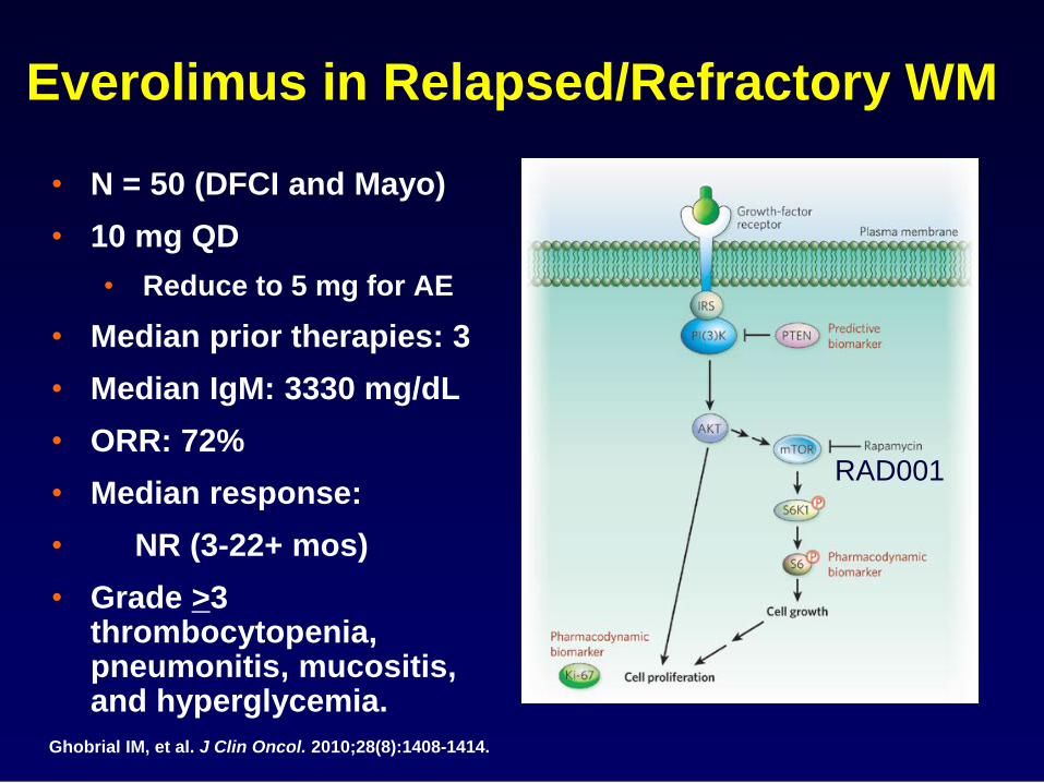
• N = 50 (DFCI and Mayo)
• 10 mg QD
• Reduce to 5 mg for AE
• Median prior therapies: 3
• Median IgM: 3330 mg/dL
• ORR: 72%
• Median response:
• NR (3-22+ mos)
• Grade >3 thrombocytopenia, pneumonitis, mucositis, and hyperglycemia.
Everolimus in Relapsed/Refractory WM
Ghobrial IM, et al. J Clin Oncol. 2010;28(8):1408-1414.
RAD001
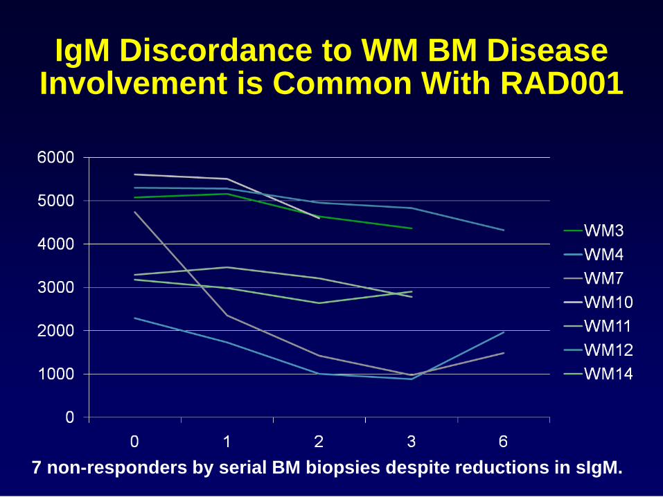
IgM Discordance to WM BM Disease Involvement is Common With RAD001
7 non-responders by serial BM biopsies despite reductions in sIgM.

Advances in the Biology of Waldenstrom’s Macroglobulinemia
“Medicine is not only a science; it is also an art. It does not consist of compounding pills and plasters; it deals with the very processes of life, which must be understood before they may be guided.”
Phillipus Aureolus Paracelsus
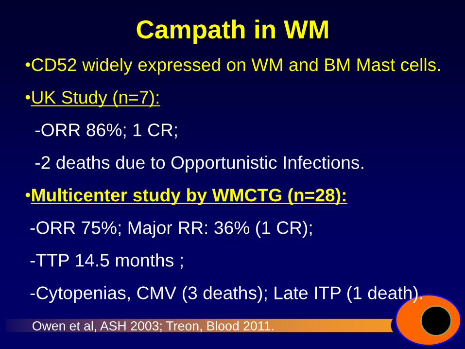
Campath in WM
•CD52 widely expressed on WM and BM Mast cells.
•UK Study (n=7):
-ORR 86%; 1 CR;
-2 deaths due to Opportunistic Infections.
•Multicenter study by WMCTG (n=28):
-ORR 75%; Major RR: 36% (1 CR);
-TTP 14.5 months ;
-Cytopenias, CMV (3 deaths); Late ITP (1 death).
Owen et al, ASH 2003; Treon, Blood 2011.
Novel Directions
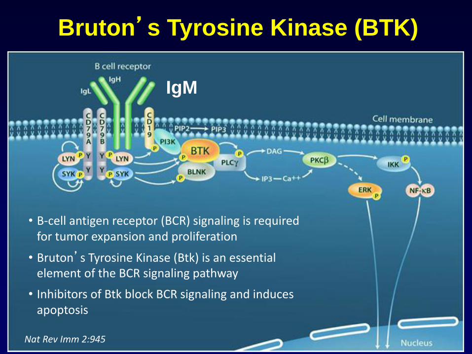
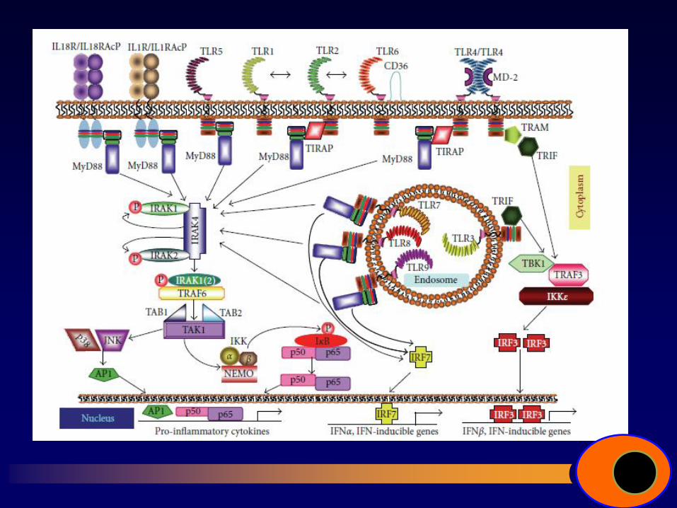
Bruton’s Tyrosine Kinase (BTK)
Nat Rev Imm 2:945
• B-cell antigen receptor (BCR) signaling is required for tumor expansion and proliferation
• Bruton’s Tyrosine Kinase (Btk) is an essential element of the BCR signaling pathway
• Inhibitors of Btk block BCR signaling and induces apoptosis
IgM
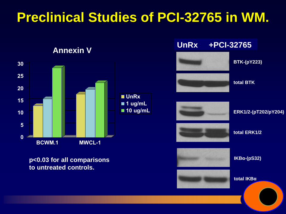
Preclinical Studies of PCI-32765 in WM.
0
5
10
15
20
25
30
BCWM.1 MWCL-1
UnRx
1 ug/mL
10 ug/mL
p<0.03 for all comparisons
to untreated controls.
Hogan Hogan +
PCI-32765
BTK-(pY223)
total BTK
ERK1/2-(pT202/pY204)
total ERK1/2
IKBα-(pS32)
total IKBα
UnRx +PCI-32765 Annexin V
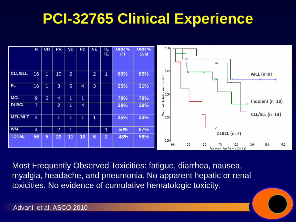
PCI-32765 Clinical Experience
N CR PR SD PD NE TE
TE
ORR %
ITT
ORR %
Eval
CLL/SLL 16 1 10 2 2 1 69% 85%
FL 16 1 3 5 4 3 25% 31%
MCL 9 3 4 1 1 78% 78%
DLBCL 7 2 1 4 29% 29%
MZL/MLT 4 1 1 1 1 25% 33%
WM 4 2 1 1 50% 67%
TOTAL 56 5 22 11 10 6 2 48% 56%
Advani et al. ASCO 2010
MCL (n=9)
CLL/SLL (n=13)
Indolent (n=20)
DLBCL (n=7)
Most Frequently Observed Toxicities: fatigue, diarrhea, nausea,
myalgia, headache, and pneumonia. No apparent hepatic or renal
toxicities. No evidence of cumulative hematologic toxicity.
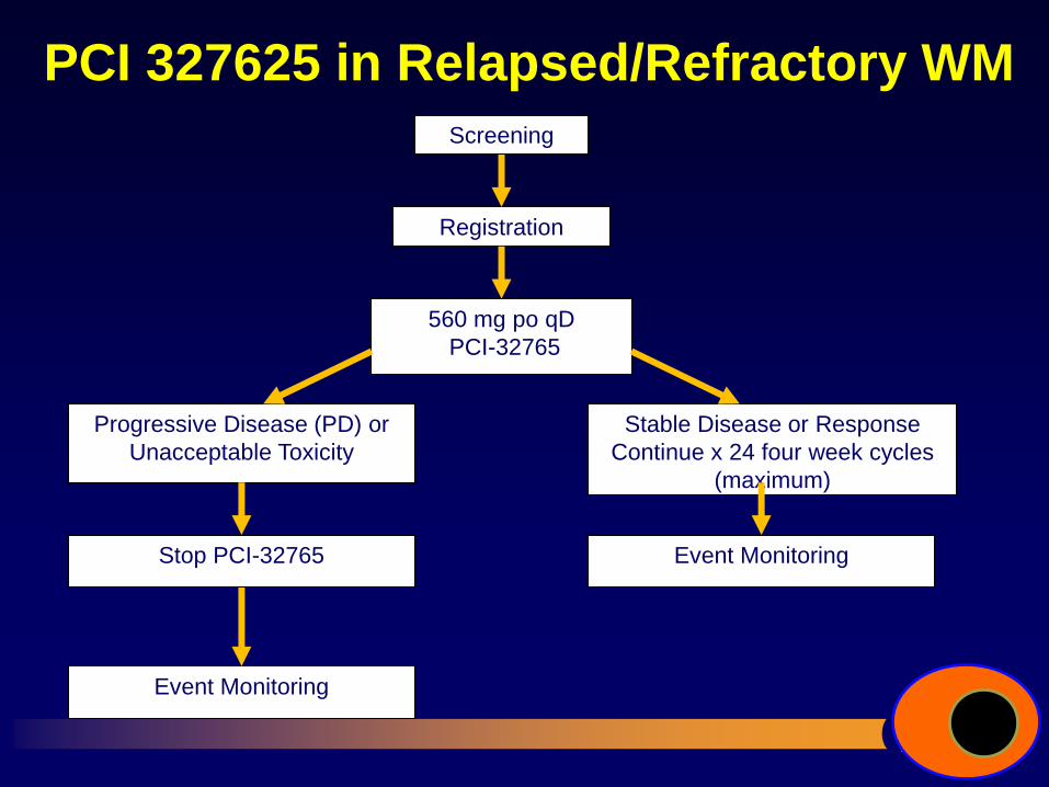
PCI 327625 in Relapsed/Refractory WM
Screening
Registration
560 mg po qD
PCI-32765
Progressive Disease (PD) or
Unacceptable Toxicity
Stable Disease or Response
Continue x 24 four week cycles
(maximum)
Stop PCI-32765
Event Monitoring
Event Monitoring
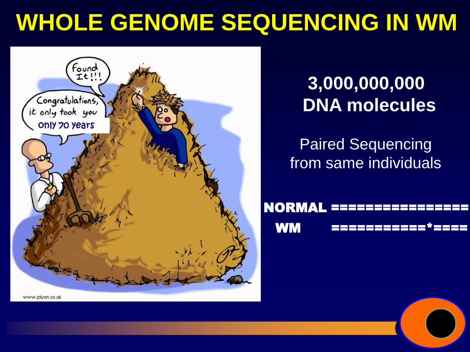
WHOLE GENOME SEQUENCING IN WM
only 70 years
WM ===========*====
Paired Sequencing
from same individuals
NORMAL ================
3,000,000,000
DNA molecules
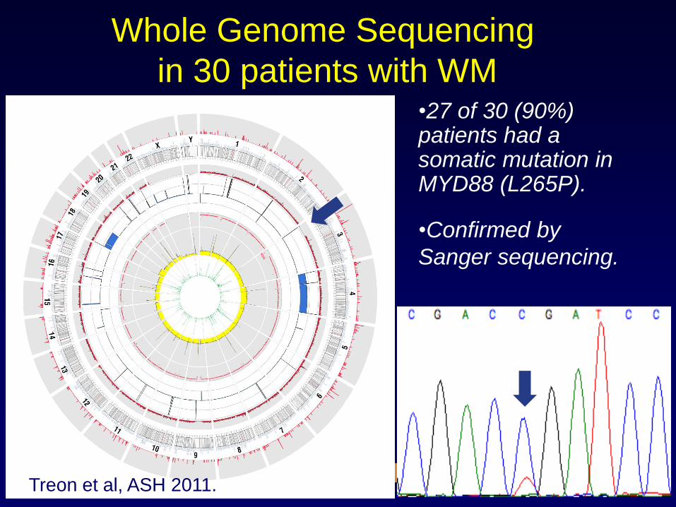
Whole Genome Sequencing
in 30 patients with WM •27 of 30 (90%) patients had a somatic mutation in MYD88 (L265P). •Confirmed by
Sanger sequencing.
Treon et al, ASH 2011.
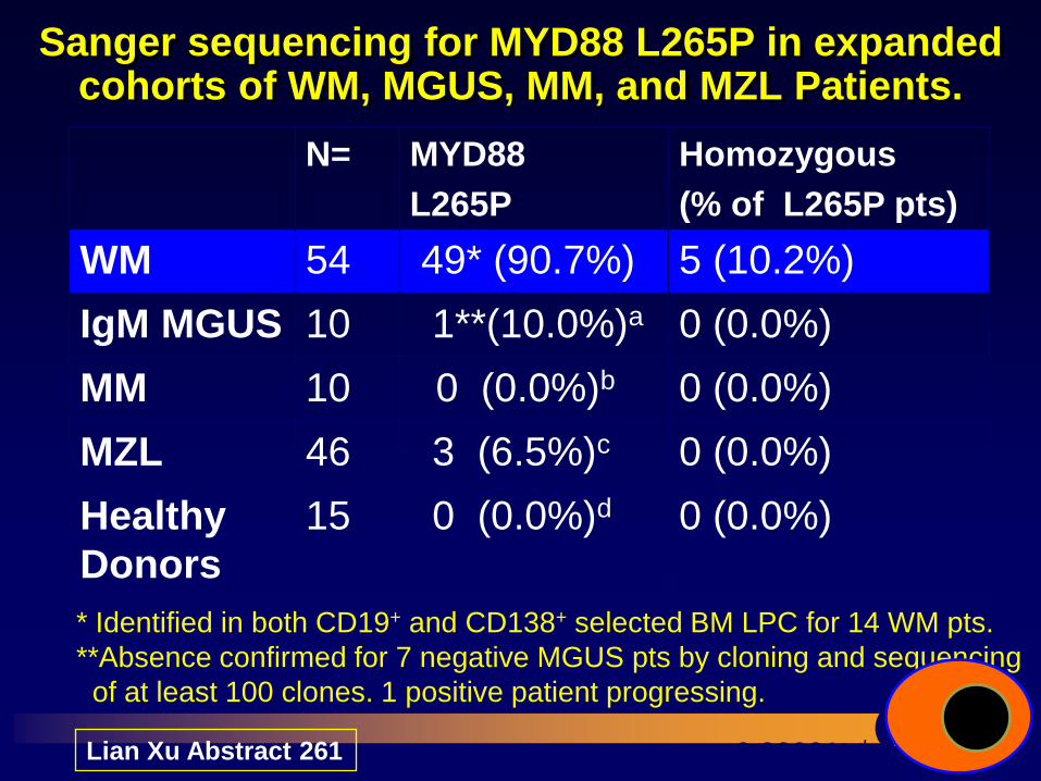
Sanger sequencing for MYD88 L265P in expanded cohorts of WM, MGUS, MM, and MZL Patients.
N= MYD88
L265P
Homozygous
(% of L265P pts)
WM 54 49* (90.7%) 5 (10.2%)
IgM MGUS 10 1**(10.0%)a 0 (0.0%)
MM 10 0 (0.0%)b 0 (0.0%)
MZL 46 3 (6.5%)c 0 (0.0%)
Healthy
Donors
15 0 (0.0%)d 0 (0.0%)
p<0.00001a-d versus WM.
**Absence confirmed for 7 negative MGUS pts by cloning and sequencing
of at least 100 clones. 1 positive patient progressing.
* Identified in both CD19+ and CD138+ selected BM LPC for 14 WM pts.
Lian Xu Abstract 261
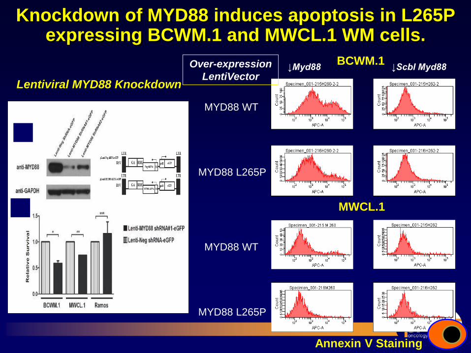
Knockdown of MYD88 induces apoptosis in L265P expressing BCWM.1 and MWCL.1 WM cells.
Lentiviral MYD88 Knockdown
BCWM.1
MWCL.1
MYD88 L265P
MYD88 WT
MYD88 L265P
MYD88 WT
Over-expression
LentiVector ↓Myd88 ↓Scbl Myd88
59.6%
46.4%
18.3%
16.9%
22.1%
11.3%
14.2%
12.1%
Annexin V Staining
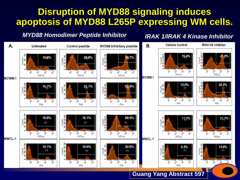
Disruption of MYD88 signaling induces apoptosis of MYD88 L265P expressing WM cells.
MYD88 Homodimer Peptide Inhibitor IRAK 1/IRAK 4 Kinase Inhibitor
Guang Yang Abstract 597
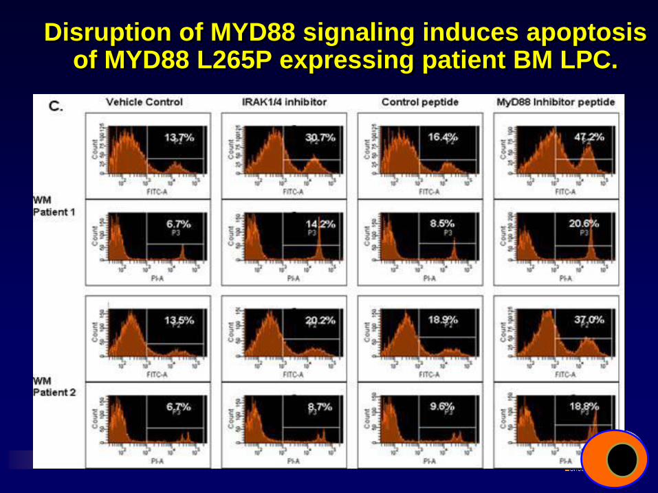
Disruption of MYD88 signaling induces apoptosis of MYD88 L265P expressing patient BM LPC.
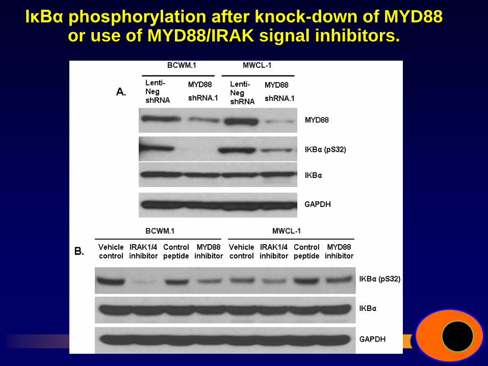
IκBα phosphorylation after knock-down of MYD88 or use of MYD88/IRAK signal inhibitors.
Take Home
• Familial predisposition is common in WM and impacts therapy.
• Bendamustine, bortezomib, cyclophosphamide, and thalidomide–based rituximab therapies are active, and can be used for symptomatic WM.
• Use of nucleoside analogues should be carefully weighed against other options.
• Better categorical responses are associated with improved PFS in rituximab treated patients, and reflect FCGR3A polymorphisms.
• WGS has revealed a somatic mutation in MYD88 in 91% of WM patients and represents a novel target for therapy of WM.

“Do not go where the path may lead, go instead where there
is no path and leave a trail”
Ralph Waldo Emerson