Embed Size (px)
Citation preview
Postgraduate Medical Journal (June 1971) 47, 354-360.
CLINICAL REVIEW
Waardenburg's syndrome and familial periodic paralysis
C. H. TAYA.M., M.B., B.S., M.R.C.P.(Glas.)
Senior Medical Registrar and Clinical Teacher, Medical Unit II, Department of Clinical Medicine,University of Singapore, Outram Road General Hospital, Singapore, 3
SummaryNine members in three generations of a Chinesefamily were found to have Waardenburg's syndromecomprising, mainly, lateral displacement of the innercanthi, broadening of the nasal root and hyper-trichosis of the eyebrows. Other minor features werealso found.Two patients had in addition, hypokalemic periodic
paralysis of the familial type, one had prominentfrontal bossing and another, bilateral cleft lips andpalate. These associated anomalies have not beenpreviously documented and the presence of two auto-somal dominant genetic defects in this family is ofparticular interest.
IntroductionIn 1951, Waardenburg described a rare hereditary
disease characterized by the following features:(1) lateral displacement of the inner canthi withdystopia canthrum, (2) broad nasal root, (3) con-fluent eyebrows with hypertrichosis of the medialends, (4) heterochromia irides, (5) congenital deaf-ness, (6) white forelock and (7) an autosomal domi-nant inheritance with variable penetrance.Many complete and incomplete forms of this
syndrome have been reported since the originaldescription (Keizer, 1952; Wildervanck, 1957;
McKenzie, 1958; Fisch, 1959; Arnvig, 1958;Partington, 1959; Di George, Olmsted & Harley,1960; Campbell, Campbell & Swift, 1962; Chew,Chen & Tan, 1968).
It is also known as a variant of the first archsyndrome (McKenzie, 1958; Campbell et al., 1962)and later other minor characteristics of the syndromewere added: (1) abnormal depigmentation of theskin (Klein, 1950; Mende, 1926; Partington, 1959;Campbell et al, 1962), (2) pigmentary changes of thefundi (Waardenburg, 1951; Di George et al., 1960)and (3) abnormal facial appearance to maldevelop-ment of the maxilla and mandible (Fisch, 1959;Campbell et al., 1962).
In this paper, nine members in three generations ofone Chinese family were found to have Waarden-burg's syndrome. The interesting aspect of thisfamily is the presence of hypokalemic periodicparalysis in two patients, frontal bossing in one andbilateral cleft lips and palate in another. None ofthese congenital anomalies has been previouslydocumented. The association with familial periodicparalysis is of particular genetic interest, since thelatter is also an autosomal dominant disease.
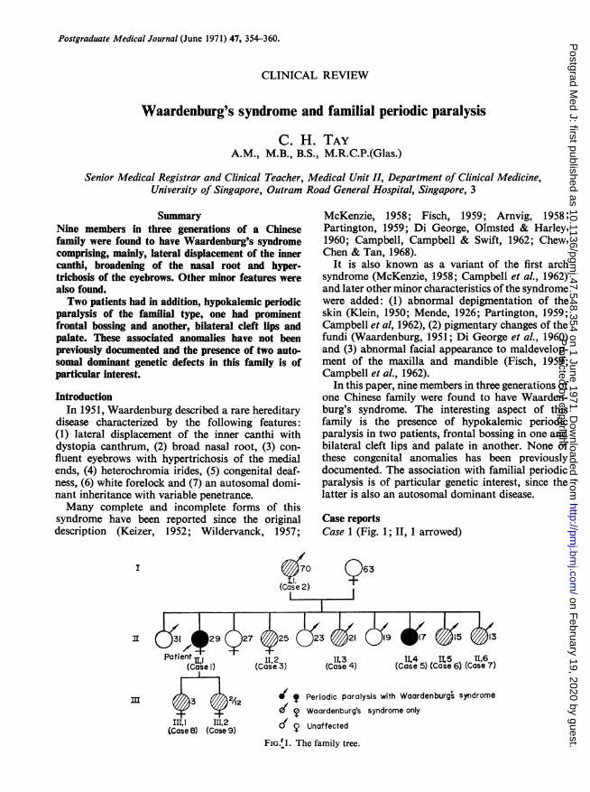
Case reportsCase 1 (Fig. 1; II, 1 arrowed)
(5 70 63
(Case 2)I ,-I
3I (j1 429(&
Patient II.(Case I)
m 2
II,1 I11.2(Case 8) (Case 9)
27 425 ( 21
11(2 (C as3(Case 3) (ase 4)
;17 5 13
11(4 S L5 11,6(Case 5) (Case 6) (Case 7)
?? Periodic paralysis with Waardenburgs syndrome0 T Waardenburg's syndrome only
dc 4 Unaffected
FIG.r1. The family tree.
Protected by copyright.
on February 19, 2020 by guest.
http://pmj.bm
j.com/
Postgrad M
ed J: first published as 10.1136/pgmj.47.548.354 on 1 June 1971. D
ownloaded from

Clinical review
The propositus, a 27-year-old Chinese housewife,was admitted on 3 February 1970 when she suddenlybecame paralysed of all four limbs early thatmorning. Her speech, swallowing, breathing andsphincteric functions were not affected. There wasno previous similar attack. She gave no history ofvomiting, diarrhoea or excessive sweating and shedenied taking any purgatives or drugs. Her generalhealth had always been good.
Examination revealed a healthy adult with noevidence of dehydration, wasting or stunting. Thethyroid was palpable but there were no signs oftoxicity. The pulse was 86/min, regular and of fullvolume, BP 110/80 mmHg. No abnormalities weredetected in the heart, lungs or abdomen. All fourlimbs, including the neck, were flaccid, and themotor power, with the exception of that of the face,was markedly diminished. Sensory functions re-mained intact. All the deep tendon reflexes werereduced. The plantar responses were equivocal.
Incidental findings in this patient were: (1) in-creased intercanthal distance, 46 mm (normalChinese adults-37-40 mm (Chew et al., 1968);there was also lateral displacement of lacrimalpunctae. The interpupillary distance was normal (65mm) (normal adult range-54-75 mm), (2) broaden-ing of nasal root, (3) hyperplasia of medial end ofeyebrows, (4) heterochromia of the left iris and (5) aspray of white forelock (which she often concealedby combing) (Fig. 2). There was no deafness, con-firmed by the audiogram, and no skin, fundal orskeletal changes. The following laboratory studieswere within normal limits: complete blood count,complete urinalysis, ESR, blood urea nitrogen, basalmetabolic rate, 1311-labelled triiodothyronine up-take, plasma PB 131I, thyroid antibodies (tanned redcell method), bilateral audiograms, X-rays of theskull, chest and abdomen, intravenous pyelograms,chromosome karyotypes and urinary electrolyteexcretions.
FIG. 2. Case 1 showing lateral displacement of innercanthi, broad nasal root, confluent eyebrows with hyper-trichosis of the medial ends, and unilateral left hetero-chromia irides.
Serum potassium on admission was 1 8 mEq/l,sodium 139 mEq/l and chloride 113 mEq/l. ECG:prolonged PR and QT intervals and prominent Uwaves, consistent with hypokalemia.The patient was treated with oral potassium
chloride 3-6 g daily in divided doses and the motorpower of her limbs rapidly returned to normal.Serum potassium on the third day was 3-6 mEq/l,sodium 136 mEq/l and chloride 120 mEq/l. ECG wasnow normal.
After discharge, she defaulted as she was stayinga long distance away from the clinic. When sheexhausted the supply of potassium tablets, she beganto experience periodic weakness of her limbs. Atfirst, these attacks were mild and readily aborted bydrinking orange and other fruit juices, but later, theparalysis was severe and she was readmitted inMarch 1970. Serum potassium was found to be 2-1mEq/l and the serum sodium and chloride werenormal. Again she improved with oral potassiumsupplements. Since then she has been on main-tenance dose of potassium chloride 1 g thrice a dayand remained symptom-free. The family tree of thepatient was traced (Fig. 1) revealing eight moremembers in three generations with similar physicalabnormalities. One brother (Case 5) also had periodicweakness of the limbs for about 6 months. Thesignificant features of this family are summarized inTable 1.
Case 2 (Fig. 1; I, 1)This 70-year-old male, father of the propositus,
had been in excellent health. At a very young age,he migrated to Singapore from China and could notrecall any disease present in his family. When seen,he had grey hair and was partially deaf because ofhis old age. The intercanthal distance was increasedto 50 mm but the interpupillary measurement wasnormal (66 mm). There was broadening of the nasalroot and confluent eyebrows with hypertrichosis ofthe medial ends. No ocular, fundal or skin changeswere present and he gave no history of periodicweakness. His hearing had been good until the last5 years.
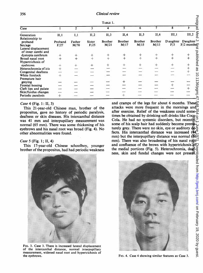
Case 3 (Fig. 1; II, 2)This 25-year-old Chinese girl, sister of the pro-
positus, had no deafness or weakness of the limbs,and she had been enjoying good health. She wasalso found to have an increase in the intercanthaldistance (43 mm) with lateral displacement of thelacrimal points, but with a normal interpupillarymeasurement. Her nasal root was somewhat promi-nent and there was marked hypertrichosis of the eye-brows (Fig. 3). There were no eye, skin, hair orauditory lesions.
355P
rotected by copyright. on F
ebruary 19, 2020 by guest.http://pm
j.bmj.com
/P
ostgrad Med J: first published as 10.1136/pgm
j.47.548.354 on 1 June 1971. Dow
nloaded from
356 Clinical review
TABLE 1.
Case 1 2 3 4 5 6 7 8 9
Generation II,1 I,1 11,2 II,3 11,4 11,5 11,6 111,1 111,2Relationship toproband Proband Father Sister Brother Brother Brother Brother D aughter Daughter
Sex/age F/27 M/70 F/25 M/21 M/17 M/15 M/13 F/3 F/2 monthsLateral displacementof inner canthi anddystopia canthrum + + + + + + + + +Broad nasal root + ± + + + + + + +Hypertrichosis ofeyebrows + + + + + + + + +Heterochromia of iris + - +Congenital deafness - - - +White forelock + - -Premature hairgreying +
Frontal bossing +Cleft lips and palate - - - +Skin/fundus changesPeriodic paralysis + - +
Case 4 (Fig. 1: 11. 3)This 21-year-old Chinese man, brother of the
propositus, gave no history of periodic paralysis,deafness or skin diseases. His intercanthal distancewas 41 mm and interpupillary measurement wasnormal (65 mm). There was some thickening of hiseyebrows and his nasal root was broad (Fig. 4). Noother abnormalities were found.
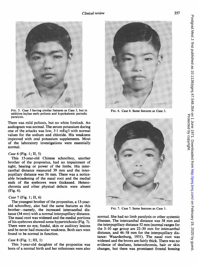
Case 5 (Fig. 1; II, 4)This 17-year-old Chinese schoolboy, younger
brother of the propositus, had had periodic weakness
....j/is .:.: 44E::::v
FIG. 3. Case 3. There is increased lateral displacementof the intercanthal distance, normal interpupillarymeasurement, widened nasal root and hypertrichosis ofthe eyebrows.
and cramps of the legs for about 6 months. Theseattacks were more frequent in the mornings andafter exercise. Relief of the weakness could some-times be obtained by drinking soft drinks like Coca-Cola. He had no systemic disorders, but recently,some of his scalp hair had suddenly become prema-turely grey. There were no skin, eye or auditory de-fects. His intercanthal distance was increased (44mm) but the interpupillary distance was normal (56mm). There was also broadening of his nasal rootand confluence of the brows with hypertrichosis ofthe medial portions (Fig. 5). Heterochromia, deaf-ness, skin and fundal changes were not present.
... ....
FIG. 4. Case 4 showing similar features as Case 3.
Protected by copyright.
on February 19, 2020 by guest.
http://pmj.bm
j.com/
Postgrad M
ed J: first published as 10.1136/pgmj.47.548.354 on 1 June 1971. D
ownloaded from
Clinical review 357
FIG. 5. Case 5 having similar features as Case 3, but inaddition he has early poliosis and hypokalemic periodicparalysis.
There was mild poliosis, but no white forelock. Anaudiogram was normal. The serum potassium duringone of the attacks was low, 3d1 mEq/l with normalvalues for the sodium and chloride. His weaknessimproved with oral potassium supplements. Mostof the laboratory investigations were essentiallynormal.
Case 6 (Fig. 1; II, 5)This 15-year-old Chinese schoolboy, another
brother of the propositus, had no impairment ofsight, hearing or power of the limbs. His inter-canthal distance measured 39 mm and the inter-pupillary distance was 56 mm. There was a notice-able broadening of the nasal root and the medialends of the eyebrows were thickened. Hetero-chromia and other physical defects were absent(Fig. 6).Case 7 (Fig. 1; II, 6)The youngest brother of the propositus, a 13-year-
old schoolboy, also had the same features as thisbrother-namely, the increased intercanthal dis-tance (34 mm) with a normal interpupillary distance.The nasal root was widened and the medial portionsof his eyebrows showed some hypertrichosis (Fig. 7).There were no eye, fundal, skin or auditory lesionsand he never had muscular weakness. Both ears werefound to be normal in function.
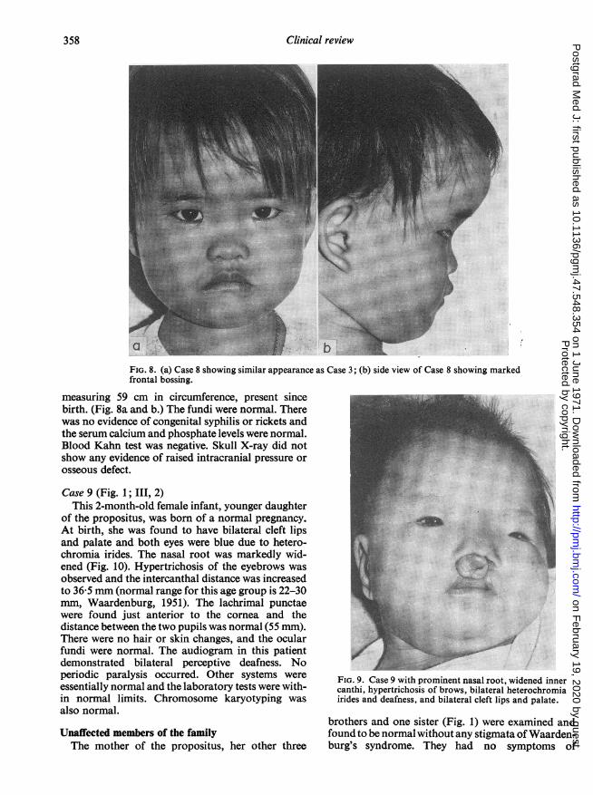
Case 8 (Fig. 1; III, 1)This 3-year-old daughter of the propositus was
born of a normal birth and her milestones were also
.............
.....
FIG. 6. Case 6. Same features as Case 3.
..::. ...
X..... .. ... .. ..... ; ...
FIG. 7. Case 7. Same features as Case 3.
normal. She had no limb paralysis or other systemicillnesses. The intercanthal distance was 38 mm andthe interpupillary distance 52 mm (normal ranges forthe 3-10 age group are 22-30 mm for intercanthaldistance, and 46-58 mm for the interpupillary dis-tance: Waardenburg, 1951). The nasal root waswidened and the brows are fairly thick. There was noevidence of deafness, heterochromia, hair or skinchanges, but there was prominent frontal bossing
Protected by copyright.
on February 19, 2020 by guest.
http://pmj.bm
j.com/
Postgrad M
ed J: first published as 10.1136/pgmj.47.548.354 on 1 June 1971. D
ownloaded from
Clinical review
FIG. 8. (a) Case 8 showing similar appearance as Case 3; (b) side view of Case 8 showing markedfrontal bossing.
measuring 59 cm in circumference, present sincebirth. (Fig. 8a and b.) The fundi were normal. Therewas no evidence of congenital syphilis or rickets andthe serum calcium and phosphate levels were normal.Blood Kahn test was negative. Skull X-ray did notshow any evidence of raised intracranial pressure orosseous defect.
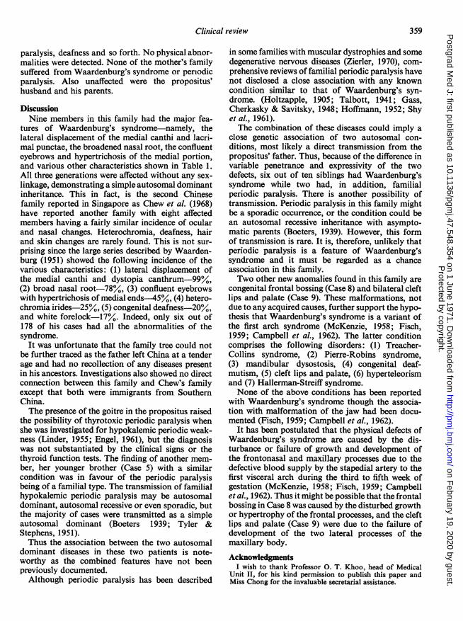
Case 9 (Fig. 1; III, 2)This 2-month-old female infant, younger daughter
of the propositus, was born of a normal pregnancy.At birth, she was found to have bilateral cleft lipsand palate and both eyes were blue due to hetero-chromia irides. The nasal root was markedly wid-ened (Fig. 10). Hypertrichosis of the eyebrows wasobserved and the intercanthal distance was increasedto 36 5 mm (normal range for this age group is 22-30mm, Waardenburg, 1951). The lachrimal punctaewere found just anterior to the cornea and thedistance between the two pupils was normal (55 mm).There were no hair or skin changes, and the ocularfundi were normal. The audiogram in this patientdemonstrated bilateral perceptive deafness. Noperiodic paralysis occurred. Other systems wereessentially normal and the laboratory tests were with-in normal limits. Chromosome karyotyping wasalso normal.
Unaffected members of the familyThe mother of the propositus, her other three
FIG. 9. Case 9 with prominent nasal root, widened innercanthi, hypertrichosis of brows, bilateral heterochromiairides and deafness, and bilateral cleft lips and palate.
brothers and one sister (Fig. 1) were examined andfound to be normal without any stigmata of Waarden-burg's syndrome. They had no symptoms of
358P
rotected by copyright. on F
ebruary 19, 2020 by guest.http://pm
j.bmj.com
/P
ostgrad Med J: first published as 10.1136/pgm
j.47.548.354 on 1 June 1971. Dow
nloaded from
Clinical review
paralysis, deafness and so forth. No physical abnor-malities were detected. None of the mother's familysuffered from Waardenburg's syndrome or periodicparalysis. Also unaffected were the propositus'husband and his parents.
DiscussionNine members in this family had the major fea-
tures of Waardenburg's syndrome-namely, thelateral displacement of the medial canthi and lacri-mal punctae, the broadened nasal root, the confluenteyebrows and hypertrichosis of the medial portion,and various other characteristics shown in Table 1.All three generations were affected without any sex-linkage, demonstrating a simple autosomal dominantinheritance. This in fact, is the second Chinesefamily reported in Singapore as Chew et al. (1968)have reported another family with eight affectedmembers having a fairly similar incidence of ocularand nasal changes. Heterochromia, deafness, hairand skin changes are rarely found. This is not sur-prising since the large series described by Waarden-burg (1951) showed the following incidence of thevarious characteristics: (1) lateral displacement ofthe medial canthi and dystopia canthrum-99°/,(2) broad nasal root-78°/, (3) confluent eyebrowswith hypertrichosis of medial ends-455/0, (4) hetero-chromia irides-25%/, (5) congenital deafness-20°/,and white forelock-17%. Indeed, only six out of178 of his cases had all the abnormalities of thesyndrome.
It was unfortunate that the family tree could notbe further traced as the father left China at a tenderage and had no recollection of any diseases presentin his ancestors. Investigations also showed no directconnection between this family and Chew's familyexcept that both were immigrants from SouthernChina.The presence of the goitre in the propositus raised
the possibility of thyrotoxic periodic paralysis whenshe was investigated for hypokalemic periodic weak-ness (Linder, 1955; Engel, 1961), but the diagnosiswas not substantiated by the clinical signs or thethyroid function tests. The finding of another mem-ber, her younger brother (Case 5) with a similarcondition was in favour of the periodic paralysisbeing of a familial type. The transmission of familialhypokalemic periodic paralysis may be autosomaldominant, autosomal recessive or even sporadic, butthe majority of cases were transmitted as a simpleautosomal dominant (Boeters 1939; Tyler &Stephens, 1951).Thus the association between the two autosomal
dominant diseases in these two patients is note-worthy as the combined features have not beenpreviously documented.Although periodic paralysis has been described
in some families with muscular dystrophies and somedegenerative nervous diseases (Zierler, 1970), com-prehensive reviews of familial periodic paralysis havenot disclosed a close association with any knowncondition similar to that of Waardenburg's syn-drome. (Holtzapple, 1905; Talbott, 1941; Gass,Cherkasky & Savitsky, 1948; Hoffmann, 1952; Shyet al., 1961).The combination of these diseases could imply a
close genetic association of two autosomal con-ditions, most likely a direct transmission from thepropositus' father. Thus, because of the difference invariable penetrance and expressivity of the twodefects, six out of ten siblings had Waardenburg'ssyndrome while two had, in addition, familialperiodic paralysis. There is another possibility oftransmission. Periodic paralysis in this family mightbe a sporadic occurrence, or the condition could bean autosomal recessive inheritance with asympto-matic parents (Boeters, 1939). However, this formof transmission is rare. It is, therefore, unlikely thatperiodic paralysis is a feature of Waardenburg'ssyndrome and it must be regarded as a chanceassociation in this family.Two other new anomalies found in this family are
congenital frontal bossing (Case 8) and bilateral cleftlips and palate (Case 9). These malformations, notdue to any acquired causes, further support the hypo-thesis that Waardenburg's syndrome is a variant ofthe first arch syndrome (McKenzie, 1958; Fisch,1959; Campbell et al., 1962). The latter conditioncomprises the following disorders: (1) Treacher-Collins syndrome, (2) Pierre-Robins syndrome,(3) mandibular dysostosis, (4) congenital deaf-mutism, (5) cleft lips and palate, (6) hyperteleorismand (7) Hallerman-Streiff syndrome.None of the above conditions has been reported
with Waardenburg's syndrome though the associa-tion with malformation of the jaw had been docu-mented (Fisch, 1959; Campbell et al., 1962).
It has been postulated that the physical defects ofWaardenburg's syndrome are caused by the dis-turbance or failure of growth and development ofthe frontonasal and maxillary processes due to thedefective blood supply by the stapedial artery to thefirst visceral arch during the third to fifth week ofgestation (McKenzie, 1958; Fisch, 1959; Campbellet al., 1962). Thus it might be possible that the frontalbossing in Case 8 was caused by the disturbed growthor hypertrophy of the frontal processes, and the cleftlips and palate (Case 9) were due to the failure ofdevelopment of the two lateral processes of themaxillary body.
AcknowledgmentsI wish to thank Professor 0. T. Khoo, head of Medical
Unit II, for his kind permission to publish this paper andMiss Chong for the invaluable secretarial assistance.
359P
rotected by copyright. on F
ebruary 19, 2020 by guest.http://pm
j.bmj.com
/P
ostgrad Med J: first published as 10.1136/pgm
j.47.548.354 on 1 June 1971. Dow
nloaded from
360 Clinical review
ReferencesARNVIG, J. (1959) The syndrome of Waardenburg. Acta
genetica et statistica medica, 9, 41.BOETERS, H. (1939) Erbleiden des Nerren systems beim
Menschen. In: Handbuck der Erbbiologie des Menschen(Ed. by E. Just), p. 59. Springer Verlag, Berlin.
CAMPBELL, B., CAMPBELL, N.R. & SWIFT, S. (1962) Waarden-burg's syndrome-a variation of the first arch syndrome.Archives of Dermatology, 86, 718.
CHEW, K.L., CHEN, A.J. & TAN, K.H. (1968) A Chinesefamily with Waardenburg's syndrome. American Journalof Ophthalmology, 65, 174.
Di GEORGE, A.M., OLMSTED, R.W. & HARLEY, R.D. (1960)Waardenburg's syndrome-a syndrome of heterchromiaof the irides, lateral displacement of the medial canthi andlacrimal puncta, congenital deafness and other charac-teristic associated defects. Journal of Pediatrics, 57, 649.
ENGEL, A.G. (1961) Thyroid function and periodic paralysis.American Journal of Medicine, 30, 327.
FisCH, L. (1959) Deafness as part of an hereditary syndrome.Journal ofLaryngology and Otology, 73, 355.
GASS, H., CHERKASKY, M. & SAVITSKY, N. (1948) Potassiumand periodic paralysis. Medicine, 27, 105.
HOFMANN, W.W. (1952) The syndrome of periodic paralysis,Neurology, 2, 1.
HOLTZAPPLE, G.E. (1905) Periodic paralysis. Journal of theAmerican Medical Association, 45, 1224.
KEIZER, D.P.R. (1952) Een Nieuwe Vorn Van CongenitaleEnfelijke Doofheid (Syndroom Van Waardenburg). Neder-landsch tijdschrift voor geneeskunde, 96, 2541.
KLEIN, D. (1950) Albinisme, partiel (leucisme) avec surdi-mutite, blepharophimosis et dysplasie myo-osteo-articu-lasie. Helvetica paediatrica acta, 5, 38.
LINDER, M.A. (1955) Periodic paralysis with hyperthyroidism.Annals of Internal Medicine, 43, 241.
McKENZIE, J. (1958) The first arch syndrome. Archives ofDiseases of Childhood, 73, 477.
MENDE, I. (1926) Uber eine familie hereditar-degenerativerTaubstummer mit mongoloidiem Einschlag and teilwesemLeukismus der Hant und Haare. Archiv fur Kinderheil-kunde, 70, 214.
PARTINGTON, M.W. (1959) An English family with Waarden-burg's syndrome. Archives of Diseases of Childhood, 34,154.
SHY, G.M., WANKO, T., ROWLEY, P.T. & ENGEL, A.G. (1961)Studies in familial periodic paralysis. ExperimentalNeurology, 3, 53.
TALBOTT, J.H. (1941) Periodic paralysis-a clinical syndrome.Medicine, 20, 85.
TYLER, F.H. & STEPHENS, F.E. (1951) The clinical manifesta-tions and inheritance of a type of periodic paralysis with-out hypopotassemia. Journal of Clinical Investigations, 30,492.
WAARDENBURG, P.J. (1951) A new syndrome combining de-velopmental anomalies of the eyelids, eyebrows and noseroot with pigmentary defects of the iris and head hair andwith congenital deafness. American Journal of HumanGenetics, 3, 195.
WILDERVANCK, L.S. (1957) Doof Stomme Kinderen met betSyndroom van Waardenburg-Klein. Nederlandsch tijd-schrift geneeskunde, 101, 120.
ZIERLER, K.L. (1970) Diseases of the muscles. In: BiochemicalDisorders in Human Disease (Ed. by R. H. S. Thompson &I. D. Wootton), 3rd edn, p. 489. J. & A. Churchill, London.
Protected by copyright.
on February 19, 2020 by guest.
http://pmj.bm
j.com/
Postgrad M
ed J: first published as 10.1136/pgmj.47.548.354 on 1 June 1971. D
ownloaded from