Embed Size (px)
Citation preview

VULVAR CANCER - GUIDELINES 1
VULVAR CANCER
GUIDELINES
- Complete report -
VULVAR CANCER - GUIDELINES 1
VULVAR CANCER
GUIDELINES
- Complete report -
VULVAR CANCER - GUIDELINES 1
VULVAR CANCER
GUIDELINES
- Complete report -

VULVAR CANCER - GUIDELINES 2
TABLE OF CONTENTS
1 Introduction................................................................................................................................................................ 4
2 Acknowledgements .................................................................................................................................................... 4
3 Method........................................................................................................................................................................ 4
3.1 Nomination of multidisciplinary international development group ............................................................................ 5
3.2 Identification of scientific evidence ............................................................................................................................ 5
3.3 Formulation of guidelines ........................................................................................................................................... 5
3.4 External evaluation of the guidelines - International review....................................................................................... 6
3.5 Integration of international reviewers comments ........................................................................................................ 6
4 Management of conflicts of interest.......................................................................................................................... 6
5 Summary of guidelines .............................................................................................................................................. 7
5.1 Diagnosis and referral ................................................................................................................................................. 7
5.2 Staging system............................................................................................................................................................ 7
5.3 Preoperative investigations ......................................................................................................................................... 7
5.4 Surgical management.................................................................................................................................................. 8
5.5 Sentinel lymph node procedure................................................................................................................................... 9
5.6 Radiation therapy........................................................................................................................................................ 9
5.7 Chemoradiation......................................................................................................................................................... 10
5.8 Systemic treatment.................................................................................................................................................... 10
5.9 Treatment of recurrent disease.................................................................................................................................. 10
5.10 Follow-up ................................................................................................................................................................. 11
6 Diagnosis and referral ............................................................................................................................................. 12
6.1 Summary of available scientific evidence................................................................................................................. 12
6.2 Previous initiatives.................................................................................................................................................... 12
6.3 Development group comments ................................................................................................................................. 12
6.4 Guidelines................................................................................................................................................................. 12
7 Staging system.......................................................................................................................................................... 13
7.1 Summary of available scientific evidence................................................................................................................. 13
7.2 Previous initiatives.................................................................................................................................................... 13
7.3 Development group comments ................................................................................................................................. 13
7.4 Guidelines................................................................................................................................................................. 13
8 Preoperative investigations ..................................................................................................................................... 15
8.1 Summary of available scientific evidence................................................................................................................. 15
8.2 Previous initiatives.................................................................................................................................................... 16
8.3 Development group comments ................................................................................................................................. 16
8.4 Guidelines................................................................................................................................................................. 17
9 Surgical management .............................................................................................................................................. 19
9.1 Summary of available scientific evidence................................................................................................................. 19
9.2 Previous initiatives.................................................................................................................................................... 23
9.3 Development group comments ................................................................................................................................. 23
9.4 Guidelines................................................................................................................................................................. 24
10 Sentinel lymph node procedure .............................................................................................................................. 25
10.1 Summary of available scientific evidence................................................................................................................. 25
10.2 Previous initiatives.................................................................................................................................................... 28

VULVAR CANCER - GUIDELINES 3
10.3 Development group comments ................................................................................................................................. 29
10.4 Guidelines................................................................................................................................................................. 29
11 Radiation therapy .................................................................................................................................................... 31
11.1 Summary of available scientific evidence................................................................................................................. 31
11.2 Previous initiatives.................................................................................................................................................... 32
11.3 Development group comments ................................................................................................................................. 33
11.4 Guidelines................................................................................................................................................................. 33
12 Chemoradiation ....................................................................................................................................................... 35
12.1 Summary of available scientific evidence................................................................................................................. 35
12.2 Previous initiatives.................................................................................................................................................... 37
12.3 Development group comments ................................................................................................................................. 37
12.4 Guidelines................................................................................................................................................................. 37
13 Systemic treatment .................................................................................................................................................. 43
13.1 Summary of available scientific evidence................................................................................................................. 43
13.2 Previous initiatives.................................................................................................................................................... 43
13.3 Development group comments ................................................................................................................................. 43
13.4 Guidelines................................................................................................................................................................. 43
14 Treatment of recurrent disease............................................................................................................................... 45
14.1 Summary of available scientific evidence................................................................................................................. 45
14.2 Previous initiatives.................................................................................................................................................... 45
14.3 Development group comments ................................................................................................................................. 45
14.4 Guidelines................................................................................................................................................................. 45
15 Follow-up.................................................................................................................................................................. 48
15.1 Summary of available scientific evidence................................................................................................................. 48
15.2 Previous initiatives.................................................................................................................................................... 48
15.3 Development group comments ................................................................................................................................. 48
15.4 Guidelines................................................................................................................................................................. 48
16 Acronyms and abbreviations .................................................................................................................................. 49
17 References................................................................................................................................................................. 51
18 Appendices ............................................................................................................................................................... 66
18.1 Appendix 1 - People involved in the development of the guidelines ........................................................................ 66
18.2 Appendix 2 - List of evidence-based medicine websites consulted .......................................................................... 73
18.3 Appendix 3 - Key to evidence statements and grades of recommendations.............................................................. 74
VULVAR CANCER - GUIDELINES 4
1 IntroductionVulvar cancers are relatively uncommon and affect predominantly elderly women. The vast majority aresquamous cell carcinomas. The objectives of the guidelines are to improve and to homogenize the managementof patients with vulvar cancer. The guideline is intended for use by gynaecological oncologists, generalgynaecologists, surgeons, pathologists, radiotherapists, medical and clinical oncologists, general practitioners,palliative care teams, and allied health professionals.
The guideline covers diagnosis and referral, preoperative investigations, surgical management (local treatment,groin treatment, reconstructive surgery), sentinel lymph node procedure, radiation therapy, chemoradiation,systemic treatment, treatment of recurrent disease (vulvar recurrence, groin recurrence, distant metastases), andfollow-up for patients with vulvar cancer and provides information for discussion with patients and carers. Thiscomplete report does not include any economic analysis of the strategies. These guidelines apply to adults overthe age of 18 years with squamous cell carcinoma of the vulva. These guidelines do not address patients withother vulvar cancer histologies.
Any clinician seeking to apply or consult these guidelines is expected to use independent medical judgment inthe context of individual clinical circumstances to determine any patient’s care or treatment.
2 AcknowledgementsThe European society of gynaecological oncology (ESGO) would like to thank the international developmentgroup for their constant availability, work, and for making possible the development of these guidelines for themanagement of patients with vulvar cancer. ESGO is also very grateful to the external panel of physicians andpatients (international reviewers) for their participation. The names of the participants in each group are listed onAppendix 1.
ESGO also wishes to express sincere gratitude to the Institut National du Cancer (INCa, France) for providingthe main funding for this work.
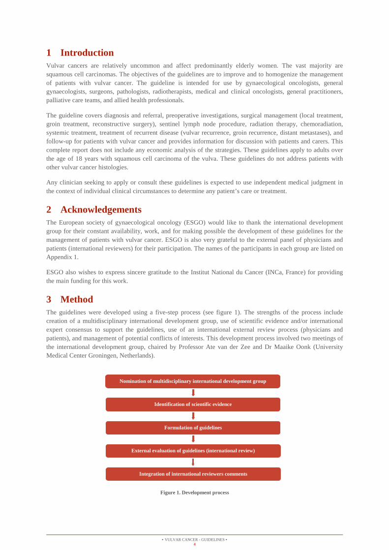
3 MethodThe guidelines were developed using a five-step process (see figure 1). The strengths of the process includecreation of a multidisciplinary international development group, use of scientific evidence and/or internationalexpert consensus to support the guidelines, use of an international external review process (physicians andpatients), and management of potential conflicts of interests. This development process involved two meetings ofthe international development group, chaired by Professor Ate van der Zee and Dr Maaike Oonk (UniversityMedical Center Groningen, Netherlands).
Figure 1. Development process
External evaluation of guidelines (international review)
Nomination of multidisciplinary international development group
Identification of scientific evidence
Integration of international reviewers comments
Formulation of guidelines

VULVAR CANCER - GUIDELINES 5
3.1 Nomination of multidisciplinary international development groupThe ESGO Council nominated practicing clinicians that care for vulvar cancer patients and have demonstratedleadership in clinical management of patients through research, administrative responsibilities, and/or committeemembership to serve on the expert panel. The objective was to assemble a multidisciplinary panel. It wastherefore essential to include professionals from relevant disciplines (gynaecological oncology, medicaloncology, pathology, radiation oncology, surgery) so that their perspectives would contribute to the validity andacceptability of the guidelines. The list of the development group is available in Appendix 1.1.
3.2 Identification of scientific evidenceTo ensure that the statements made in this document are evidence based, the current literature was reviewed andcritically appraised. A systematic literature review of the studies published between January 1980 and September2015 was carried out using the MEDLINE database. This search used indexing terms as follows: accuracy,adverse effects, bilateral en bloc dissection, biopsy, chemotherapy (primary, neoadjuvant, adjuvant),chemoradiation (primary, neoadjuvant, adjuvant), chemotherapeutic agents, detection rate, diagnosis, en blocdissection, exenteration (anterior, posterior, total), follow-up, frozen sections, groin lymph node involvement,groin node metastasis, histology, histological examination, imaging, inguinofemoral lymph node dissection,laboratory testing, local excision, lymph node dissection, lymphadenectomy, (inguinofemoral or deep, inguinalor superficial, ipsilateral, pelvic), lympho-vascular invasion, margin, node dissection, operation, pathology,pathology report, pelvic-lymph node dissection, perioperative care, physical examination, postoperativecomplications, preoperative care, preoperative workup, quality of life, radiotherapy (primary, neoadjuvant,adjuvant), radiation (primary, neoadjuvant, adjuvant), radical local excision, reconstructive surgery, sensibility,sentinel lymph node assessment, sentinel lymph node biopsy, sentinel lymph node dissection, specificity,staging, surgical management, surgical outcome, surgical procedures, surgical resection, surveillance, survivalrate, survival analysis, systemic treatment, targeted therapy, toxicity, treatment outcome, tumour margin, vulvarcancer (early and/or advanced stages), vulvectomy (radical, simple, modified, hemi).
The literature search was limited to publications in English. Priority was given to high-quality systematicreviews, meta-analyses, and randomized controlled trials but lower levels of evidence were also evaluated. Thesearch strategy excluded editorials, letters, and in vitro studies. The reference list of each identified article wasreviewed for other potentially relevant papers. The bibliography was also to be supplemented by additionalreferences provided by the international development group.
Another bibliographic search was carried out to identify previous initiatives using a systematic literature searchin MEDLINE database (no restriction in the search period, indexing terms: clinical practice guidelines, evidence-based medicine, guidelines, methodology, recommendations, vulvar cancer) and a bibliographic search usingselected websites (see Appendix 2). All retrieved articles have been methodologically and clinically appraised.After the selection and critical appraisal of the articles, a summary of the scientific evidence has been developed.
3.3 Formulation of guidelinesDuring the first meeting (December 4, 2015), the Development group developed guidelines for diagnosis andreferral, preoperative investigations, surgical management (local treatment, groin treatment, reconstructivesurgery), sentinel lymph node procedure, radiation therapy, chemoradiation, systemic treatment, treatment ofrecurrent disease (vulvar recurrence, groin recurrence, distant metastases), and follow-up.
The guidelines were retained if they were supported by sufficient high level scientific evidence and/or when alarge consensus among experts was obtained. By default, a guideline is the clinical approach that is unanimouslyrecognized by the Development group as being the criterion-standard clinical approach. If an approach is judgedto be acceptable but is not unanimously recognized as a criterion-standard clinical approach, indication is giventhat it is still subject to discussion and/or evaluation. In the absence of any clear scientific evidence, judgmentwas based on the professional experience and consensus of the development group (expert agreement). Thereliability and quality of the evidence given throughout this document has been graded following the SIGNgrading system (see Appendix 3).

VULVAR CANCER - GUIDELINES 6
3.4 External evaluation of the guidelines - International reviewThe ESGO Council established a large panel of practicing clinicians that provide care to vulvar cancer patientsand patients. The objective was to assemble a multidisciplinary panel. These international reviewers areindependent from the development group. International reviewers were asked to evaluate each guidelineaccording to their relevance and feasibility in clinical practice (only physicians). Quantitative and qualitativeevaluations of the guidelines were proposed to be performed. Patients were asked to qualitatively evaluate eachguideline (according their experience, preferences, feelings, etc.). The list of international reviewers (N = 181) isavailable in Appendix 1.2.
3.5 Integration of international reviewers commentsResponses were be pooled and discussed by the international development group to finalize the guidelines.
4 Management of conflicts of interestThe experts of the multidisciplinary international development group were required to complete a declaration ofinterest form, and to promptly inform the ESGO council if any change in the disclosed information occurredduring the course of this work.

VULVAR CANCER - GUIDELINES 7
5 Summary of guidelines
5.1 Diagnosis and referral
In any patient suspected for vulvar cancer, diagnosis should be established by a punch/incision biopsy.Excision biopsy should be avoided for initial diagnosis, as this may obstruct further treatment planning.
In patients with multiple vulvar lesions, all lesions should be biopsied separately (with cleardocumentation of mapping).
All patients with vulvar cancer should be referred to a Gynaecological oncology centre (GOC) andtreated by a multidisciplinary gynaecological oncology team.
5.2 Staging system
Vulvar cancer should be staged according to FIGO and/or TNM classification1.
5.3 Preoperative investigations
Preoperative work-up should at least include clear documentation of clinical exam (size of lesion,distance to the midline/clitoris/anus/vagina/urethra and palpation of lymph nodes). Picture or clinicaldrawing is advised (see below).
Evaluation of the cervix/vagina/anus is recommended.
C Prior to sentinel lymph node biopsy, clinical examination and imaging of the groins (either byultrasound, (positron emission tomography-)computed tomography ((PET-)CT), or magnetic resonanceimaging (MRI)) are required to identify potential lymph node metastases.
Suspicious nodes (at palpation and/or imaging) should be analysed by fine-needle aspiration (FNA) orcore biopsy when this would alter primary treatment.
1 Throughout these recommendations advanced stage of disease is defined as clinical T3 and/or N3.
VULVAR CANCER - GUIDELINES 8
Further staging with CT thorax/abdomen and pelvis is recommended where there is a clinical suspicionof, or proven (nodal) metastatic disease and/or advanced stage disease.
The pathology report on preoperative biopsy should at least include histological type and depth ofinvasion.
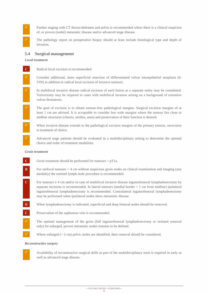
5.4 Surgical managementLocal treatment
C Radical local excision is recommended.
Consider additional, more superficial resection of differentiated vulvar intraepithelial neoplasia (d-VIN) in addition to radical local excision of invasive tumours.
In multifocal invasive disease radical excision of each lesion as a separate entity may be considered.Vulvectomy may be required in cases with multifocal invasion arising on a background of extensivevulvar dermatosis.
The goal of excision is to obtain tumour-free pathological margins. Surgical excision margins of atleast 1 cm are advised. It is acceptable to consider less wide margins where the tumour lies close tomidline structures (clitoris, urethra, anus) and preservation of their function is desired.
When invasive disease extends to the pathological excision margins of the primary tumour, reexcisionis treatment of choice.
Advanced stage patients should be evaluated in a multidisciplinary setting to determine the optimalchoice and order of treatment modalities.
Groin treatment
C Groin treatment should be performed for tumours > pT1a.
B For unifocal tumours < 4 cm without suspicious groin nodes on clinical examination and imaging (anymodality) the sentinel lymph node procedure is recommended.
C For tumours ≥ 4 cm and/or in case of multifocal invasive disease inguinofemoral lymphadenectomy byseparate incisions is recommended. In lateral tumours (medial border > 1 cm from midline) ipsilateralinguinofemoral lymphadenectomy is recommended. Contralateral inguinofemoral lymphadenectomymay be performed when ipsilateral nodes show metastatic disease.
D When lymphadenectomy is indicated, superficial and deep femoral nodes should be removed.
C Preservation of the saphenous vein is recommended.
The optimal management of the groin (full inguinofemoral lymphadenectomy or isolated removalonly) for enlarged, proven metastatic nodes remains to be defined.
Where enlarged (> 2 cm) pelvic nodes are identified, their removal should be considered.
Reconstructive surgery
Availability of reconstructive surgical skills as part of the multidisciplinary team is required in early aswell as advanced stage disease.

VULVAR CANCER - GUIDELINES 9
5.5 Sentinel lymph node procedurec
B The sentinel lymph node procedure is recommended in patients with unifocal cancers of < 4 cm,without suspicious groin nodes.
B Use of radioactive tracer is mandatory, use of blue dye is optional.
C Lymphoscintigram is advised to enable the preoperative identification, location and number of sentinellymph nodes.
C Intraoperative evaluation and/or frozen sectioning of the sentinel lymph node can be performed in anattempt to prevent a second surgical procedure. Caution is warranted because of an increased risk ofmissing micrometastases on final pathology due to the loss of tissue arising from processing for frozensection assessment.
When a sentinel lymph node is not found (method failure), inguinofemoral lymphadenectomy shouldbe performed.
c
C Where metastatic disease is identified in the sentinel lymph node (any size): inguinofemorallymphadenectomy in the groin with the metastatic sentinel lymph node.
For tumours involving the midline: bilateral sentinel lymph node detection is mandatory. Where onlyunilateral sentinel lymph node detection is achieved, an inguinofemoral lymphadenectomy in thecontralateral groin should be performed.
c
C Pathological evaluation of sentinel lymph nodes should include serial sectioning at levels of at leastevery 200 µm. If the H&E sections are negative, immunohistochemistry should be performed.
5.6 Radiation therapy
Adjuvant radiotherapy should start as soon as possible, preferably within 6 weeks of surgical treatment.
When invasive disease extends to the pathological excision margins of the primary tumour, and furthersurgical excision is not possible, postoperative radiotherapy should be performed.
In case of close but clear pathological margins, postoperative vulvar radiotherapy may be considered toreduce the frequency of local recurrences. There is no consensus for the threshold of pathologicalmargin distance below which adjuvant radiotherapy should be advised.
B Postoperative radiotherapy to the groin is recommended for cases with > 1 metastatic lymph nodeand/or presence of extracapsular lymph node involvement.
Adjuvant radiotherapy for metastatic groin nodes should include the ipsilateral groin area and wherepelvic nodes are non-suspicious on imaging, the distal part of the iliac nodes with an upper limit at thelevel of the bifurcation of the common iliac artery.
C Based on evidence from other squamous cell cancers such as cervical, head & neck, and anal cancer,the addition of concomitant, radiosensitising chemotherapy to adjuvant radiotherapy should beconsidered.

VULVAR CANCER - GUIDELINES 10
5.7 Chemoradiation
C Definitive chemoradiation (with radiation dose escalation) is the treatment of choice in patients withunresectable disease.
C In advanced stage disease neoadjuvant chemoradiation should be considered in order to avoidexenterative surgery.
C Radiosensitising chemotherapy, preferably with weekly cisplatin, is recommended.
5.8 Systemic treatment
D Data in vulvar cancer are insufficient to recommend a preferred schedule in a palliative setting.
5.9 Treatment of recurrent diseaseTreatment of vulvar recurrence
Radical local excision is recommended.
For vulvar recurrence with a depth of invasion > 1 mm and previous sentinel lymph node removalonly, inguinofemoral lymphadenectomy should be performed.
The indications for postoperative radiotherapy are comparable to those for the treatment of primarydisease.
Treatment of groin recurrence
Restaging by CT (or PET-CT) of the thorax/abdomen/pelvis is recommended.
Preferred treatment is radical excision when possible, followed by postoperative radiation inradiotherapy naïve patients.
Based on evidence from other squamous cell cancers such as cervical and anal cancer, the addition ofradiosensitising chemotherapy to postoperative radiotherapy should be considered.
Definitive chemoradiation when surgical treatment is not possible.
Treatment of distant metastases
Systemic (palliative) therapy may be considered in individual patients (see systemic treatment).

VULVAR CANCER - GUIDELINES 11
5.10 Follow-up
The optimal follow-up schedule for vulvar cancer is undetermined.
After primary surgical treatment the following follow-up schedule is suggested:
First follow-up 6-8 weeks postoperative
First two years every three-four months
Third and fourth year biannually
Afterward, long-term follow-up, especially in case of predisposing vulvar disease.Follow-up after surgical treatment should include clinical examination of vulva and groins.2
After definitive (chemo)radiation the following follow-up schedule is suggested:
First follow-up visit 10-12 weeks post completion of definitive (chemo)radiation.
First two years every three-four months
Third and fourth year biannually
Afterward, long-term follow-up, especially in case of predisposing vulvar disease.At first follow-up visit 10-12 weeks post definitive (chemo)radiation CT or PET-CT is recommendedto document complete remission.
2 Despite the well-recognized low sensitivity of palpation to identify groin recurrences, currently available datado not support routine use of imaging of the groins in follow-up.

VULVAR CANCER - GUIDELINES 12
6 Diagnosis and referral
6.1 Summary of available scientific evidenceNo directly applicable clinical studies have been identified.
6.2 Previous initiativesFour previous1-4 initiatives presenting guidelines on diagnosis and referral were identified.
6.3 Development group commentsFor accurate treatment planning (sentinel lymph node (SLN) procedure: yes/no; expected uni-or bilateral lymphdrainage; visibility of scar; etc.) the localization of the primary tumour is important. Therefore excision biopsyshould be avoided.
In case of multifocal macroinvasive vulvar cancer, the patient is not eligible for SLN detection, andinguinofemoral lymphadenectomy should be performed.
Because vulvar cancer is a rare disease and outcome of e.g. the SLN procedure is related to experience of thetreating physician, treatment should be centralized in centres with adequate experience in the treatment of thisdisease.
6.4 Guidelines
In any patient suspected for vulvar cancer, diagnosis should be established by a punch/incision biopsy.Excision biopsy should be avoided for initial diagnosis, as this may obstruct further treatment planning.
In patients with multiple vulvar lesions, all lesions should be biopsied separately (with cleardocumentation of mapping).
All patients with vulvar cancer should be referred to a GOC and treated by a multidisciplinarygynaecological oncology team.

VULVAR CANCER - GUIDELINES 13
7 Staging systemThe TNM classification5 and the FIGO staging system6,7 classify vulvar cancer on the basis of the size of thetumour (T), whether the cancer has spread to lymph nodes (N), and whether it has spread to distant sites (M)(Table 1). By convention, the depth of invasion is defined from the epithelial-stromal junction of the mostsuperficial adjacent dermal papilla to the deepest point of invasion of the tumour8. Inguinal and femoral nodesare the initial sites of regional spread and involvement of pelvic lymph nodes is considered distant metastasis.
The FIGO staging system was last reviewed in 2009 by the FIGO Committee on gynecologic oncology6,7 inclose collaboration with the American joint commission on cancer and the Union of international cancer control.It should be noted that as part of this revised FIGO staging system, the pathologist must report not only thenumber of nodes with metastatic disease but also the size of the metastases and the presence or absence ofextranodal spread.
7.1 Summary of available scientific evidenceNo studies assessing the performance of the TNM classification have been identified.
Three retrospective studies9-11 assessing the performance of the revised FIGO staging system havebeen identified. The new staging system has generally been considered appropriate. This has seen amajor downstaging of between 18.3% to 42% of patients. This has mainly involved old patients withstage II disease being downstaged to stage IB. Among the 1,131 patients enrolled in these studies,only 6 patients were upstaged by the new system (< 1%). Nevertheless, Tabbaa et al.10 suggested thattumours > 4 cm in diameter had a less favourable prognosis. A potential limitation with the revisedFIGO staging system is that the number of patients with stage II disease will be very low. From thethree retrospective studies above9-11, about 20% of patients were classified as stage II in the oldFIGO staging system, whereas it is likely to be less than 5% in the revised system.
LoE 2-
7.2 Previous initiativesNo previous initiative presenting guidelines on the staging system to use was identified.
7.3 Development group commentsThe development group recommends using the TNM classification because it more accurately reflects the statusof the primary tumour and lymph nodes.
7.4 Guidelines
Vulvar cancer should be staged according to FIGO and/or TNM classification3.
3 Throughout these recommendations advanced stage of disease is defined as clinical T3 and/or N3.

VULVAR CANCER - GUIDELINES 14
Table 1. Staging systems of squamous cell vulvar cancer
PRIMARY TUMOUR (T)
TNM categories5 FIGO stages6 Definition
TX Primary tumour cannot be assessed
T0 No evidence of primary tumour
Tis* Carcinoma in situ
T1a IA Lesions ≤ 2 cm in size, confined to the vulva or perineum and with stromal invasion ≤ 1.0 mm**, no nodal metastasis
T1b IB Lesions > 2 cm in size or with stromal invasion > 1.0 mm*, confined to the vulva or perineum, with negative nodes
T2*** II Tumour of any size with extension to adjacent perineal structures (1/3 lower urethra, 1/3 lower vagina, anus) withnegative nodes
T3**** IVA Tumour invades upper urethral and/or vaginal mucosa, bladder mucosa, rectal mucosa, or fixed to pelvic bone
REGIONAL LYMPH NODES (N)
TNM categories5 FIGO stages6 Definition
NX Regional lymph nodes cannot be assessed
N0 No regional lymph node metastasis
N1 One or two regional lymph nodes with the following features
N1a IIIA One or two node metastasis(es), each 5 mm or less
N1b IIIA One lymph node metastasis 5 mm or greater
N2 IIIB Regional lymp node metastasis with the following features
N2a IIIB Three or more lymph node metastases each less than 5 mm
N2b IIIB Two or more lymph node metastases 5 mm or greater
N2c IIIC Lymph node metastasis with extracapsular spread
N3 IVA Fixed or ulcerated regional lymph nodes
DISTANT METASTASIS (M)
TNM categories5 FIGO stages6 Definition*
M0 No distant metastasis
M1 IVB Distant metastasis (incluing pelvic lymph node metastasis)
* FIGO no longer includes stage 0 (Tis), ** the depth of invasion is defined as the measurement of the tumour from the epithelial-stromal junction of the adjacentmost superficial dermal papilla to the deepest point of invasion, *** FIGO uses the classification T2/T3. This is defined as T2 in TNM, **** FIGO uses theclassification T4. This is defined as T3 in TNM.

VULVAR CANCER - GUIDELINES 15
8 Preoperative investigations
8.1 Summary of available scientific evidencePathology review: two studies enrolling at least 50 pathology reports of vulvar tissues wereidentified. As part of a retrospective pathology report review, Beugeling et al.12 assessed 1) theimpact of pathology review on patient management and 2) the adequacy of the pathology reports,with regard to tumour type, infiltration depth, and, for excision biopsies, resection margins on 121pathology reports from 112 patients. Two discrepancies have been reported (1.7%) but the hugemajority of reviewed reports showed no discrepancy (98.3%). In this study, a report statinghistological type and depth of infiltration was considered “adequate”. Using this criterion, 56% ofthe original reports and 83% of the review reports were adequate. In the second identified study13,113 pathology reports were reviewed and 4 major discrepancies were reported.
Results from the 4 other identified studies14-17 are limited by the small number of pathology reportstaken into account. These studies show a rate between 0% and 15.8% for major discrepancy (Table 2).Among the 6 identified studies, it was not possible to estimate how many histology reviews wouldbe necessary to find one major discrepancy. Half of the authors from the 6 identified studies12,15,16
have expressed doubt concerning the necessity of pathology report review for vulvar cancer.
LoE 2+
Accuracy of clinical palpation to assess the lymph nodes status: four studies18-21 assessing the valueof clinical palpation of the groin lymph nodes were identified. But only two studies18,21 have accruedin excess of 50 patients:
In a series of 258 patients treated with radical vulvectomy and bilateral groin lymphadenectomy,Iversen et al.18 reported metastases to the superficial and/or deep inguinal lymph nodes in 100cases. Only 64 of which were detected by clinical examination. A false positive rate of 15.5%among the patients with clinically suspicious groin lymph nodes has been reported.
Podratz et al.21 reported that the preoperative clinical staging efforts were incorrect in 25% ofthe cases (56/224).
Among the 50 patients enrolled in the study published by Piura et al.19, data with respect to bothclinical palpation and histopathologic examination of groin lymph nodes were available in 20 of the26 patients who had radical vulvectomy and groin lymph node dissection. Authors have noticed thatclinical palpation was not very reliable in detecting groin lymph node metastases. Overdiagnosis andunderdiagnosis were present in 55.5% and 27.3% of patients (sensitivity: 57.1%, specificity: 61.5%).
Thirty-nine patients out of the 59 patients enrolled in the fourth identified study20 hadinguinofemoral lymphadenectomy and all except one had bilateral groin node excision. Clinicalfindings were compared with histology result to assess test accuracy for a total of 77 groin nodes. Inthis study published by Singh et al.20, clinical examination has a sensitivity of 35% and specificity of94.3%.
LoE 2+
Accuracy of MRI to assess the lymph nodes status: as part of a systematic review, Selman et al.22
compared the accuracy of non-invasive tests to assess the groin node status. One prospective23 andone retrospective24 studies assessing the value of the MRI have been included in this review for atotal of 60 patients. MRI has a pooled sensitivity and specificity of 86% (95% CI = 0.57-0.98) and87% (95% CI = 0.74-0.95) respectively in predicting the groin node status.
LoE 1-
Three other original studies20,25,26 were identified but only one study25 has accrued in excess of 50patients. In a retrospective study published by Bipat et al. 25, 60 patients underwent MRIexamination for preoperative evaluation of lymph nodes. MRI images were read independently andretrospectively by two radiologists, both unaware of physical examination and surgery findings. Both
LoE 2+

VULVAR CANCER - GUIDELINES 16
observers detected 12 of the 23 positive groin nodes (sensitivity: 52%). Of the 96 negative nodes, 14and 11 were scored as positive by the observers (specificity: 85% and 89% respectively). Singh etal.20 (39 patients, 77 groin nodes) reported consistent results with those described by Selman et al.22.MRI correctly identified metastatic nodal disease in 18 of the 21 positive groins and among the 56negative groin nodes, 46 nodes were correctly identified on MRI, leading to a sensitivity of 85.7%and a specificity of 82.1%.
It should to be noted that the used MRI criterion for groin lymph node metastasis prediction variedbetween the studies (short-axis diameter of the node24,25, short axis/long axis ratio, contour, andsignal intensity20,23). Kataoka et al.26 used several criteria for evaluation of lymph node metastases of49 patients (36 primary and 13 recurrent). A short axis/long axis ratio ≥ 0.75 was described as themost relevant criterion for diagnosis of groin lymph node metastasis in groin-by-groin analysis(sensitivity: 86.7% and specificity: 81.3%). The presence of necrosis within a lymph node showedthe highest specificity (87.5%), but lower sensitivity (40.0%). Furthermore, MRI accuratelyclassified 31 out of 36 primary cancers (accuracy: 86%). The addition of contrast-enhanced MRI didnot change the accuracy of the size category of primary cancers (accuracy: 85%).
Accuracy of PET to assess the lymph nodes status: Selman et al.22 pooled results of two prospectivestudies27,28 to assess the value of PET in the determination of groin nodes status (75 patients). PEThas a pooled sensitivity and specificity of 71% (95% CI = 50-86) and 72% (95% CI = 59-82)respectively.
LoE 1-
One small original study29 was also identified (20 patients). Of the 12 positive nodes, 6 were scoredas positive (sensitivity: 50%) and all the 8 negative nodes were correctly identified (specificity:100%).
LoE 3
Accuracy of Ultrasound to assess the lymph nodes status: four prospective studies30-33 assessing thevalue of ultrasound have been included in the systematic review published by Selman et al.22.However, a pooled analysis could not be performed due to the difference between studies intechniques used to discriminate positive and negative groin nodes. Combining the results of anotherstudy34 identified and independently of the test parameters used for ultrasound, the results showedsensitivity and specificity ranging from 45% to 100% and from 58% to 96% respectively (
Table 3). Moskovic et al.30 combined ultrasound with ultrasound-guided fine-needle aspirationcytology (FNAC) to improve accuracy. This combined technique could accurately predict nodalstatus in the majority of cases. Falsely negative cytology occurred when the metastatic focus was ≤ 3mm (two false-negative results out of 40 groins). Hall et al.31, who extended the study of Moskovicet al.30 to 44 patients, reported that the combination of ultrasound and FNAC provides a sensitiveand specific tool for preoperative assessment (sensitivity = 93%, specificity = 100%).
LoE 2+
Accuracy of CT to assess the lymph nodes status: no literature is available on the diagnostic value ofCT for detection of inguinofemoral lymph node metastases in patients with vulvar cancer. The onlyexperience with CT in patients with vulvar cancer is the measurement of the distance in centimetresbetween the skin and the underlying inguinofemoral lymph nodes for planning of groin radiation35,36.
LoE 4
8.2 Previous initiativesSeven previous initiatives1-4,37-39 presenting guidelines on preoperative investigations were identified.
8.3 Development group commentsSize of the lesion, distance to the midline and palpation of the lymph nodes all determine the choice for primarytreatment. Involvement of clitoris, anus, and/or urethra often means that these structures will need to be radicallyexcised together with the primary tumour. Such information is important for treatment planning and informingthe patient. In case of clitoral/anal/urethral involvement, primary radio(chemo)therapy might be an alternative.

VULVAR CANCER - GUIDELINES 17
In patients with primary unifocal vulvar cancer <4 cm, inguinofemoral lymphadenectomy can be performedimmediately instead of SLN procedure in case when lymph node metastases are diagnosed preoperatively. CT orPET/CT can be performed to rule out involvement of pelvic nodes and to decide whether or not to performpelvic nodal debulking. Presence of distant metastases should also be evaluated as their presence or absence mayinfluence the radicality of treatment of the primary tumour and the regional lymph nodes.
Treatment policy for melanomas and basal cell cancer for example is different. Depth of invasion is necessary todecide whether groin treatment is indicated, both in squamous cell cancers as well as in melanomas.
8.4 Guidelines
Preoperative work-up should at least include clear documentation of clinical exam (size of lesion,distance to the midline/clitoris/anus/vagina/urethra and palpation of lymph nodes). Picture or clinicaldrawing is advised (see below).
Evaluation of the cervix/vagina/anus is recommended.
C Prior to sentinel lymph node biopsy, clinical examination and imaging of the groins (either byultrasound, PET-CT, or MRI) are required to identify potential lymph node metastases.
Suspicious nodes (at palpation and/or imaging) should be analysed by FNA or core biopsy when thiswould alter primary treatment.
Further staging with CT thorax/abdomen and pelvis is recommended where there is a clinical suspicionof, or proven (nodal) metastatic disease and/or advanced stage disease.
The pathology report on preoperative biopsy should at least include histological type and depth ofinvasion.

VULVAR CANCER - GUIDELINES 18
Table 2. Original studies presenting data on pathology slide review
Table 3. Original studies presenting data on the accuracy of imaging to assess the groin node status
Authorreference Year N Major discrepancy Minor discrepancy
Beugeling et al.12 2014 121 1.7% (2/121) 0% (0/121)
Santoso et al.13 1998 113 3.5% (4/113) 10.6% (12/113)
Chafe et al.14 2000 28 7.1% (2/28) 32.1% (9/28)
Khalifa et al.15 2003 28 0% (0/28) 10.7% (3/28)
Selman et al.16 1999 19 15.8% (3/19) 0% (0/19)
Chan et al.17 1999 13 15.4% (2/13) 15.4% (2/13)
Authorreference Year TP FP TN FN Sensitivity Specificity
MRI
Hawnaur et al.23* 2002 8 1 10 1 89% 91%
Sohaib et al.24* 2002 4 5 30 1 80% 56%
Bipat et al.25 2006
(observer 1) 12 14 80 11 52% 85%
(observer 2) 12 11 90 11 52% 89%
Singh et al.20 2006 18 10 46 3 85.7% 82.1
Kataoka et al.26 2010
(short axis/long axis ratio ≥ 0.75) 26 3 13 4 86.7% 81.3%
(contour) 21 7 8 9 70.0% 53.3%
(necrosis) 12 2 14 18 40.0% 87.5%
(loss of fatty hilum) 24 8 8 6 80.0% 50.0%
(similarity of signal intensity to vulva lesion) 23 8 3 3 88.5% 27.3%
PET
Cohn et al.27* 2002 6 2 18 3 67% 90%
de Hullu et al.28* 1999 9 13 21 3 75% 62%
Kamran et al.29 2014 6 0 8 6 50% 100%
Ultrasound
de Gregorio et al.34 2013 29 6 63 9 76% 91%
Hall et al.31* 2003 24 2 43 4 86% 96%
Makela et al.32* 1993 9 5 34 2 81% 87%
Moskovic et al.30* 1999 11 5 25 2 85% 83%
Abang Mohammed et al.33* 2000
(short axis) 5 3 28 6 45% 90%
(long/shot axis ratio) 6 10 14 0 100% 58%
(combined) 5 3 21 1 83% 87%
* studies included in the systematic review published by Selman et al.22, FN: false negative, FP false positive, TN: truenegative, TP: true positive.

VULVAR CANCER - GUIDELINES 19
9 Surgical management
9.1 Summary of available scientific evidenceRadical/wide local excision versus radical vulvectomy : none of the five identified studies40-44
reported statistically significant differences in overall survival, disease-free survival, local or distantrecurrence rates between patients treated by radical/wide local excision and patients treated byradical vulvectomy:
In a retrospective study enrolling 74 patients (T1-2N0-1M0), Farias-Eisner et al.40 compared theeffectiveness and safety of a radical local excision (N = 56) versus radical vulvectomy (N = 18).Of women with stage I disease, the 5-year survival was similar for those patients who underwentthe more conservative operation (97%) compared with those who underwent a radicalvulvectomy (100%). The difference in the overall survival of stage II patients undergoing radicallocal excision versus radical vulvectomy did not reach statistical significance (90% versus 75%,p > 0.05). Operative morbidity was less in those undergoing a conservative operation. Seriousinfection, necrosis, or major breakdown of the primary wound occurred in 2 (11%) and 14(25%) patients undergoing radical local excision and radical vulvectomy, respectively.
Similar overall survival, local control and 5-year disease-free survival rates were reported byBalat et al.41 between 25 patients treated by wide local excision and 24 patients treated byradical vulvectomy (73% versus 67%, 83% versus 80%, and 75% versus 67%, respectively). Inthis retrospective study, all patients received irradiation combined with surgery. There werefewer complications (eg lymphedema, wound infection, lymphocyst, vulvar dystrophy) in thepatients treated by wide local excision than in those treated with radical vulvectomy. Similarlocal recurrence rates were reported by de Hullu et al.42 between patients treated by wide localexcision and patients treated by radical vulvectomy (11.4% (9/79) versus 7.5% (12/159), p =0.32). An analysis of the exact tumour free margins among 39 patients treated by wide localexcision showed that no patient with histologic tumour free margins measuring > 8 mmdeveloped a local recurrence, whereas 9 of 40 patients with at least one tumour free marginmeasuring ≤ 8 mm developed local recurrences within 2 years (p = 0.002). As Balat et al.41,there was no difference in overall survival between two groups of patients. Rutledge et al.43
undertook an analysis of 179 stage I and II lesions treated with a curative aim to see if there wasa difference in survival or in disease-free interval between those patients treated with radicalvulvectomy and those treated with radical wide local excision. No survival advantage from theradical vulvectomy procedure has been reported (data not shown).
No statistical correlation between the type of primary surgery performed and the frequency ofrecurrence to any site were described by DeSimone et al.44 in a retrospective study enrolling 122patients with lateral T1 (N = 61) and T2 (N = 61) vulvar cancer confined to the labium majusand labium minus (local: 13% versus 8%, p = 0.33, groin: 0% versus 3%, p = 0.50, distant(pulmonary): 2% versus 3%, p = 1.0, total: 15% versus 15%, p = 1.0). It should be noted thatlymphoedema occurred more commonly in patients undergoing radical vulvectomy than inpatients undergoing radical wide excision (26% versus 7.5%, p = 0.007). Likewise, both woundseparation (23% versus 7.5%) and lymphocyst formation (6.7% versus 3.2%) were morecommon in patients undergoing radical vulvectomy.
LoE 2+
As part of Cochrane systematic review, van der Velden45 also assessed the effectiveness and safetyof a radical local excision. Two observational studies46,47 enrolling 94 patients (TIN0M0: N = 51,T2N0M0: N = 43) have been included in this systematic review. No pooled analysis is described andit should be noted that details regarding radiotherapy interventions were not addressed and the gradeof complications was not defined in any study. Furthermore, an adequate description of commoncomplications was not stated in one study47. Authors reported a recurrence rate of 0%47 and 12%46.
LoE 2-

VULVAR CANCER - GUIDELINES 20
None of the patients with a local recurrence died of vulvar cancer after a median follow-up of 38months.
Three other studies48-50 documenting recurrence rates after radical/wide local excision wereidentified (0%48 (0/18 patients with stage I), 23.1%49 (28/121 patients with stage I and II), and 10%50
(5/50 patients with stage I).
Only one study comparing quality of life of patients treated by wide local excision versus radicalvulvectomy was identified. In this retrospective (57 patients), Gunther et al.51 observed tendenciesfor a better physical, role, emotional, and cognitive functioning, as well as global health status aftersurgical treatment with wide local excision. Patients who underwent radical vulvectomy sufferedfrom a significant higher level of pain than those who underwent wide local excision. In addition,these patients suffered from nausea/vomiting, fatigue, insomnia, appetite loss, and diarrhoea to ahigher degree (p > 0.05). It should be noted that after radical vulvectomy, 89% of patients havesexual complications.
LoE 2+
Omission of Inguinofemoral lymphadenectomy: the presence of pelvic node metastases is very rare inthe absence of inguinofemoral lymph node metastases. Thirty percent of all patients with vulvarcancer have inguinofemoral metastases and 20% of these patients will have pelvic metastases,too52,53. None of the seven identified studies49,54-59 described positive lymph nodes (or inguinalrecurrences after a minimal follow-up of two years) in patients with very early stage vulvar cancer,where the primary lesion measures less than 2 cm in maximum diameter and the depth of invasion isless than 1 mm (FIGO stage IA disease). Among the 30 patients who underwent surgery withoutlymphadenectomy in the study published by Magrina et al.59, one developed groin, pelvic, and aorticnode metastases 7.5 years after initial operation and 3.5 years after experiencing a vulvar recurrence(the primary lesion measured 2 x 1.5 cm, was moderately well differentiated, and was located to theleft of the clitoris with only 0.1 mm of invasion). In contrast, with infiltration of 1-2 mm, lymphnode metastases or inguinal recurrences were seen from 0 to 17%54-57.
LoE 2+
Several case reports60-65 of regional lymph node recurrences following treatment for FIGO stage IAvulvar cancer have been published but no pattern of particular risk factors can be defined from thissmall number of cases.
LoE 3
Superficial inguinal lymphadenectomy versus total inguinofemoral lymphadenectomy: as part of aretrospective study enrolling 217 patients with stage I disease (5 mm or less invasion, no vascularspace involvement, and negative inguinal and femoral nodes), Stehman et al.66 reported a groinrecurrence in 7.3% of patients treated with superficial inguinofemoral lymphadenectomy versus 0%recurrences in those treated with radical vulvectomy and bilateral inguinofemoral lymphadectomy(historic controls). The recurrent-free interval was significantly lower for patients treated withsuperficial inguinal lymphadenectomy compared to historic controls (84.2% (102/121) versus 91.8%(90/98), p = 0.0028). For survival time, the difference did not reach statistical significance (87.6%(106/121) versus 82.6% (81/98), p > 0.05).
LoE 2-
Three uncontrolled studies50,67,68 evaluating outcomes of patients treated with superficial inguinallymphadenectomy were also identified. Among the 104 patients (stage I or II, depth of invasiongreater than 1 mm) treated with radical wide excision (negative margins) and superficial inguinallymphadenectomy, Gordinier et al.67 reported that nine patients experienced recurrent disease thatinvolved one or both of the groins (8.6%). Berman et al.50 reported outcomes of 50 patients with T1vulvar cancers < 1 cm diameter with stromal invasion > 5 mm who underwent radical wide excisionand superficial inguinal lymphadenectomy. There were no isolated groin recurrences noted during afollow-up period of 36 months. The third study68 reported that three of the 65 patients with stage I/IIvulvar cancer and a pathologically negative superficial inguinal lymphadenectomy recurred in theinguinal region (4.6%).
LoE 2+

VULVAR CANCER - GUIDELINES 21
Unilateral inguinofemoral lymphadenectomy versus bilateral inguinofemoral lymphadenectomy: therisk of recurrent disease in a contralateral groin after ipsilateral groin node dissection in patients withT1 or T2 lesions confined to the labium majus or minus is very low. Among the five identifiedstudies44,46,66,69,70 for a total of 295 patients, only four recurrent diseases in a contralateral groin afteripsilateral groin node dissection have been reported (1.4%).
LoE 2+
A case report71 of a contralateral recurrence 2.5 years after wide local excision and unilateral groinnode dissection in a patient with a T1 lesion without clinically palpable groin nodes has been alsoidentified.
LoE 3
As part of a thesis, van der Velden72 found that 19 out of 489 patients (3.9%) with unilateral vulvartumours and negative ipsilateral lymph nodes had positive contralateral lymph nodes. In a subgroupanalysis taking into account patients with tumours < 2 cm, the incidence of contralateral lymphnodes is only 0.9%.
LoE 2+
Preservation of the saphenous vein: among the seven identified studies73-79, Zhang et al.73 showedthat preservation of the saphenous vein was associated with a statistically significant decrease in theoccurrence of cellulitis, short-term lower extremity lymphoedema, wound breakdown, and chronicedema (18% versus 39%, p = 0.006, 32% versus 70%, p < 0.001, 13% versus 38%, p = 0.001, 32%versus 3%, p = 0.003, respectively) compared to saphenous vein ligation without compromising thelocal or distant recurrent disease rates (data not shown). Overall, the likelihood of developing nopostoperative complications was higher in the saphenous vein preservation group compared with thesaphenous vein ligation group (56% versus 23%, p < 0.001).
More recently, Zhang et al.74 reported that preservation of the saphenous vein was associated with astatistically significant decrease by about 50% in the occurrence of chronic lower limblymphoedema, chronic lower extremity pain, chronic cellulitis, and sensory abnormalities (25.0%versus 48.3%, p < 0.01, 23.2% versus 46.6%, p < 0.01, 21.4% versus 41.4%, p < 0.05, and 19.6%versus 36.2%, p < 0.05 respectively) without compromising 5-year survival rate and groin recurrencerate (68% versus 66.7%, p > 0.05 and 8.9% versus 12.1%, p > 0.05, respectively). Short-term lowerextremity lymphoedema and short-term lower extremity phlebitis were also less frequent in patientstreated by saphenous vein sparing surgery to those treated by lymphadenectomy with saphenous veinligation (43.5 versus 66.7%, p < 0.01, and 11.3% versus 25.8%, p < 0.05, respectively).
Similarly, Rouzier et al.75 reported that lymphadenectomy with saphenous vein preservation isassociated with a significant decrease in the occurrence of wound breakdown, cellulitis andlymphoedema compared to lymphadenectomy with saphenous vein ligation (16.2% versus 36.4%, p< 0.001, 17.7% versus 29.8%, p = 0.01, and 23.1% versus 45.3%, p < 0.001, respectively). Asignificant differences in the occurrence of cellulitis and wound breakdown were also described byDardarian et al.76 in favour of saphenous vein sparing surgery (0% versus 45%, p < 0.001, and 0%versus 25%, p ≤ 0.02, respectively). Subsequently, chronic lymphoedema (> 6 months) persisted in38% of the vein-ligated group compared to 11% in the vein-spared group (p < 0.05) withoutcompromising the incidence of recurrent disease (19.3% versus 22.2%, p > 0.05)76.
However, preservation of the saphenous vein was not systematically associated with a statisticallysignificant decrease of morbidity. Zhang et al.73 observed that the difference of seroma, phlebitis,deep vein thrombosis, and hematoma in favour of saphenous vein sparing surgery did not reachstatistical significance (3% versus 8%, p = 0.29, 0% versus 3%, p = 0.50, 2% versus 5%, p = 0.38,0% versus 3%, p = 0.50, respectively). More recently, Zhang et al.74 observed also that the differenceof acute cellulitis, seroma, lymphocyst formation, chronic lower extremity phlebitis, and deepvenous thrombosis with saphenous vein sparing surgery did not reach statistical significance (67.7%versus 72.7%, p > 0.05, 30.6% versus 37.9%, p > 0.05, 25.8% versus 31.8%, p > 0.05, 10.7% versus
LoE 2+

VULVAR CANCER - GUIDELINES 22
15.5%, p > 0.05, 7.1% versus 10.3%, p > 0.05, respectively). Dardarian et al.76 showed that thedifference of short-term oedema in favour of saphenous vein ligation did not reach statisticalsignificance (67% versus 72%, p > 0.05). Finally, groin wound breakdown or cellulitis occurred in18% of patients with saphenous vein preservation, and 24% where the vein was sacrificed in thestudy published by Paley et al.77.
In contrast, some investigators73,74,77,78 described an increase of morbidity in patients with saphenousvein sparing compared to patients where it was sacrificed. Paley et al.77 described an increase of theincidence of lymphoedema and lymphocyst formation (36% versus 21%, 27% versus 14%,respectively). Zhang et al.73,74 observed a slight increase of postoperative fever, lymphocystformation, and pulmonary embolism (96.8% versus 93.9%, 10% versus 4%, 2% versus 0%,respectively) but it should be noted that the differences did not reach statistical significance (p >0.05, p = 0.19, p = 0.45, respectively). In the study published by Lin et al.78, lymphoedema occurredin 17% of patients who had preservation of the long saphenous vein during the groin dissectionversus 13% in whom the long saphenous vein was sacrificed (p = 0.50). It should be noted that therisk of groin recurrence did not change with preservation of the saphenous vein (6% versus 6%).
Finally, Soliman et al.79 did not find significant correlations between saphenous vein ligation and thedevelopment of any local complications (data not shown).
Triple incision technique versus en bloc dissection (the butterfly incision) : no randomised trials havebeen performed to evaluate whether the use of the triple incision technique is as safe as the en blocapproach, but all the identified studies42,80-83 that compared these two surgical approaches showedthat vulvectomy and inguinofemoral lymphadenectomy via three separate incisions provide similaroutcome in terms of survival compared to an en bloc butterfly resection. In multivariate analysis, vander Velden et al.81 reported that surgical technique has no impact on disease-specific survival (afteradjustment for tumour diameter, extracapsular lymph node involvement, TNM stage, and number ofnodal metastases, HR = 0.99, 95% CI = 0.43-2.30, p = 0.996) and overall survival (data not shown).After correction for tumour dimension, depth of invasion, presence or absence of lymph/vascularinvasion, and grade, de Hullu et al.42 observed that wide local excision with inguinofemorallymphadenectomy through separate incisions was not related independently to an increased risk ofdeath within 4 years related to vulvar carcinoma (OR = 1.98, 95% CI = 0.80-4.80, p > 0.05) even ifthey described more frequent fatal recurrences in the groin or the skin bridge (6.3% versus 1.3%, p =0.029).
LoE 2+
Among the seven identified studies42,80-85, a skin bridge recurrence was observed in only 1.8% ofpatients (6/336). It should be noted that Hacker et al.84 published 2 skin bridge recurrences, both inpatients with lymph node metastases. However, the majority of identified studies42,81,83 described alower local recurrence rate among patients treated by an en bloc resection. With regard to the risk ofvulvar recurrence, van der Velden et al.81 reported that patients treated by an en bloc resectionshowed a significantly lower risk of local recurrence than those treated by the triple incisiontechnique after adjustment for tumour diameter, extracapsular lymph node involvement, TNM stage,and number of nodal metastases (HR = 0.10, 95% CI = 0.02-0.44, p = 0.002). But the type ofsurgical treatment was not an independent predictor for regional recurrence (HR = 0.39, 95% CI =0.13-1.17, p > 0.05) or distant recurrence (HR = 0.97, 95% CI = 0.32-2.91, p > 0.05). In multivariateanalyses, after correction for tumour dimension, depth of invasion, presence or absence oflymph/vascular invasion, and grade, de Hullu et al.42 mentioned that wide local excision withinguinofemoral lymphadenectomy through separate incisions was associated with a higher risk ofdeveloping recurrences 2 and 4 years after primary treatment (OR = 2.29, 95% CI = 1.00-5.28, p <0.05, and OR = 2.272, 95% CI = 1.11-4.67, p < 0.05, respectively).
Fambrini et al.86 assessed the feasibility and safety of a modified triple incision total radicalvulvectomy and inguinofemoral lymphadenectomy in 57 patients with locally advanced vulvar
LoE 2+

VULVAR CANCER - GUIDELINES 23
cancer (LAVC). In all cases, two teams performed the surgery: one for total radical vulvectomy andthe other for inguinofemoral lymphadenectomy. Surgical procedures started at the same time andwere performed according to standard triple incision technique. Postoperative complicationsinvolving the surgical sites or lymphatic drainage were observed in one third of patients (19/57).None of them required surgical re-intervention. After treatment 29 patients developed local, regionalor distant recurrence of disease, with a median progression-free survival of 39.5 ± 20.9 months.Three-year and 5-year overall survival (OS) were of 60.5% and 48.6%, respectively.
9.2 Previous initiativesNine previous initiatives1-4,37-39,87,88 presenting guidelines surgical management were identified.
9.3 Development group commentsVulvectomy in addition to radical local excision can be considered in tumours with extensive premalignantdisease to reduce the risk of local recurrence. Data on surgical margins are conflicting. Therefore, thedevelopment group advises to consider narrow margins when this means clitoris/anus can be preserved.
Treatment of advanced stage vulvar cancer often involves multiple treatment modalities. Treatment planning isoften individualized in advanced stage and depends on primary tumour characteristics and presence of regionaland/or distant metastases. Also comorbidity and/or frailty of the patient influences treatment planning.Therefore, a multidisciplinary setting is needed to optimize treatment planning.
In case of enlarged groin nodes either inguinofemoral lymphadenectomy followed by radiotherapy, or groin nodedebulking followed by radiotherapy can be considered. When imaging shows enlarged pelvic nodes, debulkingof these nodes is recommended with adjuvant radiotherapy, since radiotherapy alone will probably not sterilizelarge nodal pelvic disease.

VULVAR CANCER - GUIDELINES 24
9.4 GuidelinesLocal treatment
C Radical local excision is recommended.
Consider additional, more superficial resection of d-VIN in addition to radical local excision ofinvasive tumours.
In multifocal invasive disease radical excision of each lesion as a separate entity may be considered.Vulvectomy may be required in cases with multifocal invasion arising on a background of extensivevulvar dermatosis.
The goal of excision is to obtain tumour-free pathological margins. Surgical excision margins of atleast 1 cm are advised. It is acceptable to consider less wide margins where the tumour lies close tomidline structures (clitoris, urethra, anus) and preservation of their function is desired.
When invasive disease extends to the pathological excision margins of the primary tumour, reexcisionis treatment of choice.
Advanced stage patients should be evaluated in a multidisciplinary setting to determine the optimalchoice and order of treatment modalities.
Groin treatment
C Groin treatment should be performed for tumours > pT1a.
B For unifocal tumours < 4 cm without suspicious groin nodes on clinical examination and imaging (anymodality) the sentinel lymph node procedure is recommended.
C For tumours ≥ 4 cm and/or in case of multifocal invasive disease inguinofemoral lymphadenectomy byseparate incisions is recommended. In lateral tumours (medial border > 1 cm from midline) ipsilateralinguinofemoral lymphadenectomy is recommended. Contralateral inguinofemoral lymphadenectomymay be performed when ipsilateral nodes show metastatic disease.
D When lymphadenectomy is indicated, superficial and deep femoral nodes should be removed.
C Preservation of the saphenous vein is recommended.
The optimal management of the groin (full inguinofemoral lymphadenectomy or isolated removalonly) for enlarged, proven metastatic nodes remains to be defined.
Where enlarged (> 2 cm) pelvic nodes are identified, their removal should be considered.
Reconstructive surgery
Availability of reconstructive surgical skills as part of the multidisciplinary team is required in early aswell as advanced stage disease.

VULVAR CANCER - GUIDELINES 25
10 Sentinel lymph node procedure
10.1 Summary of available scientific evidenceDiagnostic test accuracy according to the mapping method: three meta-analyses89-91 assessing thediagnostic accuracy of SLN biopsy were identified. Hassanzade et al.89, Meads et al.90, and Lawrie etal.91 included 47 studies92-138, 29 studies97,98,109,110,113-120,124-126,129,135,136,139-148, and 34 studies92,93,95,97-
99,103,104,107,109,110,112,114-119,122-127,129,135,136,140-144,149-169, respectively. It should be noted that studiesincluded in these meta-analyses had methodological limitations, such as lack of an adequatedescription of population (especially stage of disease), inclusion criteria, assessment procedure, andreference standard used. Data from different reports of the same study were also taken into account.
Two meta-analyses89,90 reported pooled patient basis detection rate of various techniques andprovided evidence that a combination of blue dye/99mTc is the most accurate technique (Table 4). Itshould to be noted that many of the studies taken into account by Meads et al.90 were also includedin the pooled analysis performed by Hassanzade et al.89, which explains the consistency of results.Only Hassanzade et al.89 published pooled groin basis detection rate data and observed that it wasalso higher with the use of the combined blue dye and 99mTc testing (Table 4).
Two of the three identified meta-analyses89,91 described per patient and per groin pooled sensitivityof the SLN biopsy and provide evidence that a combination of blue dye/99mTc is also the mostsensitive technique (Table 4). It should to be noted that many of the studies taken into account byLawrie et al.91 were also included in the pooled analysis performed by Hassanzade et al.89, whichexplains the consistency of results.
LoE 1-
Diagnostic test accuracy according to the location of the tumour: Hassanzade et al.89 reported thatdiagnostic test accuracy of the SLN procedure is also related to location of the tumour. For midlinelesions (≤ 2 cm of midline), per groin pooled detection rate was 22% lower than per patient pooleddetection rate but groin basis pooled sensitivity was 4% higher than patient basis pooled sensitivity(Table 5). However, for lateral lesions (> 2 cm from the midline plane), per patient and per groinpooled detection rates and sensitivity were similar.
LoE 1-
Diagnostic test accuracy according to the tumour size: Hassanzade et al.89 observed that pooledpatient basis sensitivity was also related to the size of the primary tumour. Indeed, the pooledsensitivity of SLN mapping in < 4 cm tumours was 7% higher than > 4 cm tumours (< 4 cm: 93%(95% CI = 87-97), > 4 cm: 86% (95% CI = 77-93)). It should be noted that, in the Groningeninternational study on sentinel nodes in vulvar cancer (GROINSS-V)170, sentinel-node detection wasdone in patients with T1-T2 (< 4 cm) squamous-cell vulvar cancer.
LoE 1-
Diagnostic test accuracy according to the inclusion of patients with palpable or suspicious inguinalnodes in the study population: Hassanzade et al.89 observed that per patient and per groin pooledpatient basis detection rate and sensitivy were lower among patients with palpable or suspiciousinguinal nodes (Table 5).
LoE 1-
Diagnostic accuracy of intraoperative pathologic analysis of frozen sections: as part of theGROINSS-V170, frozen sectioning was done in 315 and showed a low sensitivity (48%) but a highspecificity (100%).
LoE 2++
In contrast, two older and smaller studies (52 patients142 and 42 patients141) found sensitivity greaterthan 90%. It should be noted that these two studies141,142 reported a specificity for intraoperativeanalysis of SLN by frozen section greater than 90%. In the fourth identified study115, 18 positivenodes were detected in 13 of the 43 enrolled women (30.2%). In two cases, although the frozensection was negative, the definitive histopathologic examination revealed a micrometastasis
LoE 2+

VULVAR CANCER - GUIDELINES 26
(accuracy: 98%).
Diagnostic test accuracy according to histological methods: only one of the three identified meta-analyses91 described pooled estimates of sensitivity for the combined technique (blue dye/99mTc)according to histological methods:
Ultrastaging only: 95% (95% CI = 91-97) (per groin data), 95% (95% CI = 89-98) (per patientdata)
Ultrastaging and/or immunohistochemistry (IHC): 94% (95% CI = 88-97) (per groin data), 95%(95% CI = 90-98) (per patient data)
LoE 1-
In the GROINSS-V170, ultrastaging detected a positive SLN in 55 (41%) of 135 patients (66 (40%) of164 groins). After multiple sectioning, IHC identified micrometastases in 36 (12%) of 304 patientswith a negative sentinel node. The risk of metastases in non-SLN was higher when the SLN wasfound to be positive by traditional pathologic processing than when the SLN was found to bepositive only with ultrastaging (23 of 85 groins (27%) versus 3 of 56 groins (5%), p = 0.001). InGynecologic oncology group (GOG) protocol 173135, 23% of all positive SLNs were missed byroutine H&E staining of SLN tissue cut and were only detected with the addition ofimmunohistochemical stains.
LoE 2++
Nine smaller studies50,54,58,65,67,77,84,112,118 have also reported micrometastases found after ultrastagingand/or IHC among patients that were previously negative with standard H&E.
LoE 2+
Visualization of the SLN by scintigraphy: in GOG protocol 173, Coleman et al.155 reported a negativecorrelation between distance of vulvar lesion from midline and the probability of detecting bilateraldrainage in preoperative lymphoscintigraphy. Thirty percent of women with tumours invading orcrossing the midline had unilateral drainage on lymphoscintigraphy. However, authors observed thatmore than one in five patients with lateralized primary tumours (> 2 cm from the midline) hadbilateral drainage on lymphoscintigraphy.
LoE 2++
Out of 42 patients with midline tumours enrolled in the retrospective review published by Lindell etal.125, only 18 had bilateral lymphatic drainage at scintigram. The lymphoscintigraphy showedunilateral lymphatic drainage in 40 out of 58 patients, including all 16 patients with lateral lesions.Louis-Sylvestre et al.157 found that of 13 patients with lesions less than 1 cm from the midline inwhom lymphoscintigraphy identified only unilateral drainage, 3 patients had metastatic disease innodes located in the contralateral, lymphoscintigraphy-negative groin. Six identifiedstudies102,117,118,160,171,172 assessed detection rate of the preoperative visualization of the SLN byscintigraphy and all of them reported a detection rate greater than 90%.
De Cicco et al.97 used preoperative and intraoperative lymphoscintigraphy alone to successfullyidentify at least one sentinel node in each of the 37 patients in their series. There were no false-negative sentinel nodes. Eight patients had positive nodes, and the sentinel node was the onlypositive node in 5 of these cases. If lymphoscintigraphy did not identify a sentinel node in a groin, nometastases were found at surgery. Using a combination of preoperative lymphoscintigraphy andintraoperative lymphoscintigraphy, de Hullu et al.98 reported that all the 23 patients with laterallesions or with tumours primarily labial but came within 1 cm of the midline had unilateral SLNdetected in the groin on preoperative lymphoscintigraphy and at the time of surgery.
In a very small study enrolling 10 patients, DeCesare et al.93 showed that intraoperativelymphoscintigraphy correctly identified the nodal status as positive in all 4 cases of metastaticdisease and negative in all 16 groins negative for metastases.
LoE 2+

VULVAR CANCER - GUIDELINES 27
Impact of training and experience of the surgeon on the diagnostic accuracy: Severalauthors118,120,145,173,174 have suggested surgeons should perform at least 10 successful SLN biopsyprocedures followed by complete inguinofemoral lymph node dissection without any false-negativeresults prior to performing SLN biopsy alone. In order to keep the experience at a high level, van derZee et al.145 proposed that an exposure of at least five to 10 patients per year per surgeon should beregarded as a minimum figure, requiring potentially centralization of early-stage vulvar cancertreatment in oncology centres.
LoE 4
As part of a prospective study enrolling 52 patients, Levenback et al.142 reported that the number ofcases in which the sentinel node is not identified or in which there is a false-negative sentinel nodedecreases with experience. Indeed, a sentinel node could not be identified in 4 of the 25 (16%)patients and 13 of the 36 (36%) groins dissected, compared with 2 of the 27 (7%) of patients treatedand 6 of the 40 (15%) groins dissected during the first two years of the study (p = 0.034).
LoE 2+
Recurrence and survival rates following SLN procedure: in the GROINSS-V170, five-year disease-specific survival for patients with positive sentinel nodes was 64.9% when identified by routinepathology versus 92.1% when identified by ultrastaging (p < 0.0001). The update of the GROINSS-V-I175 (377 patients) highlighted that on the long-term a significant proportion of patients willdevelop a local recurrence, regardless of sentinel node status and that these local recurrences mayoccur even a long time after primary treatment. This prospective study also showed that long-termsurvival is very good for patients with early-stage vulvar cancer and a negative sentinel node. After amedian follow-up of 105 months, Te Grootenhuis et al.175 reported an overall local recurrence rate of24.6% at 5 years and 36.4% at 10 years for sentinel node negative patients, and 33.2% and 46.4% forsentinel node positive patients, respectively (p = 0.03). Disease-specific 10-year survival was 91%for sentinel node negative patients compared to 65% for sentinel node positive patients (p < 0.0001).Overall 5- and 10-year survival was also better for sentinel node negative patients (5y-OS: 81.2%versus 61.3%, 10y-OS: 68.6% versus 43.6%, p < 0.0001).
As part of a health technology assessment comparing SLN biopsy and inguinal lymph nodedissection (ILND), Reade et al.176 reported from 11 studies93,96,113,114,117,132,145-147,177,178 enrolling 591patients a groin recurrence rate after a negative SLN biopsy of 3.6% (range 0 to 22%). It should benoted that follow-up in these studies was variable, but in most was at least two years. A recurrencerate after ILND of 4.3% was also reported (13 studies46,66-68,179-187 enrolling 1,077 patients). It shouldbe noted that, in general, there was longer follow-up in these studies than in the studies of SLNbiopsy.
LoE 2++
Multivariate analyses performed from the surveillance, epidemiology, and end results database on1,094 patients188 showed that SLN biopsy was not significantly associated with an excess risk ofmortality or recurrence after adjustment for age, ethnicity, stage, grade, and lymph node status (datanot shown).
LoE 2-
Complication rates & clinical parameters: Reade et al.176 compared also complication rates betweenSLN biopsy (6 studies113,117,120,145,146,178, 532 patients) and ILND (27 studies46,66,68,73-
76,78,82,85,117,120,145,178,179,182,183,186,189-197, 2,135 patients). Wound infection, wound breakdown,lymphocysts, and chronic lymphoedema after SLN biopsy were 4.4%, 9.5%, 3.8%, and 1.5%,respectively. The rate of groin wound infection after ILND across all studies was 30.7%, groinwound breakdown occurred in 23.2%, and lymphocysts occurred in 15.5%. Chronic lymphoedemanoccurred in 22.9% accros all studies.
LoE 2++
In a retrospective study enrolling 128 patients, Brammen et al.171 reported also a higher presence oflymph cysts after ILND compared to SLN biopsy (OR = 3.4 (95% CI = 1.1-10.6), p = 0.02). Inaddition, three original studies145,171,178 reported significantly higher operation time, hospital stay orduration of inguinal drainage after ILND (Table 6).
LoE 2+

VULVAR CANCER - GUIDELINES 28
Quality of life: one study198 investigated quality of life in 62 patients who participated in theGROINSS-V study. Using the EORTC QLQ-C30 questionnaire, no difference in overall quality oflife was observed between the 35 patients who underwent the SLN-procedure alone and the 27patients who underwent an inguinofemoral lymphadenectomy. The major difference was the increasein complaints of lymphoedema of the legs after inguinofemoral lymphadenectomy (p = 0.01).Patients who underwent inguinofemoral lymphadenectomy also reported more discomfort in groins,vulva and legs (p = 0.03), and more frequent need to wear stockings (p = 0.003). Patients after theSLN procedure only were more content with the treatment they had undergone (p = 0.04). Moreover,no differences in sexual activeness were observed between SLN procedure and inguinofemorallymphadenectomy.
LoE 2+
Two smaller studies199,200 were also identified. As part of a prospective study enrolling 36 patients(12 SLN biopsy procedures and 24 inguinofemoral lymphadenectomies), Novackova et al.199
observed an increased fatigue and impaired lymphoedema in patients after inguinofemorallymphadenectomy. Among patients who underwent SLN biopsy procedures, none of the quality oflife variables worsened postoperatively. In the second small study (5 SLN biopsy procedures and 10inguinofemoral lymphadenectomies), Former et al.200 found that inguinofemoral lymphadenectomyhad a negative impact on sexual function.
LoE 2-
Preferences of patients/acceptance of the SLN procedure: three identified studies198,201,202 assessedthe preferences of women for SLN procedure versus inguinofemoral lymphadenectomy in thetreatment of vulvar cancer. Acceptance of the SLN procedure depended on the false-negative rate:
Oonk et al.198: when the false-negative rate was stated as 10%, 84% of patients who underwent aSLN procedure would recommend it, whereas only 48% of the patients who required theinguinofemoral lymphadenectomy advised it (p = 0.005). These differences were also observedwith a suggested false-negative rate of 1% (97% versus 62%, p = 0.001) and 0.1% (97% versus71%, p = 0.013).
de Hullu et al.201: sixty-six per cent of the patients who had undergone inguinofemorallymphadenectomy would recommend an inguinofemoral lymphadenectomy if the possibility ofmissing a lymph node metastasis with the SLN procedure was one out of 80 patients, while thisproportion increased to 84% if the estimated risk was 10 out of 80. Their preference was notrelated to age or the side-effects they had experienced. Investigators also assessed thepreferences on the acceptable false-negative rate of the SLN procedure in gynecologists treatingpatients with vulvar cancer. Sixty per cent of gynecologists were willing to accept a 5-20%false-negative rate of the SLN procedure.
Farrell et al.202: if the risk of missing a positive lymph node was higher than 1 in 100, 80% ofpatients who had undergone inguinofemoral lymphadenectomy chose inguinofemorallymphadenectomy and 15% of patients chose a SLN procedure (5% of patients were unable tomake a decision). An association has been reported between the choice inguinofemorallymphadenectomy or SLN procedure and the severity of lymphoedema. Of the 48 womenchoosing inguinofemoral lymphadenectomy, 4 reported moderate or severe lymphoedema,whereas of the 9 women choosing SLN procedure, 3 reported moderate or severe lymphoedema(p = 0.04). But if the risk of missing a positive lymph node was lower than 1 in 100, almost onethird of the women would prefer sentinel node biopsy.
LoE 2+
10.2 Previous initiativesFour previous initiatives2,3,39,88,203 presenting guidelines on SLN procedure were identified.
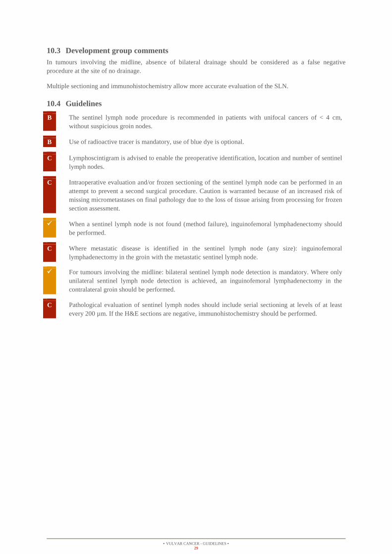
VULVAR CANCER - GUIDELINES 29
10.3 Development group commentsIn tumours involving the midline, absence of bilateral drainage should be considered as a false negativeprocedure at the site of no drainage.
Multiple sectioning and immunohistochemistry allow more accurate evaluation of the SLN.
10.4 Guidelinesc
B The sentinel lymph node procedure is recommended in patients with unifocal cancers of < 4 cm,without suspicious groin nodes.
B Use of radioactive tracer is mandatory, use of blue dye is optional.
C Lymphoscintigram is advised to enable the preoperative identification, location and number of sentinellymph nodes.
C Intraoperative evaluation and/or frozen sectioning of the sentinel lymph node can be performed in anattempt to prevent a second surgical procedure. Caution is warranted because of an increased risk ofmissing micrometastases on final pathology due to the loss of tissue arising from processing for frozensection assessment.
When a sentinel lymph node is not found (method failure), inguinofemoral lymphadenectomy shouldbe performed.
c
C Where metastatic disease is identified in the sentinel lymph node (any size): inguinofemorallymphadenectomy in the groin with the metastatic sentinel lymph node.
For tumours involving the midline: bilateral sentinel lymph node detection is mandatory. Where onlyunilateral sentinel lymph node detection is achieved, an inguinofemoral lymphadenectomy in thecontralateral groin should be performed.
c
C Pathological evaluation of sentinel lymph nodes should include serial sectioning at levels of at leastevery 200 µm. If the H&E sections are negative, immunohistochemistry should be performed.
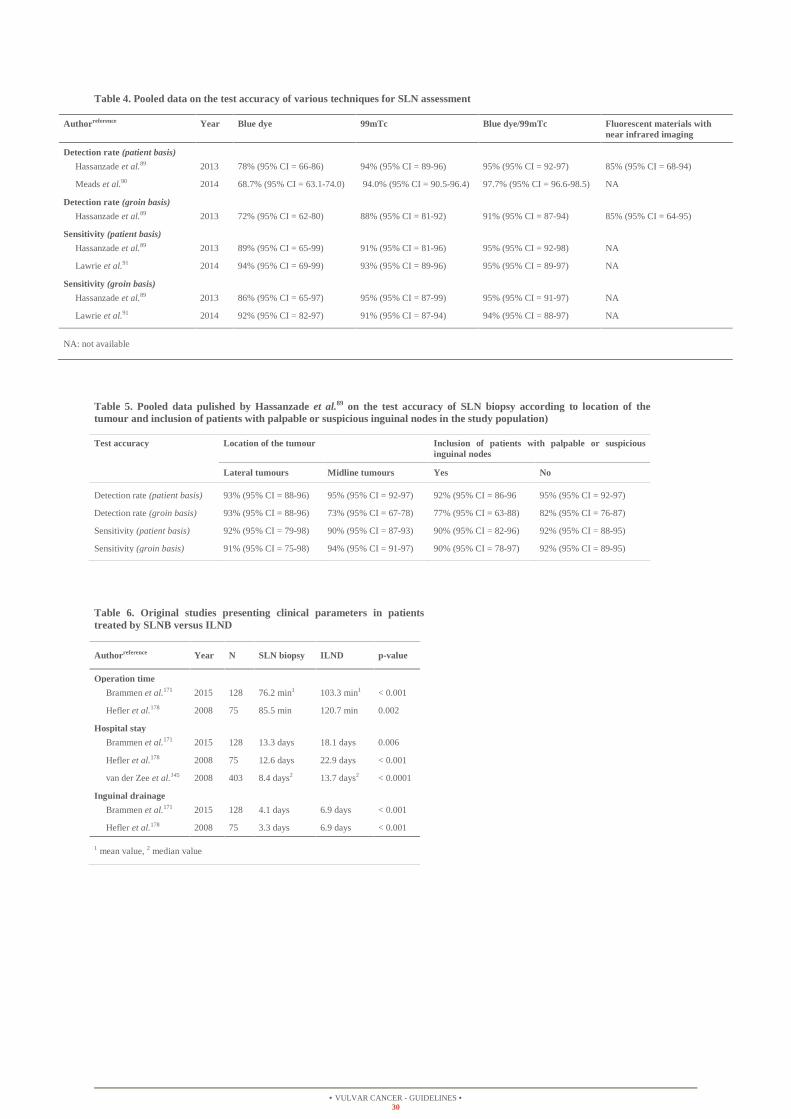
VULVAR CANCER - GUIDELINES 30
Table 4. Pooled data on the test accuracy of various techniques for SLN assessment
Table 5. Pooled data pulished by Hassanzade et al.89 on the test accuracy of SLN biopsy according to location of thetumour and inclusion of patients with palpable or suspicious inguinal nodes in the study population)
Table 6. Original studies presenting clinical parameters in patientstreated by SLNB versus ILND
Authorreference Year Blue dye 99mTc Blue dye/99mTc Fluorescent materials withnear infrared imaging
Detection rate (patient basis)
Hassanzade et al.89 2013 78% (95% CI = 66-86) 94% (95% CI = 89-96) 95% (95% CI = 92-97) 85% (95% CI = 68-94)
Meads et al.90 2014 68.7% (95% CI = 63.1-74.0) 94.0% (95% CI = 90.5-96.4) 97.7% (95% CI = 96.6-98.5) NA
Detection rate (groin basis)
Hassanzade et al.89 2013 72% (95% CI = 62-80) 88% (95% CI = 81-92) 91% (95% CI = 87-94) 85% (95% CI = 64-95)
Sensitivity (patient basis)
Hassanzade et al.89 2013 89% (95% CI = 65-99) 91% (95% CI = 81-96) 95% (95% CI = 92-98) NA
Lawrie et al.91 2014 94% (95% CI = 69-99) 93% (95% CI = 89-96) 95% (95% CI = 89-97) NA
Sensitivity (groin basis)
Hassanzade et al.89 2013 86% (95% CI = 65-97) 95% (95% CI = 87-99) 95% (95% CI = 91-97) NA
Lawrie et al.91 2014 92% (95% CI = 82-97) 91% (95% CI = 87-94) 94% (95% CI = 88-97) NA
NA: not available
Test accuracy Location of the tumour Inclusion of patients with palpable or suspiciousinguinal nodes
Lateral tumours Midline tumours Yes No
Detection rate (patient basis) 93% (95% CI = 88-96) 95% (95% CI = 92-97) 92% (95% CI = 86-96 95% (95% CI = 92-97)
Detection rate (groin basis) 93% (95% CI = 88-96) 73% (95% CI = 67-78) 77% (95% CI = 63-88) 82% (95% CI = 76-87)
Sensitivity (patient basis) 92% (95% CI = 79-98) 90% (95% CI = 87-93) 90% (95% CI = 82-96) 92% (95% CI = 88-95)
Sensitivity (groin basis) 91% (95% CI = 75-98) 94% (95% CI = 91-97) 90% (95% CI = 78-97) 92% (95% CI = 89-95)
Authorreference Year N SLN biopsy ILND p-value
Operation time
Brammen et al.171 2015 128 76.2 min1 103.3 min1 < 0.001
Hefler et al.178 2008 75 85.5 min 120.7 min 0.002
Hospital stay
Brammen et al.171 2015 128 13.3 days 18.1 days 0.006
Hefler et al.178 2008 75 12.6 days 22.9 days < 0.001
van der Zee et al.145 2008 403 8.4 days2 13.7 days2 < 0.0001
Inguinal drainage
Brammen et al.171 2015 128 4.1 days 6.9 days < 0.001
Hefler et al.178 2008 75 3.3 days 6.9 days < 0.001
1 mean value, 2 median value

VULVAR CANCER - GUIDELINES 31
11 Radiation therapy
11.1 Summary of available scientific evidencePrimary radiotherapy of the groin: as part of Cochrane systematic review, van der Velden et al.204
compared the effectiveness and safety of this therapeutic approach to the inguino-femoral lymphnodes with primary groin surgery. One randomised controlled trial205, one case-control206 and twoobservational186,207 studies have been included in this review. No pooled analysis is described and itshould be noted that two studies also included patients with non-squamous histology206,207. Thetumour recurrence rate in the groin after primary groin radiation ranged from 0% to 18.5% (Table 7).However, only the randomised controlled trial205 directly compared radiotherapy towards the groinversus surgery. In this trial, there is a difference in groin recurrence, favouring the primary groinsurgery (0% versus 18.5%). Overall survival and progression-free survival were significantly lowerin the radiation group compared with the surgery group (p = 0.04 and p = 0.03, respectively). But,the patients treated with groin radiation had substantially shorter hospitalizations than those whounderwent groin surgery (p = 0.0001). It should be noted that this trial was closed prematurely wheninterim monitoring revealed an excessive number of groin relapses on the groin radiation regimen.Criticisms could be made of the technique of radiotherapy applied in this trial (potentialinsufficiency to sterilise subclinical lymph node metastases in the groin). Maximum dose wasprescribed at 3 cm in this trial. It is likely, therefore, that the deeper groin nodes were relativelyundertreated.
LoE 1-
Neoadjuvant radiotherapy: no studies enrolling at least 50 patients were identified. Interpretation ofthe results from the 8 identified trials208-215 are limited notably by the small number of patientsevaluated (only 3 trials208-210 have accrued in excess of 10 patients) and by the heterogeneity in theradiotherapy regimens (external beam radiation and/or intracavitary brachytherapy). Althoughstudies are very small, authors reported low severe complications and high proportions of patientsalive with no evidence of disease and no recurrence (Table 8). Furthermore, this combined therapeuticapproach showed a good probability of bladder and/or rectal preservation.
LoE 3
Adjuvant radiotherapy (close surgical margins or positive margins): Faul et al.216 reported areduction of local recurrence from 58% to 16% in these patients treated with adjuvant radiationtherapy. On multivariate analysis, adjuvant radiation was a significant prognostic predictor for localcontrol (p = 0.009). However, it did not reach statistical significance for overall survival. Onsubgroup analysis, adjuvant radiation therapy significantly improved actuarial 5-year survival forpatients with positive margins (p = 0.001), but not for those with close margins (p = 0.63).
LoE 2+
Adjuvant radiotherapy (no suspicious groin nodes): Stehman et al.205 randomised 58 patients patientswith lesions clinically confined to the vulva and no suspicious groin nodes to either radicalvulvectomy followed by either groin radiation or inguinal lymphadenectomy (plus groin radiation ifnodes were involved) to compare efficacy and morbidity of the two treatment approaches. The groinswere treated daily to a dose of 50 Gy over 5 weeks (200 cGy/d). Patients randomised to the groindissection arm who where found to have metastatic carcinoma in the resected nodes received post-operative radiation therapy to the ipsilateral groin and hemipelvis. A total dose of 50 Gy wasadministered through anterior portals to the groin and through anterior and posterior portals to theiliac nodes. The study was closed prematurely when interim monitoring revealed an excessivenumber of groin relapses on the groin radiation regimen (see above).
LoE 1-
Adjuvant radiotherapy (single positive node): the benefit of adjuvant radiation in patients with asingle lymph node metastasis and micrometastatic disease to the lymph nodes is controversial. Fonset al.217 could not demonstrate a significant benefit of adjuvant radiotherapy in these patients on bothdisease-free and disease-specific survival (HR = 0.98, 95% CI = 0.45-2.14, p = 0.97 and HR = 1.02,95% CI = 0.42-2.47, p = 0.96). Recurrence rates appeared quite similar between the radiotherapy
LoE 2+

VULVAR CANCER - GUIDELINES 32
and the no-radiotherapy group (39% versus 32%). In multivariate subanalysis performed as part ofthe AGO-CaRE-1 study218 (163 patients), adjuvant radiotherapy was associated with a notstatistically significant better PFS compared to patients without adjuvant treatment (adjustment forage, Eastern cooperative oncology group (ECOG) performance status, Union internationale contre lecancer (UICC) stage, grade, and invasion depth: HR = 0.88, 95% CI = 0.50-1.56, p = 0.67). Similarresults were obtained after control for multiple confounding factors by inverse probability oftreatment weighting (HRIPTW = 0.93, 95% CI = 0.51-1.67, p = 0.79).
Parthasarathy et al.219 have for their part reported a favourable 5-y disease specific survival (DSS) inpatients receiving adjuvant radiation. Controlling for age at diagnosis and extent oflymphadenectomy, their data suggest that adjuvant radiation may improve the survival of thesepatients although this only reached borderline statistical significance (HR = 0.57, 95% CI = 0.32-1.03, p = 0.06). However, it should be noted that no information about the size and location oftumour is available in this study. Moreover, adjuvant radiation did not significantly benefit womenwho had more than 12 nodes resected (66.7 versus 77.3%, p = 0.23).
Adjuvant radiotherapy (multiple positive nodes): a randomised trial compared pelvic radiotherapywith pelvic lymphadenectomy in 114 patients with inguinofemoral lymph node metastases afterradical vulvectomy and bilateral inguinofemoral lymphadenectomy220. The difference in regional(groin) recurrence was significant, favouring the adjunctive radiation therapy group (5.1% versus23.6%, p = 0.02). Survival proved also to be better in the patients who received postoperativeradiotherapy (overall survival (p = 0.03), relative survival (0.004), progression-free interval (0.03)).In this study, the most dramatic survival advantage for radiation therapy was in patients who hadeither of the two major poor prognostic factors present: 1) clinically suspicious or fixed ulceratedgroin nodes, and 2) two or more positive groin nodes. The long time results of this trial revealed apersistent benefit for patients treated with pelvic irradiation221.
After a median survival follow-up of 74 months, the OS benefit for radiation in patients withclinically suspected or fixed ulcerated groin nodes (p = 0.04) and two or more positive groin nodes(p < 0.001) persisted. The relative risk of progression was significantly reduced in radiation patients(HR = 0.39, 95% CI = 0.17-0.88, p = 0.02) after adjustment for age and adverse tumourcharacteristics. Moreover, the cancer-related death rate was significantly higher for pelvic noderesection compared with radiation (HR = 0.49, 95% CI = 0.28-0.87, p = 0.015). The proportion ofpatients developing post-operative wound infections, urinary tract infection, and other adversesequelae were similar between treatment approaches. However, it should be noted that patients withpositive groin nodes in the surgery group in this study did not receive adjuvant radiotherapy to thegroins.
LoE 1+
In multivariate analysis of different nodal subgroups performed as part of the AGO-CaRE-1 study218
(adjustment for age, ECOG performance status, UICC stage, grade, and invasion depth) adjuvantradiotherapy was associated with statistically significant better progression-free survival (PFS) inpatients with two positive nodes (91 patients, HR = 0.31, 95 CI 0.14-0.71, p = 0.005), and in patientswith three positive nodes (56 patients, HR = 0.40, 95% CI = 0.16-0.98, p = 0.05) compared topatients without adjuvant treatment. Similar results were obtained after control for multipleconfounding factors by inverse probability of treatment weighting (two positive nodes: HRIPTW =0.24, 95% CI = 0.11-0.56, p < 0.001; three positive nodes: HPIPTW = 0.32, 95% CI = 0.13-0.79, p =0.009). The benefit of adjuvant radiotherapy among patients with more than three positive nodes didnot reach statistical significance (21 patients, HR = 0.52, 95% CI = 0.24-1.10, p = 0.09/HRIPTW =0.44, 95% CI = 0.17-1.17, p = 0.10).
LoE 2+
11.2 Previous initiativesEight previous initiatives1-4,37-39,87 presenting guidelines on radiation therapy were identified.

VULVAR CANCER - GUIDELINES 33
11.3 Development group commentsWhen possible without damaging structures such as anus, urethra and clitoris, reexcision is preferred in case ofpositive margins in the light of the significant short as well as long term morbidity associated with the necessaryrelatively high dose of radiotherapy to the vulvar skin.
11.4 Guidelines
Adjuvant radiotherapy should start as soon as possible, preferably within 6 weeks of surgical treatment.
When invasive disease extends to the pathological excision margins of the primary tumour, and furthersurgical excision is not possible, postoperative radiotherapy should be performed.
In case of close but clear pathological margins, postoperative vulvar radiotherapy may be considered toreduce the frequency of local recurrences. There is no consensus for the threshold of pathologicalmargin distance below which adjuvant radiotherapy should be advised.
B Postoperative radiotherapy to the groin is recommended for cases with > 1 metastatic lymph nodeand/or presence of extracapsular lymph node involvement.
Adjuvant radiotherapy for metastatic groin nodes should include the ipsilateral groin area and wherepelvic nodes are non-suspicious on imaging, the distal part of the iliac nodes with an upper limit at thelevel of the bifurcation of the common iliac artery.
C Based on evidence from other squamous cell cancers such as cervical, head & neck, and anal cancer,the addition of concomitant, radiosensitising chemotherapy to adjuvant radiotherapy should beconsidered.

VULVAR CANCER - GUIDELINES 34
Table 7. Original studies presenting data in patients treated with primary groin radiation
Table 8. Original studies presenting data in patients treated with neoadjuvant radiation
Authorreference Year N Radiotherapy regimen Groin recurrence Survival
Stehman et al.205 1992 52 Dose: 50 Gy at 3 cmType: 50% electrons
Radiation: 18.5% (5/27)Surgery: 0% (0/25)
Median follow-up: > 36 monthsOS: 60% versus 86%DSS: 67% versus 92%DFS: 68% versus 92%
Manavi et al.206 1997 135 Dose: 45 Gy at 5 cmType: telecobalt
Radiation: 4.6% (6/65)No radiation: 10% (7/70)
Follow-up: NA5y-OS: 62.4% versus 93.7%
Katz et al.186 2003 14 Dose: 45 GyType: electrons and photons combined
0% (0/14) Median follow-up: 98 months
Perez et al.207 1998 19 Dose: 50-70 Gy at 4 cmType: photons (electron boost)
10.5% (2/19) Median follow-up: 60 months
5y-OS: 5-year overall survival, DFS: disease-free survival, DSS: disease-specific survival, Gy: Gray, NA: not available, OS: overall survival.
Authorreference Year N Radiotherapy regimen Recurrence Survival/complications
Boronow et al.208 1987 37 External beam and intracavitary: N = 22Intracavitary only: N = 12External beam only: N = 3
Local: N = 5Pelvic: N = 1
Median follow-up: 38.4 monthsStatus: 59.5% (22/37) alive NEDSevere complications: 23%
Balat et al.209 2000 24 External beam: N = 24 Local: N = 5Distant: N = 1
Median follow-up: NAStatus: 70.8% (17/24) alive NEDSevere complications: NA
Rotmensch et al.210 1990 16 External beam: N = 16 Central: N = 4Distant: N = 2
Median follow-up: 25 monthsStatus: 56,3% (9/16) alive NEDSevere complications: 4%
Hacker et al.211 1984 8 External beam and intracavitary: N = 1External beam only: N = 7
NA Median follow-up: NAStatus: 62.5% (5/8) alive NEDSevere complications: 12%
Jafari et al.212 1981 4 External beam: N = 4 Local: N = 0Distant: N = 0
Median follow-up: NAStatus: 100% (4/4) alive NEDSevere complications: 0%
Fairey et al.213 1985 7 External beam: N = 7 Local: N = 1Distant: N = 1
Median follow-up: NAStatus: 85.7% (6/7) alive NEDSevere complications: 14%
Carlino et al.214 1984 6 Intracavitary: N = 6 Local: N = 2 Median follow-up: NAStatus: NASevere complications: NA
Pao et al.215 1988 2 NA NA Median follow-up: NAStatus: 100% (2/2) alive NEDSevere complications: 0%
NA: not available, NED: no evidence of disease and no recurrence.

VULVAR CANCER - GUIDELINES 35
12 Chemoradiation
12.1 Summary of available scientific evidencePrimary chemoradiation: as part of Cochrane systematic review, Shylasree et al.222 evaluated theeffectiveness and safety of neoadjuvant and primary chemoradiation for women with LAVC. Amongthe 3 studies included in this review223-225, only two retrospective studies224,225 looked at primarychemoradiation versus primary surgery. It should be noted that no pooled analysis is described. Thenumber of cases of tumour recurrence and deaths were too small in one study225 to allow computingan adjusted hazard ratio (the confidence interval was non-informative for all combinations ofcovariate adjustment). In the second retrospective study, Landrum et al.224 compared outcomes of 63patients with LAVC treated by primary surgery (N = 30) of by primary chemoradiation (N = 33).The general schema for chemoradiation involved weekly cisplatin (40 mg/m²) or two cycles ofcisplatin (50 mg/m²) plus 5-FU (1,000 mg/m²) concurrent with external beam radiation. Theradiation fraction size was generally 160-180 cGy delivered in a once-daily fraction with a mediandose of 4,760 cGy (range 3,690-6,300 cGy) to the whole pelvis and primary vulvar site, withadditional radiation to the inguinal regions as directed by nodal status. Patients were managedsurgically with radical (N = 11) or modified radical vulvectomy (N = 19) when adequate surgicalmargins could be obtained without urinary or colonic diversion. Adjuvant radiation orchemoradiation was completed in 19 of 25 patients in the primary surgery group with lymph nodemetastasis. Eight patients had surgical excision of residual disease following primarychemoradiation.
At a median follow-up of 31 months, there was no statistically significant difference in the risk ofdeath in patients with LAVC between patients who received primary chemoradiation and those whoreceived primary surgery, after adjustment for age, FIGO stage, size of tumour and nodal status (HR= 1.09, 95% CI = 0.37-3.17, p > 0.05). Recurrence or PFS was not reported in a multivariate analysisin this study. However, the authors reported no statistically significant difference in recurrence ratebased on treatment group (5 in the chemoradiation arm versus 7 in the primary surgery arm, p >0.05). Four patients that were treated with primary chemoradiation only had a partial response totreatment and died of the disease.
An another study enrolling at least 50 patients has been identified. In a GOG phase II study including58 patients with LAVC not amenable to surgical resection (radical vulvectomy), Moore et al.226
assessed the efficacy and toxicity of radiation therapy and concurrent chemotherapy when used forthe primary treatment. Radiation was given daily, five days per week in 1.8 Gy fractions to a totaldose of 57.6 Gy. Patients received concurrent cisplatin (40 mg/m² to maximum dose 70 mg)chemotherapy administered weekly throughout radiation therapy. Patients only underwent radicalsurgical resection after chemoradiation if they had residual disease present on biopsy. After a medianfollow-up of 24 months, 37 patients (64%) achieved a cCR. Among these patients there were 29(50%) who underwent surgical biopsy and had a pCR (Table 9). Twenty-two of these 29 patientscontinued to have no evidence of disease, while 7 patients experienced recurrence. Of the 29 patientswho had persistent disease after chemoradiation and who underwent surgical resection, 8 (28%) werealive at last follow-up with no evidence of disease recurrence. Although acute toxicity wassignificant, the protocol was considered tolerable.
LoE 2+
Results from the 16 other identified studies227-242 are limited notably by the small number of patientsevaluated (only 2 trials231,232 have accrued in excess of 20 patients) and by the heterogeneity in theprimary chemoradiation regimens. Although studies are small, chemoradiation as a primarytherapeutic approach has been reported to produce high response rates (Table 9).
LoE 3
Neoadjuvant chemoradiation: among the 3 studies223-225 included in the Cochrane systematic reviewpublished by Shylasree et al.222, only one study223 looked at neoadjuvant chemoradiation versus
LoE 1-

VULVAR CANCER - GUIDELINES 36
primary surgery. In this randomised controlled trial, 68 women with operable LAVC wererandomised to either primary radical surgery followed by radiation if more than one groin lymphnode contained metastatic disease, or to neoadjuvant chemoradiation followed by surgery.Chemoradiation comprised 50 Gy neoadjuvant radiotherapy with concurrent infusional 5-FU 750mg/m² days 1-5 and Mitomycin-C 15 mg/m² IV day 1, with two courses given three weeks apart. Inthe primary surgery arm, 15 (15/37) patients underwent adjuvant radiation. Surgery was feasible in24 out of 28 patients in the neoadjuvant arm. At a mean follow-up of 42 months, thirty recurrenceshave been reported (13 in the neoadjuvant chemoradiation arm and 17 in the primary surgery arm).The authors reported no statistically significant difference in the risk of death at 5 years between thetwo therapeutic approaches (RR = 1.39, 95% CI = 0.94-2.06, p > 0.05). Furthermore, no statisticallysignificant difference in the risk of overall treatment related morbidity was found (RR = 1.18, 95%CI = 0.71-1.96, p > 0.05). It should be noted that details regarding the extent of primary tumour andthe complexity of surgical procedures required in each group are not provided, and quality of life isnot reported.
Two other original studies243,244 enrolling at least 50 patients were identified. In a GOG phase IIstudy including 71 patients with unresectable vulvar disease, or disease requiring exenterations,Moore et al.243 investigated the role of concurrent radiotherapy and cisplatin/infusional 5-FUchemotherapy. A cCR occurred in 47% of patients. Among those patients who had surgery, 70% hada pCR. Two of 71 patients had unresectable disease after chemoradiation, and three patients requiredexenteration. After a median follow-up of 50 months, 40 patients were alive with no evidence ofdisease and no recurrence (Table 10). Toxicity from chemoradiation was estimated acceptable,although acute cutaneous reactions were almost universal. In the second identified study244, 58patients referring for primary or recurrent disease received preoperative radiotherapy to a dose of 54Gy (divided into two courses with an interval of two weeks). Concurrent preoperative chemotherapywith 5-FU (750 mg/m² daily for 5 days) and Mitomycin-C (15 mg/m² single bolus) were given at thestart of each cycle. A cCR of both the vulvar and inguinal disease occurred in 27% of patients. ApCR was confirmed in 13 patients (31%). After a median follow-up of 22 months, 28 patients werealive with no evidence of disease and no recurrence (Table 10). Like the GOG phase II study243,treatment side effects were estimated acceptable.
LoE 2+
As part of a meta-analysis including 7 studies229,234,237,245-248 for a total of 70 patients, Stuckey etal.249 investigated whether elderly patients are more likely to die of intercurrent disease or oftreatment complications. It should be noted that Stuckey et al.249 included patients receivingpreoperative or primary chemoradiation treatment with curative intent even if in the majority ofcases, this was given with neoadjuvant intent. Radiation doses ranged from 18 to 72 Gy and includedthe vulvar, inguinal, and the pelvic regions. Chemotherapy included 5-FU with or without cisplatinor Mitomycin-C (Table 11). Seventy-eight percent of patients younger than 65 years were withoutevidence of disease after treatment versus 66% of patients aged 65 years and above. Three percent ofpatients younger than 65 years of age died of intercurrent disease or treatment-related complicationsversus 11% of patients aged 65 years and above. But these differences did not reach statisticalsignificance (p = 0.30 and p = 0.37, respectively). It should be noted that 1) the small sample sizefrom included studies and 2) the changes in radiation therapy techniques and chemotherapy couldmake it difficult to statistically support the trend showing that elderly patients have lower survivaland higher intercurrent death.
LoE 1-
Results from the 11 other identified studies233,242,245-247,250-255 are limited notably by the small numberof patients evaluated (only 4 studies242,250,252,253 have accrued in excess of 20 patients) and by theheterogeneity in the chemotherapy regimens used in the neoadjuvant setting along with radiationtherapy (Table 10). Although studies are small, chemoradiation as a neoadjuvant therapeutic approachhas been reported to produce high response rates and high rates of surgical resectability withoutexenteration, regardless of chemotherapy regimen used. Overall, authors described high but
LoE 3

VULVAR CANCER - GUIDELINES 37
manageable rates of vulvar cutaneous toxicity.
Adjuvant chemoradiation: only one study256 enrolling at least 50 patients was identified. As part of alarge population-base analysis, Gill et al.256 evaluated adjuvant chemotherapy for node-positivevulvar cancer patients who received adjuvant radiotherapy. All patients (N = 1,797) receivedexternal beam radiotherapy as their radiotherapy treatment modality. Radiation modality wasavailable for 35.7% of patients. For those with modality captured, intensity-modulated radiotherapywas utilized in 6.5%. Median radiotherapy dose was 54 Gy. Median radiation length and time tochemotherapy initiation were 44 days and 76 days, respectively. Of patients receiving chemotherapy,78.5% started chemotherapy within 7 days of the start of radiotherapy.
After a median follow-up of 28.3 months, the unadjusted median survival without (N = 1,324) andwith adjuvant chemotherapy (N = 473) was 29.7 months and 44 months (p = 0.001), respectively. Onmultivariate analysis, delivery of adjuvant chemotherapy resulted in a trend towards reduction in therisk of death among patients who received adjuvant radiotherapy (HR = 0.81, 95% CI = 0.65-1.01, p= 0.059). On regression modeling with an adjustment using propensity score with IPTW, Gill etal.256 reported a statistically significant reduction in the risk of death for patients who receivedadjuvant chemotherapy (HRIPTW = 0.62, 95% CI = 0.48-0.79, p < 0.001).
LoE 2-
Results from the 4 other identified studies225,230,231,236 are limited notably by the very small number ofpatients evaluated. No study has accrued in excess of 10 patients (Table 12).
LoE 3
12.2 Previous initiativesSeven previous initiatives1,2,4,37-39,87 presenting guidelines on chemoradiation were identified.
12.3 Development group commentsNone.
12.4 Guidelines
C Definitive chemoradiation (with radiation dose escalation) is the treatment of choice in patients withunresectable disease.
C In advanced stage disease neoadjuvant chemoradiation should be considered in order to avoidexenterative surgery.
C Radiosensitising chemotherapy, preferably with weekly cisplatin, is recommended.

VULVAR CANCER - GUIDELINES 38
Table 9. Original studies presenting response and survival data in patients treated with primary chemoradiation
Authorreference Year N Chemotherapy regimen Radiotherapy regimen Response Survival
Moore et al.a,226 2012 LAVC: N = 58 Weekly CisP 40 mg/m² IV, up to 7 cycles 57.6 Gy in 1.8 Gy daily fractions cCR: 64% (37/58)pCR: 50% (29/58)
Median follow-up: 24 monthsStatus: 51% (30/58) alive NEDRecurr. 24% (7/29) with pCR
Landrum et al.224 2008 LAVC: N = 33 Either weekly CisP 40 mg/m² or two cycles ofCiSP 50 mg/m² IV d1 + 5-FU 1,000 mg/m² IV d1-4
47.6 Gy in 1.8 Gy daily fractions CR: 87% (29/33) Median follow-up: 31 monthsStatus: NARecurr.: 17% (5/29) with CR
Mak et al.231 2011 LAVC : N = 24 Either weekly CisP or 3-4 week 5-FU basedregimens
50 Gy, timing of fractions varied CR: 58% (20/34)b Median follow-up: 31.5 monthsStatus: NARecurr.: NA
Leiserowitz et al.232 1997 LAVC : N = 23 5-FU 1,000 mg/m² infusion d1-4 + CisP 100mg/m² IV d2, given 2-3 times during radiotherapy
Vulvar and inguinal region. 54 Gy in1.8 Gy BID fractions
CR : 78% (18/23) Mean follow-up: 45 monthsStatus: 60% (14/23) alive NEDRecurr.: 17% (4/23)
Tans et al.228 2011 LAVC : N = 20 5-FU 1,000 mg/m² infusion d1-4 + MMC 10mg/m² IV d1, given first week of each course ofradiotherpay
Split course 40 Gy + 20 Gy in 2 Gyfractions with 2-week break
CR: 70% (14/20) Median follow-up: NAStatus: NARecurr.: NA
Wahlen et al.234 1995 LAVC : N = 19 5-FU 1,000 mg/m² infusion d1-4 given weeks 1 +5 of radiotherapy. Six pts also given MMC 10mg/m² IV d1
45-50 Gy in 1.8 Gy daily fractions +implant or electron boost to vulva
CR: 52% (10/19)PR : 36% (7/19)
Median follow-up: 34 monthsStatus: 79% (15/19) alive NEDRecurr.: 10% (1/10) with CR
Russel et al.229 1992 LAVC: N = 18 5-FU 750-1,000 mg/m² infusion d1-4 + CisP 100mg/m² IV d1, 2-3 cycles given
54 Gy for macro and 36 Gy formicroscopic disease
CR: 50% (9/18)pCR: 44% (8/18)PR: 6% (1/18)
Median follow-up: 24 monthsStatus: 67% (12/18) alive NEDRecurr.: 11% (2/18)
Sebag-Montefiore et al.227 1994 LAVC: N = 16 5-FU 750 mg/m² infusion d1–5 + MMC 10 mg/m2IV d1, given first 5 d and last 5 d of radiotherapy
45 Gy in 2-2.5 Gy daily fractions CR : 44% (7/16)PR : 37% (6/16)
Follow-up: NAStatus: NARecurr.: NA
Koh et al.233 1993 LAVC: N = 14 5-FU 750-1,000 mg/m² IV infusion d1-4, weeklyfor 3 cycles
54 Gy in either daily or BID fractions CR: 57% (8/14)PR: 36% (5/14)
Median follow-up: 27 monthsStatus: 50% (7/14) alive NEDRecurr. 7% (1/14)
Cunningham et al.235 1997 LAVC : N = 14 5-FU 1000 mg/m² infusion d1-4 + CisP 50 mg/m²d1, given on first and last week of radiotherapy
45-50 Gy plus vulvar boost of 9-14 Gy CR : 64% (9/14)PR : 29% (4/14)
Mean follow-up: 26 monthsStatus: 28% (4/14) alive NEDRecurr. : 11% (1/9) with CR
Iversen et al.238 1982 LAVC: N = 9Recur.: N =4
Bleo 30 mg IM d 1, 3, 5 repeated after 2 weeks 36-40 Gy in 3 Gy daily fractions NA Follow-up: 112 monthsStatus: 30% (4/13) alive NEDRecurr. : NA
a Radiotherapy given to the vulva, groin and pelvis unless otherwise stated, b treatment response among the 34 patients treated with initial chemoradiation (data not available for patients treated by primary chemoradiationspecifically), 5-FU: 5-fluorouracil, cCR: clinical complete response, CR: complete response, CisP: cisplatin, Gy: Gray, LAVC: locally advanced vulvar cancer, MMC: mitomycin C, NA: not available, NED: no evidenceof disease and no recurrence, pCR: pathologic complete response, PR: partial response, Recurr.: recurrence.

VULVAR CANCER - GUIDELINES 39
Original studies presenting response and survival data in patients treated with primary chemoradiation (continued)
Authorreference Year N Chemotherapy regimen Radiotherapy regimen Response Survival
Han et al.236 2000 LAVC : N =12
5-FU 1,000 mg/m² infusion d1-4 + MMC 10 mg/m² IV d1,given week 1 and 5 of radiotherapy
45 Gy (vulva, pelvic and inguinal lymph nodes), 6-17 Gy to gross disease
CR: 42% (5/12)PR: 58% (7/12)
Follow-up: NAStatus: NARecurr.: NA
Berek et al.a,237 1991 LAVC: N = 12 5-FU 1,000 mg/m² infusion d1-4 + CisP 100 mg/m² d1every 28 d for 2 cycles
40-52 Gy in 1.6-1.8 Gy daily fractions, with boost tovulva (up to 74 Gy)
CR: 67% (8/12)PR: 25% (3/12)
Median follow-up: 37 monthsStatus: 83% (10/12) alive NEDRecurr. : 17% (2/12)
Akl et al.239 2000 NA: N = 12 5-FU 1,000 mg/m²/24h as continuous infusion days 1–4 and29-32 + MMC 15 mg/m² IV day 1
Vulva only (all pts node negative). 30–36 Gy in 2Gy daily fractions
CR: 100% (12/12) Mean follow-up: 41 monthsStatus: 66% (8/12) alive NEDRecurr.: 16% (2/12)
Thomas et al.230 1989 LAVC: N = 9 5-FU 1,000 mg/m² infusion d1-4 ± MMC 6 mg/m² (4/6 oneinjection, and 2/6 two injections 4 weeks apart)
40-64 Gy in 1.6-1.8 Gy twice daily fractions CR: 67% (6/9) Median follow-up: 20 monthsStatus: 67% (6/9) alive NEDRecurr.: NA
Beriwal et al. 242 2013 LAVC: N = 9 CisP 40 mg/m² d1 (N = 6) and 5-FU 1,000 mg/m² infusion,d1-5 (N = 36). Two cycles, given the first and last week ofradiotherapy
IMRT 46 Gy in 1.6 Gy BID fractions for 5d, then1.8 Gy daily for 7-8d then a break of 10-14 d, then1.6 Gy BID for 5 d
cCR: 44.4% (4/9) Follow-up: NAStatus: NARecurr.: NA
Mulayim et al.225 2004 LAVC : N = 7 5-FU 1,000 mg/m² infusion d1-4 + MMC 10 mg/m² IV d1,given weeks 1 and 4 of radiotherapy
60 Gy for macro and 45 Gy for microscopic disease CR: 85% (6/7) Median follow-up: 31 monthsStatus: 42% (3/7) alive NEDRecurr.: 28% (2/7)
Evans et al.a,240 1988 LAVC: N = 4 5-FU 1,000 mg/m² continous infusion d1-4 + MMC 10mg/m² IV d1
25-50 Gy in 2 Gy daily fractions CR: 50% (2/4)PR: 50% (2/4)
Mean follow-up: 33 monthsStatus: 50% (2/4) alive NEDRecurr.: 0% (0/3)
Kalra et al.a,241 1985 LAVC: N = 2 MMC 10 mg/m² IV d1 + 5-FU 1,000mg/m² infusion d1-5,given weeks 1 and 4 of radiotherapy
50 Gy in 2 Gy daily fractions CR: 100% (2/2) Mean follow-up: 33 monthsStatus: 100% (2/2) alive NEDRecurr.: 0% (0/2)
a Radiotherapy given to the vulva, groin and pelvis unless otherwise stated, 5-FU: 5-fluorouracil, Bleo: bleomycin, CR: complete response, CisP: cisplatin, Gy: Gray, LAVC: locally advanced vulvar cancer, MMC: mitomycinC, NA: not available, NED: no evidence of disease and no recurrence, PR: partial response, Recurr.: recurrence.

VULVAR CANCER - GUIDELINES 40
Table 10. Original studies presenting data in patients treated with neoadjuvant chemoradiation
Authorreference Year N Chemotherapy regimen Radiotherapy regimen Response Survival
Moore et al.a,243 1998 LAVC: N = 71 5-FU 1,000 mg/m² infusion d1-4 + CisP 50 mg/m²IV d1, given week 1 of each course of radiotherapy
2 courses of 23.8 Gy, given as 1.7 Gy BID for 4days and daily for 6 days with 2 weeks break
CR: 47% (34/71) Median follow-up: 50 monthsStatus: 56% (40/71) alive NEDRecurr. : 34% (24/69)
Landoni et al.a,244 1996 LAVC: N = 41Recurr.: N = 17
5-FU 750 mg/m² infusion d1-5 + MMC 15 mg/m²IV d1 given week 1 of each course of radiotherapy
54 Gy in 2 courses (36 Gy + 18 Gy) with 14 dtreatment break
cCR: 27% (14/52)pCR: 31% (13/42)
Median follow-up: 22 monthsStatus: 48% (28/58) alive NEDRecurr.: 27% (16/58)
Montana et al.a,250 2000 LAVC: N = 46 5-FU 1,000 mg/m² infusion d1-4 + CisP 50 mg/m²IV d1, given week 1 of each course of radiotherapy
2 courses of 23.8 Gy, given as 1.7 Gy BID for 4days and daily for 6 days with planned 2 weeksbreak
pCR (nodes): 40% (15/37)pCR (vulva): 52% (20/38)
Median follow-up: 78 monthsStatus: 26% (12/46) alive NEDRecurr.: 51% (19/37)
Beriwal et al.242 2013 LAVC: N = 42 CisP 40 mg/m² d1 (N = 6) and 5-FU 1,000 mg/m²infusion, d1-5 (N = 36). Two cycles, given the firstand last week of radiotherapy
IMRT 46 Gy in 1.6 Gy BID fractions for 5d,then 1.8 Gy daily for 7-8d then a break of 10-14d, then 1.6 Gy BID for 5 d
cCR: 51.2% (21/41)pCR: 48.5% (16/33)pCR: 48.8% (20/41)
Median follow-up: 15 monthsStatus: 45.5% (15/33) alive NEDRecurr.: 24.2% (8/33)
Lupi et al.a,252 1996 LAVC: N = 24 5-FU 750 mg/m² infusion d1-5 + MMC 15 mg/m²IV d1, given for 2 cycles
54 Gy in 2 courses with 14 d treatment break CR: 42% (10/24)PR: 54% (13/24)pCR: 36% (8/22)
Median follow-up: 34 monthsStatus: 65.5% (15/24) alive NEDRecurr.: 29% (7/24)
Gaudineau et al.253 2012 LAVC: N = 22 Carbo AUC 2 weekly during radiotherapy 50 Gy in 2 Gy daily fractions pCR: 27% (6/22)ORR: 95% (21/22)
Median follow-up: 28 monthsStatus: 54% (12/2) alive NEDRecurr.: 32 % (7/22)
Scheistroen et al.251 1993 LAVC: N = 20 Bleo 30 mg IV d1, 3, 5 during weeks 1 + 3 ofradiotherapy
30-45 Gy in 3 Gy daily fractions CR: 25% (5/20)PR: 50% (10/20)
Follow-up: NAStatus: 5% (1/20) alive NEDRecurr.: 80% (4/5) of pts with CR
Gerszten et al.254 2005 LAVC: N = 18 5-FU 1,000 mg/m² infusion d1-4 + CisP 50 mg/m²IV d1, given first and last week of radiotherapy
44.6 Gy in 1.6 Gy BID fractions for 5 d, then1.8 Gy daily for 7d, with 1-2 weeks break, then1.6 Gy BID for 5 d
cCR: 72% (13/18)pCR: 39% (7/18)
Mean follow-up: 24 monthsStatus: 83% (15/18) alive NEDRecurr.: 17% (3/18)
Eifel et al.245 1995 LAVC: N = 12 CisP 4 mg/m²/d infusion d1-4 + 5-FU 250 mg/m²/dinfusion d1-4, given weekly for 4 weeks
40 Gy in 2 Gy daily fractions CR: 50% (6/12)PR: 41% (5/12)
Mean follow-up: 18 monthsStatus: 50% (6/12) alive NEDRecurr.: 16% (1/6) of pts with CR
Whitaker et al.246 1990 LAVC: N = 9Recurr.: N = 3
5-FU 750-1,000 mg/m² infusion d1-4 + MMC 10-12 mg/m² IV d1, week 1 of each course ofradiotherapy
25 Gy in 2.5 Gy fractions CR : 42% (5/12)PR : 58% (7/12)
Follow-up: NAStatus: 25% (3/12) alive NEDRecurr.: 60% (3/5) of pts with CR
a Radiotherapy given to the vulva, groin and pelvis unless otherwise stated, 5-FU: 5-fluorouracil, AUC: area under the curve, BID: twice a day, Carbo: carboplatin, cCR clinical complete response, CisP: cisplatin, cPR clinicalpartial response, CR: complete response, d: days, Gy: Gray, IMRT: intensity-modulated radiation therapy, LAVC: locally advanced vulvar cancer, MMC: mitomycin C, NA: not available, NED: no evidence of disease and norecurrence, ORR: overall response rate, pCR: pathologic complete response, PR: partial response, pts: patients, Recurr: recurrence.

VULVAR CANCER - GUIDELINES 41
Original studies presenting data in patients treated with neoadjuvant chemoradiation (continued)
Authorreference Year N Chemotherapy regimen Radiotherapy regimen Response Survival
Carson et al.a,255 1990 LAVC: N = 6Recurr.: N = 2
5-FU 750 mg/m² infusion d1-5 + MMC 7.5 mg/m²IVd4 + CisP 0mg/m² IV d1, given weekly duringradiotherapy.
45-50 Gy in 1.75 Gy daily fractions pCR: 75% (6/8) Mean follow-up: 10 monthsStatus: 25% (2/8) alive NEDRecurr. or prog.: 50% (4/8)
Levin et al.247 1986 LAVC: N = 6 5-FU 1000 mg/m² infusion d1-4 + MMC 10 mg/²IV d1. 1-2 cycles
20-40 Gy in 2 Gy daily fraction NA Mean follow-up: 11 monthsStatus: 66% (4/6) alive NEDRecurr.: NA
Koh et al.233 1993 LAVC: N = 4 5-FU 750-1,000 mg/m²/d for 3-4 d 40-44.8 Gy CR: 25% (1/4)PR: 50% (2/4)
Mean follow-up: 29.8 monthsStatus: 25% (1/4) alive NEDRecurr.: 0% (0/4)
a Radiotherapy given to the vulva, groin and pelvis unless otherwise stated, 5-FU: 5-fluorouracil, CisP: cisplatin, CR: complete response, d: days, Gy: Gray, LAVC: locally advanced vulvar cancer, MMC: mitomycin C, NA: notavailable, NED: no evidence of disease and no recurrence, pCR: pathologic complete response, PR: partial response, prog.: progression, Recurr: recurrence.

VULVAR CANCER - GUIDELINES 42
Table 11. Original studies included in the meta-analysis published by Stuckey et al.249
Table 12. Original studies presenting response and survival data in patients treated with adjuvant chemoradiation
Authorreference Year N Median age Chemotherapy Radiotherapy Median follow-up DOD DOT DICD NED
(years) regimen regimen (months) (%) (%) (%) (%)
Eifel et al.245 1995 11 55 (37-85) 5-FU/CisP 40-50 Gy 21 27.3 0.0 9.1 63.6
Wahlen et al.234 1995 15 64 (37-89) 5-FU±MMC 45-50.4 Gy 36 13.3 0.0 13.1 73.3
Berek et al.237 1991 12 69 (52-76) 5-FU/CisP 46.64 Gy 34 16.7 0.0 0.0 83.3
Whitaker et al.246 1990 7 73 (65-87) 5-FU/MMC 25-50 Gy 7 57.1 14.3 0.0 28.8
Levin et al.247 1986 5 60 (44-66) 5-FU/MMC 18-60 Gy 5 0.0 0.0 0.0 80.0
Beriwal et al.248 2006 4 66.5 (54-84) 5-FU/CisP 43-49 Gy 19.5 0.0 0.0 0.0 100.0
Russel et al.229 1992 16 71 (13-90) 5-FU ± CisP 46-72 Gy 17 months 6.3% 6.3 0.0 75.0
5-FU: 5-fluorouracil, CisP: cisplatin, DICD: dead of intercurrent disease, DOD: dead of disease, DOT: dead of treatment, Gy: Gray, MMC: mitomycin C,NED: no evidence of disease and no recurrence.
Authorreference Year N Chemotherapy regimen Radiotherapy regimen Survival
Mak et al.231 2011 LAVC : N = 10 Either weekly CisP or 3-4 week 5-FU based regimens
50 Gy, timing of fractions varied Median follow-up: 31.5 monthsStatus: NARecurr.: NA
Thomas et al.230 1989 LAVC: N = 9 5-FU 1,000 mg/m² infusion d1-4 ±MMC 6 mg/m² (4/6 one injection,and 2/6 two injections 4 weeks apart)
40-64 Gy in 1.6-1.8 Gy twicedaily fractions
Median follow-up: 21 monthsStatus: 78% (7/9) alives NEDRecurr.: 22% (2/7)
Mulayim et al.225 2004 LAVC: N = 6 5-FU 1,000 mg/m² infusion d1-4 and21-24 of radiotherapy + MMC 10mg/m² IV d1 and d21 ofradiotherapy
60 Gy for macro and 45 Gy formicroscopic disease
Median follow-up: 20 monthsStatus: 0% (0/6)Recurr.: 33% (2/6)
Han et al.236 2000 LAVC: N = 6 5-FU 1,000 mg/m² infusion d1-4 +MMC 10 mg/m² IV d1, given week1 and 5 of radiotherapy
40-62 Gy Median follow-up: 17 monthsStatus: 83% (5/6) alive NEDRecurr.: 17% (1/6)
5-FU: 5-fluorouracil, CisP: cisplatin, Gy: Gray, LAVC: locally advanced vulvar cancer, MMC: mitomycin C, NA: not available, NED: no evidence of diseaseand no recurrence, Recurr.: recurrence.

VULVAR CANCER - GUIDELINES 43
13 Systemic treatment
13.1 Summary of available scientific evidenceNeoadjuvant chemotherapy: no studies enrolling at least 50 patients were identified. Results from the8 identified studies257-264 are limited notably by the heterogeneity and the number of patientsevaluated (only 3 studies258,262,263 have accrued in excess of 20 patients), and by the heterogeneity inthe chemotherapy regimens. Although studies are very small, agents showing response includebleomycin, cisplatin, and most notably infusional 5-FU (Table 13). It should be noted that responserates differ quite extensively among the studies. But, the identified trials have not shown significantevidence of improved survival. Additionally, some effective agents produce high toxicity, such asBleomycin, that is a significant issue.
LoE 3
Adjuvant chemotherapy: only one very small study265 was identified. To assess the use ofchemotherapy alone in the adjuvant setting, Bellaty et al.265 included 14 patients with inguinal nodemetastases after radical surgery. Cisplatin (100 mg/m²) was administered every 21 days for 4 cycles.Four of 14 patients recurred (29%) at a median of 57 months of follow-up, including two recurrencesin the groin. Three-year OS and PFS were 86% and 71%, respectively.
LoE 3
Targeted therapy: only one small study was identified. Horowitz et al.266 evaluated the efficacy andtoxicity of erlotinib (150 mg daily), a selective epidermal growth factor receptor tyrosine kinaseinhibitor, among 41 patients with locally advanced, primary, recurrent or metastatic vulvar squamouscell carcinoma. In this first phase II trial, overall clinical benefit rate was 67.5% including partialresponse (27.5%) and stable disease (40%). No complete response has been observed. It should to benoted that 1) responses were of relatively short duration and toxities were significant, and 2) qualityof life evaluation was not assessed in this study.
LoE 3
13.2 Previous initiativesThree previous initiatives1,3,39 presenting guidelines on systemic treatment were identified.
13.3 Development group commentsNone.
13.4 Guidelines
D Data in vulvar cancer are insufficient to recommend a preferred schedule in a palliative setting.

VULVAR CANCER - GUIDELINES 44
Table 13. Original studies presenting data in patients treated with neoadjuvant chemotherapy
Authorreference Year N Chemotherapy regimen Nb of cycles Response Survival
Aragona et al.263 2012 LAVC: N = 35 CisP + 5-FU (n = 12) or CisP + Tax (n = 6) or CisP +5-FU + Tax (n = 6) or VinC + Bleo + CisP (n = 6) orBleo alone (n = 5)
3 PR: 86% (30/35) Median follow-up: 49 monthsStatus: 68% (24/35) alive NEDRecurr. : 14% (4/29) of pts undergoing surgery
Domingues et al.262 2010 LAVC: N = 25A) N = 10B) N = 5C) N = 10
A) Bleo 20 mg/m² IV d1-10 continuous infusionB) Tax 100 mg/m² IV weeklyC) 5-FU 750 mg/m² d1-4 continuous infusion + CisP60–80 mg/m² IV d1, weekly
3 A) CR: 10% (1/10), PR: 50% (5/10)B) PR: 40% (2/5)C) PR: 20% (2/10)
Mean follow-up: 22 monthsStatus: A) 30% (3/10) alive NED, B) 20% (1/5)alive NED, C) 10% (1/10) alive NEDRecurr.: NA
Benedetti-Panici et al.258 1993 LAVC: N = 21 CisP 100 mg/m² day 1 + Bleo 15 mg days 1 and 8 +MTX 300 mg/m² day 8 every 21 days
Up to 3 PR in 14% (3/21)SD in 81% (17/21)
Median follow-up: 33 monthsStatus : NARecurr. : NA
Durrant et al.257 1990 LAVC: N = 18 Bleo 5 mg IM d1–5 + MTX 15 mg PO d1 and 4 +CCNU 40 mg PO d5-7 week 1, then Bleo 5 mg IM d1and 4 + MTX 15 mg PO d1 and 4 weeks 2-5
Up to 4 ORR: 67% (12/18) Follow-up: NAStatus: NARecur.: NA
Geisler et al.261 2006 LAVC: N = 13A) N = 10B) N = 3
A) 5-FU 1,000 mg/m²/24 h infusion d1-5 + CisP 50mg/m² IV d1, q3 weeksB) CisP 50 mg/m² IV q3 weeks
3-4 A) PR: 60% (6/10), pCR: 40% (4/10)B) 0% response
Median follow-up: 49 monthsStatus: A) 90% (9/10) alive NED, B) 0% aliveNEDRecur.: NA
Wagenaar et al.259 2001 LAVC: N = 12 Week 1: Bleo 5 mg IM d1-5 + CCNU 40 mg PO d5-7+ MTX 10 mg PO d1+4Weeks 2-6: Bleo 5 mg IM d1 + 4 + MTX 15 mg POd1.
Up to 3 ORR: 58% (7/12) Median follow-up: 8 monthsStatus: NARecurr.: NA
Bafna et al.260 2004 LAVC: N = 9 Cyclo 500 mg + MTX 50 mg + 5-FU 500 mg days 1,8 every 14 d
3 pCR: 11% (1/9)PR: 89% (8/9)
Follow-up: NAStatus: NARecur.: NA
Han et al.264 2012 LAVC: N = 4 Tax 60 mg/m² IV + Carbo AUC 2.7 IV weekly Up to 9 ORR = 0% Mean follow-up: 12 monthsStatus: 50% (2/4) alive NEDRecurr. : -
5-FU: 5-fluorouracil, Bleo: bleomycin, Carbo: carboplatin, CisP: cisplatin, CCNU: lomustine, CR: complete response, Cyclo: cyclophosphamide, LAVC: locally-advanced vulvar cancer, MTX: methotrexate, NA: not available,NED: no evidence of disease and no recurrence, ORR: overall response rate, pCR: pathologic complete response, PR: partial response, Recurr. : recurrence, Tax: paclitaxel, VinC: vincristine.
VULVAR CANCER - GUIDELINES 45
14 Treatment of recurrent disease
14.1 Summary of available scientific evidenceChemoradiation: no studies enrolling at least 50 patients were identified. Results from the 8identified studies227-230,241,244,251,252 are limited notably by the small number of patients evaluated(only one study251 has accrued in excess of 20 patients) and by the heterogeneity in thechemoradiation regimens (Table 14).
LoE 3
Chemotherapy: no studies enrolling at least 50 patients were identified. Results from the 7 identifiedtrials257,259,264,267-270 are limited notably by the small number of patients evaluated (only 2 trials267,268
have accrued in excess of 20 patients) and by the heterogeneity in the chemotherapy regimens (Table
15).
LoE 3
14.2 Previous initiativesFour previous initiatives1-3,39 presenting guidelines on treatment of recurrent disease were identified.
14.3 Development group commentsLocal recurrences should be treated as primary tumours with wide local excision and inguinofemorallymphadenectomy in case of depth of invasion >1 mm and not performed previously.
CT thorax/abdomen or PET/CT thorax/abdomen is recommended to examine the presence of additionalmetastases, which presence may influence treatment planning. Imaging might also be helpful in determining thepossibility of surgical resection.
14.4 GuidelinesTreatment of vulvar recurrence
Radical local excision is recommended.
For vulvar recurrence with a depth of invasion > 1 mm and previous sentinel lymph node removalonly, inguinofemoral lymphadenectomy should be performed.
The indications for postoperative radiotherapy are comparable to those for the treatment of primarydisease.
Treatment of groin recurrence
Restaging by CT (or PET-CT) of the thorax/abdomen/pelvis is recommended.
Preferred treatment is radical excision when possible, followed by postoperative radiation inradiotherapy naïve patients.
Based on evidence from other squamous cell cancers such as cervical and anal cancer, the addition ofradiosensitising chemotherapy to postoperative radiotherapy should be considered.
Definitive chemoradiation when surgical treatment is not possible.
Treatment of distant metastases
Systemic (palliative) therapy may be considered in individual patients (see systemic treatment).

VULVAR CANCER - GUIDELINES 46
Table 14. Original studies presenting response and survival data in recurrent patients treated with chemoradiation
Authorreference Year Chemotherapy regimen Radiotherapy regimen Response Survival
Scheistroen et al.251 1993 22 Bleo 30 mg IV d1, 3, 5 during weeks 1 + 3 ofradiotherapy
30-45 Gy in 3 Gy daily fractions CR: 9% (2/22)PR: 50% (11/22)
Follow-up: NAStatus: NARecurr.: NA
Landoni et al.a,244 1996 17 5-FU 750 mg/m² infusion d1-5 + MMC 15mg/m² IV d1 given week 1 of each course ofradiotherapy
54 Gy in 2 courses (36 Gy + 18Gy) with 14 d treatment break
pCR: 18% (3/17)pPR: 35% (6/17)
Follow-up: NAStatus: 29% (5/17) alive NEDRecurr.: NA
Sebag-Montefiore et al.227 1994 16 5-FU 750 mg/m² infusion d1–5 + MMC 10mg/m2 IV d1, given first 5 d and last 5 d ofradiotherapy
45 Gy in 2-2.5 Gy daily fractions CR : 50% (8/16)PR : 31% (5/16)
Follow-up: NAStatus: NARecurr.: NA
Thomas et al.230 1989 15 5-FU 1,000 mg/m² infusion d1-4 ± MMC 6mg/m² (4/6 one injection, and 2/6 twoinjections 4 weeks apart)
40-64 Gy in 1.6-1.8 Gy twice dailyfractions
CR: 53% (8/15) Follow-up: 5-45 monthsStatus: 47% (7/15) alive NEDRecurr.: NA
Tans et al.228 2011 8 5-FU 1,000 mg/m² infusion d1-4 + MMC 10mg/m² IV d1, given first week of each courseof radiotherpay
Split course 40 Gy + 20 Gy in 2 Gyfractions with 2-week break
CR: 75% (6/8) Median follow-up: NAStatus: NARecurr.: NA
Russel et al.229 1992 7 5-FU 750-1,000 mg/m² infusion d1-4 + CisP100 mg/m² IV d1, 2-3 cycles given
54 Gy for macro and 36 Gy formicroscopic disease
CR: 57% (4/7) Mean follow-up: 17.9 monthsStatus: 29% (2/7) alive NEDRecurr.: 14% (1/7) in pts with pCR
Lupi et al.a,252 1996 7 5-FU 750 mg/m² infusion d1-5 + MMC 15mg/m² IV d1, given for 2 cycles
54 Gy in 2 courses with 14 dtreatment break
CR: 71% (5/7)PR: 29% (2/7)
Median follow-up: 38 monthsStatus: 57% (4/7) alive NEDRecurr.: NA
Kalra et al.a,241 1985 1 MMC 10 mg/m² IV d1 + 5-FU 1000mg/m²infusion d1-5, given weeks 1 and 4 ofradiotherapy
50 Gy in 2 Gy daily fractions CR: 100% (1/1) Follow-up: NAStatus: 100% (1/1) alive NEDRecurr. : 0% (0/1)
a Radiotherapy given to the vulva, groin and pelvis unless otherwise stated, 5-FU: 5-fluorouracil, Bleo: bleomycin, CR: complete response, CisP: cisplatin, Gy: Gray, MMC: mitomycin C, NA:not available, NED: no evidence of disease and no recurrence, pCR: pathologic complete response, pPR pathologic partial response, PR: partial response, Recurr. recurrence.

VULVAR CANCER - GUIDELINES 47
Table 15. Original studies presenting data in recurrent patients treated with chemotherapy alone
Authorreference Year N Chemotherapy regimen Response Survival
Witteveen et al.267 2009 29 Tax 175 mg/m² IV q3 weeks; up to 9 cycles ORR: 13.8% (4/29)CR: 6% (2/29)PR: 6% (2/29)
Median PFS: 2.6 monthsMedian OS: 6.8 months
Thigpen et al.268 1986 22 CisP 50 mg/m² IV q3 weeks ORR: 0%CR: 0%PR: 0%
NA
Cormio et al.269 2009 15 CisP 80 mg/m2 IV d1 + Vinorelbine 25 mg/m² IV d1 and d8,q21 d for up to 6 cycles
ORR: 40% (6/15)CR: 27% (4/15)PR: 13% (2/15)
Median PFS: 10 monthsMedian OS: 19 months
Thigpen et al.268 1986 13 Piperazinedione 9 mg/m² IV q3 weeks ORR: 0%CR: 0%PR: 0%
PFS: NAOS: NA
Wagenaar et al.259 2001 13 Week 1: Bleo 5 mg IM d1-5 + CCNU 40 mg PO d5-7 + MTX 10mg PO d1+4Weeks 2-6: Bleo 5 mg IM d1 + 4 + MTX 15 mg PO d1.
ORR: 54% (7/13) Median follow-up: 8 monthsMedian PFS: 4.8 monthsa
Median OS: 7.8 monthsa
Muss et al.270 1989 11 Mitoxantrone 12 mg/m² IV q3 weeks ORR: 0%CR: 0%PR: 0%
Median PFS: 1.3 monthsMedian OS: 3.2 months
Durrant et al.257 1990 11 Bleo 5 mg IM d1–5 + MTX 15 mg PO d1 and 4 + CCNU 40 mgPO d5-7 week 1, then Bleo 5 mg IM d1 and 4 + MTX 15 mg POd1 and 4 weeks 2-5
ORR: 60% (6/10)CR : NAPR : NA
PFS: NAOS: NA
Han et al.264 2012 2 Tax 60 mg/m² IV + Carbo AUC 2.7 IV weekly ORR = 0% Mean follow-up: 3.5 monthsPFS: -OS: NA
a median survival among 12 patients with primary locally advanced disease and 13 with locoregional recurrence (data not available for patients with locoregionalrecurrence specifically), Bleo bleomycin, Carbo: carboplatin, CCNU: lomustine, CisP: cisplatin, CR: complete response, LAVC: locally advanced vulvar cancer,NA: not available, ORR: overall response rate, OS: overall survival, PR: partial response, PFS: progression-free survival, MTX: methotrexate, Tax: paclitaxel.

VULVAR CANCER - GUIDELINES 48
15 Follow-up
15.1 Summary of available scientific evidenceNo directly applicable clinical studies have been identified.
15.2 Previous initiativesSix previous initiatives1-3,38,39,271 presenting guidelines on follow-up were identified.
15.3 Development group commentsThere is no evidene for best follow-up schedule. Since local recurrences may occur many years after primarytreatment, lifelong follow-up is advised.
Since patients with associated vulvar intraepithelial neoplasia or lichen sclerosus/planus have a higher risk onlocal recurrence, more intensive follow-up may be indicated.
15.4 Guidelines
The optimal follow-up schedule for vulvar cancer is undetermined.
After primary surgical treatment the following follow-up schedule is suggested:
First follow-up 6-8 weeks postoperative
First two years every three-four months
Third and fourth year biannually
Afterward, long-term follow-up, especially in case of predisposing vulvar disease.Follow-up after surgical treatment should include clinical examination of vulva and groins.4
After definitive (chemo)radiation the following follow-up schedule is suggested:
First follow-up visit 10-12 weeks post completion of definitive (chemo)radiation.
First two years every three-four months
Third and fourth year biannually
Afterward, long-term follow-up, especially in case of predisposing vulvar disease.At first follow-up visit 10-12 weeks post definitive (chemo)radiation CT or PET-CT is recommendedto document complete remission.
4 Despite the well-recognized low sensitivity of palpation to identify groin recurrences, currently available datado not support routine use of imaging of the groins in follow-up.

VULVAR CANCER - GUIDELINES 49
16 Acronyms and abbreviations5-FU 5-fluorouracil
99mTc technectium-99m
ACPG Alberta clinical practice guidelines
AGDH Australian government department of health
AHRQ agency for healthcare research and quality
AquAS agència de qualitat i avaluació sanitàries deCatalunya
ASCO American society of clinical oncology
AUC area under the curve
BCCA British Columbia cancer agency
BID twice a day
Bleo bleomycin
CADTH Canadian agency for drugs andtechnologies in health
Carbo carboplatin
CCO cancer care Ontario
CCNU lomustine
cCR clinical complete response
CEPO comité de l’évolution des pratiques enoncologie
CI confidence interval
CisP cisplatin
CoCanCPG coordination of cancer clinical practiceguidelines in Europe
COMPAQ-HPST coordination pour la mesure de laperformance et l’amélioration de la qualité, hôpital,patient, sécurité, territoire
CR complete response
CT computed tomography
Cyclo cyclophosphamide
DICD dead of intercurrent disease
DOD dead of disease
DOT dead of treatment
DSS disease specific survival
ECOG Eastern cooperative oncology group
ESGO European society of gynaecologicaloncology
ESMO European society of medical Oncology
FIGO international federation of gynecology andobstetrics
FN false negative
FNA fine-needle aspiration
FNAC fine-needle aspiration cytology
FP false positive
GIN guidelines international network
GOC gynaecological oncology centre
GOG gynecologic oncology group
GROINSS-V Groningen international study onsentinel nodes in vulvar cancer
H&E haematoxylin and eosin
HAS haute autorité de santé
HR hazard ratio
IHC immunohistochemistry
ILND inguinal lymph node dissection
IMRT intensity-modulated radiation therapy
INAHTA international network of agencies forhealth technology assessment
INCa institut national du cancer
INESSS institut national d’excellence en santé et enservices sociaux
IPTW inverse probability of treatment weighting
KCE centre fédéral d’expertise des soins de santé
LAVC locally advanced vulvar cancer

VULVAR CANCER - GUIDELINES 50
MMC mitomycin C
MRI magnetic resonance imaging
MSAC medical services advisory committee
MTX methotrexate
NA not available
NCCN national comprehensive cancer network
NED no evidece of disease and no recurrence
NHMRC national health and medical researchcouncil
NHS national health service
NICE national institute for health and careexcellence
NZGG New Zealand guidelines group
OR odd ratio
ORR overall response rate
OS overall survival
pCR pathologic complete response
PET positron emission tomography
PET-CT positron emission tomography-computedtomography
PFS progression-free survival
PR partial response
Recurr recurrence
RCT randomised controlled trial
SIGN Scottish intercollegiate guidelines network
SLN sentinel lymph node
Tax paclitaxel
TN true negative
TP true positive
UICC union internationale contre le cancer
VinC vincristine

VULVAR CANCER - GUIDELINES 51
17 References1. NHS. Medical Protocol - Oncological
Management of Vulvar Cancerhttp://www.cmscnsenate.nhs.uk/index.php/download_file/view/415/1178/ (2015).
2. UHN. Princess Margaret Cancer Centre -Clinical Practice Guidelines - GynecologicCancer.http://www.uhn.ca/PrincessMargaret/Health_Professionals/Programs_Departments/Gynecologic/Pages/clinical_practice_guidelines.aspx (2015).
3. RCOG. Guidelines for the Diagnosis andManagement of Vulval Carcinoma.https://www.rcog.org.uk/en/guidelines-research-services/guidelines/vulval-carcinoma-guidelines-for-the-diagnosis-and-management-of/ (2014).
4. GMCT. Gynaecological CancerGuidelines.http://www.aci.health.nsw.gov.au/__data/assets/pdf_file/0010/154549/go_clinical_guidelines.pdf (2009).
5. Sobin, L.H., Gospodarowicz, M.K. &Wittekind, C.H. TNM classification ofmalignant tumours. 7th edition. Oxford:Whiley-Blackwell (2009).
6. Pecorelli, S. Revised FIGO staging forcarcinoma of the vulva, cervix, andendometrium. Int J Gynaecol Obstet 105,103-104 (2009).
7. Petru, E., et al. Gynecologic CancerIntergroup (GCIG) proposals for changesof the current FIGO staging system. Eur JObstet Gynecol Reprod Biol 143, 69-74(2009).
8. Wilkinson, E.J., Rico, M.J. & Pierson,K.K. Microinvasive carcinoma of thevulva. Int J Gynecol Pathol 1, 29-39(1982).
9. van der Steen, S., de Nieuwenhof, H.P.,Massuger, L., Bulten, J. & de Hullu, J.A.New FIGO staging system of vulvarcancer indeed provides a better reflectionof prognosis. Gynecol Oncol 119, 520-525(2010).
10. Tabbaa, Z.M., et al. Impact of the newFIGO 2009 staging classification forvulvar cancer on prognosis and stagedistribution. Gynecol Oncol 127, 147-152(2012).
11. Tan, J., et al. Validation of the FIGO 2009staging system for carcinoma of the vulva.Int J Gynecol Cancer 22, 498-502 (2012).
12. Beugeling, M., Ewing-Graham, P.C.,Mzallassi, Z. & van Doorn, H.C.Pathology slide review in vulvar cancerdoes not change patient management.ISRN Surg 2014, 385386 (2014).
13. Santoso, J.T., et al. Pathology slide reviewin gynecologic oncology. Obstet Gynecol91, 730-734 (1998).
14. Chafe, S., Honore, L., Pearcey, R. &Capstick, V. An analysis of the impact ofpathology review in gynecologic cancer.Int J Radiat Oncol Biol Phys 48, 1433-1438 (2000).
15. Khalifa, M.A., Dodge, J., Covens, A.,Osborne, R. & Ackerman, I. Slide reviewin gynecologic oncology ensurescompleteness of reporting and diagnosticaccuracy. Gynecol Oncol 90, 425-430(2003).
16. Selman, A.E., Niemann, T.H., Fowler,J.M. & Copeland, L.J. Quality assuranceof second opinion pathology ingynecologic oncology. Obstet Gynecol 94,302-306 (1999).
17. Chan, Y.M., et al. Pathology slide reviewin gynecologic oncology: routine orselective? Gynecol Oncol 75, 267-271(1999).
18. Iversen, T. The value of groin palpation inepidermoid carcinoma of the vulva.Gynecol Oncol 12, 291-295 (1981).
19. Piura, B., Rabinovich, A., Cohen, Y.,Friger, M. & Glezerman, M. Squamouscell carcinoma of the vulva in the south ofIsrael: a study of 50 cases. J Surg Oncol67, 174-181 (1998).

VULVAR CANCER - GUIDELINES 52
20. Singh, K., et al. Accuracy of magneticresonance imaging of inguinofemorallymph nodes in vulval cancer. Int JGynecol Cancer 16, 1179-1183 (2006).
21. Podratz, K.C., Symmonds, R.E., Taylor,W.F. & Williams, T.J. Carcinoma of thevulva: analysis of treatment and survival.Obstet Gynecol 61, 63-74 (1983).
22. Selman, T.J., Luesley, D.M., Acheson, N.,Khan, K.S. & Mann, C.H. A systematicreview of the accuracy of diagnostic testsfor inguinal lymph node status in vulvarcancer. Gynecol Oncol 99, 206-214(2005).
23. Hawnaur, J.M., Reynolds, K., Wilson, G.,Hillier, V. & Kitchener, H.C.Identification of inguinal lymph nodemetastases from vulval carcinoma bymagnetic resonance imaging: an initialreport. Clin Radiol 57, 995-1000 (2002).
24. Sohaib, S.A., et al. MR imaging ofcarcinoma of the vulva. AJR Am JRoentgenol 178, 373-377 (2002).
25. Bipat, S., et al. Is there a role for magneticresonance imaging in the evaluation ofinguinal lymph node metastases in patientswith vulva carcinoma? Gynecol Oncol103, 1001-1006 (2006).
26. Kataoka, M.Y., et al. The accuracy ofmagnetic resonance imaging in staging ofvulvar cancer: a retrospective multi-centrestudy. Gynecol Oncol 117, 82-87 (2010).
27. Cohn, D.E., et al. Prospective evaluationof positron emission tomography for thedetection of groin node metastases fromvulvar cancer. Gynecol Oncol 85, 179-184(2002).
28. De Hullu, J.A., et al. Noninvasivedetection of inguinofemoral lymph nodemetastases in squamous cell cancer of thevulva by L. Int J Gynecol Cancer 9, 141-146 (1999).
29. Kamran, M.W., et al. Whole-body[18F]fluoro-2-deoxyglucose positronemission tomography scan as combinedPET-CT staging prior to planned radical
vulvectomy and inguinofemorallymphadenectomy for squamous vulvarcancer: a correlation with groin nodemetastasis. Eur J Gynaecol Oncol 35, 230-235 (2014).
30. Moskovic, E.C., et al. The role of highresolution ultrasound with guided cytologyof groin lymph nodes in the managementof squamous cell carcinoma of the vulva: apilot study. Br J Obstet Gynaecol 106,863-867 (1999).
31. Hall, T.B., et al. The role of ultrasound-guided cytology of groin lymph nodes inthe management of squamous cellcarcinoma of the vulva: 5-year experiencein 44 patients. Clin Radiol 58, 367-371(2003).
32. Makela, P.J., Leminen, A., Kaariainen, M.& Lehtovirta, P. Pretreatment sonographicevaluation of inguinal lymph nodes inpatients with vulvar malignancy. JUltrasound Med 12, 255-258 (1993).
33. Abang Mohammed, D.K., Uberoi, R., de,B.L.A. & Monaghan, J.M. Inguinal nodestatus by ultrasound in vulva cancer.Gynecol Oncol 77, 93-96 (2000).
34. de Gregorio, N., et al. The role ofpreoperative ultrasound evaluation ofinguinal lymph nodes in patients withvulvar malignancy. Gynecol Oncol 131,113-117 (2013).
35. McCall, A.R., Olson, M.C. & Potkul, R.K.The variation of inguinal lymph nodedepth in adult women and its importancein planning elective irradiation for vulvarcancer. Cancer 75, 2286-2288 (1995).
36. Koh, W.J., et al. Femoral vessel depth andthe implications for groin node radiation.Int J Radiat Oncol Biol Phys 27, 969-974(1993).
37. Kidd, E., et al. ACR AppropriatenessCriteria(R) management of locoregionallyadvanced squamous cell carcinoma of thevulva. Am J Clin Oncol 36, 415-422(2013).

VULVAR CANCER - GUIDELINES 53
38. AHS. Squamous Cell Carcinoma of theVulva.http://www.albertahealthservices.ca/hp/if-hp-cancer-guide-gyne006-vulva.pdf(2013).
39. NCCN. Vulvar Cancer (Squamous CellCarcinoma). Version 1.http://www.nccn.org/professionals/physician_gls/f_guidelines.asp (2016).
40. Farias-Eisner, R., et al. Conservative andindividualized surgery for early squamouscarcinoma of the vulva: the treatment ofchoice for stage I and II (T1-2N0-1M0)disease. Gynecol Oncol 53, 55-58 (1994).
41. Balat, O., Edwards, C. & Delclos, L.Complications following combinedsurgery (radical vulvectomy versus widelocal excision) and radiotherapy for thetreatment of carcinoma of the vulva: reportof 73 patients. Eur J Gynaecol Oncol 21,501-503 (2000).
42. De Hullu, J.A., et al. Vulvar carcinoma.The price of less radical surgery. Cancer95, 2331-2338 (2002).
43. Rutledge, F.N., et al. Prognostic indicatorsfor invasive carcinoma of the vulva.Gynecol Oncol 42, 239-244 (1991).
44. DeSimone, C.P., et al. The treatment oflateral T1 and T2 squamous cellcarcinomas of the vulva confined to thelabium majus or minus. Gynecol Oncol104, 390-395 (2007).
45. van der Velden, J. Surgical interventionsfor early squamous cell carcinoma of thevulva. Cochrane Database Syst Rev,CD002036 (2000).
46. Burke, T.W., et al. Surgical therapy of T1and T2 vulvar carcinoma: furtherexperience with radical wide excision andselective inguinal lymphadenectomy.Gynecol Oncol 57, 215-220 (1995).
47. DiSaia, P.J., Creasman, W.T. & Rich,W.M. An alternate approach to earlycancer of the vulva. Am J Obstet Gynecol133, 825-832 (1979).
48. Heaps, J.M., Fu, Y.S., Montz, F.J., Hacker,N.F. & Berek, J.S. Surgical-pathologicvariables predictive of local recurrence insquamous cell carcinoma of the vulva.Gynecol Oncol 38, 309-314 (1990).
49. Tantipalakorn, C., Robertson, G.,Marsden, D.E., Gebski, V. & Hacker, N.F.Outcome and patterns of recurrence forInternational Federation of Gynecologyand Obstetrics (FIGO) stages I and IIsquamous cell vulvar cancer. ObstetGynecol 113, 895-901 (2009).
50. Berman, M.L., Soper, J.T., Creasman,W.T., Olt, G.T. & DiSaia, P.J.Conservative surgical management ofsuperficially invasive stage I vulvarcarcinoma. Gynecol Oncol 35, 352-357(1989).
51. Gunther, V., et al. Impact of radicaloperative treatment on the quality of life inwomen with vulvar cancer--a retrospectivestudy. Eur J Surg Oncol 40, 875-882(2014).
52. Curry, S.L., Wharton, J.T. & Rutledge, F.Positive lymph nodes in vulvar squamouscarcinoma. Gynecol Oncol 9, 63-67(1980).
53. Hacker, N.F., Berek, J.S., Lagasse, L.D.,Leuchter, R.S. & Moore, J.G. Managementof regional lymph nodes and theirprognostic influence in vulvar cancer.Obstet Gynecol 61, 408-412 (1983).
54. Hacker, N.F., Berek, J.S., Lagasse, L.D.,Nieberg, R.K. & Leuchter, R.S.Individualization of treatment for stage Isquamous cell vulvar carcinoma. ObstetGynecol 63, 155-162 (1984).
55. Hoffman, J.S., Kumar, N.B. & Morley,G.W. Microinvasive squamous carcinomaof the vulva: search for a definition. ObstetGynecol 61, 615-618 (1983).
56. Ross, M.J. & Ehrmann, R.L. Histologicprognosticators in stage I squamous cellcarcinoma of the vulva. Obstet Gynecol70, 774-784 (1987).

VULVAR CANCER - GUIDELINES 54
57. Iversen, T., Abeler, V. & Aalders, J.Individualized treatment of stage Icarcinoma of the vulva. Obstet Gynecol57, 85-89 (1981).
58. Kelley, J.L., 3rd, et al. Minimally invasivevulvar carcinoma: an indication forconservative surgical therapy. GynecolOncol 44, 240-244 (1992).
59. Magrina, J.F., et al. Squamous cellcarcinoma of the vulva stage IA: long-termresults. Gynecol Oncol 76, 24-27 (2000).
60. Van Der Velden, J., Kooyman, C.D., VanLindert, A.C. & Heintz, A.P. A stage Iavulvar carcinoma with an inguinal lymphnode recurrence after local excision. Acase report and literature review. Int JGynecol Cancer 2, 157-159 (1992).
61. Sidor, J., Diallo-Danebrock, R., Eltze, E.& Lelle, R.J. Challenging the concept ofmicroinvasive carcinoma of the vulva:report of a case with regional lymph noderecurrence and review of the literature.BMC Cancer 6, 157 (2006).
62. Volgger, B., et al. Fulminant course of amicroinvasive vulvar carcinoma in animmunosuppressed woman. GynecolOncol 65, 177-179 (1997).
63. Thangavelu, A., Andrew, A. & Buxton,E.J. Groin recurrence following Stage 1Asquamous cell carcinoma of the vulva.Gynecol Oncol 101, 172-174 (2006).
64. Atamdede, F. & Hoogerland, D. Regionallymph node recurrence following localexcision for microinvasive vulvarcarcinoma. Gynecol Oncol 34, 125-128(1989).
65. Hicks, M.L., Hempling, R.E. & Piver,M.S. Vulvar carcinoma with 0.5 mm ofinvasion and associated inguinal lymphnode metastasis. J Surg Oncol 54, 271-273(1993).
66. Stehman, F.B., Bundy, B.N., Dvoretsky,P.M. & Creasman, W.T. Early stage Icarcinoma of the vulva treated withipsilateral superficial inguinallymphadenectomy and modified radical
hemivulvectomy: a prospective study ofthe Gynecologic Oncology Group. ObstetGynecol 79, 490-497 (1992).
67. Gordinier, M.E., et al. Groin recurrence inpatients with vulvar cancer with negativenodes on superficial inguinallymphadenectomy. Gynecol Oncol 90,625-628 (2003).
68. Kirby, T.O., et al. Outcomes of Stage I/IIvulvar cancer patients after negativesuperficial inguinal lymphadenectomy.Gynecol Oncol 98, 309-312 (2005).
69. Andrews, S.J., et al. Therapeuticimplications of lymph nodal spread inlateral T1 and T2 squamous cell carcinomaof the vulva. Gynecol Oncol 55, 41-46(1994).
70. Tham, K.F., Shepherd, J.H., Lowe, D.G.,Hudson, C.N. & Van Dam, P.A. Earlyvulval cancer: the place of conservativemanagement. Eur J Surg Oncol 19, 361-367 (1993).
71. Jackson, K.S., Das, N., Naik, R., Lopes, A.& Monaghan, J.M. Contralateral groinnode metastasis following ipsilateral groinnode dissection in vulval cancer: a casereport. Gynecol Oncol 89, 529-531 (2003).
72. Van der Velden, J. Some aspects of themanagement of squamous cell carcinomaof the vulva. Thesis. Amsterdam, TheNetherlands (1996).
73. Zhang, S.H., Sood, A.K., Sorosky, J.I.,Anderson, B. & Buller, R.E. Preservationof the saphenous vein during inguinallymphadenectomy decreases morbidity inpatients with carcinoma of the vulva.Cancer 89, 1520-1525 (2000).
74. Zhang, X., et al. Sparing of saphenousvein during inguinal lymphadenectomy forvulval malignancies. Gynecol Oncol 105,722-726 (2007).
75. Rouzier, R., Haddad, B., Dubernard, G.,Dubois, P. & Paniel, B.J. Inguinofemoraldissection for carcinoma of the vulva:effect of modifications of extent and

VULVAR CANCER - GUIDELINES 55
technique on morbidity and survival. J AmColl Surg 196, 442-450 (2003).
76. Dardarian, T.S., Gray, H.J., Morgan, M.A.,Rubin, S.C. & Randall, T.C. Saphenousvein sparing during inguinallymphadenectomy to reduce morbidity inpatients with vulvar carcinoma. GynecolOncol 101, 140-142 (2006).
77. Paley, P.J., et al. The effect of sartoriustransposition on wound morbidityfollowing inguinal-femorallymphadenectomy. Gynecol Oncol 64,237-241 (1997).
78. Lin, J.Y., DuBeshter, B., Angel, C. &Dvoretsky, P.M. Morbidity and recurrencewith modifications of radical vulvectomyand groin dissection. Gynecol Oncol 47,80-86 (1992).
79. Soliman, A.A., Heubner, M., Kimmig, R.& Wimberger, P. Morbidity ofinguinofemoral lymphadenectomy invulval cancer. ScientificWorldJournal2012, 341253 (2012).
80. Fotiou, S.K., et al. "Butterfly" operation vstriple incision technique in vulvar cancer. acomparison of morbidity and clinicaloutcome. Eur J Gynaecol Oncol 17, 67-73(1996).
81. van der Velden, J., Schilthuis, M.S., Hyde,S.E., Ten Kate, F.J. & Burger, M.P.Squamous cell cancer of the vulva withoccult lymph node metastases in the groin:the impact of surgical technique onrecurrence pattern and survival. Int JGynecol Cancer 14, 633-638 (2004).
82. Helm, C.W., et al. A matched comparisonof single and triple incision techniques forthe surgical treatment of carcinoma of thevulva. Gynecol Oncol 46, 150-156 (1992).
83. Siller, B.S., et al. T2/3 vulva cancer: acase-control study of triple incision versusen bloc radical vulvectomy and inguinallymphadenectomy. Gynecol Oncol 57,335-339 (1995).
84. Hacker, N.F., Leuchter, R.S., Berek, J.S.,Castaldo, T.W. & Lagasse, L.D. Radical
vulvectomy and bilateral inguinallymphadenectomy through separate groinincisions. Obstet Gynecol 58, 574-579(1981).
85. Grimshaw, R.N., Murdoch, J.B. &Monaghan, J.M. Radical vulvectomy andbilateral inguinal-femorallymphadenectomy through separateincisions-experience with 100 cases. Int JGynecol Cancer 3, 18-23 (1993).
86. Fambrini, M., et al. Two-teamsimultaneous total radical vulvectomy andinguino-femoral lymphadenectomy viaseparate incisions for locally advanced(FIGO stages IB >/= 4 cm-III) squamousvulvar cancer. Anticancer Res 34, 7345-7350 (2014).
87. SOGC. Management of Squamous CellCancer of the Vulva.http://sogc.org/fr/guidelines/prise-en-charge-du-cancer-spinocellulaire-de-la-vulve-remplace-2-mai-1993/ (2006).
88. CCO. Sentinel Lymph Node Biopsy inVulvar Cancer.https://www.cancercare.on.ca/common/pages/UserFile.aspx?fileId=313084 (2014).
89. Hassanzade, M., Attaran, M., Treglia, G.,Yousefi, Z. & Sadeghi, R. Lymphaticmapping and sentinel node biopsy insquamous cell carcinoma of the vulva:systematic review and meta-analysis of theliterature. Gynecol Oncol 130, 237-245(2013).
90. Meads, C., et al. Sentinel lymph nodebiopsy in vulval cancer: systematic reviewand meta-analysis. Br J Cancer 110, 2837-2846 (2014).
91. Lawrie, T.A., et al. Sentinel nodeassessment for diagnosis of groin lymphnode involvement in vulval cancer.Cochrane Database Syst Rev 6, CD010409(2014).
92. Levenback, C., et al. Intraoperativelymphatic mapping for vulvar cancer.Obstet Gynecol 84, 163-167 (1994).

VULVAR CANCER - GUIDELINES 56
93. Decesare, S.L., et al. A pilot studyutilizing intraoperativelymphoscintigraphy for identification ofthe sentinel lymph nodes in vulvar cancer.Gynecol Oncol 66, 425-428 (1997).
94. Bowles, J., Terada, K.Y., Coel, M.N. &Wong, J.H. Preoperativelymphoscintigraphy in the evaluation ofsquamous cell cancer of the vulva. ClinNucl Med 24, 235-238 (1999).
95. Ansink, A.C., et al. Identification ofsentinel lymph nodes in vulvar carcinomapatients with the aid of a patent blue Vinjection: a multicenter study. Cancer 86,652-656 (1999).
96. Rodier, J.F., et al. Sentinel node biopsy invulvar malignancies: a preliminaryfeasibility study. Oncol Rep 6, 1249-1252(1999).
97. De Cicco, C., et al. Sentinel node biopsyin early vulvar cancer. Br J Cancer 82,295-299 (2000).
98. de Hullu, J.A., et al. Sentinel lymph nodeprocedure is highly accurate in squamouscell carcinoma of the vulva. J Clin Oncol18, 2811-2816 (2000).
99. Sideri, M., et al. Detection of sentinelnodes by lymphoscintigraphy and gammaprobe guided surgery in vulvar neoplasia.Tumori 86, 359-363 (2000).
100. Tavares, M.G., et al. The use of 99mTc-phytate for sentinel node mapping inmelanoma, breast cancer and vulvarcancer: a study of 100 cases. Eur J NuclMed 28, 1597-1604 (2001).
101. Trope, C., et al. [Surgery and sentinel nodeexamination in early vulvar cancer].Tidsskr Nor Laegeforen 121, 2723-2727(2001).
102. Molpus, K.L., Kelley, M.C., Johnson, J.E.,Martin, W.H. & Jones, H.W., 3rd. Sentinellymph node detection and microstaging invulvar carcinoma. J Reprod Med 46, 863-869 (2001).
103. Sliutz, G., et al. Lymphatic mapping ofsentinel nodes in early vulvar cancer.Gynecol Oncol 84, 449-452 (2002).
104. Zambo, K., et al. Preliminary experienceswith sentinel lymph node detection incases of vulvar malignancy. Eur J NuclMed Mol Imaging 29, 1198-1200 (2002).
105. Radziszewski, J., et al. Sentinel lymphnode in vulvar cancer-a pilot study toidentify and assess the diagnostic value.NOWOTWORY 53, 268-274 (2003).
106. Moore, R.G., Granai, C.O., Gajewski, W.,Gordinier, M. & Steinhoff, M.M.Pathologic evaluation of inguinal sentinellymph nodes in vulvar cancer patients: acomparison of immunohistochemicalstaining versus ultrastaging withhematoxylin and eosin staining. GynecolOncol 91, 378-382 (2003).
107. Boran, N., Kayikcioglu, F. & Kir, M.Sentinel lymph node procedure in earlyvulvar cancer. Gynecol Oncol 90, 492-493(2003).
108. Hakam, A., et al. Value of multilevelsectioning for improved detection ofmicrometastases in sentinel lymph nodesin invasive squamous cell carcinoma of thevulva. Anticancer Res 24, 1281-1286(2004).
109. Basta, A., et al. Sentinel node ingynaecological oncology. Rep Prac OncolRadiother 10, 91-94 (2005).
110. Merisio, C., et al. Radioguided sentinellymph node detection in vulvar cancer. IntJ Gynecol Cancer 15, 493-497 (2005).
111. Carcopino, X., et al. [Feasibility andmorbidity of sentinel lymph node detectionin patients with vulvar carcinoma]. BullCancer 92, 489-497 (2005).
112. Louis-Sylvestre, C., et al. [Interpretationof sentinel node identification in vulvarcancer]. Gynecol Obstet Fertil 34, 706-710(2006).
113. Terada, K.Y., Shimizu, D.M., Jiang, C.S.& Wong, J.H. Outcomes for patients with

VULVAR CANCER - GUIDELINES 57
T1 squamous cell cancer of the vulvaundergoing sentinel node biopsy. GynecolOncol 102, 200-203 (2006).
114. Vidal-Sicart, S., et al. Validation andapplication of the sentinel lymph nodeconcept in malignant vulvar tumours. EurJ Nucl Med Mol Imaging 34, 384-391(2007).
115. Rob, L., et al. Further data on sentinellymph node mapping in vulvar cancer byblue dye and radiocolloid Tc99. Int JGynecol Cancer 17, 147-153 (2007).
116. Nyberg, R.H., Iivonen, M., Parkkinen, J.,Kuoppala, T. & Maenpaa, J.U. Sentinelnode and vulvar cancer: a series of 47patients. Acta Obstet Gynecol Scand 86,615-619 (2007).
117. Johann, S., Klaeser, B., Krause, T. &Mueller, M.D. Comparison of outcomeand recurrence-free survival after sentinellymph node biopsy and lymphadenectomyin vulvar cancer. Gynecol Oncol 110, 324-328 (2008).
118. Hampl, M., Hantschmann, P., Michels, W.& Hillemanns, P. Validation of theaccuracy of the sentinel lymph nodeprocedure in patients with vulvar cancer:results of a multicenter study in Germany.Gynecol Oncol 111, 282-288 (2008).
119. Klat, J., et al. Characteristics of sentinellymph nodes' metastatic involvement inearly stage of vulvar cancer. Aust N Z JObstet Gynaecol 49, 672-676 (2009).
120. Achimas-Cadariu, P., et al. Assessment ofthe sentinel lymph node in patients withinvasive squamous carcinoma of the vulva.Acta Obstet Gynecol Scand 88, 1209-1214(2009).
121. Garcia, J.D., et al. First results in thesentinel lymph node procedure in vulvarsquamous cell carcinoma. Prog ObstetGinecol 52, 675-680 (2009).
122. Li, B., et al. [An initial experience of theuse of sentinel lymph node biopsy insquamous cell cancer of the vulva].
Zhonghua Fu Chan Ke Za Zhi 44, 364-368(2009).
123. Sawicki, S., Romanowicz, G., Wydra, D.& Lass, P. The usefulness of sentinellymph node detection in vulvar cancer - ashort communication. Nucl Med Rev CentEast Eur 13, 81-83 (2010).
124. Radziszewski, J., et al. The accuracy of thesentinel lymph node concept in early stagesquamous cell vulvar carcinoma. GynecolOncol 116, 473-477 (2010).
125. Lindell, G., et al. Evaluation ofpreoperative lymphoscintigraphy andsentinel node procedure in vulvar cancer.Eur J Obstet Gynecol Reprod Biol 152,91-95 (2010).
126. Crosbie, E.J., et al. The accuracy of thesentinel node procedure after excisionbiopsy in squamous cell carcinoma of thevulva. Surg Oncol 19, e150-154 (2010).
127. Akrivos, N., et al. Detection andcredibility of sentinel node in vulvarcancer: a single institutional study andshort review of literature. Arch GynecolObstet 284, 1551-1556 (2011).
128. Guedec-Ghelfi, R., Papathanassiou, D. &Bruna-Muraille, C. Impact of theSPECT/CT in the sentinel lymph nodedetection. Apropos of 32 cases. Med Nucl35, 208-218 (2011).
129. Klar, M., et al. Sentinel lymph nodedetection in patients with vulvarcarcinoma; Feasibility of intra-operativemapping with technetium-99m-labelednanocolloid. Eur J Surg Oncol 37, 818-823 (2011).
130. Ennik, T.A., Allen, D.G., Bekkers, R.L.,Hyde, S.E. & Grant, P.T. Effects ofprevious surgery on the detection ofsentinel nodes in women with vulvarcancer. Int J Gynecol Cancer 21, 1679-1683 (2011).
131. Crane, L.M., et al. Intraoperative near-infrared fluorescence imaging for sentinellymph node detection in vulvar cancer:

VULVAR CANCER - GUIDELINES 58
first clinical results. Gynecol Oncol 120,291-295 (2011).
132. Devaja, O., et al. A prospective study ofsentinel lymph node detection in vulvalcarcinoma: is it time for a change inclinical practice? Int J Gynecol Cancer 21,559-564 (2011).
133. Woelber, L., et al. Secondary sentinelnode biopsy after previous excision of theprimary tumor in squamous cell carcinomaof the vulva. Ann Surg Oncol 20, 1701-1706 (2013).
134. Hutteman, M., et al. Optimization of near-infrared fluorescent sentinel lymph nodemapping for vulvar cancer. Am J ObstetGynecol 206, 89 e81-85 (2012).
135. Levenback, C.F., et al. Lymphaticmapping and sentinel lymph node biopsyin women with squamous cell carcinomaof the vulva: a gynecologic oncologygroup study. J Clin Oncol 30, 3786-3791(2012).
136. Zekan, J., et al. Reliability of sentinel nodeassay in vulvar cancer: the first Croatianvalidation trial. Gynecol Oncol 126, 99-102 (2012).
137. Garcia-Iglesias, A., et al. Sentinel nodedisection in the treatment of early stages ofvulvar cancer. Eur J Gynaecol Oncol 33,151-154 (2012).
138. Schaafsma, B.E., et al. Near-infraredfluorescence sentinel lymph node biopsyin vulvar cancer: a randomised comparisonof lymphatic tracers. BJOG 120, 758-764(2013).
139. Brunner, A.H., et al. The accuracy ofintraoperative frozen section of theinguinal sentinel lymph node in vulvarcancer. Anticancer Res 28, 4091-4094(2008).
140. Camara, O., et al. Sentinel lymph nodebiopsy in vulvar cancer: a pilot study. EurJ Gynaecol Oncol 30, 622-624 (2009).
141. Hauspy, J., et al. Sentinel lymph node invulvar cancer. Cancer 110, 1015-1023(2007).
142. Levenback, C., et al. Intraoperativelymphatic mapping and sentinel nodeidentification with blue dye in patientswith vulvar cancer. Gynecol Oncol 83,276-281 (2001).
143. Martinez-Palones, J.M., et al. Comparisonof recurrence after vulvectomy andlymphadenectomy with and withoutsentinel node biopsy in early stage vulvarcancer. Gynecol Oncol 103, 865-870(2006).
144. Pitynski, K., et al. [Lymph node mappingand sentinel node detection in carcinomaof the cervix, endometrium and vulva].Ginekol Pol 74, 830-835 (2003).
145. Van der Zee, A.G., et al. Sentinel nodedissection is safe in the treatment of early-stage vulvar cancer. J Clin Oncol 26, 884-889 (2008).
146. Moore, R.G., et al. Isolated sentinel lymphnode dissection with conservativemanagement in patients with squamouscell carcinoma of the vulva: a prospectivetrial. Gynecol Oncol 109, 65-70 (2008).
147. Vakselj, A. & Bebar, S. The role ofsentinel lymph node detection in vulvarcarcinoma and the experiences at theInstitute of Oncology Ljubljana. RadiolOncol 41, 167-173 (2007).
148. Van den Eynden, J., Lannoo, L., Amant,F., Stroobants, S. & Vergote, I. Sentinelnode biopsy in the treatment of vulvarcancer. Tijdschr voor Geneeskunde 59,1169-1178 (2003).
149. Akrivos, N., et al. Sentinel node in vulvarcancer. To use or not to use technetium?Int J Gynecol Cancer 21, S905 (2011).
150. De Hullu, J.A., et al. Sentinel lymph nodeidentification with technetium-99m-labeled nanocolloid in squamous cellcancer of the vulva. J Nucl Med 39, 1851-1855 (1998).
VULVAR CANCER - GUIDELINES 59
151. Echt, M.L., et al. Detection of sentinellymph nodes with lymphazurin in cervical,uterine, and vulvar malignancies. SouthMed J 92, 204-208 (1999).
152. Goni, E., et al. Sentinel lymph node biopsyin vulvar cancer: our experience. Eur JNucl Med Mol Imaging 38, S368-369(2011).
153. Levenback, C., et al. Potential applicationsof intraoperative lymphatic mapping invulvar cancer. Gynecol Oncol 59, 216-220(1995).
154. Coleman, R.L., et al. When is bilaterallymphadenectomy for midline squamouscell carcinoma of the vulva necessary? Anancillary analysis from GOG-173. Int JGynecol Cancer 21, S50 (2011).
155. Coleman, R.L., et al. Is bilaterallymphadenectomy for midline squamouscarcinoma of the vulva always necessary?An analysis from Gynecologic OncologyGroup (GOG) 173. Gynecol Oncol 128,155-159 (2013).
156. Levenback, C.F., et al. Sentinel node (SN)biopsy in patients with vulvar cancer: aGynecologic Oncology Group (GOG)study. J Clin Oncol 27, 5505 (2009).
157. Louis-Sylvestre, C., et al. Sentinel nodelocalization should be interpreted withcaution in midline vulvar cancer. GynecolOncol 97, 151-154 (2005).
158. Martinez-Palones, J.M., et al. Comparisonof recurrence after vulvectomy andlymphadenectomy with and withoutsentinel node biopsy in early stage vulvarcancer: Commentary. Obstet Gynecol Surv62, 240-242 (2007).
159. Moore, R.G., et al. Sentinel nodeidentification and the ability to detectmetastatic tumor to inguinal lymph nodesin squamous cell cancer of the vulva.Gynecol Oncol 89, 475-479 (2003).
160. Morotti, M., et al. Role of routinepreoperative lymphoscintigraphy insentinel node biopsy for vulvar cancer. IntJ Gynecol Cancer 21, S917 (2011).
161. Rob, L., et al. [Sentinel lymph nodesidentification in vulvar cancer--methodsand technique]. Ceska Gynekol 71, 298-301 (2006).
162. Sawicki, S., Kobierski, J. & Wydra, D.Sentinel lymph node detection in earlyvulvar cancer. Int J Gynecol Cancer 21,S935 (2011).
163. Wydra, D., Sawicki, S., Emerich, J. &Romanowicz, G. Evaluation of sentinelnode detection in vulvar cancer. Nucl MedRev Cent East Eur 8, 128-130 (2005).
164. Trifiro, G., et al. Sentinel node detectionby lymphoscintigraphy and sentinel lymphnode biopsy in vulvar melanoma. Eur JNucl Med Mol Imaging 37, 736-741(2010).
165. Puig-Tintore, L.M., et al. Further data onthe usefulness of sentinel lymph nodeidentification and ultrastaging in vulvarsquamous cell carcinoma. Gynecol Oncol88, 29-34 (2003).
166. Vidal-Sicart, S., et al. [Sentinel node ingynaecological cancers. Our experience].Rev Esp Med Nucl 28, 221-228 (2009).
167. Vidal-Sicart, S., Puig-Tintore, L.M., Ordi,J. & Lejarcegui, J.A. Localization ofsentinel lymph nodes in cancer of thevulva. Ginecologica y Obstetricia Clinica3, 67-71 (2002).
168. Schmidt, E., Zambo, K., Hartmann, T.,Dehghani, B. & Bodis, J. Preliminaryexperiences with sentinel node detection incases of vulvar malignancy. MagyarNoorvosok Lapja 66, 177-180 (2003).
169. Corusic, A., et al. Safety of sentinel nodebiopsy in vulvar cancer FIGO staging IBand II. Int J Gynecol Cancer 21, S900(2011).
170. Oonk, M.H., et al. Size of sentinel-nodemetastasis and chances of non-sentinel-node involvement and survival in earlystage vulvar cancer: results fromGROINSS-V, a multicentre observationalstudy. Lancet Oncol 11, 646-652 (2010).
VULVAR CANCER - GUIDELINES 60
171. Brammen, L., et al. Sentinel lymph nodedetection in vulvar cancer patients: a 20years analysis. Hell J Nucl Med 17, 184-189 (2014).
172. Terada, K.Y., Coel, M.N., Ko, P. & Wong,J.H. Combined use of intraoperativelymphatic mapping andlymphoscintigraphy in the management ofsquamous cell cancer of the vulva.Gynecol Oncol 70, 65-69 (1998).
173. Levenback, C.F., et al. Sentinel lymphnode biopsy in patients with gynecologiccancers Expert panel statement from theInternational Sentinel Node SocietyMeeting, February 21, 2008. GynecolOncol 114, 151-156 (2009).
174. de Hullu, J.A., et al. Pitfalls in the sentinellymph node procedure in vulvar cancer.Gynecol Oncol 94, 10-15 (2004).
175. Te Grootenhuis, N.C., et al. Sentinel nodesin vulvar cancer: Long-term follow-up ofthe GROningen INternational Study onSentinel nodes in Vulvar cancer(GROINSS-V) I. Gynecol Oncol (2015).
176. Reade, C.J., Jimenez, W., O'Reilly, D. &Covens, A. Sentinel lymph node biopsy invulvar cancer: a health technologyassessment for the canadian health carecontext. J Obstet Gynaecol Can 34, 1053-1065 (2012).
177. de Hullu, J.A., et al. Vulvar melanoma: isthere a role for sentinel lymph nodebiopsy? Cancer 94, 486-491 (2002).
178. Hefler, L.A., et al. Inguinal sentinel lymphnode dissection vs. complete inguinallymph node dissection in patients withvulvar cancer. Anticancer Res 28, 515-517(2008).
179. Gori, J.R., Fritsches, H.W., Castano, R.,Toziano, M. & Habich, D. Ipsilateralsuperficial inguinal lymphadenectomy forthe treatment of early cancer of the vulva.J Low Genit Tract Dis 6, 150-154 (2002).
180. Frumovitz, M., et al. Characteristics ofrecurrence in patients who underwent
lymphatic mapping for vulvar cancer.Gynecol Oncol 92, 205-210 (2004).
181. Woolderink, J.M., et al. Patterns andfrequency of recurrences of squamous cellcarcinoma of the vulva. Gynecol Oncol103, 293-299 (2006).
182. Bell, J.G., Lea, J.S. & Reid, G.C.Complete groin lymphadenectomy withpreservation of the fascia lata in thetreatment of vulvar carcinoma. GynecolOncol 77, 314-318 (2000).
183. Rodolakis, A., et al. Squamous vulvarcancer: a clinically based individualizationof treatment. Gynecol Oncol 78, 346-351(2000).
184. Gonzalez Bosquet, J., et al. Long-termsurvival and disease recurrence in patientswith primary squamous cell carcinoma ofthe vulva. Gynecol Oncol 97, 828-833(2005).
185. Butler, J.S., et al. Isolated groin recurrencein vulval squamous cell cancer (VSCC).The importance of node count. Eur JGynaecol Oncol 31, 510-513 (2010).
186. Katz, A., Eifel, P.J., Jhingran, A. &Levenback, C.F. The role of radiationtherapy in preventing regional recurrencesof invasive squamous cell carcinoma ofthe vulva. Int J Radiat Oncol Biol Phys 57,409-418 (2003).
187. Sznurkowski, J.J. & Emerich, J.Characteristic features of recurrences ofsquamous cell carcinoma of the vulva.Ginekol Pol 81, 12-19 (2010).
188. Canlorbe, G., Rouzier, R., Bendifallah, S.& Chereau, E. [Impact of sentinel nodetechnique on the survival in patients withvulvar cancer: analysis of the Surveillance,Epidemiology, and End Results (SEER)database]. Gynecol Obstet Fertil 40, 647-651 (2012).
189. Gaarenstroom, K.N., et al. Postoperativecomplications after vulvectomy andinguinofemoral lymphadenectomy usingseparate groin incisions. Int J GynecolCancer 13, 522-527 (2003).

VULVAR CANCER - GUIDELINES 61
190. Judson, P.L., et al. A prospective,randomized study analyzing sartoriustransposition following inguinal-femorallymphadenectomy. Gynecol Oncol 95,226-230 (2004).
191. Carlson, J.W., et al. A randomized phaseIII trial of VH fibrin sealant to reducelymphedema after inguinal lymph nodedissection: a Gynecologic OncologyGroup study. Gynecol Oncol 110, 76-82(2008).
192. Manci, N., et al. Inguinofemorallymphadenectomy: randomized trialcomparing inguinal skin access above orbelow the inguinal ligament. Ann SurgOncol 16, 721-728 (2009).
193. Sawan, S., Mugnai, R., Lopes Ade, B.,Hughes, A. & Edmondson, R.J. Lower-limb lymphedema and vulval cancer:feasibility of prophylactic compressiongarments and validation of leg volumemeasurement. Int J Gynecol Cancer 19,1649-1654 (2009).
194. Gould, N., et al. Predictors ofcomplications after inguinallymphadenectomy. Gynecol Oncol 82,329-332 (2001).
195. Magrina, J.F., et al. Primary squamous cellcancer of the vulva: radical versusmodified radical vulvar surgery. GynecolOncol 71, 116-121 (1998).
196. Leminen, A., Forss, M. & Paavonen, J.Wound complications in patients withcarcinoma of the vulva. Comparisonbetween radical and modifiedvulvectomies. Eur J Obstet GynecolReprod Biol 93, 193-197 (2000).
197. Abbas, S. & Seitz, M. Systematic reviewand meta-analysis of the used surgicaltechniques to reduce leg lymphedemafollowing radical inguinal nodesdissection. Surg Oncol 20, 88-96 (2011).
198. Oonk, M.H., et al. A comparison ofquality of life between vulvar cancerpatients after sentinel lymph nodeprocedure only and inguinofemoral
lymphadenectomy. Gynecol Oncol 113,301-305 (2009).
199. Novackova, M., et al. A prospective studyin the evaluation of quality of life aftervulvar cancer surgery. Int J GynecolCancer 25, 166-173 (2015).
200. Forner, D.M., Dakhil, R. & Lampe, B.Quality of life and sexual function aftersurgery in early stage vulvar cancer. Eur JSurg Oncol 41, 40-45 (2015).
201. de Hullu, J.A., Ansink, A.C., Tymstra, T.& van der Zee, A.G. What doctors andpatients think about false-negative sentinellymph nodes in vulvar cancer. JPsychosom Obstet Gynaecol 22, 199-203(2001).
202. Farrell, R., Gebski, V. & Hacker, N.F.Quality of life after completelymphadenectomy for vulvar cancer: dowomen prefer sentinel lymph node biopsy?Int J Gynecol Cancer 24, 813-819 (2014).
203. Covens, A., et al. Sentinel lymph nodebiopsy in vulvar cancer: Systematicreview, meta-analysis and guidelinerecommendations. Gynecol Oncol 137,351-361 (2015).
204. van der Velden, J., Fons, G. & Lawrie,T.A. Primary groin irradiation versusprimary groin surgery for early vulvarcancer. Cochrane Database Syst Rev,CD002224 (2011).
205. Stehman, F.B., et al. Groin dissectionversus groin radiation in carcinoma of thevulva: a Gynecologic Oncology Groupstudy. Int J Radiat Oncol Biol Phys 24,389-396 (1992).
206. Manavi, M., Berger, A., Kucera, E.,Vavra, N. & Kucera, H. Does T1, N0-1vulvar cancer treated by vulvectomy butnot lymphadenectomy needinguinofemoral radiation? Int J RadiatOncol Biol Phys 38, 749-753 (1997).
207. Perez, C.A., et al. Irradiation in carcinomaof the vulva: factors affecting outcome. IntJ Radiat Oncol Biol Phys 42, 335-344(1998).

VULVAR CANCER - GUIDELINES 62
208. Boronow, R.C., Hickman, B.T., Reagan,M.T., Smith, R.A. & Steadham, R.E.Combined therapy as an alternative toexenteration for locally advancedvulvovaginal cancer. II. Results,complications, and dosimetric and surgicalconsiderations. Am J Clin Oncol 10, 171-181 (1987).
209. Balat, O., Edwards, C.L., Verschraegen, C.& Delclos, L. The long term results ofradiotherapy with or without surgery inmanagement of advanced vulvar cancer:report of 76 patients. Eur J GynaecolOncol 21, 426-429 (2000).
210. Rotmensch, J., et al. Preoperativeradiotherapy followed by radicalvulvectomy with inguinallymphadenectomy for advanced vulvarcarcinomas. Gynecol Oncol 36, 181-184(1990).
211. Hacker, N.F., Berek, J.S., Juillard, G.J. &Lagasse, L.D. Preoperative radiationtherapy for locally advanced vulvarcancer. Cancer 54, 2056-2061 (1984).
212. Jafari, K., Magalotti, F. & Magalotti, M.Radiation therapy in carcinoma of thevulva. Cancer 47, 686-691 (1981).
213. Fairey, R.N., MacKay, P.A., Benedet, J.L.,Boyes, D.A. & Turko, M. Radiationtreatment of carcinoma of the vulva, 1950-1980. Am J Obstet Gynecol 151, 591-597(1985).
214. Carlino, G., Parisi, S., Montemaggi, P. &Pastore, G. Interstitial radiotherapy withIr192 in vulvar cancer. Eur J GynaecolOncol 5, 183-185 (1984).
215. Pao, W.M., et al. Radiation therapy andconservation surgery for primary andrecurrent carcinoma of the vulva: report of40 patients and a review of the literature.Int J Radiat Oncol Biol Phys 14, 1123-1132 (1988).
216. Faul, C.M., et al. Adjuvant radiation forvulvar carcinoma: improved local control.Int J Radiat Oncol Biol Phys 38, 381-389(1997).
217. Fons, G., et al. Adjuvant radiotherapy inpatients with vulvar cancer and one intracapsular lymph node metastasis is notbeneficial. Gynecol Oncol 114, 343-345(2009).
218. Mahner, S., et al. Adjuvant therapy inlymph node-positive vulvar cancer: theAGO-CaRE-1 study. J Natl Cancer Inst107(2015).
219. Parthasarathy, A., et al. The benefit ofadjuvant radiation therapy in single-node-positive squamous cell vulvar carcinoma.Gynecol Oncol 103, 1095-1099 (2006).
220. Homesley, H.D., Bundy, B.N., Sedlis, A.& Adcock, L. Radiation therapy versuspelvic node resection for carcinoma of thevulva with positive groin nodes. ObstetGynecol 68, 733-740 (1986).
221. Kunos, C., Simpkins, F., Gibbons, H.,Tian, C. & Homesley, H. Radiationtherapy compared with pelvic noderesection for node-positive vulvar cancer:a randomized controlled trial. ObstetGynecol 114, 537-546 (2009).
222. Shylasree, T.S., Bryant, A. & Howells,R.E. Chemoradiation for advancedprimary vulval cancer. Cochrane DatabaseSyst Rev, CD003752 (2011).
223. Maneo, A., Landoni, F., Colombo, A.,Villa, A. & Caspani, G. Randomised studybetween neoadjuvant chemoradiotherapyand primary surgery for the treatment ofadvanced vulval cancer. Int J GynecolCancer 13 (Suppl 1), Abstract PL 19(2003).
224. Landrum, L.M., Skaggs, V., Gould, N.,Walker, J.L. & McMeekin, D.S.Comparison of outcome measures inpatients with advanced squamous cellcarcinoma of the vulva treated withsurgery or primary chemoradiation.Gynecol Oncol 108, 584-590 (2008).
225. Mulayim, N., Foster Silver, D., Schwartz,P.E. & Higgins, S. Chemoradiation with 5-fluorouracil and mitomycin C in thetreatment of vulvar squamous cell

VULVAR CANCER - GUIDELINES 63
carcinoma. Gynecol Oncol 93, 659-666(2004).
226. Moore, D.H., et al. A phase II trial ofradiation therapy and weekly cisplatinchemotherapy for the treatment of locally-advanced squamous cell carcinoma of thevulva: a gynecologic oncology groupstudy. Gynecol Oncol 124, 529-533(2012).
227. Sebag-Montefiore, D.J., et al. Treatmentof advanced carcinoma of the vulva withchemoradiotherapy - can exenterativesurgery be avoided? Int J Gynecol Cancer4, 150-155 (1994).
228. Tans, L., Ansink, A.C., van Rooij, P.H.,Kleijnen, C. & Mens, J.W. The role ofchemo-radiotherapy in the management oflocally advanced carcinoma of the vulva:single institutional experience and reviewof literature. Am J Clin Oncol 34, 22-26(2011).
229. Russell, A.H., et al. Synchronous radiationand cytotoxic chemotherapy for locallyadvanced or recurrent squamous cancer ofthe vulva. Gynecol Oncol 47, 14-20(1992).
230. Thomas, G., et al. Concurrent radiationand chemotherapy in vulvar carcinoma.Gynecol Oncol 34, 263-267 (1989).
231. Mak, R.H., et al. Outcomes after radiationtherapy with concurrent weekly platinum-based chemotherapy or every-3-4-week 5-fluorouracil-containing regimens forsquamous cell carcinoma of the vulva.Gynecol Oncol 120, 101-107 (2011).
232. Leiserowitz, G.S., et al. Prophylacticchemoradiation of inguinofemoral lymphnodes in patients with locally extensivevulvar cancer. Gynecol Oncol 66, 509-514(1997).
233. Koh, W.J., et al. Combined radiotherapyand chemotherapy in the management oflocal-regionally advanced vulvar cancer.Int J Radiat Oncol Biol Phys 26, 809-816(1993).
234. Wahlen, S.A., et al. Concurrent radiationtherapy and chemotherapy in the treatmentof primary squamous cell carcinoma of thevulva. Cancer 75, 2289-2294 (1995).
235. Cunningham, M.J., et al. Primaryradiation, cisplatin, and 5-fluorouracil foradvanced squamous carcinoma of thevulva. Gynecol Oncol 66, 258-261 (1997).
236. Han, S.C., Kim, D.H., Higgins, S.A.,Carcangiu, M.L. & Kacinski, B.M.Chemoradiation as primary or adjuvanttreatment for locally advanced carcinomaof the vulva. Int J Radiat Oncol Biol Phys47, 1235-1244 (2000).
237. Berek, J.S., Heaps, J.M., Fu, Y.S., Juillard,G.J. & Hacker, N.F. Concurrent cisplatinand 5-fluorouracil chemotherapy andradiation therapy for advanced-stagesquamous carcinoma of the vulva. GynecolOncol 42, 197-201 (1991).
238. Iversen, T. Irradiation and bleomycin inthe treatment of inoperable vulvalcarcinoma. Acta Obstet Gynecol Scand 61,195-197 (1982).
239. Akl, A., Akl, M., Boike, G., Hebert, J. &Graham, J. Preliminary results ofchemoradiation as a primary treatment forvulvar carcinoma. Int J Radiat Oncol BiolPhys 48, 415-420 (2000).
240. Evans, L.S., Kersh, C.R., Constable, W.C.& Taylor, P.T. Concomitant 5-fluorouracil, mitomycin-C, andradiotherapy for advanced gynecologicmalignancies. Int J Radiat Oncol Biol Phys15, 901-906 (1988).
241. Kalra, J., et al. Effective multimodalitytreatment for advanced epidermoidcarcinoma of the female genital tract. JClin Oncol 3, 917-924 (1985).
242. Beriwal, S., et al. Preoperative intensitymodulated radiation therapy andchemotherapy for locally advanced vulvarcarcinoma: analysis of pattern of relapse.Int J Radiat Oncol Biol Phys 85, 1269-1274 (2013).

VULVAR CANCER - GUIDELINES 64
243. Moore, D.H., et al. Preoperativechemoradiation for advanced vulvarcancer: a phase II study of theGynecologic Oncology Group. Int JRadiat Oncol Biol Phys 42, 79-85 (1998).
244. Landoni, F., et al. Concurrent preoperativechemotherapy with 5-fluorouracil andmitomycin C and radiotherapy (FUMIR)followed by limited surgery in locallyadvanced and recurrent vulvar carcinoma.Gynecol Oncol 61, 321-327 (1996).
245. Eifel, P.J., Morris, M., Burke, T.W.,Levenback, C. & Gershenson, D.M.Prolonged continuous infusion cisplatinand 5-fluorouracil with radiation forlocally advanced carcinoma of the vulva.Gynecol Oncol 59, 51-56 (1995).
246. Whitaker, S.J., Kirkbride, P., Arnott, S.J.,Hudson, C.N. & Shepherd, J.H. A pilotstudy of chemo-radiotherapy in advancedcarcinoma of the vulva. Br J ObstetGynaecol 97, 436-442 (1990).
247. Levin, W., Goldberg, G., Altaras, M.,Bloch, B. & Shelton, M.G. The use ofconcomitant chemotherapy andradiotherapy prior to surgery in advancedstage carcinoma of the vulva. GynecolOncol 25, 20-25 (1986).
248. Beriwal, S., et al. Intensity-modulatedradiotherapy for the treatment of vulvarcarcinoma: a comparative dosimetric studywith early clinical outcome. Int J RadiatOncol Biol Phys 64, 1395-1400 (2006).
249. Stuckey, A., Schutzer, M., Rizack, T. &Dizon, D. Locally advanced vulvar cancerin elderly women: is chemoradiationbeneficial? Am J Clin Oncol 36, 279-282(2013).
250. Montana, G.S., et al. Preoperative chemo-radiation for carcinoma of the vulva withN2/N3 nodes: a gynecologic oncologygroup study. Int J Radiat Oncol Biol Phys48, 1007-1013 (2000).
251. Scheistroen, M. & Trope, C. Combinedbleomycin and irradiation in preoperativetreatment of advanced squamous cell
carcinoma of the vulva. Acta Oncol 32,657-661 (1993).
252. Lupi, G., et al. Combined preoperativechemoradiotherapy followed by radicalsurgery in locally advanced vulvarcarcinoma. A pilot study. Cancer 77,1472-1478 (1996).
253. Gaudineau, A., et al. Neoadjuvantchemoradiotherapy followed by surgery inlocally advanced squamous cell carcinomaof the vulva. Oncol Lett 4, 719-722 (2012).
254. Gerszten, K., Selvaraj, R.N., Kelley, J. &Faul, C. Preoperative chemoradiation forlocally advanced carcinoma of the vulva.Gynecol Oncol 99, 640-644 (2005).
255. Carson, L.F., Twiggs, L.B., Adcock, L.L.,Prem, K.A. & Potish, R.A. Multimodalitytherapy for advanced and recurrent vulvarsquamous cell carcinoma. A pilot project.J Reprod Med 35, 1029-1032 (1990).
256. Gill, B.S., et al. Impact of adjuvantchemotherapy with radiation for node-positive vulvar cancer: A National CancerData Base (NCDB) analysis. GynecolOncol 137, 365-372 (2015).
257. Durrant, K.R., et al. Bleomycin,methotrexate, and CCNU in advancedinoperable squamous cell carcinoma of thevulva: a phase II study of the EORTCGynaecological Cancer Cooperative Group(GCCG). Gynecol Oncol 37, 359-362(1990).
258. Benedetti-Panici, P., Greggi, S., Scambia,G., Salerno, G. & Mancuso, S. Cisplatin(P), bleomycin (B), and methotrexate (M)preoperative chemotherapy in locallyadvanced vulvar carcinoma. GynecolOncol 50, 49-53 (1993).
259. Wagenaar, H.C., et al. Bleomycin,methotrexate, and CCNU in locallyadvanced or recurrent, inoperable,squamous-cell carcinoma of the vulva: anEORTC Gynaecological CancerCooperative Group Study. EuropeanOrganization for Research and Treatmentof Cancer. Gynecol Oncol 81, 348-354(2001).

VULVAR CANCER - GUIDELINES 65
260. Bafna, U.D., et al. Carcinoma of the vulva:a retrospective review of 37 cases at aregional cancer centre in South India. JObstet Gynaecol 24, 403-407 (2004).
261. Geisler, J.P., Manahan, K.J. & Buller, R.E.Neoadjuvant chemotherapy in vulvarcancer: avoiding primary exenteration.Gynecol Oncol 100, 53-57 (2006).
262. Domingues, A.P., et al. Neoadjuvantchemotherapy in advanced vulvar cancer.Int J Gynecol Cancer 20, 294-298 (2010).
263. Aragona, A.M., et al. Tailoring thetreatment of locally advanced squamouscell carcinoma of the vulva: neoadjuvantchemotherapy followed by radical surgery:results from a multicenter study. Int JGynecol Cancer 22, 1258-1263 (2012).
264. Han, S.N., Vergote, I. & Amant, F.Weekly paclitaxel/carboplatin in thetreatment of locally advanced, recurrent, ormetastatic vulvar cancer. Int J GynecolCancer 22, 865-868 (2012).
265. Bellati, F., et al. Single agent cisplatinchemotherapy in surgically resected vulvarcancer patients with multiple inguinallymph node metastases. Gynecol Oncol 96,227-231 (2005).
266. Horowitz, N.S., et al. Phase II trial oferlotinib in women with squamous cellcarcinoma of the vulva. Gynecol Oncol127, 141-146 (2012).
267. Witteveen, P.O., et al. Phase II study onpaclitaxel in patients with recurrent,metastatic or locally advanced vulvarcancer not amenable to surgery orradiotherapy: a study of the EORTC-GCG(European Organisation for Research andTreatment of Cancer--GynaecologicalCancer Group). Ann Oncol 20, 1511-1516(2009).
268. Thigpen, J.T., Blessing, J.A., Homesley,H.D. & Lewis, G.C., Jr. Phase II trials ofcisplatin and piperazinedione in advancedor recurrent squamous cell carcinoma ofthe vulva: a Gynecologic Oncology GroupStudy. Gynecol Oncol 23, 358-363 (1986).
269. Cormio, G., et al. Cisplatin andvinorelbine chemotherapy in recurrentvulvar carcinoma. Oncology 77, 281-284(2009).
270. Muss, H.B., Bundy, B.N. &Christopherson, W.A. Mitoxantrone in thetreatment of advanced vulvar and vaginalcarcinoma. A gynecologic oncology groupstudy. Am J Clin Oncol 12, 142-144(1989).
271. Salani, R., et al. Posttreatment surveillanceand diagnosis of recurrence in women withgynecologic malignancies: Society ofGynecologic Oncologistsrecommendations. Am J Obstet Gynecol204, 466-478 (2011).
VULVAR CANCER - GUIDELINES 66
18 Appendices
18.1 Appendix 1 - People involved in the development of the guidelines
18.1.1 Appendix 1.1 - List of the international development group
Name Specialty Affiliation
Ate van der Zee Gynecologic Oncologist (chair) University Medical Center, Groningen (Netherlands)
Maaike Oonk Gynecologic Oncologist (co-chair) University Medical Center, Groningen (Netherlands)
François Planchamp Methodologist Institut Bergonié, Bordeaux (France)
Peter Baldwin Gynecologic Oncologist Addenbrooke’s Hospital, Cambridge (United Kingdom)
Mariusz Bidzinski Gynecologic Oncologist Hollycross Oncology Center, Kielce (Poland)
Mats Brännström Gynecologic Oncologist University of Göteborg, Göteborg (Sweden)
Fabio Landoni Gynecologic Oncologist European Institute of Oncology, Milano (Italy)
Sven Mahner Gynecologic Oncologist University of Munich, Munich (Germany)
Sergio Martinez Gynecologic Oncologist Hospital Clinic, Barcelona (Spain)
Umesh Mahantshetty Radiation Oncologist Tata Memorial Hospital, Mumbai (India)
Mansoor Mirza Medical Oncologist Finsen Centre, Rigshospitalet, Copenhagen (Denmark)
Cordula Petersen Radiation Oncologist University Medical Center, Hambourg (Germany)
Sigrid Regauer Pathologist Medical University, Graz (Austria)
Lukas Rob Gynecologic Oncologist Charles University , Prague (Czech Republic)
Roman Rouzier Surgeon Pierre and Marie Curie University, Paris (France)
Elena Ulrikh Gynecologic Oncologist St. Petersburg N.N Petrov Research Institut, St. Petersburg (Russia)
Jacobus van der Velden Gynecologic Oncologist Academic Medical Center, Amsterdam (Netherlands)
Ignace Vergote Gynecologic Oncologist University Hospital, Leuven (Belgium)
Linn Woelber Gynecologic Oncologist University Clinic, Hamburg (Germany)

VULVAR CANCER - GUIDELINES 67
18.1.2 Appendix 1.2 - List of external panel of physicians and patients (international reviewers)
Name Physician/Patient Country
Anonymous patient Germany
Reem Abdallah gynaecological oncology Lebanon
Ieera Aggarwal gynaecology Singapore
Diogo Alpuim Costa medical oncology Portugal
Roberto Altamirano gynaecological oncology Chile
Georgios Angelopoulos gynaecological oncology United Kingdom
Pérez Benavente Assumpcio gynaecological oncology Spain
Beyhan Ataseven gynaecological oncology Germany
Annika Auranen gynaecological oncology Finland
Gabriela Baiocchi obstetric & gynaecology Italy
Marc Barahona gynaecological oncology Spain
Manel Barahona Orpinell gynaecological oncology Spain
Lisa Barbera radiation oncology Canada
Jana Barinoff gynaecological oncology Germany
Margarida Barros gynaecological oncology Portugal
Ali Ergin Bengisu gynaecological oncology Turkey
Virginia Benito gynaecological oncology Spain
Farouk Benna radiation oncology Tunisia
Jonathan Berek gynaecological oncology United States of America
Margarida Bernadino gynaecological oncology Portugal
David Bernshaw radiation oncology Australia
Ruben Betoret obstetric & gynaecology Spain
Claudia Bessa Pereira Chaves gynaecological oncology Brazil
Line Bjorge gynaecology Norway
Pawel Blecharz gynaecological oncology Poland
Michaela Bossart gynaecological oncology Germany
Jacky Botterman clinical oncology Belgium
Jiri Bouda obstetric & gynaecology Czech Republic
Katharina Buser medical oncology Switzerland
Silvia Cabrera Diaz gynaecology Spain

VULVAR CANCER - GUIDELINES 68
Name (continued) Physician/Patient Country
Sonia Carballo Rastrilla obstetric & gynaecology Spain
Carmine Carriero obstetric & gynaecology Italy
Ghee Kheng Chew gynaecological oncology Singapore
Vesna Colakovic-Popovic gynaecological oncology Montenegro
Lucia Correia gynaecological oncology Portugal
Margaret Cummings pathology Australia
Maite Cusido gynaecological oncology Spain
Caetano da Silva Cardial gynaecological oncology Brazil
Grisaru Dan gynaecological oncology Israel
Elsie Rodriguez Dancel gynaecological oncology Philippines
Horanyi Daniel obstetric & gynaecology Hungary
Nagindra Das gynaecological oncology United Kingdom
Joanne de Hullu gynaecological oncology Netherlands
Philippe de Sutter gynaecological oncology Belgium
Grigorios Derdelis gynaecology Greece
Begona Diaz de la Noval obstetric & gynaecology Spain
Violante Di Donato gynaecological oncology Italy
Santiago Domingo gynaecological oncology Spain
Jelena Dotlic obstetric & gynaecology Serbia
Geanina Elena Dragnea obstetric & gynaecology Romania
Paula Ambrosio Duarte gynaecological oncology Portugal
Sally Sayed El-Tawab gynaecological surgery Egypt
Nour El-Etreby gynaecological oncology Egypt
Henrik Falconer gynaecological oncology Sweden
Farah Farzaneh obstetric & gynaecology Iran
Ani Mihaljevic Ferari radiation oncology Croatia
José Alberto Fonseca-Moutinho gynaecological oncology Portugal
Dirk Michael Forner gynaecological oncology Germany
Christina Fotopoulou gynaecological oncology United Kingdom
Ligita Froding gynaecological oncology Denmark

VULVAR CANCER - GUIDELINES 69
Name (continued) Physician/Patient Country
Katrine Fuglsang gynaecological oncology Denmark
Ketan Gajjar gynaecological oncology United Kingdom
Prafull Ghatage gynaecological oncology Canada
Nidal Ghaoui Dit Ebef gynaecology United Kingdom
Ronny Goethals obstetric & gynaecology Belgium
Andreja Gornjec gynaecological oncology Slovenia
Mikel Gorostidi gynaecological oncology Spain
Andreas Gunthert gynaecological oncology Switzerland
Wolfgang Hamm gynaecological oncology Germany
Philipp Harter gynaecological oncology Germany
Adnan Hassan gynaecological oncology Jordan
Thomas Hebert gynaecological oncology France
Reda Hemida obstetric & gynaecology Egypt
Cathrine Holland gynaecological oncology United Kingdom
Christoph Honegger gynaecological oncology Switzerland
Brigitte Honhon medical oncology Belgium
Sara Iacoponi gynaecology Spain
Christos Iavazzo gynaecological oncology United Kingdom
Ibon Jaunarena gynaecological oncology Spain
Marcin Jedryka gynaecological oncology Poland
Silke Johann gynaecological oncology Switzerland
Matias Jurado gynaecological oncology Spain
Preben Kjolhede obstetric & gynaecology Sweden
Malgorzata Klimek radiation oncology Poland
Pawel Knapp gynaecological oncology Poland
Petra Kohlberger gynaecological oncology Austria
Jan Kotarski gynaecological oncology Poland
Kalpana Kothari gynaecological oncology India
Antonio Augusto Carvalho Lagoa gynaecological oncology Portugal
Ignacio Lobo gynaecological oncology Spain

VULVAR CANCER - GUIDELINES 70
Name (continued) Physician/Patient Country
Alberto Lopes gynaecological oncology United Kingdom
Domenica Lorusso gynaecological oncology Italy
Lene Lundvall gynaecological oncology Denmark
Mathieu Luyckx gynaecological oncology Belgium
José Claudio Maanon obstetric & gynaecology Spain
Beata Mackowiak-Matejczyk gynaecological oncology Poland
Aljosa Mandic gynaecological oncology Serbia
Slobodan Maricic gynaecological oncology Serbia
Nuno Nogueira Martins gynaecology Portugal
Ladislav Masak gynaecological oncology Slovakia
Jane McMeilage gynaecological oncology Australia
Sebastjan Merlo gynaecological oncology Slovenia
Manfred Miehe gynaecology Germany
Swarupa Mitra radiation oncology India
Milos Mlyncek gynaecological oncology Slovakia
Michael Mueller gynaecological oncology Switzerland
Seoud Muhieddine gynaecological oncology Lebanon
Eva Myriokefalitaki gynaecological oncology United Kingdom
Purushothaman Natarajan gynaecological oncology United Kingdom
Krassimir Nedialkov gynaecological oncology Bulgaria
Andy Nordin gynaecological oncology United Kingdom
Reita Nyberg gynaecological oncology Finland
Felipe Ojeda gynaecology Spain
Gitte Ortoft obstetric & gynaecology Denmark
Borja Otero gynaecological oncology Spain
Pablo Padilla Iserte gynaecological oncology Spain
Dimitrios Papatheodorou gynaecological oncology Greece
Eduardo Paulino medical oncology Brazil
Tamar Perri gynaecological oncology Israel
Suzana Pessini gynaecological oncology Brazil

VULVAR CANCER - GUIDELINES 71
Name (continued) Physician/Patient Country
Imre Pete gynaecological oncology Hungary
Stamatios Petousis obstetric & gynaecology Greece
Maria Cristina Petrella gynaecological oncology Italy
Jurgen Piek gynaecological oncology Netherlands
Evelin Pinto gynaecology Portugal
Robert Poka obstetric & gynaecology Hungary
Stephan Polterauer gynaecological oncology Austria
Jordi Ponce gynaecological oncology Spain
Sonia Prader gynaecological oncology Germany
Denis Querleu gynaecological oncology France
Rajeev Ramanah gynaecological surgery France
Isabelle Ray Coquard medical oncology France
Daniel Reimer gynaecological oncology Austria
Enzo Ricciardi obstetric & gynaecology Italy
Isabel Rodriguez radiation oncology Spain
Philip Rolland gynaecological oncology United Kingdom
Ingo Runnebaum gynaecological oncology Germany
Azmat Sadozye clinical oncology United Kingdom
Alfonso Lenin Salinas Miranda gynaecological oncology Nicaragua
Angel Sanchez del Rio obstetric & gynaecology Spain
Fernanda Santos gynaecology Portugal
Marcia Schmidt gynaecological oncology United States of America
Tine Schnack gynaecological oncology Denmark
Stephanie Schneider gynaecological oncology Germany
Henk Schreuder gynaecological oncology Netherlands
Alejandro Soderini gynaecological oncology Brazil
Amr Soliman gynaecological oncology Germany
Rita Mafalda Sousa gynaecological oncology Portugal
Bogdan Ioan Stefanescu gynaecological oncology Romania
Regina Strueber gynaecology Germany

VULVAR CANCER - GUIDELINES 72
Name (continued) Physician/Patient Country
Sudha Sundar gynaecological oncology United Kingdom
Grzegorz Szewczyk obstetric & gynaecology Poland
Karl Tamussino gynaecological oncology Austria
Ai Ling Tan gynaecological oncology New Zealand
Ingrid Thranov gynaecological oncology Denmark
John Tidy surgery United Kingdom
Tayfun Toptas gynaecological oncology Turkey
Anna Torrent gynaecological oncology Spain
Nicholas Trip Reed clinical oncology United Kingdom
Irina Tripac gynaecological oncology Moldova
Elisa Tripodi obstetric & gynaecology Italy
Nataliya Tsip gynaecological oncology Ukraine
Dimitrios Tsolakidis gynaecological oncology Greece
Arno Uppin gynaecological oncology Estonia
Giorgio Valabrega medical oncology Italy
Ales Vakselj gynaecological oncology Slovenia
Helena van Doorn gynaecology Netherlands
Johan van Ginderachter gynaecology Belgium
Katrien Vandecasteele radiation oncology Belgium
Dogan Vatansever obstetric & gynaecology Turkey
Ingvild Vistad gynaecology Norway
Khadija Mohamed Warfa gynaecological oncology Kenya
Anne Westermann medical oncology Netherlands
Peter Widschwendter gynaecology Germany
Edward Wight gynaecological oncology Switzerland
Pauline Wimberger gynaecological oncology Germany
Diana Zach gynaecological oncology Sweden
Vanna Zanagnolo gynaecological oncology Italy
Giuliano Carlo Zanni gynaecological oncology Italy
Ignacio Zapardiel gynaecological oncology Spain
Vibeke Zobbe gynaecological oncology Denmark

VULVAR CANCER - GUIDELINES 73
18.2 Appendix 2 - List of evidence-based medicine websites consulted
Organism/agency Website
ACPG http://www.topalbertadoctors.org/home/
AGDH http://www.health.gov.au/
AHRQ http://www.guideline.gov/
AQuAS http://aquas.gencat.cat/ca/
ASCO http://www.asco.org/
BCCA http://www.bccancer.bc.ca/default.htm
CADTH http://www.cadth.ca/
CCO https://www.cancercare.on.ca/
CEPO http://www.msss.gouv.qc.ca/index.php
CoCanCPG http://www.cocancpg.eu/
COMPAQ-HPST http://www.compaqhpst.fr/fr/
ESMO http://www.esmo.org/
GIN http://www.g-i-n.net/
HAS http://www.has-sante.fr/portail/jcms/fc_1249588/fr/accueil
INAHTA http://www.inahta.org/
INCa http://www.e-cancer.fr/
INESSS http://www.inesss.qc.ca/
KCE https://kce.fgov.be/fr
MSAC http://www.msac.gov.au/
NCCN http://www.nccn.org/
NHMRC http://www.nhmrc.gov.au/
NHS http://www.nhs.uk/Pages/HomePage.aspx
NICE http://www.nice.org.uk/
NZGG http://www.health.govt.nz/
SIGN http://www.sign.ac.uk/
ACPG Alberta Clinical Practice Guidelines, AGDH Australian Government Department of Health, AHRQ Agency for Healthcare Researchand Quality, AQuAS Agència de Qualitat i Avaluació Sanitàries de Catalunya, ASCO American Society of Clinical Oncology BCCABritish Columbia Cancer Agency, CADTH Canadian Agency for Drugs and Technologies in Health, CCO Cancer Care Ontario, CEPOComité de l’Evolution des Pratiques en Oncologie, CoCanCPG Coordination of Cancer Clinical Practice Guidelines in Europe, COMPAQ-HPST Coordination pour la Mesure de la Performance et l’Amélioration de la Qualité, Hôpital, Patient, Sécurité, Territoire, ESMOEuropean Society of Medical Oncology, GIN Guidelines International Network, HAS Haute Autorité de santé, INAHTA InternationalNetwork of Agencies for Health Technology Assessment, INCa Institut National du Cancer, INESSS Institut National d’Excellence enSanté et en Services Sociaux, KCE Centre fédéral d’expertise des soins de santé, MSAC Medical Services Advisory Committee, NCCNNational Comprehensive Cancer Network, NHMRC National Health and Medical Research Council, NHS National Health Service, NICENational Institute for Health and Care Excellence, NZGG New Zealand Guidelines Group, SIGN Scottish Intercollegiate GuidelinesNetwork.

VULVAR CANCER - GUIDELINES 74
18.3 Appendix 3 - Key to evidence statements and grades of recommendations5
LEVELS OF EVIDENCE
1++ High quality meta-analyses, systematic reviews of randomized controlled trials (RCTs), or RCTs with avery low risk of bias
1+ Well conducted meta-analyses, systematic reviews, or RCTs with a low risk of bias
1- Meta-analyses, systematic reviews, or RCTs with a high risk of bias
2++ High quality systematic reviews of case control or cohort studies
High quality case control or cohort studies with a very low risk of confounding or bias and a highprobability that the relationship is causal
2+ Well conducted case control or cohort studies with a low risk of confounding or bias and a moderateprobability that the relationship is causal
2- Case control or cohort studies with a high risk of confounding or bias and a significant risk that therelationship is not causal
3 Non-analytic studies, eg case reports, case series
4 Expert opinion
GRADES OF RECOMMENDATIONS
A At least one meta-analysis, systematic review, or RCT rated as 1++, and directly applicable to the targetpopulation; or
A body of evidence consisting principally of studies rated as 1+, directly applicable to the targetpopulation, and demonstrating overall consistency of results
B A body of evidence including studies rated as 2++, directly applicable to the target population, anddemonstrating overall consistency of results; or
Extrapolated evidence from studies rated as 1++ or 1+
C A body of evidence including studies rated as 2+, directly applicable to the target population anddemonstrating overall consistency of results; or
Extrapolated evidence from studies rates as 2++
D Evidence level 3 or 4; or
Extrapolated evidence from studies rated as 2+
GOOD PRACTICE POINTS
Recommended best practice based on the clinical experience of the guideline development group
5 http://www.sign.ac.uk/guidelines/fulltext/50/annexoldb.html

VULVAR CANCER - GUIDELINES 75