Embed Size (px)
Citation preview

iologicalsychiatry
Archival Report BP
Vulnerable and Resilient Phenotypes in a MouseModel of Anorexia Nervosa
Jeff A. Beeler, Devry Mourra, Roseanna M. Zanca, Abigail Kalmbach, Celia Gellman,Benjamin Y. Klein, Rebecca Ravenelle, Peter Serrano, Holly Moore, Stephen Rayport,Susana Mingote, and Nesha S. BurghardtISS
ABSTRACTBACKGROUND: Increased physical activity is a common feature of anorexia nervosa (AN). Although high activitylevels are associated with greater risk of developing AN, particularly when combined with dieting, most individualswho diet and exercise maintain a healthy body weight. It is unclear why some individuals develop AN while most donot. A rodent model of resilience and vulnerability to AN would be valuable to research. Dopamine, which is believedto play a crucial role in AN, regulates both reward and activity and may modulate vulnerability.METHODS: Adolescent and young adult female C57BL/6N mice were tested in the activity-based anorexia (ABA)model, with an extended period of food restriction in adult mice. ABA was also tested in dopamine transporterknockdown mice and wild-type littermates. Mice that adapted to conditions and maintained a stable body weightwere characterized as resilient.RESULTS: In adults, vulnerable and resilient phenotypes emerged in both the ABA and food-restricted mice withoutwheels. Vulnerable mice exhibited a pronounced increase in running throughout the light cycle, which dramaticallypeaked prior to requiring removal from the experiment. Resilient mice exhibited an adaptive decrease in totalrunning, appropriate food anticipatory activity, and increased consumption, thereby achieving stable body weight.Hyperdopaminergia accelerated progression of the vulnerable phenotype.CONCLUSIONS: Our demonstration of distinct resilient and vulnerable phenotypes in mouse ABA significantly ad-vances the utility of the model for identifying genes and neural substrates mediating AN risk and resilience. Modu-lation of dopamine may play a central role in the underlying circuit.
Keywords: Activity-based anorexia, Anorexia nervosa, Dopamine, Exercise, Food restriction, Hyperdopaminergic,Resilience, Starvation, Vulnerability
https://doi.org/10.1016/j.biopsych.2020.06.030
Anorexia nervosa (AN) is characterized by severe restriction offood intake and fear of gaining weight, leading to life-threatening weight loss. The disorder tends to be chronic,resistant to treatment, and associated with high mortality (1–4).Neural mechanisms underlying the disorder remain poorlyunderstood, and there are no approved pharmacologicaltreatments (5).
Excessive physical activity has been associated with ANsince its earliest description (6), with 31% to 81% of AN pa-tients exhibiting high activity levels, depending on how it isdefined (7,8). Although characterized as compulsive (9–12) orcompensatory (13) voluntary exercise, increased nonexerciseactivity, such as fidgeting, has also been observed (14). Ex-ercise is associated with poorer outcomes, including greaterrisk of relapse, longer hospitalizations, and increased chro-nicity (15–19), indicating a role for exercise in maintenance ofthe disorder. Additionally, higher levels of premorbid activityhave been associated with greater risk of developing AN(20–22), even among athletes (23–25), supporting a role for
ª 2020 Society oCC BY-NC-
N: 0006-3223
physical activity in the development of AN. However, the ma-jority of individuals who combine diet and exercise do notdevelop AN, and the underlying factors mediating AN vulner-ability or resilience are not understood.
Alterations in dopamine and associated changes in rewardhave been implicated in AN (26–31). Importantly, dopaminealso modulates physical activity (32–34), as exemplified byincreased psychomotor activity resulting from increaseddopamine transmission with psychostimulants. The relation-ship between altered dopamine and the increased activityobserved in AN has not been empirically characterized, thoughit is potentially important to understanding the disorder.
Activity-based anorexia (ABA), a widely used rodent modelof AN, assesses the interaction between food restriction andphysical activity. The model combines limited access to foodwith unlimited access to a running wheel, leading to hyper-activity, self-starvation, rapid weight loss, and death unlessremoved from the experiment (35–37). We conducted adetailed analysis of running behavior of adolescent and adult
f Biological Psychiatry. This is an open access article under theND license (http://creativecommons.org/licenses/by-nc-nd/4.0/).
1
Biological Psychiatry - -, 2020; -:-–- www.sobp.org/journal

Anorexia Nervosa Model of Vulnerability and ResilienceBiologicalPsychiatry
female C57BL/6 mice in the ABA model and assessed theimpact of genetically increasing dopamine. We discovereddistinct vulnerable and resilient phenotypes, with the lattershowing adaptation to ABA and weight stabilization. Incontrast, vulnerable mice exhibited severely dysregulatedrunning activity, inadequate consumption, and catastrophicweight loss. Vulnerability to ABA was increased in hyper-dopaminergic mice, indicating that dopamine may play acentral role in the development of AN.
METHODS AND MATERIALS
Animals
Female C57BL/6N mice (Taconic Biosciences, Germantown,NY) were purchased at postnatal day (PND) 21 and PND 56 forthe adolescent and early adulthood behavioral studies,respectively. Male and female DAT-cre (dopamine transporter–Cre recombinase) heterozygous mice (DAT knockdown [KD];DATcre/1; Cat# JAX: 020080; C57BL/6J genetic background)(38) and wild-type (WT) littermates (DAT1/1) were bred in-house (New York State Psychiatric Institute, New York, NY;Hunter College, New York, NY). Only female KD and WT micewere used in behavioral studies. Prior to experiments, micewere group housed on a 12-hour light/dark cycle with ad libi-tum chow (Prolab Isopro 3000 5P75; W.F. Fisher & Son,Somerville, NY). Experiments were approved by the Institu-tional Animal Care and Use Committees of the New York StatePsychiatric Institute, Hunter College, and Queens College.
ABA Procedure
Mice were distributed into 4 groups: ABA (2 hours/day foodaccess; unlimited wheel access), wheel control (WH) (unlimitedfood and wheel access), food-restricted control (FR) (2 hours/day food access; unlimited access to a locked wheel), andhome cage control (HC) (unlimited access to food and a locked
2 Biological Psychiatry - -, 2020; -:-–- www.sobp.org/journal
wheel). Mice were weighed immediately preceding dark cycleonset and removed when they lost 25% of their baselineweight (Supplemental Methods). Mice that did not requireremoval after 10 days were characterized as resilient.
Fast-Scan Cyclic Voltammetry
Evoked dopamine release was measured in DATcre/1 and WTlittermates of both sexes using slice fast-scan cyclic voltam-metry. For adolescent and young adult female mice (C57BL/6N), fast-scan cyclic voltammetry was done in intact, anes-thetized animals (Supplemental Methods).
Statistical Analyses
Data were analyzed with a t test, analysis of variance withBonferroni post hoc correction, or Mantel-Cox log-rank test forsurvival analyses. For analyses across days of restriction, inwhich mice dropped out on different days, the linear mixed-effects (LME) model was used with the LME4 package (39) inR, version 3.6.2 (R Foundation for Statistical Computing,Vienna, Austria), computing F values using lmerTest (40). Wemodeled mouse as a random effect, including intercept andslope across experimental days.
See the Supplement for immunohistochemistry, messengerRNA, and protein quantification methods.
RESULTS
Adolescent C57 Female Mice Are Vulnerable to ABA
We first tested adolescent female mice (PND 43 on ABA day 1).There were no baseline differences in body weight (F3,60 = 0.23,p = .87). During the restriction phase, the FR (locked wheel) andABA (food restricted; freely moving wheel) groups lost sub-stantial weight compared with non-FR groups (LME model:day3 group [F3,66 = 13.89, p , .001]) (Figure 1A). Although ABAand FR groups did not significantly differ in weight loss (LME
Figure 1. Adolescent female mice are vulnerableto activity-based anorexia (ABA). (A) Body weightacross days of food restriction. (B) Survival curvesfor ABA and food-restricted control (FR) mice.Numbers indicate number of surviving mice that day.(C) Average food intake across 5 days of restriction.(D) Average and (E) individual (light traces) foodintake of ABA and FR mice prior to removal vs. theday of removal (bold lines in E show group average).n = 16 per group. ***p , .001 vs. FR at removal;†survival curves, p , .001. Error bars indicate SEM.
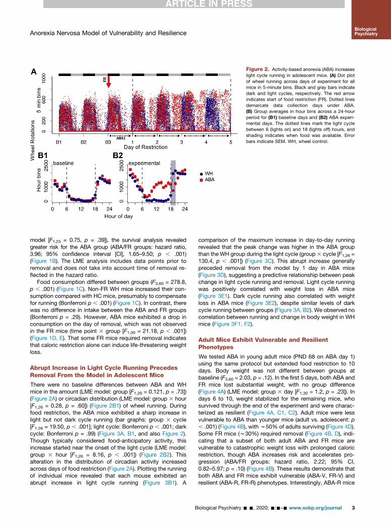
Figure 2. Activity-based anorexia (ABA) increaseslight cycle running in adolescent mice. (A) Dot plotof wheel running across days of experiment for allmice in 5-minute bins. Black and gray bars indicatedark and light cycles, respectively. The red arrowindicates start of food restriction (FR). Dotted linesdemarcate data collection days under ABA.(B) Group averages in hour bins across a 24-hourperiod for (B1) baseline days and (B2) ABA experi-mental days. The dotted lines mark the light cyclebetween 6 (lights on) and 18 (lights off) hours, andshading indicates when food was available. Errorbars indicate SEM. WH, wheel control.
Anorexia Nervosa Model of Vulnerability and ResilienceBiologicalPsychiatry
model [F1,23 = 0.75, p = .39]), the survival analysis revealedgreater risk for the ABA group (ABA/FR groups: hazard ratio,3.96; 95% confidence interval [CI], 1.65–9.50; p , .001)(Figure 1B). The LME analysis includes data points prior toremoval and does not take into account time of removal re-flected in the hazard ratio.
Food consumption differed between groups (F3,60 = 278.8,p , .001) (Figure 1C). Non-FR WH mice increased their con-sumption compared with HC mice, presumably to compensatefor running (Bonferroni p , .001) (Figure 1C). In contrast, therewas no difference in intake between the ABA and FR groups(Bonferroni p = .29). However, ABA mice exhibited a drop inconsumption on the day of removal, which was not observedin the FR mice (time point 3 group [F1,30 = 21.18, p , .001])(Figure 1D, E). That some FR mice required removal indicatesthat caloric restriction alone can induce life-threatening weightloss.
Abrupt Increase in Light Cycle Running PrecedesRemoval From the Model in Adolescent Mice
There were no baseline differences between ABA and WHmice in the amount (LME model: group [F1,26 = 0.121, p = .73])(Figure 2A) or circadian distribution (LME model: group 3 hour[F1,26 = 0.28, p = .60]) (Figure 2B1) of wheel running. Duringfood restriction, the ABA mice exhibited a sharp increase inlight but not dark cycle running (bar graphs: group 3 cycle[F1,26 = 19.50, p , .001]; light cycle: Bonferroni p , .001; darkcycle: Bonferroni p = .99) (Figure 3A, B1, and also Figure 2).Though typically considered food-anticipatory activity, thisincrease started near the onset of the light cycle (LME model:group 3 hour [F1,26 = 8.16, p , .001]) (Figure 2B2). Thisalteration in the distribution of circadian activity increasedacross days of food restriction (Figure 2A). Plotting the runningof individual mice revealed that each mouse exhibited anabrupt increase in light cycle running (Figure 3B1). A
B
comparison of the maximum increase in day-to-day runningrevealed that the peak change was higher in the ABA groupthan the WH group during the light cycle (group3 cycle [F1,26 =130.4, p , .001]) (Figure 3C). This abrupt increase generallypreceded removal from the model by 1 day in ABA mice(Figure 3D), suggesting a predictive relationship between peakchange in light cycle running and removal. Light cycle runningwas positively correlated with weight loss in ABA mice(Figure 3E1). Dark cycle running also correlated with weightloss in ABA mice (Figure 3E2), despite similar levels of darkcycle running between groups (Figure 3A, B2). We observed nocorrelation between running and change in body weight in WHmice (Figure 3F1, F2).
Adult Mice Exhibit Vulnerable and ResilientPhenotypes
We tested ABA in young adult mice (PND 68 on ABA day 1)using the same protocol but extended food restriction to 10days. Body weight was not different between groups atbaseline (F3,60 = 2.03, p = .12). In the first 5 days, both ABA andFR mice lost substantial weight, with no group difference(Figure 4A) (LME model: group 3 day [F1,30 = 1.2, p = .23]). Indays 6 to 10, weight stabilized for the remaining mice, whosurvived through the end of the experiment and were charac-terized as resilient (Figure 4A, C1, C2). Adult mice were lessvulnerable to ABA than younger mice (adult vs. adolescent: p, .001) (Figure 4B), with w50% of adults surviving (Figure 4D).Some FR mice (w30%) required removal (Figure 4B, D), indi-cating that a subset of both adult ABA and FR mice arevulnerable to catastrophic weight loss with prolonged caloricrestriction, though ABA increases risk and accelerates pro-gression (ABA/FR groups: hazard ratio, 2.22; 95% CI,0.82–5.97; p = .10) (Figure 4B). These results demonstrate thatboth ABA and FR mice exhibit vulnerable (ABA-V, FR-V) andresilient (ABA-R, FR-R) phenotypes. Interestingly, ABA-R mice
iological Psychiatry - -, 2020; -:-–- www.sobp.org/journal 3
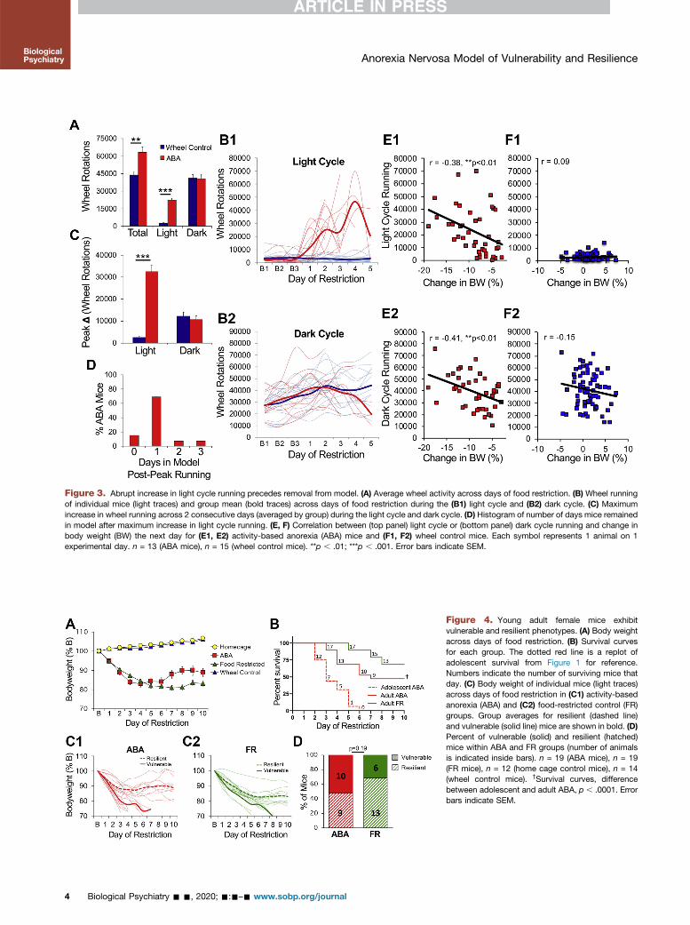
Figure 3. Abrupt increase in light cycle running precedes removal from model. (A) Average wheel activity across days of food restriction. (B) Wheel runningof individual mice (light traces) and group mean (bold traces) across days of food restriction during the (B1) light cycle and (B2) dark cycle. (C) Maximumincrease in wheel running across 2 consecutive days (averaged by group) during the light cycle and dark cycle. (D) Histogram of number of days mice remainedin model after maximum increase in light cycle running. (E, F) Correlation between (top panel) light cycle or (bottom panel) dark cycle running and change inbody weight (BW) the next day for (E1, E2) activity-based anorexia (ABA) mice and (F1, F2) wheel control mice. Each symbol represents 1 animal on 1experimental day. n = 13 (ABA mice), n = 15 (wheel control mice). **p , .01; ***p , .001. Error bars indicate SEM.
Figure 4. Young adult female mice exhibitvulnerable and resilient phenotypes. (A) Body weightacross days of food restriction. (B) Survival curvesfor each group. The dotted red line is a replot ofadolescent survival from Figure 1 for reference.Numbers indicate the number of surviving mice thatday. (C) Body weight of individual mice (light traces)across days of food restriction in (C1) activity-basedanorexia (ABA) and (C2) food-restricted control (FR)groups. Group averages for resilient (dashed line)and vulnerable (solid line) mice are shown in bold. (D)Percent of vulnerable (solid) and resilient (hatched)mice within ABA and FR groups (number of animalsis indicated inside bars). n = 19 (ABA mice), n = 19(FR mice), n = 12 (home cage control mice), n = 14(wheel control mice). †Survival curves, differencebetween adolescent and adult ABA, p , .0001. Errorbars indicate SEM.
Anorexia Nervosa Model of Vulnerability and Resilience
4 Biological Psychiatry - -, 2020; -:-–- www.sobp.org/journal
BiologicalPsychiatry
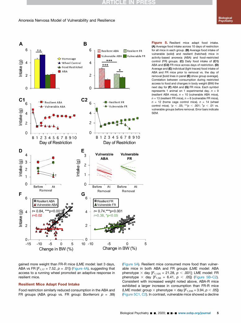
Figure 5. Resilient mice adapt food intake.(A) Average food intake across 10 days of restrictionfor all mice in each group. (B) Average food intake ofvulnerable (solid) and resilient (hatched) mice inactivity-based anorexia (ABA) and food-restrictedcontrol (FR) groups. (C) Daily food intake of (C1)ABA and (C2) FR mice across days of restriction. (D)Average and (E) individual (light traces) food intake ofABA and FR mice prior to removal vs. the day ofremoval [bold lines in panel (E) show group average].Correlation between consumption during restrictedaccess to food and changes in body weight (BW) thenext day for (F) ABA and (G) FR mice. Each symbolrepresents 1 animal on 1 experimental day. n = 9(resilient ABA mice), n = 10 (vulnerable ABA mice),n = 13 (resilient FR mice), n = 6 (vulnerable FR mice),n = 12 (home cage control mice), n = 14 (wheelcontrol mice). *p , .05; ***p , .001; †p , .01 vs.vulnerable groups before removal. Error bars indicateSEM.
Anorexia Nervosa Model of Vulnerability and ResilienceBiologicalPsychiatry
gained more weight than FR-R mice (LME model: last 3 days,ABA vs FR [F1,17 = 7.52, p = .01]) (Figure 4A), suggesting thataccess to a running wheel promoted an adaptive response inresilient mice.
Resilient Mice Adapt Food Intake
Food restriction similarly reduced consumption in the ABA andFR groups (ABA group vs. FR group: Bonferroni p = .99)
B
(Figure 5A). Resilient mice consumed more food than vulner-able mice in both ABA and FR groups (LME model: ABAphenotype 3 day [F1,125 = 21.28, p , .001]; LME model: FRphenotype 3 day [F1,56 = 6.41, p , .05]) (Figure 5B–C2).Consistent with increased weight noted above, ABA-R miceexhibited a larger increase in consumption than FR-R mice(LME model: group 3 phenotype3 day [F1,235 = 3.94, p , .05])(Figure 5C1, C2). In contrast, vulnerable mice showed a decline
iological Psychiatry - -, 2020; -:-–- www.sobp.org/journal 5

Figure 6. Vulnerable but not resilient activity-based anorexia (ABA) mice exhibit altered distribu-tion of circadian running activity. (A) Dot plot ofwheel running across days of experiment for all micein 5-minute bins. Black and gray bars indicate darkand light cycles, respectively. The red arrow in-dicates start of food restriction (FR). Dotted arrowsdemarcate data collection days under ABA.(B) Group averages in hour bins across a 24-hourperiod for (B1) baseline days and (B2) ABA experi-mental days. The dotted lines mark the light cyclebetween 6 (lights on) and 18 (lights off) hours, andshading indicates when food was available. Errorbars indicate SEM. WH, wheel control.
Anorexia Nervosa Model of Vulnerability and ResilienceBiologicalPsychiatry
in consumption that occurred earlier in ABA-V than FR-V mice(days in model [t13 = 2.86, p = .01]) (Figure 5C1, C2). Thiscannot be due to insufficient time to eat because the resilientmice increased food intake in the same amount of time. Similarto adolescent mice, consumption decreased at time of removalfor vulnerable mice of both groups (time point 3 phenotype[F1,33 = 54.12, p , .001]) (Figure 5D, E). In resilient mice,consumption correlated with changes in body weight, a cor-relation reduced in FR-V mice and absent in ABA-V mice(Figure 5F, G).
ABA-V Mice Exhibit Maladaptive Running Behavior
There were no baseline differences between ABA-V, ABA-R,and non-FR WH mice in amount (LME model: group [F2,30 =0.398, p = .67]) (Figure 6A) or circadian distribution of running(LME model: group 3 hour [F2,30 = 2.13, p = .13]) (Figure 6B1).During food restriction, ABA-R mice exhibited an adaptivereduction in total running, while ABA-V mice continued to runas much as WH mice (group [F2,30 = 5.21, p = .01]; ABA-Rgroup vs. WH group: Bonferroni p , .05) (Figure 7A). Asabove, food restriction increased light cycle running, whichwas modest in ABA-R but dramatic in ABA-V mice (bar graphs[F2,30 = 38.05, p , .001]; ABA-V mice vs. ABA-R mice: Bon-ferroni p , .001; ABA-R mice vs. WH mice: Bonferroni p = .05)(Figure 7B, D1). In contrast, both ABA groups decreased darkcycle running (bar graphs [F2,30 = 7.76, p , .01]) (Figure 7C,D2). These changes reflect a shift in running from dark tolight cycle, an effect more pronounced in ABA-V mice andpartially mitigated in ABA-R mice (LME model, light cycle: ABAphenotype 3 day [F1,22 = 10.03, p , .01]) (Figure 7E1, E2). Asobserved in adolescent mice, ABA-V mice exhibited an abruptincrease in light cycle running (bar graphs [F2,30 = 22.00,p , .001]; ABA-V mice vs. ABA-R mice: Bonferroni p , .001)
6 Biological Psychiatry - -, 2020; -:-–- www.sobp.org/journal
(Figure 7D1, F) that preceded removal by 1 to 3 days(Figure 7G), an effect greatly reduced in ABA-R mice (ABA-Rmice vs. WH mice: Bonferroni p = .19) (Figure 7D1, F). Lightcycle running positively correlated with weight loss in the ABA-V group but not in the ABA-R group (Figure 7H1). In contrast,dark cycle running positively correlated with weight loss inboth groups (Figure 7H2). Like adolescent ABA mice, the ABA-V mice exhibited an altered distribution of circadian activity(Figure 6A), with running that began 2 to 3 hours into the lightcycle (Figure 6B2). In contrast, the ABA-R mice exhibited amodest increase in activity prior to onset of the dark cycle,consistent with adaptive food-anticipatory activity (ABAphenotype 3 hour [F1,17 = 8.92, p , .01]) (Figure 6B2). Thesedata highlight an association between dysregulated increasesin light cycle running and vulnerability in the ABA model.
Hyperdopaminergic Mice Show IncreasedVulnerability to ABA
To test dopamine’s contribution to ABA vulnerability, we usedheterozygote DAT-cre mice in which 1 allele of the DAT wasreplaced with cre (DATcre/1). DAT messenger RNA (genotype[F1,22 = 4.7, p , .05]) (Figure 8A) and protein (t27 = 2.06, p =.049) (Figure 8B, C) were reduced without affecting expressionof other key dopamine-related genes (TH [tyrosine hydroxy-lase]: p = .81; VMAT2 [vesicular monoamine transporter 2]: p =.50; D2 [dopamine D2 receptor]: p = .30) (Figure 8A), renderingthe mice DAT KDs. Evoked dopamine release measured byfast-scan cyclic voltammetry revealed reduced clearance andincreased peak amplitude in KD mice across striatal regions(genotype effects for single stimulation: tau [F1,51 = 24.1, p ,
.001], peak [F1,51 = 23.1, p , .001]; genotype effects for burststimulation: tau [F1.51 = 27.6, p , .001], peak [F1,51 = 23.7, p ,
.001]; no genotype 3 region interactions) (Figure 8D),

Figure 7. Vulnerable activity-based anorexia(ABA) mice exhibit maladaptive running behavior. (A)Total, (B) light cycle, and (C) dark cycle wheelrunning averaged across days of food restriction. (D)Wheel running of individual mice (light traces) andgroup mean (bold traces) across days of food re-striction during the (D1) light cycle and (D2) darkcycle (vulnerable ABA mice, solid red; resilient ABAmice, dashed red; wheel control [WH] mice, solidblue). (E) Percentage of total running during the (E1)light cycle and (E2) dark cycle. (F) Maximum in-crease in wheel running across 2 consecutive days(averaged by group) during the light and dark cycles.(G) Histogram of the number of days mice remainedin the model after maximum increase in light cyclerunning. Correlation between (H1) light cycle or (H2)dark cycle running and change in body weight (BW)for vulnerable and resilient ABA mice. Each symbolrepresents 1 animal on 1 experimental day. n = 9(resilient ABA mice), n = 10 (vulnerable ABA mice),n = 14 (wheel control mice). *p , .05; **p , .01; ***p, .001. Error bars indicate SEM.
Anorexia Nervosa Model of Vulnerability and ResilienceBiologicalPsychiatry
indicating mild hyperdopaminergia. We compared young adult(PNDs 62–78, ABA day 1) female KD and WT littermates inABA, FR, and WH conditions separately.
At baseline, genotypes were similar in weight (F1,72 = 1.07,p = .31), food intake (F1,61 = 0.87, p = .35), and light cyclerunning (F1,45 = 0.05, p = .82). On the first day of baseline, darkcycle running was 36% higher in DATcre/1 mice than WT mice(t45 = 2.95, p, .01). This difference declined to nonsignificanceby the third day of baseline (t45 = 1.24, p = .22), indicating anincreased response to novelty rather than sustained elevatedrunning. This interpretation is supported by the lack of a
B
difference between genotypes during 10 additional days ofrunning in WH controls (Figure S1C–E). Combining data acrosstest conditions revealed no main effect of genotype on weight(LME model [F1,48 = 1.51, p = .23]). Significant genotype in-teractions are described below.
Under ABA conditions, KD mice exhibited acceleratedweight loss (LME model: genotype [F1,28 = 6.64, p , .05])(Figure 9A, B) and poorer survival (KD/WT mice: hazard ratio,2.23; 95% CI, 0.91–5.47; p , .05) (Figure 9C). All KD miceexhibited the vulnerable phenotype, while some WT mice wereresilient (Figure 9C). There were no differences between
iological Psychiatry - -, 2020; -:-–- www.sobp.org/journal 7
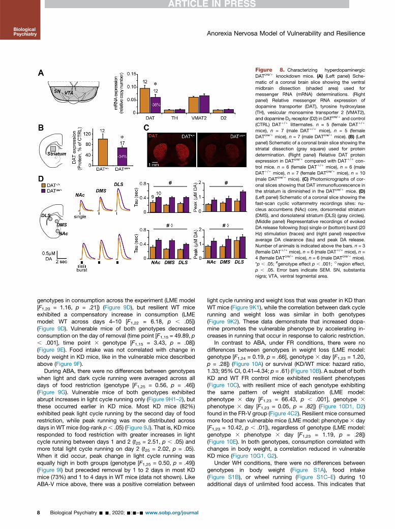
Figure 8. Characterizing hyperdopaminergicDATcre/1 knockdown mice. (A) (Left panel) Sche-matic of a coronal brain slice showing the ventralmidbrain dissection (shaded area) used formessenger RNA (mRNA) determinations. (Rightpanel) Relative messenger RNA expression ofdopamine transporter (DAT), tyrosine hydroxylase(TH), vesicular monoamine transporter 2 (VMAT2),and dopamine D2 receptor (D2) in DATcre/1 and control(CTRL) DAT1/1 littermates. n = 5 (female DAT1/1
mice), n = 7 (male DAT1/1 mice), n = 5 (femaleDATcre/1 mice), n = 7 (male DATcre/1 mice). (B) (Leftpanel) Schematic of a coronal brain slice showing thestriatal dissection (gray square) used for proteindetermination. (Right panel) Relative DAT proteinexpression in DATcre/1 compared with DAT1/1 con-trol mice. n = 6 (female DAT1/1 mice), n = 6 (maleDAT1/1 mice), n = 7 (female DATcre/1 mice), n = 10(male DATcre/1 mice). (C) Photomicrographs of cor-onal slices showing that DAT immunofluorescence inthe striatum is diminished in the DATcre/1 mice. (D)(Left panel) Schematic of a coronal slice showing thefast-scan cyclic voltammetry recordings sites: nu-cleus accumbens (NAc) core, dorsomedial striatum(DMS), and dorsolateral striatum (DLS) (gray circles).(Middle panel) Representative recordings of evokedDA release following (top) single or (bottom) burst (20Hz) stimulation (traces) and (right panel) respectiveaverage DA clearance (tau) and peak DA release.Number of animals is indicated above the bars. n = 3(female DAT1/1 mice), n = 6 (male DAT1/1 mice), n =4 (female DATcre/1 mice), n = 6 (male DATcre/1 mice).*p , .05; #genotype effect p , .001; >region effect,p , .05. Error bars indicate SEM. SN, substantianigra; VTA, ventral tegmental area.
Anorexia Nervosa Model of Vulnerability and ResilienceBiologicalPsychiatry
genotypes in consumption across the experiment (LME model[F1,20 = 1.16, p = .21]) (Figure 9D), but resilient WT miceexhibited a compensatory increase in consumption (LMEmodel: WT across days 4–10 [F1,22 = 6.18, p , .05])(Figure 9D). Vulnerable mice of both genotypes decreasedconsumption on the day of removal (time point [F1,15 = 49.89, p, .001], time point 3 genotype [F1,15 = 3.43, p = .08])(Figure 9E). Food intake was not correlated with change inbody weight in KD mice, like in the vulnerable mice describedabove (Figure 9F).
During ABA, there were no differences between genotypeswhen light and dark cycle running were averaged across alldays of food restriction (genotype [F1,25 = 0.56, p = .46])(Figure 9G). Vulnerable mice of both genotypes exhibitedabrupt increases in light cycle running only (Figure 9H1–2), butthese occurred earlier in KD mice. Most KD mice (82%)exhibited peak light cycle running by the second day of foodrestriction, while peak running was more distributed acrossdays in WT mice (log-rank p, .05) (Figure 9J). That is, KD miceresponded to food restriction with greater increases in lightcycle running between days 1 and 2 (t25 = 2.51, p , .05) andmore total light cycle running on day 2 (t25 = 2.02, p = .05).When it did occur, peak change in light cycle running wasequally high in both groups (genotype [F1,25 = 0.50, p = .49])(Figure 9I) but preceded removal by 1 to 2 days in most KDmice (73%) and 1 to 4 days in WT mice (data not shown). LikeABA-V mice above, there was a positive correlation between
8 Biological Psychiatry - -, 2020; -:-–- www.sobp.org/journal
light cycle running and weight loss that was greater in KD thanWT mice (Figure 9K1), while the correlation between dark cyclerunning and weight loss was similar in both genotypes(Figure 9K2). These data demonstrate that increased dopa-mine promotes the vulnerable phenotype by accelerating in-creases in running that occur in response to caloric restriction.
In contrast to ABA, under FR conditions, there were nodifferences between genotypes in weight loss (LME model:genotype [F1,24 = 0.19, p = .66], genotype 3 day [F1,23 = 1.20,p = .28]) (Figure 10A) or survival (KD/WT mice: hazard ratio,1.33; 95% CI, 0.41–4.34; p = .61) (Figure 10B). A subset of bothKD and WT FR control mice exhibited resilient phenotypes(Figure 10C), with resilient mice of each genotype exhibitingthe same pattern of weight stabilization (LME model:phenotype 3 day [F1,23 = 66.43, p , .001], genotype 3
phenotype 3 day [F1,23 = 0.05, p = .82]) (Figure 10D1, D2)found in the FR-V group (Figure 4C2). Resilient mice consumedmore food than vulnerable mice (LME model: phenotype3 day[F1,23 = 10.42, p , .01]), regardless of genotype (LME model:genotype 3 phenotype 3 day [F1,23 = 1.19, p = .28])(Figure 10E). In both genotypes, consumption correlated withchanges in body weight, a correlation reduced in vulnerableKD mice (Figure 10G1, G2).
Under WH conditions, there were no differences betweengenotypes in body weight (Figure S1A), food intake(Figure S1B), or wheel running (Figure S1C–E) during 10additional days of unlimited food access. This indicates that
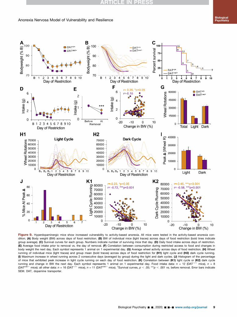
Figure 9. Hyperdopaminergic mice show increased vulnerability to activity-based anorexia. All mice were tested in the activity-based anorexia con-dition. (A) Body weight (BW) across days of food restriction. (B) BW of individual mice (light traces) across days of food restriction (bold lines indicategroup average). (C) Survival curves for each group. Numbers indicate number of surviving mice that day. (D) Daily food intake across days of restriction.(E) Average food intake prior to removal vs. the day of removal. (F) Correlation between consumption during restricted access to food and changes inbody weight the next day. Each symbol represents 1 animal on 1 experimental day. (G) Average wheel activity across days of food restriction. (H) Wheelrunning of individual mice (light traces) and group mean (bold traces) across days of food restriction for (H1) light cycle and (H2) dark cycle running.(I) Maximum increase in wheel running across 2 consecutive days (averaged by group) during the light and dark cycles. (J) Histogram of the percentageof mice that exhibited peak increase in light cycle running on each day of food restriction. (K) Correlation between (K1) light cycle or (K2) dark cyclerunning and change in BW the next day. Each symbol represents 1 animal on 1 experimental day. Food intake data: n = 12 (DAT1/1 mice), n = 8(DATcre/1 mice); all other data: n = 16 (DAT1/1 mice), n = 11 (DATcre/1 mice). †Survival curves, p , .05; ***p , .001 vs. before removal. Error bars indicateSEM. DAT, dopamine transporter.
Anorexia Nervosa Model of Vulnerability and Resilience
Biological Psychiatry - -, 2020; -:-–- www.sobp.org/journal 9
BiologicalPsychiatry
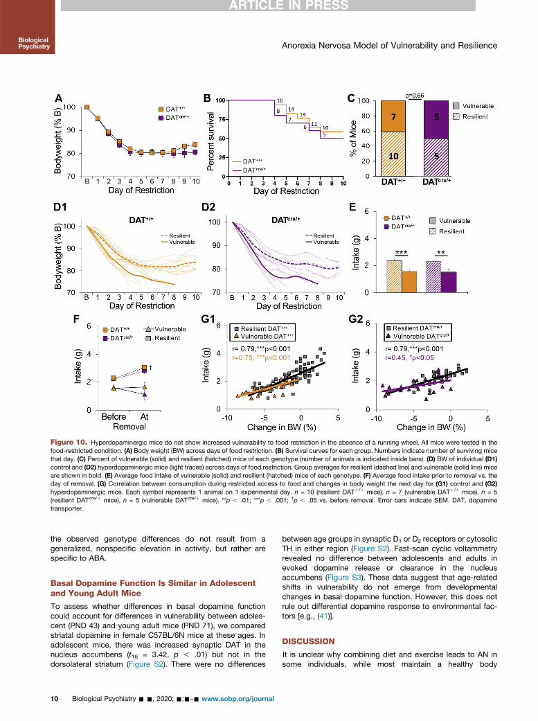
Figure 10. Hyperdopaminergic mice do not show increased vulnerability to food restriction in the absence of a running wheel. All mice were tested in thefood-restricted condition. (A) Body weight (BW) across days of food restriction. (B) Survival curves for each group. Numbers indicate number of surviving micethat day. (C) Percent of vulnerable (solid) and resilient (hatched) mice of each genotype (number of animals is indicated inside bars). (D) BW of individual (D1)control and (D2) hyperdopaminergic mice (light traces) across days of food restriction. Group averages for resilient (dashed line) and vulnerable (solid line) miceare shown in bold. (E) Average food intake of vulnerable (solid) and resilient (hatched) mice of each genotype. (F) Average food intake prior to removal vs. theday of removal. (G) Correlation between consumption during restricted access to food and changes in body weight the next day for (G1) control and (G2)hyperdopaminergic mice. Each symbol represents 1 animal on 1 experimental day. n = 10 (resilient DAT1/1 mice), n = 7 (vulnerable DAT1/1 mice), n = 5(resilient DATcre/1 mice), n = 5 (vulnerable DATcre/1 mice). **p , .01; ***p , .001; †p , .05 vs. before removal. Error bars indicate SEM. DAT, dopaminetransporter.
Anorexia Nervosa Model of Vulnerability and ResilienceBiologicalPsychiatry
the observed genotype differences do not result from ageneralized, nonspecific elevation in activity, but rather arespecific to ABA.
Basal Dopamine Function Is Similar in Adolescentand Young Adult Mice
To assess whether differences in basal dopamine functioncould account for differences in vulnerability between adoles-cent (PND 43) and young adult mice (PND 71), we comparedstriatal dopamine in female C57BL/6N mice at these ages. Inadolescent mice, there was increased synaptic DAT in thenucleus accumbens (t16 = 3.42, p , .01) but not in thedorsolateral striatum (Figure S2). There were no differences
10 Biological Psychiatry - -, 2020; -:-–- www.sobp.org/journal
between age groups in synaptic D1 or D2 receptors or cytosolicTH in either region (Figure S2). Fast-scan cyclic voltammetryrevealed no difference between adolescents and adults inevoked dopamine release or clearance in the nucleusaccumbens (Figure S3). These data suggest that age-relatedshifts in vulnerability do not emerge from developmentalchanges in basal dopamine function. However, this does notrule out differential dopamine response to environmental fac-tors [e.g., (41)].
DISCUSSION
It is unclear why combining diet and exercise leads to AN insome individuals, while most maintain a healthy body

Anorexia Nervosa Model of Vulnerability and ResilienceBiologicalPsychiatry
weight. One approach for studying this is to examine indi-vidual differences in ABA (42,43). Here, we demonstratedistinct vulnerable and resilient phenotypes, providing arobust animal model for investigating the physiological andneural adaptations underlying resilience and vulnerability toAN.
ABA resilience is associated with a progressive increase infood intake and decrease in overall wheel activity, leading toweight stabilization. Similar decreases have been reported inolder C57BL/6 female mice (44,45), possibly reflecting an age-related increase in resilience. Moreover, we found that ABA-Rmice actually eat more than FR-R mice and consequently gainmore weight, despite the energy expenditure from wheelrunning. Though counterintuitive, this parallels evidence fromclinical studies demonstrating that appropriate exercise maybe beneficial in clinical treatment (46–49).
In contrast, vulnerable mice fail to increase their con-sumption, reducing intake as caloric restriction continues.This failure cannot be attributed to insufficient time to eat,because resilient mice increase consumption in the same 2-hour period of food access. While low food intake correlatedwith daily weight loss in FR-V mice, this was not found inABA-V mice. Instead, daily weight loss correlated withamount of wheel activity. Vulnerability did not arise frompreexisting differences in activity, as vulnerable and resilientmice ran similarly at baseline. During food restriction, runningdramatically increased throughout the light cycle, withvulnerable animals primarily running instead of sleeping.These findings are consistent with prior studies demon-strating that caloric restriction can induce activity that ispartially diurnal (50–52) and may reflect exaggerated food-anticipatory activity or a shift in circadian activity to pro-mote foraging (51). Disruption of circadian rhythms has beenobserved in AN (53). This, as well as hyperactivity, couldarise from activation of starvation-induced foraging mecha-nisms that promote physical activity (26,54–56). Individualdifferences in this response might arise from genetic variationin a human foraging gene (57).
Our studies with hyperdopaminergic mice indicate thatincreased dopamine promotes vulnerability to ABA by acceler-ating increases in activity that occur in response to caloric re-striction. Vulnerability was not increased in KD mice testedunder FR conditions, suggesting a critical interaction betweendopamine, physical activity, and vulnerability to caloric restric-tion. As the KD is global, such effects may arise from increasedstriatal dopamine or dopamine actions elsewhere. This couldinclude the hypothalamus or prefrontal cortex, though dopaminereuptake in the prefrontal cortex is mediated primarily by thenorepinephrine transporter rather than the DAT (58–60).
ABA studies have primarily used rats, with fewer studiestesting mice (44,45,61–63). In contrast to the stable bodyweight typically found in FR rats [(35,64,65), but see (66,67)],we found that FR control mice can exhibit life-threateningweight loss (Supplemental Discussion). This suggests thatcaloric restriction is the primary driver of vulnerability, withwheel access accelerating and augmenting expression of thevulnerable phenotype. Increased home cage activity may havepromoted weight loss in the FR mice, which would furthersuggest a central role for activity in the emergence of thevulnerable phenotype.
Bio
Consistent with rat studies (68) and AN in humans (69), wefind reduced vulnerability to ABA with age. Smaller animalsgenerally have higher metabolic rates, which in combinationwith less body mass may increase vulnerability in youngermice. The rapidity of decline in adolescent mice may precludeemergence of a resilient phenotype, while greater initial bodyweight in young adult mice may slow the decline sufficiently fora resilient phenotype to emerge. Extending daily food restric-tion allowed detection of the vulnerable phenotype in FR mice.Single housing likely exacerbates vulnerability in smaller ani-mals, as the energetic cost of maintaining body temperature isincreased without group huddling, compounding increasedsusceptibility associated with low body mass and high meta-bolic rate.
Although ABA vulnerability is increased by dopamine, thegreater vulnerability we observe in adolescent mice is not dueto differences in basal levels of striatal dopamine (Figure S3).Vulnerability during adolescence may instead result from age-related differences in how caloric restriction and exerciseinduce dopamine adaptations (see below). Alternatively,maturation of dopamine-prefrontal cortical innervation, whichcontinues through young adulthood, may play a role byaffecting inhibitory control. The smaller size of the adolescentmouse may be an additional factor.
Dopamine and AN Vulnerability
Dopamine has been implicated in AN, but its contributionremains poorly understood. Recovered AN patients exhibitreduced dopamine metabolites, suggesting decreased turn-over (70), but show increased dopamine D2 receptor binding,reflecting increased D2 receptor expression and/or decreaseddopamine transmission (71). Patients often remain symp-tomatic after recovery (72), making it difficult to determinewhether differences in dopamine observed in clinical studiesrepresent preexisting risk factors or potentially reversibleabnormalities induced by starvation (26). Our finding thathyperdopaminergic mice are more vulnerable to ABA sug-gests that enhanced dopamine increases AN risk. However,this does not necessarily mean that vulnerability is mediatedby basal or trait differences in dopamine function. In AN,several factors arising from caloric restriction can upregulatedopamine, including increased glucocorticoids (73–77),enhanced insulin sensitivity (78–81), increased ghrelin(82–84), and decreased leptin (85–90). Indeed, studies havelinked decreased leptin with hyperactivity [(87,90–93) but see(94)], with some implicating dopamine (87,90,93). Such ad-aptations to caloric restriction presumably arise to promotephysical activity required to forage for food to obtain neededcalories (26), making them potential therapeutic targets forreducing hyperactivity in AN. Dopamine also contributes tothermoregulation (95–97), which is altered in AN (6,98) andABA (94,99). Hypothermia arising from weight loss may alsoinduce changes in dopamine. Vulnerability to AN might varydue to individual differences in any of these responses tocaloric restriction, differences that could result from variationin dopamine genes or genes that modulate dopamine (e.g.,leptin, insulin, ghrelin, glucocorticoid, orexin). Of course,factors independent of dopamine may also modulatevulnerability [e.g., (100)].
logical Psychiatry - -, 2020; -:-–- www.sobp.org/journal 11

Anorexia Nervosa Model of Vulnerability and ResilienceBiologicalPsychiatry
Dopamine function may change over the course of AN, asobserved in addiction, in which dopamine contributes differ-ently in different stages of the disorder (101–104). Such dy-namic changes may account for why a recent study found thatpharmacogenetic activation of dopamine protects rats fromABA (64). Artificial activation of dopamine cells could haveblocked normally occurring changes in dopamine and pro-gressive adaptations to caloric restriction that underlie ABA,dynamic changes facilitated in our hyperdopaminergic mice.The pharmacogenetic activation of some nondopaminergiccells may have also contributed to the discrepancy betweenour findings. The nature of progressive changes in dopamineacross AN/ABA is unknown but merits further study.
A robust model of vulnerability and resilience, as reportedhere, could aid in the discovery of genes that mediate AN riskand/or contribute to the progression of the disorder. Such workmay lead to the identification of biomarkers for early diagnosisand the discovery of novel therapeutic targets.
ACKNOWLEDGMENTS AND DISCLOSURESThis work was supported by National Institute on Drug Abuse Grant No.DA046058 (to JB); National Institute of Mental Health Grant Nos.R21MH114182 (to NSB) and T32MH19970 (to AK); National Institute onMinority Health and Health Disparities of the National Institutes of HealthGrant No. G12MD007599 (to NSB); PSC-CUNY Awards jointly funded bythe Professional Staff Congress and the City University of New York (to JBand NSB); and Conte Grant No. P50 MH086404 (to SR).
All authors report no biomedical financial interests or potential conflictsof interest.
ARTICLE INFORMATIONFrom the Department of Psychology (JAB, DM), Queens College, City Uni-versity of New York, Flushing; Psychology Program (JAB, DM, RMZ, PS,NSB), Biology Program (JAB, RR), and Advanced Science Research Center(SM), The Graduate Center, City University of New York; Department ofPsychology (RMZ, PS, NSB), Hunter College, City University of New York;Department of Psychiatry (AK, CG, BYK, HM, SR, SM, NSB), ColumbiaUniversity, New York; Department of Molecular Therapeutics (CG, SR, SM),Integrative Neuroscience (HM), and Developmental Neuroscience (BYK),New York State Psychiatric Institute, New York, New York; Department ofMicrobiology and Molecular Genetics (BYK), Hebrew University, Jerusalem,Israel; and National Institute on Drug Abuse (HM), National Institutes ofHealth, Bethesda, Maryland.
HM is currently affiliated with the National Institute on Drug Abuse, Na-tional Institutes of Health, Bethesda, Maryland.
NSB is currently affiliated with Department of Psychology, Hunter Col-lege, and The Graduate Center, City University of New York, New York, NewYork.
Address correspondence to Jeff A. Beeler, Ph.D., at [email protected], or Nesha S. Burghardt, Ph.D., at [email protected].
Received Aug 8, 2019; revised Jun 26, 2020; accepted Jun 30, 2020.Supplementary material cited in this article is available online at https://
doi.org/10.1016/j.biopsych.2020.06.030.
REFERENCES1. Steinhausen H-C (2002): The outcome of anorexia nervosa in the
20th century. Am J Psychiatry 159:1284–1293.2. Khalsa SS, Portnoff LC, McCurdy-McKinnon D, Feusner JD
(2017): What happens after treatment? A systematic review ofrelapse, remission, and recovery in anorexia nervosa. J Eat Disord5:20.
3. Hay P, Mitchison D, Collado AEL, González-Chica DA, Stocks N,Touyz S (2017): Burden and health-related quality of life of eating
12 Biological Psychiatry - -, 2020; -:-–- www.sobp.org/journal
disorders, including avoidant/restrictive food intake disorder (ARFID),in the Australian population. J Eat Disord 5:21.
4. Fichter MM, Quadflieg N, Crosby RD, Koch S (2017): Long-termoutcome of anorexia nervosa: Results from a large clinical longitu-dinal study. Int J Eat Disord 50:1018–1030.
5. Frank GKW, Shott ME (2016): The role of psychotropic medications inthe management of anorexia nervosa: rationale, evidence and futureprospects. CNS Drugs 30:419–442.
6. Gull W (1888): Clinical notes: Medical, surgical, obstetrical, andtherapeutical. Anorexia Nervosa. Lancet 516–517.
7. Davis C, Katzman DK, Kaptein S, Kirsh C, Brewer H, Kalmbach K,et al. (1997): The prevalence of high-level exercise in the eating dis-orders: Etiological implications. Compr Psychiatry 38:321–326.
8. Rizk M, Lalanne C, Berthoz S, Kern L, EVHAN Group, Godart N(2015): Problematic exercise in anorexia nervosa: testing potentialrisk factors against different definitions. PLoS One 10:e0143352.
9. Holland LA, Brown TA, Keel PK (2014): Defining features of unhealthyexercise associated with disordered eating and eating disorder di-agnoses. Psychol Sport Exerc 15:116–123.
10. Cook BJ, Hausenblas HA (2008): The role of exercise dependence forthe relationship between exercise behavior and eating pathology:Mediator or moderator? J Health Psychol 13:495–502.
11. Dittmer N, Jacobi C, Voderholzer U (2018): Compulsive exercise ineating disorders: Proposal for a definition and a clinical assessment.J Eat Disord 6:42.
12. Schlegl S, Dittmer N, Hoffmann S, Voderholzer U (2018): Self-re-ported quantity, compulsiveness and motives of exercise in patientswith eating disorders and healthy controls: Differences and similar-ities. J Eat Disord 6:17.
13. Colleen Stiles-Shields E, Labuschagne Z, Goldschmidt AB,Doyle AC, Grange DL (2012): The use of multiple methods ofcompensatory behaviors as an indicator of eating disorder severity intreatment-seeking youth. Int J Eat Disord 45:704–710.
14. Belak L, Gianini L, Klein DA, Sazonov E, Keegan K, Neustadt E, et al.(2017): Measurement of fidgeting in patients with anorexia nervosausing a novel shoe-based monitor. Eat Behav 24:45–48.
15. Solenberger SE (2001): Exercise and eating disorders: A 3-yearinpatient hospital record analysis. Eat Behav 2:151–168.
16. Carter JC, Blackmore E, Sutandar-Pinnock K, Woodside DB (2004):Relapse in anorexia nervosa: A survival analysis. Psychol Med34:671–679.
17. Strober M, Freeman R, Morrell W (1997): The long-term course ofsevere anorexia nervosa in adolescents: Survival analysis of recov-ery, relapse, and outcome predictors over 10-15 years in a pro-spective study. Int J Eat Disord 22:339–360.
18. Casper RC, Jabine LN (1996): An eight-year follow-up: Outcome fromadolescent compared to adult onset anorexia nervosa. J YouthAdolesc 25:499–517.
19. Kostrzewa E, van Elburg AA, Sanders N, Sternheim L, Adan RAH,Kas MJH (2013): Longitudinal changes in the physical activity of ado-lescents with anorexia nervosa and their influence on body compositionand leptin serum levels after recovery. PLoS One 8:e78251.
20. Kostrzewa E, Eijkemans MJC, Kas MJ (2013): The expression ofexcessive exercise co-segregates with the risk of developing aneating disorder in women. Psychiatry Res 210:1123–1128.
21. Davis C, Kennedy SH, Ravelski E, Dionne M (1994): The role ofphysical activity in the development and maintenance of eating dis-orders. Psychol Med 24:957–967.
22. Davis C, Blackmore E, Katzman DK, Fox J (2005): Female adoles-cents with anorexia nervosa and their parents: A case-control studyof exercise attitudes and behaviours. Psychol Med 35:377–386.
23. Holm-Denoma JM, Scaringi V, Gordon KH, Van Orden KA, Joiner TE(2009): Eating disorder symptoms among undergraduate varsityathletes, club athletes, independent exercisers, and nonexercisers.Int J Eat Disord 42:47–53.
24. Bratland-Sanda S, Sundgot-Borgen J (2013): Eating disorders inathletes: Overview of prevalence, risk factors and recommendationsfor prevention and treatment. Eur J Sport Sci 13:499–508.
25. Smolak L, Murnen SK, Ruble AE (2000): Female athletes and eatingproblems: A meta-analysis. Int J Eat Disord 27:371–380.
Anorexia Nervosa Model of Vulnerability and ResilienceBiologicalPsychiatry
26. Södersten P, Bergh C, Leon M, Zandian M (2016): Dopamine andanorexia nervosa. Neurosci Biobehav Rev 60:26–30.
27. O’Hara CB, Campbell IC, Schmidt U (2015): A reward-centred modelof anorexia nervosa: A focussed narrative review of the neurologicaland psychophysiological literature. Neurosci Biobehav Rev 52:131–152.
28. Frank GKW, DeGuzman MC, Shott ME (2019): Motivation to eat andnot to eat – The psycho-biological conflict in anorexia nervosa.Physiol Behav 206:185–190.
29. Berner LA, Brown TA, Lavender JM, Lopez E, Wierenga CE, Kaye WH(2019): Neuroendocrinology of reward in anorexia nervosa andbulimia nervosa: Beyond leptin and ghrelin. Mol Cell Endocrinol497:110320.
30. Cowdrey FA, Park RJ, Harmer CJ, McCabe C (2011): Increasedneural processing of rewarding and aversive food stimuli in recoveredanorexia nervosa. Biol Psychiatry 70:736–743.
31. Scaife JC, Godier LR, Reinecke A, Harmer CJ, Park RJ (2016): Dif-ferential activation of the frontal pole to high vs low calorie foods: Theneural basis of food preference in anorexia nervosa? Psychiatry ResNeuroimaging 258:44–53.
32. Kravitz AV, O’Neal TJ, Friend DM (2016): Do dopaminergic impair-ments underlie physical inactivity in people with obesity? Front HumNeurosci 10:514.
33. Beeler JA, Frazier CRM, Zhuang3 (2012): Putting desire on a budget:Dopamine and energy expenditure, reconciling reward and re-sources. Front Integr Neurosci 6:49.
34. Wang S, Tan Y, Zhang J-E, Luo M (2013): Pharmacogenetic activa-tion of midbrain dopaminergic neurons induces hyperactivity. Neu-rosci Bull 29:517–524.
35. Routtenberg A, Kuznesof AW (1967): Self-starvation of rats living inactivity wheels on a restricted feeding schedule. J Comp PhysiolPsychol 64:414–421.
36. Epling WF, Pierce WD, Stefan L (1983): A theory of activity-basedanorexia. Int J Eat Disord 3:27–46.
37. Gutierrez E (2013): A rat in the labyrinth of anorexia nervosa: Con-tributions of the activity-based anorexia rodent model to the under-standing of anorexia nervosa. Int J Eat Disord 46:289–301.
38. Zhuang X, Masson J, Gingrich JA, Rayport S, Hen R (2005): Targetedgene expression in dopamine and serotonin neurons of the mousebrain. J Neurosci Methods 143:27–32.
39. Bates D, Maechler M, Bolkler B, Walker S (2015): Fitting linear mixed-effects models using lme4. J Stat Softw 67:1–48.
40. Kuznetsova A, Brockhoff P, Christensen R (2017): lmerTest package:Tests in linear mixed effecs models. J Stat Softw 82:1–26.
41. Montagud-Romero S, Nuñez C, Blanco-Gandia MC, Martínez-Laorden E, Aguilar MA, Navarro-Zaragoza J, et al. (2017): Repeatedsocial defeat and the rewarding effects of cocaine in adult andadolescent mice: Dopamine transcription factors, proBDNF signalingpathways, and the TrkB receptor in the mesolimbic system. Psy-chopharmacology (Berl) 234:2063–2075.
42. Chen Y-W, Wable GS, Chowdhury TG, Aoki C (2016): Enlargement ofaxo-somatic contacts formed by GAD-immunoreactive axon termi-nals onto layer V pyramidal neurons in the medial prefrontal cortex ofadolescent female mice is associated with suppression of foodrestriction-evoked hyperactivity and resilience to activity-basedanorexia. Cereb Cortex 26:2574–2589.
43. Barbarich-Marsteller NC, Underwood MD, Foltin RW, Myers MM,Walsh BT, Barrett JS, Marsteller DA (2013): Identifying novel phe-notypes of vulnerability and resistance to activity-based anorexia inadolescent female rats: Vulnerability to activity-based anorexia. Int JEat Disord 46:737–746.
44. Gelegen C, Collier DA, Campbell IC, Oppelaar H, van den Heuvel J,Adan RAH, Kas MJH (2007): Difference in susceptibility to activity-based anorexia in two inbred strains of mice. Eur Neuro-psychopharmacol 17:199–205.
45. Gelegen C, van den Heuvel J, Collier DA, Campbell IC, Oppelaar H,Hessel E, Kas MJH (2008): Dopaminergic and brain-derived neuro-trophic factor signalling in inbred mice exposed to a restrictedfeeding schedule. Genes Brain Behav 7:552–559.
Bio
46. Dittmer N, Voderholzer U, von der Mühlen M, Marwitz M, Fumi M,Mönch C, et al. (2018): Specialized group intervention for compulsiveexercise in inpatients with eating disorders: Feasibility and pre-liminary outcomes. J Eat Disord 6:27.
47. Cook BJ, Wonderlich SA, Mitchell JE, Thompson R, Sherman R,McCallum K (2016): Exercise in eating disorders treatment: System-atic review and proposal of guidelines. Med Sci Sports Exerc48:1408–1414.
48. Rizk M, Kern L, Lalanne C, Hanachi M, Melchior J-C, Pichard C, et al.(2018): High-intensity exercise is associated with a better nutritionalstatus in anorexia nervosa. Eur Eat Disord Rev 27:391–400.
49. Schlegel S (2015): The Freiburg sport therapy program for eatingdisordered outpatients: A pilot study. Eat Weight Disord 20:319–327.
50. Challet E (2010): Interactions between light, mealtime and calorierestriction to control daily timing in mammals. J Comp Physiol B180:631–644.
51. van der Vinne V, Riede SJ, Gorter JA, Eijer WG, Sellix MT,Menaker M, et al. (2014): Cold and hunger induce diurnality in anocturnal mammal. Proc Natl Acad Sci U S A 111:15256–15260.
52. Acosta-Rodríguez VA, de Groot MHM, Rijo-Ferreira F, Green CB,Takahashi JS (2017): Mice under caloric restriction self-impose atemporal restriction of food intake as revealed by an automatedfeeder system. Cell Metab 26:267–277.e2.
53. Menculini G, Brufani F, Bello VD, Moretti P, Tortorella A (2019):Circadian rhythms disruptions and eating disorders: Clinical impactand possible psychopathological correlates. Psychiatr Danub31:497–502.
54. Scheurink AJW, Boersma GJ, Nergårdh R, Södersten P (2010):Neurobiology of hyperactivity and reward: Agreeable restlessness inanorexia nervosa. Physiol Behav 100:490–495.
55. Södersten P, Brodin U, Zandian M, Bergh C (2019): Eating behaviorand the evolutionary perspective on anorexia nervosa. Front Neuro-sci 13:596.
56. Guisinger S (2003): Adapted to flee famine: Adding an evolutionaryperspective on anorexia nervosa. Psychol Rev 110:745–761.
57. Struk AA, Mugon J, Huston A, Scholer AA, Stadler G, Higgins ET,et al. (2019): Self-regulation and the foraging gene (PRKG1) inhumans. Proc Natl Acad Sci U S A 116:4434–4439.
58. Sesack SR, Hawrylak VA, Guido MA, Levey AI (1998): Cellular andsubcellular localization of the dopamine transporter in rat cortex. AdvPharmacol 42:171–174.
59. Morón JA, Brockington A, Wise RA, Rocha BA, Hope BT (2002):Dopamine uptake through the norepinephrine transporter in brainregions with low levels of the dopamine transporter: Evidence fromknock-out mouse lines. J Neurosci 22:389–395.
60. Mundorf ML, Joseph JD, Austin CM, Caron MG, Wightman RM(2008): Catecholamine release and uptake in the mouse prefrontalcortex: Catecholamines in mouse prefrontal cortex. J Neurochem79:130–142.
61. Avraham Y, Hao S, Mendelson S, Berry EM (2001): Tyrosine improvesappetite, cognition, and exercise tolerance in activity anorexia. MedSci Sports Exerc 33:2104–2110.
62. Klenotich SJ, Seiglie MP, McMurray MS, Roitman JD, Le Grange D,Dugad P, Dulawa SC (2012): Olanzapine, but not fluoxetine, treat-ment increases survival in activity-based anorexia in mice. Neuro-psychopharmacology 37:1620–1631.
63. Chowdhury TG, Wable GS, Sabaliauskas NA, Aoki C (2013):Adolescent female C57BL/6 mice with vulnerability to activity-basedanorexia exhibit weak inhibitory input onto hippocampal CA1 pyra-midal cells. Neuroscience 241:250–267.
64. Foldi CJ, Milton LK, Oldfield BJ (2017): The role of mesolimbicreward neurocircuitry in prevention and rescue of the activity-basedanorexia (ABA) phenotype in rats. Neuropsychopharmacology42:2292–2300.
65. Scharner S, Prinz P, Goebel-Stengel M, Kobelt P, Hofmann T,Rose M, Stengel A (2016): Activity-based anorexia reduces bodyweight without inducing a separate food intake microstructure oractivity phenotype in female rats—Mediation via an activation ofdistinct brain nuclei. Front Neurosci 10:475.
logical Psychiatry - -, 2020; -:-–- www.sobp.org/journal 13
Anorexia Nervosa Model of Vulnerability and ResilienceBiologicalPsychiatry
66. Aoki C, Sabaliauskas N, Chowdhury T, Min J-Y, Colacino AR,Laurino K, Barbarich-Marsteller NC (2012): Adolescent female ratsexhibiting activity-based anorexia express elevated levels of GABAAreceptor a4 and d subunits at the plasma membrane of hippocampalCA1 spines. Synapse 66:391–407.
67. Aoki C, Chowdhury TG, Wable GS, Chen Y-W (2017): Synapticchanges in the hippocampus of adolescent female rodents associ-ated with resilience to anxiety and suppression of food restriction-evoked hyperactivity in an animal model for anorexia nervosa. BrainRes 1654:102–115.
68. Woods SC, Routtenberg A (1971): “Self-starvation” in activity wheels:Developmental and chlorpromazine interactions. J Comp PhysiolPsychol 76:84–93.
69. Bulik CM (2002): Eating disorders in adolescents and young adults.Child Adolesc Psychiatr Clin N Am 11:201–218.
70. Kaye W (1999): Altered dopamine activity after recovery fromrestricting-type anorexia nervosa. Neuropsychopharmacology21:503–506.
71. Frank GK, Bailer UF, Henry SE, Drevets W, Meltzer CC, Price JC,et al. (2005): Increased dopamine D2/D3 receptor binding after re-covery from anorexia nervosa measured by positron emission to-mography and [11C]raclopride. Biol Psychiatry 58:908–912.
72. Wagner A, Barbarich-Marsteller NC, Frank GK, Bailer UF,Wonderlich SA, Crosby RD, et al. (2006): Personality traits after re-covery from eating disorders: Do subtypes differ? Int J Eat Disord39:276–284.
73. Holly EN, DeBold JF, Miczek KA (2015): Increased meso-corticolimbic dopamine during acute and repeated social defeatstress: Modulation by corticotropin releasing factor receptors inthe ventral tegmental area. Psychopharmacology (Berl) 232:4469–4479.
74. Wanat MJ, Hopf FW, Stuber GD, Phillips PEM, Bonci A (2008):Corticotropin-releasing factor increases mouse ventral tegmentalarea dopamine neuron firing through a protein kinase C-dependentenhancement of I h: CRF increases VTA dopamine neuron firing.J Physiol 586:2157–2170.
75. Graf EN, Wheeler RA, Baker DA, Ebben AL, Hill JE, McReynolds JR,et al. (2013): Corticosterone acts in the nucleus accumbens toenhance dopamine signaling and potentiate reinstatement of cocaineseeking. J Neurosci 33:11800–11810.
76. Saal D, Dong Y, Bonci A, Malenka RC (2003): Drugs of abuse andstress trigger a common synaptic adaptation in dopamine neurons.Neuron 37:577–582.
77. Wei N-L, Quan Z-F, Zhao T, Yu X-D, Xie Q, Zeng J, et al. (2019):Chronic stress increases susceptibility to food addiction byincreasing the levels of DR2 and MOR in the nucleus accumbens.Neuropsychiatr Dis Treat 15:1211–1229.
78. Caravaggio F, Borlido C, Hahn M, Feng Z, Fervaha G, Gerretsen P,et al. (2015): Reduced insulin sensitivity is related to less endogenousdopamine at D2/3 receptors in the ventral striatum of healthy non-obese humans. Int J Neuropsychopharmacol 18:pyv014.
79. Cai W, Xue C, Sakaguchi M, Konishi M, Shirazian A, Ferris HA, et al.(2018): Insulin regulates astrocyte gliotransmission and modulatesbehavior. J Clin Invest 128:2914–2926.
80. Könner AC, Hess S, Tovar S, Mesaros A, Sánchez-Lasheras C,Evers N, et al. (2011): Role for insulin signaling in catecholamin-ergic neurons in control of energy homeostasis. Cell Metab13:720–728.
81. Stouffer MA, Woods CA, Patel JC, Lee CR, Witkovsky P, Bao L, et al.(2015): Insulin enhances striatal dopamine release by activatingcholinergic interneurons and thereby signals reward. Nat Commun6:8543.
82. Cone JJ, Roitman JD, Roitman MF (2015): Ghrelin regulates phasicdopamine and nucleus accumbens signaling evoked by food-predictive stimuli. J Neurochem 133:844–856.
83. Jerlhag E (2008): Systemic administration of ghrelin induces condi-tioned place preference and stimulates accumbal dopamine. AddictBiol 13:358–363.
84. Jerlhag E, Egecioglu E, Dickson SL, Douhan A, Svensson L, Engel JA(2007): Ghrelin administration into tegmental areas stimulates
14 Biological Psychiatry - -, 2020; -:-–- www.sobp.org/journal
locomotor activity and increases extracellular concentration ofdopamine in the nucleus accumbens. Addict Biol 12:6–16.
85. Krügel U, Schraft T, Kittner H, Kiess W, Illes P (2003): Basal andfeeding-evoked dopamine release in the rat nucleus accumbens isdepressed by leptin. Eur J Pharmacol 482:185–187.
86. Hommel JD, Trinko R, Sears RM, Georgescu D, Liu ZW, Gau XB,et al. (2006): Leptin receptor signaling in midbrain dopamine neuronsregulates feeding. Neuron 51:678–680.
87. Verhagen LAW, Luijendijk MCM, Adan RAH (2011): Leptin reduceshyperactivity in an animal model for anorexia nervosa via the ventraltegmental area. Eur Neuropsychopharmacol 21:274–281.
88. Davis JF, Choi DL, Benoit SC (2010): Insulin, leptin and reward.Trends Endocrinol Metab 21:68–74.
89. Fulton S, Pissios P, Manchon RP, Stiles L, Frank L, Pothos EN, et al.(2006): Leptin regulation of the mesoaccumbens dopamine pathway.Neuron 51:811–822.
90. Fernandes MFA, Matthys D, Hryhorczuk C, Sharma S, Mogra S,Alquier T, Fulton S (2015): Leptin suppresses the rewarding effects ofrunning via STAT3 signaling in dopamine neurons. Cell Metab22:741–749.
91. Exner C, Hebebrand J, Remschmidt H, Wewetzer C, Ziegler A,Herpertz S, et al. (2000): Leptin suppresses semi-starvation inducedhyperactivity in rats: Implications for anorexia nervosa. Mol Psychi-atry 5:476–481.
92. Hillebrand JJG, Koeners MP, de Rijke CE, Kas MJH, Adan RAH(2005): Leptin treatment in activity-based anorexia. Biol Psychiatry58:165–171.
93. Evans MC, Kumar NS, Inglis MA, Anderson GM (2018): Leptin andinsulin do not exert redundant control of metabolic or emotivefunction via dopamine neurons. Horm Behav 106:93–104.
94. Fraga A, Carreira MC, Gonzalez-Izquierdo A, Diéguez C, López M,Gutiérrez E (2020): Temperature but not leptin prevents semi-starvation induced hyperactivity in rats: Implications for anorexianervosa treatment. Sci Rep 10:5300.
95. Folgueira C, Beiroa D, Porteiro B, Duquenne M, Puighermanal E,Fondevila MF, et al. (2019): Hypothalamic dopamine signalling reg-ulates brown fat thermogenesis. Nat Metab 1:811–829.
96. Balthazar CH, Leite LHR, Ribeiro RMM, Soares DD, Coimbra CC(2010): Effects of blockade of central dopamine D1 and D2 receptorson thermoregulation, metabolic rate and running performance.Pharmacol Rep 62:54–61.
97. Hasegawa H, Meeusen R, Sarre S, Diltoer M, Piacentini MF,Michotte Y (2005): Acute dopamine/norepinephrine reuptake inhi-bition increases brain and core temperature in rats. J Appl Physiol99:1397–1401.
98. Nishita JK, Knopes KD, Ellinwood EH Jr, Rockwell WJK (1986): Hy-pothermia and abnormalities in thermoregulation in patients withanorexia nervosa. Int J Eat Disord 5:713–725.
99. Gutiérrez E, Vázquez R, Boakes RA (2002): Activity-based anorexia:Ambient temperature has been a neglected factor. Psychon Bull Rev9:239–249.
100. Aoki C, Chen Y-W, Chowdhury TG, Piper W (2018): a4bd-GABAA re-ceptors in dorsal hippocampal CA1 of adolescent female rats traffic tothe plasma membrane of dendritic spines following voluntary exerciseand contribute to protection of animals from activity-based anorexiathrough localization at excitator. J Neurosci Res 96:1450–1466.
101. Caprioloi D, Calu D, Shaham Y (2014): Loss of phasic dopamine: Anew addiction marker? Nat Neurosci 17:644–646.
102. Carelli RM, West EA (2014): When a good taste turns bad: Neuralmechanisms underlying the emergence of negative affect andassociated natural reward devaluation by cocaine. Neuropharma-cology 76:360–369.
103. Rose EJ, Ross TJ, Salmeron BJ, Lee M, Shakleya DM, Huestis M,Stein EA (2012): Chronic exposure to nicotine is associated withreduced reward-related activity in the striatum but not the midbrain.Biol Psychiatry 71:206–213.
104. Willuhn I, Burgeno LM, Groblewski PA, Phillips PEM (2014):Excessive cocaine use results from decreased phasicdopamine signaling in the striatum. Nat Neurosci 17:704–709.