Embed Size (px)
Citation preview
Volumetric Changes in Edentulous Alveolar Ridge Sites Utilizing Guided Bone
Regeneration and a Custom Titanium Ridge Augmentation Matrix (CTRAM).
by
Jared Cameron Beck, DMD Lieutenant Commander, Dental Corps
United States Navy
A Thesis submitted to the Faculty of the
Periodontics Graduate Program Naval Postgraduate Dental School
Uniformed Services University of the Health Sciences in partial fulfillment of the requirements for the degree of
Master of Science in Oral Biology
June 2016

Naval Postgraduate Dental School Uniformed Services University of the Health Sciences
Bethesda, Maryland
CERTIFICATE OF APPROVAL
MASTER'S THESIS
This is to certify that the Master's thesis of
Jared Cameron Beck
has been approved by the Examining Committee for the thesis requirement for the Master of Science degree in Oral Biology at the June 2016 graduation.
Thesis Committee: (\ J!...,__ ~ \=iw Petef M. Bert <l:Ws CAPT(Ret), DC, USN, Professor, Dental Research Thesis Sup rvisor
Ivan R' an, DDS, MS CAPT, DC, USN Chairman, NPDS Periodontics
Thu P. Getka, DDS, MS CAPT(Ret), DC, us/ Je~es~S CDR,DC, USN

COPYRIGHT STATEMENT
The author hereby certifies that the use of any copyrighted material in the thesis manuscript entitled:
"Volumetric Changes in Edentulous Alveolar Ridge Sites Utilizing Guided Bone Regeneration and a Custom Titanium Ridge Augmentation Matrix (CTRAM)"
is appropriately acknowledged and, beyond brief excerpts, is with the permission of the copyright owner.
Jared Cameron Beck Periodontics Graduate Program Naval Postgraduate Dental School June 2016
DISCLAIMER
The views presented here are those of the author and are not to be construed as official
or reflecting the views of the Uniformed Services University of the Health Sciences, the
Depmiment of Defense or the U.S. Government.
ii
iii
ABSTRACT
Volumetric Changes in Edentulous Alveolar Ridge Sites Utilizing Guided Bone Regeneration and a Custom Titanium Ridge Augmentation Matrix (CTRAM).
Jared Cameron Beck, DMD Periodontics, 2016
Thesis directed by: Peter M. Bertrand, DDS
CAPT (Ret), DC, USN Professor Naval Postgraduate Dental School
Introduction:
Patients with missing teeth often lack sufficient bone to receive a dental implant and require
bone augmentation (grafting). This can be accomplished using a custom titanium ridge
augmentation matrix (CTRAM). The matrix is designed via 3D computer aided design (CAD)
software using a virtual jaw model derived from pre-surgical cone beam computerized tomography
(CBCT). The matrix is then printed in titanium alloy and sterilized. The CTRAM is surgically
fixated to the deficient alveolar ridge, filled with freeze-dried bone allograft (FDBA), and covered
with a resorbable collagen membrane. In this prospective observational study, up to 14 patients
treatment planned to receive one or more dental implants will be consented for evaluation of the
clinical outcome of bone augmentation using CTRAM. The changes in their alveolar ridge
dimensions will be assessed by three methods.
iv
Methods:
First: A second CBCT will be taken 7 months following CTRAM placement and bone
grafting. Using 3D modeling, the second CBCT accounts for change in jaw morphology afforded
by grafting, allows precise implant placement at approximately 8 months, and comparison to the
pre-surgical CBCT to quantify volumetric change. Second: Periodontal probe measurements to the
alveolar ridge below the CTRAM will be recorded: 1) At initial fixation, and 2) at 8 months (prior
to CTRAM removal and implant placement) for comparison. Third: Impressions will be made of
the alveolar ridge before CTRAM placement, at 2-4 weeks and 4 months following implant
placement. Stone models will be digitally scanned and compared using computer software to
assess dimensional alveolar ridge changes.
Conclusion:
It is anticipated that the measurements recorded from the CBCT and stone model
scans will correlate with the clinical measurements to demonstrate complete bone fill
utilizing the CTRAM ridge augmentation technique. The data from this IRB approved
study may show that CTRAM predictably produces complete ridge regeneration, such that
a second post-graft CBCT may not be necessary when implant therapy is planned.

v
TABLE OF CONTENTS
Page
LIST OF FIGURES ................................................................................................... vi
LIST OF ABBREVIATIONS.................................................................................... vii
CHAPTER
I. INTRODUCTION ........................................................................ 1
II. REVIEW OF THE LITERATURE .............................................. 4
III. MATERIALS AND METHODS ................................................. 20
IV. CONCLUSION ............................................................................ 29
APPENDIX
A. CTRAM PROSPECTIVE STUDY FLOW CHART ................... 32
B. PERIODONTAL SURGICAL PROCEDURES .......................... 33
C. ONE PAGE STUDY BRIEF ........................................................ 38
D. PERIODONTAL PROBE MEASUREMENT MAP ................... 40
E. DATA COLLECTION SHEETS .................................................. 41
REFERENCES ......................................................................................................... 45

vi
LIST OF FIGURES
Figure Page
1. Deficient alveolar ridge............................................................................... 2
1A. Flap elevated............................................................................................... 2
1B. CTRAM fixated... ....................................................................................... 2
1C. Graft placed..................................................................................................... 2
1D. Resorbable membrane placed..................................................................... 2
1E. Flap sutured................................................................................................. 2
1F. CTRAM removed........................................................................................ 3
1G. Implant........................................................................................................ 3
2. Implant on deficient ridge........................................................................... 4
3. Purulence due to peri-implantitis................................................................ 5
4. CBCT slice of deficient ridge..................................................................... 17
5. CBCT slice after CTRAM with bone.......................................................... 17
6. Custom titanium matrix.............................................................................. 17
7. Periodontal Probing.................................................................................... 22
8. Probe measurement.................................................................................... 22
9. Digital scan of stone model......................................................................... 24
vii
LIST OF ABBREVIATIONS
AAOMR American Academy of Oral and Maxillofacial Radiology
CAD Computer Aided Design
CAM Computer Aided Milling
CBCT Cone Beam Computed Tomography
CT Computed Tomography
CTRAM Custom Titanium Ridge Augmentation Matrix
DMLS Direct Metal Laser Sintering
EBM Electron Beam Melting
EMD Enamel Matrix Derivative
e-PTFE Expanded Polytetrafluoroethylene
FDBA Freeze-Dried Bone Allograft
GBR Guided Bone Regeneration
mg Milligram
mm Millimeter
NSAIDS Non-steroidal Anti-Inflammatory Drugs
PACS Picture Archiving and Communication System
q6h Every 6 Hours
q8h Every 8 Hours
stl Stereolithography
TBSP Tablespoon
WRNMMC Walter Reed National Medical Military Center

1
CHAPTER I: INTRODUCTION
Bone grafting at the time of tooth extraction helps prevent alveolar ridge atrophy.
In its absence, ridge deficiency is likely and can be problematic when a fixed restorative
option, afforded by a dental implant, is eventually desired. Bone grafting using a fixed,
rigid material that will maintain space to stabilize the bone graft placed at the edentulous
site is desired for a predictable augmentation outcome.
Off the shelf titanium mesh (or matrix) is not a new technique and has been used
to achieve good clinical results in horizontal, and in some cases, vertical bone growth
(Louis, 2008).
The downsides to commercial titanium mesh are (Jensen, 2014; Louis 2008):
1. The time required to trim the material increases duration of surgery.
2. Potentially sharp points on the trimmed mesh may protrude the tissue flap.
3. The inability to precisely form fit the mesh to create ideal bone contours.
Now computer aided design (CAD) and 3D modeling allow fabrication of a thin
custom titanium ridge augmentation matrix (CTRAM) that defines ideal ridge dimension,
reduces the surgical time to fixate the mesh, enables easy placement of particulate freeze-
dried bone allograft (FDBA), and appears to achieve ideal restoration of lost jaw bone.
The CTRAM approach eliminates harvesting autogenous bone causing an additional
surgical site, and negates the need to resort to block grafts that are prone to significant
resorption during healing.
Figure 1 shows how the loss of the molar teeth results in a deficient alveolar ridge.
Figure 1A reveals the patient’s ridge after a surgical flap has been reflected. Figure1B
depicts the CTRAM fixated to the ridge and the periodontal probe illustrates at one point, the
2
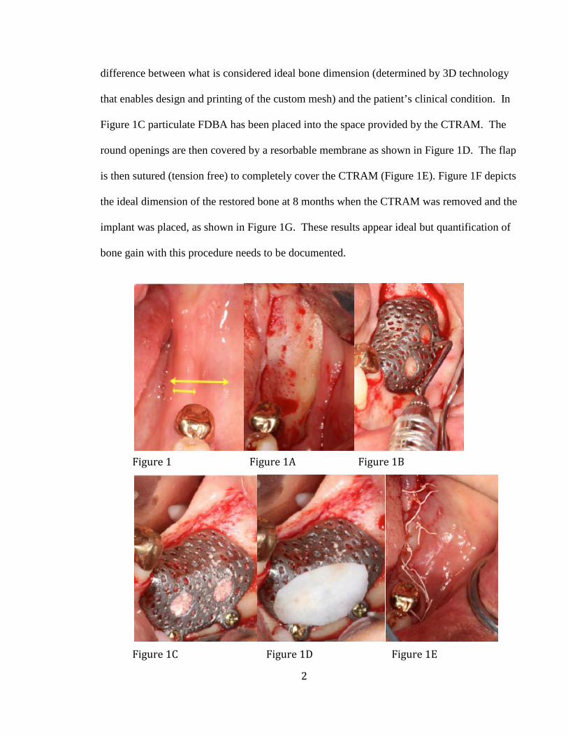
difference between what is considered ideal bone dimension (determined by 3D technology
that enables design and printing of the custom mesh) and the patient’s clinical condition. In
Figure 1C particulate FDBA has been placed into the space provided by the CTRAM. The
round openings are then covered by a resorbable membrane as shown in Figure 1D. The flap
is then sutured (tension free) to completely cover the CTRAM (Figure 1E). Figure 1F depicts
the ideal dimension of the restored bone at 8 months when the CTRAM was removed and the
implant was placed, as shown in Figure 1G. These results appear ideal but quantification of
bone gain with this procedure needs to be documented.
Figure 1 Figure 1A Figure 1B
Figure 1C Figure 1D Figure 1E

3
At this time no studies have reported the volumetric change gained when utilizing a
custom titanium matrix (CTRAM) with bone grafting to augment an edentulous ridge for
dental implant placement.
The observed, non-quantified, results for ridge augmentation provided with
CTRAM appear to completely restore deficient ridges to ideal dimension. Additionally,
if the data in this prospective study indeed show predictable complete augmentation then
it may be feasible to consider eliminating a second CBCT recommended by the American
Academy of Oral and Maxillofacial Radiology (AAOMR) to assess success of
regeneration (Tyndall, 2012).
Figure 1F Figure 1G
4
CHAPTER II: REVIEW OF THE LITERATURE
Successful restoration of dental function and health in an edentulous area utilizing
dental implants requires several considerations. Of major importance is adequate width
and height of alveolar bone for implant placement. A study of forty-six patients
(Schropp, 2003) utilizing linear and subtraction radiography techniques, along with stone
models found once a tooth is extracted, the height and width of the alveolar bone is
reduced. Their findings quantified the loss to be approximately 50% of the width of the
alveolar ridge after one year—two thirds of which was lost in the first three months.
Figure 1 (Introduction section) illustrates how thin the alveolar ridge becomes after teeth
are extracted. The solid line shows the width of the alveolar crest while the dashed line
approximates the width prior to molar extraction. Without intervention at the time of
extraction, a large portion of the bone that retained the tooth will atrophy following tooth
extraction.
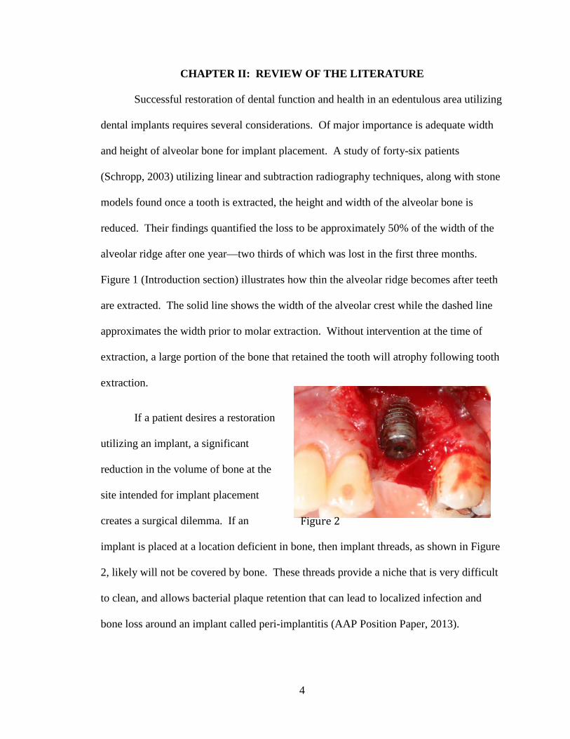
If a patient desires a restoration
utilizing an implant, a significant
reduction in the volume of bone at the
site intended for implant placement
creates a surgical dilemma. If an
implant is placed at a location deficient in bone, then implant threads, as shown in Figure
2, likely will not be covered by bone. These threads provide a niche that is very difficult
to clean, and allows bacterial plaque retention that can lead to localized infection and
bone loss around an implant called peri-implantitis (AAP Position Paper, 2013).
Figure 2

5
Figure 3 shows the implant in Figure
2 before flap reflection and depicts peri-
implantitis. Purulence is evident as it exudes
through the gingiva surrounding an implant.
A bone graft procedure to maximize alveolar
ridge dimension would have reduced the potential for incomplete coverage of the implant
threads with bone. Such a surgical result threatens long-term stability of the implant, and
indicates that augmentation of the alveolar bone prior to implant placement would be
preferred.
Evidence that alveolar bone resorbs following extraction of teeth, raises the
question of how best to preserve alveolar bone—or regenerate bone that has been lost
over time at the location where the tooth or teeth were removed. Maintaining alveolar
ridge dimension after extraction or augmenting bone in clinical presentations, as in
Figure 1 and Figure 2, is necessary to insure healthy long-term restoration with dental
implants.
Guided Bone Regeneration
According to the AAP Glossary of Terms (2001), Guided Bone Regeneration
(GBR) refers to augmenting lost jaw bone using a bone graft, covered by a barrier
membrane, which aims to exclude epithelial cells and gingival fibroblasts from invading
the location where bone augmentation is targeted. Exclusion of epithelial cells and
gingival fibroblasts provides slower growing bone cells a better opportunity to
differentiate into mature bone. The concept of GBR was introduced in 1959 (Hurley,
Figure 3
6
1959) using occlusive membranes to cover bone grafts during spinal fusion procedures on
dogs. Buser (1993) developed a surgical technique to augment jaw bone employing a
fixated membrane to achieve GBR. The authors describe a staged approach to ridge
augmentation where the site was left to heal over approximately nine months. After the
nine months passed, the membrane and fixation screws were removed and implants were
then placed in the area of regenerated bone. Four critical principles were stated in the
article to ensure GBR technique success: 1) obtain primary closure of the tissue flap, 2)
utilize a barrier membrane to prevent epithelial and gingival cell migration into the graft
site, 3) stabilize the membrane, and 4) maintain space under the membrane for bone
regeneration. McAllister (2007) discussed the four concepts related to GBR success, the
use of resorbable and non-resorbable membranes, as well as with available bone graft
materials which are explained in further detail in the “Membranes” and “Bone Graft
Materials” sections to follow.
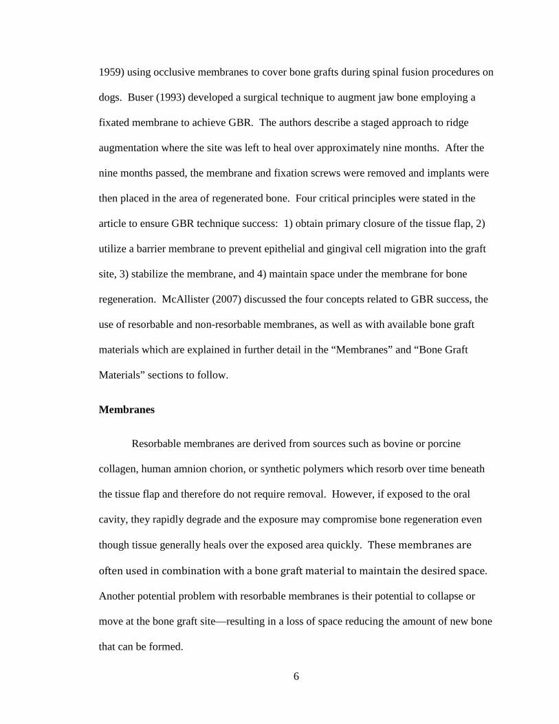
Membranes
Resorbable membranes are derived from sources such as bovine or porcine
collagen, human amnion chorion, or synthetic polymers which resorb over time beneath
the tissue flap and therefore do not require removal. However, if exposed to the oral
cavity, they rapidly degrade and the exposure may compromise bone regeneration even
though tissue generally heals over the exposed area quickly. These membranes are
often used in combination with a bone graft material to maintain the desired space.
Another potential problem with resorbable membranes is their potential to collapse or
move at the bone graft site—resulting in a loss of space reducing the amount of new bone
that can be formed.
7
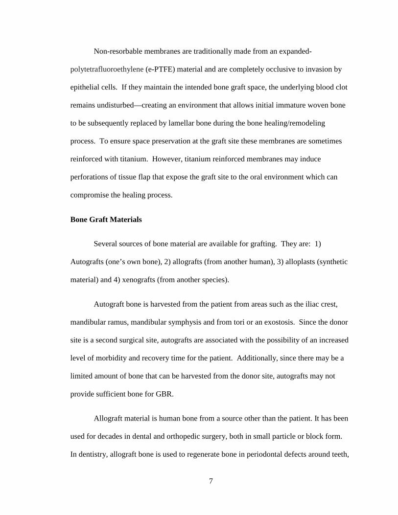
Non-resorbable membranes are traditionally made from an expanded-
polytetrafluoroethylene (e-PTFE) material and are completely occlusive to invasion by
epithelial cells. If they maintain the intended bone graft space, the underlying blood clot
remains undisturbed—creating an environment that allows initial immature woven bone
to be subsequently replaced by lamellar bone during the bone healing/remodeling
process. To ensure space preservation at the graft site these membranes are sometimes
reinforced with titanium. However, titanium reinforced membranes may induce
perforations of tissue flap that expose the graft site to the oral environment which can
compromise the healing process.
Bone Graft Materials
Several sources of bone material are available for grafting. They are: 1)
Autografts (one’s own bone), 2) allografts (from another human), 3) alloplasts (synthetic
material) and 4) xenografts (from another species).
Autograft bone is harvested from the patient from areas such as the iliac crest,
mandibular ramus, mandibular symphysis and from tori or an exostosis. Since the donor
site is a second surgical site, autografts are associated with the possibility of an increased
level of morbidity and recovery time for the patient. Additionally, since there may be a
limited amount of bone that can be harvested from the donor site, autografts may not
provide sufficient bone for GBR.
Allograft material is human bone from a source other than the patient. It has been
used for decades in dental and orthopedic surgery, both in small particle or block form.
In dentistry, allograft bone is used to regenerate bone in periodontal defects around teeth,
8
preserve bone dimensions at dental extraction sites and augment deficient alveolar ridges.
As opposed to autogenous bone, allograft bone is available in essentially limitless
quantities and eliminates the need for a second surgical site for harvesting.
Two main forms of particulate allograft are currently utilized in GBR: Freeze-
Dried Bone Allograft (FDBA) and Demineralized Freeze Dried Bone Allograft
(DFDBA). These have been shown to have similar results when used with augmentation
for implant site development. In a study of 72 FDBA grafted sites and 21 DFDBA
grafted sites in 93 patients receiving ridge augmentation, the mean percentages of new
bone formed were 41.89% for FDBA and 41.74% for DFDBA (Cammack, 2005). A
statistical difference between the two graft materials was not found regardless of where
the graft was used in this particular study. A different study suggested that dental
implants placed and loaded in bone regenerated from DFDBA, FDBA or autogenous
sources had success rates similar to “native” bone. A total of 526 implants were placed
in 352 patients who were followed from six to seventy-four months. Only 8 implants (all
in the maxilla) were lost, yielding a 97.5% implant survival rate (Nevins, 1998).
Block autografts and allografts have also been used to augment deficient alveolar
ridges. As mentioned previously, autogenous blocks require a second surgical site to
harvest the graft and are associated with some morbidity including the potential for
traumatization or de-vitalization of adjacent nerves. Resorption of block grafts after
placement is also a concern that several studies have assessed.
Fifty block autografts from human symphysis or ramus locations were followed
through a 4-6 month healing time end point. Although a 100% survival rate was
achieved, resorption of these autograft blocks ranged from 0 - 25% (Misch, 1997).

9
Keith (2004) studied mineralized block allografts in partially edentulous patients.
No resorption was observed in 69% (52/75) of subjects and 0.5 - 2.0 mm of localized
resorption around block fixation screws or around the blocks themselves occurred in 31%
(23/75) of subjects. In a case report, freeze-dried cancellous block allografts covered by
a membrane were placed at five deficient sites that were re-entered after 6 months healing
time. A gain of 2.0 - 4.0 mm in ridge width was achieved with these grafts however,
despite the increase in ridge width, 1.0 - 2.0 mm of surface resorption was recorded at
three of the five grafted areas (Lyford, 2003). These studies point to the fact that
autogenous or allograft blocks can increase width of deficient ridges but are prone to
resorption, which can affect the predictability of the bone augmentation and ultimately
implant placement.
Synthetic graft materials known as alloplasts have been utilized in augmentation
for over 100 years. Calcium phosphate and calcium sulfate are compounds that have
shown some biologic success in regard to biocompatibility and additionally are readily
available in regard to supply and cost. However, because alloplasts are not completely
remodeled into natural bone, they are less suitable for future implant placement.
Xenograft materials have been implemented in association with GBR techniques
for many years. These materials may come from equine or bovine sources and have been
noted to be quite effective in space maintenance at the graft site due to a long resorptive
and remodeling period. They have been heavily utilized in sinus augmentation
procedures where they maintain the desired space for implant placement. Wallace (1996)
reported that after taking histologic samples of sinuses grafted with xenograft (80%) and
autogenous bone (20%), a 12 - 20 month period was needed in order to see the graft

10
remodel to “vital bone”.
Membranes, as stated earlier, have been utilized to contain the bone material at
the grafted site, exclude epithelial cell and gingival fibroblast migration into the graft site
and attempt to maintain the space necessary for the graft to remain undisturbed.
Resorbable and non-resorbable membranes are currently used in a number of
regenerative periodontal procedures. A potential problem with a flexible membrane is
the potential for collapse. This loss of space over the course of regeneration directly
affects the amount of new bone that can be formed, and has prompted efforts to develop
membranes which are better able to maintain space.
Titanium Mesh in Cranioplasty
Neurosurgeons have employed titanium mesh or plates as biocompatible space
maintainers following cranioplasty. Gundeslioglu (2013) submitted a case report of a 58-
year-old patient who had a titanium mesh cranial implant, which became exposed, shortly
after placement. Eight years later the patient presented for treatment due to odor and
“discharge” from the area. When the titanium mesh implant was removed, significant
osteogenesis had occurred underneath the mesh and had dramatically decreased the bone
defect in the skull. Pathological analysis revealed that the odor and discharge were due to
chronic inflammatory responses attributed to “reactions against the inverted scalp” and
not the titanium mesh material. This report demonstrates the biocompatibility of titanium
even in the event of exposure.
Wiggins (2013) examined the results of custom titanium mesh or plates used in
cranioplasty over a 14-year period. Eighteen of 113 patients (16%) developed infections

11
that occurred approximately 120 days after placement. It was found that “the only
variable that significantly influenced the risk of infection was the size of the titanium
cranioplasty.” The authors noted that most infections stemmed from either exposure to
microbes (mainly S. aureus) in a hospital environment, or from problems related to
residual effects of previous infections associated with flap procedures that had failed.
The larger the original cranial defects exposed to the open environment, the higher the
incidence of infection.
Titanium Mesh in Dental Surgery
Titanium mesh has been used in dental surgery to overcome flexible membrane
deformation (collapse) and create solid structural support that allows optimal space
maintenance and stabilization for bone growth. Titanium mesh ridge augmentation was
outlined in a case report (Sumi, 2000). Bone defects were measured and the titanium
mesh was trimmed by hand. Autogenous bone grafts were placed to fill approximately
50% of the space between the mesh and crestal bone. Corticotomies (small holes through
the cortical plate) were made at the recipient site to allow blood vessels to grow into the
graft from medullary bone. The mesh was fixated into place with screws and primary
closure was achieved. Six to nine months later, the mesh and screws were removed and a
3.5 mm mean gain in alveolar crest width was recorded. Implants in all three patients
were stable at 6 - 18 months following placement.
Von Arx (1998) assessed the clinical outcome of 10 dental implants placed in 6
patients who needed autogenous bone harvested from the symphysis or ramus, and
grafted over the implants at the time of implant placement. Despite pre-surgical CT

12
scans to assess bone morphology, these surgical sites had deficient alveolar ridges that
caused implant surface exposures (4-10 mm) upon implant placement. After the implants
were placed, titanium mesh was adapted over the exposed implant threads and bone
defect to contain the autogenous bone graft and fixated with screws for stability. During
6 - 9 months of healing, no complications—including mesh exposures, were reported.
The mesh and screws were retrieved and all sites were found to have >90% of the grafted
bone remaining over the implants.
Alveolar ridge defects were treated using titanium mesh in a prospective study
(Pieri, 2008). Sixteen subjects with 19 alveolar ridge bony defects were treated.
Commercially available titanium mesh was trimmed and fixated over the graft composed
of autogenous and bovine bone. Of the 19 sites treated, 18 (94.7%) had uneventful
healing. One site had a mesh exposure at 2 months. The mesh exposure, however, did
not interfere with subsequent implant placement. After 8 - 9 months, the mesh at all sites
was removed and 44 implants were placed. Post healing CT imaging was compared to a
baseline scan and revealed a 3.71 mm average gain in vertical bone growth and 4.16 mm
mean horizontal gain. Following 2 years of functional loading, all implants were
retained. A 93.2% success rate and a 100% survival rate were observed. Success was
determined by absence of pain, suppuration, radiolucency, mobility and bone loss <1.5
mm within one year, and <0.2 mm each subsequent year (Albrektsson, 1986). Survival
was defined as the absence of pain, suppuration, radiolucency and mobility with the
additional criteria that implants demonstrated bone loss that exceeded the parameters
designated for success.

13
A review of six articles by Ricci (2013) assessed 79 patients who received 82
titanium grids (meshes) and bone grafts for various alveolar bone deficiencies. Eighteen
patients (22.78%) had grid exposures between 5 and 12 weeks after surgery and 9
patients required grid removal. Following 4 - 10 months of bone graft healing time, and
despite the aforementioned findings, 141 implants were placed in the 79 patients. All 141
implants survived. Success and survival were determined according to similar criterion
used by Pieri (2008). The overall success rate of bone grafting utilizing titanium grids
was greater than 98%. Only one site out of the 82 grids had complete loss of the graft
resulting in bone graft failure rate of 1.21%.
Louis (2008) retrospectively studied titanium mesh ridge augmentation in 44
patients who underwent maxillary or mandibular rehabilitation. Findings after 7 months
of healing showed a 97.7% bone grafting success rate that allowed placement of 174
implants. Twenty-three patients (52%) experienced some degree of mesh exposure.
Seven patients had exposures related to localized infection that required mesh removal.
However, the authors stated that sharp mesh edges created by trimming the mesh to fit
the defects might have contributed more to the large incidence of mesh exposure than did
infection. After 17 months of follow up, only one patient lost implants (3 total) due to
local infection.
Another retrospective study (Her, 2012) evaluated 26 patients for complications
associated with titanium mesh used for ridge augmentation by bone grafting. Twenty-
seven sites were treated with titanium mesh and various bone grafting materials. At these
sites, 69 implants were placed after 4 - 11 months of bone graft healing time. A 26%
mesh exposure rate (7 sites) was reported. Mesh exposures were treated by smoothing

14
the protruding surface with a diamond bur and healing of these sites ensued within a few
weeks. No mesh required removal, and only slight resorption of grafted bone seemed to
occur at exposed areas. The mesh exposures did not prevent implant placement.
Titanium mesh has been demonstrated to be a biocompatible material that
preserves needed space for alveolar ridge bone regeneration. A disadvantage to using
commercially prefabricated titanium mesh is the time required during surgery to
manually cut the material and adapt it to the graft site. Additionally, regardless of the
ability of the surgeon to shape the mesh to the site, there will always be some areas where
the interface between the mesh and bone will be less than ideal. There is also potential
for sharp edges that may irritate the soft tissue flap.
Customizing Titanium Mesh Using 3D Technology
Joffe (1999) reported findings using 3D generated images from CT to fabricate
titanium plates for 148 patients undergoing cranioplasty for skull injuries. Ninety-seven
percent of the fabricated plates “fit passively over the defect” or had the border of the
plate within 2 mm of the bone margin. The authors also noted that 96% of cases only
required 30 minutes to fixate. In 57% of patients no symptoms were reported post
operatively, while 31% reported mild headache, dizziness or pain. Significant discomfort
(7%), localized pain (2.5%) and severe headaches (2.5%) were reported by the remaining
12% of patients. Only one case involved the removal of a plate due to infection.
Computer-aided design/computer-aided manufacturing (CAD/CAM) of customized
titanium plates in the treatment of skull defects was efficacious, demonstrated a high
level of precision, reduced operative time (as the plates did not need to be adapted by

15
hand to the site) and resulted in minimal post-operative symptoms. Titanium was also
shown to lower the rate of infection versus other materials, such as acrylic, where
infection rate has been reported as high as 10%.
In an article by Hou (2011), seven patients had CT’s of bilateral mandibular
defects. These were reconstructed with the aid of CAD/CAM rapid prototyping using the
CT information to fabricate pre-operative 3D jaw models upon which commercial, off the
shelf, 0.5 mm thick titanium mesh frameworks were manually adapted. No
complications were reported for these 7 patients who were followed from 11 months to 3
years. This combination of CT and CAD/CAM technologies allowed for reduced
intraoperative time required and improved the accuracy of mesh placement.
A case report by Ciocca (2011) showed how the integration of CT and
CAD/CAM data could be used by Direct Metal Laser Sintering (DMLS) to create custom
titanium mesh to reconstruct a deficient maxillary alveolar anterior ridge. A pre-
operative CT was taken of the patient and linked with implant planning software to
calculate placement parameters and volume of required bone. The mesh was designed
with a 0.6 mm thickness and square 1.0 mm pores. Using titanium alloy (Ti6AlV4),
DMLS created the customized grid. A 3D resin model was produced and a trial fit was
conducted to verify satisfactory adaptation. The mesh was sterilized. Particulate bone
graft material was placed within the mesh, and then the mesh was implanted at the site.
No fixation screws were utilized, as the mesh was reportedly non-mobile via bony
undercuts. A CT was taken after 8 months when the mesh was removed. A mean
increase of 2.57 mm in bone height and 3.41 mm in width was reported, allowing for
placement of four pre-planned dental implants.
16
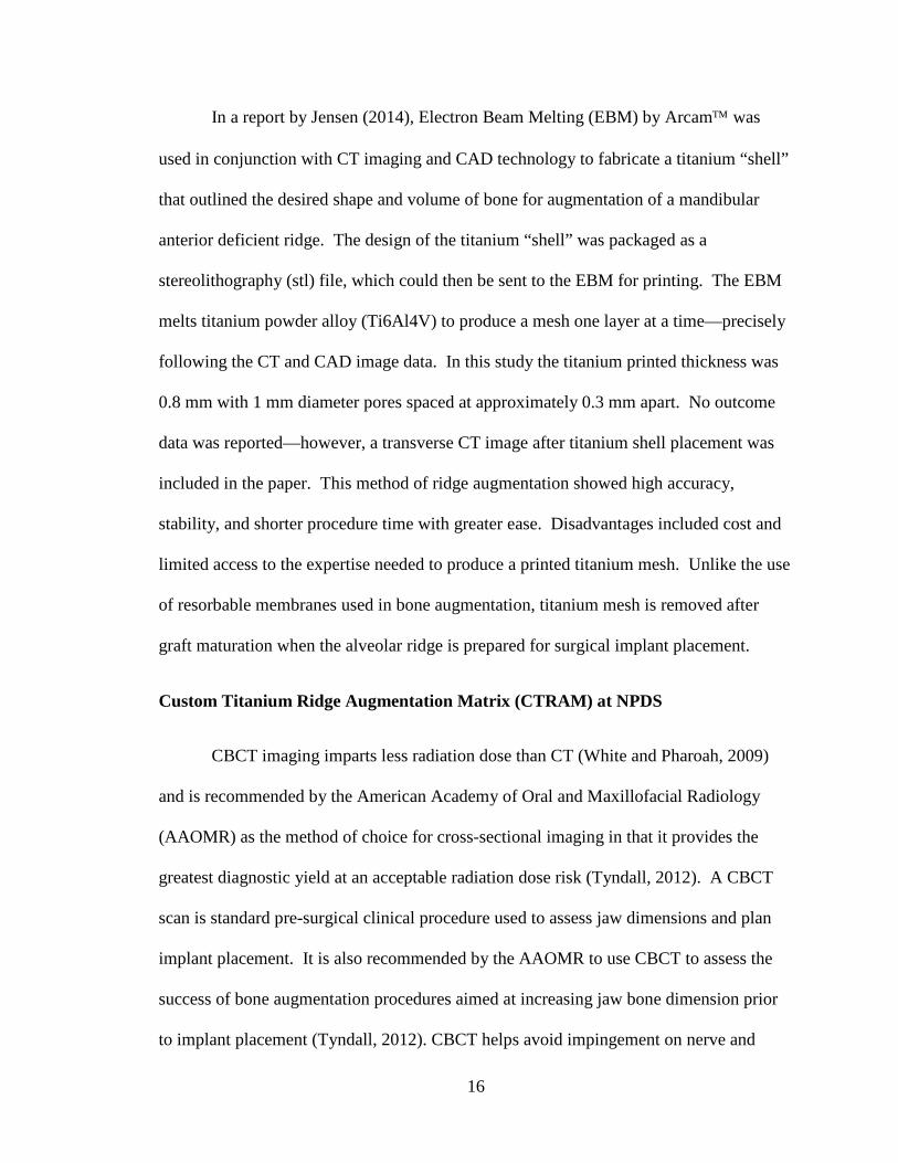
In a report by Jensen (2014), Electron Beam Melting (EBM) by Arcam was
used in conjunction with CT imaging and CAD technology to fabricate a titanium “shell”
that outlined the desired shape and volume of bone for augmentation of a mandibular
anterior deficient ridge. The design of the titanium “shell” was packaged as a
stereolithography (stl) file, which could then be sent to the EBM for printing. The EBM
melts titanium powder alloy (Ti6Al4V) to produce a mesh one layer at a time—precisely
following the CT and CAD image data. In this study the titanium printed thickness was
0.8 mm with 1 mm diameter pores spaced at approximately 0.3 mm apart. No outcome
data was reported—however, a transverse CT image after titanium shell placement was
included in the paper. This method of ridge augmentation showed high accuracy,
stability, and shorter procedure time with greater ease. Disadvantages included cost and
limited access to the expertise needed to produce a printed titanium mesh. Unlike the use
of resorbable membranes used in bone augmentation, titanium mesh is removed after
graft maturation when the alveolar ridge is prepared for surgical implant placement.
Custom Titanium Ridge Augmentation Matrix (CTRAM) at NPDS
CBCT imaging imparts less radiation dose than CT (White and Pharoah, 2009)
and is recommended by the American Academy of Oral and Maxillofacial Radiology
(AAOMR) as the method of choice for cross-sectional imaging in that it provides the
greatest diagnostic yield at an acceptable radiation dose risk (Tyndall, 2012). A CBCT
scan is standard pre-surgical clinical procedure used to assess jaw dimensions and plan
implant placement. It is also recommended by the AAOMR to use CBCT to assess the
success of bone augmentation procedures aimed at increasing jaw bone dimension prior
to implant placement (Tyndall, 2012). CBCT helps avoid impingement on nerve and
17
vascular pathways and enables positioning of the implant in the most functional and bone
supported location.
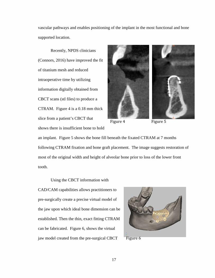
Recently, NPDS clinicians
(Connors, 2016) have improved the fit
of titanium mesh and reduced
intraoperative time by utilizing
information digitally obtained from
CBCT scans (stl files) to produce a
CTRAM. Figure 4 is a 0.18 mm thick
slice from a patient’s CBCT that
shows there is insufficient bone to hold
an implant. Figure 5 shows the bone fill beneath the fixated CTRAM at 7 months
following CTRAM fixation and bone graft placement. The image suggests restoration of
most of the original width and height of alveolar bone prior to loss of the lower front
tooth.
Using the CBCT information with
CAD/CAM capabilities allows practitioners to
pre-surgically create a precise virtual model of
the jaw upon which ideal bone dimension can be
established. Then the thin, exact fitting CTRAM
can be fabricated. Figure 6, shows the virtual
jaw model created from the pre-surgical CBCT
Figure 4 Figure 5
Figure 6

18
and the designed CTRAM for clinical use. An EBM machine by Arcam then prints the
actual CTRAM that is used by clinicians at NPDS.
This combination of CT imaging and CAD/CAM technology to create CTRAM
from virtual 3D models of the jaw provides many advantages when augmenting deficient
alveolar ridges with bone grafting. When dental implants are desired but the jaw site has
an alveolar bone defect, this technology allows for precise pre-surgical planning that
allows ideal restoration of the patient’s defect. This technology enables 1) determination
of the ideal amount of required bone for ridge augmentation for implants, 2) reduction of
intraoperative time, and 3) application of the critical principle of rigid space maintenance.
To date, quantification of the change in bone volume at deficient alveolar ridges
accomplished by bone grafting using this technology has not been reported.
Assessing Volumetric Change Using Digital Scanning
One method that can assess volumetric change utilizes a digital scanner to acquire
images. This method is non-invasive and patients are not exposed to radiation.
Presently, digital intraoral scans of jaws and teeth are directly used in dental offices and
dental laboratories to fabricate crowns and other fixed prosthetic restorations for patients.
Akyalcin (2013) demonstrated the accuracy of this technology in comparing changes in
volume over time using a Cadent iTero scanner to scan 60 dry skulls and converted the
images into stl files. These stl files were used to compare digital scanning measurements
of three teeth in each arch to measurements made via CBCT images and hand held
calipers. Teeth were measured in three dimensions—apical-coronal, mesio-distal and
bucco-lingual. Intra-class correlation analysis (ICC) was conducted for the three

19
measurement techniques. Caliper measurements versus a digital intraoral scanner yielded
high ICC values ranging from 0.92-0.99. CBCT compared with calipers yielded ICC
values ranging from 0.88 to 0.98. A mean difference of 0.16 mm was found between the
caliper method versus digital scanner and a 0.28 mm difference between caliper and
CBCT. This study demonstrates the high level of accuracy that can be achieved using
digital capture methods for documenting the dimensions of intra-oral anatomy.
Rebele (2014) recently demonstrated the ability to document changes in the
volume of soft tissue with a digital scanning device. Six patients, from a pool of 24 study
participants, were followed for 12 months after receiving root coverage flap surgery.
Digital scans using the Imetric D103 scanner were made on dental stone models
obtained from impressions of the treated sites at baseline, 1, 2, 3, 6 and 12 months post-
operatively. The digital images were superimposed (registered) on one another by
orienting images to a specific site on the buccal surface of treated teeth. This registration
allowed quantification of tissue volume changes. Volume in mm3 was converted into a
percentage for convenience in comparing changes over time. Digital measurement of
volume change over time permits precise comparisons between the original and
augmented sites.

20
CHAPTER III: MATERIALS AND METHODS
This will be a prospective pilot study of 14 subjects to document the volumetric
changes in the alveolar ridge which occur when utilizing the CTRAM technique for
guided bone regeneration. Bone dimension, using different techniques, will be assessed
at 1) baseline, 2) at 7 and 8 months post bone grafting with CTRAM and 3) at 2 - 4 weeks
and 4 months post implant placement.
The study’s flow chart, titled “CTRAM Prospective Study Flow Chart” (Appendix
A), outlines how procedures (marked by superscript numbers) for research measurement are
sequenced within the normal clinical and laboratory procedures for treatment of deficient
alveolar ridges using CTRAM. The superscript numbers are indicated in the specific aims
below.
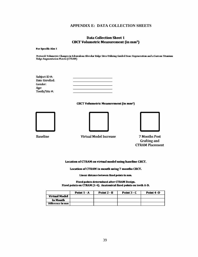
Specific Aim 1
Compare stereolithography (stl) files of the pre-surgical CBCT1 of the
deficient alveolar ridge with a) the virtual jaw model to show how much
added bone is ideal, and b) with the post-bone graft CBCT5 at 7 months to
determine actual volumetric gain. Differences between ideal bone and actual
bone gain will be recorded in cubic mm.
• Additionally, stl files will be used to compare location of the
CTRAM on the virtual jaw model to its actual location after
fixation to the jaw.
Specific Aim 2
Measure bone fill achieved by CTRAM by comparing periodontal probe
measurements to the bone under CTRAM at baseline before bone grafting and

21
at 8 months:
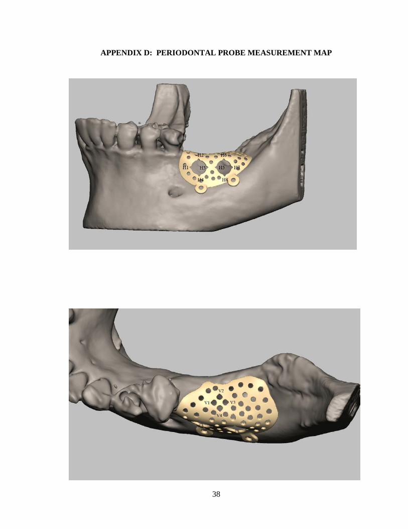
• At CTRAM design on the virtual jaw model, a digital image of the
CTRAM design (Appendix D: “Periodontal Probe Measurement Map”)
will be made that designates exactly which pores (small openings) and
windows (large openings) investigators will measure space below the
CTRAM just before bone grafting and 8 months later.
o At the two large windows, ridge width measurements will
be made at the noon, 3, 6 and 9 o’clock positions.
Please note: Since the CTRAM is custom made,
each participant’s mapping that designates which
windows and pores will be used for measurement
with the periodontal probe will also be unique.
Each data collection sheet (Appendix E) for probing
bone levels will reflect the custom nature of each
CTRAM design.
• Measure space under CTRAM after fixation using marked windows on
buccal surface (horizontal)3 and pores on ridge crest (vertical)4.
o This will be accomplished using a UNC 15 periodontal probe
fitted with a rubber stopper (Figure 7) and measured with a
ruler marked at 0.5 mm increments. The depth will be
recorded to the nearest 0.5 mm (Figure 8).

22
• Measure space under CTRAM at same locations3*, 4* for bone fill at 8
months following bone graft surgery just before the CTRAM is removed
and the implant is placed.
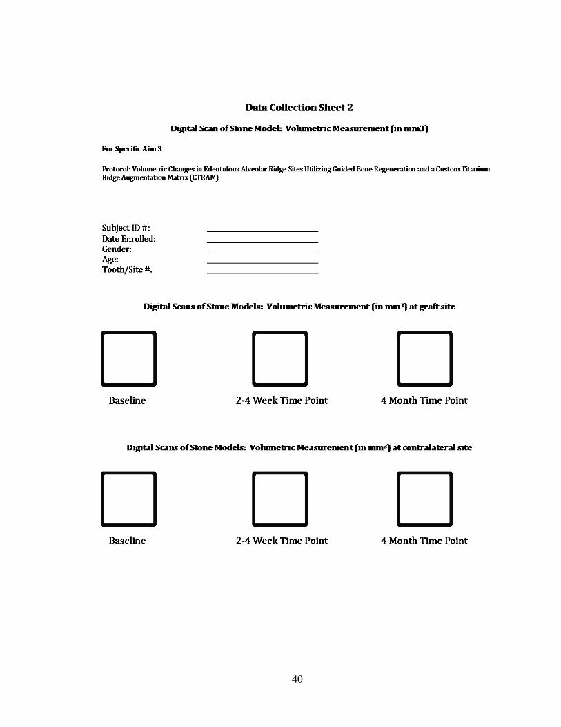
Specific Aim 3
Compare stl files of the digital scans of the stone models made from
impressions made pre-surgically2, 2-4 weeks (after any post-surgical swelling
has abated) after the implant is placed6, and at 4 months after implant
Figure 7
Figure 8
Figure 8
23
placement7 to note volume changes on models. Differences in volume will be
recorded in cubic mm.
• Comparison of stl files will be made for the surgical site and at
another location on the models where teeth are intact and no
surgery was performed. The comparison of stl files at a non-
surgical site where no tissue dimension changes are anticipated is
being performed to assess reliability of the stl file comparison on
study models.
Before Study: Initial Clinical Sequence with Screening and Consent:
1. Patients are referred to Naval Postgraduate Dental School,
Department of Periodontics for alveolar ridge augmentation and
subsequent implant therapy.
2. A treatment plan (based on the clinical exam, diagnostic stone
models and CBCT) may be developed for ridge augmentation
using CTRAM with GBR leading to implant placement.
3. Patients for whom CTRAM with GBR is treatment planned will
be asked by their provider if they would like to read a one page
brief (Appendix C) that describes the study that is measuring
how well CTRAM works to regenerate bone.
4. If the patient is not interested in hearing more about the study the
provider begins the CTRAM treatment process.
24
5. If the patient expresses interest in study participation after
reading the one page brief, an investigator will be asked to meet
with the patient to fully discuss the study.
a. If the patient does not consent to be in the study, therapy
will continue as planned by the patient’s surgeon, but
measurements of volumetric bone changes will not be made
as described in this study.
b. If the patient consents to be in the study, the therapy under
the research protocol will continue as stated below.
Following Consent:
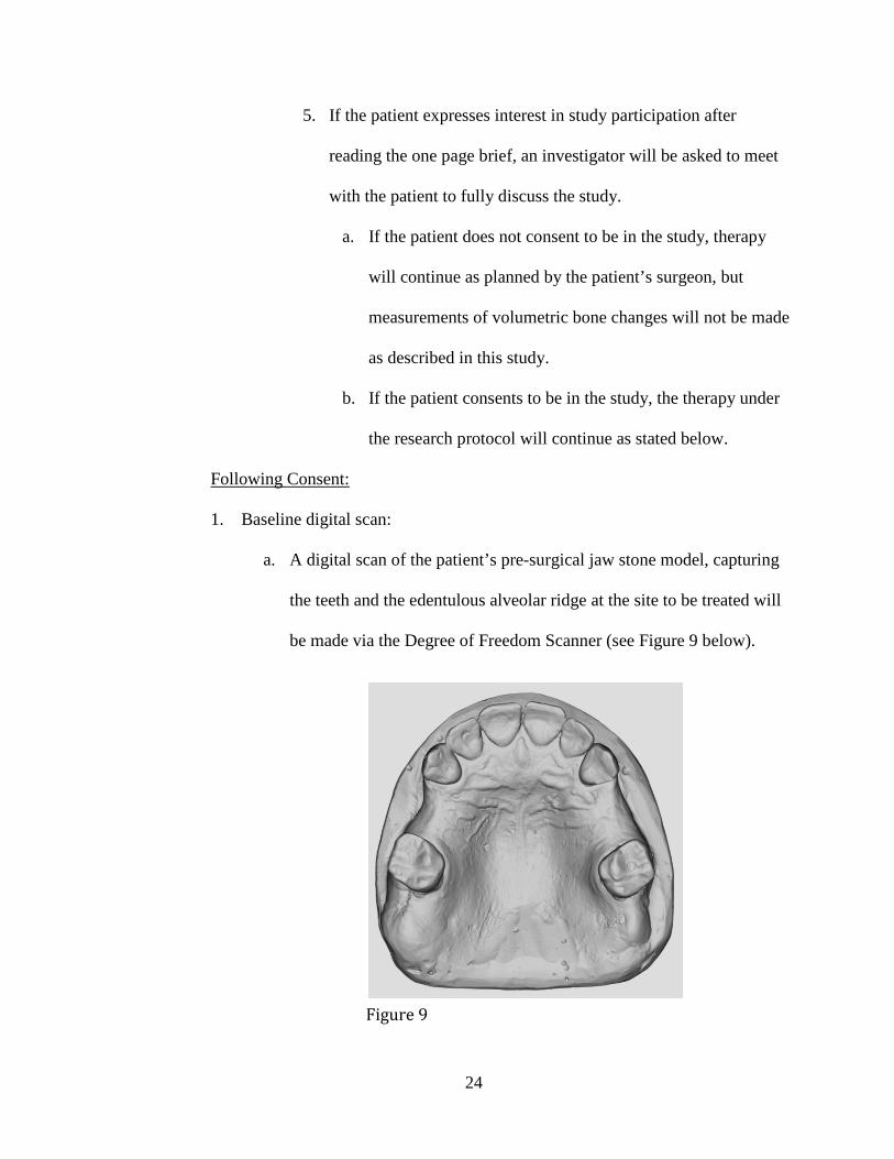
1. Baseline digital scan:
a. A digital scan of the patient’s pre-surgical jaw stone model, capturing
the teeth and the edentulous alveolar ridge at the site to be treated will
be made via the Degree of Freedom Scanner (see Figure 9 below).
Figure 9
25
i. The image will be saved as an stl file under the patient’s study
number and date the image was taken on a removable storage
device that only stores digital scans and is not connected to the
web. The device will be locked by the PI in a secure office
drawer at NPDS.
ii. The baseline stl file2 image will be compared to digital images
that will be taken at time points (stl files6, 7) listed in the
methods protocol (See Appendix A). The 3 stl digital image
files will be transferred to a secure computer in 3D Medical
Applications Department for comparison. (See Appendix A).
2. The 3D virtual model of the ridge defect will be created (stl file):
a. Using the software in the 3D Medical Applications Department the
patient’s CBCT images (stored on the WRNMMC Radiology Picture
Archiving and Communication System (PACS) for radiology) will be
uploaded and used to create a virtual model of the teeth and the
deficient ridge to plan the augmentation of the ridge.
b. A virtual 3D model of deficient ridge is produced.
3. 3D Medical Applications will design a CTRAM using the computer generated
model to virtually augment the ridge to ideal bone contours, and determine
bone volume from the underside of the CTRAM to the surface of the
edentulous ridge (stl file). Following the CTRAM design phase, it is printed
(see Chapter II, Figure 6).

26
4. Once the CTRAM has been printed, the cameo and intaglio surfaces will be
polished and the peripheral edges smoothed to a “feather edge” with slow
speed polishing burs.
5. The CTRAM is then sterilized for surgery. (Please see Appendix B: Surgical
Protocol for details concerning the surgical treatment phase of the study.)
6. 3D Medical Applications will make a photo image (mapping – see Appendix
D) of the CTRAM on which markings will be made to show investigators
where measurements are made using the periodontal probe at CTRAM
fixation and at 8 months just before the CTRAM is removed.
7. The surgical protocol will be followed (Appendix B) to insert the CTRAM,
take clinical measurements and remove the CTRAM at the end of 8 months,
prior to implant placement.
Data Analysis Plan
This study is a pilot study with 12 anticipated subjects (plus 2 additional
enrollees in case some subjects are exited from the study). Thus, the results
presented from this study will primarily be descriptive, with means and 95%
confidence intervals (or medians and ranges) presented to summarize continuous
data and counts and percentages for categorical data.
1. A patient flow diagram will present the number of subjects recruited,
consented, and the number of subjects at each follow up time point.
2. A table will present demographic and clinical characteristics of the
subjects. The pattern of any adverse events or loss to follow-up over the
duration of the study will be described.

27
3. Specific Aim 1: For each subject, the hypothetical ideal volume of bone
augmentation will be calculated and summarized by comparing the pre-
surgical CBCT of the deficient alveolar ridge with the virtual model
created for fabrication of the CTRAM.
4. Specific Aim 1: The post-graft CBCT (taken at 7 months) will be
compared with the pre-surgical CBCT to measure actual bone growth.
The differences between actual bone growth and hypothetical bone growth
will be calculated for each patient and summarized. Paired comparisons
(Wilcoxon signed-rank tests or paired t-tests) will be done to evaluate the
statistical significance of bone growth as assessed by the CBCT.
5. Specific Aim 1: STL files will be used to compare the actual and ideal
placement of the CTRAM. The distance between the actual placement and
the ideal placement will be calculated and summarized.
6. Specific Aim 2: Mean space between the CTRAM and the alveolar ridge
at initial placement will be measured using a periodontal probe and
summarized for each subject on both the vertical and horizontal extent.
The same locations will be re-sampled immediately prior to CTRAM
removal. Paired comparisons (Wilcoxon signed-rank tests or paired t-
tests) will be done to evaluate the statistical significance of bone growth as
assessed by the periodontal probe.
7. Specific Aim 3: Volumetric measurements taken from STL files of digital
scans of stone models will be summarized and compared across three time
points: pre-surgically, 2-4 post surgically, and 4 months after implant

28
placement. Paired comparisons (Wilcoxon signed-rank tests or paired t-
tests) will be done to assess change at each time point, with significance
levels adjusted for three comparisons.
8. Specific Aim 3: For each patient, a region of approximately equivalent
size contralateral to the surgical site will be identified for use as a split-
mouth, paired control region. A paired test (Wilcoxon signed-rank tests or
paired t-tests) will be used to compare volumetric change from baseline in
the surgical site versus the control site over the two time points.

29
CHAPTER IV: CONCLUSION
It is anticipated that the measurements recorded from the CBCT and stone model
scans will correlate with the clinical measurements to demonstrate complete bone fill
utilizing the CTRAM ridge augmentation technique. The data from this IRB approved
study may show that CTRAM predictably produces complete ridge regeneration, such
that a second post-graft CBCT may not be necessary when implant therapy is planned.

30
APPENDIX A: CTRAM PROSPECTIVE STUDY FLOW CHART

31
APPENDIX B: PERIODONTAL SURGICAL PROCEDURES
Phase I CTRAM Surgical Procedure:
Females of childbearing age will be asked to complete a HCG urinalysis prior to
the surgical procedure. If the results of the HCG test are positive, the surgery will be
deferred until after pregnancy and thus the subject will be exited from the study.
Prior to surgical procedure, in line with standard procedure at the Periodontics
Department, participants will be offered the option of having the surgery performed using:
1) Only local anesthesia, or 2) a combination of oral anxiolysis with Triazolam and local
anesthesia, or 3) a combination of IV moderate sedation with Versed and Fentanyl and
local anesthesia. The use of sedation will not affect the surgical procedure.
1. All surgical providers will be briefed in the protocol. All surgeries will follow the
same steps listed below:
a. Surgical set-up is standardized for all surgeries done at the Naval
Postgraduate Dental School Periodontics Department.
b. Surgical Procedure Steps:
i. Administration of oral anxiolysis or IV moderate sedation if patient
desires and such treatment is indicated
ii. Administration of IV Dexamethasone if considered needed
iii. Administration of topical and local anesthetic with any combination
of 2% Lidocaine with 1:100K epinephrine, 4% Articaine with
1:100K epinephrine, and 0.5% Marcaine with 1:200K epinephrine

32
iv. Sulcular incision around teeth adjacent to the edentulous site,
connected via a crestal or paracrestal incision along the edentulous
ridge. Vertical releasing incisions may be made for surgical access.
v. Full thickness flaps will be reflected to expose the deficient ridge.
vi. The CTRAM will be tried in at the site and examined for correct fit.
If fit is appropriate, corticotomies will be made into the bone to
induce bleeding and the CTRAM will be fixated with the required
number of surgical fixation screws for proper stability—ensuring
that there is no movement.
vii. A periodontal probe fitted with a rubber stopper will record the
space from the surface of the CTRAM to the alveolar ridge through
specific pores as shown in Appendix D. The distance will be
measured via an endodontic style ruler marked at 0.5 mm
increments. At the same pores, a second set of measurements will
be made 8 months later just before the CTRAM is removed and the
implant is placed.
viii. FDBA will be hydrated with sterile saline as per the manufacturer’s
instructions, or enamel matrix derivative (EMD) and placed into the
alveolar defect—level with the pores and access port(s) of the
titanium matrix (see Figure 1C).
ix. A resorbable collagen membrane will be placed over the access
port(s) on the external aspect of the matrix.

33
x. If needed, a periosteal releasing incision will be made in the buccal
and/or lingual flap to allow for primary closure.
xi. The area of the flap directly covering the matrix will be sutured
with a monofilament, non-resorbable suture material. Areas which
do not directly cover the matrix may employ this same suture type,
or may require a resorbable suture instead.
2. Standard post-operative care will be provided: (See next section: Post-operative
Care below)
Post-operative Care:
1. All participants receive the following post-operative regimen:
a. Pain medication consisting of any of the following alone or in
combination:
i. Ibuprofen 800 mg , Take 1 tab PO q6-8h for moderate pain OR
ii. Hydrocodone/Acetaminophen 5/325 mg, Take 1-2 tab PO q6h
prn severe/breakthrough pain OR
iii. Oxycodone/Acetaminophen 5/325mg, Take 1-2 tab PO q6h prn
severe/breakthrough pain
b. Pain medication for patients who cannot take NSAIDS will be
prescribed any of the following alone or in combinations:
i. Acetaminophen 325 mg, Take 1-2 tabs PO q4h for moderate
pain
ii. Oxycodone 5mg, Take 1 tab PO q4h prn severe/breakthrough
pain

34
c. Antibiotics consisting of either of the following:
i. Amoxicillin 500mg, Take 1 tab PO q8h for 10 days
ii. Clindamycin 300 mg, Take 1 tab PO q8h for 10 days
d. 0. 12% Chlorhexidine, 1 bottle, Rinse and spit bid with 1 TBSP as
directed on the bottle
e. Medrol 4 mg Dosepak if needed. Use as directed on package.
3. All patients are provided with the standard post-operative instructions. Patients
will be evaluated at weeks 1, 2, 4 and 8 weeks and then at 3, 5 and 7 months for
routine post-operative care.
At 7 months post-insertion of the CTRAM device, the patient has a second CBCT
taken to assess the grafted site and plan for the second stage of surgery involving CTRAM
and fixation screw removal and placement of the dental implant(s) at the grafted site.
Phase II Surgical Procedure at Approximately 8 Months:
1. Surgical steps will be completed as outlined in the Phase I surgical procedure
section up until step “v”.
2. Measurements with a UNC-15 periodontal probe will be taken at the augmented
site using the access port(s) and pores of the CTRAM to assess bone fill as
described in the specific aims.
3. The fixation screw(s) and the CTRAM device will be removed.
4. The implant(s) will be placed based upon the manufacturer’s guidelines. A cover
screw or healing abutment will be hand tightened and a peri-apical radiograph will
be taken to ensure proper seating contact between implant and cover screw or
healing abutment.

35
5. Standard post-operative care will be provided at weeks 1, 2, 4 and 8 and at 4
months.
a. During a postoperative visit 2 - 4 weeks when any post-surgical edema has
abated, an impression of the surgical site will be made and a digital scan of
the stone model will be made.
b. At approximately 4 months post-operatively the patient will have another
jaw impression made and a final digital scan of the model will be made.
6. This completes the subject’s participation in this prospective case series. Patients
will be exited from the study and followed by their periodontist and primary care
dentist for subsequent maintenance therapy and implant restoration.
36
APPENDIX C: ONE PAGE STUDY BRIEF
You and your doctor have decided that you are going to have a bone graft placed
under a custom titanium mesh where a tooth was removed and your jawbone shape
decreased in size. This procedure is called guided bone regeneration. It can restore the
jawbone contours needed to place a dental implant. Although custom titanium mesh
technique appears to work great, how much new bone is actually restored has not been
scientifically measured.
The Periodontics Department is conducting a research study to measure how
much new jawbone is restored when a custom titanium mesh is used to hold a bone graft.
If you choose to enroll in the study you will have the same number of appointments, and
receive the same treatment as patients who do not enroll in the study. The only
differences are that we are asking study volunteers to let us make 3 measurements and
take 2 extra jaw impressions.
Normal clinical treatment with a custom titanium mesh needs a pre-surgical 3D x-
ray scan of the missing tooth area and a post grafting 3D x-ray scan to see the bone
growth. Measurement #1 in this study will compare these x-rays on a computer to
determine how much new bone is gained.
The custom titanium mesh and bone graft are placed in your jaw during the first
surgery. The mesh is removed and the implant is placed in the second surgery 8 months
later. Measurement #2 uses a periodontal probe to measure the space under your mesh to
your jawbone before the bone graft is placed, and 8 months later before the matrix is
removed.

37
An impression for a model of your teeth and jaw is made before surgery. For the
study we are asking you to let us take an impression for jaw models at 2 to 4 weeks and at
4 months after implant placement. Measurement #3 uses digital photo scans of these 3
jaw models and compares them on a computer to measure how jaw contours change.
If you are interested in being is this study to help us document how bone volume
changes using the custom titanium mesh, tell your doctor. He or she will have an
investigator come over to discuss the study.
Speaking with an investigator does not obligate you to be in the study. If you are
not interested, that is okay and you will start your treatment with your doctor as a non-
study patient.
Thank you for your consideration.
38
APPENDIX D: PERIODONTAL PROBE MEASUREMENT MAP
39
APPENDIX E: DATA COLLECTION SHEETS
40

41
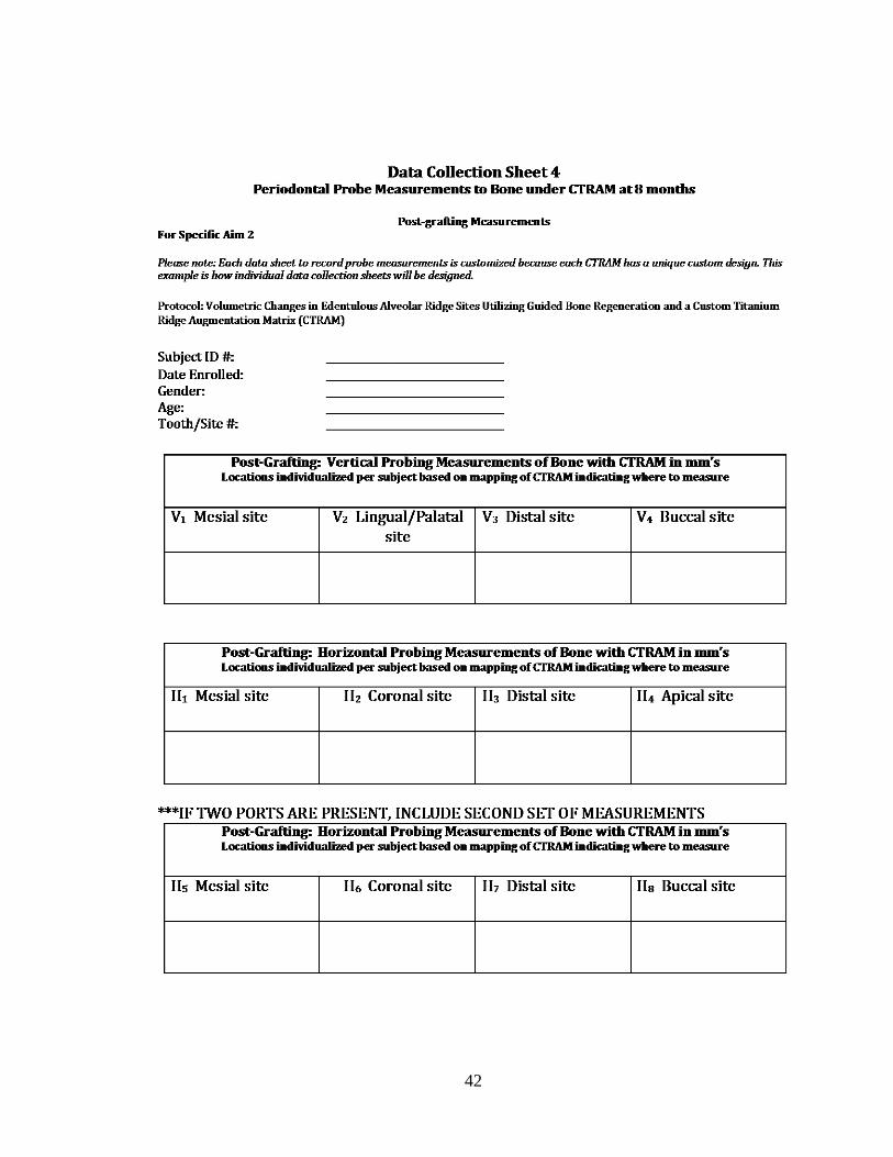
42

43
REFERENCES
Albrektsson T, Zarb G, Worthington P, Eriksson AR. The Long-Term Efficacy of Currently Used Dental Implants: A Review and Proposed Criteria of Success. Int J Oral Maxillofac Implants 1986; 1(1):11-25. American Academy of Periodontology Glossary of Periodontal Terms, 4th Edition, 2001, Chicago, Illinois 60611-2690 American Academy of Periodontology Position Paper. Peri-Implant Mucositis and Peri-Implantitis: A Current Understanding of Their Diagnoses and Clinical Implications. J Periodontol 2013; 84(4):436-443. Akyalcin S, Cozad BE, English JD, Colville CD, Laman S. Diagnostic Accuracy of Impression-Free Digital Models. Am J Orthod Dentofacial Orthop 2013; 144:916-22. Buser D, Dula K, Belser U, Hirt H, Berthold H. Localized Ridge Augmentation Using Guided Bone Regeneration. I. Surgical Procedure in the Maxilla. Int J Periodont Rest Dent 1993; 13:29-45. Cammack GV, Nevins M, Clem DS, Hatch JP, Mellonig JT. Histologic Evaluation of Mineralized and Demineralized Freeze-Dried Bone Allograft for Ridge and Sinus Augmentations. Int J Periodont Rest Dent 2005; 25:231-237. Ciocca L, Fantini M, De Crescenzio F, Corinaldesi G, Scotti R. Direct Metal Laser Sintering (DMLS) of a Customized Titanium Mesh for Prosthetically Guided Bone Regeneration of Atrophic Maxillary Arches. Med Biol Eng Comput 2011; 49:1347-1352.
Connors CA, Liacouras PC, Grant GT. Custom Titanium Ridge Augmentation Matrix (CTRAM). Int J Periodont Rest Dent 2016; (in press). Gundeslioglu OA. Exposed Titanium Mesh and Dura Persisting for 8 Years After Cranioplasty. J Craniofac Surg 2013; 24(2):655-656. Her S, Taeheon K, Fien M. Titanium Mesh as an Alternative to a Membrane for Ridge Augmentation. J Oral and Maxillofac Surg 2012; 70:803-810. Hou J, Chen M, Pan C, Tao Q, Wang J, Wang C, Zhang B, Huang H. Immediate Reconstruction of Bilateral Mandibular Defects: Management Based on Computer-Aided Design/Computer-Aided Manufacturing Rapid Prototyping Technology in Combination with Vascularized Fibular Osteomyocutaneous Flap. J Oral Maxillofac Surg 2011; 69:1792-1797.
44
Hurley LA, Stinchfield FE, et al. The role of soft tissues in osteogenesis. An experimental study of canine spine fusions. J Bone Joint Surg Am 1959;41A: 1243-54. Jensen OT, Lehman H, Ringeman JL, Casap N. Fabrication of Printed Titanium Shells for Containment of BMP-2 Composite Graft Materials for Alveolar Bone Reconstruction. J Oral Maxillofac Implants 2014; 29e103-e105. Joffe J, Harris M, Kahugu F, Nicoll S, Linney A, Richards R. A Prospective Study of Computer-Aided Design and Manufacture of Titanium Plate for Cranioplasty and its Clinical Outcome. Br J Neurosurg 1999; 13(6):576-580.
Keith JD Jr, Petrungaro P, Leonetti JA, Elwell CW, Zeren KJ, Caputo C, Nikitakis NG, Schöpf C, Warner MM. Clinical and histologic evaluation of a mineralized block allograft: results from the developmental period (2001-2004). Int J Periodontics Restorative Dent 2006; Aug:26(4):321-7. Louis P, Gutta R, Said-Al-Naief N, Bartolucci AA. Reconstruction of the Maxilla and Mandible with Particulate Bone Graft and Titanium Mesh for Implant Placement. J Oral Maxillofac Surg 2008; 66:235-245. Lyford RH, Mills MP, Knapp CL, Scheyer ET, Mellonig JT. Clinical evaluation of freeze-dried block allografts for alveolar ridge augmentation: a case series. Int J Periodontics Restorative Dent 2003; Oct;23(5):417-25. McAllister BS, Haghighat K. Bone Augmentation Techniques. J Periodontol 2007; 78(3): 377-396.
Misch C. Comparison of Intraoral Donor Sites for Onlay Grafting Prior to Implant Placement. Int J Oral Maxillofac Implants 1997; 12:767-776.
Nevins M, Mellonig JT, Clem DS, Reiser GM, Buser DA. Implants in Regenerated Bone: Long-Term Survival. Int J Periodont Rest Dent 1998; 18:35-45. Pieri F, Corinaldesi G, Fini M, Aldini NN, Giardino R, Marchetti C. Alveolar Ridge Augmentation with Titanium Mesh and a Combination of Autogenous Bone and Anorganic Bovine Bone: A 2-Year Prospective Study. J Periodont 2008; 79:2093-2103. Rebele SF, Zuhr O, Schneider D, Jung RE, Hurzeler MB. Tunnel Technique with Connective Tissue Graft Versus Coronally Advanced Flap with Enamel Matrix Derivative for Root Coverage: A RCT using 3D Digital Measuring Methods. Part II. Volumetric Studies on Healing Dynamics and Gingival Dimensions. J Clin Periodontol 2014; 41: 593-603.
45
Ricci L, Perrotti V, Ravera L, Scarano A, Piattelli A, Iezzi G. Rehabilitation of Deficient Alveolar Ridges Using Titanium Grids Before and Simultaneously with Implant Placement: A Systematic Review. J Periodontol 2013; 84:1234-1242. Schropp L, Wenzel A, Kostopoulos L, Karring T. Bone Healing and Soft Tissue Contour Changes Following Single-Tooth Extraction: A Clinical and Radiographic 12-Month Prospective Study. Int J Periodontics Restorative Dent 2003; 23:313-323. Sumi Y, Miyaishi O, Tohnai I, Ueda M. Alveolar Ridge Augmentation with Titanium Mesh and Autogenous Bone. Oral Surg Oral Med Oral Pathol Oral Radio Endod 2000; 89:268-70. Tyndall DA, Price JB, Tetradis S, Ganz SD, Hildebolt C, Scarfe WC. Position statement of the American Academy of Oral and Maxillofacial Radiology on Selection Criteria for the Use of Radiology in Dental Implantology with Emphasis on Cone Beam Computed Tomography. Oral Surg Oral Med Oral Pathol Oral Radiol 2012;113:817-826.
von Arx T, Kurt B. Implant Placement and Simultaneous Peri-Implant Bone
Grafting Using a Micro Titanium Mesh for Graft Stabilization. Int J Periodont Rest Dent 1998; 18:117-127.
Wallace SS, Froum SJ, Tarnow DP. Histologic evaluation of a sinus elevation
procedure: a clinical report. Int J Periodont Rest Dent 1996; Feb:16(1):46-51. White SC and Pharoah MJ. Oral Radiology: Principles and Interpretation. 7th
Edition. 2009.
Wiggins A, Austerberry R, Morrison D, Ho KM, Honeybul S. Cranioplasty With Custom-Made Titanium Plates—14 Years Experience. Neurosurgery 2013; 72:248-256.