Embed Size (px)
Citation preview

ISSN 2041-6539
rsc.li/chemical-science
ChemicalScience
EDGE ARTICLEQuli Fan, Aixi Yu, Zhen Cheng et al.Diketopyrrolopyrrole-based semiconducting polymer nanoparticles for in vivo second near-infrared window imaging and image-guided tumor surgery
Volume 9 Number 12 28 March 2018 Pages 3071–3256
ChemicalScience
EDGE ARTICLE
Ope
n A
cces
s A
rtic
le. P
ublis
hed
on 0
6 Fe
brua
ry 2
018.
Dow
nloa
ded
on 3
/21/
2022
9:3
8:32
AM
. T
his
artic
le is
lice
nsed
und
er a
Cre
ativ
e C
omm
ons
Attr
ibut
ion
3.0
Unp
orte
d L
icen
ce.
View Article OnlineView Journal | View Issue
Diketopyrrolopyr
aDepartment of Orthopedics, Zhongnan Hosp
430071, China. E-mail: [email protected] Imaging Program at Stanford
Radiology, Canary Center at Stanford
University, California 94305-5344, USA. E-mcKey Laboratory for Organic Electronics
Advanced Materials (IAM), Jiangsu Nati
Advanced Materials (SICAM), Nanjing Uni
Nanjing 210023, China. E-mail: iamqlfan@dCAS Key Laboratory of Receptor Research,
Laboratory (SOMCL), Shanghai Institute o
Sciences, No. 555 Zuchong Road, Pudong N
† Electronic supplementary informa10.1039/c8sc00206a
‡ These authors contributed equally to th
Cite this: Chem. Sci., 2018, 9, 3105
Received 13th January 2018Accepted 5th February 2018
DOI: 10.1039/c8sc00206a
rsc.li/chemical-science
This journal is © The Royal Society of C
role-based semiconductingpolymer nanoparticles for in vivo second near-infrared window imaging and image-guided tumorsurgery†
Kangquan Shou,‡ab Yufu Tang,‡c Hao Chen,‡b Si Chen,b Lei Zhang, b Ao Zhang, d
Quli Fan, *c Aixi Yu*a and Zhen Cheng *b
A diketopyrrolopyrrole-based semiconducting polymer nanoparticle (PDFT1032) has been developed as
a NIR-II (near infrared window II, 1000–1700 nm) fluorescent probe. It shows high photostability,
a favorable absorption peak at 809 nm, a large Stokes shift of 223 nm, outstanding biocompatibility and
minimal in vivo toxicity. More importantly, the versatile use of PDFT1032 for several important
biomedical applications in the NIR-II window has been demonstrated, including the NIR-II optical
imaging of tumors on a subcutaneous osteosarcoma model, assessing the vascular embolization therapy
of tumors, and NIR-II image-guided orthotopic tumor surgery and sentinel lymph node biopsy (SLNB)
with high spatial and temporal resolution. Overall, excellent biocompatibility, favorable hydrophilicity, and
desirable chemical and optical properties make the semiconducting polymer nanoparticle PDFT1032
a highly promising NIR-II imaging probe with the potential to be widely applicable in clinical imaging and
the surgical treatment of malignancy.
Introduction
In vivo uorescence imaging in the second near-infraredwindow region (NIR-II, 1000–1700 nm) holds great promisefor providing deeper tissue penetration and higher spatialresolution and signal-to-noise ratios than those obtained at theconventional near-infrared window I region (NIR-I, 650–900nm), due to reduced tissue auto-uorescence and photon scat-tering and low levels of long-wavelength photon absorption.1 Todate, various ‘hard’ inorganic nanoparticles (NPs), such assingle-walled carbon nanotubes (SWCNs), quantum dots (QDs)and rare-earth NPs, have been developed as NIR-II uorescentagents for in vivo imaging.2 However, their potential long-term
ital of Wuhan University, Wuhan, Hubei,
(MIPS), Bio-X Program, Department of
for Cancer Early Detection, Stanford
ail: [email protected]
and Information Displays, Institute of
onal Synergetic Innovation Center for
versity of Posts & Telecommunications,
njupt.edu.cn
Synthetic Organic & Medicinal Chemistry
f Materia Medica, Chinese Academy of
ew Area, Shanghai, P. R. China, 201203
tion (ESI) available. See DOI:
is work.
hemistry 2018
toxicity remains the primary roadblock for further biomedicalapplications. To achieve safety in preclinical models andpotential clinical use, a handful of ‘so’ organic materials havebeen reported for NIR-II uorescence imaging which mainlyrely on two types of organic small molecule, cyanine derivatives(IR1061) and benzobisthiadiazole derivatives.3,4 Unfortunately,most of the organic small molecules still suffer from limitationssuch as a low extinction coefficient and quantum yield, whichmay hinder the broad use of small molecule based NIR-IIuorescence imaging technology in biomedicine domains.5
Semiconducting polymer NPs represent a new class of uo-rescent nanomaterial with excellent brightness that can beorders of magnitude higher than that of small-molecule uo-rophores.6–11 Currently, due to their mostly emission wave-length (< 900 nm), semiconducting polymer NPs are particularlyapplied in NIR-I uorescence imaging and exhibit goodperformance.7 However, in virtue of their limitations inmolecular design and synthesis, the use of semiconductingpolymer NPs for NIR-II imaging is still far behind that for NIR-Iimaging. Furthermore, it is generally believed that the excita-tion wavelength at 808 nm can balance absorption and scat-tering to achieve a maximum tissue penetration depth greaterthan that of short wavelengths.12 The maximum permissibleexposure for skin at a light intensity of 808 nm is also strongerthan short wavelengths.13 Consequently, developing semi-conducting polymer NPs with a strong absorption peak at
Chem. Sci., 2018, 9, 3105–3110 | 3105

Fig. 2 Characterization of PDFT1032. (a) Schematic drawing ofa PDFT1032 nanoparticle composed of semiconducting polymer DFTand a hydrophilic DSPE-mPEG shell. (b) Absorbance and fluorescencespectrum of PDFT1032 showing an absorption peak at 809 nm anda fluorescence peak at 1032 nm with an 808 nm excitation laser. (c) ADLS spectrum and TEM image (inset) of PDFT1032, showing a hydro-dynamic size of 68 nm. (d) NIR-II signals of PDFT1032 with sequentiallong-pass filters (1000–1200 nm), indicating that the fluorescencesignals of PDFT1032 were distinct at the 1000 and 1050 nm filters, andeven above 1200 nm. A photostability test of PDFT1032 using NIR-IIfluorescence imaging (e) and quantified fluorescence intensity (f) forPDFT1032 in PBS, FBS and DMEM (cell culture media) with 808 nmexcitation laser radiation for up to 2 h. The photobleaching ofPDFT1032 in PBS, FBS and DMEM culture media was almost negligible.
Chemical Science Edge Article
Ope
n A
cces
s A
rtic
le. P
ublis
hed
on 0
6 Fe
brua
ry 2
018.
Dow
nloa
ded
on 3
/21/
2022
9:3
8:32
AM
. T
his
artic
le is
lice
nsed
und
er a
Cre
ativ
e C
omm
ons
Attr
ibut
ion
3.0
Unp
orte
d L
icen
ce.
View Article Online
808 nm and excellent photostability and biocompatibility ishighly desired for efficient in vivo NIR II imaging.14
Diketopyrrolopyrrole (DPP), a planar electron acceptor, caneasily couple with various electron donors to modify thebandgap and implement expected optical properties.15 Notably,DPP derivatives functionalized with electron-donor groups canexhibit red to NIR emission and a large Stokes shi, which areboth quite suitable for biomedical imaging.16,17 Due to theexcellent properties of maximum absorption between 600 and800 nm and favorable light and thermal stability,18 DPP-basedsemiconducting polymers have been widely evaluated for pho-toacoustic (PA) imaging.19 Recently, DPP-based polymers havebeen applied to NIR-I uorescence imaging, exhibiting goodimaging performance including strong absorption in the NIR-Iregion, high biocompatibility, and excellent light and thermalstability.16 However, they are limited in permitting the visuali-zation of microscopic biological structures within tissues inliving objects because the emission wavelengths of all DPP-based polymers reported thus far are less than 900 nm,17,18,20
which is consequently difficult for the separation of excitationof uorophores and image acquisition for real-time imaging. Inthis study, a semi-conducting polymer nanoparticle (named“PDFT1032”) was successfully designed and synthesized basedon furan-containing diketopyrrolopyrrole polymers (PDFT)(Fig. 1), with an emission at the NIR-II window of 1032 nm.Then, we further evaluated and demonstrated its potentialapplications for efficient NIR-II in vivo imaging using multiplesignicant pathological models in living mice including NIR-IItumor imaging on a subcutaneous osteosarcoma model,assessing the vascular embolization therapy of tumors, andNIR-II image-guided orthotopic tumor surgery and real-timesentinel lymph node biopsy (SLNB) with high spatial andtemporal resolution.
Results and discussion
In our study, PDFT1032 exhibits an excellent maximumabsorption wavelength at 809 nm (Fig. 2). For the rst time, wedesigned and prepared the novel DPP-based semiconductingpolymer (PDFT) with a low bandgap through donor–acceptor(D–A) alternating copolymerization. The PDFT used thiophene
Fig. 1 Synthetic route to PDFT. Detailed synthesis procedures can befound in the ESI.†
3106 | Chem. Sci., 2018, 9, 3105–3110
(T) as the donor and 2,5-dihexadecyl-3,6-di(furan-2-yl)pyrrolo[3,4-c]pyrrole-1,4(2H,5H)-dione (DPP-2F) as the acceptor.Hydrophobic PDFTs were then encapsulated with the amphi-philic PEGylated phospholipid (DSPE-mPEG, Mw ¼ 5 kDa) toprovide excellent water-soluble and biocompatible PDFT1032s.The PDFT1032s showed high monodispersity and homogeneitywith a particle size of 68 nm in PBS (Fig. 2c and S5 in ESI†).Impressively, the particle size of PDFT1032 was almostunchanged in PBS at 30 days, indicating good stability in PBS(Fig. S6 in ESI†).
Furthermore, PDFT1032 showed a unique absorption peakat 809 nm (Fig. 2b). As expected, the uorescence emissionspectrum of PDFT1032 was in the NIR-II region (a main emis-sion peak at 1032 nm in PBS), exhibiting a large Stokes shi of223 nm (Fig. 2b). The NIR-II uorescence emission signals ofPDFT1032 under different long pass (LP) lters (1000–1200 nm)showed that the uorescence signals of PDFT1032 wereapparent at the 1000 and 1100 nm lters, and even at 1200 nm(Fig. 2d and S6 in ESI†). In addition, the decay of uorescenceintensity of PDFT1032 in PBS, FBS and DMEM culturing mediawas almost negligible under continuous 808 nm light at a powerdensity of 140 mW cm�2, even over 2 h (Fig. 2e and f).
This journal is © The Royal Society of Chemistry 2018

Fig. 3 NIR-II imaging and image-guided surgery on mice with oste-osarcoma using PDFT1032. (a) The images from both the lateral andprone position in mice (n ¼ 3) demonstrated clear visualization of thesubcutaneous tumor up to 7 days. (b) High tumor-to-background ratioof the lateral position (TBR, up to 3.5� 0.39) and the prone position (upto 2.6 � 0.16) due to the excellent EPR effect of PDFT1032. (c) Brightfield photograph of a nude mouse with an orthotopic osteosarcoma.The whole tumor was visualized using NIR-II imaging with excellentcontrast ((d) and (e)). The white dashed circle contours the location ofthe tumor. (f) The tumor body was resected. The NIR-II signal stillremained around the knee joint, indicating that there were adjacenttumor metastases located at the metaphysis of the tibia. The whitearrow indicates the residual lesion. (g) The micro-metastasis of thetumor is identified using NIR-II imaging, and (h) the bright fieldphotograph of the surgical scope. (i) The micro-metastasis was thencompletely resected using NIR-II imaging, which was furtherconfirmed by the histological analysis (j). Finally, the lymph node wasalso visualized and resected with the help of PDFT1032 (k) and (l). Scalebar: 1.5 cm.
Edge Article Chemical Science
Ope
n A
cces
s A
rtic
le. P
ublis
hed
on 0
6 Fe
brua
ry 2
018.
Dow
nloa
ded
on 3
/21/
2022
9:3
8:32
AM
. T
his
artic
le is
lice
nsed
und
er a
Cre
ativ
e C
omm
ons
Attr
ibut
ion
3.0
Unp
orte
d L
icen
ce.
View Article Online
Previously, a semiconducting polymer NP named ‘pDA-PEG’ hasbeen used for NIR-II imaging.1 However, serious concerns stillremain regarding the low photostability (the reported uores-cence intensity of pDA-PEG declines by 20% under an excitationof 808 nm for 1 h) and unfavorably short absorption wavelength(an absorption peak at 654 nm) of pDA-PEG.1 In comparison,PDFT1032 showed maximum absorption at 809 nm, which ismuch more favorable for in vivo imaging owing to deeper tissuepenetration depth and stronger maximum permissible expo-sure for skin at a light intensity of 800 nm. Rao et al.21 previouslyreported a NIR-I uorescent nanoprobe, synthesised by encap-sulating NIR-I dye into the conjugated polymer (MEH-PPV), andthe in vivo biodistribution and bioimaging of the nanoprobe inliving mice were demonstrated. However, the photostability ofthe MEH-PPV probe is poor. On the contrary, the PDFT1032probe reported here has very high photostability. Overall, ourresults shown here suggest that PDFT1032 is obviously wellsuited for bioimaging in the NIR-II window on account of itshigh photostability and favorable absorption.
To further use PDFT1032 as a NIR-II imaging agent in vivo, itis necessary to evaluate its potential toxicity. In vitro cytotoxicitywas rst studied by standard MTT analysis. No apparent cyto-toxicity of PDFT1032 was observed even in concentrations up to150 mg mL�1, indicating its low cytotoxicity (Fig. S7a†). The IC50
of PDFT1032 was further determined to be 347 mg mL�1, muchhigher than that of pDA-PEG (30 mg mL�1). To evaluate the longterm effects of PDFT1032 on animals, C57BL/6 mice wereintravenously injected with PDFT1032 in doses of 0.5 and1mg kg�1, which are close to the normal dosage and overloadeddosage used in in vivo experiments, and PBS solution was usedas a control. Almost no differences in body weights between thecontrol and experimental groups each day for 30 days wasobserved (Fig. S7b†). In addition, H&E staining of major organs,including the heart, liver, spleen, lung, and kidney, demon-strated no obvious hydropic damage or necrotic lesions at 21days aer intravenous injection of PDFT1032, indicating itspotentially good long term biocompatibility in vivo (Fig. S7c†).Thus, our ndings clearly demonstrate that PDFT1032 exhibi-ted much more favorable biocompatibility compared withpreviously reported pDA-PEG, indicating that PDFT1032 is moreapplicable for clinical translation as an organic polymer basedNIR-II nanoprobe.
To understand the in vivo clearance and biodistribution ofPDFT1032, the nanoprobe (200 mL, 50 mg mL�1) was injectedinto C57BL/6 mice (n ¼ 3) through the tail vein. An Ex vivobiodistribution study was performed by NIR-II imaging of thevital organs, including the heart, liver, spleen, lung, kidney,brain and skin, 24 h post-injection of PDFT1032. The resultsrevealed low uptakes in the heart, brain and skin (Fig. S8a andb in ESI†). All these data implied that PDFT1032 is suitable forin vivo NIR-II imaging. Additionally, NIR-II imaging ofPDFT1032 on mice hind limb vessels (femoral arteries) was alsoinvestigated. It was found that the hind limb vessel was dis-tinguishingly observed under NIR-II imaging and the uores-cence signal of PDFT1032 was maintained for several minutes(Fig. S9 in ESI†), indicating that PDFT1032 is appropriate forvascular imaging during surgery.
This journal is © The Royal Society of Chemistry 2018
To examine the capability of PDFT1032 for NIR-II tumorimaging and image-guided surgery in vivo, the nanoprobe (200mL, 50 mg mL�1) was injected into mice (n ¼ 3) with a subcuta-neous osteosarcoma at the right shoulder, through the tail vein.The NIR-II images of both the lateral and prone positions atdifferent time-points demonstrated the clear visualization ofthe tumor for up to 3 days with a high tumor-to-backgroundratio (TBR, up to 3.4 � 0.16 for the lateral position and 2.4 �0.15 for the prone position), which is attributed to the accu-mulation of the nanoprobe through the enhanced permeabilityand retention (EPR) effect (Fig. 3a and b). Ex vivo uorescenceimages also demonstrated the favorable contrast between thetumor and the skin due to the low uorescence signal of skin(Fig. S10, ESI†). The dynamic imaging and biodistributionstudies clearly show that the nanoprobe can be cleared by boththe kidneys and the hepatobiliary systems. High tumor uptakesand retention were also observed. Overall, PDFT1032 exhibiteda desirable NIR-II tumor imaging capability owing to good TBR(�3.5), which also suggests it is suitable for NIR-II image-guidedsurgery.
To conrm the ability of PDFT1032 in NIR-II image-guidedtumor surgery, the mice with an orthotopic osteosarcoma(n ¼ 3) were visualized using NIR-II imaging with excellent
Chem. Sci., 2018, 9, 3105–3110 | 3107

Fig. 4 NIR-II imaging for the assessment of the vascular embolizationtherapy of osteosarcoma with PDFT1032. (a) The vascular mappingand the hemodynamic status of the tumor and the femoral artery weredetermined. The white dashed circle contours the location of thetumor. ((b) and (c)) The branch of the femoral artery that supports thetumor and the vascular network of the tumor (exhibited as a clawshape) were clearly identified. (d) A vessel clamp was used to block theblood flow (red arrow) and the signal of the vascular network vanished.(e) After 5 minutes, the clamp was removed and the blood flow of thetumor was still devoid because a temporary thrombus was formed(blue arrowhead). (f) Magnification of (c). The vascular network of thetumor was clearly identified (white arrowheads). (g) Magnification of(d). (h) Themajor artery was surgically incised (blue arrowhead). (i) NIR-II imaging exhibited the absence of the residual tumor fluorescenceand normal circulation (femoral artery) was successfully maintained.Inset is the histological analysis of the osteosarcoma. Scale bar: 8 mm.
Chemical Science Edge Article
Ope
n A
cces
s A
rtic
le. P
ublis
hed
on 0
6 Fe
brua
ry 2
018.
Dow
nloa
ded
on 3
/21/
2022
9:3
8:32
AM
. T
his
artic
le is
lice
nsed
und
er a
Cre
ativ
e C
omm
ons
Attr
ibut
ion
3.0
Unp
orte
d L
icen
ce.
View Article Online
contrast (Fig. 3c and d). Thanks to the micron-scale spatialresolution and high temporal resolution (>25 frames persecond), the NIR-II window imaging showed high sensitivity forthe delineating orthotopic tumor. Aer the imaging-guidedtumor resection was completed, there were still NIR-II signalsremaining around the knee joint, which were otherwise notvisible to the naked eye of the surgeon (Fig. 3f and h). Therefore,the resection bed was surveyed to eliminate all of the residuallesions. Interestingly, the lesions, including adjacent orthotopictumor micro-metastasis (Fig. 3g–i) (which was furtherconrmed by histological analysis, Fig. 3j) as well as the lymphnode (Fig. 3k–l), were then identied and completely resectedunder the guidance of our NIR-II imaging technique. Impres-sively, the NIR-II imaging PDFT1032 displayed a high sensitivitytowards visualizing orthotopic tumors and their micro-metastasis (satellite lesions), thereby providing us with theability to resect residual lesions and micro-metastasis accu-rately, thus eliminating relapses as much as possible. Interest-ingly, a series of nanoparticles using the DPP-basedsemiconducting polymer were synthesized and used for tumorphotoacoustic imaging aer an intravenous injection intomice.19 However, it is a challenge to use photoacoustic imagingfor tumor resection due to its low temporal resolution,22
because the long interval time between the action of thesurgeon and the feedback of the signal would evidently inter-vene or distract the intraoperative judgement and the decisionof the surgeon. Hence, the excellent temporal resolution of NIR-II imaging renders PDFT1032 a reliable and promising probefor real-time image-guided surgery on patients.
Furthermore, for patients who show a poor response tochemotherapy or have recurrent or unresectable tumors,vascular embolization may be an alternative therapy to reducepain in a short period of time.23,24 To evaluate the ability ofPDFT1032 as a NIR-II probe for imaging the major blood vesselsof tumors in order to implement embolotherapy, PDFT1032(200 mL, 50 mg mL�1) was injected intravenously into mice(n¼ 3) with a tumor located at the proximal femur. Five minuteslater, the vascular mapping and the hemodynamic status of thetumor, as well as the femoral artery, were well-determined usingthe PDFT1032 nanoprobe (Fig. 4a and b). It is of note that thebranch of the femoral artery that supports the tumor and thevascular network of the tumor (exhibited as a claw shape) wereclearly identied (Fig. 4c and f). Next, the procedure mimickingvascular embolization was performed by a skilled surgeonaccording to the NIR-II imaging results with a 1000 nm LP lterand 200 ms exposure time. To accomplish the occlusion of themajor blood supply and the collateral circulation, a vesselclamp was utilized to block the blood ow and hence the signalof the vascular network vanished successfully (Fig. 4d and g).Aer 5 min, the clamp was removed and the blood ow of thetumor was still devoid because a temporary thrombus wasformed (Fig. 4e). Next, the major artery was surgically incised(Fig. 4h) and the tumor was resected, while normal circulation(femoral artery) was successfully maintained with the help ofthe NIR-II image-guided surgery and further conrmed bypathological examination (Fig. 4i).
3108 | Chem. Sci., 2018, 9, 3105–3110
Sentinel lymph node biopsy (SLNB) mapping has beenwidely applied in clinical practice for predicting the metastaticspread of tumors.25,26 For visualizing axillary lymph nodes,PDFT1032 (50 mL, 50 mg mL�1) was injected intradermally intothe dorsal skin of the forepaws of four to six-week-old nudemice(n¼ 3). With the help of NIR-II imaging, the axillary lymph nodewas clearly identied within a short time (Fig. S11a–c in ESI†),dissected from the ambient tissue (Fig. S11d and e in ESI†), andthen conrmed by histological analysis (Fig. S11m in ESI†). Tofurther test the feasibility of using PDFT1032 as a uorescenttracer on melanoma B16F10-bearing C57BL/6 mice (n ¼ 3),PDFT1032 (50 mL, 50 mg mL�1) was injected into the forepawnear the melanoma located at the right shoulder. Ten minuteslater, the axillary lymph node was notably identied (Fig. S11gin ESI†). Interestingly, the afferent lymphatic vessels for boththe sentinel lymph node and the secondary lymph node grad-ually became visible and distinguishable at 7 min post-injection(Fig. S11h in ESI†), demonstrating the successful visualizationof SLNB using PDFT1032 (Fig. S11i and j in ESI†). During thesurgical procedure, the sentinel lymph nodes were distinctlyidentied and distinguished from the visual eld because of thefavorable signal-to-background ratio (SBR) generated by theNIR-II PDFT1032 probe (Fig. S11k and l in ESI†). Hence, ourndings demonstrated that the NIR-II probe PDFT1032exhibited excellent imaging quality for the identication ofSLNs both on normal mice and tumor-bearing mice.
This journal is © The Royal Society of Chemistry 2018
Edge Article Chemical Science
Ope
n A
cces
s A
rtic
le. P
ublis
hed
on 0
6 Fe
brua
ry 2
018.
Dow
nloa
ded
on 3
/21/
2022
9:3
8:32
AM
. T
his
artic
le is
lice
nsed
und
er a
Cre
ativ
e C
omm
ons
Attr
ibut
ion
3.0
Unp
orte
d L
icen
ce.
View Article Online
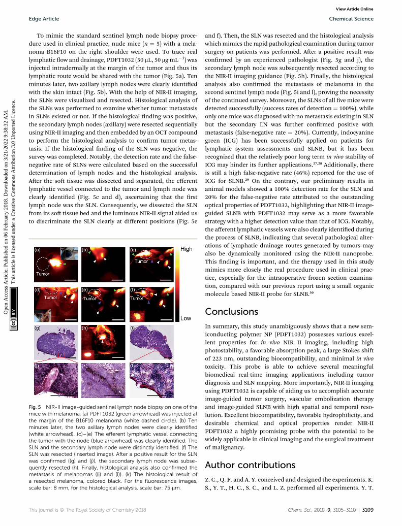
To mimic the standard sentinel lymph node biopsy proce-dure used in clinical practice, nude mice (n ¼ 5) with a mela-noma B16F10 on the right shoulder were used. To trace reallymphatic ow and drainage, PDFT1032 (50 mL, 50 mgmL�1) wasinjected intradermally at the margin of the tumor and thus itslymphatic route would be shared with the tumor (Fig. 5a). Tenminutes later, two axillary lymph nodes were clearly identiedwith the skin intact (Fig. 5b). With the help of NIR-II imaging,the SLNs were visualized and resected. Histological analysis ofthe SLNs was performed to examine whether tumor metastasisin SLNs existed or not. If the histological nding was positive,the secondary lymph nodes (axillary) were resected sequentiallyusing NIR-II imaging and then embedded by an OCT compoundto perform the histological analysis to conrm tumor metas-tasis. If the histological nding of the SLN was negative, thesurvey was completed. Notably, the detection rate and the false-negative rate of SLNs were calculated based on the successfuldetermination of lymph nodes and the histological analysis.Aer the so tissue was dissected and separated, the efferentlymphatic vessel connected to the tumor and lymph node wasclearly identied (Fig. 5c and d), ascertaining that the rstlymph node was the SLN. Consequently, we dissected the SLNfrom its so tissue bed and the luminous NIR-II signal aided usto discriminate the SLN clearly at different positions (Fig. 5e
Fig. 5 NIR-II image-guided sentinel lymph node biopsy on one of themice with melanoma. (a) PDFT1032 (green arrowhead) was injected atthe margin of the B16F10 melanoma (white dashed circle). (b) Tenminutes later, the two axillary lymph nodes were clearly identified(white arrowhead). (c)–(e) The efferent lymphatic vessel connectingthe tumor with the node (blue arrowhead) was clearly identified. TheSLN and the secondary lymph node were distinctly identified. (f) TheSLN was resected (inserted image). After a positive result for the SLNwas confirmed ((g) and (j)), the secondary lymph node was subse-quently resected (h). Finally, histological analysis also confirmed themetastasis of melanomas ((i) and (l)). (k) The histological result ofa resected melanoma, colored black. For the fluorescence images,scale bar: 8 mm, for the histological analysis, scale bar: 75 mm.
This journal is © The Royal Society of Chemistry 2018
and f). Then, the SLN was resected and the histological analysiswhichmimics the rapid pathological examination during tumorsurgery on patients was performed. Aer a positive result wasconrmed by an experienced pathologist (Fig. 5g and j), thesecondary lymph node was subsequently resected according tothe NIR-II imaging guidance (Fig. 5h). Finally, the histologicalanalysis also conrmed the metastasis of melanoma in thesecond sentinel lymph node (Fig. 5i and l), proving the necessityof the continued survey. Moreover, the SLNs of all ve mice weredetected successfully (success rates of detection ¼ 100%), whileonly one mice was diagnosed with nometastasis existing in SLNbut the secondary LN was further conrmed positive withmetastasis (false-negative rate ¼ 20%). Currently, indocyaninegreen (ICG) has been successfully applied on patients forlymphatic system assessments and SLNB, but it has beenrecognized that the relatively poor long term in vivo stability ofICG may hinder its further applications.27,28 Additionally, thereis still a high false-negative rate (46%) reported for the use ofICG for SLNB.29 On the contrary, our preliminary results inanimal models showed a 100% detection rate for the SLN and20% for the false-negative rate attributed to the outstandingoptical properties of PDFT1032, highlighting that NIR-II image-guided SLNB with PDFT1032 may serve as a more favorablestrategy with a higher detection value than that of ICG. Notably,the afferent lymphatic vessels were also clearly identied duringthe process of SLNB, indicating that several pathological alter-ations of lymphatic drainage routes generated by tumors mayalso be dynamically monitored using the NIR-II nanoprobe.This nding is important, and the therapy used in this studymimics more closely the real procedure used in clinical prac-tice, especially for the intraoperative frozen section examina-tion, compared with our previous report using a small organicmolecule based NIR-II probe for SLNB.30
Conclusions
In summary, this study unambiguously shows that a new sem-iconducting polymer NP (PDFT1032) possesses various excel-lent properties for in vivo NIR II imaging, including highphotostability, a favorable absorption peak, a large Stokes shiof 223 nm, outstanding biocompatibility, and minimal in vivotoxicity. This probe is able to achieve several meaningfulbiomedical real-time imaging applications including tumordiagnosis and SLN mapping. More importantly, NIR-II imagingusing PDFT1032 is capable of aiding us to accomplish accurateimage-guided tumor surgery, vascular embolization therapyand image-guided SLNB with high spatial and temporal reso-lution. Excellent biocompatibility, favorable hydrophilicity, anddesirable chemical and optical properties render NIR-IIPDFT1032 a highly promising probe with the potential to bewidely applicable in clinical imaging and the surgical treatmentof malignancy.
Author contributions
Z. C., Q. F. and A. Y. conceived and designed the experiments. K.S., Y. T., H. C., S. C., and L. Z. performed all experiments. Y. T.
Chem. Sci., 2018, 9, 3105–3110 | 3109
Chemical Science Edge Article
Ope
n A
cces
s A
rtic
le. P
ublis
hed
on 0
6 Fe
brua
ry 2
018.
Dow
nloa
ded
on 3
/21/
2022
9:3
8:32
AM
. T
his
artic
le is
lice
nsed
und
er a
Cre
ativ
e C
omm
ons
Attr
ibut
ion
3.0
Unp
orte
d L
icen
ce.
View Article Online
contributed to the design and synthesis of the semi-conductingpolymer. K. S. and H. C. contributed to the preparation of theorganic nanoparticles, the optical characterization of thenanoparticles and the animal experimentation. K. S., Z. C., Y. T.,A. Z., and H. C. analyzed the data and wrote the manuscript. Allauthors discussed the results and commented on themanuscript.
Conflicts of interest
There are no conicts to declare.
Acknowledgements
This work was supported by the Office of Science (BER), U.S.Department of Energy (DE-SC0008397), the InternationalCooperative Program of the Chinese Academy of Sciences(Grant No. GJHZ1622), the National Basic Research Program ofChina (No. 2012CB933301), the National Natural ScienceFoundation of China (No. 21674048, 21574064, 81572163 and61378081), the Synergetic Innovation Center for Organic Elec-tronics and Information Displays, the Natural Science Foun-dation of Jiangsu Province of China (No. NY211003), and theHuanghe Talents Project of Wuhan City (2014). All animalexperimentation was performed under the approval of StanfordUniversity’s Administrative Panel on Laboratory Animal Care.Moreover, all experiments were performed in accordance withthe National Institutes of Health’s Guide for the Care and Use ofLaboratory Animals.
Notes and references
1 G. Hong, Y. Zou, A. L. Antaris, S. Diao, D. Wu, K. Cheng,X. Zhang, C. Chen, B. Liu, Y. He, J. Z. Wu, J. Yuan,B. Zhang, Z. Tao, C. Fukunaga and H. Dai, Nat. Commun.,2014, 5, 4206.
2 A. L. Antaris, H. Chen, K. Cheng, Y. Sun, G. Hong, C. Qu,S. Diao, Z. Deng, X. Hu and B. Zhang, Nat. Mater., 2016,15, 235–242.
3 K. Welsher, S. P. Sherlock and H. Dai, Proc. Natl. Acad. Sci. U.S. A., 2011, 108, 8943–8948.
4 A. M. Smith, M. C. Mancini and S. Nie, Nat. Nanotechnol.,2009, 4, 710–711.
5 D. J. Naczynski, M. C. Tan, M. Zevon, B. Wall, J. Kohl,A. Kulesa, S. Chen, C. M. Roth, R. E. Riman andP. V. Moghe, Nat. Commun., 2013, 4, 2199.
6 Z. Tao, G. Hong, C. Shinji, C. Chen, S. Diao, A. L. Antaris,B. Zhang, Y. Zou and H. Dai, Angew. Chem., Int. Ed., 2013,52, 13002–13006.
7 Q. Yang, Z. Ma, H. Wang, B. Zhou, S. Zhu, Y. Zhong, J. Wang,H. Wan, A. Antaris, R. Ma, X. Zhang, J. Yang, X. Zhang,H. Sun, W. Liu, Y. Liang and H. Dai, Adv. Mater., 2017, 29,1605497.
8 Q. Miao, C. Xie, X. Zhen, Y. Lyu, H. Duan, X. Liu, J. V. Jokerstand K. Pu, Nat. Biotechnol., 2017, 35, 1102–1110.
3110 | Chem. Sci., 2018, 9, 3105–3110
9 Y. Lyu, X. Zhen, Y. Miao and K. Pu, ACS Nano, 2017, 11, 358–367.
10 Y. Jiang, P. K. Upputuri, C. Xie, Y. Lyu, L. Zhang andQ. Xiong, Nano Lett., 2017, 17, 4964–4969.
11 Y. Jiang and K. Pu, Small, 2017, 13, 1700710.12 S. Zhu, Q. Yang, A. L. Antaris, J. Yue, Z. Ma, H. Wang,
W. Huang, H. Wan, J. Wang, S. Diao, B. Zhang, X. Li,Y. Zhong, K. Yu, G. Hong, J. Luo, Y. Liang and H. Dai,Proc. Natl. Acad. Sci. U. S. A., 2017, 114, 962–967.
13 G. Hong, S. Diao, J. Chang, A. L. Antaris, C. Chen, B. Zhang,S. Zhao, D. N. Atochin, P. L. Huang, K. I. Andreasson,C. J. Kuo and H. Dai, Nat. Photonics, 2014, 8, 723–730.
14 A. L. Antaris, H. Chen, S. Diao, Z. Ma, Z. Zhang, S. Zhu,J. Wang, A. X. Lozano, Q. Fan and L. Chew, Nat. Commun.,2017, 8, 15269.
15 X. D. Zhang, H. Wang, A. L. Antaris, L. Li, S. Diao, R. Ma,A. Nguyen, G. Hong, Z. Ma, J. Wang, S. Zhu,J. M. Castellano, T. Wyss-Coray, Y. Liang, J. Luo andH. Dai, Adv. Mater., 2016, 28, 6872–6879.
16 K. Pu, A. J. Shuhendler and J. Rao, Angew. Chem., Int. Ed.,2013, 52, 10325–10329.
17 Y. Hang, X. P. He, L. Yang and J. Hua, Biosens. Bioelectron.,2015, 65, 420–426.
18 H. Chen, J. Zhang, K. Chang, X. Men, X. Fang, L. Zhou, D. Li,D. Gao, S. Yin, X. Zhang, Z. Yuan and C. Wu, Biomaterials,2017, 144, 42–52.
19 K. Pu, J. Mei, J. V. Jokerst, G. Hong, A. L. Antaris,N. Chattopadhyay, A. J. Shuhendler, T. Kurosawa, Y. Zhou,S. S. Gambhir, Z. Bao and J. Rao, Adv. Mater., 2015, 27,5184–5190.
20 Y. Cao, J. Yi, X. Yang, L. Liu, C. Yu, Y. Huang, L. Sun, Y. Baoand Y. Li, Biomacromolecules, 2017, 18, 2306–2314.
21 M. Palner, K. Pu, S. Shao and J. Rao, Angew. Chem., Int. Ed.,2015, 54, 11477–11480.
22 A. Maeda, J. Bu, J. Chen, G. Zheng and R. S. DaCosta, Mol.Imaging, 2015, 14, 7290201400043.
23 Y. Lyu and K. Pu, Adv. Sci., 2017, 4, 1600481.24 L. Krauel, A. Albert, J. Mora, T. Sola, O. Cruz, C. Mortera and
J. M. Ribo, J. Pediatr. Surg., 2009, 44, 1848–1855.25 I. Kim, J. M. Ryu, J. M. Kim, H. J. Choi, S. K. Lee, J. H. Yu,
J. E. Lee, S. W. Kim and S. J. Nam, J. Breast Cancer, 2017,20, 270–278.
26 E. P. Mamounas, T. Kuehn, E. J. T. Rutgers and G. vonMinckwitz, Lancet, 2017, DOI: 10.1016/s0140-6736(17)31451-4.
27 S. Wu and H. J. Butt, Adv. Mater., 2016, 28, 1208–1226.28 J. C. Rasmussen, I. C. Tan, M. V. Marshall, C. E. Fife and
E. M. Sevick-Muraca, Curr. Opin. Biotechnol., 2009, 20, 74–82.29 I. Miyashiro, M. Hiratsuka, M. Sasako, T. Sano, J. Mizusawa,
K. Nakamura, A. Nashimoto, A. Tsuburaya andN. Fukushima, Gastric Cancer, 2014, 17, 316–323.
30 K. Shou, C. Qu, Y. Sun, H. Chen, S. Chen, L. Zhang, H. Xu,X. Hong, A. Yu and Z. Cheng, Adv. Funct. Mater., 2017, 23,1700995.
This journal is © The Royal Society of Chemistry 2018