Embed Size (px)
DESCRIPTION
UWOMJ University of Western Ontario Schulich School of Medicine & Dentistry
Citation preview

University of Western Ontario
MEDICAL JOURNAL
VOL. IX, No. 3
CONTENTS
Modern Cancer Re earch Myra MacKenzie, M.A ., '40
The Significance of Harvey's Contribution to Physiology and Medicine
March, 1939
Page
............ .... .. 85
E. M. Watson, M.D., (L on.) .... ........... .......... .. ............ ... .... 95
On the Diagno is of Pulmonary Tuberculosis W. E. Crysler, M.D. ... .. ........ ... .. .. ... .. ... ... ........ .... . ... ... .... .. ..... 98
The Patient With a Headache L. D . Wilcox, M.D., F.R.C.P. (C.) .... ... ... ..... ....................... .. . 103
Drug Allergy Gordon Calder, B.A., M.D. (Lon.) ............... .. .. ........ ... 107
The Modern Management of Pneumonia B. L. H ession, M.D . ....... .... .. ...... . . ... ·········· ············· ... 110
Anatomy and Physiology- A Short History Ray Stubbing, '42 ........ . . . . . ................. ... ... .. .. .. .......... ......... ..... 116
Abstracts .... .... ................ . .. ............ ··········· ........ ..... ....... ....... .... 120
Editorial ... ...... ...... .... ......... .......... .. ...... ......... .. ....................... ........ .. 124
Book Reviews ... .. . . ............ .... ...................... .......... ....... .......... .... 125
Recent Accessions to the Medical School Library .. .............................. 127
•

UNIVERSITY OF WESTE RN ONTARIO MEDICA L JO U RNAL
ROYAL VICTORIA HOSPITAL MONTREAL
F . R. C. S. (Can.) Special t utor ial clas es for the F inal F . R. C. S. (Can.)
will be given at the Royal Victoria Hospital, Montreal, from August 28th t o October 21st, 1939, consisting of daily lecture demonstration, ward rounds and surgical pathology .
The Course will be given by : DR. G. G. Mll..LER, F .R.C.S. (Can.) ... ...................... ...... Clin ical S m·ge1y DR. J. C. L UKE, F .R.C.S. ( E ng. ) .. .. .. ... . .. ... ... ... . ..... .... Abdominal S u1·gery DR. H. S. MORTON, F.R.C.S. ( Eng. a nd Can.) ....... ... T horacic S m·gery DR. H . F . MOSELEY, F .R. .S. ( Eftg. ) ... ... 0 1·tkopmdic S m·gery DR. A. ELVIDGE, F .R.C.S. (Can.) ... eurosurgery DR. A. B. HAWTHOR N E, F .A.C.S. . ........... ... .. . ........... Urology PROF. T. R. W AUG H , M.D. . ... . ....... S urgical P athology
The fee for the Course will be $100.00 Further particulars may be obtained from the Secretary
of the Course, H. S. MORTON, F.R.C.S.,
1374 Sherbrooke Street West , Montreal, P .Q.
Your Stationery Supplies Everything that the " doctor" needs in Office
Stationery. Your requirements executed m a d istinctive manner at a reasonable cost.
HUNTER PRINTING COMPANY Printers and Publishers
226 King Street LONDON Phone Met. 1724
Note Paper .. En'l'elopes BiUh eads Statements Ledger S h eets .. I ndex Cards Circulars
and Printing o f E'l'ery Description
When w·riting advertisers please mention University of Western Ontario Med.ical Journal.
The University of W estern Ontario
MEDICAL SCHOOL \"0Lu~1E IX. NU MBER 3
Modern Cancer Research By MYRA MACKENZIE, M.A., Meds '40
A PPROACHES to the cancer problem, as it is now visualized, are indeed diverse. The aid of all the sciences is being freely called
upon by the medical profession which, realizing t hat this is "the age of cancer progress," is resolved to "maintain at the highest level the momentum now gained in this field." The ramifications of the interest in cancer reach into many branches of human knowledge and form a broad basis which assures sound progress, providing the resources be not dissipated over so wide a territory as to lose practical value. Experimental investigation requires the labours of chemists, physicists, biologists, eugenists, physiologists and pathologists for, to make progress in this complex subject, specialization is imperative.
A vast body of knowledge, accumulated from investigations into the fundamental problems of cancer in the past 40 years, has cleared away much of the mystery associated with the disease. In the first scientific report of the Imperial Cancer Research Fund in 1904 was the statement that" ... fundamental common attributes relate the malignant new growths to one another in a natural group. The explanation of these common fundamental characters is bound up with the mode of origin of malignant new growths and is the problem awaiting solution, to be followed by the specific explanation of the individual variations, as its natural corollary." Although, in its broader aspects, the same problem still confronts us, it has been more accurately defined and brought to a practical stage by experimental pathology. The mode of origin of malignant new growths, if it be a single factor, has not yet been discovered but numerous modes of production of malignancy in vivo and in vitro are now available. A study of the relationship of these various way of producing cancer is one of the most urgent needs at present.
THE VffiUS HYPOTHESIS The results of certain bacteriological investigations tend to support
a virus hypothesis of the origin of cancer. Recently, interest in the virus origin has been greatly revived and valuable possibilities have been disclosed.
Following the discovery by Rous, in 1910, that a sarcoma occurring
85
86 UNIVERSITY OF WESTERN ONTARIO MEDICAL JOURNAL
spontaneously in a domestic fowl was transmissible to other fowls of the same species by a . cell-free filtrate, numerous investigators sought to attribute similar properties to mammalian tumors. With the exception of a lympho~?arcoma of dogs and a papillocarcinoma of rabbits, it has not been possible to inoculate mammalian tumors by any other method than cell transfer.
A long series of experiments, performed in England and America, de igned to determine the nature of the agents in t he Rous and allied tumors, have disclosed some interesting features. Incredibly small amounts of the filtrates were found to be capable of stimulating normal cells to assume malignant ch.anges and to form tumors. In behaviour and in appearance . under the ultra-violet ·ultra-microscope, marked similarity to known ' vil·uses was pointed out by Gye and later workers. Both the Rous and Shope papilloma agents have been demonstrated as particulate in nature and approaching the dimensions of 90 uu. in diameter.
More recently, . the known viruses have been subjected to closer scrutiny and the crystalline nature of some of them revealed. Coincident work on the Rous agent has also disclosed its chemical nature. However, in the light of modern conceptions the chemical, non-living, protein molecules grade imperceptibly into the living viruses which are of quite comparable molecular complexity, so that it is impossible to say, as yet, in what sphere the Rous agent belongs.
The various "agents" st imulate the production of pecific agglutinins in tumor-bearing animals, indicating that they are, in whole or in part, extrinsic to the animal. Certain instances of tumor agent-neutralizing antibodies in normal fowls have been recorded, a phenomenon which is analogous to the occasional spontaneous regressions of mammalian tumors.
One view of the virus hypothesis is that this extrinsic, antibodyproducing agent require the presence of a second specific factor in order to initiate tumor growth. In support of this proposal is the fact that the intravenous injection of the virus fail to produce growths, although it can be readily isolated from all the ti sues of. the body. If, however, a muscle or other tissue be traumatized after injection of the virus then a tumor develops at the site of the injury.
This peculiar property bears an interesting relationship to the chemicals which induce cancer. The Shope virus produces active proliferation of warts in wild rabbits. In domestic rabbits these warts often become fissured, ulcerate and develop a low-grade carcinoma. If benzpyrene, a chemical carcinogen, be rubbed on the skin of rabbits into which the virus has been injected intravenously the resulting warts are more numerous, grow more actively, and a much greater percentage become malignant.
These findings suggest the possibility of multiple factors, one of

MODERN CANCER RESEARCH 87
which may be a virus, being concerned in the origin of some cancers. Viruses may come to be considered as a partial factor in the etiology of certain epithelial tumors of man for the infectious behaviour of some papillary mucous membrane tumors has long been recognized clinically.
CHEMICAL CARCINOGENESIS
One of the great landmarks in cancer research was the production of papillomata in rabbits by prolonged tar application . Since the epochal report of Yamigawa and Ichikawa, in 1918, tumors have been experimentally induced by a great variety of agents. Highly potent carcinogenic chemicals have been isolated and still others have been synthesized. Leaders in the chemistry of cancer production have been Kenna way, Cook and their associates in England; Fieser, Shear and co-workers in America; and Wieland in Germany.
The active carcinogenic agent in tar was isolated in 1933 by Cook, Hewett and Rieger. It proved to be 3 :4-benzpyrene. In the course of the next few years the English school synthesized a large number of related aromatic hydrocarbons for a sy tematic study of the relation between chemical structure and carcinogenic properties. Many of these are derivatives of 1 :2-benzanthracene. All the theoretically possible derivatives have been synthesized. A new member, 9 :10-dimethyl-1 :2-benzanthracene, is the most active carcinogen known, producing tumors in mice within 50 days.
Substitution of simple (methyl) groups in the 10-position of the 1 :2-benzanthracene system increases the carcinogenic potency while the presence of elaborate (isopropyl) groups in this position practically destroys it. Methyl cholanthrene is one of the most significant substances discovered in the course of studying cancer-producing activity. It is a derivative of 1 :2-benzanthracene with substituents at positions 5 and 10 and produces cancer in mice in 2.5 months. Originally it was prepared from desoxycholic acid, a bile acid, by a four-step reaction. This fact suggested the possibility of a pathological deviation in bile or cholesterol metabolism leading to the production of a carcinogenic chemical but the presence of methyl cholanthrene has never been demonstrated in the animal body. Fieser and his collaborators have hown, however, that the 5-carbon ring characteristic of the bile acids is not essential for carcinogenicity. He has also postulated that even if methyl cholanthrene is formed in the degradation of bile acids it may be further acted upon to form an innocuous hydrocarbon. Such a conver ion has not yet been accomplished in the laboratory but even modern chemistry fails to perform many chemical transformations which the tissues accomplish with ease.
Recent work has shown that carcinogenic chemicals inhibit the breakdown of cholesterol esters and possibly cause esterification of cholesterol in animals. It has been sugge ted that the sterols, bile acids and sex hormones, all structurally related by the five-membered ring to

88 UNIVERSITY OF WESTERN ONTARIO MEDICAL JOURNAL
methyl cholanthrene, may be involved in the incidence of some forms of spontaneous cancer but at present there is no good reason for presuming the presence of active carcinogens in the body. While most of the active substance so far examined are benzanthracene derivatives, compounds of widely different structure are al o carcinogenic. Carcinogenic power, though not dependent on a specific chemical structure, may be determined by a particular kind of reactivity shared by compounds of dissimilar structure. Fieser's recent work demonstrates that all the active compounds undergo rapid change in the body and are exhausted long before tumors begin to appear, but they may initiate a complicated chain of events leading ultimately to malignant growth. He has found that "the most potent carcinogens have unique chemical propertie , surpassing all other known aromatic hydrocarbons in chemical reactivity of a special kind, notably a high degree of susceptibility to substitutions of a special type and to oxidation with lead tetra-acetate." The suggestion is made that these highly active chemicals conjugate with substances present in the organism, by hydroxylation or substitution, thereby forming a basis for later cancer production in the animal body.
While the carcinogenic potency of certain substances is established, the same chemical does not uniformly produce cancer in all type of animals, nor even in all strains of a susceptible species. Some strain of mice are highly susceptible to kin cancer, chemically induced, while others, with a high incidence of pontaneous lung tumors, respond to carcinogens on the skin by developing lung tumors in greater numbers and at earlier than normal age periods. Old rabbits are liable to uterine cancer and it has been found that in many of these to which carcinogens were administered by various routes, no tumors arose at the site of application but the percentage of uterine tumors rose abruptly. Conversely, in studying a fluorescent carcinogen, Chalmers and Peacock (1938) have reported that it is excreted in the bile, urine and skin fat but produces tumors only at the site of application and never along the routes of excretion. These observation point to an inheritance factor, either chromosomal or extra-chromosomal, but certainly with a localization of malignancy. They corroborate the theory of localization characters in pontaneous tumors of all animal type and again suggest the operation of two factors, an intrinsic hereditary susceptibility and an extrinsic chemical agent.
Some chemicals entirely foreign to the animal body can also induce malignant tumors. A variety of these have been reported but in numerous cases repetition failed to substantiate early claims. Attempts to induce bladder tumor growth in animals by administering aniline dyes have finally been rewarded after many years of experiment. Increasing doses of aniline, benzidine and betanaphtylamine were given to dogs both by mouth and subcutaneously for 1112 to 2 years. Periodic cystoscopic examinations and biopsies followed the histopathological process leading to the formation of benign papillomata and finally to true
MODERN CANCER RESEARCH 89
carcinoma. The time to induce these tumors is significantly long and is consistent with the observation that dye workers have been exposed for many years before developing bladder symptoms.
Another dye, amino azotoluene, produces liveo tumors in rats either when administered in the diet for 10 months or when subcutaneously implanted in solid form. The high solubility of the dye in fats and lipoids may be a factor in its selective action on the liver. A recent paper reported that "butter yellow," an azo dye closely related to amino azotoluene, also induces liver tumors in rats. If this be confirmed, the discontinuance of the use of this dye would be justified.
Excessive e"rposure to X-rays and radium has long been known to give rise to cancerous changes in the body. The use of radioactive substances in medicine has consequently been frowned upon but no evidence of their carcinogenic action was brought forth . Now, however, there is proof that "Thorotrast" (colloidal thorium hydroxide), a radio-opaque substance injected into the brain and venous system for diagnostic purposes, is definitely carcinogenic. In the rats, mice and guinea pigs tested there was a long delay before malignant change occurred, but it finally did appear in two and one-half to three years. Since thorotrast has not been long in use it is premature to conclude that it is harmless to man as 10 to 15 years would be required before cancerous change could be expected.
Lacassagne (1932) reported the first instance of the production of malignant tumors by a naturally occurring chemical of known structure. Mammary cancer normally occurs only in female mice but Lacassagne succeeded in inducing the development of mammary cancer in male mice by long-continued treatment with large do es of oestrin. Subsequent research has shown that the carcinogenic action of the oestrogens is proportional to the dosage and that they produce cancer in other sites, namely, the cervix uteri and subcutaneous tissue.
Cramer and Horning (1938) have reported the effects of prolonged oestrone administration on mice and rats. The anterior pituitary gland enlarged with almost complete disappearance of the granular acidophil cells, the adrenals degenerated and the mammary glands progressed to malignancy. When thyrotropic hormone was given simultaneously with oe trone these changes did not occur. This result led to a test of the effect of thyrotropic hormone on spontaneous mammary cancer in mice. F emale mice of a highly susceptible strain were treated with thyrotropic hormone from an early age and none of them developed tumors although they all lived well into the cancer age period. The possibility of specific prophylaxis is enticing but it must not be overestimated while so much r emains to be known about the pituitary gland.
An intere ting ob ervation was made by Cook in 1933 when several synthetic carcinogens were discovered to be oestrogenic. However, the most potent carcinogens have only weakly oestrogenic properties and enormous dosages of oestrogenic hormones are required to induce

90 UNIVERSITY OF WESTERN ONTARIO MEDICAL JOURNAL
malignant growth. Therapeutic quantities of these hormones are extremely unlikely to give rise to such changes. The underlying law governing the power of these related substances to influence epithelial proliferation and the oestrus cycle must be disclosed before their shared activity can be explained.
Still another naturally occurring body constituent, acetyl choline, was reported by Franks and Hall (1938) to have produced osteosarcoma in animals following repeated subcutaneous injection of large doses. This claim has not yet been confirmed but it conjures up very interesting possibilities because of the specialized function of acetyl choline.
Detailed exploration of the entire field of inter-cellular and intracellular body chemistry must eventually bring to light the factors influencing normal and malignant growth expression. Great advances have been made in the past five years; much remains to be learned.
THE INFLUENCE OF INHERITANCE A fact long disputed, but now generally accepted, is that certain
constitutional factors predispose some animals to form cancer and predispose others against its formation. These constitutional factors, which are transmitted from one generation to another, have been extensively studied in laboratory animals. Close inbreeding has produced many pure strains, especially in mice and rats, and has permitted controlled investigation of numerous inherited characteristics. By thus reducing the genetic variability it has become possible to determine the effects of other influences such as age, sex, and diet.
In certain cases the Mendelian laws of dominance and recessiveness have been demonstrated but these phenomena are by no means universally applicable to neoplasms. Slye (1937) maintains that malignancy is transmitted as a localized recessive factor, each type of malignancy (carcinoma, sarcoma, leukemia) being a unit character and capable of suppression by a dominant. Breast cancer, for instance, requires two recessive characters for expression, one for malignancy and one for location according to her calculations. Little (1933), Murray (1935) and Bittner (1937) have, on the other hand, presented convincing evidence that breast cancer in mice is not dependent on genetic influences. While most non-mammary tumors seem to be distributed in essentially the same way as are the chromosomes, mammary tumors do not follow this pattern.
In strains susceptible to mammary adenocarcinoma the potentiality for neoplastic growth varies from 50 to 100 per cent. The incidence in virgin females, in the majority of these strains, is considerably lower than in breeding females, indicating that lactation may have an irritative effect. Forced breeding, with no opportunity to nurse the young, also appears to raise the t umor incidence by chronic irritation (Bagg, 1937). The practice is, therefore, to consider the incidence in breeding females as characteristic of the strain.

MODERN CANCER RESEARCH 91
When females from an established high-tumor stock were crossed with males from a low-tumor stock a higher than expected percentage of the female progeny developed mammary tumors (Little, 1933). The reciprocal cross of low-tumor females with high-tumor males yielded a significantly low percentage of mammary cancers. These results indicated a strong maternal influence located outside of the chromo omes. At first the cytoplasm of the egg cell was considered to be the source of this extra-chromosomal element. Bittner (1937), however, revealed the source of at least one agent not located in the egg cell. He used low-t umor stock, lactating females to nurse young mice separated from their high-tumor stock mothers. Of these fostered mice only 7.4 per cent of the females developed tumors in contradistinction to 83.6 per cent of their sisters and parent stock. Reversing the arrangement, by having high-tumor stock females nur e the young of low-tumor tock mothers, he found the cancer incidence was raised almost to the level of the foster mothers. (By eliminating any chances of the young nursing their own mothers for the first few hours of life he expects to reduce the 7 per cent incidence of tumors to zero.) The average tumor age remained the same in all cases and, as the mice which did not develop tumors lived three to four months longer than the others there was greater opportunity for expression of the cancer tendency.
Bittner (1939) has extended his investigations to include the effects of foster nursing on the hybrid stock in which the extra-chromosomal influences were first noted. Further confirmation of the findings in the genetically homogeneous stocks was obtained. Mice from the high-tumor stock females-low-tumor stock males, when nursed by low-tumor stock females, showed a drop in tumor incidence as compared with sister mice raised by their own high-tumor stock mothers. Potentially high-tumor mice, therefore, transmit a "breast-cancer producing influence" in their milk. The nature of this influence, whether chemical, virus or hormonal, has yet to be determined.
MacDowell (1936) found that the incidence of leukemia in mice was significantly higher when the mother carried the leukemic heredity. The source of the leukemia-producing influence has not yet been reported. Lynch (1937) investigated the possibility of extra-chromosomal influence on inheritance of lung cancers. She found that both spontaneous and induced lung tumors were subject to genetic control but suggested that this control might be upset by external agents of great potency.
Very little is known of the comparative importance of extrinsic and intrinsic factors in most tumors. Heredity and environment do not appear to have the same relative effects on all types of tumors. A strong hereditary influence is recognized in human beings in whom retinal glioma or intestinal polyposis occur, while some other neoplasia, as lip cancer, appear to depend more on environmental factors. In special industries and in association with certain diseases causal connections
92 UNIVERSITY OF WESTERN ONTARIO MEDICAL JOURNAL
have been traced between the inciting factor and the neoplastic reaction in human beings. Where certain individuals display a resistance to a powerful inciting agent we must conclude that inherited characteristics are operating in their defence.
Certain principles, which are characteristic of all mammal , have been established by the study of cancer genetics in pure strains:
1. Constitutional differences in susceptibility are inherited. 2. Susceptibility is specific for organs. 3. External agents can induce neopla tic changes. 4. In some sites the response to the agent is controlled by the
genetic constitution of the individual. Genetic factor exert a con iderable influence on the transplant
ability of tumors. Some tumors require only one genetic factor to permit their propagation while others require as many as five to seven. Tumors requiring few factors can be readily transplanted in nearly all strains of the species in which they arose; those tumors which require multiple genetic factors can only grow in the strains which carry all or most of the necessary factors. In some cases it has been possible to adapt the tumors to grow in strain which would not at first harbour them.
While transplantability within the original species can be carried on indefinitely, almo tall attempts to transfer tumors from mice to rats, cats to dogs and man to lower forms, have failed. Under rigid control some mammalian tumors have been grown in developing chick embryos and a mouse sarcoma ha been carried for several generations by transplanting it into new-born rats. Apart from these two instances heteroplastic transfer of mammalian tissue has been considered impossible and useless for experimental purpo e . The succes of Greene and Saxton (1938) in propagating rabbit adenocarcinoma in guinea pigs and human breast cancer in rabbits has changed this attitude. By their method human cancer can be indefinitely cultivated in the anterior chamber of the eye of the rabbit. This new technique may be applied to a wide range of phy iological and pathological problems and opens a whole new field of inve tigation into human cancer.
THE BASIS OF MALIGNANT CHANGE An apparently unrestrained capacity for proliferation is the out-
tanding biological characteristic of malignant tumors. A normal cell is transformed into a malignant cell by some action f rom without but once the transformation has occurred the external timulus is no longer neces ary for the maintenance of the malignant character. Cancer cells are permanently altered, as evidenced by their abnormal intensity of growth and their ability to retain indefinitely t hose characteristics which first distinguished them from surrounding normal cells.
Malignant change can be induced by a wide variety of extrinsic agents, uch as hormones, pure chemicals, X-rays and parasites. These diverse agents seem to initiate a proce s of mutation within the normal cell. Actually, visible distortion of the chromosomes from heavy
MODERN CANCER RESEARCH 93
X-radiation ha been demonstrated in plants, with a resultant change in the character of the affected cell . The other carcinogenic substances, however, appear to work more indirectly on the chemistry of the cells, probably producing an ultimate chromosomal upset. Their effects are conditioned by intrinsic factors of the organism which may be termed "susceptibility." On this premise it is easy to explain why different pure strain animals react differently to the same stimuli. The threshold of mutation may be lower in " usceptible" strains, or lower in certain organs of such strains, so that a change in external or internal irritants may induce mutations in these organs.
In support of the mutation theory of cancer origin is the fact that intermediary phases between normalcy and malignancy have been difficult to demonstrate in cells. Certain precancerous changes can be pointed out by the pathologist but the stage whereat the cell actually assumes malignant characteristics is still unknown. Various attempts have been made to induce normal cells in tis ue culture to take on malignant properties by expo ure to carcinogenic agent . When the transformation occurred, as it did very infrequently, it wa so sudden that the intermediate stages were obscured. Lower animals have been experimented upon in further attempt to clarify the problem but, while some growth stimulation of bacteria (Goldstein, 1937), Planaria (Owen, 1938) and Obelia (Hammett and Reimann, 1935) was evident after exposure to carcinogens, no new information was revealed.
In tissue cultures those chemical which are very potent in producing tumor re ponses in vivo appear to retard the growth of normal fibroblasts and even exhibit toxic properties. Con equently Reimann (1938) has stated that "stimulation of cell proliferation alone does not lead to neoplasia." A new approach to this phenomenon was found by Murphy (1937) when he discovered the presence of inhibiting sub-tances in extracts of placenta and embryo skin. The intraperitoneal
injection of these extract into animal with spontaneou mammary cancer caused the arrest of growth in 70 per cent and actual regression in 22 per cent of the tumors. More recently, there has also been isolated a potent inhibiting fraction from the prelactating mammary tissue of the cow and rabbit. Some chemical have been tested for inhibition of tumor growth and significant results have been obtained with heptyl aldehyde (the active principle of Wintergreen), with colchicine and with essential amino acids. These substances may have imilar effects to the inhibitor of unknown chemical nature, discovered by Murphy, in retarding mitosis or other cell functions.
Murphy (1939) postulates that normal tissue growth is the result of a balance between stimulating and inhibiting factors but that the removal of one or more inhibitors permits neoplastic proliferation. These stimulators and inhibitors are possibly chemical in nature and their role in the conversion of cells from normal to malignant behaviour is probably based on profound metabolic changes.
• • •

94 UNIVERSITY OF WESTERN ONTARIO MEDICAL JOURNAL
The aim of cancer research is, of course, the discovery of the cause of cancer. The successful treatment of a disease may be planned without knowledge of its cause but it is essential that the cause be known before preventive measures can be devised. So many approaches to the problem have been disclosed by research work in the past few years that a survey of the entire field leads only to confusion. In his 1938 report on the British Empire Cancer Campaign, Sir Alfred Webb-Johnson adequately describes the present situation: "The truth must be demonstrated by some rare genius with a faculty for seeing the hidden relations between the various phenomena." Since normal and malignant cells have many characteristics in common, it is conceivable that when the laws of typical growth are discovered the atypical or cancerous growth will also yield up its secret.
BIBLIOGRAPHY Andervont, H. B.: Pub. Health Rep. 52: 212-221, 1937. Andervont, H . B.: Pub. Health Rep. 52: 1584-1589, 1937. Andrewes, C. H.: J. Path. and Bact. 47: 87-99, 1938. Bagg, H. J.: Am. J. Cancer 30 : 539-548, 1937. Bittner, J. J.: Am. J. Cancer 30: 530-538, 1937. Bittner, J. J.: Am. J. Cancer 35: 90-96, 1939. British Empire Cancer Campaign Report: Brit. M. J. 2: 1216-1217, 1938. Chalmers, J. G., and Peacock, P . R.: Biochem. J. 32: 271-278, 1938. Cook, J. W., Hewett, C. L., and Hieger, 1.: Nature 130: 926, 1932. Cook, J. W., Hewett, C. L., and Dodds, E. C. : Nature 131: 56-57, 1933. Cook, J. W., and Kennaway, E. L.: Am. J. Cancer 33 : 50-97, 1938. Cramer, W.: Brit. M. J. 1: 829- 34, 1938. Cramer, W., and Horning, E. S. : Lancet 1: 72-76, 1938. Dodds, E . C.: Lancet 2: 351-355, 1938. Earle, W. R., and Voegtlin, C.: Am. J. Cancer 34: 373-390, 1938. Fieser, L. F.: Am. J. Cancer 29: 260-268, 1937. Fieser, L. F.: Am. J. Cancer 34 : 37-124, 1938. Goldstein, S.: Science 86; 176-177, 1937. Greene, S. N.: Science 88: 357-358, 1938. Greene, S. N., and Saxton, J. A.: J. Exper. Med. 67: 691-708, 1938. Gye, W. E.: Lancet 1: 989-995, 1926. Gye, W. E . : Brit. M. J. 1: 551-554, 1938. Halter, C. R.: Am. J. Cancer 33 : 218-222, 1938. Hammett, F. S., and Reimann, S. P. : Am. J. Cancer 25: 807-808, 1935. Hora, J.: Ztschr. f. Krebsforsch. 47: 303-324, 1938. Imperial Cancer Research Fund Reports 1, 1904; 36, 1938. Jobling, J. W., and Sproul, E. E. : Science 85: 270-271, 1937. Lacassagne, A.: Com pt. rend. oc. de bioi. 114: 427-429, 1933. Ledingham, J. C. G.: Brit. J. Exper. Path. 18: 436-449, 1937. Lewis, W. H. : Cancer Symposium pp. 119-120, 1937. Little, C. C.: Science 78: 465-466, 1933. Ludford , R. J. : Am. J. Cancer 35: 63-71 , 1939. Lynch, C. J.: Am . J. Cancer 31 : 77-84, 1937. MacDowell, E. C.: Am. J. Cancer 26: 85-101, 1936. Mellanby, E.: J . Path. and Bact. 46: 495-515, 1938. Murphy, J. B.: Cancer Symposium pp. 104-107, 1937. Murphy, J. B.: Diplomate 11: 25-31, 1939. Murray, W. S .. and Little, C. C.: Science 82: 228-230, 1935. Owen. S. E., Weiss, H. A .. and Prince. L. H.: Science 87: 261-262, 1938. Orr, J . W.: J. Path. and Bact. 46: 495-515, 1938. Reimann; S. P.: Cancer Symposium pp. 114-134, 1938. Rous, P .. and Kidd, J. G. : J. Exper. Med. 67: 399-428, 1938. Shear. M. J.: Am. J. Cancer 33 : 499-537, 1938. Slye. M.: Cancer Symposium op. 3-16, 1937. Voee-tlin, C.: Science 88: 41-48. 1938. Wells, H. G., Syle, M., and Holmes, H. F . : Am. J. Cancer 33: 223-228, 1938.
The Significance of Harvey's Contribution to Physiology and Medicine·=·
By E. M. WATSON, M.D. London, On tario
H IPPOCRATES taught that the functioning of the organism was the proper subject of the phy ician' study. Many centuries after
ward, William Harvey, by his tudies of the circulation of the blood, was the first to show that vital phenomena are related to the operations of definite organs. Harvey followed the trail opened up by Vesalius and other early anatomists until it led to a great physiological truth. His course was the course of ever y true scientist since his day, that is, reasoning ba ed upon experiment and observation.
Sir Michael Foster chose to regard Harvey not as the " Discoverer of the Circulation" but rather a "he who was the first to demonstrate the circulation of the blood." Harvey's methods of investigation were those which may be called strictly physiological methods and Fulton has remarked that "the introduction of the experimental method into physiological science, for which Harvey was responsible, wa no le s significant than the discovery of the circulation."
Judged by modern standards of medical r esearch, his technique might not rate very highly, but viewed in the light of the learning of his day it was remarkable. We must not forget that Harvey had no instruments of precision to aid him in hi experiments. The science of physics was in its infancy and there were not any perfect physical conceptions. There was no chemistry except that of the ancients and there were no certain biological principles to which he could appeal. There was, however, plenty of philosophizing about Nature. There were super titions and a tenacious adherence to the doctrines of Galen . It was under such circumstances and in the face of much criticism that Harvey had the courage to record precisely, logically, modestly, what he saw and what he thought concerning the movements of the heart and the blood. His work has not been rendered obsolete by ubsequent knowledge. William Harvey, therefore, was the effective founder of modern physiology without which there could be no Science of Medicine.
In our day, it i inconceivable that an investigator, having made original ob ervations of the importance of those of Harvey, should wait twelve years before publishing hi results. Perhaps some modern researchers might be guided by Harvey's reticence in this regard. It is true there was no incentive for Harvey to rush into print. Competition in hi particular field of research was not active and the opportunities
*Read at the Twentieth Annual Banquet of the Harvey Club, London, Ontario, F ebruary 28th, 1939.
95
96 UNIVERSITY OF WESTERN ONTARIO MEDICAL JOURNAL
for the speedy publication of experimental observations were not what they are today. ("Publicationitis" assumed epidemic proportions only in recent years.) Furthermore, his ideas were not generally accepted. In common with certain reports of research activities subsequently, "his views pleased some, displeased others." As a matter of fact, his announcement regarding the circulation was greeted in some quarters with scepticism and derision and he was subjected to scorn for daring to question the philosophical authority of Galen and his disciples. Although Harvey suffered materially, his demonstration of the circulation of the blood afforded the death-blow to the Galenic physiology, including the "spirits," and to the old humoral pathology, an achievement sufficient in itself to place him among the Immortals.
Following the publication in 1628, in a foreign country, of Harvey's views on the circulation of the blood, the growth of physiology was slow and it did not have immediately any great effect on medical practice. Fre h impetus to physiological re earch was supplied by certain other scientific developments, namely, the birth of physics during the early part of the 17th century followed somewhat later by the evolution of rational chemistry from a mystic alchemy. In addition, there came to the aid of those who were inquiring into the problems of life the microscope, which opened up an entirely new world of Nature. One hundred years ago (1839) Theodor Schwann enunciated the "cellular theory" as applied to animal structures and at the same time Liebig introduced chemistry to physiology. Such advances marked an epoch so aptly described by Daly, "when the men intere ted in the pure sciences wholeheartedly joined hands with those in Medicine, thereby inaugurating a liai on, the importance of which can only be e timated in the light of our present knowledge."
While Harvey grasped the truth of the circulation, he was never able to follow it throughout, for he could not ee the capillary vessels. It remained for another to find the missing link. Three years after Harvey's death, Malpighi demonstrated the existence of the connecting channels between the terminal branches of the arteries and the smallest tributaries of the veins.
Sir Michael Foster wrote that "all the deeper problems of physiology turn on the mutual action of the tissues and the blood, as the stream of the latter sweeps among the elements of the former. In science no man's results are wholly his own; like other living things they come from something which lived before. Harvey showed that the blood did sweep through t he tissues; Malpighi showed how the blood swept through them." Schwann showed what the normal tissues were and invented the word metabolism. Virchow applied the cell theory to diseased tissues. "Thus the way was opened for those inquiries into the ways in which the blood acts on the tissues and the tissues act on the blood, inquiries the results of which are the pride of modern times and the hope of times to come."
THE SIGNIFICA CE OF HARVEY'S CONTRIBUTION TO 97 PHYSIOLOGY AND MEDICINE
Harvey was the first of a notable group of medical men in whom the urge to discover new truths took them away from the mere routine of practice into the realm of r e earch and who e careers have brought "physiology and medicine into that close and special relation indicated by the common etymology of the words 'physician' and 'physiology'." A compilation of the names of tho e who have been both "clinician" and "scientist" would include: Marcello Malpighi, Hermann Boerhaave, Richard Lower, Jean Conrad P eyer, von Brunner, James Black, William Cruikshank, William Beaumont, Thoma Willis, Francis Glisson, Alexander Stewart, Charles Bell, Robert Whytt. Marshall Hall, BrownSequard, David Ferrier and, in our own time, Leonard Rowntree, Sir Frederick Banting, Harvey Cushing and Sir Thomas Lewis. The last-named, like Harvey a student of cardiovascular phenomena, probably more than any other individual by his work and teaching has bridged the gap between the laboratory and the clinic.
The true ignificance of Harvey's contribution to physiology and medicine has been stated by inger in the following words: "The knowledge of the circulation of the blood has been the basis of the whole of modern physiology and with it the whole of modern rational medicine. The blood is a carrier, always going round and round on the arne beat. What it carries and why, how and where it takes up its load , and how, where and why it parts with them, these are the questions the answering of which has been the main task of physiology in the centuries that have followed. As each of the questions ha obtained a more and more rational answer, so clinical medicine has always made a step forward and has come to approach more nearly to a true science. Thus it is that the work of Harvey lies at the back of almost every important medical advance."
BIBLIOGRAPH Y Clendening, L.: The Romance of Medicine, New York, Garden City Pub. Co., Inc., 1933. Daly, I. de Burgh : Per pectives in Physiology, Edin. Med. J. 41 , 341 ; 1934. Dempster, J. H . : Pathfinders of Physiology, Detroit, Detroit Med. Jour. Co., 1914. Foster, Sir M.: The History of Physiology, Cambridge University Pres , 1901. Fulton, J . F .: Selected Readings in the History of Physiology, pringfield, Ill .,
Charles C. Thomas, 1930. Malloch, A.: William Harvey, New York, Paul B. Roeber, Inc., 1929. P ower, D'Arcy: William Harvey, London, T. Fisher Unwin , 1897. Singer, C.: A Short History of Medicine, Oxford, Clarendon Press, 192 . Stirling, W.: Some Apostles of Physiology, London, Waterlow & Son , Ltd ., 1902.
In the nineteenth century, Kalesnikoff, a Russian shoemaker, disguised himself as a doctor and rapidly rose to the office of chief surgeon at the Kieff Ho pital. He performed 600 major operations before his imposture wa uncovered.
* * * In remote sections of India patient contemplating an operation
demand the burning of incense in the operating room -otherwise devils might enter the body through the opening.-FACT DIGEST.

On the Diagnosis of Pulmonary Tuberculosis
By W. E . CRYSLER, M.D. Instructor in Anatomy
University of Western Ontario Medical School, London, Ontario
W HEN one attempts t o add something to the overburdened literature concerning this subject, it is done with apology. However,
it is felt that there are certain changes which have occurred in our knowledge of the nature of pulmonary tuberculosis during the past two or three decades which are of sufficient importance to command the attention of the general practitioner, the man upon whose shoulders rest s the task of decreasing one of the chief causes of death among our youth.
Since the advent of the science of Roentgenology and the employment of routine chest plates under certain circumstances, the earliest lesion which has been observed is the so-called Assmann infiltrate. Radiographs indicative of t he lesion are shown in figures 1 and 2.
F igure 1 F igure 2
These two radiographs indicate the characteristics of a somewhat advanced Assmann infi ltrate. Note that in either case the lesion is located chiefl y in the axillary half of the fi rst intercostal space and that t he spr ead of the disease has occurred from t hat point.
The radiographic appearance of this lesion is that of a soft, fluffy opacity approximately one t o t wo em. in diameter. It may be located anywhere in the pulmonary parenchyma, t he site usually being in the axillary half of the clavicular region. In some cases, the characters of
98
ON THE DIAGNOSIS OF PULMONARY TUBERCULOSIS 99
this mall area have been observed to change rapidly. Exceptionally, it may completely resolve. Conversely, the infection may advance rapidly to involve the upper lung in an acute tuberculous process, frequently accompanied by cavitation. Few pathological findings have been described. Those which have been reported correspond somewhat to those of the primary infection if the lesion is exudative in type. Certain characteristics of the secondary invasion are retained.
The hi tory elicited from patients suffering from this process is extremely variable. There may be scarcely any alteration from the usual state of health. The majority tell of fatigue which is most noticeable at the end of the day; that is, they are more fatigued than usual at the end of a day's work. Upon awaking, they feel miserable and at this time may suffer from coughing spasms. Blood-streaked sputum or frank haemoptysis may now appear but its absence is not indicative. Pleurisy may have been present previously or assert itself now for the first time. As the condition advances, low-grade mid-afternoon P'!Jrexia, accompanied by the characteristic flushed face supervenes. All early symptoms become progressively aggravated. A past history frequently reveals an "influenza} attack" shortly before the onset of the present illness. Excessive perspiration is an additional manifestation of weakness. ·
This symptom-disease complex is surely of a different nature than we formerly understood. As the disease advances it resembles the chronic type of pulmonary tuberculosis. With such case , the presence of the low-grade chronic infection i known to cause a mild degree of secondary anaemia. This, coupled with the weakness induced by reduced exercise, brings about another train of symptoms characteristic of the chronic apical tubercula is which our texts describe. Earlier diagnosis has allowed us to recognize certain symptoms which have previously passed unnoticed. However, this fact does not appear to explain the entire problem. It is our opinion that certain cases of pulmonary tuberculosis, such as we see today, repre ent a different type of disease than was recognized several years ago.
In the first place, it is a conceded fact that in this country, particularly in Ontario, the incidence of reactors to the intracutaneous injection of old tuberculin among youths and young adults has materially dropped. This reassuring fact is conceivably explained by the vigorous public health measures which have been so prominent during the past two or three decades. As a result, the cases of so-called primary or childhood infection are becoming steadily decreased. In other words, we must be encountering a group of young adults who are suffering from their primary or initial infection. We believe that it is this group who present the symptom-disease complex described above. This is confirmed by Rich and McMurdoch's enlightening work on allergy and immunity with respect to tuberculosi . They have shown that the primary infection, when it affects the adult, is in many respects similar

100 UNIVERSITY OF WE TERN ONTARIO MEDICAL JOURNAL
in nature to secondary infection. The infecting dose, according to t heir observations, i the single factor which determines whether the di ease produced will have "primary" or " econdary infection" characteristics. On the other hand, other maintain that the uniformly high upper lobe localization and the earline of cavity formation are clearly reinfection characteristics.
One wonders, since primary infection in childhood is usually a benign process, why it as umes a more serious nature when it affects the adult. It is an accepted fact that the chief known line of defense that the body can mobilize against tuberculosis lies in the lymphatic system of vessels and glands. We know that in the child the lymphat ic system is abundant, the pulmonary network being no exception. As age advances, the lymphoid ti ue throughout the body regresses, ultimately becoming atrophic. Further, when one considers the amount of pulmonary lymphoid tis ue damaged from the effects of anthracosis as age progresses, it is not difficult to understand that the chief line of defense has been rendered less efficient. Thus the invading tubercle bacilli, so to speak, are left without foe and destruction supervenes. On the other hand, it may be po sible that the child's open lymphatic system favors spread of the disea e, whereas the adult's relatively closed pulmonary lymphatic system tends to localize it.
DIAGNOSIS There remains little to be said regarding this problem. The chief
difficultie which hinder early recognition may be summed up as: Failure to recognize the condition in the early stages because of ilence of the disease and, therefore, when treatment is of most value; lack of co-operation of the patient ; the high cost of diagnostic procedures.
Recognition of the condition i admittedly difficult during the early days of the disease. As a result, diagnosis has been largely turned over to chest specialists who operate free clinics in regional zone . Since the physician refers these ca es, it is obvious that he must po sess a rea onable degree of accuracy in the evaluation of symptoms which point to the chest.
A history of contact with a case of pulmonary tuberculosis is important circumstantial evidence. Persons with such a history must be subjected to a periodic chest examination until a definite conclusion is reached. The number of cases found among children under fifteen, examined because of contact or symptom , has been found to be almost negligible. In case-finding programs, greater emphasis mu t be placed upon securing the examinat ion of adult contacts. Occasionally one chest plate alone is valueless. Although it is our most precise instrument for the recognition of the condition, it is only an aid to diagnosis and must always be regarded as such. No radiologist is prepared to give a positive diagnosis from a doubtful shadow with no further information. Moreover, there are certain indefinite shadows which require serial
ON THE DIAGNOSIS OF PULMO ARY TUBERCULOSIS 101
studies before a definite opinion can be given. We often expect a roentgenogram to make the diagnosis for us after we have failed to do so on clinical grounds.
A history suggestive of the condition in the absence of contact likewise must be thoroughly investigated. One important point to bear in mind is that the use of the stethoscope with regard to early disease of this type must be, of necessity, discarded. True, it is of value, but negative physical findings in a suspicious case should never be allowed to deceive one. The discovery of respiratory lag is the best single sign of a pulmonary tuberculous proces , either recent or old.
H aemoptysis and pleurisy are to be regarded as manifestations of pulmonary tuberculosis until definitely proven otherwise. A glaring example is the case of a young wife with a history of haemoptysis on two occasions, each occurring after wimming. Other symptoms were not prominent. A physician informed her that a small vessel in her throat had ruptured and so the case was dismissed. On admission to Queen Alexandra Sanatorium, radiological examination revealed the presence of a large cavity near a lung hilus. Pneumothorax was attempted on several occasions without success, due to the presence of adhesions between the parietal and visceral pleurae.
The red blood cell sedimentation index, and red blood cell sedimentation time, although by no means specific for pulmonary tuberculous, is a far more accurate index of the activity of the disease than either the temperature chart or the white blood cell count. Early cases will usually show a red blood cell sedimentation index between 10-25 mm. in the first hour as determined by Cutler's vein method. This procedure should be performed on all suspected cases even prior to the far more costly X-ray examination.
Lack of co-operation is a factor which, in most cases, is due to a horror of the disease. Here the physician must exert his whole-hearted persuasiveness in order to dispel such fears. Sanatorium life is pleasant; the majority of inmates are contented.
The high cost of diagnostic procedures is a factor which has been met to a large extent. The availability of free clinics throughout the country has done much. However, under our present system, it is the middle-class individuals who must absorb a great proportion of the expenses incurred, in addition to paying for their own. Surely this one fact is sufficient evidence to direct attention to the serious need for some type of social insurance system.
Finally, routine examination of the chests of all young adults in every walk of life is perhaps an ideal which will take years to reach. However, there are various institutions, particularly nursing schools, which have shown us the value of this procedure. The method consists of tuberculin-testing all entrants and the subjection of positive reactors to X-ray examination. One has only to ee cases identified in this manner, which later yield almost universally favorable results, to be

102 UNIVERSITY OF WESTERN O NTARIO MEDICAL JOURNAL
convinced that it is the only method of eliminating this terrible scourge, according to our present knowledge.
SUMMARY
1. A new form of pulmonary tuberculosis has been described. It is not inferred that it is a new disease. Modern diagnostic procedures have allowed us to recognize the early manifestations of the onset of the condition.
2. It is difficult, however , to state whether we are seeing tuberculosis earlier than before, or whether this disease is the result of a so-called primary infection in the adult.
3. The routine method of diagnosis is described. * * * *
The author is indebted to P. M. Andrus, M.D., F.R.C.P. (C). for reading and criticising this paper.
BIBLIOGRAPHY Andrus, P. M. : Amer. Rev. Tuberc., 38: 2, 1938. Boyd, Wm.: " Pathology of Internal Diseases," Lea & Febiger, 1936. Fishberg, M.: Amer. Rev. Tuberc., 17: 1, 1928. Fischel, K. : Amer. Rev. Tuberc., 23: 139, 1931. Kolmer and Boerner: "Approved Laboratory Technique," D. Appleton & Co., 1931. Krause, A. K.: A mer. Rev. Tuberc., 1: 65, 1917. Musser: " Internal Medicine-Its Theory and Practice," Lea and Febiger, 1934. Myers, J . A. : Amer. Rev. Tuber c., 27: 121, 1932. Opie, E. L.: Amer. Rev. Tuberc., 32: 617, 1929. Pinner, M., et a!.: Amer. Rev. Tuberc., 19: 153, 1929. Pinner, M., et a!.: A mer. Rev. Tuberc., 31: 162, 1935. Plunkett, R. E.: A mer. Rev. Tuberc., 39: 256, 1939. Rich, M. : Bull. Johns Hopk. Hosp., 44: 273, 1929. Sante, L. R.: "The Chest," P . B. Roeber, Inc., New York. 1931. Sweeny, H. C.: A mer. Rev. Tuberc., 27: 559, 1932. Sweeny, H . C.: Amer. Rev. Tuberc., 27: 575, 1932.
MENTAL EFFICIENCY People think better lying down. Further than that, the chap who
sits with his feet up on t he desk is doing a good job of thinking for the boss. Upright evolution was a major mistake in human history because the erect posture deprives the brain of blood. These are some of the discoveries of Dr. Donald Anderson Laird, psychologist of Colgate University, in whose laboratory the role of guinea pigs is played by the human being and his mental processes.
Dr. Laird's work is described by G. Edward Pendray in Today magazine. The writer gives the psychologist credit for another great discovery, namely that nobody sleeps "like a log." The ordinary sleeper squirms and turns ten to twenty times a night, which is something a log cannot do. Six hours of sleep are enough for anybody. That is another discovery of the Colgate professor, who adds the other two hours on each day's sleeping allowance are sheer luxury, amounting to waste of ninety-one eight-hour days a year.

The Patient With a Headache By L. D. WILCOX, M.D., F.R.C.P . (C)
London, Ontario
H EAD pain is a common experience to most of us, although in the majority of instances it does not have a serious significance.
However, in some patients it comprises the chief complaint of a erious condition, and so calls forth our best diagnostic and therapeutic efforts. The purpose of this paper is to outline briefly some of the common cau es of headache, and an approach to patients who complain of chronic or recurring headache. The treatment of the condition is usually fairly definite once the cause has been established, except perhaps in the migraine type of headache. Therefore, a short account of this malady along with its now very effective therapy will be included.
COMMON CAUSES OF HEAD PAIN Brain: Concussion, tumor, ab ces , diffuse inflammation, hydro
cephalus, vascular changes. Meninges: Infections, adhesions, haemorrhages, neoplasms. Skull: Tumor, suppurative states in the bone or sinuses, dental
conditions, metabolic disea es of bone. Disturbances of the Organs of Special Sense : Eyes, ear , nose. Toxic Causes: Carbon monoxide, alcohol, tobacco, drugs such as
luminal, opium, salicylates, acetanilide, constipation, uraemia, cholaemia. Functional Conditions: High or low blood pressure, mental or
emotional stres es, migraine.
THE INVESTIGATION OF THE PATIENT History of the Pain : Its time of onset, duration, type, location,
precipitating factors, everity (whether or not it prevents sleep, work or play), measure that give relief, and associated symptoms should all be noted.
Family History: Thi is positive in three out of four migraine patients. The presence of vascular disease or of psycho-neurotic trends in other members of the family should be ascertained.
Past History : This covers the general health of the patient, infections, head injuries, and operations, especially tho e involving the nose or sinu es.
Functional Inquiry: This should touch on all symptoms referable to the eyes, ear , nose, sinu es, teeth, gastro-intestinal y tern, with particular emphasis on the functioning of the tomach and colon, cardiovascular system, genito-urinary tract, skeletal structures, and the central nervou sy tern. The patient's performance at work or play deserves attention at this point, as do his environment, the drugs he uses, sleep, and weight.
103

104 UNIVERSITY OF WESTERN ONTARIO MEDICAL JOURNAL
PHYSICAL EXAMINATION
General Appearance: This often gives one a real clue as in the case of the acromegalic, the anxiety neurotic, and, to the experienced diagnostician, the patient with a brain tumor.
Skull: The contour, findings on palpation, percussion, and auscultation are important.
· Eyes: External ocular muscles, visual fields, tension, fundi, refraction (short-sighted people do not have headaches while those who are far-sighted may have intense ones after eye work if the error has not been corrected with suitable glasses).
Nose: Obstructions are looked for. Sinuses: Tenderness, pain experienced on stooping, and clouding
on transillumination. Ears: Hearing acuity, presence or absence of a discharge must be
investigated. Chest: It is a common site of the primary focus in tuberculous
meningitis, brain abscess, and brain tumor. Circulation: The heart, the condition of the arteries, and the blood
pressure are recorded. Meningeal Signs: A stiff neck and a Kernig are suggestive.
NEUROLOGICAL EXAMINATION
Speech, gait, muscular power, cranial nerves, reflexes, sensory findings, cerebellar signs are investigated.
GENERAL SURVEY
The neck, breasts, chest (X-ray), skin (moles and fibromata), kidney and adrenal areas, and the prostate are examined with a view to detecting primary sites of neoplasms that frequently metastasize to the head region.
SPECIAL STUDIES
Urine : It is examined for albumen, sugar, and sediment. Blood: A white cell count and sedimentation rate will exclude most
active infections. The hemoglobin estimation will point to polycythemia or anaemia that may have gone unrecognized. The blood N. P. N. and Wassermann should be done routinely.
Lumbar Puncture: When all other findings are negative, and a careful examination of the fundi shows no evidence of increased intracranial pressure, this procedure is indicated. It is of greatest aid in diffuse inflammations of the brain or meninges, whereas in tumor or abscess it is rarely helpful and may even carry a serious risk. When doing a spinal tap one should always be equipped with a simple water manometer with which the cerebro-spinal pressure can be measured. The old "drop method" and the mercury manometer are both grossly inaccurate. The fluid should be cultured, and then studied for cells, globulin, chlorides, and serological abnormalities.
X-Rays of the Skull: These should be obtained whenever possible;
THE PATIENT WITH A HEADACHE 105
a single or tereoscopic lateral view often gives much information as in the following conditions : Enlargement of the ella turcica, thinning of the cranial vault by the raised intracranial pressure of tumor, dislocation of a calcified pineal gland, di eases of the skull itself-as with metastatic le ions or metabolic disturbances such as hypero tosis frontali (a diffu e or localized thickening of the skull that is usually a ssociated with severe headache), and brain tumors that have become calcified.
A i r I njection : Thi may be done by the lumbar route when a visualization of the ub-arachnoid space (and the. ventricles occasionally) i desired. The large t field of u efulne s for this technique is in the po t-traumatic group of cases. It is not correct to use it in patients who have increased intracranial pressure, and it is not worth doing unless one has the facilities for very accurate X-ray work. Of much greater value is the ventriculogram, which i the method of choice for tumor patients. Cerebrospinal fluid is withdrawn and replaced by air after a brain needle has been introduced into the posterior horn of one or other lateral ventricle through a mall trephine opening. It is a dangerous procedure and not to be contemplated unless a neuro-surgical unit ready for immediate operation is at hand .
MIGRAINE
This di turbance is characterized by attacks of inten e pain on one or both sides of the head and is as ociated usually with nausea and vomiting, visual symptom , and sympathetic or psychic phenomena. Great variations occur in different epi ode in the same individual. The one constant feature i an entire relief from pain between attacks.
Mental or physical fatigue, emotional up et , or menstruation may act as precipitating factors of the headache. The frequency may vary from one or more attacks a week to everal in a year , and the economic loss will often therefore be marked. A foreboding or "aura" is common as a feeling of tension, anxiety, fatigue, or abnormal weakness. The pain may appear at any time of the day or night.
The headache often begins behind the eyes; it may involve the frontal, temporal, or vertex area and in many instances radiates back to the occiput and even down to the neck and into the shoulders. Pain over one side of the head is encountered in perhaps one-half the number of the case . It increa es gradually in inten ity and may be described as dull, boring, pressing, throbbing, hammering, or vise-like.
Vomiting tends to occur at the height of the attack so that many believe it heralds the end of the headache. Actually the duration of the pain i measured in hours or occa ionally in days. The condition is rare after the age of 45. True migraine is not often helped by codeine and aspirin mixture or by pyramidon.
Before resorting to special drugs one should always attend to the

106 UNIVERSITY OF WESTERN ONTARIO MEDICAL JOURNAL
physical, mental, or emotional factors that may militate against successful treatment if not corrected. The specific treatment for migraine is ergotamine tartrate put up under the trade name of Gynergen. It can be used by mouth, subcutaneously, or intravenously.
Symptoms which accompany its use are nausea and vomiting, weakness of the legs, stiff joints, constriction in the throat, heaviness in the chest, and sometimes tingling in the toes; the pulse tends to fall and the blood pressure may rise. It is therefore unwise to use it in the face of heart disease, hypertension, or pregnancy.
Gynergen gives rise to a fullness in the head, loss of the headache, and a feeling of healthy fatigue within one-half hour of its use by vein; within two hours after hypodermic injection; and within perhaps four hours of oral administration. Severe headaches often fail to respond to the drug by mouth.
It is best to give 0.5 cc. (0.25 mgm.) subcutaneously and a similar amount by vein as early in the attack as possible; or the 1 cc. may be given subcutaneously with a slightly greater degree of safety.
Success will follow the use of the drug by vein or subcutaneously in more than 90 per cent of cases if it is used in adequate dosage, and it will continue to r elieve the headaches even after 100 attacks. It is well to remember that variations in the severity of the headache are found in any one individual. When the patient feels he is "in for a bad one" the maximum dose (1 cc.) should be given; otherwise 0.5 cc. subcutaneously will often prove sufficient. If the pain has attained a peak before treatment, a full dose should be given at once. As a rule, the earlier the drug is given the smaller the dose required. If the headache does recur, a second injection may be given safely and successfully. Such repeats are often needed if the first treatment has not been given early in the attack.
The dose of Gynergen by mouth is 1 to 4 mgms. at the beginning of an attack. It will give good results in more than 60 per cent of the cases. Its use as a prophylactic measure is inadvisable.
No patient should be given the drug unless he is absolutely quiet at bed rest. A few patients who are distressed by much nausea and vomiting after drug treatment may escape these symptoms if given atropine sulphate gr. V!oo by hypodermic injection after the Gynergen has been given.
Gynergen probably acts by constricting the blood vessels in the scalp or meninges. It is safe to add that its use marks the greatest gain that has ever been achieved for migraine sufferers.
Among the awards offered at a Los Angeles charity ball held last year was a free operation.

Drug Allergy By GORDON CALDER, B.A., M.D.
London, Ontario
CASE REPORTS CASE 1-A woman in her third confinement wa given an injection
of ergot extract at the stage at which this is usually done. There was nothing abnormal about the patient nor the labour, and up to the time of the injection everything was going along normally. Within a few minutes of the injection she went into a state of collapse, became pulseless and uncon cious. It looked as though she were going to die within a few minutes. For several hours she remained in this tate of collapse (a period which must have seemed like weeks to her physician).
Gradually her pulse became apparent, consciousness returned and she became normal again. Later, it was learned that a omewhat similar, though not so severe, reaction had occurred during her previous pregnancy, when she had been attended by a different doctor but had been given a similar injection
CASE 2-A man was admitted to hospital for removal of nasal polyps and for inve tigation of his chronic bronchitis and asthma. Among other points in the history, he told the interne that he could not take a pirin because it gave him violent asthma.
On account of the poliomyelitis epidemic which occurred at this time all non-urgent operations were postponed, o the man was sent home and told to return in two or three months to have the polyps removed.
He was re-admitted several weeks later on account of an acute bronchitis, or pos ibly broncho-pneumonia. He was sick when admitted after supper one evening, but his general condition, pulse rate, etc., did not suggest that he was eriously ill.
The interne had left the service since his first admission and a new interne was in charge. The patient complained of soreness and aching in the chest muscles and an aspirin compound tablet was prescribed.
One hour and a half later the man was dead.
DISCUSSION Anyone who administers drugs for any purpose in any disease is
not only liable but bound to run into the problem of drug allergy from time to time. Not all allergic reactions to drugs are as spectacular as those mentioned in the above two case reports. They may be quite mild, as well as fatal.
We have all known patients who have said, "I can't take aspirin (or ipecac, or arsenic, or some other drug)." And we all know patients who have brought back a bottle of medicine, or a prescription, after taking one or two doses and said, "I can't take this, Doctor; there's something about it which doesn't seem to agree with me at all." Such cases are most likely instances of allergy to drugs. When a drug is prescribed in regular sized doses there are three different responses possible. First,
107

108 UNIVERSITY OF WESTERN ONTARIO MEDICAL JOURNAL
and most likely, we will obtain the usual expected therapeutic result. Secondly, occasionally we get t he same result but in an abnormally exaggerated degree. For example, a usual dose of quinine may cause buzzing in the ears, deaf ness or nausea . These are the usual toxic effects of la rge doses. In the e cases of drug intolerance we can get the proper degree of therapeutic response with a fraction of the usual dose; or we can get the ordinary toxic symptom with the regular dose. Thirdly, a lso occasionally, we get an unu ual and often spectacular reaction. For instance, one aspirin tablet may be followed suddenly by severe bronchial constriction, collap e, and perhaps death. These are true cases of drug allergy or idiosyncrasy.
A table is appended, giving in compact form the main allergic reactions one may expect f rom the more commonly used drugs.
Drug are given and taken o frequently these days that, even if allergy to drugs occurs in only a minute percentage of patients, the actual . number of these people is quite large and they are frequently encountered. If we realize that between 50 and 60 per cent of all people a re potentially allergic to some drug, we are impressed with the fact that we must keep t he po ibility of drug allergy constant ly in the back of our heads-and not too far back in our heads, either.
To skin test for drug ensitivity is not possible, except in the case of sera (antitetanic, scarlet fever antitoxin, etc.) and glandular products (insulin, thyroid, pituitrin, etc.). Therefore we are forced to depend upon what the patient tell us regarding pa t experiences with drugs, or else upon observation of the actual effect of taking a small quantity of the drug. A pirin can be t ested by placing half a grain on the tongue and observing what happens.
If we discover that a certain patient is allergic to a certain drug, it is our duty to tell them t he name of the drug, what sort of preparations it i likely to be found in, and explain the trouble they are bound to get into if they ever take it in the future.
If someone tells us that they cannot take a certain drug or prescription, let us not commit the unforgivable error of disbelieving him or, what is worse t ill, delibera tely mi leading him by aying that there is none of that drug in a pre cription when we know there is. "What they don't know won't hurt them" may hold true in a number of circumstances but not in drug allergy, where "What t hey don 't know may kill them."
DRUG Acetanilid
Antipyrine
SYMPTOMS OF I TOLERA CE ALLERGIC REACTIONS
1ausea , vomi ting, cyanosis, Maculo-papular eruption, urti-dullness, conf usion. caria, edema, eczema , er ythe
ma-multiforme-like eruption. Catarrh, burning, swelling of E r yth em a- multiforme - like mouth and throat. eruptions. Fixed or circum-
cribed , erythema tous or bullous, polychromat ic, pig mented eruptions, urticarial and purpuri c les ions, edema of lips.
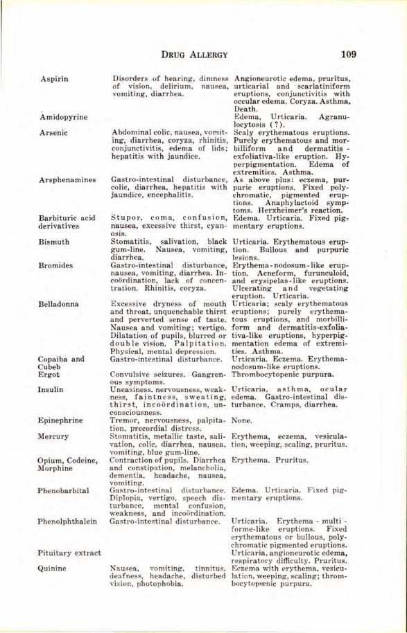
Aspirin
Amidopyrine
Arsenic
Arsphenamines
Barbituric acid derivatives
Bismuth
Bromides
Belladonna
Copaiba and Cubeb Ergot
Insulin
Epinephrine
Mercury
Opium, Codeine, Morphine
Phenobarbital
Phenolphthalein
Pituitary extract
Quinine
DRUG ALLERGY
Disorders of hearing, dimness of vision, delirium, nausea, vomiting, diarrhea.
Abdominal colic, nausea, vomiting, diarrhea, coryza, rhinitis, conjunctiviti , edema of lids; hepatitis with jaundice.
Gastro-intestinal disturbance, colic, diarrhea, hepatitis with jaundice, encephalitis.
Stupor, coma, confu ion, nausea, excessive thirst, cyanosis. Stomatitis, salivation, black gum-line. ausea, vomiting, diarrhea. Gastro-inte tina! disturbance, nausea, vomiting, diarrhea. Incoordination, lack of concentration. Rhinitis, coryza.
Excessive dryness of mouth and throat, unquenchable thirst and perverted ense of taste. Nausea and vomiting; vertigo. Dilatation of pupils, blurred or double vision. Palpita tion. Physical, mental depression. Gastro-intestinal disturbance.
Convulsive seizures. Gangrenous symptoms. Uneasiness. nervousness, weakness, faintn ess, sweating, thirst, incoordination, unconsciousness. Tremor, nervousness, palpitation, precordial distress. Stomatitis, metallic taste, salivation, colic, diarrhea, nausea, vomiting, blue gum-line. Contraction of pupils. Diarrhea and constipation, melancholia, dementia, headache, nausea, vomiting. Gastro-intestinal disturbance. Diplopia, vertigo, speech disturbance, mental confusion, weakness, and incoordination. Gastro-intestinal disturbance.
Nausea, vomiting, tinnitus, deafness, headache, disturbed vision, photophobia.
109
Angioneurotic edema, pruritus, urticarial and scarlatiniform eruptions, conjunctivitis with occular edema. Coryza. Asthma, Death. Edema, Urticaria. Agranulocytosis ( ? ) . Scaly erythematous eruptions. Purely erythematous and morbilliform and dermatitis -exfoliativa-like eruption. Hyperpigmentation. Edema of extremities. Asthma. As above plus: eczema, purpuric eruptions. Fixed polychromatic, pigmented eruptions. Anaphylactoid symptoms. Herxheimer's reaction. Edema. Urticaria. Fixed pigmentary eruptions.
Urticaria. Erythematous eruption. Bullous and purpuric lesions. Erythema- nodosum -like eruption. Acneform, furunculoid, and erysipelas-like eruptions. Ulcerating and vegetating eruption. Urticaria. Urticaria; scaly erythematous eruptions; purely erythematous eruptions, and morbilliform and dermatitis-exfolia tiva-like eruptions, hyperpigmentation edema of extremities. Asthma. Urticaria. Eczema. Erythemanodosum-like eruptions. Thrombocytopenic purpura .
Urticaria, asthma, ocular edema. Gastro-intestinal disturbance. Cramps, diarrhea.
one.
Erythema, eczema, vesiculation, weeping, scaling, pruritus.
Erythema. Pruritus.
Edema. Urticaria. Fixed pigmentary eruptions.
Urticaria. Erythema - multi -forme-like eruptions. Fixed erythematou or bullous, polychromatic pigmented eruptions. Urticaria, angioneurotic edema, respiratory difficulty. Pruritus. Eczema with erythema, vesiculation, weeping, scaling ; thrombocytoprenic purpura.

The Modern Management of Pneumonia By B. L. HESSION, M.D.
Senior Medical Interne, Victoria Hospital, London, Ontario
T HE purpose of this paper is not, as the title may suggest, to give a complete outline of the management of pneumonia. Only a few
r ecent advance in therapy are outlined and these pertain only to the pneumococcal pneumonia . Even in the year 1939, pneumonia still ranks third as the commone t cau e of death. It is preceded only by cardiovascular diseases and cancer. Few of the prevalent acute conditions have such a high mortality or are so expensive to treat. When we consider that 25 per cent of our pneumonia patients die and that the majority of them come from our most productive age group, we then realize just how much skillful therapy is indicated in these cases.
In the past, the unhappy victim of lobar pneumonia perforce must cough, perspire, gasp for a ir for days on end until resolution or, at times, death ended hi sufferings. Today, the picture changes. No longer is our conduct of the pneumonic patient one of watchful waiting. Pneumonia is a medical emergency. Active rational therapy carried out in the early tages of the disease will ameliorate and sometimes practically eliminate those toxic manifestation so distres ing and injurious to the patient. Three factors contribute to this change: (1) Improvements in symptomatic treatment, (2) The introduction of chemical therapy, (3) The u e of specific anti-sera.
SYMPTOMATIC TREATMENT This aspect of the treatment, even in the face of changing therapy,
still remains the ba is and framework of pneumonic management. ( 1) General Measures : Pneumonia is a contagious disease. As
such, its victims should be i olated. In some centres, attendants wear rna ks when near the patient and follow the special technique associated with the treatment of communicable disease . The room should be well ventilated, free of draught and maintained at a temperature of 68° F. Good nursing is of prime importance.
(2) Pain: The pleuritic pain of pneumonia may be mild or severe, transitory or prolonged. To combat this, the opium derivatives, codeine and morphine, are of common use, the drug being suited to the degree of pain. Strapping the chest with adhesive is not advised since it is a barrier which the physical signs must penetrate to reach the physician's ear. A Scultete binder is just a beneficial and does not hinder the physical examination. When the pain i very severe, the introduction of 200-300 cc. of air between the pleural surfaces with a pneumothorax apparatus gives complete and rapid relief.
(3) Sleep : The patient must sleep. A restless night caused by coughing, dyspnoea, and chest pain produces in the morning a much
110

THE MODERN MA AGEMENT OF PNEUMONIA 111
more toxic and more miserable patient. A judicious use of sedatives will obviate thi . Many ob ervers will que tion the use of morphine on the ground that it increa es the abdominal distension and is a depressant to the respiratory centre. They would favour the use of the barbiturates and codeine. Since we are able to produce uch a rapid change for the better in the pneumonic patient, it i eldom necessary to u e morphine for more than two to three nights. From then on, codeine and the barbiturates are quite adequate. But three factors keep t he pneumonic patient awake, namely, cough, pain and dyspnoea. In the early acute case, no one drug can control the e quite as well as morphine.
( 4) Cough: This distressing symptom u ually can be controlled with codeine either in the form of tablet or syrup. Seldom do we need the strong respiratory sedatives such as heroin or morphine. During the stage of resolution, expectorant cough mixtures are indicated. Here we have recourse to the various mixtures containing potas ium iodide, ammonium chloride and ipecac listed in the B. P.
(5) Diet : For many years the diet in the early stages has been soft food with abundant high caloric fluids, the latter usually consisting of fruit juices. As the pat ient progressed, the diet was increased to light diet and then general. Some authors would prefer a full diet from the beginning, stating that the patient mu t have his full quota of calories to offset the demands of the body. At any rate, fluids are essential, up to 3,000 cc. per diem being the minimum.
(6} The Bowels : Abdominal distension must be treated actively and, if possible, prevented. It greatly embarrasses the patient's respiration. This di tension is due to a paralytic ileus resulting from the generalized toxaemia present in pneumonia. Peristaltic stimulants such as castor oil, pituitrin or eserine may be given once or twice per diem. The use of castor oil may be questioned but it is an excellent mall bowel stimulant. Small saline enemas every second day are advised. Along with the above, turpentine stupes and the rectal tube deserve a place.
(7) Oxygen : The role of oxygen in pneumonia is an important one. It decreases the cyanosis, r elieves the dyspnoea, and lower s the temperature and pulse rate. The oxygen tent, especially the airconditioned type, where there is a uniform inflow of oxygen carried in a circulating medium of ice-cooled air, is in general u e. The one great advantage i that once placed in position little adjusting is needed. For days on end, the patient is assured of a uniform intake of oxygen. There is, however, the occasional patient who grows restless to the point of frenzy when placed in what he consider to be an oxygen "cage." Oxygen per nasal catheter serve well here and has the added feature of making the patient more acce sible to treatment and physical examination. The rate of flow is easily adjusted and there is little waste. In England, nasal oxygen is much in vogue. They use a double nasal catheter in much the same manner as in intranasal anaesthesia. The aving in oxygen is apparently great.

112 UNIVERSITY OF WESTERN ONTARIO MEDICAL JOURNAL
( 8) The Heart : Two conditions focus attention on the heart : auricular fibrillation which is uncommon and vasomotor collapse which is responsible for most of the deaths in pneumonia. The former can be well controlled with digitalis. The latter condition should be treated as a peripheral vascular collapse. Here we use adrenalin hydrochloride, coramine, camphor in oil, caffeine sodium benzoate, and intravenous fluids in small repeated do es.
CHEMOTHERAPY In the past few years two drugs somewhat chemically allied have
made their appearance in t he treatment of pneumonia. The first was Sulphanilamide introduced in 1935 and the second Sulphanilyl amino pyridene introduced in 1937. Sulphanilamide, which began its career as an anti-streptococcus haemolyticus agent, later showed a beneficial action on other cocci infections. According to the experimental work done by Long of Baltimore on rats, Sulphanilamide exerts a bacteriostatic action on pneumococci. Osgood claims it has an antitoxic action also. No one can doubt that Sulphanilamide preparations do benefit the pneumonic patient. When given in large doses early, there is a decrease in the temperature, pulse and respirations within 24 to 48 hours. As Long has pointed out, we must provide an effective concentration of the drug in the blood early, continue with the maintenance dose daily and then withdraw the drug slowly only after a marked improvement is noted. Failure to do t his usually results in a recurrence of the patient's symptoms and signs plus an elevation of the T.P.R. Today, we are able to estimate the blood content of the drug by colorimetric methods. An effective concentration is considered to be 10 mgm~ · per 100 cc. of blood. This level must be reached early and maintained daily till marked improvement is shown. To give a working idea of how the drug is given let us say we have a man weighing 150 pounds with a definite lobar pneumonia. An initial dose by mouth would be 80 grains. The maintenance daily dose is 20 grains every four hours for 24 hours. The total dose for one day would be 180 grains. This dosage is maintained until marked improvement is noted. It is then decreased slowly, i.e., 15 grains q.4.h.; 10 grains q.4.h. until it is stopped entirely.
There are a few toxic manifestations to sulphanilamide which warrant our consideration. The first group, consist.ing of nausea, vomiting, cyanosis and mild anaemia, is relatively unimportant. In the second group we have simple fever, dermatitis, acidosis, jaundice and acute haemolytic anaemia. The drug must be stopped if these occur. The simple fever occurs as a sharp rise in a lowered temperature during the administration of the drug. The dermatitis may imitate measles, scarlet fever or urticaria. Acido is may be combatted by giving sodium bicarbonate in the ratio of 1: 3 of the drug. The best way to control the dosage and to guard against toxic manifestations is as follows:
(1) The daily estimation of the sulphanilamide content of the blood. (2) Daily haemoglobin estimation.
THE MODERN MANAGEMENT OF PNEU MO IA 113
(3) Four-hourly temperature record. ( 4) Careful examination of the patient. Our second chemical agent, sulphanilyl amino pyridene, is also
known by the experimental number M. & B. 693 and by the trade name Dagenan. Ju t a sulphanilamide i considered to be a specific antihaemolytic streptococcu agent, o i amino pyridene considered to be a specific anti-pneumococcal agent. Two workers, Evan and Gar ford, in England, are perhaps mostly responsible for the wave of enthusiasm which accompanied this drug in Canada. During the first clinical trials they ob erved a erie of 100 case of lobar pneumonia, using as controls 100 comparable cases receiving non-specific treatment. The mortality in the controls was 27 per cent, while the mortality in those cases receiving amino pyridene was only eight per cent.
The mode of action, as in the case of sulphanilamide, seems to be bacteriostatic. This is borne out by animal experimentation. Clinically, amino pyridene does appear to have a beneficial effect on the pneumonic patient. When given in full do es early, we have a marked drop in the T. P.R. with improvement in the patient's condition. Failure to give the drug in sufficient dosage or withdrawal too rapidly allows a recurrence of symptoms.
Dosage : Each tablet of amino pyridene contain 0.5 grams of active principle. In an adult patient, the following dosage is advised:
2 grams at once followed by 2 grams in four hours; 1 gram every four hours for thirty-six hours.
The above treatment should produce marked improvement. We then continue with 1 gram every four hours for 36 hours and complete the course with 1.5 gram per day till 20 grams in all have been given. As might be expected, the do age mu t be adjusted to the individual ca e.
Amino pyridene also ha toxic manifestation , but less so than sulphanilamide. The following symptoms are listed, viz., headache, nausea, vomiting, dermatitis, imple fever and cyano is. Nausea is very common, whereas the remainder are uncommon. Being relatively new, undoubtedly there is much to be learned concerning its use. We might, at thi point, stress a few advantages of the use of the chemical agents in the treatment of pneumonia :
(1) Can be given the moment diagnosis is made. (2) Relatively inexpensive. (3) Readily available. ( 4) Can be given orally.
SEROTHERAPY Specific anti-pneumococcic erum was used first in 1892. It enjoyed
but a fair reputation until 1924, when Felton succeeded in concentrating the antibodies to one-tenth their original volume. This did away with the severe reactions previously encountered and did much to stimulate the use of serum. In the bacteriology of the pneumococcus many
114 UNIVERSITY OF WESTERN ONTARIO MEDICAL JOURNAL
advances have been made. In 1913, the pneumococci were first divided into three specific types, I , II and III, and a non-specific group IV. Sixteen years later, this group IV was divided into 29 specific types. Today, we have 32 types of pneumococci each with its specific ant iserum. Up to three years ago, horse serum was in general use. Today rabbit serum is gradually supplanting it. Rabbit serum is more readily obtained, is less expensive, causes fewer reactions and is said to have a more penetrating action.
In the treatment of lobar pneumonia, serum has a very wide scope. 95.5 per cent of our pneumonias are caused by pneumococci. Of these, 70 to 80 per cent are caused by types I, II and III. The latter varies in that some communities will have a preponderance of types V, VII or VIII. Happily, in the commonest type, i.e. , type I, the best results are obtained.
With the above interesting facts in mind, we now come to the question: How shall we proceed to treat the pneumonic patient with serum? A definite routine must be followed. A careful history and a complete physical examination are essential. Physical findings should be checked by X-rays. Blood cultures should be done at once.
S ecuring the Sputum-This may be done in three ways : (1) Collecting coughed-up sputum in a sterile container. (2) Taking a pharyngeal swab. ( 3) Lung puncture.
Lung puncture is a skilled procedure wherein the bacteria are aspirated directly from the pneumonic lobe by a needle. The dislodgment of an embolus, or pulmonary bleeding, occur rarely with good technique.
Typing the S putum-The most common and rapid method was devised by Neufeld. When combined with its specific anti-serum, and only then, the pneumococcus capsule swells markedly. A drop of sputum, a drop of Methylene blue, plus a drop of typing serum, is aJl that is needed. Results are obtained in five minutes.
When to Give Serum-Serum is given at the earliest possible moment. The ideal time i within 24 hours of the onset of the disease. When given within the first three days, the mortality r at e from pneumonia drops from 25 to 4.8 per cent. Waiting one more day doubles the rate. It is a common belief that beyond 3 days serum is useless. Results are poorer the farther we draw away from the onset, yet, according to Bullowa, it is never too late.
Dosage-This depends on: ( 1) Date of commencing treatment; (2) Type of infection; (3) Age of patient; (4) Presence or absence of bacteraemia. In a type I infection, where treatment begins within the first 24 to 48 hours, 100,000 units are usually sufficient. After 48 hours the dosage must be doubled, tripled, etc., t he farther we draw away from the onset. In types I, III and VII infections and many others 200,000 units are usually t he minimum. In patients past 40 years of age usually we have to double the dosage. In the presence of a bacteraemia we must double the dosage from the beginning.

THE MODERN MANAGEMENT OF PNEUMONIA 115
Method of Giving Serum-Before giving serum we must test the patient for sensitivity towards the serum employed. This may be done in three ways. The first method, the ophthalmic test, consists in placing one drop of the normal rabbit serum diluted 1:10 into the conjunctival sac. A positive reaction, read in 20 minutes, is evident by engorgement of the conjunctival vessels. The intradermal test, using 0.1 cc. of 1: 10 dilution, shows up as a reddening of the skin at the site of the injection. The intravenous test, using a 1:5 dilution, 0.5 cc., is postive if the blood pressure falls 30 mms. Hg. in 10 minutes. Any patient with a positive reaction must be desensitized. Providing the sensitivity tests are negative, serum may be given. All serum i given intravenously so that we may give sufficient in the shortest possible time. The fir t dose varies, but at the Victoria Hospital we give 10,000 units. Great caution must be exercised here. It is usually with the first or second dose where reactions occur. To avoid these, we give the first dose along with 200 ccs. of normal saline solution. Then any reaction will be slight and easily controllable. The second dose, given in two hours, is usually 20,000 units and may be given with 20 cc. of saline. Ten minutes are taken to inject this amount. In the absence of reactions, we then give 20,000 to 40,000 units directly into the vein every two hours. This is continued till we get a marked drop in pulse rate, temperature and respirations. In some cases 400,000 to 800,000 units are needed to affect a response. Following a marked drop in the T. P.R. an additional 20,000 to 40,000 units may be given to ensure success. In the early ideal case the effect of serum is dramatic. On admission, we have a dyspnoeic, toxic and very miserable patient. Within 12 to 24 hours of serum treatment we have a quietly breathing, non-toxic and very grateful patient. Serum does not stop the pneumonic process. Indeed it is surprising how well the patient looks yet has all the signs of a consolidated lung. Biologically, the use of serum has a sound basis. Primarily, it is an anti-toxin. Secondly, it fixes the pneumococci in such a way as to render them innocuous and induces the phagocytes to complete their destruction.
As one would expect, serum therapy is attended by many reactions, varying in type and degree. Some of the common ones are chills, hyperpyrexia, shock, collapse, vomiting, dyspnoea, cyanosis plus anginal, lumbar or chest pain. Acetyl salicylic acid gr. 20 given 20 minutes prior to injection prevents chills and thermal reactions. Epinephrine min. 30 given before, during or after the reaction is an excellent drug. If reactions occur during the injection, stop at once, wait for one hour and then begin again, using diluted serum and giving it very slowly. To begin carefully and slowly usually results in a very rapid successful ending.
BIBLIOGRAPHY 1. Bullowa, I. G. M. : J.A.M.A., Dec. 18, 1937. 2. Cecil, R. T., and Bullowa, I. G. M.: J.A.M.A., Oct 23, 1937. 3. Evans, G. M., and Gaisford, W. F .: Lancet, July 2, 1938. 4. Long, P. H., Baltimore: J.A.M.A., Jan. 14, 1939.

Anatomy and Physiology.-.-.A Short History
By RAY STUBBING, '42
A LL history is colourful! It deals with facts and figures and with famous people; it traces the course of progress and development;
it is the record of an everlasting search for truth and knowledge. The student of history becomes engros ed in the life stories of individuals, for history is not a record of geographical, mechanical or scientific facts, but rather it is a tale of the desires, action and achievements of men and women.
This is an historical essay about the fundamental sciences of modern medicine, anatomy and physiology. Because it is a history, it deals with people; because the sciences have developed over the course of centuries, it is possible to touch only the beginnings.
ANATOMY During the first century there arose Claudius Galen, a native of
Greece, tall of stature, dark, bearded, vain of appearance, destined to become the renowned physician and medical authority of the Roman world. He dissected the bodies of animals such as dogs and pigs, and accepted the arrangement of the body structures of these animals as being that of the human body. His knowledge was essentially empirical, yet he was t he outstanding healer of diseases in his time. His system of medicine and his books were to be accepted as classics for many years.
Galen's influence lasted for fifteen centuries. His system was taught in all schools, and if, as often happened during the course of a dissection, the structure of the animal did not corre pond to Galen's teaching, the animal was called an anomaly. Galen was never questioned, so generally was his authority accepted. Medicine was, during that period, chiefly "according to Galen." And then suddenly, as it seems, men thrust aside both tradition and authority. They began to build a knowledge of the human body and of disease on a ound, scientific basis. Henri de Mondeville, in 1304, exclaimed, "God did not exhaust all his power in making Galen."
The Renaissance of medicine had begun. Andreas Vesalius, born in Belgium in 1514, became the founder of Gro s Anatomy. He studied medicine in the University of Louvain, and from there he went to Paris which was then the centre of medical education.
Vesalius' nature made him dissatisfied with the halting approach to anatomy which his professors presented. Neither could he be sympathetic with the church's prohibitory stand on the dissection of the human body. He became a skilled dissector, and was often able to confuse the professors with searching questions regarding some structure. By stealing the bodies of executed criminals, he was able, finally, to dis-
116
ANATOMY A D PHYSIOLOGY- A SHORT HISTORY 117
sect and tudy t he human body. He secured the position of Professor of Anatomy at the University of Padua, in Italy, by naming the individual bone of the human body while blindfolded. In Italy, thought and teaching were more progre sive, and with the assistance of Jan van Kalkar, a pupil of the artist Titian, he published in 1543 his wellillustrated book, "De Humani Corporis Fabrica." Thi book revolutionized the science of human anatomy by introducing the method of direct observation.
Vesaliu had several important contemporaries: Dryander of Mar burg; Miguel Servetus, the Spani h theologian who di sected human bodies primarily to find the seat of the soul; Leonardo da Vinci, the artist who did human dis ections in order to improve his sense of proportion in art. There were many immediate successors to Vesalius who were able to avail themselves of his work and to make further discoveries; Fallopius, and the Fallopian tubes; Eu tachius, and the Eustachian tubes; Varolius, and the pons Varolii, and many others. Thu in the course of a century, the greater portion of human anatomy was investigated and explained. Sir William Osler said of Vesalius' book: "The greatest book ever printed from which modern medicine dates."
In 1590, Hans Janssen, a Dutch pectacle-maker, made a small metal tube with a simple lens in erted. He called it a flea glass. As seen through it, a flea resembled a grotesque, hairy monster. It was considered a toy but there was the germ of the microscope.
Antonj van Leeuwenhoek, born in Delft in 1632, is looked upon as the inventor of the micro cope. He was an ingenious young man who devised a combination of len e · which would magnify an object to many diameters. Thus the cience of microscopic anatomy (histology), and of developmental anatomy (embryology), were made possible. In 1677, van Leeuwenhoek himself reported the discovery of "little animals" in the male seminal fluid. He found the triations in muscular ti sue and he discovered that scraping from the tongue revealed countless animalcules under the microscope. Salenpatrius, in 1699, asserted that he had observed a minute human form in each spermatozoon as supporting evidence to the doctrine of of preformation. However, William Harvey, with whose work in physiology we shall later deal, had published earlier a book entitled "Studies on the Generation of Animals." It contradicted the doctrine of preformation by bowing that the embryo went through stages of development until it gradually evolved the parent form. He may thus be regarded as a founder of embryology.
Marcello Malpighi, born in Bologna in 1628, was the investigator who first used the microscope as a erious scientific weapon. He was curious regarding the minute structure of the various organs of the body. On examining prepared lung tissue, he saw that the arteries which entered the tis ue broke up into even finer vessels, as fine as hairs. He called them capillaries. He followed the e and ob erved that they

118 UNIVERSITY OF WESTERN ONTARIO MEDICAL JOURNAL
gradually became larger until they formed vein . Here was a great histological discovery. William Harvey had earlier proved that blood left the heart by arterie and returned by veins. But how did it get from the arteries to the veins? Malpighi supplied the answer.
Malpighi did not s top until he had thoroughly established the science of histology. From his investigations of the spleen, we have the Malpighian bodie ; of t he kin, we have one layer called the rete Malpighii; of the kidney, we have the cortex and the renal pelvis. From his studies he was able to assume correctly that the kidneys extracted urinous elements from the blood.
Great strides were now possible in the treatment of di eases, and in the progress of medical science, because of these developments in gross, microscopic, and developmental anatomy.
PHYSIOLOGY Towards the end of the 16th century a young Englishman entered
the University of Padua, made famous for its anatomy by Vesalius. He was William Harvey, who was to establish the true facts of blood circulation, and thus to create the beginnings of physiology.
Various fanciful theories existed. Galen had observed that after death the arteries contain air and the veins blood. So he had advanced the statement of a circulation depending on the pa sage of blood in the veins and air in the arteries; the flow of blood through minute orifices in the interventricular sy tern; with the liver, as well as the heart, a pumping-organ. Vesalius had had serious doubts about the correctness of this view yet he was unable to improve upon it.
William Harvey is rightly given the credit for establishing the true facts of circulation: that the blood travels in a circle. However, it is interesting to observe that Miguel Servetus had already described the circulation of the blood from the right to t he left side of the heart in "The Restitution of Christianity," published in 1553. Servetus was a radical theologian and the heretical views which he expressed led to his being burned at the stake in the same year, and to the destruction of the five hundred copie of the book. Several copies were preserved, but it wa not until 141 years later that medical attention was called to his descriptive explanation of the circulation.
Harvey conducted numerous experiments in the early years of the 17th century. He tied off the artery of a sheep and made a nick in it on the side toward the heart. Blood spurted with each heart-beat until gradually no more flowed. He rea oned that none was returning to the heart. He tied off a large artery close to the heart and watched that organ become engorged until all the blood bad gathered in it. He chose a vein in the arm and "milked" it toward the heart, watching it rapidly fill up again from the extremity. So he showed the direction of flow in the veins and arteries, and thus logically observed that the blood travelled in a circle which led from the heart to the lung and back

ANATOMY AND PHYSIOLOGY- A SHORT HISTORY 119
again. We have seen how the discovery of capillaries later substantiated his work. These results were published in 1628 as "Anatomical Exercises on the Motions of the Heart and Blood in Animals." It ranks in medical importance with the anatomical publication of Vesal ius .
Harvey had enunciated the first major generalization of physiology. It was of great significance because it demonstrated the value of the experimental methods; it made possible the control of haemorrhage in surgery; it explained the function of the placental after-birth; and it permitted further progress in the physiology of the various organs of the body.
The second major problem of physiology was to explain the function of respiration, and numerous men made discoveries which finally led to its solution. In 1657, Robert Boyle exhibited to the "Philosophical Associations" at Oxford an apparatus which demonstrated that air has weight, so that if air were pressed against a column of mercury, the level of the column would rise. Robert Hooke, in 1667, showed that the act of respiration is of secondary importance, and John Mayhow, in 1668, demonstrated the mechanism of respiration. In a pamphlet, "Tractatus de Corde," he explained that venous blood is changed to bright red arterial blood because of the air mingling with it. Gradually men were approaching the truth.
Chemistry at last supplied the an wer. Joseph Priestly, a Unitarian minister, actually isolated a gas which is necessary for all combustion, and he called it "phlogiston." Finally a brilliant French chemist, Lavoisier, in 1777, isolated oxygen from the air and explained correctly how the blood conveys oxygen to the body tissues and takes up carbon dioxide in exchange.
The progress of physiology wa most rapid. By the middle of the 19th century investigators had discovered the function of digestion, the natur e of muscle contraction and of nerve impulses, and the process of excre tion. Since then the great unifying principle of nutrition has been explained.
Medicine has come a long way since the time when the discoveries of Vesalius and Harvey gave impetus and direction to the major sciences. Medical science and practice are now recognized as necessary accompaniments to any real social progress, and the application of medical knowledge to the health of a community is slowly becoming the concern of all people.
BIBLIOGRAPHY
Garrison, F. H.: An Introduction to the History of Medicine. Osler, William: Evolution of Modern Medicine. Clendening, L.: The Romance of Medicine.

il l l ll l ll l ll l ll l ll l lll ll l ll l ll l ll l ll l l ll lll lfl ll l lll ll l ll l ll l ll l ll l lll ll l llllll lll ll l ll l ll l ll l ll l ll l ll l ll~
81! ~bstr acts !l8 ~ ~
~~~ ~ ~~ · ~~ · ~~ · ~~ · ~~ · ~~ · ~~ · · ~ · ~ ~ · ~ ~ · ·~ · ~~ · ~~ · ~~ · ·~ · ~~ · ~~ · ~~ · ~~ · ~~ · .. · ~~ · ~~ · ~~ · ~~ · ~~ · ~~ · ~~ · ~~ · ~~ · ~~ · ~~ · ~ ~ · ~~ z
HUMAN STERILITY By w. P. TEW
Can. Med. Assn. J.; 40:116, 1939 A thorough and comprehensive account
of the main features of human sterility is given. The author has indicated that sterility is the result of not one but several factors and that usually both parties are partly at fault. The general factors influencing human infertility: race, age, climate and season, civilization and constitution, are listed and briefly discussed. An excellent clinical classification of sterility based on etiology and a method of investigating a sterile mating are outlined. The treatment of infertility in both the female and the male are outlined.
-R. JOLLIFFE, '41.
OBSERVATIONS ON THE NEUFELD REACTION (QUELLUNG TEST)
FOLLOWING DESICCATION OF PNE UM OCOCCUS
PREPARATIONS By H. R. BROWN
Jour. Lab. Clin. Med., 23, 12, 1938 A method of preparing dried smears of
sputum, and also of using dried smears of type-specific sera, is herein described. By this method loss of time in detecting the presence of a specific type of pneumococcus can be avoided, and danger of destruction or damage by capsular change to the specimen by autolysis or contaminating overgrowth of other bacteria can also be obviated. A very comprehensive series of experiments shows that dried smears of sputum containing pneumococci will give positive
eufeld reactions after being exposed either t o high or low temperatures for periods of a year. It is also shown that specific sera may be dried on slides at room or incubator temperature and used whenever desired. The Quellung reaction develops as well as, or better than, in cases where the less simple method of using liquid sera is used.
--J. G. STAPLETON, '41.
TREATMENT OF PNEUMONIA WITH 2 (P. AMINOBENZENESUL
PHONAMIDO)-PYRIDINE By G. M. EvANS and WILFRED S. GARSFORD
J.A.M.A. ; 108:1483, 1937 The authors administered this drug in
one hundred cases of lobar pneumonia and forty cases of broncho-pneumonia. As a r esult the mortality rate dropped from 27 % in control cases which did not receive the drug to 8% in the test cases which did receive the drug.
Method of Administration: At the beginning the dosage was largely experimental. A common practice was to give one tablet (0.5 grams) every four hours for three or four days, followed by one tablet twice a day for an additional two or three days. The average amount given per case by this method was 12 grams. Later larger initial doses were g iven. At the same time a close watch was kept on the blood count for signs of a granulocytopoenia. Doses as large as four tablets (two grams) were given on admission, followed by one tablet every four hours. The average amount given per case by this method was 25 grams. In a f ew cases as much as 9 grams has been given in the first 24 hours. The temperature and toxic symptoms, especially the delirium, subsided very rapidly.
To>..'ic symptoms were not pronounced and cleared up immediately upon withdrawal of the drug. Cyanosis appeared in 25% of the intensely treated cases. Spect roscopic examination of the blood showed a methhaemoglobinaemia in 6% of the cases.
No cases of sulphhaemoglobinaemia were encountered.
-D. WOLLIN, '39.
CONTROL OF PUERPERAL FEVER By L. COLEBROOK
B.M.J.; 4069:1378, 1938 Dr. Colebrook pointed out that ap
proximately 40 per cent of the cases of puerperal fever are brought about by the
120

-----------------------------
ABSTRACTS 121
haemolytic streptococcus, the source of infection in at least 50 per cent of cases being an attendant contact--that is, doctor, nurse, or midwife. Other sources of infection include members of the mother's household and the mothers themselves. Respiratory infections are probably the chief offenders.
Dr. Colebrook listed the following preventive methods: 1. The wearing of efficient masks by
attendants. 2. The use of strict antiseptic toilet, in
the attaining and maintaining of which soap and water are of prime importance.
3. The use of iodine and dettol as antiseptics.
4. Institutional confinements r a t her than home confinements. ( I n four years at Queen Charlotte's, only one patient in 700 had developed haemolytic strept ococcus infection, whereas in that district the incidence had been one in 135) 0
In discussing active therapeutic measures, Dr. Colebrook contented himself with outlining t he remarkable drop in the death rate in puerperal fever at Queen Charlotte's following the introduction of sulphanilamide therapy.
--H. T. McALPINE, '39.
DIVERTICULITIS OF THE S IGMOID COLON
B y J . H. GEDDES
Can. Med. Assn. J.; 40:157, 1939 The author has dealt briefly but com
prehensively with the main aspects of diverticulitis of the sigmoid colon. He has divided the condition into five clinical groups. The importance of prophylactic treatment in the first group, diverticulosis, to prevent progression of the condition to symptomatic diverticulitis is indicated. The necessity for prolonged bed rest in the acute phase is stressed a nd the medical treatment of this phase outlined. The indications for surgery in the complications attending diverticulitis of the sigmoid colon are noted with the necessity for skilled surgery. As the author has pointed out, t he development of ca rcinoma in diverticulitis, t hough rare, is possible and with all ca rcinoma of the sigmoid must be differentiated from the hyperplastic stenosing type of
diverticulitis. The different ial diagnosis of these two condit ions with t he difficulties encountered is thoroughly discussed.
--J. GALLOWAY, '40.
TEACHING OF MEDICAL ECONOMICS TO UN DERGRADUATE
MEDICA L STUDENTS B y KINGSLEY ROBERTS and
MICHAEL M. SAIRS
J. A ssn . of Amer. M ed. Colleges; 13:359; 1938
The results of an inquiry made to 64 medical schools in Canada and t he United States regarding the teaching of medical economics are here analyzed.
Forty per cent of the schools give no attention to the subject, while in t he others it is handled in various ways. Of these r emaining 37 schools, 15 give a series of several lectures on phases of medical economics and ethics; 6 schools reported a rather systematic course ent itled "Medical Economics"; 8 schools are giving a broader course dealing with the economic and social relations of the medical services. Only 4 schools were offering a really comprehensive lecture and discussion course dealing with medical ethics, the economics of medical practice and various studies of socialized medicine.
The article shows that about one-half of the medical schools are paying serious attention to teaching of medical economics and that attention to the teaching of this important subject is on the increase.
-RAY STUBBING, '42.
THE TR EATMENT OF PAR ALYTIC BLADDE R IN CASES OF SPINAL
CORD INJ URY By F. H I NMAN
S urg ery; 4:649, 1938 Four phases characterize the common
effect on micturition of a transverse lesion at any level of the cord: ( 1) retention, (2) overflow incontinence, (3) automaticity, ( 4) true incontinence. In cases of fracture above the lumbar center the first three occur in succession; an automatic bladder never develops as the late and final effect after fractures below this center, a nd incontinence may follow destruction of the cord at t h is level.
Injudicious treatment of the paralyzed

122 UNIVERSITY OF W ESTERN ONTARIO MEDICAL JOURNAL
bladder results in urinary infection and bed sores and will foil the outcome to a maximum of over 80 per cent. The intent of all care, therefore, is to prevent or control these two evils. Treatment should be based on sound urological principles and it is impractical to treat all paralyzed bladders alike. The accepted methods are:
(1) Nancatheterization and Manual E x pression: Pressure should be applied regularly every 6-8 hours with the open hand above the symphysis. This will ward off urinary epsi and if successful will hasten automa ticity.
(2) Supmpubic Cystostomy : This method gives good drainage a nd tidal irrigations may be given t hrough it or suction applied on occasion.
(3) R etention Cathete1· with or without Tidal Irriga.tion: Continuous care and attention is required for a retained catheter; if this is possible a retention catheter may save time, control the· infection and keep the patient dry and comfortable until such time a s automaticity develops. It must be emphasized that all authorities condemn intermittent catheterization.
-CLEMENT DELETSKY, '40.
VARICOSE ULCERS AND INSULIN By L. G. PAYNE
Can. Med. Assn. J.; 38:269, 1938
Varicose ulcers are not uncommonly encountered in general pract ice, their mo t important a spect being treatment.
There are many methods of treatment, most of them dating back to many years ago--rest with elevation of the leg : antiseptic powders and salves, various ointments, and occlusion of adjacent veins. Local treatment with in ul in has a definite value. Apply to t he ulcer, cleansed with sa line compresses and dried, a small amount of insulin (usually U20 strength) for approximately 20 minutes. Then the insulin which has not been absorbed is removed, the base of the ulcer dried again, and the dressing applied. Repeat three to four days, and keep leg elevated.
In younger persons, or where the margin is deep or the base fibrosed, the treatment is less satisfactory, and requires infection or ligation of the regional veins.
Where the ulcer is shallow, the patient older, or rest with elevation impossible, insulin gives the best results. For the initial treatment where it is usually infected, dressing of iodine dusting powder, covered by elastic adhesive for three days, is good.
-L. C. BARTLETT, '41.
OBSERVATIONS ON THE TREATMENT OF EMPYEMA IN CHILDREN
By H. L. WALLACE
B.M.J.; 4053:560, 1938 The author has investigated the treat
ment of empyema in 348 children admitted to the Royal Edinburgh Hospital for Sick Children, over a period of fifteen years. He has attempted to compare the radical and conservative methods of treatment.
The conservative treatment consisted of repeated aspiration, drainage by cannula aspiration, and drainage by closed suction. The radical treatmen t consisted of thoracotomy with open drainage and drainage by rib resection. The most striking and significant difference is in t he children under two years of age. By conservative methods, the mortality was 57.4 per cent, and by radical methods the mortality was 37.1 per cent. This is significant in that this age group usually is treated by conservative methods. The mortality, using radical treatment, in the age group from two to five years of age wa slightly lower, and in the age group from five to twelve years of age was s lightly higher than by conservative treatment.
The other factor investigated was the duration of stay in hospital. The cases treated by conservative methods averaged from eight to fourteen weeks, depending upon the method used. The cases treated by radical methods averaged five weeks.
In conclus ion, the author deduced from this eries of cases that the best treatment wa the operation of rib resection performed immediately after localization of the empyema had been achieved by means of repeated aspiration. It is emphasized that the ideal treatment has not yet been evolved, and that no finality has been reached on this important problem.
- R. H. CRAM, '41.

ABSTRACTS 123
T REATMENT OF INFECTED WOUN DS WITH U REA
By L. F . MULDAVIN and J . M. HOLTZMANN Lancet; 234:549, 1938
Urea in saturated solution has a solvent power on slough or debris which, being removed, prevents a potential breeding ground for bacteria. In addition, solutions of 8 - 25% are found to be bacteriostatic and stronger solutions are bactericidal.
It was used with success in abscesses, carbuncles, whitlows, cellulitis, infected burns and wounds. The technique consists of syringing away free pus and necrotic material with a saturated solution of urea, excessive moisture removed and urea crystals then liberally applied. Waxed paper is placed next to the crystals to keep them in contact with the wound and to prevent dressings from becoming soaked. Adjacent kin is protected with zinc cream.
The disadvantage of the treatment are:
(1) pain is sometimes experienced on application.
(2) urea does not directly stimula te epithelialisation.
Results were so encouraging that it has been adopted as routine treatment of infected wounds in the casualty department of the Royal Free Hospital.
- D. STATE, '39.
TH E T REAT MENT OF BACI LLA RY DYSENTERY WITH BACT E RIOPHAGE
By J . E. MURRAY The Practi tioner; cxli, 199, 1938
The author treated 146 cases of bacillary dy entery. The majority were cured within two weeks. The bacteriophage used must act on all dysentery groupsShiga, Flexner, and Sonne. Four patients suffered from the same complaint for two successive summers, but probably represented cases of re-infection. Bacteriophage does not confer immunity like vaccine. The author points out that many cases of chronic colitis are really bacillary dysentery, and should be investigated by a bacteriologist. The incidence is seasonal and greatest from July to September. The treatment was one ampoule o.r bacteriophage in alkaline medium before meals. The diet shot:ld be
nourishing, fluid and fat-free, containing no red meat, alcohol, or seedy fruits. The author concludes that bacteriophage is the best treatment for bacillary dysentery and that thereapeutic failures are due to an unreliable bacteriophage.
- A. JOHNSON, '40.
DISLOCATIONS AN D F RACTU RE DISLOCATIONS OF TH E PELVIS By R. WATSON-JONES, Liverpool
Brit. J. Surg. 25; 773-781, 1938 The author divides fractures of the
pelvis into injuries not involving t he pelvic ring, e.g., fractures of t he crest and spines of the ilium, fractures of the acetabulum, etc., and fractures involving the pelvic ring. He then goes into a consideration of the various types of injury to the pelvic ring and for each type gives the method of treatment which he has found to be most satisfactory.
Various types of injury to the pelvic ring mentioned are:
( 1) Isolated Injuries: Include fract ure of the body of the ilium, unilateral fracture of one or both pubic rami, and slight separation of the symphysi pubis. In this group, since there cannot be seri ous displ acement, no treatment is required except recumbency in bed for a few weeks; immobilization is unnecessary.
(2) Combined Injuries of Pubic S egment of the Pelvic Ring: Both fractures in anterior segment of pelvic ring, e.g., bilateral fracture of both pubic rami. These injuries are produced by compression of the pelvi s in its lateral axis. The displacement is limited, due to the large amount of muscular attachment in this area. The patient is to be treated in recumbency lying on hi s back for four to six weeks.
( 3) Combined Injuries of Iliac and Pubic S egments of the Pelvic Ring: These injuries cause complete disruption and wide displacement. They are produced by antero-posterior compression of the pelvi!'. The displacement is most easily corrected by manipulation in lateral recumbency and the application of a double plaster spica. The patient must lie on one side and not on his back. The functional result depends on the accuracy of reduction of the sacro-iliac dislocation, which may easily be overlooked.
-N. J. ENGLAND, '39.
.-----------------
~l l ll l ll l ll l ll l ll l ll l ll l ll l ll i ii i ii i ii i ii i ii i ii i ii i ii i ii i ii i iii ii i ii i ii i ii i ii i ii i ii i i i i ii i ii i U I II I O I II I_!:
2l! ~bitorial ilv ~ ~ . ~ .l l l ll l l l l ll l ll l ll l ll l ll l ll l ll l ll l ll l ll l l l l ll l ll l ll l ll l ll l ll l ll l ll l l l l l l l ll l l l l ll ill l ll l ll l ll l l l l l l l ll l l~
CANCER IN NEON LIGHTS
Statistics show that cigarette sales Yary directly as the amount of advertising done per year. Of the three most popular brands offered for sale in the United States, the one that has the most advertising is invariably the leading cigarette of that year. If you wish to sell something to somebody you must keep it before their attention. If you wish to sell cancer treatment to the public you must let them know about it. For this reason the Canadian Society for the Control of Cancer was formed.
Recent surveys have shown that hope for immediate improvement of cancer mortality lies in education of the public. It is granted that ultimately the ideal and only true way to eradicate cancer is through research. However, before the benefits of this field become available many thousands of people will have died of the disease.
Radiation and surgery are now accepted as the only measures of proven value in the treatment of cancer. The earlier the lesion is demonstrated and treated, the better the patient's chance becomes of cure. It is then essential that the patient comes to the doctor early; that the doctor make an immediate tentative diagnosis; and that treatment be started without delay. Of t hese three link in the chain of successful cancer therapy, the first two can be greatly improved. By education, the patient can be taught to recognize cancer early. Education is best carried out by keeping before the public the early signs and symptoms of cancer. Thus women would realize the significance of a painless single nodule in the breast; men would appreciate the importance of a change in gastric habit. It would then be up to the physician to institute treatment early.
Doctors throughout the province have already indicated their interest and enthusiasm for this new organization. An Ontario Society for the Control of Cancer has been formed to act in conjunction with the Canadian Society. It is expected that local Lay-Medical organizations will be set up in all the cities and towns of Ontario. Three of these have already been organized and plans to circularize the public have been started. If there is not a local society in your community already there should be one soon. For information write Dr. C. C. Ross, Secretary, Canadian Society for the Control of Cancer, 43 St. George St., Toronto.
124

~t i U I II I II I II I II I U I U I U I II I U i n l n l n l n l u l n l n i ii i ii i ii i ii i ii i ii i U I U I U I II I II I II I II Iu l n l tt l n l n l ll l n l u l n l u l u l ll ti;
~ ~
I moolt ~ebieWS I _, ll l ll l ll l ll l ll l ll l ll l lll ll l lll ll l ll l ll l lll lll lll ll l ll l ll l lll ll l ll l ll l ll l ll l ll l ll l lll ll l ll l ll l ll l lll ll l ll l ll l ll l ll l li i ii i ii i iii Ui
THE BRAIN AND ITS ENVffiONMENT By JOSEPH BARCROFT,
Professor of Physiology, Cambridge.
(111 pp., $2.00. Yale University P ress, 1938.)
This book represents the script of the Dwight Harrington Terry Foundation Lectures delivered by Professor Barcroft at Yale University. An attempt has been made to correlate central nervous system activity with the physical and chemical state of t he blood passing through the cerebral circulatory system. The tory i told in a vivid and entertaining manner. Chapter III, dealing with the relation of mental efficiency to properties of the blood, will stimulate many new lines of thought. The volume is mall and is recommended as a "week-end book" to those especially interested in the nervous system.
DIABETES INSIPIDUS AND THE NEURO-HORMONAL CONTROL
OF WATER BALANCE - A CONTRIBUTION TO THE STRUCTURE
AND FUNCTION OF THE HYPOTHALAMICO-HYPOPHYSEAL
SYSTEM
By C. FISHER, PH.D ., W. R. I NGRAM, PH.D. ,
and S. W. R ANSON, PH.D., M.D. Institute of Neurology, Northwestern Univ. Medical School, Chicago.
(212 pp., Illustrated, $5.00. Edwards Bros., Ann Arbor, 1938.)
This monograph sets for t h in detail the r esults obtained dur ing everal year of intensive research on the anatomy and physiology of
the hypothalamus. Working chiefly with the cat and monkey, circumscribed lesion were placed in various parts of t he hypot halamus with the aid of the Hor sely-Clarke stereotaxic instrument. When such lesions involved the hypophy eo-hypothalamic tract, diabetes insipidus invariably resulted. Inter ruption of the nerve fibres passing between the hypothalamus and the hypophysis produced profound degeneration of the post erior lobe of the latter. The pars intermedia and pars anterior were not affected.
125
126 BOOK REVIEWS
In female animals with experimental diabetes insipidus partuition was also disturbed, presumably due to a deficiency in the elaboration of the oxytocic hormone.
The e authors have amply demon trated the clo e anatomical and physiological relationship between the hypothalamus and the hypophysis. The monograph is printed by a proces of photolithography with excellent reproduction of the type-script and illustrations.
-M. L. BARR, M.D.
DISEASES OF THE NOSE, THROAT AND EAR By W. H. WALLACE MORRISON, M.D.,
Clinical Professor and Chief of Clinic, Department of Otola'ryngology, N ew York Polyclinic Medical School and Hospital.
(675 pp., 629 Illustrations, Indexed, 5.50. W. B. Saunders Co., Philadelphia, 1938.)
Dr. Morri on's is an extremely atisfactory text on this subject. Written, as the author tate , out of fifteen year's experience as a teacher of graduate students, it is intended for the undergraduate student and general practitioner. The conciseness and simplicity of exposition coupled with a completeness of material renders the book of great practical value.
The treatment of each disea e has been evaluated, and only those r-!asures that have proven ucces ful de cribed. Every procedure is described in detail. A bibliography at the end of each chapter renders available ready access to the re earche upon which the statements in the book are based.
A striking feature about the book is the abundance and clarity of the illu tration , the simplicity of which renders them extremely effective. It is interesting to note that they are all the work of the author him elf. They are line drawing ~howing not only anatomy and pathology, but the exact technique of both diagnostic and therapeutic procedures.
These attributes insure a wide popularity for the book. -J. GALLOWAY, '40.
EDITOR'S NOTE: Dt·. Morrison is a graduate of t he University of Western Ontario Medical School, Class of '19.
SURGICAL " BELIEVE-IT-OR-NOTS" A post-mortem recently performed on an English woman disclosed
a pair of forceps left in the body thirteen years before. A jury completely vindicated the surgeon of any charges of negligence.

--- ---------------------------------------~
To Study the Phenomena of Disease without Books
Is to Sail an Uncharted Sea.
-Otler.
RECENT ACCESSIONS TO THE MEDICAL SCHOOL LIBRARY American Association for the Advancement of Science--Tuberculosis and Leprosy,
the Mycobacterial Diseases. 1938. Association for Research in Nervous and Mental Disease--The Pituitary Gland; An
Investigation of the Most Recent Advances. 1938.
Berkeley, Bonney and MacLeod-The Abnormal in Obstetrics. 1938. Buckley-Arthritis, Fibrositis and Gout. 1938.
Clark, Beattie, Riddoch and Dott-The Hypothalamus. 1938. Crothers-A P ediatrician in Search of Mental Hygiene. 1937. Cutler & Buschke---Cancer ; Its Diagnosis and Treatment. 1938.
Dick & Dick-Scarlet Fever. 1938. Dilling-The Pharmacology and Therapeutics of Materia Medica. 15th ed. 1939.
Glueck- Later Criminal Careers. 1937. Great Britain Ministry of Health- Report on an Investigation into Maternal Mor-
tality. 1937.
Hale-White--Keats as Doctor and Patient. 1938. Henry- E ssentials of Psychiatry. 3rd ed. 1938.
Jordan- Text-book of Embryology. 3rd ed. 1937.
Luisada-Cardiologia. 1938.
Mann-Developmental Abnormalities of the Eye. 1937. Menninger-Man Against Himself. 1938.
Rhine--New Frontiers of the Mind. 1937. Rosanoff-Manual of Psychiatry and Mental Hygiene. 7th ed. 1938.
Schilder-Psychotherapy. 1938. Sharpey-Schafer-Experimental Physiology. 6th ed. 1938. Shryock-The Development of Modern Medicine. 1936.
Wakeley & Orley-A Text-book of Neuro-radiology. 1938. Walker- The Primate Thalamus. 1938. White--William Alanson White; the Autobiography of a Purpose. 1938. SUBSCRIPTIONS:-American Journal of Medical Jurisprudence. Anesthesia Abstr acts.
British Heart J ournaL
Canadian Science Digest.
Enzymologia.
127

UNIVERSITY OF WESTERN ONTARIO MEDICAL JOURNAL
FOOD
TONIC
QUICK ENERGY New pep in next to no time!- VI
TONE is quickly converted into en
ergy and nourishment. A highly
palatable, digestible, alkalizing drink.
Eminently recommended for children,
adults and convalescents.
VI-TONE
Sole dist r ibut ing agent in Western Ontar io for
THE ORIGINAL
DR. WARREN CROWE'S VACCINES
for the treatment of
CHRONIC RHEUMATIC DISEASES
Literat ure and Full Information available
]IDliEA\N JIR11U~~liEJILUL London, Ontario
264 Dundas Street Phone Met. 4880
\Vhen writing advert isers please mention University of Western Ontario Medical Journal.

BLOOD DONORS
Like innumerable other things, that medical miracle - blood transfusion - has its titleholders.
A P arisian has put a claim on the world champion hip with t he boast that he has given transfusions 264 times, a total of 13.5 gallons, though the normal body has only five quarts. An American presents evidence he has given 125 quarts in more than two hundred t ransfusions since 1914 - for a total of five thousand dollars, incidentally.
Scattered statistics on transfusion change rapidly, but it is certain that among the bona fide "champions" on the receiving end is a fourteenyear-old New York boy who had 89 operations, at least 83 recorded transfusions. To combat a disease which impaired his ability to manufacture blood, a nine-year-old North Carolinian has taken others' blood regularly for six years, and leads his class in school. A man in Missour i had more than a hundred transfusions within a year.
W. E. Saunders, Limited PHYSICIANS' and HOSPITAL SUPPLIES
Complete stock of Pharmaceuticals, Surgical Instruments, Tablets, Elixirs, Dressings
and Students' Supplies.
Our Motto
HQUALITY AND SERVICE"
Established 1858
186 King Street London, Ontario
When writing advertisers please mention U niversity of Western Onta rio Medical Journal.

UNIVERSITY OF WESTERN ONTARIO MEDICAL JOURNAL
The University of Ontario Medical
is Published by
Western Journal
The Staff of the University of Western Ontario Medical Journal
GENERAL INFORMATION Articles and communications for publication in the
J ournal should be addressed to the Editor, University of Western Ontario Medical School, London, Canada.
All correspondence regarding subscriptions, advertisements, etc .. should be addressed to the Business Manager, University of Western Ontario Medical Journal, Medical School. London, Canada.
Papers for publication must be typewritten with double spacing and the original copy only be submitted. The author of a paper should always keep a copy.
Charts, diagrams, etc .. will be reproduced free of charge. All lines in drawings are to be in black ink.
All references to publications should be given in the following order-name of author; name of periodical ; volume; pages; year . Example:
Miller. J. A.: The Pathogenesis of Bronchiectasis. J. Thor. Surg. ; 3; 246-269 ; 1934.
Reprints. which can be supplied from each article, should be ordered from the editor. Prices will be submitted on r equest.
The annual subscription is $1.00, payable in advance .. Remittances should be made payable to the Subscription Manager, University of Western Ontario Medical School, London. Canada.
Non-receipt of copie8: Subscribers who have not received each copy are requested to not ify the business manager promptly so as to ensure delivery.
\Vhen writing advertisers please mention University of Western Ontario Medical Journal.