Embed Size (px)
Citation preview

VOLUME 7 • NUMBER 1 March 2017
ReconstructiveREVIEW
OFFICIAL JOURNAL OF THE
Joint Implant Surgery and Research Foundation
Strategic Alliance with
An Open Access Journal

VOLUME 7 • NUMBER 1 March 2017
ReconstructiveREVIEW

ReconstructiveReview.org • JISRF.org • Joint Implant Surgery & Research Foundation
3
An Announcement From:
Dr Rami M Sorial FRACS FAOrthA President, Asia Pacific Arthroplasty Society & Associate
Editor-in-Chief, Pacific Rim, Reconstructive Review&
Timothy McTighe, Dr. H.S. (hc)Executive Director, JISRF,
& Editor-in-Chief, Reconstructive Review
We are pleased to announce that JISRF’s journal Reconstructive Review will become the official journal for APAS. We welcome its Members to open free access to all publications and encourage its Members to submit manuscripts for publication in one of four quarterly issues.
We also welcome interested Members to become reviewers for the Reconstructive Review.
Please visit our websites for more information:
www.jisrf.org • www.reconstructivereview.org
Reconstructive Review Editor-in-Chiefs Role has been Expanded Providing Global Outreach
Dr. Keith Berand, USA
Dr. Evert Smith, UK
Dr. Rami Sorial, Pacific Rim

4 JISRF • Reconstructive Review • Vol. 7, No. 1, March 2017
Joint Implant Surgery & Research Foundation • JISRF.org • ReconstructiveReview.org
DARF, founded in 2005 by Dr. Thomas K. Donald-son, has a focus on outcome studies and basic science with major emphasis on implant retrievals. His ongoing collaboration with Ian Clarke, PhD provides a syner-gy between the laboratory and clinical surgical science. Both men are Board Members of JISRF and have a sig-nificant working relationship with its Executive Director Timothy McTighe Dr. HS (hc).
JISRF, founded in 1971, has had significant experi-ence with continuing medical education, product devel-opment, and clinical surgical evaluation of total joint implant devices.
The long term relationships JISRF has with to-tal joint surgeons world wide and the experience of its Co-Directors and research evaluation equipment of the DARF Retrieval Center make for a strong long-term re-lationship.
Together both groups will provide unprecedented analysis of your Retrievals.
www.jisrf.org • www.darfcenter.org
Strategic Alliance
Joint Implant Surgery & Research Foundation
is Pleased to Continue a Strategic Alliance with the
Donaldson Arthritis Research Foundation
Ian Clarke, PhD & Thomas K. Donaldson, MD
Metal on metal retrieval

ReconstructiveReview.org • JISRF.org • Joint Implant Surgery & Research Foundation
5
Our new website provides a more user friendly platform for
viewing and searching all past and current articles. It’s based on open source software called Open Journal Systems (OJS) created by the Public Knowledge Project.
OJS was designed for the management and online presentation of open access, peer-reviewed academic journals. The software has a ‘plugin’ architecture allowing easy integration of key features including tools to facilitate indexing in online directories such as Google Scholar and PubMed Central.
Reconstructive Review – Promoted on Five WebsitesLinks to Reconstructive Review and its articles are available on these websites:• APASonline.org Asian Pacific Arthroplasty Society• COA.org California Orthopaedic Association• ICJR.net International Congress for Joint Reconstruction• JISRF.org Joint Implant Surgery & Research Foundation• ReconstructiveReview.org
Abstracts Indexed On:
And Searchable In:Google and Google Scholar
.orgNow with a
Responsive DesignReconstructiveReview.org has been redesigned to be responsive - it will adjust its size and layout for any device from smart phone, to tablet, to desktop computer.
Articles now include the CrossMark (by CrossRef) Button
It gives readers the information they need to verify that they are using the most recent
and reliable versions of a document.

6 JISRF • Reconstructive Review • Vol. 7, No. 1, March 2017
Joint Implant Surgery & Research Foundation • JISRF.org • ReconstructiveReview.org
Reconstructive ReviewA Journal Published by the Joint Implant Surgery & Research Foundation
Editor-in-ChiefTimothy McTighe, Dr. HS (hc)Executive Director, JISRFChagrin Falls, OH, [email protected]
Associate Editor-in-Chief USAKeith R. Berend, MDJoint Implant SurgeonsNew Albany, OH, USA
Associate Editor-in-Chief UKEvert J. Smith, MD
Associate Editor-in-Chief Pacific RimRami M Sorial, FRACS FAOrthA
Editor EmeritusM.A.R. Freeman, MD, FRCSLondon, UK
Managing EditorDavid FarooChagrin Falls, OH, [email protected]
Copy EditorMegan McTigheCleveland, OH, USA
USA Editorial Board
Daniel C. Allison, MDKeith R. Berend, MDCharles Bryant, MDHarbinder S. Chadha, MDEdward Cheal, PhDTerry Clyburn, MDDouglas Dennis, MDThomas K. Donaldson, MDChris Drinkwater, MDMark Froimson, MDRon Hillock, MDEric Hirsch, MDRiyaz Jinnah, MDRichard “Dickey” Jones, MD
International Editorial Board
Declan Brazil, PhDWarwick Bruce, MDHugh U. Cameron, MB, ChB, FRCSDavid Campbell, MDDermot Collopy, MDDr. John M. Harrison AMChristian Kothny, MD
Kristaps J. Keggi, MDJohn M. Keggi, MDRobert “Ted” Kennon, MDLouis Keppler, MDStefan Kreuzer, MD James Kudrna, MD, PhDRichard Kyle, MDJeremy Latham, MA MCh FRCSAudley Mackel, MDDavid Mauerhan, MDMichael B. Mayor, MDJoseph McCarthy, MDEd McPherson, MDJon Minter, DO
Russell Nevins, MDLee Rubin, MDFrank Schmidt, MDH. Del Schutte, MDW. Norman Scott, MDDavid Stulberg, MDSam Sydney, MDRobert L. Thornberry, MDThomas Tkach, MDBradley K. Vaughn, MDBradley Walter, MD
Lafayette Lage, MDLewis Samuels, MDJasmeet Saren, MDSuresh Siva, MD, FRCSEvert Smith, Bsc, MBBCh, FRCSRami M Sorial, MDRobert M. Streicher, PhD
Prof. Emer. Panayot Tanchev, MD Allen Turnbull, MDAdrian van der Rijt, MDPeter Walker, MDDuncan Whitwell, MDDavid Wood, MDIan Woodgate, MD
Co-Directors of Research & Development, JISRF Declan Brazil, PhDNSW, Australia, BranchProfessor Ian Clarke, PhDOrthopaedic Research at Loma Linda University & Co-Director, DARF Implant Retrieval Center

ReconstructiveReview.org • JISRF.org • Joint Implant Surgery & Research Foundation
7
JISRF Board MembersCharles O. Bechtol, MD (Founder 1971-1998)Louise Bechtol, R.N. (Founding member)Keith Berend, MD Hugh U. Cameron, MB, ChBIan Clarke, PhDJack Diamond, Esq.Thomas Donaldson, MDKristaps J. Keggi, MDDr. John M. Harrison AMEdward James McPherson, MDRichard E. Jones, MDTimothy McTighe, Dr. HS (hc) H. Del Schutte, MD
Lifetime Achievement Honorees1991 Charles O. Bechtol, MD1992 Charles O. Townley, MD1993 Irwin S. Leinbach, MD1994 Bruce D. Shepherd, MB1995 James E. Bateman, MD1996 Roderick H. Turner, MD1997 William R. Murray, MD2003 Thomas H. Mallory, MD2007 Ian Clarke, PhD2010 Kristaps J. Keggie, MD 2014 John H. Harrison, PM, MD
Clinical/Surgical Research Advisors:Warwick Bruce, MDTerry Clyburn, MD John Keggi, MD Louis Keppler, MDS. David Stulberg, MD Thomas Tkach, MDAllan Turnbull, MDBradley K. Vaughn, MD
Regional OfficesCalifornia DivisionDirectorEdward J. McPherson, MD, FACS1414 S. Grand Ave.Suite #123Los Angeles, CA 90015
Co-Directors of ResearchDeclan Brazil, PhD, Sydney, AustraliaProfessor Ian Clarke, PhD, Loma Linda, California
Members of the TSI™ Study Group posted on www.jisrf.org.
Charles AlexanderDaniel AllisonHani AlnakhliChristopher AndersonAsaad AsaadKeith BerendDeclan BrazilWarwick BruceHugh CameronDavid CampbellEdward ChealMichael ChristieIan ClarkeTerry ClyburnSimon CoffeyRichard CookPaul Della TorrePaul DiCesareThomas DonaldsonScott DunitzC. Anderson Engh
Mark FroimsonJerry GorskiKenneth GreeneWilliam GriffinRonald HillockKirby HittJohn IrelandRobert JamiesonRiyaz JinnahRichard JonesMaurice JoveMichael KaplanStephen KayiarosJohn KeggiKristaps KeggiRobert KennonLouis KepplerStefan KreuzerLafayette LageJeremy LathamAudley Mackel
Michael ManleyDavid MauerhanMichael MayorJoseph McCarthyLorcan McGonagleHarry McKellopEdward McPhersonTimothy McTigheJon MinterRussell NevinsSteven NishiyamaPhilip NobelMary O’ConnorJulio PalacioChristopher PetersDerek PupelloLee RubinMark SacarisLewis SamuelsKent SamuelsonFrank Schmidt
W. Norman ScottRaj SinhaEvert SmithRami SorialPanayot TanchevPanayot Tanchev, Jr.Richard TarrJeffery TaylorRobert ThornberryPatrick TreacyAllen TurnbullAnthony UngerAdrian van der RijtBradley WalterWilliam WalterBill WalterAndrew WassefRichard WelchDuncan WhitwellSumesh Zingde
ReviewersThe goal of JISRF and Reconstructive Review is to provide peer-reviewed, open-access orthopaedic articles focusing on total joint arthroplasty. To achieve this goal we rely on those individuals who are willing to take on the responsibility, and privilege, to review articles written by their peers. The following is Reconstructive Review’s current list of reviewers.

8 JISRF • Reconstructive Review • Vol. 7, No. 1, March 2017
Joint Implant Surgery & Research Foundation • JISRF.org • ReconstructiveReview.org
The Reconstructive Review (ISSN 2331-2262 print, ISSN 2331-2270 online) will be published four times a year by the Joint Implant Surgery & Research Founda-tion (JISRF), 46 Chagrin Plaza #117, Chagrin Falls, Ohio 44023.
Editorial Correspondence
Please direct any requests for inclusion, editorial com-ments or questions to Timothy McTighe, Dr. HS (hc), Ex-ecutive Director, JISRF, 46 Chagrin Plaza #117, Chagrin Falls, Ohio 44023, [email protected].
Correspondence
Direct any questions regarding the submission process, or requests for reprints to David Faroo, Director of Com-munications, JISRF, 46 Chagrin Plaza #117, Chagrin Falls, Ohio 44023, [email protected].
There is no subscription charge for receipt of this pub-lication. This is done as a service keeping with the overall mission of JISRF.
For information on how to submit articles to the Re-constructive Review please review the following or visit http://www.reconstructivereview.org.
Submit Articles to the Reconstructive Review
Please visit ReconstructiveReview.org to submit an ar-ticle for review and publication in the Reconstructive Re-view. All material to be considered for publication should be submitted via this online submission system.
Before submitting an article to Reconstructive Review, please follow the instructions below.
Article typesReconstructive Review accepts the following catego-
ries of articles:• Original Articles• Basic Science• Case Reports• Clinical/Surgical• Commentary• Controversial Issues (i.e. modularity, tapers, MoM)• Healthcare Policy/Economics • Reviews• Letters to the Editor• SurveysThe emphasis for these subjects is to address real life
orthopaedics in a timely fashion and to encourage the par-ticipation from a broad range of professionals in the ortho-paedic health care field.
We will strive to be responsible and reactive to the needs expressed to our editors and all members of JISRF. We an-ticipate our format will evolve as we move forward and gain more experience with this activity. Your opinion is a critical step to our motivation and overall success, please do not hesitate to communicate with us.
instructions for submitting ArticlesPlease read the following information carefully to en-
sure that the review and publication of your paper is as effi-cient and quick as possible. The editorial team reserves the right to return manuscripts that have not been submitted in accordance with these instructions.
File Formats• All articles must be submitted as Word files (.doc/.
docx) with lines of text numbered. PDF’s are not ac-ceptable for submission.
• Figures, images, and photographs should be high quality .JPG images (at least 150 dpi, 300 dpi if pos-sible). All illustrations and line art should be at least 1200 dpi.
Article PreparationArticles submitted will need to be divided into separate files including cover page and manuscript. Figures, im-ages, and photographs should be submitted separately.

ReconstructiveReview.org • JISRF.org • Joint Implant Surgery & Research Foundation
9
• Cover Page - includes article title, lists all authors that have contributed to the submission and pro-vides all authors information including their title, full name, their association with the paper, their full post-al address and email. Please list all authors in the or-der that you want them to appear.
• Manuscript - EXCLUDES ALL AUTHOR INFOR-MATION. The manuscript is used in creating the file for peer review – a double blind process. Your sub-mission should follow this structure:- Title- Abstract (ALL ARTICLES MUST INCLUDE
AN ABSTRACT)- Introduction- Materials and Methods- Results- Discussion- References (for styles please refer to the website
http://www.nlm.nih.gov/bsd/uniform_require-ments.html)
• Figures, Images and Photographs - Please do not embed figures, images, and photographs in the main manuscript. They should be uploaded as individual files.
Once you have prepared your manuscript according to the information provided above, please go to our web-site ReconstructiveReview.org and click on the Register link. Once you have registered you will click on the Sub-mit New Manuscript link. Detailed instructions on how to submit your manuscript can be found at Reconstructi-veReview.org.
informed consentAny manuscript dealing with human subjects must in-
clude a statement that proper disclosure was given and pa-tient consent was received.
copyright AgreementAuthors retain copyright and grant the journal right of
first publication with the work. Reconstructive Review follows the Creative Commons Attribution-NonCommer-cial CC BY-NC. This license allows anyone to download works, build upon the material, and share them with others for non-commercial purposes as long as they credit the se-nior author, Reconstructive Review, and the Joint Implant Surgery & Research Foundation (JISRF). An example credit would be: “Courtesy of (senior author’s name), Re-constructive Review, JISRF, Chagrin Falls, Ohio”. While works can be downloaded and shared they cannot be used commercially.
disclosure stAtementAs part of the online submission process, correspond-
ing authors are required to confirm whether they or their co-authors have any conflicts of interest to declare, and to provide details of these. If the Corresponding author is un-able to confirm this information on behalf of all co-authors, the authors in question will then be required to submit a completed Disclosture Statement form to the Editorial Of-fice ([email protected]). It is the Corre-sponding author’s responsibility to ensure that all authors adhere to this policy.
Reconstructive Review Production Specifications
The Reconstructive Review is currently constructed using InDesign running on a Mac. The document is pub-lished on the web, available for download as a PDF, and printed in limited quantities.
• Trim Size: 8.5” x 11”• Live Area: 7.25” x 9.25”• No BleedsAd Specification• Full color or black and white - available sizes:• Full Page, 7.25” x 9.25”• Half Page Horizontal, 7.25” x 4.25”• Half Page Vertical, 3.25” x 9.25”Any questions regarding these specifications should be
directed to [email protected].
General StatementThe ideas, opinions and statements expressed in the Re-
constructive Review do not necessarily reflect those of the publisher and or editor of this publication. Publication of advertisement does not indicate an endorsement of prod-uct or service by the publisher or editor of JISRF. The pub-lisher and editor assume no responsibility for any injury or damage resulting out of any publication of material within the Reconstructive Review. The reader is advised to review and regard with balance any information published within this publication with regard to any medical claim, surgical technique, product features or indications and contraindi-cations. It is the responsibility of the professional treating medical physician to review any and all information be-fore undertaking any change of treatment for their patients.

10 JISRF • Reconstructive Review • Vol. 7, No. 1, March 2017
Joint Implant Surgery & Research Foundation • JISRF.org • ReconstructiveReview.org
Signature Orthopaedics Europe88 Harcourt St Dublin Ireland
T+353 1 691 5293F+353 1 691 5010
Signature Orthopaedics USASunset Road Las Vegas NV United States
T+1 702 750 2600
Signature Orthopaedics Australia7 Sirius Rd Lane Cove West NSW Australia
T+61 2 9428 5181 F+61 2 8456 [email protected]
Signature Orthopaedics is a design, development and manufacturing company for orthopaedic implants and instruments. The head office located in Sydney Australia, with offices in Europeand North America. We have years of experience in taking concepts right through design and development and into certification, whether it be the FDA, BSI or the TGA.
We are routinely supplying parts for the Hip, Knee, foot and ankle, spine, shoulder, both to the locally and international markets.With the added capability of making custom implants for specificcases, using the latest software to guarantee the perfect fit.
We are happy to design and develop both instruments and prosthesis for your needs, or we can supply one of our many FDA approved solutions as an OEM vendor.Our product, your box!
Call or email to discuss which solution is right for you!
Design Develop Manufacture CertificationPrototype

ReconstructiveReview.org • JISRF.org • Joint Implant Surgery & Research Foundation
11
Tibia After Resection
Almost Clean: Tibia After Pulsatile Saline Lavage
Fully Clean: SameTibia After CarboJet®
CarboJet® U.S. Patent No. 8,100,851; 8,712,595. Additional US & International Patents Pending. ©2015 Kinamed® Inc. B00140F JBJS
For additional information or to schedule a product evaluation, please give us a call at 800-827-5775. To view a video demonstration, visit us on the Web at: www.kinamed.com
Expect Innovation.
CarboJet ®
CO2 Bone Preparation System
Increased Cement Penetration: Goldstein (2007) Improvement of cement mantle thickness with pressurized carbon dioxide lavage. ISTA. Paris, France.
Increased Bone-Cement Interface Strength: Stanley (2010) Bone-Cement interface strength in distal radii using two medullary canal preparation techniques. Hand Surg 15:95.
Reduced Opportunity for Micro-Emboli: Lassiter (2010) Intraoperative embolic events during TKA with use of pulsatile saline versus carbon dioxide lavage. ORS. New Orleans, USA.
Facilitates Tourniquet-free TKA: Jones (2011) Total Knee Arthroplasty without the use of a tourniquet. Seminars in Arthroplasty 22:176.
“Almost” is Not Clean Enough.
Got Radiolucent Lines?
Bone bed prepared with pulsatile saline lavage. Arrows indicate radiolucent lines.
Bone bed prepared with CarboJet.No radiolucent lines visible.
Discover why so many surgeons are making CarboJet a standard part of their cement bed preparation technique. The CarboJet’s CO2 gas jet quickly and thoroughly cleans and dries the bone surface by bringing blood, saline and, most importantly, fatty marrow elements to the surface where they are easily collected and removed. Cleaning and drying with CarboJet takes less time than is typically required for drying with lap sponges.
In clinical use since 1993, CarboJet has been shown to be safe and effective in multiple clinical studies and in tens of thousands of joint reconstructive procedures. Nozzles are available for use in TKA, UKA, THA, TSA and other cemented applications. Give it a try and see what a really clean bone bed is all about!
Clinically Proven
B00140F JBJS CarboJetAd.indd 1 7/27/2015 2:49:57 PM
12 JISRF • Reconstructive Review • Vol. 7, No. 1, March 2017
Joint Implant Surgery & Research Foundation • JISRF.org • ReconstructiveReview.org
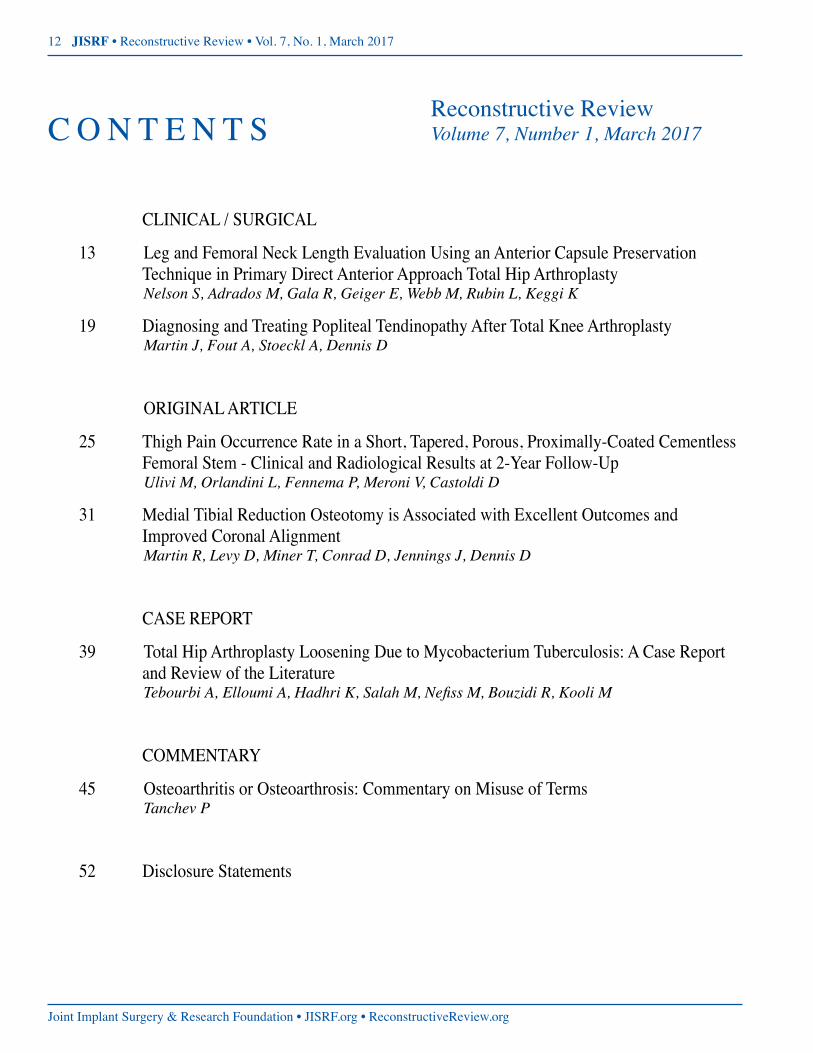
Reconstructive ReviewC O N T E N T S Volume 7, Number 1, March 2017
CLINICAL / SURGICAL
13 Leg and Femoral Neck Length Evaluation Using an Anterior Capsule Preservation Technique in Primary Direct Anterior Approach Total Hip Arthroplasty
Nelson S, Adrados M, Gala R, Geiger E, Webb M, Rubin L, Keggi K
19 Diagnosing and Treating Popliteal Tendinopathy After Total Knee Arthroplasty Martin J, Fout A, Stoeckl A, Dennis D
ORIGINAL ARTICLE
25 Thigh Pain Occurrence Rate in a Short, Tapered, Porous, Proximally-Coated Cementless Femoral Stem - Clinical and Radiological Results at 2-Year Follow-Up
Ulivi M, Orlandini L, Fennema P, Meroni V, Castoldi D
31 Medial Tibial Reduction Osteotomy is Associated with Excellent Outcomes and Improved Coronal Alignment
Martin R, Levy D, Miner T, Conrad D, Jennings J, Dennis D
CASE REPORT
39 Total Hip Arthroplasty Loosening Due to Mycobacterium Tuberculosis: A Case Report and Review of the Literature
TebourbiA,ElloumiA,HadhriK,SalahM,NefissM,BouzidiR,KooliM
COMMENTARY
45 Osteoarthritis or Osteoarthrosis: Commentary on Misuse of Terms Tanchev P
52 Disclosure Statements

Volume 7, Number 1March 2017An Open Access Journal
ReconstructiveReview.org • JISRF.org • Joint Implant Surgery & Research Foundation
C L I N I C A L / S U R G I C A L http://dx.doi.org/10.15438/rr.7.1.153
Leg and Femoral Neck Length Evaluation Using an Anterior Capsule Preservation
Technique in Primary Direct Anterior Approach Total Hip Arthroplasty
Nelson S 1, Adrados M 1, Gala R 1, Geiger E 1, Webb M 1, Rubin L 1 , Keggi K 1
1 Stephen J Nelson MD, Murillo Adrados MD, Raj J Gala MD, Erik J Geiger MD, Matthew L Webb MD, Lee Rubin MD, Kristaps J Keggi MD
Department of Orthopaedics and Rehabilitation, Yale School of Medicine, 800 Howard Avenue, New Haven, CT, 06510, USA
(Direct reprint requests to Stephen J Nelson)
Abstract
Background: Achieving correct leg and femoral neck lengths remains a challenge during total hip arthroplasty (THA). Several methods for intraoperative evaluation and restoration of leg length have been proposed, and each has inaccuracies and shortcomings. Both the supine positioning of a patient on the operating table during the di-rect anterior approach (DAA) THA and the preservation of the anterior capsule tissue are simple, readily avail-able, and cost-effective strategies that can lend themselves well as potential solutions to this problem.
Technique: The joint replacement is performed through a longitudinal incision (capsulotomy) of the anterior hip joint capsule, and release of the capsular insertion from the femoral intertrochanteric line. As trial components of the prosthesis are placed, the position of the released distal capsule in relationship to its original insertion line is an excellent guide to leg length gained, lost, or left unchanged.
Methods: The radiographs of 80 consecutive primary THAs were reviewed which utilized anterior capsule preservation and direct capsular measurement as a means of assessing change in leg/femoral neck length. Preop-eratively, the operative legs were 2.81 +/- 8.5 mm (SD) shorter than the nonoperative leg (range: 17.7 mm longer to 34.1 mm shorter). Postoperatively, the operative legs were 1.05 +/- 5.64 mm (SD) longer than the nonopera-tive leg (range: 14.9 mm longer to 13.7 mm shorter).
Conclusion: The preservation and re-assessment of the native anterior hip capsule in relationship to its point of release on the femur is a simple and effective means of determining leg/femoral neck length during DAA THA.Keywords: total hip arthroplasty; leg length discrepancy; femoral neck length; direct anterior approach; hip capsule; capsulotomy; capsulectomy; capsulorrhaphylevel of evidence: AAOS Therapeutic Level IV
© 2017 Nelson, Adrados, Gala, Geiger, Webb, Rubin, Keggi. All rights reserved.Authors retain copyright and grant the journal right of first publication with the work. Reconstructive Review follows the Creative Commons Attribution-NonCommercial CC BY-NC. This license allows anyone to download works, build upon the material, and share them with others for non-commercial purposes as long as they credit the senior author, Reconstructive Review, and the Joint Implant Surgery & Research Foundation (JISRF). An example credit would be: “Courtesy of (senior author’s name), Reconstructive Review, JISRF, Chagrin Falls, Ohio”.

14 JISRF • Reconstructive Review • Vol. 7, No. 1, March 2017
Joint Implant Surgery & Research Foundation • JISRF.org • ReconstructiveReview.org
Introduction
Maintaining both hip stability and appropriate leg lengths is one of the great challenges of total hip arthro-plasty (THA). Incorrect leg length is a major cause of mor-bidity after THA and has been associated with back pain, sciatica, neuritis, gait disorders, general dissatisfaction, early loosening of components and revision surgery [1-7]. Additionally, patients who perceive leg length discrepancy have been found to have worse Oxford Hip scores. [8,9] With such morbidity, LLD is second only to nerve injury as the most common cause of litigation after THA [10]. As such, the potential for iatrogenic leg length discrepan-cy is a known risk of THA that should be discussed with patients before surgery and documented accordingly, ex-plaining that a minor increase in leg length is not uncom-mon and perhaps preferable to a dislocated hip.
Preoperative assessment and implant templating are im-portant considerations. Templating is an important guide to intraoperative decision-making, but excellent planning does not guarantee excellent execution [11]. Actual sizing of implanted components has been reported to match pre-operative templating in only 60% of cases [12], and there-fore should not be relied upon as the only means of deter-mining leg or femoral neck length.
There have been over 20 intraoperative methods de-scribed for evaluating limb length and include the Ober test, the shuck test, and the drop kick test [13,14]. These methods utilize soft tissue tension with components in place as an indicator for limb length, but anesthesia in-duced muscle relaxation may limit the trustworthiness of these tests. Direct leg-to-leg comparison can also be useful but the palpation of anatomic landmarks may be inaccurate under surgical drapes, especially during lateral decubitus patient positioning for THA.
Fluoroscopically guided THA has increased in popular-ity recently, as it can ensure correct component position ra-diographically and may facilitate proper leg lengths. How-ever, this method potentially adds operative time, requires both a radiolucent table and an image intensifier, and may increase required personnel and equipment traffic, all of which can carry an increased risk of wound contamination and surgical site infection. The additional radiation expo-sure to patient and staff is also a matter to consider [15].
Intraoperative navigation and length measurement tech-niques are usually based on 2 reference points marked on the pelvis and femur. This can be achieved via iliac fixation pins, intraoperative calipers, infracotyloid pins, or fixed suture lengths [16]. None of these methods are perfect, and many inconsistencies have been described [11].
Computer navigation and robot assisted surgery have
also been developed to reconstruct normal anatomy and proper leg length. However, these methods are expensive, not widely available, and do not address the key issue of restoring soft tissue tension. For the present, they pale to the intraoperative judgement of an experienced surgeon.
The senior author and his colleagues have used the an-terior, internervous, muscle-sparing surgical approach since 1973, in some 15,000 primary and revision hip ar-throplasties [17-19]. This anterior approach was presented as a scientific exhibit at the Annual Meeting of the Ameri-can Academy of Orthopaedic Surgeons (AAOS) in 1977, and published in 1980 [17]. During the first 30 years of his work with the DAA, a total or subtotal capsulectomy was the norm. The results were satisfactory, but to more ac-curately restore leg length and attempt to further increase joint stability, a technique for anterior hip capsulotomy with anatomic capsule preservation, whenever possible, was evolved. It has helped to decrease soft tissue dissec-tion, reduce the dead space created by a capsulectomy, and has the additional benefit of allowing for the intraopera-tive assessment of leg/femoral neck length, as is described within this paper.
Surgical Technique
With the patient supine on the operating table, a short oblique incision is made distal and lateral to the anterior superior iliac spine overlying the femoral neck. The me-dial border of the tensor fascia lata muscle is identified. The muscle and its fascia are then split longitudinally, ap-proximately 1 cm lateral to its medial border. The medial strip of fascia and muscle can add to safety by protecting the small branches of the lateral femoral cutaneous nerve and the femoral neurovascular bundle. Cobra retractors are then placed on the superior and inferior aspects of the hip capsule. The superior Cobra retracts the tensor fascia lata and the abductors. The inferior Cobra retracts the rectus, sartorius, and iliopsoas. These two Cobras provide excel-lent exposure of the anterior hip capsule, which can then be further defined by the removal of its overlying fatty tissues.
An assessment is then made if further capsular visual-ization is necessary, which can be achieved by dissection with elevation and/or release of the reflected head of the rectus femoris by the proximal placement of a third blunt Cobra under its fibrous tendon with the tip of the Cobra in place over the anterior acetabulum with the tip just over the anterior rim of pelvis. In most cases of elderly patients without excessive acetabular or femoral head osteophytes and an atrophied reflected rectus, this third Cobra is not necessary and the capsule can be incised by starting the

Leg and Femoral Neck Length Evaluation Using an Anterior Capsule Preservation Technique in Primary Direct… 15
ReconstructiveReview.org • JISRF.org • Joint Implant Surgery & Research Foundation
incision underneath the tendon, but the morphology of the reflected head can be larger and more muscular, particular-ly in younger males.
The exposed anterior capsule is incised in line with the long axis of the underlying femoral neck, and released from its distal lateral insertion on the intertrochanteric line (Figure 1). If necessary and if it is tight, the medial distal portion can also be released, creating an inverted T-capsu-lotomy. The two Cobras are then placed inside of the cap-sule on the superior and inferior portions of the femoral
neck. The pre-planned base of the femoral neck cut is then performed, the femoral head removed, the acetabulum pre-pared, and the prosthetic components inserted. Depending on the exposure and mobilization needed, more, or all, of the femoral capsule can be dissected and released, yet pre-served for subsequent repair. The placement of the compo-nents can be performed in a variety of ways, ranging from their insertion without any trial components, to total fluo-roscopic control. Based on our surgical experience, excel-lent visualization of the hip and the anatomical position of the patient’s spine, pelvis, and legs, we rarely use tri-al components other than femoral heads, but recommend them if there is any question about achieving satisfactory component position.
After the trial femoral head is placed, femoral neck length is then assessed by approximation of the hip cap-sule to the distal intertrochanteric line with the leg held in a neutral position, with slight flexion and internal rotation (Figure 2). If the capsule overhangs its release point on the intertrochanteric line, then the pre-operative limb/femoral neck length has been shortened. If the capsule does not reach the point where it was released, then the limb/femo-
ral neck has been lengthened by the components. Thus, a simple look at the restored position of the re-
leased capsule will allow the selection of the final femoral head to be used to achieve the leg length correction deter-mined by the pre-operative x-rays. Intrinsic kinematic sta-bility of the THA is of paramount importance, and it must be tested by putting the hip through a complete arcs of mo-tion. This can only be achieved when no traction table is used, because the leg is draped free and can be moved in all planes during the procedure to test a full range of motion and ensure there is no impingement or subluxation dur-ing the procedure prior to closure. If the joint is found to be unstable, the instability can then be corrected by an in-crease of the neck length by a few millimeters, or upsizing the head and liner diameter, which can be the alternative to more extreme surgical measures.
Figure 1. Planned capsular incisions performed during the DAA to THA. (Image courtesy of Kristaps J. Keggi, MD)
Figure 2. Illustrations demonstrating capsule position when assessing leg length. Illustration A demonstrates the native capsule position aftercapsulotomy.InillustrationB,thecapsuleoverhangsitssiteof release along the intertrochanteric line demonstrating that the leg is shorter than it was pre-operatively. Illustration C demonstrates a gap between the proximal and distal capsular limbs, indicating the leg has been lengthened. Illustration D shows the capsule position to be unchanged. (Illustration by Genevra Garrett)

16 JISRF • Reconstructive Review • Vol. 7, No. 1, March 2017
Joint Implant Surgery & Research Foundation • JISRF.org • ReconstructiveReview.org
Methods
After obtaining IRB approval to perform a medical re-cord review, the records database was queried for prima-ry, unilateral total hip arthroplasty (CPT 27130) performed between 2011 and 2012. A retrospective review was per-formed and radiographs of 80 consecutive patients with complete preoperative and postoperative images were re-viewed on the radiology program PACS (Picture Archiving and Communication System). Direct capsular measure-ment was the primary determinant of leg length in these patients. For radiographic review, a line was drawn be-tween the base of the teardrops and the vertical distance between this line and the lesser trochanters was measured for both the operative and nonoperative legs (Figure 3). Limb lengths were recorded in millimeters and analyzed using Microsoft Excel (Redmond, Washington, USA).
Exclusion criteria included cases performed with a si-multaneous bilateral technique, cases with incomplete re-cords or radiographs, or cases with severe deformity. More specifically in cases involving major deformities, bone loss, or severe flexion contractures, we would expect a pathologically contracted, adherent, and compromised an-terior capsule that is best managed with a radical capsulec-tomy during the procedure, precluding the use of a routine capsulotomy.
Results and Discussion
Preoperatively, many patients are shorter on the affect-ed side due to cartilage destruction and bone remodeling secondary to the disease process. On average, our results revealed that preoperatively, the operative legs were 2.81 +/- 8.5 mm (SD) shorter than the nonoperative leg (range 17.7 mm longer to 34.1 mm shorter). Postoperatively, the operative legs were 1.05 +/- 5.64 mm (SD) longer than the nonoperative leg (range 14.9 mm longer to 13.7 mm shorter) (Table 1). Seven patients had a postoperative hip height discrepancy of greater than 10 mm, however each of these patients had a similar discrepancy of over 10 mm preoperatively (Figure 4). Preoperative deformity, compo-nent choice, and implant position were taken into consid-eration for these patients in an effort to not over correct. Leg length equality was sought in every case unless it sac-rificed hip stability or would alter an otherwise compensat-ed pelvic balance in those patients with a concurrent fixed scoliotic deformity.
Several novel alternative methods for assessing femoral neck length have been described including comparing the trial head and neck implants with the osteotomized femoral head using visual assessment [20]. Similar to anterior cap-sule assessment, this method may also be expeditious, and
Table 1. Patient demographics and limb length discrepancies Male 36 (45%)Age (SD) 67 (+/- 12.4)Left leg surgery 32 (40%)Preoperative limb length discrepancy (SD) -2.81 (+/- 8.50) mmPostoperative limb length discrepancy (SD) 1.05 (+/- 5.64) mm
Figure 3. Patient radiographs demonstrating preoperative limb shortening due to destruction of cartilage and subchondral bone and postoperative reproduction of hip length to less than a millimeter.
Figure 4. Frequencies of hip length discrepancies preoperatively and postoperatively.

Leg and Femoral Neck Length Evaluation Using an Anterior Capsule Preservation Technique in Primary Direct… 17
ReconstructiveReview.org • JISRF.org • Joint Implant Surgery & Research Foundation
cost-effective, but may require additional equipment or specialized measurement jig. In general during our cases, a second subcapital osteotomy is created in addition to the primary basicervical osteotomy, creating a “napkin ring” cut of femoral neck bone. Thus, to measure the excised head and neck, one would need to place these together with two free saw blades to gain an accurate measurement, which is objective, but perhaps more subjectively difficult to assess than our capsular assessment as described within this paper.
A number of technical factors have been associated with leg length discrepancy, including uncemented femo-ral stems [21]. All patients in this evaluation underwent THA with uncemented components. The direct anterior ap-proach for THA with a capsular sparing lends itself well to component positioning and the achievement of consistent, accurate leg length restoration.
Other potential benefits of capsular repair include in-creased stability and infection protection. To our knowl-edge, no studies have examined the stability augmented by capsular repair of THA performed through the DAA, how-ever, Hughes et al. examined the effect of capsular repair in cadavers following hip hemiarthroplasty through a direct lateral approach and found that capsular repair required a 4-fold higher peak torque force to dislocate anteriorly [22]. Infection reduction in capsular repair has also not been di-rectly evaluated, however, preserving additional anatomic layers might assist in microbial blockade.
In cases of major deformities, bone loss, or severe flex-ion contractures with compromised anterior capsules, the method we have described to “fine tune” the average hip to a few millimeters will not apply and the selection of pros-thetic components and femoral heads to achieve optimum leg length and joint stability will depend on x-rays, opera-tive findings and clinical judgment.
This paper discusses a novel technique for assessment of leg length discrepancy after THA and provides objec-tive numerical analysis to support the accuracy of this technique at our institution. The limitations of this article include the small size of its series and that all cases were performed by a single surgeon at a single institution. Addi-tionally, radiographs were not standardized and there may be small differences in magnification and rotation among the plain films. Due to variances in rotation of the images, we were unable to assess femoral offset which is critical to stability and abductor function, which enhance hip func-tion after THA [23].
Notably, this is a retrospective evaluation, and thus mul-tiple factors aside from capsular measurement were likely used for intraoperative evaluation of leg length; most nota-bly these included the surgeon’s assessment of hip stabil-
ity, shuck, and intra-operative use of the medial malleoli to measure the operative limb to the non-operative limb dur-ing the supine DAA. Future prospective evaluations com-paring the various intraoperative assessments of leg length are thus necessary to determine the most accurate and re-producible method from among these options to produce reliable leg length equality. In addition, comparison of the capsular preservation technique described here to technol-ogies such as fluoroscopy or navigation guidance would lend additional insight to this topic in the future.
disclosureThe authors declare that there is no conflict of interest
regarding the publication of this paper. For full disclosures refer to last page of this journal.
References1. FRIBERG O. Clinical symptoms and biomechanics of lumbar spine and hip-joint in leg length
inequality. Spine. 1983;8(6):643-651. doi: 10.1097/00007632-198309000-00010.2. Mihalko W, Phillips M, Krackow K. Acute sciatic and femoral neuritis following to-
tal hip arthroplasty - A case report. Journal of Bone and Joint Surgery-American Volume. 2001;83A(4):589-592.
3. Rosler J, Perka C. The effect of anatomical positional relationships on kinetic parameters after total hip replacement. Int Orthop. 2000;24(1):23-27. doi: 10.1007/s002640050006.
4. Ranawat C. The pants too short, the leg too long! Orthopedics. 1999;22(9):845-846.5. WOO R, MORREY B. Dislocations after total hip-arthroplasty. Journal of Bone and Joint Sur-
gery-American Volume. 1982;64(9):1295-1306.6. Amstutz H, Ma S, Jinnah R, Mai L. Revision of aseptic loose total hip arthroplasties. Clin
Orthop. 2004(420):2-9.7. Austin M, Hozack W, Sharkey P, Rothman R. Stability and leg length equality in total hip ar-
throplasty. J Arthroplasty. 2003;18(3):88-90. doi: 10.1054/arth.2003.50073.8. Konyves A, Bannister G. The importance of leg length discrepancy after total hip arthro-
plasty. Journal of Bone and Joint Surgery-British Volume. 2005;87B(2):155-157. doi: 10.1302/0301-620X.87B2.14878.
9. Wylde V, Whitehouse SL, Taylor AH, Pattison GT, Bannister GC, Blom AW. Prevalence and functional impact of patient-perceived leg length discrepancy after hip replacement. Int Or-thop. 2009;33(4):905-909. doi: 10.1007/s00264-008-0563-6.
10. Upadhyay A, York S, Macaulay W, McGrory B, Robbennolt J, Bal BS. Medical malpractice in hip and knee arthroplasty. J Arthroplasty. 2007;22(6):2-7. doi: 10.1016/j.artli.2007.05.003.
11. Ng VY, Kean JR, Glassman AH. Limb-length discrepancy after hip arthroplasty. Jour-nal of Bone and Joint Surgery-American Volume. 2013;95A(15):1426-1436. doi: 10.2106/JBJS.L.00433.
12. Knight J, Atwater R. Preoperative planning for total hip arthroplasty: Quantitating it’s utility and precision. J Arthroplasty. 1992;7:403-409.
13. Ranawat C, Rodriguez J. Functional leg-length inequality following total hip arthroplasty. J Arthroplasty. 1997;12(4):359-364. doi: 10.1016/S0883-5403(97)90190-X.
14. Charles M, Bourne R, Davey J, Greenwald A, Morrey B, Rorabeck C. Soft-tissue balancing of the hip - the role of femoral offset restoration. Journal of Bone and Joint Surgery-American Volume. 2004;86A(5):1078-1088.
15. McArthur BA, Schueler BA, Howe BM, Trousdale RT, Taunton MJ. Radiation exposure dur-ing fluoroscopic guided direct anterior approach for total hip arthroplasty. J Arthroplasty. 2015;30(9):1565-1 568. doi: 10.1016/j.arth.2015.03.029.
16. Desai A, Dramis A, Board T. Leg length discrepancy after total hip arthroplasty: A review of literature. Curr Rev Musculoskelet Med. 2013;6(4):336-341.
17. Light TR, Keggi KJ. Anterior approach to hip-arthroplasty. Clin Orthop. 1980(152):255-260.18. Kennon R, Keggi J, Zatorski L, Keggi K. Anterior approach for total hip arthroplasty: Be-
yond the minimally invasive technique. Journal of Bone and Joint Surgery-American Vol-ume. 2004;86A:91-97.
19. Kennon R, Keggi J, Wetmore R, Zatorski L, Huo M, Keggi K. Total hip arthroplasty through a minimally invasive anterior surgical approach. Journal of Bone and Joint Surgery-American Volume. 2003;85A:39-48.
20. Alazzawi S, Douglas SL, Haddad FS. A novel intra-operative technique to achieve ac-curate leg length and femoral offset during total hip replacement. Ann R Coll Surg Engl. 2012;94(4):281-282.
21. Ahmad R, Sharma V, Sandhu H, Bishay M. Leg length discrepancy in total hip arthroplasty with the use of cemented and uncemented femoral stems. A prospective radiological study. Hip International. 2009;19(3):264-267.
22. Hughes AW, Clark D, Carlino W, Gosling O, Spencer RF. Capsule repair may reduce disloca-tion following hip hemiarthroplasty through a direct lateral approach. Bone & Joint Journal. 2015;97B(1):141-144. doi: 10.1302/0301-620X.97B1.34038.
23. Liebs TR, Nasser L, Herzberg W, Ruether W, Hassenpflug J. The influence of femoral off-set on health-related quality of life after total hip replacement. Bone & Joint Journal. 2014;96B(1):36-42.

18 JISRF • Reconstructive Review • Vol. 7, No. 1, March 2017
Joint Implant Surgery & Research Foundation • JISRF.org • ReconstructiveReview.org
PRODUCT NO:
2741-00 [Set]Also Available Individually
PRODUCT NO’S:
4910 [Rake Retractor]4920-01 [Cement Scraper – Right]4920-02 [Cement Scraper – Left]4930-01 [Offset Osteotome]4930-02 [Offset Osteotome – Double Bent]
Enhances minimally invasive exposure of the medial femoral condyle
Engh Intercondylar Notch RetractorDesigned by Gerard A. Engh, MD
Scott Uni & Total Knee Cement Removing Curette
Usage guide available online.
Engh Unicondylar Minimally Invasive Knee Surgery Instruments
Designed for use in unicondylar minimally invasive knee surgery
Designed by Gerard A. Engh, MD
Placed in the medial/lateral tibial recess while making the horizontal tibial cut during unicompartmental knee arthroplasty—helping to retract and protect the medial and lateral collateral ligaments
Uni Medial/Lateral Ligament RetractorDesigned by Kurt Kramer, PA-C
Rosenstein Tibial Fragment Grasper for UKA
Designed to help remove the tibial bone fragment in one piece during Unicompartmental Knee Arthroplasty
Designed by Alexander D. Rosenstein, MD
PRODUCT NO’S:
3230-01 [Small - 9 mm]3230-02 [Medium – 10 mm]3230-03 [Large – 12 mm]
PRODUCT NO:
3632
PRODUCT NO:
1720
Patient Self Stress Assembly SetDesigned to help position a patient for X-ray evaluation to help determine candidacy for UKA
Designed by Kyle Cook, RTR and David Mauerhan, MD
FREE TRIAL ON MOST INSTRUMENTS
1.800.548.2362103 Estus Drive, Savannah, GA 31404www.innomed.net [email protected]
912.236.0000 Phone 912.236.7766 Fax
Innomed-Europe Tel. +41 41 740 67 74 Fax +41 41 740 67 71© 2016 Innomed, Inc.
Scan to Launch Our
WebsiteISO 9001:2008 • ISO 13485:2003
Kamath Uni Knee Tibial ImpactorAssists in MIS unicompartmental cemented tibial tray impaction
Designed by Atul F. Kamath, MD
Sized, shaped and angled 90° to help with retrieval of posteriorly extruded cement behind the tibial component in both total and unicompartmental knee arthroplasty
Designed by Richard D. Scott, MD
Titanium nitride
coatied
PRODUCT NO:
4247
PRODUCT NO:
1129
Reconstructive Review Ads.indd 9 2/3/17 4:33 PM

Volume 7, Number 1March 2017An Open Access Journal
ReconstructiveReview.org • JISRF.org • Joint Implant Surgery & Research Foundation
C L I N I C A L / S U R G I C A L http://dx.doi.org/10.15438/rr.7.1.172
Diagnosing and Treating Popliteal Tendinopathy After Total Knee Arthroplasty
Martin J 1, Fout A 2, Stoeckl A 2, Dennis D 1
1 J. Ryan Martin, MD; Douglas A Dennis, MD Colorado Joint Replacement, Denver, CO 80210
2 Allison Fout, PA-C; Andrew C Stoeckl, MD Excelsior Orthopaedics, Amherst, NY 14226
(Direct reprint requests to J. Ryan Martin)
Abstract
The following office tip describes four patients that underwent primary total knee arthroplasty and developed posterolateral knee pain at a mean follow-up duration of 1.6 months postoperatively. The first patient in this series noted substantial pain lying in bed (in a lateral decubitus position with the operative leg up) while attempting to abduct her leg to adjust her sheet in bed. A thorough clinical and radiographic work-up was performed. This patient’s posturing in bed (and subsequent physical exam maneuver) led to a presumptive diagnosis of popliteal tendinopathy. The diagnosis was confirmed arthroscopically by identifying a frayed and inflamed popliteal tendon. After undergoing arthroscopic popliteal tendon re-lease, the patient noted complete pain relief while retaining coronal stability in both flexion and exten-sion. The following office tip defines a previously undescribed clinical diagnostic examination for pop-liteal tendinopathy that was identified based on a patient’s symptomatology and subsequently utilized to identify three additional cases of arthroscopically confirmed popliteal tendinopathy.
Keywords: total knee arthroplasty; popliteal tendinosis; popliteal tendonitis; tendinopathy; posterolateral knee painlevel of evidence: AAOS Therapeutic Level IV
© 2017 Martin, Fout, Stoeckl, Dennis. All rights reserved.Authors retain copyright and grant the journal right of first publication with the work. Reconstructive Review is an open access publication and follows the Creative Commons Attribution-NonCommercial CC BY-NC. This license allows anyone to download works, build upon the material, and share them with others for non-commercial purposes as long as they credit the senior author, Reconstructive Review, and the Joint Implant Surgery & Research Foundation (JISRF). An example credit would be: “Courtesy of (senior author’s name), Reconstructive Review, JISRF, Chagrin Falls, Ohio”.
Introduction
Pain following primary total knee arthroplasty (TKA) is unfortunately a common finding [1]. There are numer-ous causes for a painful TKA, which make diagnosing the source of the pain even more challenging [2,3]. Popliteal tendinopathy is an uncommon cause of pain, which can present with posterolateral knee pain, often associated with motion-induced crepitus in the region of the popliteal ten-don. The first description of popliteal tendon dysfunction during primary TKA was noted intraoperatively at the time of capsular closure [4]. The authors noted a “popping” that occurred during flexion and extension. After release of the popliteal tendon, they noted that the “popping” disappeared and that the knee was stable. Since this study, Insall et. al.

20 JISRF • Reconstructive Review • Vol. 7, No. 1, March 2017
Joint Implant Surgery & Research Foundation • JISRF.org • ReconstructiveReview.org
proposed arthroscopic popliteal tendon release for patients diagnosed with popliteal tendon dysfunction following pri-mary TKA [5]. However, to our knowledge, other than the presence of patient-reported posterolateral discomfort, we are unaware of any single clinical examination maneuver to precisely diagnose popliteal tendinopathy following pri-mary TKA. The following office tip describes a clinical ex-amination maneuver utilized to diagnose four patients with popliteal tendinopathy, and secondarily confirmed at the time of arthroscopic release of the popliteal tendon.
Office Tip
The diagnosis of post-operative popliteal tendinopathy following total knee arthroplasty can be made with a sim-ple clinical test. Each case of popliteal tendinopathy was confirmed arthroscopically and after release of the tendon, resulted in complete relief of symptoms.
Case Examples
All but one of the patients in this series underwent pri-
mary TKA by one of the senior authors at a single institu-tion. The primary TKA was performed utilizing a gap bal-ancing approach with a posterior stabilized mobile bearing Attune TKA (Warsaw, IN). The postoperative course was uneventful in each case. However, approximately 2 months postoperatively, the first patient in this series presented with posterolateral knee pain. This patient was a 70 year old female with a preoperative diagnosis of osteoarthri-tis of her right knee. Preoperative radiographs of her right knee demonstrated tricompartmental arthritis and chon-drocalcinosis (Figure 1). Postoperative radiographs of her right knee demonstrated a well-fixed, well-aligned TKA without evidence of loosening, lysis, lateral overhanging femoral component, or lateral femoral osteophytes (Fig-ure 2).
During her clinical evaluation, she localized the pain to the posterolateral aspect of her knee, and noted no signifi-cant pain when ambulating or sitting. Her pain could not be reproduced on clinical examination, although the pos-terolateral aspect was diffusely painful to palpation. Inter-estingly, she noted substantial pain lying in a lateral decu-bitus position in bed with the operative extremity up when abducting her leg while attempting to adjust the bed sheets (Figure 3). Repeat of this maneuver consistently repro-
Figure 1. AP, lateral and patellar view of the right knee demonstrates severe patellofemoral arthritis with chondrocalcinosis.
Figure2.PostoperativeAPandpatellarviewdemonstrateawell-fixedwell-alignedrighttotalkneearthroplasty.Theredoesnotappeartobeevidence of lateral femoral osteophytes or femoral component overhang.

Diagnosing and Treating Popliteal Tendinopathy After Total Knee Arthroplasty 21
ReconstructiveReview.org • JISRF.org • Joint Implant Surgery & Research Foundation
duced the posterolateral pain. This position applied a varus stress to a flexed knee, a position where the popliteal ten-don acts an important static restraint in varus stability [6]. Therefore, it was hypothesized that this maneuver could be utilized to assess for popliteal tendinopathy in the setting of TKA. The patient was treated with conservative mea-sures including activity modification, icing, and NSAID use for three months with continued pain.
After discussing the proposed mechanism for postero-lateral pain to this patient, an arthroscopic release of the popliteal tendon was recommended and performed seven months after primary TKA. Intraoperatively, the popliteal tendon was visualized and noted to have substantial ten-donitis, fraying, and was shown to impinge slightly on the femoral component of the TKA (Figure 4). Utilizing the ar-throsopic shaver, the popliteal tendon was released without any additional procedures performed.
After the arthroscopic popliteal tendon release, the pa-tient noted complete relief of her posterolateral pain within 48 hours following the operative procedure. On her most recent clinical appointment, six months after surgery, she was noted to have complete resolution of her pain. Addi-tionally, her knee was stable to varus and valgus stress-es in flexion and extension. Her radiographs demonstrated no change, with continued excellent alignment and no evi-dence of radiolucencies or lysis.
Since the first patient presentation, three additional pa-
tients presented with similar posterolateral knee pain. Uti-lizing the clinical examination maneuver described above with a varus stress applied to the patient’s operative ex-tremity in a lateral decubitus position, each patient’s pain was readily reproduced. The average duration from TKA to the diagnosis of popliteal tendonitis was 1.6 months. The arthroscopic findings in each patient were diagnostic of popliteal tendinopathy. Subsequently, all patients noted complete or near complete resolution of their pain without any evidence of coronal or sagittal instability identified at final follow-up.
Discussion
Postoperative knee pain following primary TKA is un-fortunately a common finding [1]. Furthermore, identify-ing the source of pain can occasionally be a diagnostic di-lemma due to the wide variety of pathologies that can lead to knee pain in this patient population [2,3,7]. One uncom-mon source of postoperative knee pain is popliteal tendi-nopathy [5]. Although there have been case reports de-scribing treatments for popliteal tendon dysfunction, little is known about how to best identify this clinical condition precisely [4]. To our knowledge, a diagnostic clinical ex-amination maneuver for popliteal tendinopathy following TKA has yet to be described prior to this study. Therefore, the following case series describes a unique clinical tech-nique of identifying popliteal tendon dysfunction with sub-
Figure 4. Arhtroscopic views of the right knee. A: The lateral compartmentoftherightdemonstratessubstantialsynovitisand(B)frayingofthepoplitealtendon.C:Thearthroscopicshaverisutilizedto resect the popliteal tendon. D: The popliteal tendon has been completely released.
Figure 3. The above clinical photographs demonstrate the clinical exam maneuver for diagnosing popliteal tendonitis. (A) The patient is placed in a lateral decubitus position with the operative extremity up. The patient then abducts their hip against gravity resistance with theirkneeinfullextension.(B)Withtheirhipabductedthepatientflexiontheirkneefrom0to90degrees.
(A)
(B)
(A)
(C)
(B)
(D)

22 JISRF • Reconstructive Review • Vol. 7, No. 1, March 2017
Joint Implant Surgery & Research Foundation • JISRF.org • ReconstructiveReview.org
sequent arthroscopic confirmation of the diagnosis. Addi-tionally, a short-term clinical and radiographic follow-up was performed on this patient population to ensure resolu-tion of symptoms.
The proposed diagnostic maneuver requires the pa-tient to lie supine with their operative extremity elevated. The examiner then has the patient abduct the operative ex-tremity while an adduction force is applied to the lower leg close to the ankle joint. This force results in a varus stress through the knee. As was discussed previously, the popliteal tendon is an important varus stabilizer in knee flexion [6]. Therefore, this clinical test appears to specifi-cally stress the popliteal tendon. Each patient in this series had nonspecific posterolateral knee pain, which could not be reliably reproduced with common clinical examination maneuvers that apply varus and valgus stresses in flexion and extension. After discovering this maneuver during the first patient encounter in this series, all subsequent patients stated that their pain was reliably reproduced.
The source for popliteal tendinopathy has been attrib-uted to a retained lateral femoral condylar osteophyte or posterior femoral component impingement, often associ-ated with excessive lateral positioning of the femoral com-ponent beyond the osseous perimeter of the lateral femoral condyle [5]. Both of these conditions can increase tension on the traversing popliteal tendon resulting in irritation. Additionally, arthroscopic release of the popliteal tendon was proposed as a standard treatment for patients diag-nosed with popliteal tendinopathy [8]. The release of the tendon in this patient population was not associated with an increase in instability and had reliable pain relief. How-ever, a recent study demonstrated a decrease in patient out-comes at two to three years postoperatively in a cohort of 15 patients that had accidental iatrogenic intraoperative popliteal tendon release [9]. Although assessing a different outcome, these results appear to differ from Kesman et. al., who prospectively enrolled eighteen patients to randomly have their popliteal tendon released by the first assistant, while the senior surgeon was blinded to the presence or absence of the release [10]. The senior surgeon, with tri-als in place, would assess ligamentous stability. No subjec-tive difference in static stability was noted between the two cohorts. It is possible that the arthroscopic release and the intraoperative isolated release of the popliteus tendon are more precise methods for releasing the popliteal tendon. Whereas the iatrogenic, saw blade release, of the popliteal tendon could potentially release the posterolateral corner of the knee resulting in worse outcomes. However, longer outcomes are necessary to determine if an arthroscopic re-lease of the popliteal tendon will impact patient outcomes.
There are several notable limitations to the following
study. First, this was a retrospective case series, and there-fore has inherent limitations of both the study design and the small patient population. However, we still believe this diagnostic maneuver is useful in identifying popliteal ten-dinopathy, an uncommon cause of postoperative knee pain. Second, this clinical test has not been validated or com-pared to other diagnostic tests. Therefore the diagnostic ac-curacy is unknown. Although we did note arthroscopic ev-idence of popliteal tendinopathy and complete pain relief after release in each patient, a comparative study includ-ing imaging and other exam maneuvers to determine the validity of this exam are warranted. Third, it is possible that each patient’s tendinopathy may have resolved with-out surgical treatment. Future studies should be centered on the natural progression of popliteal tendinopathy after total knee arthroplasty which may help direct the timing of surgical intervention in this patient population. Finally, long-term clinical follow-up is necessary to determine if there are any clinical consequences of popliteal tendon re-lease. As was discussed previously, one study noted worse outcomes in patients that had iatrogenic popliteal tendon release during primary TKA [9]. This may represent a pos-terolateral corner release and therefore may not be gener-alizable to the isolated popliteal tendon release noted in this study.
Summary
The following study demonstrates a clinical examina-tion method for diagnosing popliteal tendon dysfunction. This exam was performed on four patients in this series and the diagnosis of popliteal tendinopathy was confirmed arthroscopically in each case. An arthroscopic popliteal tendon release was subsequently performed and resulted in complete pain relief in all patients. Additionally, no post-operative instability was identified. This clinical examina-tion should be considered and utilized when confronting the common diagnostic dilemma of a painful TKA, espe-cially posterolateral knee pain that is difficult to reproduce clinically.
disclosureThe authors declare that there is no conflict of interest
regarding the publication of this paper. For full disclosures refer to last page of this journal.

Diagnosing and Treating Popliteal Tendinopathy After Total Knee Arthroplasty 23
ReconstructiveReview.org • JISRF.org • Joint Implant Surgery & Research Foundation
References:1. Gandhi R, Davey JR, Mahomed NN. Predicting patient dissatisfaction following
joint replacement surgery. J Rheumatol. 2008;35(12):2415-2418. doi:10.3899/jrheum.080295.
2. Dennis DA. Evaluation of painful total knee arthroplasty. J Arthroplasty. 2004;19(4 Suppl 1):35-40. http://www.ncbi.nlm.nih.gov/pubmed/15190547. Ac-cessed June 17, 2016.
3. Potty AGR, Tzeng TH, Sams JD, et al. Diagnosis and Management of Intra-articu-lar Causes of Pain After Total Knee Arthroplasty. Instr Course Lect. 2015;64:389-401. http://www.ncbi.nlm.nih.gov/pubmed/25745923. Accessed June 17, 2016.
4. Barnes CL, Scott RD. Popliteus tendon dysfunction following total knee ar-throplasty. J Arthroplasty. 1995;10(4):543-545. http://www.ncbi.nlm.nih.gov/pubmed/8523017. Accessed May 2, 2016.
5. Allardyce TJ, Scuderi GR, Insall JN. Arthroscopic treatment of popliteus tendon dysfunction following total knee arthroplasty. J Arthroplasty. 1997;12(3):353-355. http://www.ncbi.nlm.nih.gov/pubmed/9113555. Accessed June 17, 2016.
6. Nielsen S, Helmig P. The static stabilizing function of the popliteal tendon in the knee. An experimental study. Arch Orthop Trauma surgery Arch für orthopä-dische und Unfall-Chirurgie. 1986;104(6):357-362. http://www.ncbi.nlm.nih.gov/pubmed/3964042. Accessed June 17, 2016.
7. Manning BT, Lewis N, Tzeng TH, et al. Diagnosis and Management of Ex-tra-articular Causes of Pain After Total Knee Arthroplasty. Instr Course Lect. 2015;64:381-388. http://www.ncbi.nlm.nih.gov/pubmed/25745922. Accessed June 17, 2016.
8. Westermann RW, Daniel JW, Callaghan JJ, Amendola A. Arthroscopic Manage-ment of Popliteal Tendon Dysfunction in Total Knee Arthroplasty. Arthrosc Tech. 2015;4(5):e565-e568. doi:10.1016/j.eats.2015.06.006.
9. de Simone V, Demey G, Magnussen RA, Lustig S, Servien E, Neyret P. Iatrogen-ic popliteus tendon injury during total knee arthroplasty results in decreased knee function two to three years postoperatively. Int Orthop. 2012;36(10):2061-2065. doi:10.1007/s00264-012-1631-5.
10. Kesman TJ, Kaufman KR, Trousdale RT. Popliteus tendon resection during total knee arthroplasty: an observational report. Clin Orthop Relat Res. 2011;469(1):76-81. doi:10.1007/s11999-010-1525-z.
7 Sirius Rd Lane Cove West NSW AustraliaT+61 2 9428 5181 F+61 2 8456 6065
[email protected] • www.signatureortho.com.au

24 JISRF • Reconstructive Review • Vol. 7, No. 1, March 2017
Joint Implant Surgery & Research Foundation • JISRF.org • ReconstructiveReview.org
Joint Implant Surgery and Research Foundation46 Chagrin Shopping Plaza, #117 • Chagrin Falls, OH 44022
Tissue Sparing Total Hip Arthroplasty Study GroupThe Joint Implant Surgery and Research Foundation has a long history in the study of THA. It began back in 1971 when Professor Charles O. Bechtol, M.D. established JISRF as a nonprofit scientific and educational foundation.
JISRF continues this study with the formation of a new study group of international surgeons and scientists. Findings will be posted on the foundation’s web site at www.jisrf.org.
Surgeons interested in learning more
contact the Executive Director at www.JISRF.org
JISRF Mission Statement
The specific and primary endeavors are to operate for scientific purposes by conducting medical research of potential improvements in medical surgical methods and materials for preserving and restoring the functions of the
human body joints and associated structures which are threatened or impaired by defects, lesions or diseases.
This Journal as all activities conducted by JISRF are available to all interested surgeons, scientists and educators. Our focus is on new cutting edge technologies, science – all with the intent to raise the level of discussion and discovery. Please become a part of this endeavor, we look forward to your interest and participation.

Volume 7, Number 1March 2017An Open Access Journal
ReconstructiveReview.org • JISRF.org • Joint Implant Surgery & Research Foundation
O R I G I N A L A R T I C L E http://dx.doi.org/10.15438/rr.7.1.167
Thigh Pain Occurrence Rate in a Short, Tapered, Porous, Proximally-Coated
Cementless Femoral Stem - Clinical and Radiological Results at 2-Year Follow-Up
Ulivi M 1, Orlandini L 1, Fennema P 2, Meroni V 1, Castoldi D 3
1 Dr Michele Ulivi, Dr Luca Claudio Orlandini, Dr Valentina Meroni IRCCS Istituto Ortopedico Galeazzi Milano, Via Riccardo Galeazzi, 4, 20161 Italy (Direct reprint requests to Luca Claudio Orlandini)2 Peter Fennema
AMR Advanced Medical Research GmbH, Hofenstrasse 89B, 8708 Männedorf, Switzerland
3 Daniele Castoldi, Residency Program In Orthopedics and Traumatology University of Milan, Via Festa del Perdono, 7, 20122 Italy
Abstract
introduction: Short stems have been designed with the purpose of preserving bone tissue, decreasing the incidence of thigh pain and facilitating surgical techniques. The aim of our study was to assess whether a shortened tapered conventional stem was able to reduce the incidence of thigh pain.
methods: Between March 2010 and December 2012, 200 patients were enrolled in the study. Visual ana-logue scale (VAS) that included mapping of the pain, Harris Hip Score (HHS), Short Form-12 (SF-12) and radiographic outcomes were evaluated prior to surgery as well as at 6, 12 and 24 months post-operatively.
results: After 6 months, 6 patients (3%) had thigh pain. After 12 months, 3 patients (1.5%) complained about thigh pain. After 2 years, 2 patients (1%) had thigh pain. There was no correlation between pain and clinical, radiological, or demographic variables.
conclusion: The shortened tapered conventional stem resulted in a lower incidence of thigh pain for up to 2-years following surgery, compared with conventional or other short stems.
Keywords: hip osteoarthritis; osseointegration ; radiographic evaluation; short stem prosthesis; thigh pain; total hip arthroplastylevel of evidence: AAOS Therapeutic Level III
Introduction
There has been increasing interest for minimally inva-sive total hip replacement (THR) [1,2] and most of the at-tention has been focused on reducing surgical exposure [3].
© 2017 Ulivi, Orlandini, Fennema, Meroni, Castoldi. All rights reserved.Authors retain copyright and grant the journal right of first publica-tion with the work. Reconstructive Review follows the Creative Commons Attribution-NonCommercial CC BY-NC. This license allows anyone to download works, build upon the material, and share them with others for non-commercial purposes as long as they credit the senior author, Reconstructive Review, and the Joint Implant Surgery & Research Foundation (JISRF). An example credit would be: “Courtesy of (senior author’s name), Reconstructive Review, JISRF, Chagrin Falls, Ohio”.
The main reason is related to expectations after joint re-placements particularly with regard to restoring quality of life, involving high-activity recreational interests. Conse-quently patients may face revision procedures within their lifetime [4,5] therefore also preserving bone stock is par-

26 JISRF • Reconstructive Review • Vol. 7, No. 1, March 2017
Joint Implant Surgery & Research Foundation • JISRF.org • ReconstructiveReview.org
ticularly important [6].The incidence of thigh pain after cementless hip pros-
thesis has been reported in the literature as being present in 1.9% to 40.4% of cases [7], while the occurrence of thigh pain in patients with the original Tri-Lock design varies from 2% to 9% [8,9].
Short stems have been designed with the theoretical advantages of preserving bone tissue, decreasing stress shielding, reducing the incidence of thigh pain post-opera-tively, facilitating minimally invasive surgical techniques, increasing long-term survival of the stem, and enabling surgical revision procedures.
The Tri-Lock stem has been available since 2009 in the USA and since 2010 in Europe in its short-stemmed (Tri-lock BPS) variant [9-11].
The primary aim of our study was to assess whether the new stem was able to reduce the incidence of thigh pain reported to vary from 2% to 9% with the original design [8,9]. Additionally, the study was also designed to assess if pain, when present, was positively associated with differ-ent clinical, radiological and demographic variables.
Methods
Between March 2010 and December 2012, 200 consec-utive patients were enrolled in the study. All implantations were performed by a single surgeon. All patients under-went Total Hip Arthroplasty (THA) using the cementless femoral Tri-lock BPS (DePuySynthes, Warsaw, IN), a Pin-nacle acetabular cup (DePuySynthes), a polyethylene in-sert (Marathon 10° hooded insert, DePuySynthes) and a ceramic head Biolox Delta 32 mm ball head (Ceramtec, Plochingen, Germany).
All patients underwent total hip replacement performed via postero-lateral approach.
Patients were assessed both clinically and radiographi-cally prior to surgery and at 6, 12 and 24 months post-operatively. Postoperative evaluation included the assess-ment of pain using the VAS, which included mapping of the pain.
On the Visual Analogue Scale pain ranging from 1 to 3 cm was classified as mild, from 3 to 7 cm as moderate, and from 7 to 10 cm as severe. For assessing thigh pain, the definition of Barrack was used, i.e. only in the anterior portion of the thigh and distally to the inguinal area [12].
Patients were also evaluated preoperatively and at each follow-up by means of Harris Hip Score (HHS) [13] and the self-administered Short Form-12 (SF-12) [14].
Radiographic assessment was conducted according to a standard radiographic protocol. The antero-posterior view of the pelvis was taken in weight-bearing conditions and with the legs internally rotated by 15°. Radiographic anal-ysis was conducted with the aid of IMPAX (Server appli-cation: CZPACS; IMPAX Version 6.4.0.3125 2011; AGFA Healthcare N.V., Septestraat 27, B-2640 Morstsel, Bel-gium).
Radiographic assessment included classification of the morphology of the femoral canal according to Dorr [15], frontal alignment of the prosthetic implant (Figure 1), along with subsidence of the stem over time [16], osteoly-sis, radiolucent lines (RLL), and heterotopic ossification according to Brooker [17]. The presence of cortical hyper-trophy was also assessed.
Table 1. Demographics of PatientsSex- Females 120 (60%)- Males 80 (40%)Age* (years) 68.7 (37.5-85)BMI* (kg/m2) 26.7(16.4-38.1)Femoral canal (Dorr type)- Type A 49 (24.9%)- Type B 139 (69.5%)- Type C 12 (6%)Offset- Standard 29 (14.6%)- High 171 (85.4%)
*Presented as mean (range)
Figure 1. X-ray based determination of varus-valgus angles. The individual varus-valgus angle is calculatedby90°minus the depicted angle.

Thigh Pain Occurrence Rate in a Short, Tapered, Porous, Proximally-Coated Cementless Femoral Stem… 27
ReconstructiveReview.org • JISRF.org • Joint Implant Surgery & Research Foundation
We considered a threshold value of 5° for varus/valgus malposition and a progressive subsidence of more than 3 mm as a negative indicator of future stability of the im-plant [18-21].
Ethics Committee approval was obtained for collection and retrospective analysis of the data regarding this cohort of patients. Informed consent was obtained from all indi-vidual participants included in the study.
For continuous data, Shapiro-Wilk tests were used to test for major violations of the normality assumption. As normal distribution could not be assumed for clinical out-come data, data are presented as median and interquar-tile range (IQR). Normally distributed continuous data are presented as means±standard deviation (SD). Categori-cal variables are presented as frequencies and percentag-es. Statistical analyses were performed using Stata/SE 12.1 (Stata Corp, College Station, TX, USA)
Results
The mean age of the study population at the time of in-dex surgery was 68.7 ± 9.8 years (range, 37.5 – 85.1 years). There were 120 females (60%) and 80 males (40%). The mean BMI was 26.7 ± 3.7 kg/m2 (range, 16.4 – 38.1 kg/m2). 49 patients (24.5%) were classified as Dorr A, 139 pa-tients (69.5%) as Dorr B, and 12 patients (6%) as Dorr C. 171 patients (85.4%) received a high offset stem, whereas 29 patients (14.6%) received a standard offset stem (Table I). Total HHS of the patients increased from a median of 44 (IQR, 35 - 52), preoperatively, to 98 (IQR, 96 - 100) af-ter 6 months, 100 (97 - 100) after 12 months and 100 (98 – 100) after 24 months. The median Harris Hip Pain score was 10 (IQR, 0-20) at the preoperative assessment, and 44 (44 – 44) at each subsequent follow-up (Figure 2).
At 12 months, one patient failed to present at follow-up;
at 24 months, two additional patients withdrew their con-sent for further participation in the study.
At 6 month follow-up, 11 out of 200 patients (5.5%) complained of pain, 6 patients (3%) had thigh pain and 5 patients (2.5%) had trochanteric pain. After 12 months, 6 out of 199 patients (3%) experienced pain, 3 patients (1.5%) had thigh pain, and 3 patients (1.5%) had trochan-teric pain. After 2 years, 5 out of 197 patients (2.5%) expe-rienced pain, 2 patients (1%) with thigh pain and 3 patients (1.5%) with trochanteric pain. At 6 months, 2 patients ex-perienced moderate pain, and at 12 and 24 months, 1 pa-tient experienced moderate pain (Figure 3).
Trochanteric pain was mild in 3 patients and moder-ate in 2 patients at 6 months. At both 12 as 24 months, tro-chanteric pain was mild in 1 case and moderate in 2 cases. We had no occurrence of inguinal pain in this cohort of pa-tients.
The mean preoperative SF-12 Physical Health Com-posite Scale scores (PCS) was 27.4 ± 3.5; at 6, 12 and 24 months of follow-up, 53.5 ± 4.5, 53.9 ± 4.7, and 54.1 ±5.2, respectively. The mean preoperative SF-12 Mental Health Composite Scale scores (MCS) was 56.4 ± 5.7; at 6, 12 and 24 months of follow-up 59.7 ± 2.7, 59.6 ±3.4, and 59.6 ± 3.4, respectively. At the final radiological follow-up, sub-sidence of 3 mm or more was observed in 8 patients, with a mean (SD) subsidence value of 3.4 (SD, 0.5, range, 0-4) mm. In 87 patients, the stem was positioned at the slight varus (mean angle 1.5°) in 106 patients in slight valgus, (mean angle of 2.5°). In 4 patients in neutral alignment.
Implant positioning in the frontal plane could not be determined in 3 patients. No RLLs or osteolytic lesions were seen. Pedestal formation was observed in 18 out of 197 patients at the 2 year follow-up (9.1%). Concomitant pedestal formation did not occur in any of the patients with subsidence of at least 3 mm. In 1 patient (0.5%), at the 2-year follow-up, detachment of the greater trochanter oc-Figure 2. Harris Hip Score at each visit
Figure 3. VAS score for thigh pain

28 JISRF • Reconstructive Review • Vol. 7, No. 1, March 2017
Joint Implant Surgery & Research Foundation • JISRF.org • ReconstructiveReview.org
curred. In a second patient (0.5%), partial avulsion of the apex of the greater trochanter took place. In both cases, treatment was conservative and successful. Two patients presented grade III heterotopic bone formation, but neither of these cases reported thigh or trochanteric pain.
In total, we observed 10 cases of cortical hypertrophy (5%), 3 cases at 12 months and 7 cases at 24 months. No correlation with pain was observed. We had no cases of periprosthetic fracture to report.
No patients underwent revision surgery during the 2 years of observation.
Discussion
The incidence of pain in the thigh with the original Tri-Lock design was reported as being between 2-9% [22]and 9.5% at 1 year and 8.7% at 2 years [23]. In 2008, the Tri-Lock femoral stem was modified so as to preserve the proximal femoral bone stock.
The aim of the study was to assess if the modified Tri-Lock BPS stem was able to reduce the incidence of thigh pain.
The literature evidence on these type of stems is limit-ed. In the work of Khanuja et al. for Type-4 stem designs, which we refer to for Tri-Lock BPS, there are four reports altogether, involving 563 hips [8] reporting favourable clinical outcome results.
In a paper recently published [24], investigators report-ed favorable clinical and radiological results with Tri-Lock BPS at 5 years in a cohort of 119 out of 124 originally en-rolled patients with a low (1.6%) incidence of thigh pain.
In our experience at 6 months, 2 patients (1%) experi-enced moderate pain, and at 12 and 24 months 1 of these 2 patients (0.5%) still reported moderate pain. Trochanteric pain was reported in 9 different patients (4.5%). At 12 and 24 months, trochanteric pain was mild in 1 case (0.5%) and moderate in 2 cases (1%).
In agreement with observations from other authors that in these Type-4 stems an extremely low incidence of peri-prosthetic fractures was reported (mean, 0.2%; range, 0% to 0.6%) [8], we also had no cases of periprosthetic frac-ture to report.
The total HHS, increased from a median preoperative value of 44 to 100 at 24 months.
The SF-12 in its two components mental (MCS) and physical (PCS) showed the following pattern: we reported a mean preoperative SF-12 MCS of 56.4 ± 5.7; and at 6, 12 and 24 months the SF-12 MCS were 59.7 ± 2.7, 59.6 ±3.4, and 59.6 ± 3.4, respectively.
Conversely, the PCS, which is centered on concrete
activities shows a more dynamic and positive pattern be-tween pre- and post-operative values, with mean preopera-tive SF-12 PCS of 27.4 ± 3.5; at 6, 12 and 24 months the SF-12 PCS values increased to 53.5 ± 4.5, 53.9 ± 4.7, and 54.1 ±5.2, respectively.
Strengths of this study are its completeness of pre-op-erative data and the low postoperative dropout rate, along with the meticulous reporting of thigh pain. Study limita-tions include the lack of a control group as well as the short follow-up period of the study. Due to the low incidence of postoperative pain, we were unable to highlight any factors that might be associated with postoperative pain.
Conclusion
Tri-lock BPS DePuy proved to be an easy-to-use de-vice. Results obtained up to 2 years of follow-up show ex-cellent radiographic osseointegration, no cases of septic loosening, and no images of progressive RLL or peripros-thetic osteolysis. Furthermore, compared to the literature, there was a low percentage of thigh pain at 6, 12 and 24 months of follow-up.
These are the first clinical and radiological results on a relevant cohort of patients with Tri-lock BPS, which in this primary phase (2-year follow-up), concentrated primarily on incidence and prevalence of thigh pain. This observa-tion deserves longer-term follow-up on clinical outcome, radiological appearance and implant survival.
AcknowledgementsThe authors declare that no benefits or funds were re-
ceived in direct or indirect support of this article.
disclosureThe authors declare that there is no conflict of interest
regarding the publication of this paper. For full disclosures refer to last page of this journal.

Thigh Pain Occurrence Rate in a Short, Tapered, Porous, Proximally-Coated Cementless Femoral Stem… 29
ReconstructiveReview.org • JISRF.org • Joint Implant Surgery & Research Foundation
References1. Dorr LD. The appeal of the mini-incision. Orthopedics. 2004;27(9):937-8.2. Sculco TP, Jordan LC. The mini-incision approach to total hip arthroplasty. Instr
Course Lect. 2004;53:141-7.3. Kalteis T, Handel M, Herold T, Perlick L, Baethis H, Grifka J. Greater accuracy
in positioning of the acetabular cup by using an image-free navigation system. Int Orthop. 2005;29(5):272-6.
4. Learmonth ID. Conservative stems in total hip replacement. Hip Int. 2009;19(3):195-200.
5. Rometsch E, Bos PK, Koes BW. Survival of short hip stems with a “modern”, tro-chanter-sparing design - a systematic literature review. Hip Int. 2012;22(4):344-54.
6. DiGioia AM, 3rd, Blendea S, Jaramaz B, Levison TJ. Less invasive total hip ar-throplasty using navigational tools. Instr Course Lect. 2004;53:157-64.
7. Brown TE, Larson B, Shen F, Moskal JT. Thigh pain after cementless total hip ar-throplasty: evaluation and management. J Am Acad Orthop Surg. 2002;10(6):385-92.
8. Khanuja HS, Banerjee S, Jain D, Pivec R, Mont MA. Short bone-conserving stems in cementless hip arthroplasty. J Bone Joint Surg Am. 2014;96(20):1742-52.
9. Healy WL, Tilzey JF, Iorio R, Specht LM, Sharma S. Prospective, randomized comparison of cobalt-chrome and titanium trilock femoral stems. J Arthroplasty. 2009;24(6):831-6.
10. Burt CF, Garvin KL, Otterberg ET, Jardon OM. A femoral component inserted without cement in total hip arthroplasty. A study of the Tri-Lock component with an average ten-year duration of follow-up. J Bone Joint Surg Am. 1998;80(7):952-60.
11. Teloken MA, Bissett G, Hozack WJ, Sharkey PF, Rothman RH. Ten to fifteen-year follow-up after total hip arthroplasty with a tapered cobalt-chromium femo-ral component (tri-lock) inserted without cement. J Bone Joint Surg Am. 2002;84-A(12):2140-4
12. Barrack RL, Paprosky W, Butler RA, Palafox A, Szuszczewicz E, My-ers L. Patients’ perception of pain after total hip arthroplasty. J Arthroplasty. 2000;15(5):590-6.
13. Harris WH. Traumatic arthritis of the hip after dislocation and acetabular frac-tures: treatment by mold arthroplasty. An end-result study using a new method of result evaluation. J Bone Joint Surg Am. 1969;51(4):737-55.
14. Ware J, Jr., Kosinski M, Keller SD. A 12-Item Short-Form Health Survey: con-struction of scales and preliminary tests of reliability and validity. Med Care. 1996;34(3):220-33.
15. Dorr LD, Faugere MC, Mackel AM, Gruen TA, Bognar B, Malluche HH. Structural and cellular assessment of bone quality of proximal femur. Bone. 1993;14(3):231-42.
16. Swanson RL, Evarts CM. Dual-lock total hip arthroplasty. A preliminary experi-ence. Clin Orthop Relat Res. 1984(191):224-31.
17. Brooker AF, Bowerman JW, Robinson RA, Riley LH, Jr. Ectopic ossification fol-lowing total hip replacement. Incidence and a method of classification. J Bone Joint Surg Am. 1973;55(8):1629-32.
18. Barreca S, Ciriaco L, Ferlazzo M, Rosa MA. Mechanical and biological results of short-stem hip implants: consideration on a series of 74 cases. Musculoskelet Surg. 2015;99(1):55-9.
19. Kim YH, Kim JS, Joo JH, Park JW. A prospective short-term outcome study of a short metaphyseal fitting total hip arthroplasty. J Arthroplasty. 2012;27(1):88-94.
20. Kim YH, Kim JS, Oh SH, Kim JM. Comparison of porous-coated titanium fem-oral stems with and without hydroxyapatite coating. J Bone Joint Surg Am. 2003;85-A(9):1682-8.
21. Min BW, Song KS, Bae KC, Cho CH, Kang CH, Kim SY. The effect of stem align-ment on results of total hip arthroplasty with a cementless tapered-wedge femoral component. J Arthroplasty. 2008;23(3):418-23.
22. Pellegrini VD, Jr., Hughes SS, Evarts CM. A collarless cobalt-chrome femoral component in uncemented total hip arthroplasty. Five- to eight-year follow-up. J Bone Joint Surg Br. 1992;74(6):814-21.
23. Lavernia C, D’Apuzzo M, Hernandez VH, Lee DJ. Patient-perceived out-comes in thigh pain after primary arthroplasty of the hip. Clin Orthop Relat Res. 2005;441:268-7
24. Albers A, Aoude AA, Zukor DJ, Huk OL, Antoniou J, Tanzer M. Favorable Re-sults of a Short, Tapered, Highly Porous, Proximally Coated Cementless Femoral Stem at a Minimum 4-Year Follow-Up. J Arthroplasty. 2016;31(4):824-9.
We welcome your on-going support and
encourage you to submit any new papers via our website: ReconstructiveReview.org.
Topics include:• OriginalArticles• BasicScience• CaseReports• Clinical/Surgical• Commentary• ControversialIssues
(i.e. modularity, tapers, MoM)
• HealthcarePolicy/Economics
• Reviews• LetterstotheEditor• Surveys
If you require any assistance please contact David Faroo, Managing Editor at [email protected].
ACallforPapers
We are also looking to expand our base of reviewers. IfyouwouldliketobecomeareviewerforReconstructive Review please visit our website to register.

30 JISRF • Reconstructive Review • Vol. 7, No. 1, March 2017
Joint Implant Surgery & Research Foundation • JISRF.org • ReconstructiveReview.org
Make ICJR Your Source for Orthopaedic Education
www.icjr.net
Attend any one of our live events, including Global Congresses, CME Courses and Resident Training Programs.
Interact with experts and colleagues on hot topics in orthopaedics, benefit from enhanced access to on-line content, practice marketing support, and discounted text books.
Access a wealth of educational content anytime, anywhere from your computer or mobile device.
Join ICJR and help shape this growing global community giving back to orthopaedics!
CME
“Excellent course! I learned many new innovative ideas that I will take back to my practice”
9TH ANNUAL
WINTER HIP & KNEE COURSEVAIL CASCADE | VAIL, CO
JANUARY 12-16, 2017
Register at www.icjr.net/2017winter
Mark your calendar for the ICJR Winter Hip & Knee Course in beautiful Vail, Colorado!
The 2016 course brought together more than 250 orthopaedic surgeons and allied healthcare professionals who learned about the latest trends in hip & knee surgery from our internationally renowned faculty.
For 2017, we have planned an exciting, interactive program featuring didactic presentations, video vignettes, case-based panel discussions, and live surgeries.

Volume 7, Number 1March 2017An Open Access Journal
ReconstructiveReview.org • JISRF.org • Joint Implant Surgery & Research Foundation
O R I G I N A L A R T I C L E http://dx.doi.org/10.15438/rr.7.1.166
Medial Tibial Reduction Osteotomy is Associated with Excellent Outcomes and
Improved Coronal AlignmentMartin R 1, Levy D 1, Miner T 1, Conrad D 2, Jennings J 1, Dennis D 1
1 J. Ryan Martin MD, Daniel Levy BS, Todd M. Miner MD, Jason M. Jennings MD DPT, Douglas A. Dennis MD
Colorado Joint Replacement, 2535 S. Downing Street, Denver, CO USA2 David Conrad MD
Thedacare Orthopedics, 820 E Grant St, Appleton, WI 54911 (Direct reprint requests to J. Ryan Martin)
Abstract
Background: The medial tibial reduction osteotomy (MTRO) was introduced to achieve coronal liga-mentous balance in total knee arthroplasty (TKA) patients with substantial preoperative varus deformity. Limited data exists on the outcomes of patients requiring an MTRO. This study compares outcomes of a matched cohort of patients that either required or did not require an MTRO during TKA.
Methods: A retrospective review was performed on 67 patients that underwent an MTRO during pri-mary TKA to achieve coronal balance. This patient population was matched 1:1 to another cohort of TKA patients by age, gender, and BMI that did not require an MTRO. A clinical and radiographic evalu-ation was utilized to compare the two cohorts.
Results: Preoperatively, the tibiofemoral angle was 3.42° valgus versus 6.12° varus in the control and MTRO cohorts respectively (p=0.01). Mean postoperative tibiofemoral angles were 3.40° versus 2.43° valgus respectively. Postoperative Knee Society Scores were superior in the MTRO cohort (183.84 ver-sus 174.58; p=0.04). Intraoperatively, no superficial MCL releases were required to achieve coronal bal-ance in either cohort. Complications were similar and limited in both groups. Medial tibial bone resorp-tion was observed in 64% of MTRO subjects averaging 2.02mm versus only 0.3mm in the control cohort ( p=0.01).
Conclusion: Patients requiring an MTRO achieved similar alignment and superior knee scores com-pared to a control cohort with less varus deformity. This procedure eliminated the need for release of the superficial MCL. Resorption of medial tibial bone was commonly observed, possibly secondary to saw-induced thermal necrosis associated with performing an MTRO.
Keywords: varus deformity; medial release; total knee replacement; alignment; stabilitylevel of evidence: AAOS Therapeutic Level II
© 2017 Martin, Levy, Miner, Conrad, Jennings, Dennis. All rights reserved.Authors retain copyright and grant the journal right of first publica-tion with the work. Reconstructive Review follows the Creative Commons Attribution-NonCommercial CC BY-NC. This license allows anyone to download works, build upon the material, and share them with others for non-commercial purposes as long as they credit the senior author, Reconstructive Review, and the Joint Implant Surgery & Research Foundation (JISRF). An example credit would be: “Courtesy of (senior author’s name), Reconstructive Review, JISRF, Chagrin Falls, Ohio”.

32 JISRF • Reconstructive Review • Vol. 7, No. 1, March 2017
Joint Implant Surgery & Research Foundation • JISRF.org • ReconstructiveReview.org
Introduction
A fixed varus deformity is commonly seen in patients undergoing primary total knee arthroplasty (TKA). The se-verity of the deformity may lead to longer surgical times, increased difficulty in soft tissue balancing, and trouble achieving optimal bony alignment [1–4]. There are numer-ous methods of achieving soft tissue balance in the var-us knee including a variety of ligament releases, [5,6] and more recently a medial tibial reduction osteotomy [7–9] (MTRO) has been described. This technique consists of lateralizing the tibial tray with resection of the uncapped medial tibial bone. In some cases, this is associated with downsizing of the tibial tray. The utilization of the MTRO may result in a decrease in operative times when compared to soft tissue releases alone [10] and typically lessens the amount of medial soft tissue releasing required to correct the varus deformity. Additionally, aggressive medial soft tissue release can lead to attenuation of the medial collat-eral ligament [11] (MCL). The following study describes a treatment algorithm for balancing a fixed varus knee de-formity by incorporating a series of medial soft tissue re-leases and a MTRO to achieve coronal mechanical align-ment. Secondary outcomes for this study include: 1) to report what patient or radiographic factors are associated with a need to perform a MTRO, 2) to review the radio-graphic and clinical outcomes of patients that underwent an MTRO, and 3) to compare the results of patients that re-quired an MTRO to a matched cohort of TKA patients with a varus deformity that did not require a MTRO.
Materials and Methods
This analysis was initiated after Institutional Review Board approval. A retrospective review of prospectively collected data of patients undergoing primary TKA with varus deformities from 2009 to 2011 at our institution was performed. All patients underwent a primary TKA implanted by the senior author (DAD). All subjects were implanted with a cemented posterior cruciate substituting TKA (PFC Sigma, DePuy, Warsaw, IN). Sixty-seven pa-tients were identified that required an MTRO to achieve coronal balance. This patient population was then matched by age, gender, and BMI to a cohort of 67 TKA patients with preoperative varus deformities that did not require an MTRO during this time period.
surgical ApproachEach knee was exposed utilizing a standard medial
parapatellar arthrotomy. The distal femoral and proximal
tibial resections were initially performed to create the ex-tension gap. Coronal balance was assessed with a spacer block. If the medial extension gap was noted to be tight-er than the lateral aspect of the extension gap, a series of steps were performed to achieve a rectangular extension gap (Table 1). A stepwise medial release was performed beginning with releasing the deep medial collateral lig-ament to the mid-coronal plane of the tibia and removal of femoral and tibial osteophytes which create tension on tightened medial structures. Removal of any distal femoral osteophytes located underneath the superficial medial col-lateral ligament at this point is particularly important (Fig-ure 1). A spacer block is then inserted into the extension gap to assess gap balance. If medial tightness persists, re-lease of the posteromedial capsule (posterior oblique liga-ment) was performed. Gap symmetry was reassessed with a spacer block after each release. Occasionally, there were large (>1cm) posterior femoral osteophytes that may tent the posteromedial capsule contributing to gap asymmetry. In these cases, a preliminary four millimeter resection of the posterior aspect of the medial femoral condyle was ex-ecuted with the knee flexed at 90 degrees and a laminar spreader positioned in the lateral aspect of the flexion gap to allow access to the posterior osteophytes (Figure 2A). A curved osteotome was then used to remove the poste-rior femoral osteophytes (Figure 2B). This step is crucial as these osteophytes can mislead the surgeon into think-ing a supporting soft tissue structure is too tight, result-ing in a surgical release of that “tight” structure. If the os-teophytes are removed late, after soft tissue releases have been performed, unwanted laxity of the released structures is incurred that results in asymmetry of the flexion and ex-tension gaps leading to potential malrotation of the femo-ral component if a gap balancing surgical technique is uti-lized.
If an asymmetric extension gap remains after these se-quential techniques, a MTRO was utilized. Initially, the tibial base plate trial was lateralized as much as possible
Table1.Authors’SequenceOfStepsUtilizedToCorrectAVarusDeformity And Obtain A Rectangular Extension Gap
1. Release of the deep medial collateral ligament to the mid-coronal plane of the tibia
2. Removal of all femoral and tibial osteophytes3. Release of the posteromedial capsule (posterior oblique
ligament)4. Perform a medial tibial reduction osteotomy5. Release of the semimembranosus tendon6. Perforation of the superficial medial collateral ligament with
an 18 gauge needle7. Release of the distal attachment of the superficial medial
collateral ligament

Medial Tibial Reduction Osteotomy is Associated with Excellent Outcomes and Improved Coronal Alignment 33
ReconstructiveReview.org • JISRF.org • Joint Implant Surgery & Research Foundation
without overhanging the perimeter of the lateral tibial pla-teau (Figure 3A). A marking pen was used to outline the medial extent of the tibial tray on the medial tibial plateau (Figure 3B). The bone that was exposed medial to the out-line of the tibial tray was then removed utilizing a com-bination of an oscillating saw and a rongeur (Figures 4A and 4B). This reduces tension on the medial collateral liga-ment, facilitating correction of the varus deformity. Lastly, if asymmetry persists after a substantial MTRO was per-formed (>2 mm difference in gaps), consideration for re-lease of the superficial MCL was considered.
Figure 1. Removal of any distal femoral osteophytes located underneaththesuperficialmedialcollateralligament.
Figure 2A. Four millimeter resection of the posterior aspect of the medial femoral condyle.
Figure 3A. Initially, the tibial base plate trial was lateralizedas much as possible without overhanging the perimeter of the lateral tibial plateau.
Figure3B.Amarking pen was used to outline the medial extent of the tibial tray on the medial tibial plateau.
Figure2B.Acurvedosteotomewasthenusedtoremovetheposteriorfemoral osteophytes.

34 JISRF • Reconstructive Review • Vol. 7, No. 1, March 2017
Joint Implant Surgery & Research Foundation • JISRF.org • ReconstructiveReview.org
post-operative protocolAll patients followed a standardized postoperative re-
habilitation protocol, allowing full-weight bearing on the operative extremity immediately after the operative proce-dure. Patients ambulated with the use of two crutches or a walker for 2 weeks with gradual progression to ambulation without an assistive device over the next 2-4 weeks. Out-patient physical therapy three times weekly for six weeks was utilized for all patients. Postoperative clinic evalua-tions were performed at 2 weeks, 6 weeks, 3 months, and at 1, 2 and 5- years. A functional analysis, physical examina-tion, and radiographic assessment were performed at each follow-up appointment.
radiographic AnalysisPreoperative and postoperative radiographs including a
standing AP view of both knees, lateral and merchant pa-tellar views, as well as a long leg radiograph were obtained on all subjects. The following data points were analyzed on preoperative films: femoral and tibial articular surface angles and tibiofemoral angle (Figure 5). Six-week full-length radiographs were reviewed to determine the align-ment of the tibial and femoral components, the femoral-tibial angle and to assess the status of the medial tibial bone early following MTRO. Final follow-up radiographs were reviewed to determine component position and fixa-tion, overall alignment, and the presence of any medial tib-ial bone resorption.
patient outcomesKnee Society scores were recorded at the preoperative
and all subsequent postoperative clinical visits. Addition-ally, all revisions, complications and or reoperations were continually recorded after TKA implantation.
patient demographic dataWe identified 67 patients that underwent an MTRO at
the time of TKA. This patient population was then matched to a control cohort of 67 patients with preoperative varus deformity that did not require an MTRO at the time of pri-mary TKA by age, gender, and BMI (Table 2).
The implant data for both cohorts can be seen below in Table 3. Mobile bearings were typically utilized in young-er patients and fixed bearing in more senior patients (> 65-70 years).
Figure4A&4B.Thebonethatwasexposedmedialtotheoutlineofthetibialtraywasthenremovedutilizingacombinationofanoscillatingsaw and a rongeur.
Figure 4A Figure 4B
Figure 5. Femoral and tibial articular surface angles and tibiofemoral angle.

Medial Tibial Reduction Osteotomy is Associated with Excellent Outcomes and Improved Coronal Alignment 35
ReconstructiveReview.org • JISRF.org • Joint Implant Surgery & Research Foundation
ance was assessed using Levene’s test. A multiple linear regression analysis was used to assess the risk between preoperative radiographs and patient demographics to re-quiring a MTRO. Pearson product moment correlation was used to assess the correlation between preoperative radio-graphic results to medial tibial bone resorption at final fol-low-up evaluation. The level of significance for all infer-ential statistics was set at (alpha = 0.05). All inferential statistics were performed using SPSS v22.0 (IBM Inc.).
Results
patient factors associated with increased mtroUtilizing the patient’s preoperative radiographs to ana-
lyze preoperative tibiofemoral alignment, a higher degree of varus deformity was observed in the MTRO cohort than the control cohort, (3.88 degrees varus vs. 3.42 degrees valgus respectively, p=0.01) (Table 4). The tibial articular surface angle was aligned in a greater varus posture in the MTRO cohort (85.79 vs. 87.54 degrees, p=0.01). There was no statistically significant difference in the femoral articular surface angle between the two groups (96.27 vs. 96.99 degrees, p=0.07). No patient in either group required a superficial MCL release to obtain coronal alignment.
radiographic outcomesThe postoperative alignment at final follow-up of the
MTRO cohort was statistically significantly different for overall tibiofemoral angle, femoral articular surface an-gle, and tibial articular surface angle, respectively (Table 5). There was significantly more correction in the MTRO cohort. Medial tibial bone resorption was significantly greater in the MTRO cohort. In the MTRO cohort, 64% of subjects were noted to have medial tibial bone resorption versus only 3% in the control cohort (p=0.008). Addition-ally, within the MTRO cohort, 77% had bone resorption with a cobalt chromium tibial tray vs 8% with an all poly-ethylene tibial tray (p<0.01). In the control cohort, 4% of patients with a cobalt chromium tray had resorption vs. 0% in the all polyethylene cohort.
clinical outcomesThe preoperative clinical evaluation identified no statis-
tically significant difference in pain scores, extension, flex-ion, total function, or Knee Society Scores. However, there was a statistically significantly increase in valgus align-ment (p=0.01) and total knee score (p=0.004) in the control cohort. Postoperatively, there were no statistically signif-icant differences in pain score, extension, flexion, align-ment or total Knee Society Scores. There was a statistically
Table 2. Patient DemographicsMTRO Cohort Control Cohort P-value
Gender (13F, 54M) (19F, 48M)Age (years) 65.81 (±7.73) 64.90 (±7.87) 0.50BMI (kg/m2) 29.49 (±4.74) 29.70 (±5.30) 0.81Follow-up (years) 4.62 (±1.14) 4.60 (±1.32) 0.91
Table 4. Preoperative Radiographic ParametersMTRO Cohort Control Cohort P-value
Overall 86.12 (±4.17) 93.42 (±7.08) 0.01Femoral 96.27 (±2.14) 96.99 (±2.34) 0.07Tibial 85.79 (±2.83) 87.54 (±2.04) 0.01
Table 3. Implant Data
Implant TypeMTRO Cohort
Control Cohort
PFC Sigma Rotating Platform 48 39PFC Sigma Fixed Bearing 7 9PFC Sigma All Polyethylene Tibia 12 18PFC Sigma TC3 Rotating Platform 0 1
Table 5. Postoperative Radiographic EvaluationMTRO Cohort Control Cohort P-value
Tibiofemoral angle 92.43 (±1.55) 93.40 (±1.62) 0.001Femoral Angle 93.69 (±1.41) 94.33 (±1.72) 0.02Tibial Articulation Surface Angle
89.55 (±1.10) 89.00 (±0.95) 0.002
Amount of Correction
6.31 (±4.37) -0.02 (±6.75) 0.01
Number of Patients with Medial Tibial Bone Resorption
43/67 2/67 N/A
Medial Tibial Bone Resorption (mm)
2.02 (±2.18) 0.03 (±1.03) 0.01
statistical AnalysisPreoperative radiographic image dependent variables
analyzed included femoral-tibial angle, femoral compo-nent angle, and tibial component angle. Immediate postop-erative dependent variables evaluated included the amount of correction (difference between preoperative and post-operative femoral-tibial alignment), overall femoral-tibial angle, femoral component angle, tibial component angle, and the presence of medial tibial bone-cement radiolucent lines. Knee Society scores were considered as dependent variables at preoperative and final follow-up evaluations. Dependent variables were compared across groups using Student’s t-test (for equal variances) and Welch’s analysis of variance test (for unequal variances). Equality of vari-

36 JISRF • Reconstructive Review • Vol. 7, No. 1, March 2017
Joint Implant Surgery & Research Foundation • JISRF.org • ReconstructiveReview.org
significant increase in the total function (p=0.04) and total knee score in the MTRO cohort (p=0.04; Table 6).
complications/revisionsThe overall complication rate was similar and limited in
both cohorts (4%). The most common complication was a closed manipulation for arthrofibrosis (Table 7).
We identified one revision surgery in a 66 year old fe-male, with a preoperative BMI of 39.0 and no other signifi-cant medical comorbidities, six years postoperatively. The patient was noted to have increasing medial tibial resorp-tion and then collapse as well as aseptic tibial loosening. This patient was revised to a stemmed tibial component two years postoperatively with no additional loosening at final follow-up.
Discussion
Numerous studies have evaluated methods for obtain-ing precise coronal alignment in the varus knee [1,5]. Re-cently, the MTRO has been described as an alternative or
Table 7: ComplicationsMTRO Cohort Control Cohort
Manipulation 2 1DVT/PE 1 1Blood transfusion 0 1Total 3 3
additive procedure to improve the correction achieved in arthritic knees with severe fixed varus deformities [7,8]. Dixon et. al. performed an MTRO on 10 patients with sub-stantial varus deformity (mean 24 degrees). They noted improvements in Knee Society and function scores (24 and 34 to 94 and 85) with no patient requiring revision sur-gery. The average follow-up tibiofemoral angle was 4 de-grees. More recently, Mullaji and Shetty evaluated 71 pri-mary TKAs in patients that required an MTRO [12]. They specifically wanted to determine the amount of deformi-ty correction achieved with an MTRO and correlate the amount of tibia osteotomized with the amount of correc-tion achieved. For every 2mm of tibial bone resected, they achieved one degree of deformity correction.
We performed a retrospective review of patients with varus deformities requiring a TKA to determine the patient and radiographic factors that are associated with a need to perform a MTRO, to review the radiographic and clini-cal outcomes of patients that underwent an MTRO, and lastly to compare the results of patients that required an MTRO to a matched cohort of TKA patients with a varus deformity that did not require a MTRO. At our institution, the senior author proceeds with a systematic approach for achieving coronal alignment. The MTRO is utilized for se-vere fixed varus deformities that are uncorrectable with se-quential medial soft tissue releases.
We noted several limitations to our study. First, this was a retrospective review and therefore suffers all of the limi-tations of this type of study. Secondly, we have relatively short-term follow-up duration. However, we did not be-lieve longer follow-up was necessary to show the ability of the MTRO to safely and accurately achieve coronal align-ment. Thirdly, the radiographic measurements obtained from this study were measured utilizing standing AP knee radiographs (14” X 17” films) as opposed to full-length standing films. The AP knee radiographs have been shown to have no statistically significant difference in measuring alignment in patients compared with full-length standing-films in previous studies [13]. Additionally, we only rou-tinely obtain full length standing films at the six week post-operative evaluation. Therefore, we elected to review the AP knee radiographs at all time periods to standardize the measurements throughout the duration of the study. Next, we did not compare similar cohorts with regards to preop-erative varus deformity. However, we note that preopera-tive deformity is associated with requiring an MTRO and therefore we were unable to match the cohort by this vari-able. Finally, we performed a matched cohort for age, gen-der, and BMI to control for patient demographic data that may affect outcomes. However, there may be several oth-er patient comorbidities that could impact outcomes that
Table 6: Pre- and Postoperative Clinical EvaluationsMTRO Cohort Control Cohort P-value
PreoperativePain Score 12.38 (±10.35) 11.36 (±6.84) 0.53Extension 7.52 (±5.57) 6.67 (±6.23) 0.44Flexion 119.44 (±8.89) 118.96 (±9.95) 0.78Valgus Alignment 16.71 (±7.25) 10.26 (±9.11) 0.01Total Function 58.57 (±15.49) 60.00 (±16.50) 0.63Total Knee 36.30 (±14.89) 43.87 (±13.16) 0.004Total Score 94.87 (±26.02) 103.87 (±23.51) 0.05Final Follow-upPain Score 45.00 (±9.84) 44.27 (±10.11) 0.69Extension 0.79 (±1.73) 1.84 (±4.69) 0.12Flexion 124.94 (±16.50) 121.93 (±14.79) 0.30Valgus Alignment 0.32 (±2.52) 0.17 (±0.91) 0.68Total Function 90.00 (±14.59) 82.27 (±23.07) 0.04Total Knee 93.84 (±10.62) 92.31 (±11.12) 0.45Total Score 183.84 (±20.83) 174.58 (±28.18) 0.04

Medial Tibial Reduction Osteotomy is Associated with Excellent Outcomes and Improved Coronal Alignment 37
ReconstructiveReview.org • JISRF.org • Joint Implant Surgery & Research Foundation
were not controlled.Utilizing a systematic approach for obtaining coronal
balance in the varus knee potentially avoids the need to re-lease the superficial MCL. Preoperatively, there was a sta-tistically significant increase in varus tibiofemoral angles in the MTRO cohort. Although there was a statistical dif-ference in alignment between the two groups, there was no clinically significant difference in the final postopera-tive tibiofemoral alignment. We were able to obtain align-ment within approximately one degree of the control co-hort. Additionally, no patient in the MTRO group required implantation of a constrained implant or required a superfi-cial MCL release. We believe that the MTRO offers a safe and reliable modality for achieving a well-balanced TKA, while avoiding the potential risks of injury to the super-ficial MCL that can occur with both traditional and pie-crusting techniques of release [14].
The pre-operative tibiofemoral and tibial articular sur-face angles were noted to be statistically in more varus alignment in the MTRO cohort. The majority of the varus deformity seen in varus gonarthrosis was derived from the tibia, with minimal contribution from the femoral anatomy. The femoral articular surface angle was not significantly different between the cohorts. Additionally, we did not identify any patient-specific risk factors including weight, age, or gender. The pre-operative tibiofemoral deformity identified on pre-operative radiographs is likely the best predictor for patients that will require an MTRO.
Both cohorts exhibited excellent clinical results with mean Knee Society total knee scores of 183.84 and 174.58 for the MTRO and the control cohorts, respectively. The combined and function Knee Society scores were statisti-cally better in the MTRO group but likely do not represent substantial clinical differences. Additionally, both groups demonstrated similar range of motion and pain scores. The revision rates and complication rates were low and simi-lar between these groups showing the MTRO can be per-formed without increased risk at short- to mid-term follow-up evaluation.
An interesting post-operative radiographic finding was the presence of medial tibial bone resorption underneath the medial periphery of the tibial baseplate, observed in 64% of the MTRO patients (Figures 6A and 6B). Only two patients in the control cohort were noted to have medial tibial bone resorption (p =0.008). Initially, we speculated that the presence of medial tibial bone resorption was due to thermal necrosis from the saw blade used to trim the sclerotic medial tibia during an MTRO. However, it also could be related to stress shielding as we noted a statisti-cally significant increase in medial tibial bone resorption in patients with a metal backed rotating platform tibial tray
compared to an all polyethylene tibial component (37/48 (77%) vs 1/12 (8%), p<0.01). This has led the authors to speculate that the medial tibial bone resorption may be a function of stress shielding in combination with potential thermal necrosis. The mechanism may be related to un-loading the medial tibial plateau by correcting the varus alignment and further stress-shielding by utilizing a cobalt chromium tibial base plate. Additionally, the two patients that had medial tibial bone resorption in the control cohort were implanted with a cobalt chromium tray. Further in-vestigation is required to determine the exact etiology of this unique phenomenon. At mid-term follow-up duration, this does not appear to be clinically significant, however, close monitoring of these patients will continue at our in-stitution. In light of this observation, the authors now per-form the MTRO with a rongeur or add irrigation if the scle-rotic medial tibial is being removed using a saw to lessen the potential for thermal necrosis.
Conclusion
A MTRO was found to be a safe and effective method of achieving coronal balance and alignment in patients with fixed varus knee deformity requiring a TKA. No patient in the MTRO group required a release of the superficial MCL or required increased implant constraint. The MTRO was associated with statistically significantly improved Knee Society scores and similar final range of motion. Ad-ditionally, there was no substantial difference in revision
Figure6A&B:Radiographicfindingofthepresenceofmedialtibial bone resorption underneath the medial periphery of the tibial baseplate.
Figure 6A Figure 6B

38 JISRF • Reconstructive Review • Vol. 7, No. 1, March 2017
Joint Implant Surgery & Research Foundation • JISRF.org • ReconstructiveReview.org
or complication rates between the MTRO and control co-horts. Medial tibial bone resorption was present in over half of the patients in which a MTRO was performed. The exact mechanism of this phenomenon is unknown at this time but may be related to either stress shielding or thermal necrosis associated with the osteotomy. Longer follow-up is necessary to determine if this finding will result in any adverse clinical outcomes. However, at this time, the me-dial tibial bone resorption did not impact implant survivor-ship or patient outcomes and requires continued surveil-lance. We currently recommend the use of an MTRO to achieve coronal balance in patients with substantial varus deformity.
disclosure
The authors declare that there is no conflict of interest regarding the publication of this paper. For full disclosures refer to last page of this journal.
References1. Whiteside LA. Soft tissue balancing: the knee. J Arthroplasty. 2002;17(4 Sup-
pl 1):23-27. http://www.ncbi.nlm.nih.gov/pubmed/12068398. Accessed August 9, 2015.
2. Teeny SM, Krackow KA, Hungerford DS, Jones M. Primary total knee arthro-plasty in patients with severe varus deformity. A comparative study. Clin Orthop Relat Res. 1991;(273):19-31. http://www.ncbi.nlm.nih.gov/pubmed/1959270. Ac-cessed August 9, 2015.
3. Matsumoto T, Muratsu H, Kubo S, Matsushita T, Kurosaka M, Kuroda R. The influence of preoperative deformity on intraoperative soft tissue balance in pos-terior-stabilized total knee arthroplasty. J Arthroplasty. 2011;26(8):1291-1298. doi:10.1016/j.arth.2011.01.003.
4. De Muylder J, Victor J, Cornu O, Kaminski L, Thienpont E. Total knee arthro-plasty in patients with substantial deformities using primary knee components. Knee Surg Sports Traumatol Arthrosc. September 2014. doi:10.1007/s00167-014-3269-x.
5. Seo J-G, Moon Y-W, Jo B-C, Kim Y-T, Park S-H. Soft Tissue Balancing of Varus Arthritic Knee in Minimally Invasive Surgery Total Knee Arthroplasty: Compari-son between Posterior Oblique Ligament Release and Superficial MCL Release. Knee Surg Relat Res. 2013;25(2):60-64. doi:10.5792/ksrr.2013.25.2.60.
6. Meloni MC, Hoedemaeker RW, Violante B, Mazzola C. Soft tissue balancing in total knee arthroplasty. Joints. 2(1):37-40. http://www.pubmedcentral.nih.gov/ar-ticlerender.fcgi?artid=4295665&tool=pmcentrez&rendertype=abstract. Accessed August 9, 2015.
7. Dixon MC, Parsch D, Brown RR, Scott RD. The correction of severe varus de-formity in total knee arthroplasty by tibial component downsizing and resection of uncapped proximal medial bone. J Arthroplasty. 2004;19(1):19-22. http://www.ncbi.nlm.nih.gov/pubmed/14716645. Accessed August 9, 2015.
8. Krackow KA, Raju S, Puttaswamy MK. Medial over-resection of the tibia in total knee arthroplasty for varus deformity using computer navigation. J Arthroplasty. 2015;30(5):766-769. doi:10.1016/j.arth.2014.11.034.
9. Niki Y, Harato K, Nagai K, Suda Y, Nakamura M, Matsumoto M. Effects of Re-duction Osteotomy on Gap Balancing During Total Knee Arthroplasty for Severe Varus Deformity. J Arthroplasty. July 2015. doi:10.1016/j.arth.2015.06.061.
10. Ahn JH, Back YW. Comparative Study of Two Techniques for Ligament Bal-ancing in Total Knee Arthroplasty for Severe Varus Knee: Medial Soft Tissue Release vs. Bony Resection of Proximal Medial Tibia. Knee Surg Relat Res. 2013;25(1):13-18. doi:10.5792/ksrr.2013.25.1.13.
11. Matsueda M, Gengerke TR, Murphy M, Lew WD, Gustilo RB. Soft tissue re-lease in total knee arthroplasty. Cadaver study using knees without deformi-ties. Clin Orthop Relat Res. 1999;(366):264-273. http://www.ncbi.nlm.nih.gov/pubmed/10627744. Accessed August 9, 2015.
12. Mullaji AB, Shetty GM. Correction of varus deformity during TKA with reduction osteotomy. Clin Orthop Relat Res. 2014;472(1):126-132. doi:10.1007/s11999-013-3077-5.
13. Skyttä ET, Haapamäki V, Koivikko M, Huhtala H, Remes V. Reliability of the hip-to-ankle radiograph in determining the knee and implant alignment after total knee arthroplasty. Acta Orthop Belg. 2011;77(3):329-335. http://www.ncbi.nlm.nih.gov/pubmed/21846000. Accessed September 29, 2015.
14. Meneghini RM, Daluga AT, Sturgis LA, Lieberman JR. Is the pie-crusting tech-nique safe for MCL release in varus deformity correction in total knee arthroplas-ty? J Arthroplasty. 2013;28(8):1306-1309. doi:10.1016/j.arth.2013.04.002.

Volume 7, Number 1March 2017An Open Access Journal
ReconstructiveReview.org • JISRF.org • Joint Implant Surgery & Research Foundation
C A S E R E P O R T http://dx.doi.org/10.15438/rr.7.1.160
Total Hip Arthroplasty Loosening Due to Mycobacterium Tuberculosis: A Case Report
and Review of the LiteratureTebourbi A 1, Elloumi A 1, Hadhri K 2, Salah M 2, Nefiss M 1, Bouzidi R 1, Kooli M 2
1 Anis Tebourbi, MD; Ahmed Elloumi, MD; Mouadh Nefiss, MD; Ramzi Bouzidi, MD
Department of Orthopedics and Traumatology, Mongi Slim Hospital, 2046 La Marsa, Tunisia
2 Khaled Hadhri, MD; Mohamed Ben Salah, MD; Mondher Kooli, MD Department of Orthopedics and Traumatology, Charles Nicolle Hospital, Boulevard 9 Avril, 1006 Tunis Tunisia
(Direct reprint requests to Khaled Hadhri)
Abstract
context: Prosthetic joint infection due to Mycobacterium tuberculosis (MTB) with no previous his-tory of pulmonary or extra pulmonary tuberculosis is a rare complication.
Aims: To report the case of a patient with tuberculous mycobacterial prosthetic hip infection, 14 years after surgery for post traumatic osteoarthritis, with no previous history of tuberculosis.
methods: A 46-year-old male presented with acetabular loosening of a cemented total hip arthroplas-ty with normal biologic parameters. A one stage revision surgery was planned. Intraoperative findings suggested mycobacterial tuberculous infection with presence of periacetabular yellowish rice-shaped granules.
results: A one-stage prosthesis exchange was performed; Culture on Löwenstein-Jensen medium grew MTB days after inoculation and histological examination confirmed tuberculous infection. Patient was treated with antituberculous agents for 12 months with optimal clinical and biological response and no prosthetic loosening signs at two year follow up.
conclusions: Total hip arthroplasty loosening due to mycobacterium tuberculosis is a rare entity, which should be considered even when no inflammatory signs are shown. Discovery of yellowish rice-shaped granules is suggestive of periprosthetic tuberculosis. Management of prosthetic joint infection due to M.tuberculosis must involve both medical and surgical therapy.
Keywords: total hip replacement; prosthesis loosening; mycobacterium tuberculosislevel of evidence: AAOS Therapeutic Level IV
© 2017 Tebourbi, Elloumi, Hadhri, Salah, Nefiss, Bouzidi, Kooli. All rights reserved.Authors retain copyright and grant the journal right of first publication with the work. Reconstructive Review is an open ac-cess publication and follows the Creative Commons Attribution-NonCommercial CC BY-NC. This license allows anyone to download works, build upon the material, and share them with others for non-commercial purposes as long as they credit the senior author, Reconstructive Review, and the Joint Implant Surgery & Research Foundation (JISRF). An example credit would be: “Courtesy of (senior author’s name), Reconstructive Review, JISRF, Chagrin Falls, Ohio”.

40 JISRF • Reconstructive Review • Vol. 7, No. 1, March 2017
Joint Implant Surgery & Research Foundation • JISRF.org • ReconstructiveReview.org
Introduction
Extrapulmonary tuberculosis is observed in about 20% of the total cases [1,2]. Among extrapulmonary cases ap-proximately 10% are bone and joint infections, with pe-ripheral arthritis being responsible for 30% of osteoartic-ular tuberculosis [1-4]. Usually a single joint is involved, with the hip joint being the most common [4].
Primary tubercular osteo-arthritis is well known, while periprosthetic tubercular infections are uncommon.
Prosthetic joint infection (PJI) due to MTB with no pre-vious history of pulmonary or extra pulmonary tubercu-losis is an extremely rare complication. There are only 39 cases reported in the literature.
We report the case of a patient with MTB PJI compli-cating a hip arthroplasty, 14 years after surgery for post-traumatic osteoarthritis, with no previous history of tuber-culosis.
Case Report
A 46-year-old male was admitted to our institution for acetabular loosening of a cemented total hip arthroplasty (THA) (figure1). The arthroplasty was performed 14 years previously following a diagnosis of left hip osteoarthritis after nonoperative treatment of acetabular posterior wall fracture.
On presentation the patient complained of pain and lim-itation of movement of the left hip. No fever or swelling around the hip was noted on admission. Chest radiograph was normal. There was no history of prior MTB infection. There was no known history of contact with MTP infec-tion. Blood investigations demonstrated white blood cells (WBC): 7.3 K/μl, hematocrit (Hct): 33.6 %, hemoglobin
(HGB): 12g/dl, erythrocyte sedimentation rate (ESR): 36 mm/hr and C-reactive protein (CRP): 0.02 mg/dl.
Pelvic radiograph showed acetabular cup loosening with implant medial and cranial migration and no evidence of stem loosening. Revision of the hip arthroplasty was planned because of suspected asceptic loosening.
Intraoperatively, yellowish rice-shaped granules were discovered around the acetabulum, which were withdrawn along with synovium fluid for culture and histopathologic examination (Figure 2).
The prosthesis was removed, total synovectomy was done, and total hip replacement, using acetabular rein-forcement device with morsellised grafts consisted of au-togenous bone chips from the iliac crest, was performed.
Culture on Löwenstein-Jensen medium grew MTB days after inoculation.
Histological examination showed chronic granuloma-tous inflammation with granuloma formation in the pres-ence of multinucleated giant cells, epithelioid histiocytes, and central noncaseating necrosis.
After cultures were available the patient was com-menced on a combination of four antituberculous agents (rifampicin, isoniazid, ethambutol, pyrazinamide), but de-veloped digestive and visual intolerance to ethambutol and pyrazinamide after the first week. He then continued with only rifampicin and isoniazid for 12 months.
Clinically the patient’s pain improved. Full range mo-tion was obtained after 3 months and pelvic radiograph showed no signs of prosthetic loosening after two years follow up.
Discussion
Prosthetic joint infection is a serious complication of joint arthroplasty. In patients with joint replacement, the
Figure 1. Initial radiographs showing acetabular loosening of left hip arthroplasty with cranial and medial migration of the cup.
Figure2.Peroperativefinding,yellowishrice-shapedgranulesdiscovered around the acetabulum.

Total Hip Arthroplasty Loosening Due to Mycobacterium Tuberculosis: A Case Report and Review of the Literature 41
ReconstructiveReview.org • JISRF.org • Joint Implant Surgery & Research Foundation
Table1. Summuary of the reported cases in the literatureCase Author (Reference),Year Joint Time from
Arthroplasty to Infection
Medical Therapy And Duration (months)
Surgery Follow-up Outcome
1 McCullough et al. [7],1977 Hip 7.8 years STM(2), INH, RIF(18)
Debridement 6 months Recovered
2 Bryan et al. [16],1982 Knee 8 years INH, RIF, EMB (24)
Arthrodesis 3 years Recovered
3 Zeiger et al. [17],1984 Knee 4 years NA Resection Arthroplasty
2 years Recovered
4 Levin et al. [18],1985 Hip 4 years STM (3.5), INH, RIF (36)
Resection Arthroplasty
2.5 years Recovered
5 Wolfgang et al. [24],1985 Knee 1 year INH, RIF (24) Staged Exchange
12 months Recovered
6 Baldini et al. [19],1988 Hip 1,7 years NA Resection Arthroplasty
4 months Recovered
7 Lusk et al. [20],1995 Knee 15 years INH, EMB, PZA (6)
Resection Arthroplasty
6 months Recovered
8 Al-Shaikh et al. [21],1995 Knee 8 months INH, RIF, PZA (12), EMB (9 )
Arthrodesis 1 year Recovered
9 Ueng et al. [22],1995 Case 1 Hip 1.5 years INH, RIF, EMB (24)
Staged Exchange
3 years Recovered
10 Case 2 Hip 14 years INH, RIF, EMB (12)
Resection Arthroplasty
2 years Recovered
11 Tokumoto et al. [8],1995 Case 1 Hip 38 years INH, RIF (12) Arthrodesis 2 years Recovered12 Case 2 Knee 1.7 years INH, EMB (18) Debridement 8 years Recovered13 Kreder et al. [25],1996 Hip 4 years INH, EMB, PZA
(9)Acetabulum Revised
18 months Recovered
14 Spinner et al. [11],1996 Knee 4 years INH, EMB, PZA (9)
Debridement 2.5 years Recovered
15 Berbari et al. [4],1998 Case 1 Hip 30 years INH (19), RIF (1), EMB (19)
Resection Arthroplasty
10 years Recovered
16 Case 2 Hip 23 years INH, EMB (16) Resection Arthroplasty
8 years Recovered
17 Case 3 Hip 10 years INH, RIF (15) Staged exchange
7 years Recovered
18 Case 4 Hip 1 year INH(12), RIF(7), EMB(9)
Resection Arthroplasty
20 years Recovered
19 Case 5 Hip 2 years INH, RIF (39), EMB, STM(6)
Staged exchange
20 years Recovered
20 Case 6 Hip 3 years INH(24), RIF (3), EMB (12)
Debridement 8 years Recovered
21 Case 7 Hip 2 years INH(24), RIF (6), EMB (24)
Debridement 19 years Recovered
22 Fernandez-Valencia et al. [23],2003
Hip 6 months INH, RIF (12), EMB (3 )
Resection Arthroplasty
6 years Recovered
23 Boeri et al. [12] 2003 Hip 24 months INH, RIF (13), EMB, PZA(4)
No surgery 6 years Recovered
(Table 1 continued on next page)

42 JISRF • Reconstructive Review • Vol. 7, No. 1, March 2017
Joint Implant Surgery & Research Foundation • JISRF.org • ReconstructiveReview.org
Case Author (Reference),Year Joint Time from Arthroplasty to Infection
Medical Therapy And Duration (months)
Surgery Follow-up Outcome
24 Marmor et al. [13],2004 Case 1 Knee 3 months INH, RIF, PZA (6)
Revision Arthroplasty
7 years Recovered
25 Case 2 Knee 2 months INH, RIF, PZA (6)
Revision Arthroplasty
5 years Recovered
26 Case 3 Knee 4 months INH, EMB, PZA (8)
Debridement 18 months Recovered
27 Shanbhag et al. [14],2007 Hip 14 months RIF, EMB, PYR, PZA (12)
No surgery 18 months Recovered
28 Wang et al.[10] 2007 Knee 3 years INH, RIF, EMB, PZA
Debridement Died
29 Khater et al.[26] 2007 Knee 3 months INH, EMB(18) Rev Arthroplasty
1,5 years Recovered
30 Lee et al. [15] , 2009 Knee 2 months RIF, INH, EMB (12)
No surgery 13 months Recovered
31 Carrega et al. [9], 2009 Case 1 Knee NA RIF, INH, EMB (12)
No surgery 12 months Recovered
32 Case 2 Hip 1 month RIF, INH, EMB (5)
No surgery 12 months Recovered
33 Case 3 Hip NA RIF, INH, EMB (9)
No surgery 13 months Recovered
34 Case 4 Knee 2 months RIF, INH, EMB (12)
Debridement 6 months Recovered
35 Case 5 Hand 2 years RIF, INH(12), EMB, PZA(2)
Debridement 1 years Recovered
36 Mete et al. [5], 2012 Knee NA RIF, INH(12), EMB, PZA(2)
Staged exchange
12 months Recovered
37 Kofteridis et al.[3], 2013 Hip 30 months RIF, INH(12), EMB, PZA(2)
Revision Arthroplasty
2 years Recovered
38 Burger et al.[6]. 2013 Knee 2 y RIF, INH, EMB, PZA(18)
Revision Arthroplasty
2 years Recovered
39 Montano et al.[27]. 2014 Hip 2 y RIF, INH(12), EMB, PZA(2)
Revision Arthroplasty
3,5 years Recovered
40 Present Case. 2016 Hip 14 y RIF, INH(12) Acetabulum Revised
1 year Recovered
infection rate is estimated to be less than 1% for the hip and 2 % for knees [5,6]. The common bacterial causes of prosthesis joint infections are coagulase-negative Staphy-lococcus, Staphylococcus aureus, aerobic Gram-negative bacilli and anaerobes. M.tuberculosis is a rarely seen cause of prosthesis infection.
PJI due to MTB with no previous history of pulmonary or extra pulmonary TB is a rare complication; only 40 cas-es are reported in literature (table1). In 1977, McCullough reported the first case of tuberculous infection of a THA seven years after implantation [7].
It can result from either local reactivation or less often from hematogenous spread. Local reactivation can occur as long as 38 years after initial infection and may be related to trauma associated with surgery. The risk for reactivation has been reported to be between 0% and 31%, with TKA (27%) more at risk than THA (6%). [4]. As for hematog-enous spread, reactivation at a latent tuberculous site such as the lung, kidney, or mesenteric lymph nodesresults in subsequent seeding of the prosthesis [8].
From the 39 cases reported in the literature, several clin-ical features emerge. In the majority of these cases, the pa-
(Table 1 continued)

Total Hip Arthroplasty Loosening Due to Mycobacterium Tuberculosis: A Case Report and Review of the Literature 43
ReconstructiveReview.org • JISRF.org • Joint Implant Surgery & Research Foundation
tients were over age 50 years (range 25-85 years) and were female (30 female and 10 males). The hip joint was in-volved in 22 cases and the knee in 18 cases. The time from prosthesis implantation to manifestations of infection was variable, ranging from 1 month [9] to 38 years [8]. Patients usually presented with non specific clinical findings of an infected, painful and malfunctioning prosthesis [3,10].
Inflammatory markers including erythrocyte sedimen-tation rate (ESR) and CRP were usually elevated to some degree but are non-specific.
In contrast to the reported cases in literature, our patient only suffered from hip pain and presented no inflammato-ry signs, no fever, and no signs of any infection. Even in-flammatory markers (CRP) were found negative. Pain was most likely due to mechanical loosening rather than the infectious process itself; the macroscopic aspect of the in-traarticular granuloma was thought to be related to both tuberculous infection and polyethylene induced synovitis.
Diagnosis proved difficult in nearly all the cases. Most importantly this can be attributed to the fact that the initial microbiological investigations did not include specific re-quests for tuberculosis; which depends on culture and his-topathologic examination of tissue; that may reveal acid-fast organisms or caseating granulomas. But granulomas can also represent a reaction to the prosthesis or polyethyl-ene wear debris. Acid-fast stains of the joint fluid are pos-itive in 20-25% of the cases, while cultures are positive in approximately 60 to 80%; histological features are non specific [3,10].
In our case, the discovery of the rice-shaped granules lead us to think of a septic loosening, and therefore to in-vestigate for atypical organisms. Medical management with multidrug antituberculous therapy according to prov-en sensitivities remains the mainstay of treatment. Dura-tion of therapy is controversial with five months the mini-mum [9]. The average duration was twelve months.
Surgical treatment depends on the quality of component fixation. If it remains stable components may be retained and medical treatment with or without debridement may suffice. This strategy was done and successful in fifteen cases [4,7-10,11-15]. In the presence of component loos-ening or secondary bacterial infection, removal and staged revision is advised and achieved satisfactory outcomes in twelve cases [3-6,13,22,24-27]. In twelve other cases, ex-cision arthroplasty was performed [4,8,16-23]. Staged ar-throdesis was done in three cases [8,16,21] and is aimed to eliminate the disease but is not without functional prob-lems.
In the present case, acetabular revision was combined with medical management and achieved satisfactory out-comes at 18 months followup.
Conclusions
In conclusion, the present case indicates that in any case of loosening, even when no inflammatory signs are shown, synovium should be routinely examined for pathogenic bacteria including M Tuberculosis, especially when there are atypical features. We can also consider that discovery of such yellowish rice-shaped granules is an indicator to investigate for tuberculosis.
Furthermore, the treatment of the prosthetic joint infec-tion due to M.tuberculosis must involve both medical and surgical approach.
disclosureThe authors declare that there is no conflict of interest
regarding the publication of this paper. For full disclosures refer to last page of this journal.
References1. Fitzgerald DW, Sterling TR, Haas DW. Mycobacterium tuberculosis. In Mandell,
Douglas, Bennett’s Principles and Practice of Infectious Diseases, 7th ed. Phila-delphia: Elsevier Churchill Livingston, 2010;3129-3163
2. Peto HM, Pratt RH, Harrington TA, LoBue PA, Armstrong LR. Epidemiology of extrapulmonary tuberculosis in the United States, 1993-2006. Clin Infect Dis. 2009;49: 1350-1357.
3. Kofteridis DP, Dimopoulou D, Maraki S et al. Delayed-Onset Mycobacterium tu-berculosis Prosthetic Joint Infection after Hip Hemiarthroplasty: A Case Report and Review of the Literature. Clin Microbial. 2013;2:114.
4. Berbari EF, Hanssen AD, Duffy MC, Steckelberg JM, Osmon DR. Prosthetic joint infection due to Mycobacterium tuberculosis: a case series and review of the lit-erature. Am J Orthop 1998; 27:219-27.
5. Mete B, Yemisen M, Aydin S et al. An unusual cause of prosthetic joint infection: Mycobacterium tuberculosis. J Microbiol Infect Dis. 2012;2: 72-75
6. Burger JD, De Jongh H et al. Total knee replacement infected with Mycobac-terium tuberculosis: A case study and review of the literature. SA Orthop J. 2013:12(2):64-68.
7. McCullough CJ. Tuberculosis as a late complication of total hip replacement. Acta Orthop Scand. 1977;48:508-10.
8. Tokumoto JI, Follansbee SE, Jacobs RA. Prosthetic joint infection due to Myco-bacterium tuberculosis: Report of three cases. Clin Infect Dis. 1995; 21:134-6.
9. Carrega G, Bartolacci V, Burastero G et al. Prosthetic joint infections due to My-cobacterium tuberculosis: A report of 5 cases. Int J Surg Case Rep. 2013;4:178-181.
10. Wang PH, Shih KS, Tsai CC, Wang HC. Pulmonary tuberculosis with delayed tu-berculosis infection of total knee arthroplasty. J Formos Med Assoc. 2007;106:82-85.
11. Spinner RJ, Sexton DJ, Goldner RD, Levin LS. Periprosthetic infections due to Mycobacterium tuberculosis in patients with no prior history of tuberculosis. J Ar-throplasty 1996;11:217-222.
12. Boeri C, Gaudias J, Jenny JY. Total hip replacement prosthesis infected by Myco-bacterium tuberculosis. Rev Chir Orthop. 2003;89:163-6.
13. Marmor M, Parnes N, Dekel S. Tuberculosis infection complicating total knee arthroplasty: report of 3 cases and review of the literature. J Arthroplasty 2004; 19:397-400.
14. Shanbhag V, Kotwal R, Gaitonde A, Singhal K. Total hip replacement infected with Mycobacterium tuberculosis A case report with review of literature. Acta Or-thop Belg 2007;73:268-274.
15. Lee CL, Wei YS, Ho YJ, Lee CH. Postoperative Mycobacterium tuberculosis in-fection after total knee arthroplasty. Knee 2009;16: 87-89.
16. Bryan WJ, Doherty JH Jr, Sculco TP. Tuberculosis in a rheumatoid patient. A case report. Clin Orthop. 1982; 171:206-8.
17. Zeiger LS, Watters W, Sherk H. Scintigraphic detection of prosthetic joint and soft tissue sepsis secondary to tuberculosis. Clin Nucl Med. 1984;9:638-9.
18. Levin ML. Miliary tuberculosis masquerading as late infection in total hip replace-ment. Md Med J. 1985;34:153-5.

44 JISRF • Reconstructive Review • Vol. 7, No. 1, March 2017
Joint Implant Surgery & Research Foundation • JISRF.org • ReconstructiveReview.org
19. Baldini N, Toni A, Greggi T, Giunti A. Deep sepsis from Mycobacterium tu-berculosis after total hip replacement. Case report. Arch Orthop Trauma Surg. 1988;107:186-8.
20. Lusk RH, Wienke EC, Milligan TW, Albus TE. Tuberculous and foreign-body granulomatous reactions involving a total knee prosthesis. Arthritis Rheum 1995; 38:1325-7.
21. Al-Shaikh R, Goodman SB. Delayed onset Mycobacterium tuberculosis infec-tion with staphylococcal superinfection after total knee replacement. Am J Or-thop 1995;32:302-5.
22. Ueng WN, Shih CH, Hseuh S. Pulmonary tuberculosis as a source of infection af-ter total hip arthroplasty. A report of two cases. Int Orthop. 1995;19:55-9.
23. Fernandez-Valencia JA, Garcia S, Riba J. Presumptive infection of a total hip prosthesis by Mycobacterium tuberculosis: A case report. Acta Orthop Belg. 2003; 69:193-6.
24. Wolfgang GL. Tuberculosis joint infection following total knee arthroplasty. Clin Orthop. 1985;201:162-6.
25. Kreder HJ, Davey JR. Total hip arthroplasty complicated by tuberculous infection. J Arthroplasty 1996;11:111-4.
26. Khater FJ, Samnani IQ, Mehta JB, Moorman JP, Myers JW. Prosthetic joint infec-tion by Mycobacterium tuberculosis: an unusual case report with literature review. South Med J. 2007;100:66-69.
27. Builes-Montaño C, Valderrama C, Pérez C et al. Infección por Mycobacterium tu-berculosis en una prótesis de cadera, reporte de un caso y revisión de la literatura. Rev Chilena Infectol. 2014;31(4): 473-6
BRENNAN, MANNA & DIAMOND is known nationally for its experience and expertise in
Healthcare & Hospital Law.
From physicians to hospital medical staff, from home healthcare providers to allied health professionals and
everything in between, BMD can develop and implement strategic plans specifically designed to help you meet and
navigate the ever changing healthcare environment.
We serve as legal counsel AND as business and strategic advisors to our healthcare clients.
We give our clients peace of mind so they can get back to the business of caring for their patients.
For more information contact our Health Law Department
75 E. Market Street, Akron, OH 44308 ▪ (330) 253-5060 ▪ www.bmdllc.com

Volume 7, Number 1March 2017An Open Access Journal
ReconstructiveReview.org • JISRF.org • Joint Implant Surgery & Research Foundation
C O M M E N T A R Y http://dx.doi.org/10.15438/rr.7.1.178
Osteoarthritis or Osteoarthrosis: Commentary on Misuse of Terms
Tanchev P 1
1 Panayot Tanchev, MD Professor Emeritus of Orthopedic and Trauma Surgery Former Chief Surgeon, Department of Spine Surgery Gorna Bania University Hospital of Orthopedics and Traumatology Medical University of Sofia, Bulgaria, Buzludzha Str. 40 App. 8, 1463
Sofia, BULGARIA [email protected]
(Direct reprint requests to Panayot Tanchev)
© 2017 Tanchev. All rights reserved.Authors retain copyright and grant the journal right of first publication with the work. Reconstructive Review is an open access publication and follows the Creative Commons Attribution-NonCommercial CC BY-NC. This license allows anyone to download works, build upon the material, and share them with others for non-commercial purposes as long as they credit the senior author, Reconstructive Review, and the Joint Implant Surgery & Research Foundation (JISRF). An example credit would be: “Courtesy of (senior author’s name), Reconstructive Review, JISRF, Chagrin Falls, Ohio”.
Medical terminology is an important tool for com-munication among medical practitioners, re-searchers, and scientists. The precise use of
terms ensures a successful orientation in the field of medi-cal practice contributing to the adequate treatment of pa-tients.
The subject of this commentary is the misuse of the terms “osteoarthritis” and “osteoarthrosis” in the specialty of orthopedic surgery. Contemporary literature, journals, media, etc., especially those in English language, are dom-inated by the use of the term “osteoarthritis” for the most common pathological condition, namely the degenerative joint disease. Leading speakers at prestigious internation-al scientific meetings persistently use the term “osteoar-thritis”. “Osteoarthritis” is derived from the Greek word part osteo-, meaning “bone”, combined with arthritis: ar-thr-, meaning “joint”, and –itis. Strictly taken, this applica-tion is incorrect. Because the suffix “-itis” implies the pres-ence of inflammation. Therefore, “osteoarthritis” should be considered misleading since inflammation is not a pri-mary feature of the degenerative joint disease. Here the mechanical stress, avascular necrosis, posttraumatic se-quelae, metabolic causes, etc. are the etiopathophysiologi-cal factors leading to the process of joint cartilage “tear
and wear” (degeneration, noninflammatory breakdown). The term “osteoarthrosis” is correct for degenerative joint disease. Inflammation may play only a secondary or con-comitant role in this condition. This is in contrast to rheu-matoid arthritis, which is primarily an inflammatory patho-logic entity. In short, “osteoarthritis” means inflammation of the joint, while “osteoarthrosis” means degeneration of the joint.
Unfortunately, this terminological misuse and embar-rassment in the Anglo-Saxon literature and practice is nourished by some famous medical dictionaries with mul-tiple editions (Dorland’s Illustrated Medical Dictionary, Stedman’s Medical Dictionary, Stedman’s Medical Termi-nology, etc). [1,2,3] In general, the reader may find the following terms and explanations:
- osteoarthritis - “noninflammatory degenerative joint disease… characterized by degeneration of the articu-lar cartilage, hypertrophy of the bone at the margins, and changes in the synovial membrane…”
- osteoarthrosis – “chronic arthritis of noninflammatory character”
- osteoarthritis – “inflammation of bone and joint”, but also “osteoarthrosis is another name for the chronic condi-tion known as osteoarthritis”

46 JISRF • Reconstructive Review • Vol. 7, No. 1, March 2017
Joint Implant Surgery & Research Foundation • JISRF.org • ReconstructiveReview.org
- osteoarthrosis - “chronic, noninflammatory arthritis”What is it? These definitions are plainly self-contradic-
tory per se. In fact, the attentive reader would just find him-self in a terminological trap.
I feel the term “osteoarthritis” is just so ingrained in USA, Great Britain and the other English-speaking coun-tries that there is virtually no chance that it would change. If one would switch to “osteoarthrosis” there, it would likely cause confusion. During some international scien-tific meetings (where English was the official language) I asked English-speaking colleagues why do they still use incorrectly the term “osteoarthritis” for degenerative joint disease. The answers ranged from habit, cast of mind, tra-dition, disposition, inertia, etc. to a confusing unwilling-ness for change. Some colleagues said that trying to change such a popular misuse of medical terminology would be “a lost battle”.
Recently, striving to collect some current opinions and provoke a discussion I asked a question on Research-Gate (a social networking site for medical practioners, re-searcheres and scientists): Why English-speaking col-leagues still use incorrectly the term “osteoarthritis” for degenerative joint disease? [4] I received more than 30 thoughtful posts by serious researchers and medical practi-tioners. And the general opinion was that there was enough evidence for correctional change of this obvious misuse.
I have to underline, that, in contrast, this awkward situ-ation with the terms “osteoarthritis” and “osteoarthrosis” may be juxtaposed on and compared with the never-hap-pening misuse of “spondylitis” instead of “spondylosis”, “discitis” instead of “discosis”, “periostitis” instead of “periostosis”, “osteochondritis” instead of “osteochondro-sis” and many other pathological entities where the seman-tic differentiation corresponds to the etiology, pathophysi-ology, diagnostics, therapy, etc. With these examples the misuse of terms is unwarranted and unbelievable. They are clearly and correctly defined in the medical literature and practice.
Consequently, the treatment modalities differ signifi-cantly.
On the other hand, it should be marked that Russian, German, French and most European orthopedic surgeons refer to the degenerative joint disease as “osteoarthrosis” to signify the lack of inflammatory response. This is his-torically substantiated ever since the time when the name “Orthopédie” was introduced by the French physician Nicholas Andry in his famous book “L’Orthopédie ou l’art de prevenir et corriger les difformitées du corps chez les enfants” in 1741, in which mechanical overloading was considered to be the main etiological factor for poor body posture, spinal deformity, and joint disorders.
In the 19th century and the first half of the 20th centu-ry the practice and theory of European orthopedics were strongly influenced by the French and German textbooks where the denomination “arthrosis deformans” (from Greek/Latin) was predominantly used. This term is still in use in most European countries. However, in the second half of the 20th century, when world medicine came under the strong influence of the Anglo-Saxon literature and sci-ence, “osteoarthritis” became the preferred word used for degenerative joint disease..
In fact, osteoarthrosis (in English literature “osteoar-thritis”) is a degenerative joint disease but may have dif-ferent origin. The most important point here is what is the primum movens for this variety of pathological conditions. Although primary (idiopathic) osteoarthrosis occurs, the majority of osteoarthrosis types are of secondary nature, e.g. following DDH, intraarticular fractures or cartilage in-juries, static disorders, sports overloading, state after fem-oral head osteonecrosis, metabolic and endocrine disor-ders, etc. All these are non-inflammatory conditions. The most important pathophysiological factor for degeneration of the joint is the mechanical stress leading to progressive cartilage wear and damage.
Precise terminology is very important, especially in medical practice, where it is directly related to diagnos-tics and treatment, and consequently to clinical results and outcomes. In fact, some English-speaking surgeons dem-onstrate some readiness for change. However, the majority remain stubborn explaining the wrong use of term as a bad habit, tradition, disposition, inertia... Bad habits should be subjected to correction. Although the Anglo-Saxon litera-ture is very influential, it is time for repentance, namely a correct use of terms.
Meanwhile, the English-speaking authors started using “OA” for noninflammatory joint diseases. The acronym OA seems to be a “salvage” solution of the existing termi-nological disturbance. But who can tell what hides behind OA? Probably, it makes things more comfortable and less negligent, but it does not correct the terminological cast of mind. I believe it will only change if we all keep repeating what is correct and what is incorrect. A thought-provoking discussion would be also very useful.
References: 1. Stedman’s Medical Dictionary. Williams & Wilkins, Baltimore, Philadelphia,
Hong Kong, London, Munich, Sydney, Tokyo, 28th Edition, 20052. Dorland’s Illustrated Medical Dictionary. W.B.Saunders Co., Philadelphia, Lon-
don, Toronto, Montreal, Sydney, Tokyo, 32nd Edition, 2015 3. Stedman’s Medical Terminology: Steps to Success in Medical Language. Wil-
liam& Wilkins, 2010 https://books.google.bg/books/about/Stedman_s_Medical_Terminology.html?id=YISsnVIEJgkC&redir_esc=y
4. www.researchgate.net/post/Why_English-speaking_colleagues_still_use_incor-rectly_the_term_osteoarthritis_for_degenerative_joint_disease

ReconstructiveReview.org • JISRF.org • Joint Implant Surgery & Research Foundation
47
HipToulouse 2017
au Théatre National de Toulouse, France
HIPToulouse 201721.22.23 septebre 2017Un conrès sur la chirurgie de la hanche.
Président: Philippe ChironViseprésident: PaulBonnevialle
Pendant la SFAR, vos blocs sont fermés. Venezapprendre,vousamuseretvousreposer à Toulouse.
http://www.hipnews.org/hiptoulouse
HIPToulouse 2017September 21, 22, 23 2017An expert congress on hip surgery.
Président: Philippe ChironViseprésident: PaulBonnevialle
Come learn, have fun and rest in Toulouse.
Site Information
Programme

48 JISRF • Reconstructive Review • Vol. 7, No. 1, March 2017
Joint Implant Surgery & Research Foundation • JISRF.org • ReconstructiveReview.org
GLOBAL I PERSONAL I PORTABLEMake ICJR Your Source for Orthopaedic Education
REGISTER for an ICJR Meeting!
Attend our live events, including Global Congresses, CME Courses, Resident & Fellow Programs, and our International Affiliate meetings.
JOIN our Global ICJR
Community!
Become a member of ICJR and enjoy the benefits of our global orthopaedic community. Receive discounts on meeting registrations, access premium content on ICJR.net, and help support resident & fellow education.
INSTALL the ICJR App and
visit ICJR.net! Access a wealth of educational content anytime, anywhere from your computer or mobile device.

ReconstructiveReview.org • JISRF.org • Joint Implant Surgery & Research Foundation
49
ICJR LIVE MEETINGS AROUND THE GLOBE
MEETING DATES LOCATION COURSE CHAIRS & DIRECTORS
201624th Annual Perspectives in Total Joint Arthroplasty: Updates in Knee Replacement
Oct 21-22 Fall River, KSJ. Christopher Banwart, MDKenneth A. Greene, MD
ICJR/KOA Instructional Course Oct 20Seoul, Korea
Henry D. Clarke, MDDae Bae, MD
5th Annual Shoulder Course Featuring Advanced Concepts™ in Sports Medicine
Nov 3-5Las Vegas,
NV
Richard J. Friedman, MD, FRCSCRaffy Mirzayan, MD John W. Sperling, MD, MBA Joseph D. Zuckerman, MD
ICJR Middle East Nov 3-5 Dubai, UAE Samih Tarabichi, MD
ICJR Instructional Course at the 11th International Congress of the COA
Nov 17Beijing, China
Henry D. Clarke, MDDonald Kastenbaum, MDYixin Zhou, MD
2017
9th Annual Winter Hip & Knee Course
Jan 12-15 Vail, CORaymond H. Kim, MDFred D. Cushner, MDMark W. Pagnano, MD
Current Solutions in Foot & Ankle (a collaboration between ICJR and FORE)
Jan 26-28 Tampa, FLMichael P. Clare, MDCraig S. Radnay, MD
5th Annual Revision Hip & Knee Course
Apr 6-8Rochester,
MN
Arlen D. Hanssen, MDGeorge J. Haidukewych, MDR. Michael Meneghini, MD
ICJR Japan Apr 14-15Tokyo, Japan
Shuichi Matsuda, MD
MAOA Pre-Course: The Knee Apr 19Amelia
Island, FLRyan M. Nunley, MD
5th Annual South/Real Life Orthopaedics Hip & Knee Course
May 18-20Charleston,
SC
Arlen D. Hanssen, MDGeorge J. Haidukewych, MDR. Michael Meneghini, MD
GO TO WWW.ICJR.NET FOR DETAILED INFORMATION ABOUT UPCOMING MEETINGS
MAKE YOUR SOURCE FOR ORTHOPAEDIC EDUCATION
50 JISRF • Reconstructive Review • Vol. 7, No. 1, March 2017
Joint Implant Surgery & Research Foundation • JISRF.org • ReconstructiveReview.org
Levels of Evidence For Primary Research Question1
Types of Studies Therapeutic Studies –
Investigating the results of treatment
Prognostic Studies – Investigating the effect of a patient characteristic on the outcome of disease
Diagnostic Studies – Investigating a diagnostic test
Economic and Decision Analyses – Developing an economic or decision model
Level I • High quality randomized trial with statistically significant difference or no statistically significant difference but narrow confidence intervals
• Systematic Review2 of Level I RCTs (and study results were homogenous3)
• High quality prospective study4 (all patients were enrolled at the same point in their disease with ≥ 80% follow-up of enrolled patients)
• Systematic review2 of Level I studies
• Testing of previously developed diagnostic criteria on consecutive patients (with universally applied reference “gold” standard)
• Systematic review2 of Level I studies
• Sensible costs and alternatives; values obtained from many studies; with multiway sensitivity analyses
• Systematic review2 of Level I studies
Level II • Lesser quality RCT (e.g. < 80% follow-up, no blinding, or improper randomization)
• Prospective4 comparative study5
• Systematic review2 of Level II studies or Level 1 studies with inconsistent results
• Retrospective6 study • Untreated controls
from an RCT • Lesser quality
prospective study (e.g. patients enrolled at different points in their disease or <80% follow-up.)
• Systematic review2 of Level II studies
• Development of diagnostic criteria on consecutive patients (with universally applied reference “gold” standard)
• Systematic review2 of Level II studies
• Sensible costs and alternatives; values obtained from limited studies; with multiway sensitivity analyses
• Systematic review2 of Level II studies
Level III • Case control study7 • Retrospective6
comparative study5 • Systematic review2
of Level III studies
• Case control study7 • Study of non-consecutive patients; without consistently applied reference “gold” standard
• Systematic review2 of Level III studies
• Analyses based on limited alternatives and costs; and poor estimates
• Systematic review2 of Level III studies
Level IV Case Series8 Case series • Case-control study • Poor reference
standard
• Analyses with no sensitivity analyses
Level V Expert Opinion Expert Opinion Expert Opinion Expert Opinion 1. A complete assessment of quality of individual studies requires critical appraisal of all aspects of the study design. 2. A combination of results from two or more prior studies. 3. Studies provided consistent results. 4. Study was started before the first patient enrolled. 5. Patients treated one way (e.g. cemented hip arthroplasty) compared with a group of patients treated in another way
(e.g. uncemented hip arthroplasty) at the same institution. 6. The study was started after the first patient enrolled. 7. Patients identified for the study based on their outcome, called “cases”; e.g. failed total arthroplasty, are compared to
those who did not have outcome, called “controls”; e.g. successful total hip arthroplasty. 8. Patients treated one way with no comparison group of patients treated in another way.
Levels of EvidenceReconstructive Review has adopted the American Academy of Orthopaedic Surgeons (AAOS) Levels of Evidence for
Primary Research Question. These guidelines will now be part of the review process for manuscript submission.

ReconstructiveReview.org • JISRF.org • Joint Implant Surgery & Research Foundation
51
JISRF Founder
1912-1998
Charles Bechtol, MD was internationally known in the fields of
biomechanics and orthopedic surgery. His
engineering and biomechanical research
resulted in the development of numerous joint
replacement implants and internal fracture
fixation devices – instruments that are familiar
to orthopedic surgeons the world over. His
innovations included shoulder and knee
prostheses, the Bechtol Total Hip system, the
Bechtol “fluted” bone screw, and the Bechtol
“continuous strength” bone plate.
Visit www.jisrf.org for more information.
Edward J. McPherson, MD
As an Orthopaedic surgeon in Los Angeles, CA, I’m grateful to practice medicine in an area with exceptional healthcare. My choice is to practice at St. Vincent Medical Center. My research is in
collaboration with JISRF, Founded here in L.A. in 1971 by Prof. Charles O. Bechtol, MD.
My Practice www.laoi.org
My Research Facilitywww.jisrf.org
My Medical Centerwww.stvincentmedicalcenter.com

52 JISRF • Reconstructive Review • Vol. 7, No. 1, March 2017
Joint Implant Surgery & Research Foundation • JISRF.org • ReconstructiveReview.org
Conflict of Interest Statement JISRF Orthopaedic Industry Affiliations (Past & Present)
Many Authors, Co-Authors, JISRF, or its Members have had affiliations past or present with one or more of these organi-zations.
disclosure statement
JISRF and the Reconstructive Review take disclosure very serious and often readers don’t appreciate the indirect benefit writers receive in publications. Many of our contributors are officially associated with JISRF by the membership on study groups, editorial committee and or clinical / surgical advisors. JISRF is dependent on donations and commercial funding. The overall success of this funding benefits indirectly all that are associated with activities produced by JISRF.
disclosure for Authors
Article 1, page 13. Nelson [1]; Adrados [1]; Gala [1]; Geiger [1]; Webb [1]; Rubin; Keggi [1]
Article 2, page 19. Martin [1]; Fout [1]; Stoeckl [1]; Dennis [1]
Article 3, page 25. Ulivi [1]; Orlandini [1]; Fennema [1]; Meroni [1]; Castoldi [1]
Article 4, page 31. Martin [1]; Levy [1]; Miner [1]; Conrad [1]; Jennings [1]; Dennis [1]
Article 5, page 39. Tebourbi [1]; Elloumi [1]; Hadhri [1]; Salah [1]; Nefiss [1]; Bouzidi [1]; Kooli [1]
Article 6, page 45. Tanchev [1]
Reconstructive Review Disclosure StatementThe following information will be published on each paper.
Please check one or more if pertinent of the following: 1. No benefits or funds were received in direct or indirect support of this article. 2. Benefits or funds were received in support of this article either directly or indirectly. 3. Either family, institution I am associated with, or I have received benefits or funds either directly or
indirectly regarding this article. 4. Describe:
Author’s Signature: _____________________________________ (Typed signature is valid for online submission.)
Joint Implant Surgery & Research Foundation www.jisrf.org
AAHKSAAOSAmerican Society of BiomechanicsApex SurgicalAustralian Orthopaedic AssociationBactrin International, INC.Concept Design & Development,DePuyDow Coring WrightEncore MedicalE.M. Warburg, Pincus & Co., LLCGlobal Orthopaedic TechnologyHarrington Arthritis Research CenterHowmedicaISTAJohnson & JohnsonJoint Medical Products Corp.KirschnerKenesis Medical, IncMontreal General Hospital Orthopaedic LabNASAORSOrthoDevelopmentOTIRichards ManufacturingSignature OrthopaedicsSmith & Nephew, Inc.Society for BiomaterialsZimmer

Since 1948, the Greenbrier Clinic has been recognized as an industry leader in executive health and wellness through utilizing advanced
diagnostics in the early diagnosis, prevention and treatment of disease. Building upon that history of medical excellence, Jim Justice, Chairman and owner of the Greenbrier Resort, has announced the creation of the Greenbrier Medical Institute. The institute’s 1st phase is projected to cost about $250 million, employ more than 500 people and include 3 buildings.
This phase will include an expansion of our world renowned executive health and wellness practice, The Greenbrier Clinic, which will be bolstered by a world-class sports medicine program, including an orthopedic surgery center and athletic performance/rehabilitation facility, all led by the Founder of the American Sports Medicine Institute, Dr. Jim Andrews and Chair of Cleveland Clinic Innovations, Thomas Graham. Rounding out the Institute’s services will be a first-
For more information, please contact:
Mark E. Krohn, Chief Operating OfficerGreenbrier Medical Institute, 330-697-6581
Future Site Selected For This Cutting-Edge Medical Initiative
The Greenbrier Medical InstituteWorld Class Healthcare, Orthopaedics “Sports Medicine,” Rehabilitation, Plastic Surgery, Research & Education
in-class plastic and cosmetic surgery and Lifestyle Enhancement Academy, helping people look and feel their best. Physicians, universities, research foundations, medical journals and other healthcare industry leaders, all of whom are on the cutting edge of medical technology, research and care, have committed to join the project and establish
an international research and education destination or “think tank” to stimulate research, drive innovation, force change and redefine how the world approaches health, wellness and longevity.
The Institute’s facility, designed by Willie Stokes, will feature Georgian architecture similar to the resort’s façade, a replica of the Springhouse, the site of the
famous sulphur springs and special guests suites for patients and their families. Jack Diamond, President and CEO, and Mark Krohn, COO, are leading the development of this exciting project and are actively looking for other physicians and medical thought leaders to be involved.
White Sulphur Springs, West Virginia

Joint Implant Surgery and Research Foundation46 Chagrin Shopping Plaza, #117
Chagrin Falls, Ohio 44022www.jisrf.org