Embed Size (px)
Citation preview
ChemCommChemical Communicationswww.rsc.org/chemcomm
ISSN 1359-7345
COMMUNICATIONRongqin Huang et al.Facile growth of well-dispersed and ultra-small MoS
2 nanodots in ordered
mesoporous silica nanoparticles
Volume 52 Number 67 28 August 2016 Pages 10195–10310
This journal is©The Royal Society of Chemistry 2016 Chem. Commun., 2016, 52, 10217--10220 | 10217
Cite this:Chem. Commun., 2016,
52, 10217
Facile growth of well-dispersed and ultra-smallMoS2 nanodots in ordered mesoporous silicananoparticles†
Yi Wang,b Shanshan Wang,a Chengyi Li,a Min Qian,a Juan Bu,c Jianxin Wanga andRongqin Huang*a
A facile one-step solvothermal method was developed for the
homogeneously confined growth of ultra-small (B1.5 nm) and
monodispersed 2H phase MoS2 nanodots into mesoporous silica
nanoparticles (MSNs).
Two-dimensional (2D) layered nanomaterials, such as grapheneand transition metal dichalcogenide (TMD) nanosheets havebeen of interest in research since their discovery due to theirunusual physical and chemical properties as well as their pro-mising applications in catalysis, energy storage, optoelectronicsand biomedicine.1–4 As a special derivation of 2D nanomaterials,small-sized 2D nanomaterials with a diameter below 10 nm(nanodots or quantum dots) are more fascinating compared tointrinsic 2D layers owing to their extra properties and broaderapplications as a result of stronger quantum confinement andedge effects.5–7 Therefore, many efforts have been devoted toengineering nanodots of 2D layered nanomaterials. For graphene,the technologies available for nanodot preparation are relativelyprosperous, including various ‘‘top-down’’ and ‘‘bottom-up’’methods.8,9 However, more important 2D TMD nanodots, suchas MoS2, with direct band gaps, are often prepared usingthe ‘‘top-down’’ method with labor-intensive exfoliation andbreakdown of 2D nanosheets.10,11 What’s worse, these nanodotsare usually found as byproducts during the preparation of 2Dnanosheets, which consequently have very low yields and alsoserious difficulty with their purification.11,12 Moreover, most ofthese nanodots are still large in size and have a wide distribution.The synthesis of ultra-small MoS2 nanodots whose band gap,together with their electronic and optical properties, can be
further broadened are seldom exploited. Therefore, on one hand,it is of fundamental importance and great urgency to develop afacile method to prepare ultra-small MoS2 nanodots.
On the other hand, the incorporation of optical nanodotsinto ordered mesoporous frameworks is now attracting signifi-cant attention because of the advantages of both the opticalproperties of the guest species and the large surface areas ofthe host mesoporous materials.13,14 However, the facile incorpora-tion of well-dispersed optical nanodots into ordered mesoporousframeworks is still a considerable challenge. Recently, Zhao et al.reported a direct co-assembly strategy to incorporate sub-5 nmgraphitic pencil nanodots into ordered mesoporous materialsfor excellent optoelectronic performance, which would open upnew possibilities for practical applications of nanodot-enabledordered mesoporous structures.15 Therefore, the developmentof a simple method to incorporate much more functional MoS2
nanodots into ordered mesoporous materials is still in greatdemand.
In this work, we developed a simple ‘‘bottom-up’’ method todirectly grow well-dispersed and ultra-small MoS2 nanodotsinto MSNs (UsMSND@MSN). The prepared UsMSND@MSNcan inherit the traditional virtues of MSNs. Meanwhile, it canserve as a precursor for large-scale preparation of ultra-smallMoS2 nanodots with about 3–5 unit cells. And it could also beused as an optical mesoporous material for deep ultraviolet (UV)photoluminescence emission.
UsMSND@MSN was synthesized by firstly stirring MSNs inammonium tetrathiomolybdate (NH4)2MoS4–DMF solution,which spurred (NH4)2MoS4 to be adsorbed into the orderedmesochannels of the MSNs, and then using one-step solvothermalreduction, which resulted in the confined growth (incorporation)of well-dispersed UsMSND in the MSNs. TEM images showed thatthe prepared UsMSND@MSN formed uniform nanoparticles witha size of 115 � 15 nm, similar to the MSNs. In contrast, a roughdot-like surface was observed on every UsMSND@MSN nano-particle, implying that they were composed of ultra-small nano-particles (Fig. 1A). Correspondingly, the EDX pattern revealed theemergence of Mo and S elements with an atomic ratio of 1.3/2.0
a Department of Pharmaceutics, School of Pharmacy, Key Laboratory of Smart Drug
Delivery, Ministry of Education, Fudan University, Shanghai 201203, China.
E-mail: [email protected] Center of Analysis and Measurement, Fudan University, Shanghai 200433, Chinac Department of Macromolecular Science, State Key Laboratory of Molecular
Engineering of Polymers, Fudan University, Shanghai 200433, China
† Electronic supplementary information (ESI) available: Experimental methods,STEM images, TEM images, HRTEM images, Raman spectra, FT-IR spectra, N2
adsorption–desorption isotherms and FL spectra. See DOI: 10.1039/c6cc04076d
Received 18th May 2016,Accepted 26th June 2016
DOI: 10.1039/c6cc04076d
www.rsc.org/chemcomm
ChemComm
COMMUNICATION
10218 | Chem. Commun., 2016, 52, 10217--10220 This journal is©The Royal Society of Chemistry 2016
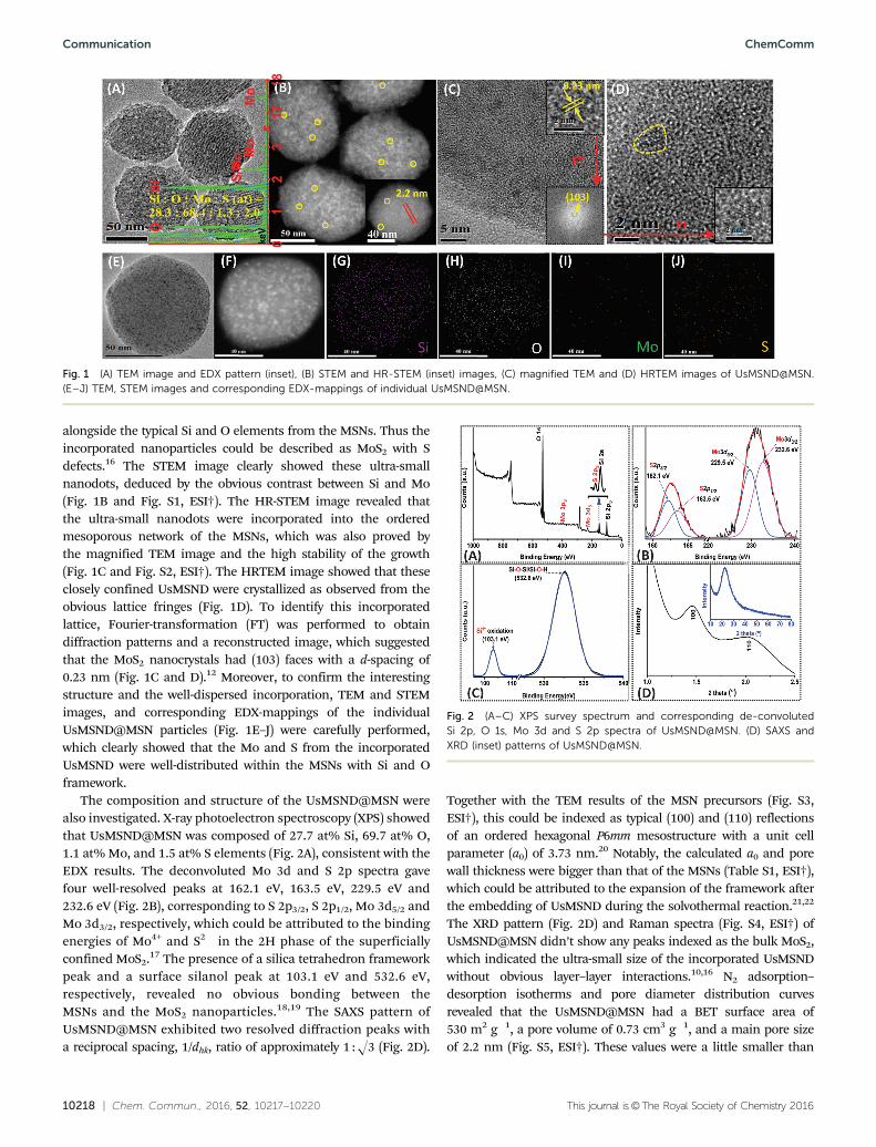
alongside the typical Si and O elements from the MSNs. Thus theincorporated nanoparticles could be described as MoS2 with Sdefects.16 The STEM image clearly showed these ultra-smallnanodots, deduced by the obvious contrast between Si and Mo(Fig. 1B and Fig. S1, ESI†). The HR-STEM image revealed thatthe ultra-small nanodots were incorporated into the orderedmesoporous network of the MSNs, which was also proved bythe magnified TEM image and the high stability of the growth(Fig. 1C and Fig. S2, ESI†). The HRTEM image showed that theseclosely confined UsMSND were crystallized as observed from theobvious lattice fringes (Fig. 1D). To identify this incorporatedlattice, Fourier-transformation (FT) was performed to obtaindiffraction patterns and a reconstructed image, which suggestedthat the MoS2 nanocrystals had (103) faces with a d-spacing of0.23 nm (Fig. 1C and D).12 Moreover, to confirm the interestingstructure and the well-dispersed incorporation, TEM and STEMimages, and corresponding EDX-mappings of the individualUsMSND@MSN particles (Fig. 1E–J) were carefully performed,which clearly showed that the Mo and S from the incorporatedUsMSND were well-distributed within the MSNs with Si and Oframework.
The composition and structure of the UsMSND@MSN werealso investigated. X-ray photoelectron spectroscopy (XPS) showedthat UsMSND@MSN was composed of 27.7 at% Si, 69.7 at% O,1.1 at% Mo, and 1.5 at% S elements (Fig. 2A), consistent with theEDX results. The deconvoluted Mo 3d and S 2p spectra gavefour well-resolved peaks at 162.1 eV, 163.5 eV, 229.5 eV and232.6 eV (Fig. 2B), corresponding to S 2p3/2, S 2p1/2, Mo 3d5/2 andMo 3d3/2, respectively, which could be attributed to the bindingenergies of Mo4+ and S2� in the 2H phase of the superficiallyconfined MoS2.17 The presence of a silica tetrahedron frameworkpeak and a surface silanol peak at 103.1 eV and 532.6 eV,respectively, revealed no obvious bonding between theMSNs and the MoS2 nanoparticles.18,19 The SAXS pattern ofUsMSND@MSN exhibited two resolved diffraction peaks witha reciprocal spacing, 1/dhk, ratio of approximately 1 :O3 (Fig. 2D).
Together with the TEM results of the MSN precursors (Fig. S3,ESI†), this could be indexed as typical (100) and (110) reflectionsof an ordered hexagonal P6mm mesostructure with a unit cellparameter (a0) of 3.73 nm.20 Notably, the calculated a0 and porewall thickness were bigger than that of the MSNs (Table S1, ESI†),which could be attributed to the expansion of the framework afterthe embedding of UsMSND during the solvothermal reaction.21,22
The XRD pattern (Fig. 2D) and Raman spectra (Fig. S4, ESI†) ofUsMSND@MSN didn’t show any peaks indexed as the bulk MoS2,which indicated the ultra-small size of the incorporated UsMSNDwithout obvious layer–layer interactions.10,16 N2 adsorption–desorption isotherms and pore diameter distribution curvesrevealed that the UsMSND@MSN had a BET surface area of530 m2 g�1, a pore volume of 0.73 cm3 g�1, and a main pore sizeof 2.2 nm (Fig. S5, ESI†). These values were a little smaller than
Fig. 1 (A) TEM image and EDX pattern (inset), (B) STEM and HR-STEM (inset) images, (C) magnified TEM and (D) HRTEM images of UsMSND@MSN.(E–J) TEM, STEM images and corresponding EDX-mappings of individual UsMSND@MSN.
Fig. 2 (A–C) XPS survey spectrum and corresponding de-convolutedSi 2p, O 1s, Mo 3d and S 2p spectra of UsMSND@MSN. (D) SAXS andXRD (inset) patterns of UsMSND@MSN.
Communication ChemComm
This journal is©The Royal Society of Chemistry 2016 Chem. Commun., 2016, 52, 10217--10220 | 10219
that of the MSNs possibly due to the incorporated MoS2 nanodotsin the meso-structure (Table S1, ESI†). All of these results demon-strated the uniform incorporation of well-dispersed and ultra-small 2H phase MoS2 nanodots (UsMSND) into MSNs, and thisincorporation didn’t obviously affect the porous properties ofthe MSNs.
The preparation of UsMSND@MSN is simple, and the key isthe employment of surface silanol-containing MSNs as carriers,which not only provide enough accessible meso-channels forthe anchoring of MoS4
2� but also prevent the aggregation andovergrowth of the nanosized MoS2 dots during the solvothermalreduction.23 In a parallel control experiment, a direct solvothermalreduction of (NH4)2MoS4 in DMF solution without MSNs onlyresulted in highly crystallized MoS2 nanosheets (Fig. S6A and S7,ESI†). Also, excess addition of (NH4)2MoS4 in the MSNs causedoverloading of the MoS4
2� species on the surfaces of the MSNsand consequently converted them into core–shell MSN@MoS2
(Fig. S6B and S8, ESI†). Moreover, using calcined MSNs as carriers,composites with MoS2 nanosheets positioned away from the MSNswere formed (Fig. S6C and S9, ESI†). This might be because theframework integrity and few surface groups on the calcined MSNswere not able to supply accessible mesochannels and anchoringsites for the nucleation and growth of ultra-small MoS2 nanodots.This was further proven by using MSN-NH2 as a carrier, whoseelectropositive surface groups were much more beneficial for theanchoring of MoS4
2�. Therefore, UsMSND@MSN-NH2 can also beobtained under conditions involving less addition of (NH4)2MoS4
(Fig. S6D, ESI†).Attributed to the dispersed incorporation of UsMSND into
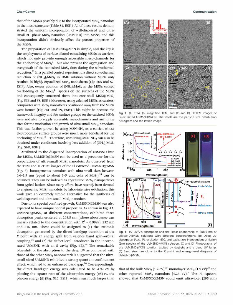
the MSNs, UsMSND@MSN can be used as a precursor for thepreparation of ultra-small MoS2 nanodots. As observed fromthe TEM and HRTEM images of the Si-extracted UsMSND@MSN(Fig. 3), homogeneous nanodots with ultra-small sizes between0.6–2.5 nm (equal to about 3–5 unit cells of MoS2),24 can beobtained. They can be indexed as crystallized MoS2 nanoparticlesfrom typical lattices. Since many efforts have recently been devotedto engineering MoS2 nanodots by labor-intensive exfoliation, thiswork gave an extremely simple alternative for the synthesis ofwell-dispersed and ultra-small MoS2 nanodots.
Due to its special confined growth, UsMSND@MSN was alsoexpected to have unique optical properties. As shown in Fig. 4A,UsMSND@MSN, at different concentrations, exhibited threeabsorption peaks centered at 208.5 nm (where absorbance waslinearly related to the concentration with R2 = 0.9999), 232 nmand 316 nm. These could be assigned to (1) the excitonicabsorption generated by the direct bandgap transition at theK point with an energy split from valence band spin–orbitalcoupling,25 and (2) the defect level introduced in the incorpo-rated UsMSND with an S cavity (Fig. 4E).16 The remarkableblue-shift of the absorption to the deep UV as compared withthose of the other MoS2 nanomaterials suggested that the ultra-small sized UsMSND exhibited a strong quantum confinementeffect, which led to an enhanced band gap.26 Correspondingly,the direct band-gap energy was calculated to be 4.92 eV byplotting the square root of the absorption energy (aE) vs. thephoton energy (E) (Fig. S10, ESI†), which was much larger than
that of the bulk MoS2 (1.2 eV),27 monolayer MoS2 (1.9 eV)28 andother reported MoS2 nanodots (4.26 eV).7 The FL spectrashowed that UsMSND@MSN could emit ultraviolet (295 nm)
Fig. 3 (A) TEM, (B) magnified TEM, and (C and D) HRTEM images ofSi-extracted UsMSND@MSN. The insets are the particle size distributionhistogram and the lattice image.
Fig. 4 (A) UV/Vis absorption and the linear relationship at 208.5 nm ofUsMSND@MSN solutions with different concentrations. (B) Deep UVabsorption (Abs), PL excitation (Ex), and excitation-independent emission(Em) spectra of the UsMSND@MSN solution. (C and D) Photographs ofthe UsMSND@MSN solution excited by daylight and a deep UV lamp.(E) Band structure close to the K point and energy-level diagrams ofUsMSND@MSN.
ChemComm Communication
10220 | Chem. Commun., 2016, 52, 10217--10220 This journal is©The Royal Society of Chemistry 2016
and violet (363 nm) photoluminescence upon excitation bydeep UV, and the intensity decreased rapidly with an increasein excitation wavelengths (Fig. 4B), which were typical featuresof the MoS2 nanodots.29 Nevertheless, being different fromcommon poly-dispersed MoS2 nanodots with excitation-dependentsingle emission,26,30 the obvious two excitation-independentemissions in UsMSND@MSN might originate from intrinsicstates and defect states of the confined UsMSND with advancedmono-dispersity.13,31 Photoluminescence excitation (PLE) spectraof the 295 nm emission gave a peak at 208.5 nm, according to theintrinsic excitonic absorption in the UV-Vis spectra, which alsosuggested that the 295 nm emission was mainly due to intrinsicstates, while the other one might come from S defects (defectstates) (Fig. 4E). Further evidence for these unique optical pro-perties was found in the FT-IR spectra, from which the surfacestates or the carbon impurities that possibly contributed to thefluorescence could be excluded (Fig. S11, ESI†).32 Attributed to thestable incorporation of UsMSND in the MSNs, UsMSND@MSNcould resist UV bleaching (Fig. S12, ESI†). Meanwhile, originatingfrom the big tail of the 363 nm emission, a yellow concentratedUsMSND@MSN solution could be observed to have blue-violetfluorescence (Fig. 4C, D and Fig. S13, ESI†). These resultssuggested that UsMSND@MSN not only had excellent lightconverting properties as compared to many compound-semiconductor-based QDs, but also could benefit from the porousand molecular-sieving behaviors of the mesoporous framework.UsMSND@MSN could be a promising material for a wide range ofoptoelectronic devices, which will be investigated in future work.
In summary, a UsMSND@MSN composite with homogeneousincorporation of ultra-small and monodispersed 2H phase MoS2
nanodots in MSNs was prepared using the one-step solvothermalreduction method. The key for this method is the employment ofMSNs as carriers, which not only provide enough accessiblemesochannels for the anchoring of MoS4
2� but also preventaggregation and overgrowth of the nanosized MoS2 dots duringthe solvothermal reaction. The prepared UsMSND@MSN can beused as precursor for facile preparation of ultra-small MoS2
nanodots, and it could also emit deep UV photoluminescencedue to the strong quantum confinement effect of the incorporatedUsMSND with a large direct band-gap energy of 4.92 eV. This workpromised a facile route to regulate MoS2 for further applications.
This work was supported by grants from the National KeyBasic Research Program (2013CB932502) of China (973 Program),National Natural Science Foundation of China (81573002 and21303022) and the Sino-German Research Project (GZ995).
Notes and references1 Y. Guo, K. Xu, C. Wu, J. Zhao and Y. Xie, Chem. Soc. Rev., 2015, 44,
637–646.2 Y. Chen, C. Tan, H. Zhang and L. Wang, Chem. Soc. Rev., 2015, 44,
2681–2701.
3 F. Bonaccorso, L. Colombo, G. Yu, M. Stoller, V. Tozzini, A. C.Ferrari, R. S. Ruoff and V. Pellegrini, Science, 2015, 347, 1246501.
4 C. Tan and H. Zhang, Chem. Soc. Rev., 2015, 44, 2713–2731.5 X. Zhang, H. Xie, Z. Liu, C. Tan, Z. Luo, H. Li, J. Lin, L. Sun, W. Chen,
Z. Xu, L. Xie, W. Huang and H. Zhang, Angew. Chem., Int. Ed., 2015,54, 3653–3657.
6 D. Kufer, I. Nikitskiy, T. Lasanta, G. Navickaite, F. H. Koppens andG. Konstantatos, Adv. Mater., 2015, 27, 176–180.
7 H. D. Ha, D. J. Han, J. S. Choi, M. Park and T. S. Seo, Small, 2014, 10,3858–3862.
8 C. K. Chua, Z. Sofer, P. Simek, O. Jankovsky, K. Klımova,S. Bakardjieva, S. Hrdlickova Kuckova and M. Pumera, ACS Nano,2015, 9, 2548–2555.
9 J. Lee, K. Kim, W. I. Park, B. Kim, J. H. Park, T. Kim, S. Bong, C. Kim,G. Chae, M. Jun, Y. Hwang, Y. S. Jung and S. Jeon, Nano Lett., 2012,12, 6078–6083.
10 S. Xu, D. Li and P. Wu, Adv. Funct. Mater., 2015, 25, 1127–1136.11 X. Zhang, Z. Lai, Z. Liu, C. Tan, Y. Huang, B. Li, M. Zhao,
L. Xie, W. Huang and H. Zhang, Angew. Chem., Int. Ed., 2015, 54,5425–5428.
12 D. Gopalakrishnan, D. Damien and M. M. Shaijumon, ACS Nano,2014, 8, 5297–5303.
13 S. Inagaki, O. Ohtani, Y. Goto, K. Okamoto, M. Ikai, K. Yamanaka,T. Tani and T. Okada, Angew. Chem., Int. Ed., 2009, 48, 4042–4046.
14 M. Guan, W. Wang, E. J. Henderson, O. Dag, C. Kubel, V. S. K.Chakravadhanula, J. Rinck, I. L. Moudrakovski, J. Thomson,J. McDowell, A. K. Powell, H. Zhang and G. A. Ozin, J. Am. Chem.Soc., 2012, 134, 8439–8446.
15 B. Kong, J. Tang, Y. Zhang, T. Jiang, X. Gong, C. Peng, J. Wei, J. Yang,Y. Wang, X. Wang, G. Zheng, C. Selomulya and D. Zhao, Nat. Chem.,2016, 8, 171–178.
16 L. Lin, Y. Xu, S. Zhang, I. M. Ross, A. C. M. Ong and D. A. Allwood,ACS Nano, 2013, 7, 8214–8223.
17 B. L. Li, L. X. Chen, H. L. Zou, J. L. Lei, H. Q. Luo and N. B. Li,Nanoscale, 2014, 6, 9831–9838.
18 A. R. Mouat, C. George, T. Kobayashi, M. Pruski, R. P. van Duyne,T. J. Marks and P. C. Stair, Angew. Chem., Int. Ed., 2015, 54,13346–13351.
19 C. Tang, J. Zhu, Q. Zhou, J. Wei, R. Zhu and H. He, J. Phys. Chem. C,2014, 118, 26249–26257.
20 Y. Wang, K. Wang, X. Yan and R. Huang, Adv. Healthcare Mater.,2014, 3, 485–489.
21 L. Miller, G. Winter, B. Baur, B. Witulla, C. Solbach, S. Reske andM. Linden, Nanoscale, 2014, 6, 4928–4935.
22 M. Kruk, M. Jaroniec and A. Sayari, J. Phys. Chem. B, 1999, 103,4590–4598.
23 R. Liu, D. Wu, S. Liu, K. Koynov, W. Knoll and Q. Li, Angew. Chem.,Int. Ed., 2009, 48, 4598–4601.
24 R. Ganatra and Q. Zhang, ACS Nano, 2014, 8, 4074–4099.25 E. P. Nguyen, B. J. Carey, J. Z. Ou, J. van Embden, E. D. Gaspera,
A. F. Chrimes, M. J. S. Spencer, S. Zhuiykov, K. Kalantar-zadeh andT. Daeneke, Adv. Mater., 2015, 27, 6225–6229.
26 X. Ren, L. Pang, Y. Zhang, X. Ren, H. Fan and S. F. Liu, J. Mater.Chem. A, 2015, 3, 10693–10697.
27 C. Lee, H. Yan, L. E. Brus, T. F. Heinz, J. Hone and S. Ryu, ACS Nano,2010, 4, 2695–2700.
28 H. Zeng, J. Dai, W. Yao, D. Xiao and X. Cui, Nat. Nanotechnol., 2012,7, 490–493.
29 D. Gopalakrishnan, D. Damien, B. Li, H. Gullappalli, V. K. Pillai,P. M. Ajayan and M. M. Shaijumon, Chem. Commun., 2015, 51,6293–6296.
30 Y. Wang and Y. Ni, Anal. Chem., 2014, 86, 7463–7470.31 F. Liu, M. Jang, H. D. Ha, J. Kim, Y. Cho and T. S. Seo, Adv. Mater.,
2013, 25, 3657–3662.32 L. Tang, R. Ji, X. Cao, J. Lin, H. Jiang, X. Li, K. S. Teng, C. M. Luk,
S. Zeng, J. Hao and S. P. Lau, ACS Nano, 2012, 6, 5102–5110.
Communication ChemComm