Embed Size (px)
Citation preview
VOLUME 27, NUMBER 3 219
ACKNOWLEDGMENTS
We are grateful for the kind assistance of Dr. Lee D. Miller (Allyn Museum of Entomology) who identified the species mentioned, and made constructive criticism on the manuscript. We also thank Dr. Alexander B. Klots, for his encouragement to present the results of our work, and Drs. Theodore D. Sargent and Allen M. Young, who gave many valuable suggestions. My younger son, Pierre, first observed oviposition in P. o. octavia. Specimens of early stages and adults are deposited with the Allyn Museum of Entomology.
LITERATURE CITED
BRUES, C. T., A. L. MELANDER & F. M. CARPENTER. 1954. Classification of Insects. Bulletin of the Museum of Comparative Zoology, at Harvard College. Vol. 108. Cambridge, Mass.
COMSTOCK, W. P. 1961. Butterflies of the American Tropics, The Genus Anaea, (Lepidoptera, N ymphalidae ).
KLOTS, A. B. 1960. A Field Guide to the Butterflies. Houghton Mifflin, Boston. LICHY, R. 1962. Apuntes sobre los Agrias Doubleday. (Nymphalidae, Charaxidi
nae). Revista de la Facultad de Agronomia. Vol. II, No.4. Maracay. Venezuela. SERRANO, FRANCISCO Y SERRANO, MIGUEL E. 1972. Las Mariposas de EI Salvador.
Primera parte: Papilionidae. Comunicaciones, 2a. Epoca. Vol. 1 #1. Universidad de EI Salvador.
TWO NEW SPECIES OF PHYCITINAE FROM TEXAS, WITH DESCRIPTION OF TWO NEW GENERA (PYRALIDAE)
ANDRE BLANCHARD
P. O. Box 20304, Houston, Texas 77025
Triozosneura A. Blanchard, new genus
Tongue well developed. Antenna (Fig. 12) simple; finely pubescent in male. Labial palpus (Fig. 4) upcurved, rough scaled, reaching level of vertex, third segment very short (on denuded pal pus (Fig. 13) it appears less than % the length of second segment). Maxillary palpus squamous. Vestiture entirely of scales.
Forewing (Fig. 11): Smooth, eleven veins, R, absent, cell longer than half the length of the wing; discocellular vein weak curved; vein Cu, from near lower angle of cell; CUt from the angle, slightly separated at base from stalk of 1\12--8; M2 and M3 stalked for about % their lengths; R, contiguous or partly fused, for about % its length, with the stalk of R3-5; R3 and R5 stalked for about % their lengths; vein R, from cell.
Hindwing (Fig. 11): With veins CUl and M3 both present; cell at lower angle about half as long as wing; discocellular vein deeply concave; vein Cu, from near lower angle of cell; vein CUl shortly united with the stalk of M2-3; M2 and M3 stalked
220 JOURNAL OF THE LEPIDOPTERISTS' SOCIETY
Figs. 1-7. Holotypes and paratypes. Figs. 1-4. Triozosneura dorsonotata: 1, holotype &, Mt. Locke, Davis Mts., 27 August 1970; 2, paratype &, Big Bend Nat. Park, Green Gulch, 31 March 1971; 3, paratype ~, Big Bend Nat. Park, Green Gulch, 6 May 1972; 4, head of holotype. Figs. 5-7. Glyphocystis viridivallis: 5, holotype &, Big Bend Nat. Park, Green Gulch, 23 March 1971; 6, paratype ~, Big Bend Nat. Park, Green Gulch 12 May 1972; 7, paratype &, Big Bend Nat. Park, Green Gulch, 28 March 1971.
for well over half their lengths; S, and R, shortly, and weakly anastomosed beyond cell.
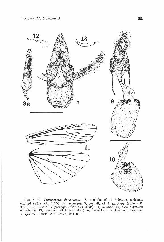
Male genitalia (Figs. 8, 8a): Uncus hoodlike subtriangular; tegumen strongly sclerotized laterally from its junction with vinculum to midheight where it supports the lateral arms of the gnathos; gnathos hoodlike, with acute apex, deeply cleft dorsally, fused apically, with semi-elliptical ventral opening; valve narrow, unarmed, with strongly sclerotized costa; transtilla absent; juxta narrow embracing % of circumference of aedeagus; aedeagus half as long as combined height of vinculum, tegumen and uncus; vesica armed with a bunch of numerous cornuti, and what
VOLUME 27, NUMBER 3 221
8a 8
~ ~ 11
Figs. 8-13. Triozosneura dorsonotata: 8, genitalia of ~ holotype, aedeagus omitted (slide A.B. 2395); 8a, aedeagus; 9, genitalia of Cjl paratype (slide A.B. 3034); 10, bursa of Cjl paratype (slide A.B. 2909); 11, venation; 12, basal segments of antenna; 13, denuded left labial palp (inner aspect) of a damaged, disearded Cjl specimen (slides A.B. 2647 A, 2647B).
222 JOURNAL OF THE LEPIDOPTERISTS' SOCIETY
appears as a small, thin, sclerotized membrane, bent as a half cylinder; vinculum about as broad as long.
Female genitalia (Figs. 9, 10): Genital opening simple, narrowly sclerotized ventrally; ductus seminalis from ductus bursae, midway between genital opening and bursa; bursa with two signa, each consisting of a disk armed with hollow spines. Figs. 9 and 10 represent the bursae of two different specimens. It is believed that the bursa of the former is over-inflated, whereas that of the latter, which is depressed all around the ductus bursae, thus forming a circular groove and a ridge, is more nearly normal. In Fig. 10 one signum only was represented for the sake of clarity: it is on the inner wall of the ridge and seen through the membrane of the bursa.
If and when other species of this genus are discovered some of the characters of this description will appear only of specific value, but it would be awkward to guess which ones at the present time.
The venation of the hindwing makes Triozosneura a member of Heinrich's Group I and venetional division B. Using Heinrich's key for division B, we are led through couplets 1, 3, 8, 9, 16, 21, 22 and 24 to couplet 25 which offers two alternatives, none of which applies satisfactorily to the case at hand: 26 is eliminated because the transtilla is absent from the male genitalia, 27 because the ductus seminalis is from the ductus bursae in the female genitalia. Thus we are led to a dead end in which, by chance, there are only four gcnera, namely: Coptarthria, Anadelosemia, Gabinius and Ceracanthia. It is enough to examine the detailed description of these four genera, and the figures of the male and female genitalia of all species belonging to them, to convince oneself of the necessity of a new genus.
Triozosneura dorsonotata A. Blanchard, new species (Figs. 1, 2, 3, 4, 8, 8a, 9, 10, 11, 12, 13)
Head and thorax covered with gray or black scales tipped with white, more contrastingly mixed on the palpus. Abdomen yellowish gray. Forewing above gray, darker along costa, variably suffused with whitish on each side of the antemedial line, in the cell, and in the fold. Antemedial line white, starting on costa Va distance from base to apex, reaching dorsal margin % distance from base to tornus, outwardly bilobed between (often indistinctly); contrastingly bordeI'd with black outwardly, between costa and radius, where it is aimed toward tornus; widely, roundly excurved between radial and anal veins, where the black outer line almost disappears except for a conspicuous black spot on cubital vein, marking the notch between the two lobes; contrastingly bordered with black on both sides between anal vein and inner margin. Subterminal line mostly indistinct. Discal dots obsolete. On some specimens a reddish or yellowish diffuse spot covers the discocellular vein. Some veins are marked in black, particularly Ro and M, and the cubital in the basal space. A black blotch beyond the middle of the inner margin is generally separated from the antemedial band by a paler spot. Terminal dots and line absent. Fringe gray, somewhat lighter than the background of the wing. Hindwing yellowish-white, a little darker along the outermargin, mainly in females; fringe concolorous with disk of wing. Beneath: forewing brownish gray, a little darker along costa, hindwing as above. Wing expanse: male 25 to 28 mm.; female 25 .5 to 29 mm.
Holotype: Male, Davis Mountains, Mount Locke, McDonald Observatory
VOLUME 27, NUMBER 3 223
14a
~.
~ 18
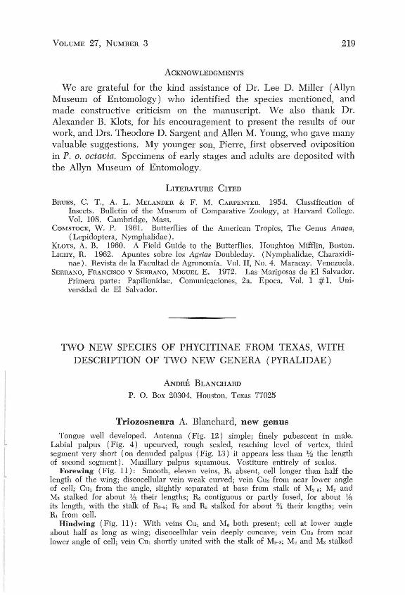
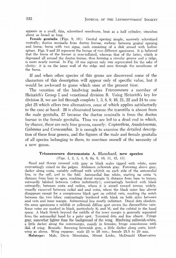
Figs. 14-18. Glyphocystis viridivallis: 14, genitalia of rt; holotype, aedeagus omitted (slide A.B. 3030); 14a, aedeagus; 15, genitalia of <;1 paratype (slide A.B . 3028); 16, venation; 17, basal segments of antenna; 18, denuded left labial palp (inner aspect) of damaged discarded specimen (slide A.B. 2649).
Grounds, Texas 27 August 1970, genitalia on slide A.B.2395, deposited in the National Museum of Natural History (No. 72379).
Para types : Big Bend Nat. Park, Basin, 7 Apdl 1967, 1 rt;, (A.B.627); Green Gulch, 25 March 1971, a, (A.B.2909); 31 March 1971, 1rt;; 6 May 1972, 1<;1, (A.B.3034); 12 May 1972, 1 rt;. Davis Mts., 5 miles SE of Mt. Livermore, 29 August 1970, 1 <;1, (A.B.2396).
224 JOURNAL OF THE LEPIDOPTERISTS' SOCIETY
Glyphocystis A. Blanchard, new genus
Tongue well developed. Antenna finely pubescent; on male with a shallow sinus at base of shaft, containing a row of minute black teeth half hidden between two rows of scales (Fig. 17). Labial palpus (Fig. 18) oblique, not grooved, smooth sca~ed, reaching level of vertex; third segment about Vs length of second, not projected forward. Maxillary palpus squamous, vestigial.
Forewing (Fig. 16): Smooth, with eleven veins; cell about % length of wing; discocellular vein concave and weak between veins M, and M2; vein Cu, from before the lower outer angle of cell; CUl from the angle, closer to M3 than to CUt, M, and M3 separated at base; M, from below upper outer angle of cell, straight; R3 and R, stalked for a little over half their lengths; R, and R2 separately from cell.
Hindwing (Fig. 16): With veins M3 and CUl both present. Cell at lower angle about half as long as wing; discocellular vein deeply concave, considerably extended at lower angle; vein Cu, from before angle; CUl from angle, connate to the stalk of M,,-"; M2 and M. anastomosed for about half their lengths; M, connate to the contiguous parts of R, and Sc, which is about the same length as the free portion of Sc.
Male genitalia (Figs. 14, 14a): Apical margin of uncus subrhomboidal; gnathos terminating in a spatulate hook; valve with strongly sclerotized costa, markedly produced at apex, about as long as combined length of tegumen and uncus; transtilla absent; juxta V -shaped; vinculum not quite as wide as long, becoming narrower and truncate anteriorly; aedeagus almost as long as combined length of vinculum, teugmen and uncus, stout; vesica armed with one stout cornutus, almost as long as aedeagus.
Female genitalia (Fig. 15): Genital opening simple; ductus bursae somewhat contracted and membranous at junction with bursa; bursa wide and sclerotized posteriorly, bulbous and membranous anteriorly, provided on its right side with a large lobe; most of the inner surface of the lobe, a collar around ductus bursae, and some of the dorsal surface of the bursa covered with an inner, densely spinose mat. Ductus seminalis from the anterior end of the lateral lobe.
In spite of the absence of a transtilla, and of substantial differences in the gnathos of the male genitalia, the following species could have been described in Catastia, but in view of considerable differences in the female genitalia this course of aotion was deemed unadvisable.
Glyphocystis viridivaIlis A. Blanchard, new species
(Figs. 5, 6, 7, 14, 14a, 15, 16, 17, 18) Head, collar, first and second segments of palpus clothed with dark gray, white
tipped scales; gray and whitc more contrasting on second scgment of palpus, third segment acute, black. Thorax concolorous with background of forewing. Forewing background, a nearly uniform slate-gray produced by dark gray, white tipped scales; generally a little darker in basal space. Antemedial line whitish, outwardly bordered by a black line which is generally much wider between costa and cubitus than in fold; varying from almost straight to a little sigmoid (outwardly convex above Cu, concave in fold) starting on costa 14 distance from base to apex, reaching inner margin % distance from base to tornus; a dark blotch of variable extent adnate to and basad of am line, along inner border. Subterminal line whitish, inwardly bordered by a thin black line (thicker and darker near costa), roundly retracted on vein M" and again, but less so in fold, reaching tornus at base of some very long fringe scales. A black triangular blotch distad of, and adnate to s.t. line near apex. Fine terminal black line. Fringe long, light gray, consisting of scales of three
f I
VOLUME 27, NUMBER 3 225
different lengths, tipped with darker gray. Hindwing pale yellowish-white, darkened near apex and, in the female, along outer margin. Beneath: forewing yellowish-gray, hindwing yellowish-white. Wing expanse: 21 to 24 mm.
Holotype: Male, Big Bend National Park, Green Gulch, Texas, 28 March 1971, gen. prep. A.B. 3030, deposited in the National Museum of Natural History, (No. 72380).
Paratypes: All from Big Bend Nat. Park, Green Gulch: 9 October 1969, 1 ~, (A.B. 2011); 28 March 1971, 5 ~ ~, (A.B. 3030, 2649, 2650); 3 May 1972, l<j>, (A.B. 3029); 12 May 1972, 1~, 1<;l, (A.B. 3028).
ACKNOWLEDGMENTS
I am deeply grateful to Dr. D. C. Ferguson of the Entomolgy Research Division, U.S.D.A., for revising the manuscript and making several very helpful suggestions. The authorizations given me by Mr. C. D. Laughlin to collect on the McDonald Observatory grounds, and by the Administration of Big Bend National Park to collect around and in the Chis os Mountains are also gratefully acknowledged.
LITERATURE CITED
HEINRICH, C. 1956. American Moths of the subfamily Phycitinae. U.S . Nat. Mus. Bull. 207.
NOTES ON THE TAXONOMIC STATUS OF HYALOPHORA COLUMBIA (SATURNIIDAE)
MICHAEL M. COLLINS
924 Mendocino Avenue, Berkeley, California 94707
The work of many authors (Sweadner, 1937; Weast, 1959; Collins & Weast, 1961; Wright, 1971) has shown that the various forms of Hyalophora are not reproductively isolated from one another. Females of any form of Hyalophora (in the restricted sense of Ferguson, 1972) will attract and mate indiscriminately with males of any other form. Generally ova laid by cross-mated females are viable and usually produce fertile F 1 males and sterile females. Backcrossing the F 1 male with a female of either parental form or even a third form again produces fertile males and generally sterile females. Occasionally these females may lay some fertile ova.
Intergrades and hybrids occur in nature. A population currently designated "kasloensis" (Cockerell) exists between H. gloveri (Strecker) and H. euryalus (Boisduval) in Idaho, western Montana, and British


![[1973] Q.B. 27.pdf](https://img.pdfslide.us/doc/110x75/577cd67d1a28ab9e789c850f/1973-qb-27pdf.jpg)