Embed Size (px)
Citation preview
THE AMERICAN PHYSIOLOGICAL SOCIETY
Founded in 1887 for the purpose of promoting the increase of
physiological knowledge and its utilization.
President
OFFICERS
David F. Bohr, Univ. of Michigan, Ann Arbor
President-Elect Ernst Knobil, Univ. of Pittsburgh
Past President William F. Ganong, Univ. of California, San Francisco
Council David F. Bohr, Ernst Knobil, William F. Ganong, Francis J.
Haddy, Walter C. Randall, Earl H. Wood, Paul C. Johnson
Executive Secretary-Treasurer Orr E. Reynolds, 9650 Rockville Pike, Bethesda, Maryland
20014
APS, Bethesda Telephone Numbers
Area Code 301 Executive Secretary - 530-7 164 Business Manager - 530-7 16 1 Publications Manager - 530-7070 Subscriptions Services - 530-7 180 Meeting Registration - 530-7010
SUSTAINING MEMBERS
Abbott Laboratories Merck Sharp & Dohme Burroughs Wellcome Co. Res. Labs. CIBA Geigy Corp. Pfizer, Inc.
Grass Instrument Co. A. H. Robins Co., Inc. Hoechst-Roussel Pharmaceu- Smith Kline & French Labs.
tical Co., Inc. Waverly Press Hoffmann-La Roche, Inc. Wyeth Laboratories, Inc. Eli Lilly and Co.
Publications
American Journal of Physiology: Cell Physiology
American Journal of Physiology: Endocrinology, Metabolism
and Gastrointestinal Physiology
American Journal of Physiology: Heart and Circulatory Physi-
dogy American Journal of Physiology: Regulatory, Integrative and
Comparative Physiology
American Journal of Physiology: Renal, Fluid and Electrolyte
Physiology
American Journal of Physiology (Consolidated)
Journal of Applied Physiology: Respiratory, Environmental
and Exercise Physiology
Journal of Neurophysiology
Physiological Reviews
The Physiologist
Handbooks of Physiology
Clinical Physiology Series
THE PHYSIOLOGIST is published bimonthly by the Ameri-
can Physiological Society at 9650 Rockville Pike, Bethesda,
Maryland 20014. Address all correspondence to this address.
Subscriptions: Distributed with The Physiology Teacher to
members as a part of their membership. Non-members and
institutions, $12.00 per year in the United States; Canada,
$12.50; Foreign and Postal Union, $13.00. The American
Physiological Society assumes no responsibility for the state-
jents and opinions advanced by contributors to THE PHYSI-
LOG I ST.
The ysiologist for Physiologists and Physiology
Orr E. Reynolds, Editor
TABLE OF CONTENTS
SOCIETY AFFAIRS Future Meetings ................................. ii David Bruce Dill. ................................. 1 Letters to the Editor. .............................. 3 Dues and Contributions. ........................... 4
Contributions from Retired Members. ................. 5 Honors and Awards. .............................. 6 Spring Meeting Symposia Support ................... 6 Hiram E. Essex ................................... 6 Journal Growth, 1975-1978 ......................... 7
STANDING COMMllTEE REPORT Notes from Capitol Hill . . . Brian A. Curtis. . . . . . . . . . . . . . 4
HISTORICAL ARTICLES Case History of a Physiologist: F. G. Hall . . . D. B. Dill. . . . 8
MEMBERSHIP NEWS News from Senior Physiologists. . . . . . . . . . . . . . . . . . . . . .22
ANNOUNCEMENTS Interamerican Medical Congress . . . . . . . . . . . . . . . . . . . . . 6 New Research Travel Grant Program. . . . . . . . . . . . . . . . . .21
Workshop on Implantable Transducers and Systems. . . . . .24 Xlth Congress on International Academy of
Legal Medicine and Social Medicine. . . . . . . . . . . . . . . . .24
First International Congress on Neurotoxicology . . . . . . . . .24 International Symposium on
Regenerative Growth Processes. . . . . . . . . . . . . . . . . . . .35 Workshop on White Noise Analysis of
Physiological Systems . . . . . . . . . . . . . . . . . . . . . . . . . . .35 Careers in Animal Biology . . . . . . . . . . . . . . . . . . . . . . . . . .41
THE PHYSIOLOGY TEACHER Affirmation of Conventional Physiology Lab Exercises
C.S.Tidball................................... 25 Forum: Integrated Functional Lab at University of Texas
J. R. Walker and D. L. Traber. . . . . . . . . . . . . . . . . . . . . .26 Editor’s Note . . . . . . . . . . . . . . . . . . . . . . . . . . . . . . . . . . . .27
TUTORIAL LECTURES Reflex Control of the Veins in Cardiovascular Function
Carl F. Rothe . . . . . . . . . . . . . . . . . . . . . . . . . . . . . . . . . .28 Blood Flow and Metabolism in Different Layers of
the Left Ventricle Douglas M. Griggs, Jr. . . . . . . . . . . . . . . . . . . . . . . . . . .36
Book Reviews Introduction to Physiology . . . R. H. Davis . . . . . . . . . . . .41
ABSTRACTS Abstracts of Educational Material . . . . . . . . . . . . . . . . . . . .42
FUTURE MEETINGS
Spring
April 13-18, 1980 - Anaheim, CA April 12-17, 1981 - Atlanta, GA April 18-23, 1982 - New Orleans, LA
October 15-19, 1979 - New Orleans, LA October 12-17, 1980 - Toronto, Canada November 1-6, 1981 - Boston, MA October 10-15, 1982 - San Diego, CA
Specialty
Relation Between Neuro transmitters and Endocrine Functions
August 22-24,1979 - East Lansing, MI
28th In terna tional Congress
July 13-19, 1980 - Budapest, Hungary
DAVID BRUCE DILL President 1950 - 1951
American Physiological Society
At the time of Henry C. Bate& death in 1950, Dr. Dill was serving as President-Elect and by vote of the Council he immediately succeeded to the office of President; the office of President-Elect was left vacant. He was Treasurer in 1947 and 1948 and then served two years as a member of Council just prior to his election as President. He served as Chairman of the Federation Executive Committee during his term as President. Dr. Dill was President during the Fall Meeting in Columbus in 1950 and during the Spring Meeting in Cleveland in 1951. During this period, the items of great- est concern to the Society seemed to be the Committee on Scientific Aid, the role of the Federation, and the status of the Board of Publication Trust- ees. Later in his term as President, Dr. Dill proposed to the Council that the President-Elect visit a number of institutions during the year for lectures and an exchange of views of the purposes and operation of the Society [con- tinued as PRESIDENT-ELECT’S TOUR.]
Excerpted from History of the American Physiological Society, The Third Quarter Century, 1937-1962.
On May 15, 16, and 17 of this year, David Bruce Dill’s 88th
birthday will be celebrated by many friends of all the past 50
years at a Symposium at the University of Nevada, Las Vegas.
They will be honoring not only his concern and efforts for physio-
logical research of a high order but also will be expressing their
affection.
The title for the three-day symposium is “Life, Heat and Alti-
tude: A Second Version” -a re-thinking, up-dating, and projec-
tion of the findings and principles which Dill presented in his clas-
sic book of 1938, “Life, Heat and Altitude.” The basis for the
book was the series of Lowell Lectures which Dill had been in-
vited to give in 1937 for the venerable Lowell Institute of Boston.
This was among the first of many honors received by Dill; he had
come a long way from an Iowa farm.
David Bruce Dill was born in Iowa on April 22, 1891 of pio-
neer and Covenanter stock. His paternal great-grandparents emi-
grated from northern Ireland in 1820 with six children to farm on
wild land in western Pennsylvania. His maternal grandparents
were farmers, born in Pennsylvania and also of Scats-Irish origin.
Dill’s father, David White Dill, was a young farmer who pio-
neered in Iowa from Pennsylvania and his mother, Lydia Walken-
shaw Dunn, had made the same move with her parents. Bruce
Dill and his four sisters, all older, were orphaned early and raised
by aunts and uncles. After their mother died, their father ar-
ranged for what he planned for them as temporary care by his
brothers and sisters but he died three years later. For Bruce Dill,
Uncle Louis Samson was, until his death 24 years later, a good,
loving foster father. In Iowa, Dill attended three different one-
room country schools near Wyman and one year of high school
at Washington, Iowa. When he was about 14, he and the Sam-
son family moved to Santa Ana, California, Uncle Lou having de-
cided to retire and also find relief for asthma.
Dill attended Santa Ana High School and Occidental College
with his uncle’s help and jobs at ranches in the Southwest and at
the Irvine Ranch. Some years ago, Dill instituted reunions of his
high school class and a few still meet every year in southern Cali-
fornia, as with a park picnic with cake, and keep in touch other-
wise. They’d received a classical education, including several
languages and sciences, at Santa Ana, and Dill remembers a
favorite teacher there, the Smithsonian ethnologist J. P. Harring-
ton, famous for his linguistic talent. High school in Iowa was also
memorable for Dill. Two stimulating teachers there were H. I.
Case in physiology and botany and E. G. Oakley who taught
mathematics and was the principal.
From Occidental College, where he was a football and track
star, Dill received the B.S. in 1913 and later an honorary D.Sc.;
from Stanford, the M.A. in 1914, and the Ph.D. in 1925. The
years away from Stanford were spent teaching chemistry (1914-
161, during which time he was married to Olive Lillian Cassel, a
classmate at Occidental and a school teacher. Through the aca-
demic year 1916-17, Dill was principal of the El Dorado County
High School in Placerville, California, where their daughter, Eliza-
beth Cassel, was born. Then for a year he was principal at the
Palo Alto High School. This led to an appointment as a chemist
with the USDA, wartime, Fisheries Research project. For this
project, Dill served in a San Pedro laboratory. The Dill family at
this time lived in south Los Angeles and here their son, David
Bruce, Jr., was born. From San Pedro, Dill was transferred to
San Diego and later, in 1921, to Seattle, where he was chemist in
what is now the Food and Drug Administration.
The years 1923-25 were at Stanford and Dill received his
Ph.D. with more help from his Uncle Lou, who had left him
$1000 and a Dodge. The doctorate was in chemistry under Carl
Alsberg, a director of the Food Research Institute. While a post-
doctoral student at Strassburg, Alsberg had become a close
friend of L. J. Henderson, a new Harvard M.D., and this led to
Dill’s obtaining an NRC fellowship in chemistry to work with
Henderson. The setting developed to be the Massachusetts Gen-
eral Hospital rather than the Medical School, since Henderson
had shifted positions from the Medical School to the University
proper. At Harvard, Henderson also shifted Dill’s interest- to the
physical chemistry of blood proteins. Through this, he estab-
lished his long-lasting friendships and working relationships with
A. V. Bock and J. H. Talbott and with many others; Fuller Al-
bright, Chester Jones, Walter Bauer, Paul White, and Howard
Means became good friends. During his two years at the Massa-
chusetts General Hospital, he and Bock wrote a 3rd edition of
Bainbridge’s monograph, Physiology of Muscular Exercise, at
the request of A. V. Hill. A secondary consequence of this effort
was the initiation of studies on exercise physiology which has
continued to this day. Exercise was considered as a stressing
agent to be superimposed upon other stressors to more clearly
identify the cardio-respiratory capabilities and adjustments of the
active organism.
Meanwhile, Henderson together with his friends, Dean Don-
ham of the Business School and Dean Edsall of the Medical
School, had conceived of the Harvard Fatigue Laboratory. (The
history of the Fatigue Laboratory was published in 1973 by Pren-
tice-Hall, Inc.) They obtained two large Rockefeller grants, the
Laboratory was given space at the Business School, and Dill was
appointed to organize and direct the program of the Laboratory.
As Dill says, “[The Laboratory] could well have been called the
Laboratory of Environmental Physiology, for that is what it be-
came.” The academic appointment was as assistant professor in
biochemistry at the School of Public Health, where he held a po-
sition from 1927-36. From 1927-47, he held professorships in in-
dustrial physiology at the Business School and was on Harvard
faculties, in one capacity or another, until retirement in 1961
from a visiting lectureship in physiology at the School of Public
Health. He continued to be director of research at the Fatigue
Laboratory until 1947, although never with an official appoint-
ment for that responsibility.
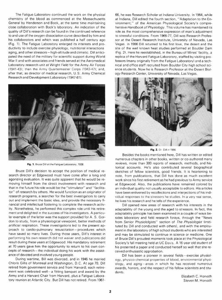
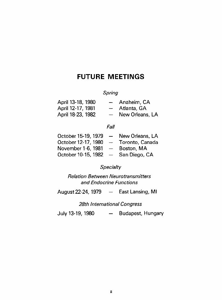
The Fatigue Laboratory continued the work on the physical chemistry of the blood as commenced at the Massachusetts General by Henderson and Bock, at the same trme maintaining close collaboration wrth Bock’s laboratory. An indtcation of the quality of Dill’s research can be found in the continued reference to and use of the oxygen dtssociation curve described by hrm and his collaborators and whrch was published a half century ago (Fig. 1). The Fatigue Laboratory enlarged its interests and pro- ductivity to include exercise physiology, nutritional interactions aging, and other stressors- high-altrtude and climatrc. Dill antrcr- pated the need of the military for scientific support during World War II and wrth associates and friends served at the Aeromedical Laboratory research unit at Wright Field for the Army Arr Forces (1941-431, then the Army Quartermaster Corps (1943-47); and, after that, as director of medical research, U.S. Army Chemical Researchand Development Laboratory (1947-61).
Fig. 1. Bruce Dillat the Fatigue Laboraton/, 1938.
Bruce Dill’s decision to accept the position of medical re- search director at Edgewood must have come after a long and agonizing evaluation. It was quite apparent that he would be re- moving himself from the direct involvement with research and that in the future his role would be the “stimulator” and “facilita- tor” of research by others. He would function as an originator of research programs, a searcher for the best investigators to carry out and implement the basic idea, and provide the necessary fi- nancial and intellectual fostering to complete the research activ- ity. Nonetheless, he performed this complex role until his retire- ment and delighted in the success of his investigators. A particu- lar example of the latter was the support provided for A. S. Gor- don to study, from a physiological viewpoint, better methods of artificial respiration. These investigations led to the present ap- proach to cardio-pulmonary resuscitation-procedures which have saved so many lives. During those years, Dill’s interest in performing his own research did not waiver and publications did result during these years at Edgewood. His mandatory retirement at 70 years gave him the opportunity to return to his own con- cepts of research- the individual doing his work with the assist- ance of devoted and involved young people.
During wartime, Bill was divorced, and in 1946 he married Chloris Gillis of Montreal and Washington, D.C. At age 70, Dill retired from government service and from Harvard; each retire- ment was celebrated well-a fitting banquet and award by the Army and a Harvard Chair from Harvard, plus a Fatigue Labora- tory reunion at Atlantic City. But Dill has not retired. From 1961-
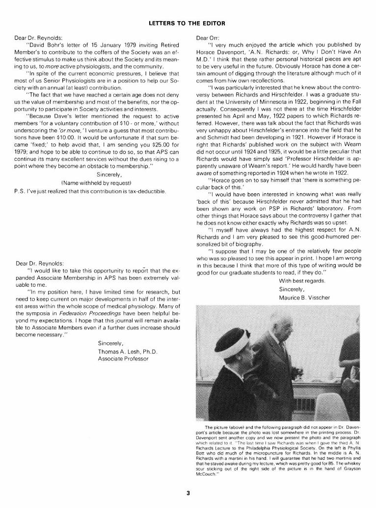
66, he was Research Scholar at Indiana University. In 1964, while at Indiana, Dill edited the fourth section, “Adaptation to the En- vironment,” of the American Physiological Socrety’s compre- hensive HandbookofPhysiology. This volume has maintained its role as the most comprehensive expression of man’s adjustment to stressful conditrons. From 1966-77, Dill was Research Profes- sor at the Desert Research Institute, University of Nevada, Las Vegas. In 1966 Dill returned to his first love, the desert and the site of the well known heat studres performed at Boulder Dam (Fig. 2). Here he reestablished, in the Bureau of Manes’ facrlrty, a replica of the Harvard Fatigue Laboratory, staffed by visiting pro- fessors (many originally from the Fatigue Laboratory) and a tech- nical and office staff recruited from Boulder Crty hrgh school sci- ence students. Now he is Research Professor at the Desert Biol- ogy Research Center, University of Nevada, Las Vegas.
Fig.2. Dr. DIII m 1966.
Besides the books mentioned here, Dill has written or edited numerous chapters in other books, written or co-authored many reviews, more than 300 reports of research, methods, and his- torical accounts. He’s also contributed several biographical sketches of fellow scientists, good friends. It is heartening to note, from publications, that Dill has done as much excellent work since his first retirement as he had previous to Army service at Edgewood. Also, the publications have remained colored by an individual quality not usually acceptable to editors. His articles have been enlivened by recollections and interjections of his indi- vidual responses to the stressors he studies. In a very real sense, he lives his research and he tells of the experience.
Dill opened new areas of research with his interests in the adaptability of the young and the aged to stressors. In fact, this adaptability principle has been examined in a couple of ways be- sides laboratory and field research forays, through the “News from Senior Physiololgists” section of The Physiologist finsti- tuted by Dill and conducted with others), and with the employ- ment in the laboratory of high school students who are interested and may be stimulated to continue in science or medicine. One of Bruce Dill’s proudest moments took place at the Physiological Society’s fall meeting held at UC Davis, A 16-year old student of his presented a paper and conducted herself so well that she re- ceived enthusiastic approbation.
Dill has been a pioneer in several fields-exercise physiol- ogy, physico-chemical properties of blood, environmental physi- ology, and aging-areas in which he has received numerous awards, honors, and the respect of his fellow scientists and stu- dents.
Elizabeth C. Horvath Steven M. Horvath
2
LElTERS TO THE EDITOR
Dear Dr. Reynolds: “David Bohr’s letter of 15 January 1979 inviting Retired
Member’s to contribute to the coffers of the Society was an ef- fective stimulus to make us think about the Society and its mean- ing to us, to more active physiologists, and the community.
“In spite of the current economic pressures, I believe that most of us Senior Physiologists are in a position to help our So- ciety with an annual fat least) contribution.
“The fact that we have reached a certain age does not deny us the value of membership and most of the benefits, nor the op- portunity to participate in Society activities and interests.
“Because Dave’s letter mentioned the request to active members ‘for a voluntary contribution of $10 - or more,’ without underscoring the ‘ormore, ‘I venture a guess that most contribu- tions have been $10.00. It would be unfortunate if that sum be- came ‘fixed;’ to help avoid that, I am sending you $25.00 for 1979; and hope to be able to continue to do so, so that APS can continue its many excellent services without the dues rising to a point where they become an obstacle to membership.”
Sincerely,
(Name withheld by request)
P.S. I’ve just realized that this contribution is tax-deductible.
Dear Dr. Reynolds: “I would like to take thus opportunrty to report that the ex-
panded Assocrate Membershrp in APS has been extremely val- uable to me.
“In my position here, I have limited time for research, but need to keep current on major developments In half of the inter- est areas within the whole scope of medrcal physrology. Many of the symposia in Federation Proceedings have been helpful be- yond my expectations. I hope that this journal WIII remain availa- ble to Associate Members even if a further dues increase should become necessary.”
Srncerely,
Thomas A. Lesh, Ph.D. Associate Professor
Dear Orr: “I very much enjoyed the article which you published by
Horace Davenport, ‘A.N. Richards: or, Why I Don’t Have An M.D.’ I think that these rather personal historical pieces are apt to be very useful in the future. Obviously Horace has done a cer- tain amount of digging through the literature although much of it comes from hiw own recollections.
“I was particularly interested that he knew about the contro- versy between Richards and Hirschfelder. I was a graduate stu- dent at the University of Minnesota in 1922, beginning in the Fall actually. Consequently I was not there at the time Hirschfelder presented his April and May, 1922 papers to which Richards re- ferred. However, there was talk about the fact that Richards was very unhappy about Hirschfelder’s entrance into the field that he and Schmidt had been developing in 1921. However if Horace is right that Richards’ published work on the subject with Wearn did not occur until 1924 and 1925, it would be a little peculiar that Richards would have simply said ‘Professor Hirschfelder is ap- parently unaware of Wearn’s report.’ He would hardly have been aware of something reported in 1924 when he wrote in 1922.
“Horace goes on to say himself that ‘there is something pe- culiar back of this.’
“I would have been interested in knowing what was really ‘back of this’ because Hirschfelder never admitted that he had been shown any work on PSP in Richards’ laboratory. From other things that Horace says about the controversy I gather that he does not know either exactly why Richardswas so upset.
“I myself have always had the highest respect for A.N. Richards and I am very pleased to see this good-humored per- sonalized bit of biography.
“I suppose that I may be one of the relatively few people who was so pleased to see this appear in print. I hope I am wrong in this because I think that more of this type of writing would be good for our graduate students to read, if they do.”
With best regards.
Sincerely,
Maurice B. Visscher
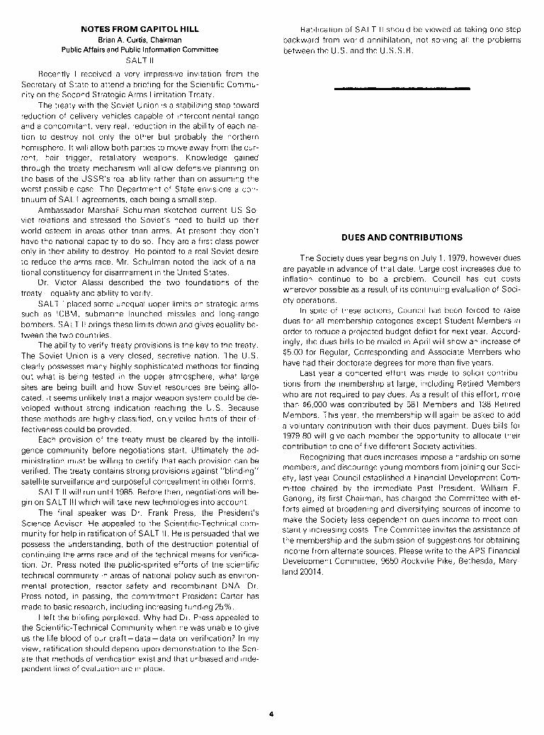
The picture (above) and the following paragraph did not appear w Dr Daven- port’s artccle because the photo was lost somewhere I” the pnntmg process Dr Davenport sent another copy and we now present the photo and the paragraph which related to It “The l:jst time I saw Richards was when I gave the third A N Richards Lecture to the Phlladelphla Physlologtcal Soctety On the left IS Phylks Bott who dtd much of the mlcropuncture for Rlchards In the mlddle IS A. N Rchards with a martvx III his hand I wfll guarantee that he had two martlms and that he stayed awake during my lecture, which was pretty good for 85. The whiskey sour stlckmg out of the right side of the picture IS I” the hand of Grayson McCouch.”
NOTES FROM CAPITOL HILL Brian A. Curtis, Chairman
Public Affairs and Public Information Committee
SALT II
Recently I received a very impressive invitation from the
Secretary of State to attend a briefing for the Scientific Commu-
nity on the Second Strategic Arms Limitation Treaty.
The treaty with the Soviet Union is a stabilizing step toward
reduction of delivery vehicles capable of intercontinental range
and a concomitant, very real, reduction in the ability of each na-
tion to destroy not only the other but probably the northern
hemisphere. It will allow both parties to move away from the cur-
rent, hair trigger, retaliatory weapons. Knowledge gained
through the treaty mechanism will allow defensive planning on
the basis of the USSR’s real ability rather than on assuming the
worst possible case. The Department of State envisions a con-
tinuum of SALT agreements, each being a small step.
Ambassador Marshall Schulman sketched current US-So-
viet relations and stressed the Soviet’s need to build up their
world esteem in areas other than arms. At present they don’t
have the national capacity to do so. They are a first class power
only in their ability to destroy. He pointed to a real Soviet desire
to reduce the arms race. Mr. Schulman noted the lack of a na-
tional constituency for disarmament in the United States.
Dr. Victor Alassi described the two foundations of the
treaty -equality and ability to verify.
SALT I placed some unequal upper limits on strategic arms
such as ICBM, submarine launched missiles and long-range
bombers. SALT II brings these limits down and gives equality be-
tween the two countries.
The ability to verify treaty provisions is the key to the treaty.
The Soviet Union is a very closed, secretive nation. The U.S.
clearly possesses many highly sophisticated methods for finding
out what is being tested in the upper atmosphere, what large
sites are being built and how Soviet resources are being allo-
cated. It seems unlikely that a major weapon system could be de-
veloped without strong indication reaching the U.S. Because
these methods are highly classified, only veiled hints of their ef-
fectiveness could be provided.
Each provision of the treaty must be cleared by the intelli-
gence community before negotiations start. Ultimately the ad-
ministration must be willing to certify that each provision can be
verified. The treaty contains strong provisions against “blinding”
satellite surveillance and purposeful concealment in other forms.
SALT II will run until 1985. Before then, negotiations will be-
gin on SALT III which will take new technologies into account.
The final speaker was Dr. Frank Press, the President’s
Science Advisor. He appealed to the Scientific-Technical com-
munity for help in ratification of SALT II. He is persuaded that we
possess the understanding, both of the destruction potential of
continuing the arms race and of the technical means for verifica-
tion. Dr. Press noted the public-spirited efforts of the scientific
technical community in areas of national policy such as environ-
mental protection, reactor safety and recombinant DNA. Dr.
Press noted, in passing, the commitment President Carter has
made to basic research, including increasing funding 25%.
I left the briefing perplexed. Why had Dr. Press appealed to
the Scientific-Technical Community when he was unable to give
us the life blood of our craft -data -data on verification? In my
view, ratification should depend upon demonstration to the Sen-
ate that methods of verification exist and that unbiased and inde-
pendent lines of evaluation are in place.
Ratification of SALT II should be viewed as taking one step
backward from world annihilation, not solving all the problems
between the U.S. and the U.S.S.R.
DUES AND CONTRIBUTIONS
The Society dues year begins on July 1, 1979, however dues
are payable in advance of that date. Large cost increases due to
inflation continue to be a problem. Council has cut costs
wherever possible as a result of its continuing evaluation of Soci-
ety operations.
In spite of these actions, Council has been forced to raise
dues for all membership categories except Student Members in
order to reduce a projected budget deficit for next year. Accord-
ingly, the dues bills to be mailed in April will show an increase of
$5.00 for Regular, Corresponding and Associate Members who
have had their doctorate degrees for more than five years.
Last year a concerted effort was made to solicit contribu-
tions from the membership at large, including Retired Members
who are not required to pay dues. As a result of this effort, more
than $6,000 was contributed by 581 Members and 138 Retired
Members. This year, the membership will again be asked to add
a voluntary contribution with their dues payment. Dues bills for
1979-80 will give each member the opportunity to allocate their
contribution to one of five different Society activities.
Recognizing that dues increases impose a hardship on some
members, and discourage young members from joining our Soci-
ety, last year Council established a Financial Development Com-
mittee chaired by the immediate Past President. William F.
Ganong, its first Chairman, has charged the Committee with ef-
forts aimed at broadening and diversifying sources of income to
make the Society less dependent on dues income to meet con-
stantly increasing costs. The Committee invites the assistance of
the membership and the submission of suggestions for obtaining
income from alternate sources. Please write to the APS Financial
Development Committee, 9650 Rockville Pike, Bethesda, Mary-
land 20014.
Contributions from Retired Members
low The generosity of Retired Members whose con tributio
are those whose contributi ons were received by press t
David I. Abramson
Harry F. Adler
Edward F. Adolph
Clarence M. Agress
J. Garrott Allen
Clifford Angerer
Sydney A. Asdell
Anna M. Baetjer
Leslie L. Bennett
Richard Bernard
Edward G. Boettiger
Walter M. Booker
Phyllis A. Bott
Emil Bozler
Pietro 0. Bramante
Ellen Brown
Frank A. Brown, Jr.
George W. Brown
J. S. L. Browne
Austin M. Brues
Howard B. Burchell
D. Bailey Calvin
Aurin M. Chase
Herbert Chasis
Leon C. Chesley
J. Kapp Clark
Robert A. Cleghorn
Kenneth S. Cole
Julius H. Comroe, Jr.
Ruth E. Conklin
Andre F. Cournand
Francis N. Craig
Anonymous
Ray G. Daggs
Lewis Dexter
Frederick L. Dey
William L. Doyle
Carl A. Dragstedt
H. Hugh Dukes
Fwu Tarng Dun
Gustav Eckstein
Leonard H. Elwell
Carl F. Essig
Gerald T. Evans
Ernst Fischer
Louis B. Flexner
Francis M. Forster
Florent E. Franke
A. Stone Freedberg
ns to the Society were received in 1979 is gratefully acknowledged. Listed be-
tme.
Robert Gaunt
Charles F. Gell
Anna Goldfeder
David E. Goldman
Helmut A. Gordon
Paul 0. Greeley
Esther M. Greisheimer
H. Roberta Hafkesbring
Henry Haimovici
Reginald E. Haist
Henry B. Hale
John F. Hall, Jr.
Chester W. Hampel
A. Sidney Harris
Helen C. Harrison
Charles C. Hassett
Franz X. Hausberger
Frances A. Hellebrandt
Raymond C. Herrin
Alexander Hollaender
Joseph P. Holt, Sr.
Olive Huber
Ernst G. Huf
Jane Sands Robb Johnson
Frederic T. Jung
Fredrick W. Kinard
Barry G. King
George F. Koepf
Kenneth G. Kohlstaedt
Eszier B. Kokas
Robert W. Lackey
Eugene M. Landis
Charles E. Lane
Henry D. Lauson
John H. Lawrence
Samuel L. Leonard
Lena A. Lewis
David R. Lincicome
Donald B. Lindsley
Frederick W. Lorenz
Anonymous
Aldo A. Luisada
Eleanor D. Mason
Evan W. McChesney
Grayson P. McCouch
Fred A. MettIer
Augustus T. Miller, Jr.
James A. Miller, Jr.
David Minard
Hugh Montgomery
Dan H. Moore
Hayden C. Nicholson
Morton J. Oppenheimer
Elizabeth E. Painter-Marcus
Ernest A. Pinson
Samuel E. Pond
Walter Redisch
Emerson A. Reed
Richard K. Richards
Lorrin A. Riggs
Richard L. Riley
David McK. Rioch
Eugene Robillard
Louise P. Roquemore
Howard H. Rostorfer
Leon J. Saul
Francis J. Saunders
V. Brown Scott
M. C. Shelesnyak
Miriam E. Simpson
Dietrich C. Smith
Falconer Smith
Samuel Soskin
George W. Stavraky
Anonymous
J. Clifford Stickney
Roy L. Swank
Katherine L. Sydnor
Oscar E. Tauber
Clara Torda
Anthonie Van Harreveld
Edward J. Van Liere
Kurt N. von Kaulla
Owen H. Wangensteen
William A. Weber
Marion E. Webster
Floyd J. Wiercinski
Richard W. Whitehead
Harold C. Wiggers
William F. Windie
Charles A. Winter
William B. Youmans
John A. Zapp, Jr.
Raymund L. Zwemer
HONORS AND AWARDS
HIRAM E. ESSEX Two APS members were elected to membership in the Na-
tional Academy of Engineering. The honor was conferred on
those who have made important contributions to engineering
theory and practice or who have demonstrated unusual accom-
plishments in the pioneering of new and developing fields of
technology.
A. Pharo Gagge, John B. Pierce Foundation Laboratory
Dr. Hiram E. Essex, President of the Society in 1954- 55, died in Rochester, Minnesota on December 15, 1978, at the age of 85. Dr. Essex was elected to Council in 1941 and served almost continuously until 1956. He also served as a member of the Board of Publication Trustees.
and Professor Emeritus of Epidemiology, Yale University was
honored for contributions to the basic principles of air condition-
ing and bioengineering of heat transfer in man.
Otto H. Schmitt, Professor of Physics and Biology, Dept.
of Electrical Engineering, University of Minnesota, was honored
for pioneering contributions in the development of bioengineer-
ing and biophysics and in the interdisciplinary science including
vectorcardiography, bioelectricity, and electronic circuitry.
INTERAMERICAN MEDICAL CONGRESS
SPRING MEETING SYMPOSIA SUPPORT
The Society gratefully acknowledges the contributions re-
ceived from various industrial donors in support of selected sym-
posia organized for the Spring Meeting. These contributions
were solicited on behalf of the Society by:
D. 0. Carpenter
J. W. Manning
C. S. Nicoll
R. K. Orkand
K. Wasserman
K. T. Weber
C. N. Woolsey
J. A. Zadunaisky
Contributions in support of the symposia were received
from:
Abbott Laboratories
Ayerst Laboratories
Beckman Instruments
Burroughs Wellcome Co.
CIBA Pharmaceuticals
CIBA-Geigy Corp. (Pharmaceuticals Div.)
Geigy Pharmaceuticals
JOEL, USA, Inc.
Anonymous
Hoffman-LaRoche, Inc.
Lilly Research Laboratories, Div. of Eli Lilly b Co.
Merck Sharp and Dohme, Inc.
Pfizer, Inc.
Roche Laboratories, Div. of Hoffman-LaRoche, Inc.
Sandoz Pharmaceuticals
Smith Kline b French Laboratories
Texas Instruments, Inc.
The Upjohn Company
Syntex Corp. (Syntex Research Div.)
The Squibb Institute for Medical Research
The Spanish American Medical Society, a 50-year-old or-
ganization of Spanish speaking physicians will sponsor the III In-
teramerican Medical Congress in New York City at the Ameri-
cana Hotel on October 5-8, 1979, celebrating “El Dia De La
Raza” (Columbus Day).
The Faculty has prominent physicians from the Spanish
speaking countries as well as American physicians.
This Congress has 20 hours’ credit for C.M.E. and will be
held annually:
October 10-13, 1980
October 9-12, 1981
October 8-l 1, 1982
October 7-10, 1983
For further information, contact: Dr. Rene F. Rodriguez,
Chairman, 37-21 -75th St., Jackson Heights, NY 11372
JOURNAL GROWH, 19751978
In April 1976 the reorganization of the Society journals was
announced. At that time, it was anticipated that the number of
new manuscripts submitted for the:
1. American Journals of Physiology (AJP) would increase, as
certain areas formerly covered in the JournalofApplied Phys-
iology were incorporated into AJP and two new journals were
formed, i.e., AJP: Cell Physiology and AJP: Regulatory, Inte-
gra rive and Comparative Physiology.
2, Journal of Applied Physiology: Respiratory, Environmental
and Exercise Physiology (JAPI would decrease, as the journal
became more specialized
3. Journal of Neurophysiology (JN) and Physiological Reviews
(PRV) would remain unchanged.
Between 1975, before the reorganization, and 1977, the first
full year after the reorganization, these predictions were followed
reasonably well. The number of new manuscripts received per
year increased by 43% for AJP, declined by 8% for JAP, and de-
clined by 9% for JN. For these journals combined there was an
increase of 19% (1869 to 2217). In 1978 the number of new
manuscripts received was higher than in 1977 for each of the
journals: AJP, +6%; JAP, +7%.; and JN, +31%. For these
journals combined there were was an increase of 8%. Between
1975 and 1978 the changes were: AJP, + 52%, JAP, - 2%; and
JN, +20%. For the journals combined, the number of new
manuscripts received per year between 1975 and 1978 increased
by 28% (from 1869 to 2400). This increase is greater than antici-
pated in 1976.
Change in Number of Manuscripts Received
1975-1977 1977-1978 1975-1978
AJP +421 + 87 + 508
JAP - 57 + 43 - 14
JN - 16 + 53 + 37
TOTAL. + 348 + 183 +531
In 1978 the number of regular articles published increased by
5% over 1977; 11% over 1976. Editor’s pages (pages used by
Editors as best suit the needs of each journal) increased by 45%
for AJP and 4% for JAP from 1977 to 1978. The total number of
text pages published changed from 1977 by + 11% (599) for
AJP, -3% (76) for JAP, +15% (221) for JN, and -11% (96)
for PRV. The overall increase in the number of pages published
was 9%. i.e., 840 pages. Similar figures comparing 1976 (before
the journals were reorganized) with 1978 are +56% (2086) for
AJP, +3% (61) for JAP, + 18% (257) for JN, and +17% (140)
for PRV. In 1977 and 1978 combined, 4248 more text pages were
published than in 1976. The increase in the number of new manu-
scripts received, and speedier reviewing and production faccept-
ante rate is sornewhat lower) have permitted this phenomenal
growth.
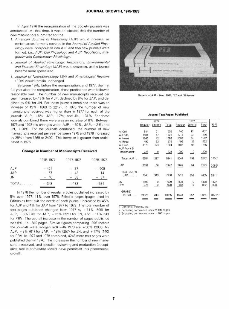
Growth of AJP - Nov. 1976, ‘77 and ‘78 issues
Journal Text Pages Published
1978 1977 Regular Edrtor’s Total qegular Editor’s Total -- -- -
A Cell 514 21 535 A Endo 1504 17 1521 A Heart 1646 42 1688 A Regu 492 83 575 A. Flutd 1170 124 1294 AJP Front b
Backmatter’ 228 0 228 ---
440 17 457 1213 23 1236 1508 34 1542 496 26 522
1187 98 1285
200 0 200 - - -
Total, AJP. 5554 287 5841
JAP 2091 56 2147 -- -
Total, AJP Et JAP. 7645 343 7988
JN 1699 0 1699 PRV 978 0 978 -- -
GRAND TOTAL 10322 343 10665
am-
5044 198 5242 3755’
2169 54 2223 - - -
7213 252 7465
1478 0 1478 882 0 882 - - -
9573 252 9825 8121’,3 ---- -
1 Contents. Indexes. etc 2 Excludmg cumulatrve index of 490 pages 3 Excluding cumulatrve Index of 260 pages
1976 -
20863 -
i84 1
1422 838 -
7

CASE HISTORY OF A PHYSIOLOGIST: F. G. HALL D. B. Dill
Desert Biology Research Center Department of Biological Sciences
University of Nevada, Las Vegas Boulder City, Nevada 83005
Introduction
A symposrum on “Physrological Adaptattons: Desert and Mountain” was held rn Las Vegas April 19 and 20, 1971. It was jorntly sponsored by the Desert Research Institute and the Uni- versity of Nevada, Las Vegas. The plan was concerved by Ray- mond J. Hock, professor, UNLV. After hrs untimely and tragrc death rt was brought to frurtron by Mohamed K. Yousef, pro- fessor, UNLV. The symposium recognized my 80th birthday. I was invited to give the dinner address My address honored my good friend and my colleague In many field studres, Frank Greg- ory Hail. The Desert Research lnstrtute in 1971 pnnted my ad- dress for drstrrbution to symposrum particrpants and friends. In 1972 the Symposium Proceedrngs, edrted by M. K. Yousef, S M. Horvath and R. W Ballard was published by Academic Press In the years since 1971 The Physiologist has become a source-journal for history of the American Physrological Soctety. Srnce my lecture touched on the activrtres of many members of the Society besides those of Hall and Dill It has been submitted to the editor for consrderatron
I remarked at the begrnnrng that there was one whom I missed above all others at the symposrum: Frank Gregory Hall - Greg. I reviewed his career hoprng to create the feelrng that he was one of us. As the story unfolds it will become evrdent that Greg and I had common beginnings and shared many expen- ences. Both of us were born In the rural mrdwest and started our education In one-room one-teacher country schools, and were orphaned young. Each of us lost a parent to tuberculosrs. We had no brothers; he had three srsters and I had four We both graduated from small colleges and recerved honorary doctorates from those colleges.
An account of hrs early years IS given in some detail herein; his experiences of those years were imprinted for lrfe HIS gradu- ate years included both JOYS and sorrows, and hrs 40 years at Duke Unrverstty brought him great honor. I have enjoyed relating our experiences of six expedrtions together to the desert, sub- tropics and mountains between the years 1935 to 1966, and during two years spent together during World War II in the Aero- Medical Laboratory. Srnce 1937 I have enjoyed the fnendshrp of hrs wife Stephanie, hrs daughter Betty, and hrs son Kenneth who IS now a professor at Duke Unrversrty. All have helped me In thts task, and Betty and Kenneth, I am happy to say, attended the dinner.
Birth To Manhood- 1896-1917
Greg was born on February 12, 1896, in a small town of Johnstown Center in southern Wrsconsin. HIS grandfather, Gregory Dexter Hall, was a dominant leader In the communrty of Johnstown Center. From his 1,000 acres of prairie, he gave land for the settlement, built the school, the church, the town hall, and a tall brick home from which he could view the town and hrs farm. From the house, Grandfather Hall sallied forth wrth hrs gold-headed cane; arrayed In a long frock coat, hrgh silk hat, a long flowing black silk tie and a beard; he was an Impressive frg- ure He died in 1906 when Greg was 10. HIS wrfe, Elrda Austrn, a granddaughter of President Tyler, died In that house In 1916 when Greg was 20 years old.
Greg’s parents, Frank Dexter Hall and Evaline (or Evelyn) Kidder, a school teacher, were married April 19, 1893. They set- tled in the “Center” where they owned the general store. The town was a more self-contained community than small towns of today. The town hall served for elections, dances, road shows and local entertainments. In back of the hall was a barn, a corral where farm stock was auctioned, and a stable for travelers’ horses. The general store sold everything but meat, and also served as a post office. There were rooms for the family and rooms to rent to travelers on the coach road between Lacrosse and Milwaukee; some of these Greg’s mother boarded.
Other buildings in the town included a Presbyterian church where Evaline played the organ, and a home for the mrnrster, his mother and two unmarried sisters. Next to the church was Dr. William Rockwell’s home. As the horse and buggy doctor for the surrounding area, he performed surgery, set broken bones, and often pulled teeth in his living room. Next to the one-room school was a small house for the teacher. The blacksmith shop had rooms in the back for the family, and the slaughter house and meat market were beside the creek. Some distance across the creek was a sorghum mill and an apple cider mill. One-half mile away Greg’s Uncle Lee ran a dairy and cheese factory.
Marjorie, Mary and Elida, Greg’s three sisters, were born in Johnstown and all but Elida went to the one-teacher school. Marjorie and Mary have written interesting letters to me concern- ing the town. There was a swimming hole in the creek and in the winter they skated there. Mary related a family anecdote-it seems Greg was paid five cents a week to carry water from the well to the house. When he was 10 and Mary was 4, he subcon- tracted with Mary to carry the water for one cent a week, a prof- itable business for him.
8
Their father died of tuberculosis in 1905. Sometime earlier,
however, he had sold the store and built a home nearby. Here
they lived until 1907 when they moved to nearby Milton. Greg
was then 11 and Elida, the youngest, was 3. In 1910 Evaline was
remarried to Ed Hurley, a neighbor who had seven children of his
own, five living with him and two away. Ed had relatives in cen-
tral Nebraska and the family moved to a ranch five miles from
North Loup. One bright spot for Greg lay in the fact that he had a
pony to ride to the North Loup High School from the ranch. After
the move, their mother took in washing to help support the
family. Marjorie writes that her mother loved all the children. But
she sent Greg back to Milton in 1911 to attend the Milton Acad-
emy. The following year, she died suddenly on October 12, and
her three daughters accompanied the body back to Milton where
she was buried beside their father. They then lived with their
uncle’s family, Mr. and Mrs. Otis Hall, who had a farm in Johns-
town Center. Uncle Otis became their guardian.
At this time Greg was starting his last year in Milton Acad-
emy. After graduation in 1913, he entered Milton College. He
often spoke of his happy years there. A fellow student, later Mil-
ton’s chemistry professor, William Burdick, recalls that in his col-
lege days Greg “revealed the intellectual curiosity and originality
which are essential to the success of a research scientist.”
Greg’s superiority in biology led to the appointment in his senior
year as a biology instructor under the direction of Professor A. R.
Crandall.
In the spring of 1917 he became engaged to a classmate,
Beth Marie Davis, who was a granddaughter of one of the foun-
ders of Milton College. Greg’s sister, Marjorie, describes Beth as
having been a wonderful person.
Greg often spoke of his visits to his sisters, all of whom set-
tled in Wisconsin. Marjorie graduated from Milton High School,
the Whitewater Normal School and taught school there until her
marriage to Joseph Weber, a farmer and carpenter. They settled
in Dorchester, Wisconsin. Marjorie has been active in community
affairs, particularly in those involving children. In recognition of
this, she was listed in “Who’s Who in American Women” in
1957. Elida taught school in Dorchester for one year before
marrying John Bochanyin. They had seven children; she died
two days after the birth of twins on March 4, 1952. Mary, the
second sister went to live with Marjorie in 1920 and in 1922
married Tom Johnston. There was a divorce a few months later,
after which she was remarried to Joseph Christie who died in
1957. She lives at Chetek, Wisconsin. Mary and Marjorie have
written interesting and informative letters to me about early days
in Johnstown and about the family. I am indebted to them for the
account of Greg’s background and early years.
Post-graduate Years - 19 17- 1923
These years encompassed notable achievements, and both
happy events and a major tragedy. Greg had been an instructor
in biology in his senior year. He spent the summer of 1917 taking
part in a biological survey of life zones and terrestrial vertebrates
of Wisconsin. This was planned by Professor George Wagner
and by Dr. Hartley H. T. Jackson who was in charge of the mam-
mal collection, U.S. Biological Survey. Jackson had graduated
from Milton in 1904 and sought a young Milton graduate to work
with him-Greg was chosen. Greg often spoke of that summer’s
work, his beginnings in research. Only a few years ago he men-
tioned discovering that some of the specimens he collected were
still available in the National Museum. Jackson has written that
Greg was “always a willing and capable worker and became one
of my closest friends.” That summer Greg visited Madison to
explore the possibility of pursuing graduate study at the
University. The plan took shape in his mind, but remained dor-
mant since he was committed to returning in the fall of 1917 to
teach biology at Milton.
With the country at war, Greg enlisted in the Signal Corps in
the spring of 1918 and was assigned to the Meteorological Sec-
tion. Shortly he was sent to Columbia University for special train-
ing. On July 4, 1918, he and Beth were married in New York
State; they had some time together in New York City. That fall
he was transferred to the University of Texas for further training
and Beth returned to Wisconsin to teach English at Watertown
High School. Greg was proud of his service in the infant aviation
organization which evolved into the Army Air Corps, later be-
coming the Army Air Forces and now the U.S. Air Force.
When Greg was relieved of active duty in the spring of 1919,
Beth joined him in Milton. Soon they went to Madison where
Greg began graduate study in the summer school. That fall they
returned to Milton where Greg had been appointed professor of
biology.
Their daughter, Betty, was born on May 12, 1920, but it had
been a difficult pregnancy and Beth never recovered from the
complications of childbirth. She died on May 30, 1920.
After this tragic loss, Greg went to Madison for graduate
study, but before leaving he arranged with one of Beth’s first
cousins, Margaret Post Bliss, to care for his daughter, Betty.
Now living in California, Betty writes that when her father went
to the hospital on May 30, 1920, he found his wife dead and his
infant daughter in precarious condition. Betty has been told that
she was tiny enough to fit into a cigar box. It was with this des-
perate situation facing him that Margaret took Betty in hand.
Margaret’s father, a doctor, predicted that the baby would not
survive, but with feedings every two hours around the clock, she
was saved. Eighteen months later Margaret Bliss (Aunt Marge)
gave birth to her first child, also a daughter and the two girls be-
came almost inseparable. Margaret’s husband became Dean of
Engineering at Marquette.
Greg received the Master’s Degree in 1921 under A. S.
Pearse. Chauncey Leake was then an instructor and graduate
student. He and Greg published a paper together in the American
Journal of Physiology which dealt with the regulation of vascular
tone (31). Chauncey wrote that Greg was a serious, hard-work-
ing student, with a quick smile, a quiet way and solid intelli-
gence. He says that Greg was a great teacher but a slow writer,
and he urged Greg to “write more- but he would only smile.”
Greg’s doctoral dissertation, The Function of the Swimblad-
derin Fishes was started in the summer of 1921 while he was em-
ployed by the U.S. Bureau of Fisheries. His observations were
made in Lake Mendota or in the laboratory, and he demonstrated
ingenuity in many phases of his experiments. Among his experi-
mental designs was an apparatus for studying the effects of
pressure on composition of gas in the swimbladder. An iron pipe
8” x 36” was fitted with a cap at one end, and a four inch view-
window at the other. An inlet pipe supplied water to this tank
from a reservoir on the 4th floor; the outlet pipe carried the over-
flow also to the 4th floor, 60 feet above the tank. By this means
he was able to demonstrate that this increase in pressure applied
for 10 hours raised the O2 in the swimbladder from 12.1 to
18.5%. He concluded that the primary role of the swimbladder is
hydrostatic. While gases ordinarily reach the swimbladder by dif-
fusion, in some species at least oxygen can be secreted into the
bladder. The thesis was published in the Biological Bulletin (201,
and reprints, in conformity with University requirements, were
deposited in the library.
Greg had spent the summer of 1922 at the Marine Biological
Laboratory (MBL), Wood’s Hole, Massachusetts, taking a

course in Invertebrate Zoology. A fellow student, I. E. Gray, was
destined to become his lifelong friend. Greg persuaded Gray to
apply for a teaching assistantship at Wisconsin; he was accepted
and started his graduate study when Greg was finishing. They
roomed together that year. Another associate, both at MBL and
Madison, was Samuel Lepkovsky, who is now professor emeri-
tus at Berkeley and is still busy in the laboratory there.
My first tie to Greg, though tenuous, was through Wiscon-
sin. My first year in high school was in Washington, Iowa, where
my stimulating teacher of physiology and botany, H. I. Case,
boasted of being a Wisconsin graduate, class of 1890. My next
three high school years were at Santa Ana, California where our
highly regarded principal and most able teacher of mathematics
was Edward B. Oakley. He, like Case, had been a poor Wisconsin
boy who earned his way through the University, graduating in
1879.
After receiving his doctorate in 1923, Greg returned to Mil-
ton as professor of biology. This was the beginning of a new era
in more ways than one. He and Stephanie Daland were married
that summer. Stephanie, a daughter of Milton’s president, had
graduated in 1917 and then worked in the Racine Library. She
later attended and ultimately became a staff member at the Li-
brary School at Madison which eventually became an integral
part of the University.
Margaret Bliss and her husband wanted to adopt Betty, but
all accepted Stephanie’s decision that Betty should be with Greg
and her. Margaret says that after separating the two girls her
daughter “did not eat for a year.”
Betty graduated from Duke University in 1942. She sang in
the glee club and choir and sings in her church choir now. A
month after graduation she married a classmate, Kent Boutwell.
He obtained a Ph.D. in mechanical engineering at the University
of Michigan and taught engineering there for 12 years. He is now
a research scientist with General Motors Research Laboratory in
Santa Barbara. (By an extraordinary set of coincidences, my
daughter’s name is Betty; she too lives in Santa Barbara, and her
husband is also a scientist. Betty Boutwell has four children; my
daughter has three, and all of the children are in the same age
range.
A t Duke University - 1926- 1967
Greg’s mentor at Wisconsin, A. S. Pearse, had accepted
Duke University’s offer of a professorship of zoology, with the re-
sponsibility for graduate study in zoology. His acceptance of this
offer was contingent upon completion of a sabbatical leave and
appointment of a suitable assistant. With the experience of three
years on the Milton faculty behind him, Greg was invited to be-
come associate in Zoology at Duke. He, Stephanie and Betty ar-
rived in the fall of 1926, and Pearse arrived at the beginning of
the second semester. They made a great team of teachers and
investigators. Within a short time, two of their graduate students
were awarded Ph.D.s, the first of these degrees to be awarded in
the young university. Both Pearse and Greg enjoyed life to the
full and both had a keen sense of humor. One of the many tales
about Pearse occurred during the latter part of the war when I
had persuaded him to act as a consultant to the Army Quarter-
master Corps. Even he, a professor and a Harvard Ph.D. was re-
quired to complete a questionnaire concerning his personal life.
To the question, “Have you ever been discharged from a posi-
tion?” he wrote, “At age 10 I was fired from my one dollar per
week job as delivery boy for a grocery store.”
This was an eventful year for the family. Kenneth was born
on October 26 of that year. I became acquainted with Kenneth
and Betty when I visited Durham in 1937 after our desert study.
When Greg brought his family to Dayton during the war years,
Kenneth was active in the Boy Scouts. After graduating from
Duke University in 1949, he entered the Duke University School
of Medicine, and interned in surgery-obstetrics-gynecology at
Rochester, N .Y. After his internship, he received special training
in anesthesiology at Duke and in the National Institutes of Health
as Chief of the Section on Pain. He then returned to Duke where
he was appointed assistant professor of anesthesiology in the
Department of Surgery. In 1968 he was appointed to a professor-
ship in that department.
Kenneth found time to work with his father in the labora-
tory, and between 1951 and 1966 they co-authored three papers.
In 1964 he accompanied Greg when they participated in our
desert study (details of which are discussed in a later section).
Kenneth and Maidi Ebel became acquainted in Rochester where
her father was an eminent physician. After their engagement,
Kenneth had to resist urging by her father to join him in the gen-
eral practice of medicine. But Kenneth had already determined
on a career in research and medical education in anesthesiology.
This he has achieved. He and Maidi and their four children are
happy in their home near the University.
During their first year at Duke, Pearse and Hall completed a
book entitled, Homoiothermism, the Origin of Warm-Blooded
Vertebrates (36). A part of their preface defined the scope, “The
physiological and ecological aspects of one type of adjustment
which has made successful life on land possible and has reached
its climax in the attainment of thermal and chemical stability
within the bodies of animals.”
In 1929-30, Pearse was visiting professor at Keio University,
Japan; he left Greg with responsibility for recruiting more scien-
tists for the department. The new recruits were George Hargitt
from Syracuse and his friend from graduate days at MBL and
Wisconsin, Gray from Tulane. The Halls and Grays were best
friends and next door neighbors for years.
Greg’s progress up the academic ladder was rapid. In 1930
he was promoted to a professorship, and during this period
Pearse and many colleagues at Duke, including Greg, founded
the Marine Laboratory of Duke University at Beaufort, North Car-
olina. This is owned and operated by the University. Greg gave a
course in physiology there one summer and gave the A. S.
Pearse Memorial Lecture there some years ago. In 1932 Greg and
others went with Pearse to Yucatan to study cenotes. This expe-
dition will be discussed later.
Of major importance to Greg’s future was his sabbatical year
with Barcroft which began in February of 1933. As an investiga-
tor, he brought great talent to bear on the problems of respira-
tory physiology that were concerning Barcroft at the time. With
Barcroft, Greg co-authored a paper on fetal circulation and had a
related paper of his own (21). During that year he also became
acquainted with Bryan Matthews and a young physiologist from
the U.S., Ancel Keys. They were to be together in 1935.
Stephanie writes that she took Betty and Kenneth to their
mother’s home in Milton late in January. The first night the tem-
perature dropped to -18’ F., quite a contrast to the mild climate
of Durham. Stephanie joined Greg in July when his studies were
nearly completed.
Those weeks were memorable. They were entertained by
the Barcrofts, the Matthews and the Adairs. Their travels took
them to Stratford-on-Avon to see a Shakespeare play and to visit
historic sites in London, Edinburgh and Paris. Greg, an expert
photographer, brought back many pictures. They returned on
the Berengaria and were reunited with Betty and Kenneth.
The next summer found Greg back at MBL. In the fall he
visited the Fatigue Laboratory to help plan the high altitude study
10
of 1935. Also, he was a subject In the control studies, partrcularly
rn propertres of arterial blood. Accounts of the 1935 study, the
Yucatan expedrtron, our desert study of 1937, our Missrssrppi
study of 1939 and the post-war expeditions to mountains and
desert in 1962, 1964 and 1966 WIII be given later.
A major event In the family history took place in the winter
of 1939-40: the building of their beautiful home which IS set In the
pines and dogwoods that cover the hills around the Unrversrty. It
IS a tribute to Greg’s devotion to hrs country that less than a year
after the home was completed, he took leave of absence for mill-
tary duty at the AeroMedrcal Laboratory (AeroMed). Stephanie
remained at Durham until 1942 when she and Kenneth torned
Greg at Dayton. They were fortunate to have responsrble mrlrtary
famrlres renting their home during the war years.
Upon hrs return to Duke, Greg was asked to become a Pro-
fessor In the Department of Physiology and Pharmacology In the
School of Medicine. He had a busy lrfe during the post-war
years. Contracts with the Arr Force enabled hrm to equip a small
laboratory away from the department. Here during weekends
and summers he continued hrs research on hypoxia, wrth particu-
lar Interest rn resprratory function. Two assrstants who jorned him
at AeroMed were Kenneth Penrod, who arrived in 1950, and
Wayland Hull, who came later Penrod turned to admrnrstratrve
tasks and IS now Vice Chancellor for Medical Affairs at the State
Unrversrty System of Florida. Hull IS now assistant to the Medical
Director, Manned Spacecraft Center, Houston.
Our common Interest In avratron medicine occasronally
brought us together, as on November 12, 1948 when Major Gen-
eral Harry Armstrong, then Commandant of the School of Avia-
tion Medicine at Randolph Arr Force Base, assembled rnvestrga-
tors, rncludrng Greg and me, In the fteld of aeromedical research.
Thus ptoneer rnqutry was titled “Ftrst Panel Meeting on
AeroMedrcal Problems of Space Travel.”
A year later Greg and I shared a notable experience in Lima,
Peru. Professors Carlos Monge and Albert0 Hurtado and their as-
sociates arranged a symposium on the Brology of High Altitudes
(5). One feature was the dedrcatron of the new high altitude
station at Morococha (14,900 ft.). The Air Force had helped sup-
port their research; this involved Major General Armstrong and
Brigadier General Benson who arranged to transport a party of
screntrsts to the symposrum. Included In the group were Bng-
adrer General Wallace Graham (the White House physrcran),
Wallace Fenn, Joseph Kaplan, Randolph Lovelace, Ashton Gray-
brel, Albert Behnke and others. Other Air Force representatives
were Major Cain and Harry Adler (see Figure 1). It had been 14
years since Greg and I had visited Lima incident to our high
altitude study In Chile. Followrng Hurtado’s example, many
students had spent a year in the U. S., rncludrng two, Juan Aste-
Salazar and Leon Contreras, who had come to the Harvard
Fatigue Laboratory. The symposrum was a great success, culmr-
natrng In the dedrcatron of the laboratory at Morococha.
A caravan was assembled to take the visrtors by car up
nearly 15,000 feet In three hours. A rank order was adopted
which placed Harry Adler, Greg, the scientrfrc attache of the
Embassy of Mexico and me in car 14. Adler sat with the driver
who disliked being 14th In line, before long he had passed cars 13
and 12, despite the many harrprn turns. Adler’s remonstratrons
only spurred him on. I remarked to the driver that his friends
must call him “el trgre,” to which he replied, “SI, si, senor.”
The Mexican and I carried on a worned conversatron, but
decided we could only hope for good luck. Greg, who sat be-
tween us, showed no sign of worry, but he drd not engage in
conversatron. The Mexican could not draw him out and at length
characterized him aptly, “El hombre del pocas palabras,” the
man of few words! When we reached the summit we were No. 3!
As soon as all had arrived we were escorted to a railroad dining
car for lunch. I sat with Behnke, Graybiel and Greg. Behnke,
proud of his physical fitness, began bantering Graybiel, but
before the meal was finished Graybiel, who was enjoying it, was
delighted that mountain sickness had overcome Behnke. He was
quite green when he left the table
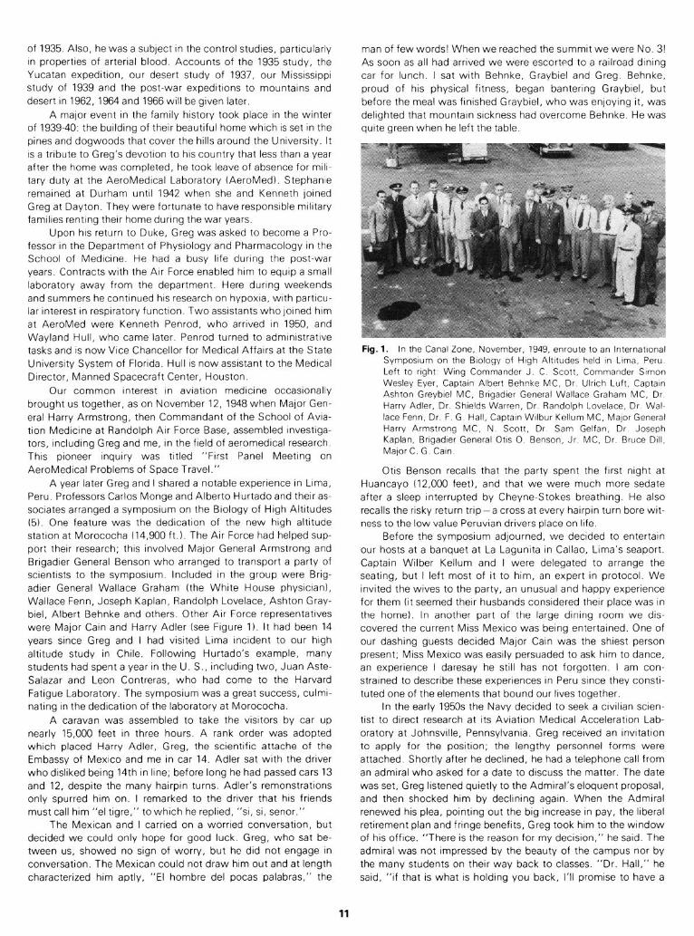
Fig. 1. In the Canal Zone, November, 1949, enroute to an lnternatronal Symposrum on the Biology of Hugh Altrtudes held In Lrma, Peru Left to rrght Wrng Commander J C Scott, Commander Srmon Wesley Ever, Captain Albert Behnke MC, Dr Ul:rch Luft, Captarn Ashton Greybrel MC, Brrgadrer General Wallace Graham MC, Dr Harry Adler, Dr Shrelds Warren, Dr Randolph Lovelace, Dr Wal- lace Fenn, Dr F G Hall, Captarn Wrlbur Kellum MC, Major General Harry Armstrong MC, N Scott, Dr Sam Gelfan, Dr Joseph Kaplan, Brrgadrer General Otrs 0 Benson, Jr MC, Dr Bruce Dtll. Major C G Cam
Otis Benson recalls that the party spent the first night at
Huancayo (12,000 feet), and that we were much more sedate
after a sleep Interrupted by Cheyne-Stokes breathing. He also
recalls the risky return trip-a cross at every hairpin turn bore wit-
ness to the low value Peruvian drivers place on Irfe.
Before the symposrum adjourned, we decrded to entertain
our hosts at a banquet at La Lagunrta In Callao, Lima’s seaport.
Captain Wilber Kellum and I were delegated to arrange the
seating, but I left most of It to him, an expert in protocol. We
Invited the wives to the party, an unusual and happy experience
for them fit seemed therr husbands considered their place was in
the home). In another part of the large dining room we drs-
covered the current MISS Mexico was being entertained. One of
our dashing guests decided Major Cam was the shiest person
present; Miss Mexico was easily persuaded to ask him to dance,
an experience I daresay he still has not forgotten. I am con-
strained to describe these experiences in Peru srnce they constr-
tuted one of the elements that bound our lives together
In the early 1950s the Navy decided to seek a crvrltan scren-
trst to direct research at its Aviation Medical Acceleratron Lab-
oratory at Johnsvrlle, Pennsylvanra. Greg recerved an rnvrtatron
to apply for the positron; the lengthy personnel forms were
attached Shortly after he declined, he had a telephone call from
an admiral who asked for a date to discuss the matter. The date
was set, Greg listened quietly to the Admiral’s eloquent proposal,
and then shocked him by declrnrng again. When the Admiral
renewed hrs plea, pointing out the brg increase in pay, the liberal
retirement plan and fringe benefits, Greg took hrm to the window
of hrs office. “There is the reason for my decrsron,” he said. The
admrral was not Impressed by the beauty of the campus nor by
the many students on their way back to classes. “Dr. Hall,” he
said, “if that IS what IS holding you back, I’ll promise to have a
11
platoon of marines march past your window every hour on the
hour.” The admiral left, still not appreciating Greg’s point of
view.
For several years I saw Greg only at meetings. In 1955 the
fall meeting of the American Physiological Society was held at
Tufts College, Medford, Massachusetts. There I heard about the
vicious mugging Greg had experienced in March of that year. At
dusk he was starting up the steps to the emergency entrance of
the Duke Hospital when he was struck over the head with a
heavy, blunt instrument. He did not collapse and his assailant,
realizing this, ran off -but not before Greg had noticed that he
was a stranger. Greg walked into the emergency room for help
without realizing that blood was streaming down his face from
the deep head wound. He was given first aid and was hospital-
ized overnight. There seemed to be no residual effects beyond
emotional shock, but it seemed to me that Greg was shaken by
the memories aroused through the telling of this experience.
Greg made what proved to be an important decision in 1956
when he had John Salzano appointed to his staff. John had re-
ceived his doctorate that year at the University of Iowa under
Wade Tuttle. Steve Horvath, then at Iowa, introduced John to
Greg. At that time Fred Zechman, Jr., was with Greg as a post-
doctoral fellow, having received his doctorate with Greg the year
before. The three worked together on studies of the role of the
vagus nerves in pulmonary mechanics, The next year Fred left,
and he is now head of the Department of Physiology and Bio-
physics in the School of Medicine, University of Kentucky. June
Barker returned to join the group after a year’s study in England.
Those were busy days, teaching several groups of students phys-
iology and pharmacology. However, with Air Force support,
Greg and John were active in aeromedical research, particularly
in the summer months. Medical students were employed as as-
sistants. John writes that, as chairman, Greg took a personal
interest in the members of the department and in the execution
of his responsibilities as department chairman.
In 1960 I was selected to edit a volume of the Physiological
Society’s Handbook. This became Volume 5, Adaptation to the
Environment. I invited Greg to write a chapter, but he replied:
“I regret that I had better not take on one of the chapters. I have
committed myself to a heavy load of work here at Duke for the
coming year. I feel that I shall not have the time for contempla-
tion that would be required for writing. I also find it much harder
to write as I get older. I shall, of course, be very glad to help on
any of the details. I have made a few suggestions as to possible
authors of chapters.”
From 1961 we corresponded frequently about our studies to-
gether of the physiology of mountain and desert; details of those
studies will follow. In June, 1965, he retired at the mandatory age
of 69. He was then free to follow his own desires and was happy
in his laboratory with John Salzano. His interests during this
period were concerned with respiratory function of the blood in
rodents. Among several papers in this field, one dealing with
hemoglobin and oxygen affinities of seven species of squirrels
was published in Science E!3).
Many honors came to Greg during his years at Duke. At the
close of the war, besides the Legion of Merit (see World War II),
he received letters of appreciation from Major General Grant, the
Air Surgeon, and from Detlev Bronk. Bronk was ‘scientific ad-
visor to the Air Force during the war, and later was president of
Rockefeller University. Greg was honored in June, 1957, by his
alma mater, Milton College, by the award of an honorary degree
of Doctor of Science. In 1966 he was elected a Fellow of the
Aerospace Medical Association in recognition of his many
achievements in the field of Aerospace Medicine. Crowning rec-
ognition came in the fall of 1965 with a dinner honoring his 40
years of service to Duke University. This was held at the Hope
Valley Country Club and about 100 of his friends and associates
were in attendance. My wife and I were privileged to be there.
His portrait was presented and now hangs in the Physiology De-
partment of the Medical School.
WORLD WAR 11-1941-1945
In the summer of 1940 Greg spent some time at the Fatigue
Laboratory studying, with Otis Benson, Will Forbes and me, res-
piratory regulation in oxygen lack as affected by amphetamine
(7). Otis, then a captain in the Medical Corps, was soon to relieve
Harry Armstrong as chief of the AeroMedical Laboratory, Wright
Field. This will be referred to subsequently as AeroMed. In prepa-
ration for this assignment, Benson spent some months with us
and at the Mayo Clinic. He convinced Greg and me that we
should apply for commissions to assist him in expanding the
research program at AeroMed. This we did, looking forward to
six months in uniform and the subsequent return to our universi-
ties.
Greg and I arrived at AeroMed early in January, 1941. I had
been commissioned a major in the Specialists Corps as it was
then called. In April, 1942, Greg was also commissioned. Aero-
Med was familiar to me. Sid Robinson, Harold Edwards and I had
collaborated with Harry Armstrong and John Heim in 1937 when
Harold and I were on our way to Boulder City (8). At the time, the
new altitude chamber had just been completed and we con-
ducted the first experiment in it.
Benson’s vision of unchartered fields is illustrated by the fact
that he introduced anthropometry to the military services. During
the summer of 1940, he sought the advice of Ernest Hooton, Pro-
fessor of Anthropology at Harvard. Growing out of this discus-
sion, four of Hooton’s students measured Air Force cadets and
gunners for purposes of equipment design. After Hooton had
analyzed their results, two of the students, Albert Damon and
Francis Randall, were commissioned and assigned to AeroMed
to apply Hooton’s statistical data. They remained’there during he
war as members of Pharo Gagge’s Biophysics Branch. Randall
was killed in an airline accident; Damon became a member of the
Anthropology Department at Harvard. Their research has played
a major role in the redesign of clothing, footgear and other items
of personal equipment developed for military use.
Benson said we must have a biophysicist and sometime later
Pharo Gagge was persuaded to fill this role. We found a strong
nucleus at AeroMed that included John Heim, John Hall and
Ernie Pinson. Ernie had received his Ph.D. at Rochester in 1940.
He rose through the ranks to Major General and became Com-
mandant of the Air Force Institute of Technology at Wright Pat-
terson Air Force Base where he began his career. Others who
were recruited as lieutenants included “Ken” Penrod, Loren
Carlson, Harvey Savely, Clarence Maaske, George Maison and
John Wilson. Some, such as Ross MacCardle, came as civilians
and were commissioned later. Our six months were stretched to
twelve and Pearl Harbor found us ordered to remain on duty for
the duration.
At the Fatigue Laboratory Will Forbes was displaying master
craftsmanship in building up an outstanding group of investiga-
tors for attack on physiological problems of the military; cold,
heat, protective clothing, footgear, load-carrying equipment and
rations. I left Harvard with a strong group working with Will: Bob
Darling, Lucien Bronha, Steve Horvath, Charlie Knehr and Frank
Consolazio. Steve had already become my son-in-law. Shortly
Will recruited Edgar Folk, H. L. Belding, Don Griffin, Paul
Robinson and Ann Hoyt. Later Sid Robinson brought his stu-
12
dent, Gene Turrell, to spend a year at the Fatigue Laboratory;
then Sid returned to start his own program at Bloomington.
Greg and I enjoyed our work together at AeroMed. We
began by studying the performance of oxygen masks then under
development. By concluding that the mask that had been devel-
oped at the Harvard School of Public Health was unsafe, I made
myself unpopular with Professor Cecil Drinker, responsible for
the project. He brought his group to AeroMed to investigate;
Greg and I proved that on some faces the fit was so poor that in
the altitude chamber at 30,000 feet the men lost consciousness
and would have died had they been in flight. Greg played a major
role in our study of mask leakage. At 30,000 feet the system
should supply the wearer with pure oxygen; a leak means that he
inspires a gas mixture containing nitrogen. Determining the
percent of nitrogen in the air in the mask during inspiration gives
a basis for calculating the percentage of leakage since air con-
tains about 79% of inert gas. For this purpose Greg adapted a
device described by Scholander (38). Five ml of gas was drawn
from the mask into a calibrated syringe. The gas was injected
through pressure tubing into a burette. The bulb held about 40 ml
of oxygen absorbent. Its upper end was a small bore tube,
capacity 5 ml calibrated to read 1 to 100. The lower end of the
burette was connected with pressure tubing to a leveling bulb
containing mineral oil. The gas sample was shaken with the oxy-
gen absorbent for one minute; then the remaining bubble was
allowed to rise into the calibrated portion. With a minor correc-
tion, the reading corresponds to percent nitrogen in the sample.
Since air is about 4/5th nitrogen, the percent leakage is 5/4ths
the percent of nitrogen in the sample. Greg demonstrated that
the method could be learned by young officers responsible for
oxygen equipment in the field. This apparatus was procured and
supplied to air bases; it was called “Unit Oxygen Officers Test
Kit.”
Greg and I made many observations on respiratory re-
sponses to altitudes ranging up to 44,000 feet. We reported that
at this altitude breathing pure oxygen is roughly eavalent to
18,000 feet breathing air (12).
Greg and I also tried out rations designed for emergency
use. For emergencies, a l-lb. bar of semisweet chocolate was
available for air crews after bailing out. We were led to evaluate
this when General “Hap” Arnold sent word from the Pacific that
survivors of air crashes reported that this bar “looks like hell and
tastes worse.” So we obtained some bars for trial. Although this
was planned for issue on the basis of 1 man-l day, we could not
find a record of its use in this way. We and some volunteers each
started on a bar at breakfast, ate part at noon and planned to
finish it for dinner. The next morning none of us had started on
the second bar. Before or after the evening portion, all had devel-
oped headache; some experienced nausea and vomiting. We
were astonished to learn that we had been poisoned by theo-
bromine. Pharmacologists could have predicted this, since they
were aware that one pound of chocolate eaten within 12 hours
raises the amount of the theobromine in the body to toxic levels.
The D-bar soon was de-standardized.
At this time the K-ration had been developed by the Quarter-
master with the advice of Ancel Keys. Greg and I decided to test
this and, simultaneously, a pemmican ration which was under
development at Wright Field. Pemmican was used by Indians
long before the white man came to America. It was commonly
made of dried berries and dried meat pounded together and
packaged in animal casings. The pemmican we were to test was
a nutritionally supplemented mixture derived from meat and
grain products in powder form. We were told that one small can
held a day’s ration, and that it could be eaten dry, mixed with a
little water to form a sludge, with more water to form a slurry, or
with still more to form a beverage.
With Sergeant Murphy and a squad of soldiers, Kenneth
Penrod, Sid Robinson, Gene Turrell, Greg and I undertook to
evaluate pemmican and the K-ration. We went by C-47 to Albu-
querque and in a short time with a light pack started across the
desert for the Sandia Mountains. We opened the cans at noon;
some ate part of the dry powder, but most decided to wait until
evening. At our camp in cottonwoods on the banks of the Rio
Grande we started eating the pemmican. Sergeant Murphy’s sol-
diers rebelled before they had downed much of it. So Murphy, a
well-disciplined soldier, added enough water to his to make a
slurry and quickly drank it; in minutes he appeared green and
then vomited. Thus ended the trial of pemmican.
Previously an expert who had developed it reported he had
tried some on the end of a knife-blade. “It isn’t tasty,” he had
said, “but a soldier could eat it if he had to.” We then lived on K-
ration quite successfully for six days.
Greg and a driver were responsible for logistics, meeting us
each night with blankets as we walked past the Santa Ana
Pueblo into the mountains, past the Jemez crater and down
towards Santa Fe. Before each evening meal Sid took blood
samples for sugar analyses. Each morning three packages that
constituted the day’s ration were issued. In the evening each
listed and commented upon the uneaten items. All had com-
ments to make but one, a soldier from the Kentucky hills with
little education. When we pressed him for an explanation, he
admitted it was less punishment to eat everything than to write
about what he had not eaten.
At the end of six days we boarded the truck and covered the
remaining miles to Santa Fe, stopping on the way for a visit with
my friend, Wo Peen, an artist at the San lldefonso Pueblo with
whom I had become acquainted in 1937. After a big meal in
Santa Fe, we continued to Albuquerque and soon were on our
way to AeroMed.
Greg and I took part in one other field study while I was at
AeroMed, a winter study at Oscoda, Michigan, of cold weather
clothing, especially foot gear. The soldiers joined Bob Johnson,
Will Forbes and me in a 23-mile march; not all of them finished
the march with us.
Our reports concerning the poor performance of some oxy-
gen equipment in the altitude chamber irritated some of the
engineers at Wright Field who had shared the responsibility for
its development. When the Office of Air Surgeon urged that re-
sponsibility for developing oxygen equipment be transferred to
AeroMed, the engineers agreed provided Dill had no part in the
plan. Colonel Doriot of the Quartermaster General’s Office re-
quested my transfer, and it was quickly arranged. In April, 1943,
Greg and I parted, meeting only infrequently during the re-
maining war years.
At about this time, Benson, then a colonel, replaced Colonel
Armstrong at the School of Aviation Medicine at Randolph Field.
Each became a major general and they both now live in retire-
ment at San Antonio, Texas. W. Randolph Lovelace succeeded
Benson at AeroMed; after the war he became Director of the
Lovelace Clinic and Research Foundation in Albuquerque. He
assembled a distinguished group, including Sam White and
Ulrich Lcuft who attended my birthday dinner. Randy’s death in
an aircraft accident in 1965 was a tragic blow for the Foundation
and for aerospace medicine.
Greg continued to be concerned not only with familiar prob-
lems but also with the effects of acceleration and protection
against it. His advice was invaluable in the design of “G” suits;
for this he was awarded the Legion of Merit. Another interest,
13
allied to his biological background, lay in hydroponics. Raising
vegetables in water containing essential nutrients seemed useful
to rocky outposts such as Ascension Island in the South Atlantic.
A system had been developed by the end of the war. Indicative
of some of Greg’s responsibilities at AeroMed are three publica-
tions (25, 26, 27).
Late in the war, Greg had become the leading authority on
many of the human problems of high altitude flight. Between
February and April, 1944, his advice was sought by the European
Theater of Operations, as a result of which he reached England in
February to confer with General Grant, Colonel Armstrong and
former associates from AeroMed, including George Maison and
J. J. Smith. He had an opportunity to visit Cambridge where he
renewed his friendships with Adair and Adrian and visited the
laboratory where he had worked 21 years earlier. Greg was fond
of telling about meeting the diener who had been the major
domo of the laboratory for a long time. Evidently time had tele-
scoped for he remarked casually to Greg, “You have been away
some little time.” From England Greg went to North Africa and
Italy where Otis Benson was the ranking Air Surgeon. He dis-
cussed problems of oxygen equipment and clothing with many
officers and air crews. At the 26th General Hospital he was
invited to give a lecture on problems of aviation physiology.
His next overseas duty took him to India in July and August
of 1945. Again he was called upon to answer questions about
oxygen equipment. His notebook records on August 2 that he
had made 200 tests on mask leakage using the kit he had devel-
oped for that purpose. He must have been happy to find no mask
with a leakage greater than 6%.
Many physiologists visited AeroMed during the years, con-
tributing greatly to the research program. Besides those men-
tioned, I can think of Walter Boothby, E. J. Baldes and Arthur
Bulbulian of the Mayo Research Foundation; Glen Millikan, then
at the University of Pennsylvania; Wallace Fenn from Rochester;
Sid Robinson, Indiana University; David Tyler from Cal Tech;
and F. J. W. Roughton, Cambridge. Experts who came to ex-
change ideas included Paul Siple, Lindbergh, Doolittle, and the
surviving member of the Wright Brothers team, Orville. A monu-
ment to the Wright Brothers’ first flight stands on the hill above
AeroMed.
Greg was relieved of active duty (but remained a reserve of-
ficer) in the fall of 1945, and returned to academic life at Duke
University. He received from General Twining the highest non-
combat award, the Legion of Merit. The Citation reads:
“His brilliant work in solving the many complex problems of
human physiology in relation to flying reflects great credit to him-
self and the military service.”
Expeditions: Yucatan- 1932
In 1932 Greg was a member of a group organized by Pearse
to study the cenotes of Yucatan (35). Their report was published
by the Carnegie Institution of Washington in 1936. The principal
authors were Pearse, Creaser (University of Michigan), and Hall.
Chapters were written by 14 collaborators dealing with life in
cenotes. Greg’s chapter was entitled, “Physical and Chemical
Survey of Cenotes of Yucatan.”
The peninsula of Yucatan consists of porous limestone cov-
ered with a thin layer of soil; there are no rivers over most of the
peninsula. From ancient times the Indians have depended on
cenotes for water. In some of them the water is one or two
meters deep, but in many it is more than 20 meters deep. Greg
observed transparency, gradients of temperature, pH and con-
tents of CO*, dissolved 02, fixed CO, and chloride. He considered
the belief held by some that subterranean rivers supply water to
cenotes, but doubted that idea. It seemed likely to him that water
enters through the porous limestone.
High Altitudes- 1935
The International High Altitude Expedition of 1935 in which
Greg played a major role was organized by Ancel Keys. In 1933
he was appointed an instructor and a tutor in biochemical sci-
ences at Harvard after three years divided between Copenhagen
with Krogh and Cambridge with Barcroft. While at Copenhagen,
Ancel and Hohwij Christensen worked together under Krogh.
They published a paper with him dealing with the organic metab-
olism of sea water (30). Ancel had discussed his dream of a high
altitude study with Krogh and Barcroft and found them recep-
tive. At Cambridge he had formed a close friendship with Bryan
Matthews, then a graduate student at Cambridge and an en-
thusiastic mountaineer. Greg’s year at Cambridge overlapped
Ancel’s study there; Greg’s rich experience in respiratory physi-
ology made him an obvious choice. Hohwti Christensen came as
a Rockefeller fellow from Krogh’s laboratory to spend 1934-35 in
the Fatigue Laboratory; he was eager to join the group. By the
summer of 1934 plans were taking shape. Forbes and Edwards of
the Fatigue Laboratory and McFarland, then of Columbia Univer-
sity, were signed up and sources of support were found.
The first site tentatively selected was the Pamirs. After two
requests for authority were ignored by the USSR Embassy, the
goal became Karakorum. Approval by British authorities seemed
routine. Keys, Edwards and Forbes put together a field labora-
tory securely packaged in plywood cases suitable for porter
transport. In December Keys left by air for India via San Fran-
cisco to make advance preparations, and Forbes left for New
York to oversee loading laboratory cases, camping, sleeping and
mountain climbing gear aboard a freighter bound for Karachi.
Before Keys reached San Francisco, a cable came from Bar-
croft to Henderson with the news that the authorities had re-
fused permission for the expedition. Henderson acted at once,
recalling Keys from San Francisco and asking Forbes to have the
equipment unloaded and stored. Henderson, Keys and I reached
agreement shortly; the site would be Northern Chile. I would take
part and hopefully Baird Hastings; the goals would be solely sci-
entific; the smaller cost would permit dispensing with an expert
photographer and newscaster. Ancel accepted these decisions
and in a short time succeeded in making arrangements for the
Chilean study.
An advance party, including Keys, Hall, Forbes, Edwards
and Christensen, left in March by Grace Line for Antofogasta
(see Figure 2). Matthews joined them two weeks later. Hastings
had accepted a professorship at Harvard; in his place he recom-
mended his associate, E. S. C. Barron (Achito), a biochemist
from Peru, a graduate of the Medical School in Lima, and a
protege of Carlos Monge, a distinguished scholar. Achito had
come down from the hills to the University and Medical School.
A poor Indian, his had been an uphill battle, but in medical school
he had become a leader of his fellow students. At one time he led
a strike demanding that professors give more time to teaching
and less to private practice.
In late April, the second group left via Grace Line; these
included McFarland, Talbott, Barron and me. Barron proved his
value early, enlivening the stopover in Panama with a dinner and
evening he had arranged at his cousin’s home, and at Lima,
introducing us to his former chief, Carlos Monge, Professor of
Medicine. We disembarked at Tocopilla and traveled by car to
Chuquicamata, the great copper mining camp owned by Gug-
genheim interests.
The advance party had made good use of their six weeks.
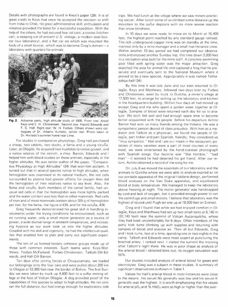
Detarls wrth photographs are found In Keys’s paper (28) It IS of
great credrt to Keys that once he accepted the decrsron to shaft
from lndra to Chile, hrs great admrnrstratrve skill, enthusiasm and
rnrttatrve provided the basis for a successful expedrtron With the
help of the others, he had secured four rarl cars’ a cocrna (kitchen
car), a sleeping car of ancrent U.S vantage, a modern steel box-
car four laboratory), and a flat car on which was mounted the
body of a small boxcar, which was to become Greg’s domarn - a
laboratory wrth quarters for anrmals.
Fig.2 Advance party, high altitude study of 1935. Front row: Amel Keys and E. H. Christensen. Second row: Harold Edwards and F. G. Hall. Right rear: W. H. Forbes. Others shown were col- leagues of Dr. Albert0 Hurtado, center rear, Photo taken at Dr. Hurtado’ssummer home near Lima.
For studies in comparative physiology, Greg had purchased
a sheep, two rabbits, two ducks, a llama and a young vicuria.
Later, at Ollagiie, he acquired two huallatas (a native goose), and
a native relative of the ostrich, a rhea. Barron, Edwards and I
helped him with blood studies on these animals, especially at the
higher altitudes. He was senior author of the paper, “Compara-
tive Physiology at High Altitudes” (24) that won him acclaim. It
turned out that in several species native to high altitudes, when
hemoglobin was examined in its natural medium, the red cells
surrounded by plasma had greater affinity for oxygen than did
the hemoglobin of their relatives native to sea level. Also, the
llama and vicuria, both members of the camel family, had un-
usual red cells in that the hemoglobin was more tightly packed
and the water content was less than in other mammals. Red cells
of man and of most mammals contain about 335 g of hemoglobin
per liter; for the llama, the figure is439, and for the vicuna, 426.
Greg frequently demonstrated his great skill in handling in-
struments under the trying conditions he encountered, such as
no running water, only a small motor generator as a source of
power, a wide diurnal range in ambient temperature, and increas-
ing hypoxia as our work took us into the higher altitudes.
Coupled with his skill and ingenuity, he had the intellectual quali-
ties that enabled him to plan and carry out significant experi-
ments.
The ten of us formed loosely cohesive groups made up of
those with common interests. Such teams were: Keys-Mat-
thews, Forbes-McFarland, Forbes-Christensen, Talbott-Dill-Ed-
wards, and Hall-Dill-Barron.
Ten days after joining forces at Chuquicamata, we loaded
our belongings onto the four cars and were pulled about 200 km
to OIlague at 12,000 feet near the border of Bolivia. The first Sun-
day we were taken by truck up 4,600 feet to a sulfur mining vil-
lage at the foot of Volcan OIlague. A dog provided proof of the
capabilities of this species to adapt to high altitudes. He not only
ran the full distance, but had energy enough for exploratory side
trips We had lunch at the village where we saw miners practrc-
rng soccer After lunch some of us clrmbed some drstance up the
mountain to the sulfur deposits with no more severe reactron
than snow blindness
In 10 days we were ready to move on to Montt at 16,400
feet, the highest point reached by any standard gauge rarlroad.
The rich underground copper mine was on standby at the time,
manned only by a mane manager and a small marntenance crew.
Within another IO-day period we had completed our observa-
tions and enjoyed another Sunday trip, this time down 3,000 feet
to a recreation area built for the mine staff A concrete swimmrng
pool filled with spring water was the major attractron Greg
searched the area for anrmal life and captured a frog that he pre-
served and eventually sent to the Natronal Museum where It
proved to be a new species Appropriately It was named Telma-
tobius halli ( 34 )
By thus time It was late June and our train returned to Ol-
lagtie. Keys and Matthews, followed two days later by Forbes
and Christensen, went by truck to Qurlcha, a mrner’s vrllage at
17,500 feet, to arrange for setting IJP the laboratory and kitchen
in the headquarters burldrng Wrthrn four days all had moved up
except Greg and me who spent a golden week together at Ol-
lagrYe Samples of blood were returned each day for us to ana-
lyze We both felt well and had enough spare time to become
better acquarnted with the people Before his departure Achrto
Barron had won us many friends among the lndrans He was a
sympathetic person devoid of class pretudrce With hrm as a me-
diator and Talbott as a physicran, we found the people of Ol-
lagtie, part lndtan and part Spanish, friendly, they considered us
“muy sympatrco ” Hall and I ate our meals In the restaurant, po-
tatoes of many vaneties were a part of most courses of every
meal, we were entertained by the hand-cranked phonograph
with Spanish songs Our favorite was “Ma1 hombre,” “bad
man” - It seemed he had deserted his grrl friend After our re-
turn, Achrto obtained a record of the song for me
On July 8 we moved the essentials of our laboratory and the
animals to Qurlcha where we were able to analyze expired arr on
our portable apparatus of the onglnal Haldane design, performed
blood analyses on the Van Slyke apparatus, and equrlrbrated
blood at body temperature We managed to keep the laboratory
above freezing at night The motor generator was handrcapped
by cold and lack of oxygen, but It provided enough power to run
the centrifuge and small motors I belreve that laboratory was the
highest of record until Pugh set one up at 19,000 feet on Everest.
Greg and I found that while we had enjoyed comfort In Ol-
lagfie, Keys and Matthews had set up two small tents at 6,140 m
(20,140 feet) near the summit of Volcan Aucanqurlcha, where
they had lived uncomfortably for 6 days Other members of the
party took turns clrmbrng up wrth supplres and returning with
samples of blood and alveolar arr Then all but Edwards, Greg
and I took turns, two at a time, spending one or two nrghts in the
camp Talbott and Edwards were most expert at puncturing the
brachral artery. I ranked next I vrsrted the summit the mornrng
after Talbott’s night there He was In poor shape as analysrs of
the arterial blood I obtained bore out, Its oxygen saturatron was
55%
Our studies Included analysis of arterial blood for gases and
electrolytes. Greg was a subject In these studies A summary of
srgnrfrcant observations IS shown In Table 1
Values for Hall’s arterial blood in most Instances were close
to the means HIS serum Na generally was low and hrs serum K
generally was the highest It IS worth emphasrztng that hrs values
for arterial pOZ and % HbO? were as hrgh or higher than the aver-
15
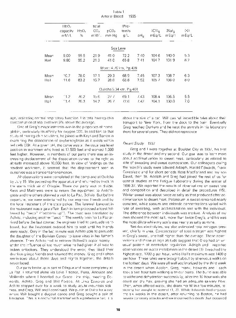
Table 1
Arterial Blood - 1935
HbO;! total (Pro-
capacity HbO, CO, PC02 tein 1s (Cl 1s (NaIs (K)
mM/L % mM/L mmHg g/L PHS mEq/L mEq/l mEq/L
Sea Level
Mean 9.00 95.5 21.9 41 .o 72.2 7.40 104.6 140.0 5.3
Hall 9.80 95.2 21.3 41 .o 65.8 7.41 104.7 131.9 8.2
Montt 4.70 km, PB 429
Mean 10.7 78.0 17.1 29.3 68.9 7.45 107.3 138.7 6.3
Hall 11.6 83.2 15.7 28.0 68.8 7.52 105.7 136.0 8.0
Mean 11.2 76.2
Hall 11.4 76.3
Quilcha 5.34 km, PB 401
15.6 27.7 69.1 7.43 108.4 136.5 5.5
14.2 26.7 72.6 7.42 104.1 130.0 7.0
age, indicating normal respiratory function. His total hemoglobin
concentration at sea level was 9% above the average.
One of Greg’s major interests was in the properties of hemo-
globin, particularly its affinity for oxygen (22). In addition to that
study of hemoglobin solutions, he joined with Keys and Barron in
examining the dissociation of oxyhemoglobin as it exists within
red cells (29). At a given pH, the curve was at the usual sea level
position in workmen who lived at 17,500 feet and worked 1,000
feet higher. However, in members of our party there was an in-
creasing displacement of the dissociation curves to the right as
altitude increased above 10,000 feet. In view of findings on the
resident workmen, it seemed that the displacement seen in
ourselves was a transient phenomenon.
All observations were completed at the camp and at Quilcha
by July 15. We packed up the apparatus and returned by truck to
the warm thick air of Ollagiie. There our party was to divide.
Keys and Matthews were to return the equipment to Antofo-
gasta and the others planned a visit to La Paz, Bolivia. But before
departure, we were entertained by our engineer friends and by
the local lieutenant of the state police. The farewelt banquet in
the restaurant was a gala affair. Each in turn proposed a toast fol-
lowed by “seco” (“bottoms up”). The toast was translated by
Achito, including another “seco.” The weekly train for La Paz ar-
rived before the last course; the engineer tried to persuade us to
board, but the lieutenant ordered him to wait until his friends
were ready. Only in the last minute was Achito able to persuade
the daughter of the Bolivian Consul to issue visas in her father’s
absence. Then Achito had to retrieve Hohwti’s paper money-
under the influence of too much wine he had given it all way to
Indian onlookers. They understood the error; they liked their drunken gringo friends and returned the money. Greg and I often
reminisced about those days and nights together; the details
never faded.
Our party broke up in part at Ollague and more completely at
La Paz. I returned alone via Lake Titicaca, Puna, Ariquipa and
Mollendo where I boarded our Grace Line ship, meeting Ed-
wards, Achito, Greg and Will Forbes. At Lima Edwards and
Achito stopped over for a week to study acute mountain sick-
ness, and Greg, Will and I continued. We put in at Quito for a day
where Will bought a dugout canoe and Greg bought a pair of
kinkajous. This is a nocturnal mammal with a prehensile tail; it is
16
about the size of a cat. Will can tell incredible tales about their
transport to New York, from the dock to the train. Eventually
Greg reached Durham and he kept the animals in his laboratory
there for several years. They did not reproduce.
Desert Srudy - 1937
Greg and I were together at Boulder City in 1937, his first
study in the desert and my second. Our goal was to learn more
about acclimatization to desert heat, particularly as related to
rate of sweating and sweat composition. Our colleagues during
the month’s study were Edward Adolph, Harold Edwards, Frank
Consolazio and for short periods Ross MacFarland and my son
David, then 18. Adolph and Greg had joined the rest of us for
control studies at the Fatigue Laboratory during the winter of
1936-37. We reported the results of observations on sweat rate
and composition and described in detail the procedures (15).
Winter sweat was about twice as salty as that produced after ac-
climatization to desert heat. Potassium in sweat remained nearly
constant, while sodium and chloride concentrations varied with
rate of sweating, with acclimatization and with the individual.
The difference between individuals was marked. Analysis of my
own showed the most salt, more than twice Greg’s, and his was
the most dilute while my son’s was nearly as salty as mine.
Besides electrolytes, we also estimated total nitrogen pres-
ent, chiefly in urea. Concentration of total nitrogen was highest
in Greg’s sweat, one-half higher than the average. These obser-
vations and those at high altitude suggest that Greg had an un-
usual pattern of electrolyte regulation. Adolph and I reported
observations on water metabolism in the desert (1). I reached the
highest rate, 1700 g per hour, while Hall’s maximum was 1400 g
per hour. These rates were brought about by strenuous walks on
the hottest days. We were all well acclimatized by the third week
in the desert when Adolph, Greg, Frank, Edwards and I each
took a two-hour turn walking with our burro. The burro was able
to withstand dehydration successfully; after the lo-hour walk she
could eat dry hay, proving she had an adequate salivary flow.
Then, when offered water, she drank her fill in a few minutes, re-
storing her weight to normal (1, 3). While Edwards had enjoyed
the six weeks in the desert, after returning to Boston, he had
severe coronary attacks and we mourned his death that occurred
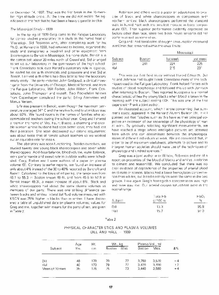
on December 14, 1937. That was the first break in the IO-mem-
ber high-altitude circle. At the time we did not realize the sig-
nificance of the fact that he had been a heavy cigarette smoker.
The Mississippi Study - 1939
In the spring of 1939 Greg came to the Fatigue Laboratory
for control studies preparatory to a study in the humid heat of
Mississippi. Sid Robinson who, after completing work for his
Ph.D. at Harvard in 1938, had returned to Indiana, organized the
study and transported a treadmill and other equipment from
Bloomington to Benoit in Mississippi, his home state. Benoit is in
the cotton belt about 20 miles north of Greenfield; Sid arranged
to set up our laboratory in the gymnasium of the high school.
Frank Consolazio took over Harold’s customary role in logistics;
he loaded his car with chemicals and glassware and met Sid at
Benoit. I arrived with others two days later to find the laboratory
nearly ready. The party included John Gedgoud, a clinical inves-
tigator from the University of Chicago, Medical School, and from
the Fatigue Laboratory, Will Forbes, John Wilson, Frank Con-
solazio, John Thompson and myself. Two Rockefeller fellows
from Copenhagen completed the party: Erling Asmussen and
Marius Nielsen.
Life was pleasant in Benoit, even though the maximum tem-
perature averaged 34” C and the relative humidity at midday was
about 60%. We found rooms in the homes of families who ac-
commodated teachers during the school year. Greg and I shared
a room in the home of Mrs. Faunt Biscoe, a charming and capa-
ble woman whose husband had seen better days; they had lost
their plantation. She soon discovered our caloric requirement
was about twice that of female school teachers so we worked
out an equitable rate for meals.
The laboratory was soon functioning. Besides ourselves, we
studied twenty-one young black sharecroppers and seven white
sharecroppers. Acid-base balance, blood volume, water balance,
work performance and sweat rate in outdoor walks were sched-
uled. Greg, Forbes and I were authors of a paper on plasma
volume (9). Contrary to earlier reports, we found an increase of
only about 4% instead of the 30 to 40% reported by Barcroft and
Bazett. Calculated to the basis of ml per kg, the range was from
40.1 to 55.3 in Boston (mean 46.41, and from 45.6 to 52.9 in
Benoit (mean 49.31, a mean increase of about 6%. Black and
white sharecroppers had about the same plasma volumes as
members of our party. There was one striking difference be-
tween blacks and whites: interstitial fluid volume measured with
KSCN was 25% higher in blacks than in whites. I have discov-
ered a table of unpublished data on plasma volumes; values for
Greg and me, together with means for the party of ten, are given
in Table 2.
Robinson and others wrote a paper on adaptations to exer-
cise of black and white sharecroppers in comparison with
northern whites; black sharecroppers performed the standard
walk in humid heat with the smallest increase in body temper-
ature (37). Their superior performance evidently depended on
factors other than race, since two black house servants did not
perform as well as some of us.
Greg and I had basal rates of oxygen consumption measured
in ml/min that remained within the usual limits:
Mississippi
Subject
Dill
Hall
Boston
241
1 st week
224
215
last week
225
231
This was our first field study without Harold Edwards. But
he and Johnson had taught Frank Consolazio many of the tech-
niques used in the Fatigue Laboratory. Frank helped Forbes with
studies of blood morphology and followed this up with Johnson
after returning to Boston. They reported leucopenia is a normal
characteristic of healthy young blacks when blood is taken in the
morning with the subject resting (19). This was one of the first
papers with Frank a joint author.
An illustrated account, which I wrote concerning that sum-
mer’s study, appeared in the Harvard Alumni Bulletin (4). In it I
pointed out that “studies such as this have as their principal ob-
jective an extension of our knowledge of the physiology of man
at work. By gradually selecting significant measurements, we
have reached a stage where intelligible pictures are obtained
from which one can discriminate between the physiological
states of different individuals at work. We are convinced that, in
order to be of maximum usefulness, attempts to picture and to
interpret human societies should make use of the techniques of
physiology and kindred sciences.”
Greg was a joint author with Wilson, Robinson and me of a
report on properties of the blood of blacks and whites in relation
to climate and season(l8). We concluded that there was no
clear evidence of dependence of the properties of arterial blood
on climate or season. Blacks had a lower hemoglobin concentra-
tion than whites, but blood electrolytes were the same in the two
groups. Here again Greg’s hemoglobin concentration was high
and mine was low. Our arterial oxygen saturations were in the
normal range:
Subject
Dill
Hall
Total Hb HbO,
g/l00 ml %
13.3 95.8
15.7 97 0
Table 2
PHYSICAL CHARACTERISTICS AND PLASMA VOLUMES
DILL AND HALL - 1939
Subject Age Yrs.
Ht.
cm
Wt., kg
Boston Miss.
Plasma Vol., ml
Boston Miss. A%
Dill 48 179 79 77 3,750 3,570 -4 Hall 40 173 79 77 3,420 3,490 +2
Mean of 10 men 35 175 75 73 3,440 3,580 +4
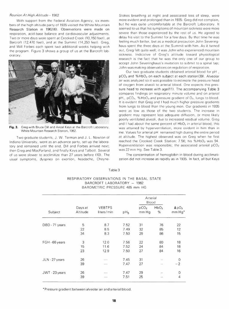
Reunion A t Hgh Altttude - 1962
With support from the Federal Avlatlon Agency, SIX mem-
bers of the high altitude party of 1935 vIsIted the White Mountan
Research Station In 1962(6). Observations were made on
respiration, acid-base balance and cardiovascular adjustments
Two or more days were spent at Crooked Creek (10,150 feet), at
Barcroft (12,470 feet), and at the Summit (14,250 feet) Greg,
and Will Forbes each spent two addItIonal weeks helping with
the program Figure 3 shows a group of us at the Barcroft lab-
oratory
Fig. 3. Greg with Bruce DIII and Ancel Keys at the Barcroft Laboratory,
White Mountain Research Station, 1962.
Two graduate students, J. W. Terman and J. L. Newton of
Indiana University, went as an advance party, set up the labora-
tory and remained until the end. Dill and Forbes arrived next;
then Greg and MacFarland, and finally Keys and Talbott. Several
of us were slower to acclimitize than 27 years before (10). The
usual symptoms, dyspnea on exertion, headache, Cheyne-
Stokes breathing at night and associated loss of sleep, were
more evident and prolonged than In 1935 Greg did not complaln,
but he was quite uncomfortable at the Barcroft Laboratory It
seemed to us that his symptoms of mountain sickness were more
severe than those experienced by the rest of us He agreed to
delay his vlslt to the Summit for a few days By that time he was
feeling much better, bllt as a medical precaution John Severlng-
haus spent the three days at the Summit with him As It turned
out, Greg felt quite well, It was John who experienced mountain
sickness lndlcntlve of Greg’s attitude toward physlologlcal
research IS the fact that he was the only one of our group to
accept John Sevennghaus’s lnvltatlon to submit to a spinal tap,
John was making observations on regulation of resplratton
Our two graduate students obtained arterial blood for pH ,
pC0, and %HbO, on each subject at each statlon(39). Alveolar
air was analyzed so It was possible to estimate the pressure head
of oxygen from alveoli to arterial blood One expects this pres-
sure head to increase with agelll). The accompanying Table 3
compares flndlnqs on respiratory mrnute volume and on arterial
pH,, pCOZ, %HbO* and pressure gradient of 02, lungs to blood
It IS evident that Greg and I had much higher pressure gradients
from lungs to blood than the young men Our gradients In 1935
were as low as those of the two students The Increase In
gradlent may represent less adequate dlffuslon, or more likely
poorly ventilated alveoli, due to Increased residual volume Greg
and I had about the same percent of HbO, In arterial blood, this
was attained by hyperventllatlon, more evident In him than In
me Values for arterial pH rernalned high durtng the entire pertod
at altitude The highest observed was on Greg when he first
reached the Crooked Creek Station 7 56; his %HbO, was 94
Hyperventllatlon was responsible, the associated arterial pCOz
was 22 mm Hg See Table 3
The concentration of hemoglobin In blood during accllmatl-
zatlon did not Increase as rapidly as In 1935. In fact, all but Keys
Table 3
RESPIRATORY OBSERVATIONS IN THE BASAL STATE
BARCROFT LABORATORY - 1962
BAROMETRIC PRESSURE 485 mm HG
Subject
Days at VEBTPS
Altitude liters/min PHS
Arterial
Blood
PC02 HbOl
mm Hg %
A PO?
mm Hg’
DBD - 71 years
FGH - 66 years
J LN - 27 years
J WT - 23 years
5 8.7 7.52 31 76 22
22 8.5 7.49 32 85 12
34 8.3 7.50 29 86 15
3 12.0 7.56 22 80 18
15 11.6 7.52 24 84 18
23 12.9 7.50 27 84 16
26 ___ 7.45 31 __ 0
39 7.47 27 -- -2
26 ___ 7.47 29 __ 0
39 7.51 25 -- 4
*Pressure gradient between alveolar air and arterial blood.
18
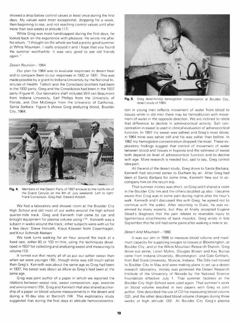
showed a drop below control values at least once during the first
days. My values were most exceptlonal, dropping for a week,
then beginnIng to rise, and not reaching control values until after
more than two weeks at altitude (17)
While Greg was most handicapped during the first days, he
looked back on the experience with pleasure He wrote me after
his return, “I thought on the whole we had a pretty good summer
at White Mountain. I really enloyed It and I hope that you found
the summer worthwhtle. It was very good to see old friends
again ”
Desert Reunion - 1964
Our plan for 1964 was to evaluate responses to desert heat
and to compare them to our responses in 1932 or 1937. This was
made possible by a grant to Indiana University by the National In-
stitutes of Health. Talbott and the Consolazio brothers had been
in the 1932 party; Greg and the Consolazios had been in the 1937
party (Figure4). Our laboratory staff included Will van Beaumont
from Indiana University, Earl Phillips from the University of
Florida, and Don McGregor from the University of California,
Santa Barbara. Figure 5 shows Greg analyzing blood, Boulder
City, 1964.
Fig.4. Members of the Desert Party of 1937 enroute to the north rim of the Grand Canyon on the 4th of July weekend Left to right, Frank Consolazlo, Greg Hall, Edward Adolph
We had a laboratory and shower room at the Boulder City
High School and did most of our walks around the high school
quarter-mile track. Greg and Kenneth Hall came by car and
brought equipment for plasma volume using 1131. Kenneth was a
subject in walks around the track; other subjects were with us for
a few days: Steve Horvath, Klaus Klausen from Copenhagen,
and Knut Schmidt-Nielsen.
We took turns walking for an hour around the track at a
fixed rate, either 80 or 100 m/min, using the techniques devel-
oped in 1937 for collecting and analyzing sweat and measuring its
volume (13).
It turned out that nearly all of us put out saltier sweat than
when we were younger 1161, though mine was still much saltier
than Greg’s, Kenneth was about the same age as Greg had been
in 1937; his sweat was about as dilute as Greg’s had been at the
same age.
Greg was joint author of a paper in which we reported the
relations between sweat rate, sweat composition, age, exercise
and environment (15). Greg and Kenneth Hall also shared author-
ship of a paper on blood volume measurements in the desert and
during a IO-day stay at Barcroft (14). This exploratory study
suggested that during the first days at altitude hemoconcentra-
Fig. 5. Greg determInIng hemoglobtn concentration at Boulder City, desert study of 1964
tlon In young men reflects movement of water from blood to
tissues while In old men there may be hemodllutlon with move-
ment of water tn the opposite dIrectIon. We are Inclined to relate
that difference to decline tn adrenocortlcal activity Salt con-
centration In sweat IS used In clInIcal evaluation of adrenocorttcal
function. In 1937 my sweat was saltiest and Greg’s most dilute;
In 1964 mine was saltier stall and his was saltier than before In
1962 my hemoglobin concentration dropped the most These ex-
ploratory findings suggest that control of movement of water
between blood and tissues In hypoxia and the saltiness of sweat
both depend on level of adrenocortlcal function and Its decline
with age. More research IS needed but, sad to say, Greg cannot
take part.
At the end of the desert study, Greg drove to Santa Barbara,
Kenneth had returned earlier to Durham by air After Greg had
been at Santa Barbara for some time, Kenneth flew out to ac-
company him on the return trip.
That summer money was short, so Greg ana I shared a room
In the Boulder City Inn and the others doubled up also I became
aware that Greg was In some pain during nights after his hour’s
walk Kenneth and I discussed this with Greg, he agreed not to
continue with the walks. After returning to Duke, he was ex-
amined by many experts, but they all finally accepted Eugene
Stead’s dlagnosls that the paln related to reversible Injury to
llgamentous attachments of back muscles Greg wrote In late
September that he still had some pains after walking a mile or so
Desert And Mountain - 1966
It was our aim in 1966 to measure blood volume and maxi-
mum capacity for supplying oxygen to tissues at Bloomington, at
Boulder City, and at the White Mountain Research Station. Greg
drove out alone; Loren Myhre, Douglas Brown and Kay Burrus
came from Indiana University, Bloomington; and Gale Gehlsen,
from Ball State University, Muncie, Indiana. The Dills had moved
to Boulder City in May and were making plans to set up a desert
research laboratory; money was promised the Desert Research
Institute of the University of Nevada by the National Science
Foundation effective July 1. That summer facilities at the
Boulder City High School were used again. That summer’s work
on blood volume resulted in two papers with Greg as loint
author. One described the use of CO for measuring blood voltlme
(321, and the other described blood volume changes during three
weeks at high altitude (33). At Boulder City Greg’s plasma
19
volume was within the normal range. His plasma volume in 1939
was 3420 ml at Boston, and 3490 in Mississippi (see Table 1). In
1964 at Boulder City, it was 3240, and in July, 1966, at Boulder
City, it averaged 3580. Again in Boulder City, after the Barcroft
experience and recuperation, it averaged 3580. Finally, in
September, after return to Durham, It was 3390. It is remarkable
that these observations from age 43 to 70 were within the limits,
3240 and 3580.
At Boulder City, Greg’s hemoglobin concentration in blood
was about 10% above mine as it had been ever since our mea-
surements at the Harvard Fatigue Laboratory in 1934. We have
made observations on others over a longer period, indicating that
this is a persistent individual characteristic.
Greg and I both were somewhat apprehensive about his
gotng to high altitude for three weeks. But he wanted to try it, so
the six of us made the trip together. He had a bad first night, but
he felt better the next day and was a subject in a blood volume
measurement. He continued to be quite miserable, sleeping little
at night. The fifth morning he went by helicopter to Bishop
(about 4,000 feet altitude). Here he made a remarkable recovery,
sleeping 14 hours the first night. After the second night, he
announced his intention to try it again. But the old symptoms
returned, and after the second miserable night, he agreed to
leave for Santa Barbara. There Stephanie joined him. After an
enloyable week at Betty’s home, they returned to Boulder City so
Greg could help complete our studies. We waved farewell early
one August morning as he and Stephanie left for Durham. We
were not to see Greg again.
Greg’s terminal illness was in the Duke Hospital where treat-
ment measures were fruitless; he died on February 19, 1967. The
final diagnosis was gradual failure of his heart. His ashes were
held until spring, and then buried in the family plot at Milton.
Stephanie has written to me about the eloquent tributes paid at
the memorial service, to his great achievements and his great
humanity.
Epl’log
I had an opportunity to review Greg’s career at the Respira-
tion session of the American Physiological Society at Chicago in
April, 1967. After paying tribute to his record of 45 years, I spoke
of his character, kindness, human understanding, great technical
skill and his judgment of what constitutes a good experiment. I
then recalled his enjoyment of life, his sense of humor and told
one of his favorite stories; it brought down the house.
Two years later I was privileged to take part in the dedication
of a magnificent memorial to Greg: The F. G. Hall Laboratory for
Environmental Research, at Duke University. There again I re-
viewed some of our experiences together and closed with the fol-
lowing verses paraphrased from sentimental verses he wrote
celebrating my retirement in 1961.
WE REMEMBER HALL
On Andean climbs our life was rough
All found sleeping very tough
But Sunday found us playing ball
That was where I first knew Hall
Singing “Ma1 Hombre,” a Gaucho tune
Dining in a miner’s saloon
Drawing blood from a llama’s leg
That is how we remember Greg
Taking turns one by one
We walked a burro in the noonday sun
Through the mesquite and over the hill
Given water he drank his fill
On the hike to Santa Fe
With Pemmican and Ration K
Camping at night near a waterfall
There is where I remember Hall
During the War it was the task
To test performance of the oxygen mask
Some were baffled by facial convolutions
But Greg was quick to find solutions
The proper study of mankind is Man
Study him where’er you can
In heat or cold or dry or wet
Drawing blood or collecting sweat
In winter and spring, summer and fall
With great esteem we remember Hall.
In closing, a quotation from a burial service composed in
1920 by Percy Dawson, is appropriate. His career tn physical edu-
cation including a professorship at Duke, 1943-46, was inter-
rupted by a pastorate in a Unitarian church. Percy died Sep-
tember 1970 at age 97; his burial service, read as he requested,
closed as follows: II
. . . all may see that even in his death his influence has not been
in vain, but in dying as in living, he has served to advance
through us the cause of truth and righteousness, of light and
love, through the growing personalities of those near and dear to
him.”
ACKNOWLEDGEMENT
Invaluable assistance has been rendered by Greg’s widow
Stephanie, by his daughter Betty Bontwell, by his son Kenneth
and by his sisters Marjorie Weber and Mary Christie. Several of
his colleagues at Duke University helped me, especially Drs. Grey
and Salzano. Useful details were found in the obituary published
in the Physiologist (2).
References:
1.
2. 3.
4.
5.
6.
7.
8.
9.
10.
11.
12.
13.
20
Adolph, E. F. and D. B. DIII. Observations on water metabolism In
the desert, Am. J. Ph ysiol., 123:369-378; 1938. Anon. Frank Gregory Hall. The Phys/blog/st 10:103-104, 1967.
0111, D. B. Life, Heat and Altitude, Harvard Untverslty Press,
Cambridge, Mass., 1938. Dill, D. B. Fatrgue studies among MISSISSIPPI sharecroppers, Harvard Alumni Bull., 42: 11 l-l 19, October 20, 1939.
Dill, D. B. Report on International Symposlom on the Biology of High Altitudes, Science, 111: 19-20, 1950.
Dill, D. B. Re union at high altitude, The Physiologist 6:40-43, 1963.
0111, D. B., 0. 0. Benson, Jr., W. H. Forbes, and F. G. Hall. Benzedrine sulphate (amphetamine) and acute anoxla; I. Respl-
ratory effects, J. Avlbtion Med., 11: 181-186; 1940. 0111, D. B., H. T. Edwards, S. Robinson, H. G. Armstrong, and
J. W. Heim. Pulmonary gaseous exchange at low barometric
pressure and In air mixed with nitrogen, J. Aviation Med., 10:3-10; 1939.
Dill, D. B., W. H. F or b es, and F. G. Hall. The effects of climate
upon the volume of blood and of tissue fluid in man, Am.
J. Ph ysiol., 130: 737-746; 1940. Dill, D. B., W. H. F or es, b J. L. Newton, and J. W. Terman.
Respiratory adaptations to high altitude as related to age, ln Re- lations of Development and Aging, Thomas, SprIngfield, III., 62-73,
1964.
0111, D. B., A. Grayblel, A. Hurtado, and A. C. Taqulni. Gaseoils exchange In the lungs in old age, J. Am. Geriatrics Sot: ,
11:1063-1076; 1963. Dtll, D. B., and F. G. Hall. Gas exchange in the lungs at high altitudes, J. Aeronautical Sci., 9:220-223, 1942.
Dill, D. B., F. G. Hall, and H. T. Edwards. Changes in compositlon
of sweat during acclimatization to heat, Am. J. Physl’ol.,
123:412-419; 1938.
14.
15.
16.
17
18.
19.
20.
21
22
23.
24.
25.
26.
27.
28.
29.
30.
31.
32.
33.
34.
35.
36.
37.
38.
39.
DIII, D B., F G Hall, K. D. Hall, C. Dawson, and J. L. Newton. Blood, plasma, and red cell volume: age, exercise and environment,
J. Appl. Physiof., 21:597-602; 1966.
0111, D. B., F. G. Hall, and W. van Beaumont. Sweat chlortde
concentration, metabolic rate, skin temperature, and age, J. Appl. Physl’ol., 21:99-106; 1966. DIII, D. B., S. M Horvath, W. van Beaumont, Gale Gehlsen, and
Kay Burrus. Sweat electrolytes In desert walks, J. Appk Physlol., 23:746-751, 1967
DIII, D. B., J. W. Terman, and F. G. Hall. Hemoglobrn at high
altrtude as related to age, C/in. Chem., 9:710-716; 1963. DIII, D B., J. W. Wrlson, F. G. Hall, and S. Robrnson. Properties of
the blood of negroes and whites tn relation to climate and season, J. Biot. Chem., 136:449-460; 1940.
Forbes ‘I. H., R E. Johnson, and F. Consolazro. Leucopenra In Negrc workmen, Am. J. Med. SC/., 2011407-412; 1941
Hall, F G. The functrons of the swimbladder in fish, Biol. &I//., 47 79-127; 1924.
Hall, F. G. Hemoglobrn functton In the developrng chick, J. Physiol.
(London), 83:222-228, 1934. Hall, F G. The effect of altrtude on the affinity of hemoglobrn for oxygen, J. Bl’ol. Chem., 115:485-490; 1936.
Hall, F. G. Hemoglobin and oxygen affinities in seven species of Sciuridae, Science, 148: 1350- 1351. 1965.
Hall, F. G., D. B. Dill, E. S. G. Barron. Comparatrve physiology in high altitudes. J. Cell. and Comp. Physiol. 8:301-313. 1936.
Hall, F. G., G. S. Maison, and K. E. Penrod. Parachute opening forces, Air Surgeon S Bull. 2:350-353. 1945.
Hall, F. G. and J. W. Wilson. Effects of physical activity and
simulated altitude on pulmonary ventilation, maximal inspiratory flow and pressure in relation to oxygen requirements. J. Aviation
Med. 151160-166. 1944. Hall, F. G., J. W. Wilson and H. F. Schwann. Physiological effects
of high negative mask pressures during simulated free falls. Proc. Am. Sot. Exp. Bl’ol. 5: 114. 1946.
Keys, A. The physiology of life at high altitudes. Sci Monthly. 43:289-312. 1936.
Keys, A., F. G. Hall and E. S. G. Barron. The position of the oxygen
dissociation curve at high altitude. Am. J. Physio/. 115:292-307. 1936. Krogh, A. and A. Keys. Methods for the determination of dissolved
organic carbon and nitrogen in sea water. Biol. Bull. 67: 132-144.
1934. Leake, C. D., F. G. Hall and A. E. Koehler. The influence of hydrion
concentration on vascular toxicity. Am. J. Physiol., 65:386-394. 1923.
Myhre, L. G., D. K. Brown, F. G. Hall and D. B. Dill. The use of carbon monoxide and T-1824 for determining blood volume. C/in.
Chem., 14:1192-1205. 1968.
Myhre, L. G., D. B. Dill, F. G. Hall, and D. K. Brown. Blood volume changes during three-week residence at high altitude: C/in. Chem.,
16:7-14. 1970. Noble, G. K. A new species of Frog of the genus Telematobius from
Chile. Am. Museum Novitates No. 973:1-3. 1938.
Pearse, A. S., C. P. Creaser, and F. G. Hall. The cenotes of
Yucatan. A zoological and hydrographic survey. Carnegie Inst.
Wash., Publ. 457. 1936. Pearse, A. S. and F. G. Hall. Homoiothermism. Wiley, New York
1928. Robinson, 4;. , D. B. Dill, P. M. Harmon, F. G. Hall and J. W.
Wilson. Adaptations to exercise in Negro and white sharecroppers in comparison with northern whites. Human Biol., 13: 139-I 58. 1941.
Scholander, P. F. Analyzer for quick estimation of respiratory gases. J. Biol. Chem., 146: 159-l 62. 1942.
Terman, J. W. and J. L. Newton. Changes in alveolar and arterial
gas tensions as related to altitude and age. J. Appl. Physiol., 79:2 7 -24. 7964.
A NEW RESEARCH TRAVEL GRANT PROGRAM
A new program of research travel grants for medical scien-
tists is being made available jointly by The Wellcome Trust in
Great Britain and The Burroughs Wellcome Fund in the United
States.
The program is so designed that a research worker engaged
in a promising investigation in the United States can cross to
Britain or Ireland to consult with or develop additional knowledge
from research workers there, with a minimum of red tape and ad-
vance timing. The same arrangement is open to workers in Great
Britain or Ireland to consult with research workers in the U.S.
In announcing the program, Lord Franks, Chairman of The
Wellcome Trust, noted that “advances in the medical sciences
might be hastened by workers being able to exchange informa-
tion rapidly. The Wellcome Research Travel Grants provide a
simple mechanism for research workers engaged in studies of an
exceptional nature to come together for consultation and the ex-
pediting of their results, perhaps for publication and for the bene-
fit of other workers in the particular field.”
The two private foundations have the knowledge to assess
proposals from each one’s own country but decided that the joint
arrangement would allow for fullest utilization of the expertise in
each country to act upon proposals with good judgment.
The Wellcome Research Travel Grants may be applied for at
any time during the academic year with the likelihood of a deci-
sion being given in a short period, thus allowing the workers to
undertake their research abroad within several months of making
application. Visits are expected to be of relatively short duration
- two weeks to a maximum of three months. The research
grants will take the form of a contribution towards the costs of
travel and subsistence. The amount of the individual grant will
vary, dependent on length of stay and location. No provision will
be made for spouses or families.
Dr. Hitchings, President of The Burroughs Wellcome Fund,
noted that “there exists a universal need for shorter term funding
and a rapid means of making funds available to research workers
with exciting leads that require collaboration with those in other
countries. Government funding generally takes a year or more
between the making of application and the receipt of money
while other competitive one-time annual awards do not respond
to the need. Our joint program will seek to fill the gap.”
The Wellcome Trust and the Burroughs Wellcome Fund
each is setting aside $100,000 per year for three years for The
Wellcome Research Travel Grants.
Inquiries originating in Britain and Ireland should be ad-
dressed to The Wellcome Trust, 1 Park Square West, London.
Those originating in the United States should be addressed to
The Burroughs Wellcome Fund, Research Triangle Park, North
Carolina, 27709.
An Advisory Committee, composed of A. McGehee Harvey,
M.D., as Consultant, Kenneth Brinkhous, M.D., and Paul L.
Munson, Ph.D., of the University of North Carolina School of
Medicine; and W. K. Joklik, D. Phil, and R. Wayne Rundles,
M.D., of Duke University Medical Center, will assist the selection
of U.S. recipients of the grants.
21
NEWS FROM SENIOR PHYSIOLOGISTS
Leslie L. Bennett to Hy:
On July 1, 1977 I finally retired, a year later than I was sup-
posed to have done so. I spent my last year in recall status as
Vice-Chancellor Academic Affairs. All in all, I spent ten of the
twelve years prior to July 1, 1977 in that position. I am very proud
of the fact that for at least eight of those twelve years I managed
to maintain some modicum of activity in research.
Since July 1, 1977, things have happened thick and fast. I
have succeeded in breaking my ankle, having major surgery to
remove three aneurysms (lower abdominal aorta and both com-
mon iliacs-no prior symptoms and an uneventful recovery),
write another biographical sketch about Herbert Evans, write a
short essay, the fate of which is undetermined, on Bennett’s
Laws of Academic Life, and complete a special assignment for
our Chancellor in the course of which I made two reasonably ex-
tensive trips across the USA to visit selected professional
schools, hopefully to gain information to help us plan more sen-
sibly for part of the future on this campus of the University of
California. Amidst all this, Mrs. Bennett and I managed to spend
eight days in Guadalajara.
As you see, my retirement has been moderately busy al-
though there have been moments of boredom. At the moment I
am debating among four options for the year 1978-79. A non-de-
batable event will be a month’s vacation in Europe in the early
Fall, a trip planned for 1977 but which had to be postponed. A
major event this year will be my seventieth birthday at which time
I plan on sharing with friends a bottle of 1908 vintage Port given
to me two years ago.
Richard L. Riley to Hy Mayerson:
I continue to pursue my dual interests in respiratory physiol-
ogy and airborne infection, but my base of operations is the type-
writer rather than the laboratory. My last effort in physiology was
a chapter for a book John West was editing, entitled “Develop-
ment of the three compartment model for dealing with uneven
distribution” of gas and blood in lungs. This was a look back at
developments during the World War II years, with Joe Lilienthal,
Fenn, Rahn and Otis, Scholander and Roughton, and others, in-
cluding a little later, Andre Cournand.
My older brother, Edward C. Riley, and I have published an
account of a measles epidemic in a school in which the index
case probably infected 26 others, with whom she had no direct
contact, by means of air recirculated through the ventilating sys-
tem. There is reason to believe that most acute respiratory infec-
tions, the greatest causes of all causes of morbidity, are airborne,
and recirculating air, as is now done in most new buildings, con-
tributes to the problem. The recent outbreaks of Legionnaires’
Disease, attributed in all probability to air conditioning systems,
is bringing this to public attention. Collaboration between micro-
biologists, engineers, epidemiologists, and renegades like myself
who try to apply physiological principles to air conditioning sys-
tems, is not easy but it needs doing. The New York Academy of
Science is sponsoring a conference on airborne infection in
November 1979, that will provide a good platform.
I am ready for anything so long as it doesn’t move me from
Petersham, Mass. The small town life suits me fine. My best to
you and all members of the Committee.
Grayson P. McCouch to Hal Davis:
Some time back we sold our little farm and moved into a vil-
lage of about 400 people over 65 years of age, called Crosslands,
22
PA, within about four miles of our former home. Here Margaret
finds an enthusiastic welcome for her flute among a variety of in-
strumentalists, and both of us find our time pleasantly occupied.
What more could a man past 90 desire? Only that his brain be
stocked with happy memories of such friends as you.
Fred W. Kasch to Bruce Dill:
I am continuing in my assignment at San Diego State Uni-
versity in teaching and operating the exercise. laboratory and
adult fitness program. Our studies have primarily been long term
or longitudinal in nature (IO-year study). I am free to move about,
preferably to the Northwest (Wash., Ore.). The Adult Fitness
Community Program is now in its 21st year (1958) and as a fac-
ulty program about the 25th year. It is self supporting, operating
12 mos. a year with approximately 115 persons. All four leaders
are women. The laboratory personnel are about half men and
half women, young people who are primarily students in training
in exercise physiology, etc. Experimentation these days includes:
hypertension, CHD, periodontics, migraine, life-style change,
diet (low fat, Na, caffeine, sugar). Talks to hospital staffs on our
work and long-range results have been rewarding as well as pre-
sentations to Dental Society, School administrators, podiatric
and industrial recreation groups, C of C and Rotary clubs. The
summer of 1979 takes me to Seattle, Walla Walla for workshop
presentations as well as Irvine and Feather River, California.
Gustav Eckstein to Horace Davenport:
I am lucky. I am able to work at what seems not far from my
pace half a lifetime ago. I keep at my “Pavlov”, which for obvi-
ous reasons is difficult and slow; and I am patchily at my auto-
biography. It interests me every day to be at both books; both
maintaining some connection to physiology; and that on myself,
as great Sophocles already found, lets me “grow old learning
new things.” What a satisfaction it is to look back over the road,
see it absurd, occasionally wise, often annoying, enlarging in any
case. About the writing: it is as difficult as it was in the begin-
ning, the same old attractive fight. I do it, God knows, not for
glory. From habit perhaps? I do not think it is that. Anyway it is
good for my body and for my mind. To look with some regularity
at one’s survival, this also has a curious interest.
W. C. McNelly (Oxford, Ohio) to Edward Adolph:
Please accept my thanks for the congratulations for my
eightieth birthday from the Committee. I get a letter each year
from Bruce Dill asking for a note. I’ll get a note to him too. I re-
member riding from the APS meeting in Memphis (1937) to Cin-
cinnati with Frank Hartman, Steve Horvath and you.
H. Stanley Bennett to Sid Robinson:
I retired as Chairman of the Department of Anatomy (Univ.
of N.C.) in June 1977 and am continuing my activities in teaching
and research as Sarah Graham Kenan Professor of Biological and
Medical Sciences and as Professor of Anatomy. Surrendering
administrative responsibilities for the Department has increased
my time for research. I continue to do a considerable amount of
editorial work for journals.
Twice in my career I have had to drop out of active research
completely. The first time was in World War II, when I served
with the Marines in the South Pacific. The second time was
when I was serving as Dean of the Medical School and of Biolog-
ical Sciences at the University of Chicago. I found it slow to re-
establish myself as an active scientist after the second lapse.
I have become interested in plant physiology, particularly in
those aspects which closely parallel the physiology of animals. In
collaboration with Professor Malcolm Brown of our Botany De-
partment we have been exploring the generality of the occur-
rence of actin in plants and have addressed our thoughts and ac-
tivities towards studies designed to give us better understanding
of the role of actin and myosin in plant physiology. The recogni-
tion of the presence of actin and myosin in many non-muscle
cells in animals and in a number of plant cells has led us to the
view that it may be necessary to reformulate in general terms the
physiological role of actin and myosin in cells.
Alerted by J. D. Pickstone’s article in The Physiologist, Vol.
20, No. 3, June, 1977, on the ideas of the French physiologists,
Magendie and Dutrochet, relating to absorption and osmosis, I
found myself looking into the writings of Dutrochet and was im-
pressed to note that in 1824 he expressed the view that plants
and animals use the same physiological mechanisms. He sug-
gested that the study of plants would, in many cases, lead to
clarification of general physiological principles which would be
applicable to animals as well. These predictions were brilliantly
fulfilled in the genetic work of Mendel on plants, though it is seen
now that Dutrochet was wrong in attempting to explain con-
tractility on the basis of the vesicle theory and osmosis. It seems
likely that he was right in postulating that plants and animals use
the same mechanisms for motility. I hope that in the remaining
years I can contribute to the development of this concept and
anticipate that there will be challenges to which I would like to
address myself for many years to come.
Best wishes to you all and especially to the Committee who
encourage letters of this sort. Most of them are my warm friends
and some were my teachers, to whom I owe much.
Paul D. Altland to Edward:
There has been no change in my research activities at NIH
except that the pace is slower than in the past. I continue to
study altitude tolerance in animals with emphasis upon enzyme
changes. I have also been studying physiological, biochemical
and pathological changes in animals exposed to individual or
multiple components of cigarette smoke. I have a post-Dot who
is a great help in this work. I am also updating my contributions
to the Biological Handbooks as well as the chapter on “Altitude
and Exercise Effects on Animals” in the textbook Progress in Bio-
meterology. I plan to continue a while longer at NIH and reside in
the Washington area after retirement.
Wanda Wyrwicka to Bruce:
I am continuing my experimental and theoretical work, and,
to tell you the truth, I do not see any ending data for it. I am in-
volved in two projects. One of them is a study of the imitation of
the mother by weaning kittens in the development of food prefer-
ences. Our former experiments showed that adult cats would eat
such unusual and inappropriate food for their species as ba-
nanas, cooked potatoes, or even tasteless and odorless jellied
agar, when eating such food was rewarded by a desired hypo-
thalamic stimulation. When 6-8 week-old-kittens accompanied
their mother-cat during the session, most of them imitated her in
eating the unusual food. After weaning, and in the absence of
the mother, most of these kittens continued to eat that unusual
food whenever offered, in the presence of meat pellets. Parts of
this study are already published.
Another project is a study of central neural influences on
gastric acid secretion in cats. This is part of a program of the
Center for Ulcer Research and Education (CURE); I am one of its
“key investigators.” Our experiments showed that electrical
stimulation of the hypothalamic sites where stimulus-bound
feeding was obtained in satiated cats, did not produce any signif-
icant increase in acid secretion in these cats when hungry. How-
ever, similar stimulation in other, non-feeding sites in the an-
terior-preoptic hypothalamic zone did produce an increase in acid
secretion. On the other hand, bilateral lesions in the lateral hypo-
thalamus which had resulted in aphagia, or lesions in the ventro-
medial hypothalamus which had resulted in hyperphagia, also
produced some changes in gastric acid secretion; these changes,
however, were not correlated with the changes in food intake
produced by these lesions. All these data suggest that the hypo-
thalamic structures related to gastric acid secretion are not
identical with structures related to eating. Some of these results
are already published.
I have decided to discontinue my work related to CURE and
to retire in December. However, I will continue my research on
the development of independent feeding and food preferences. I
also plan to join a program on the neurobehavioral aspects of al-
coholism, organized by the Brain Research Institute and Neuro-
psychiatric Institute at UCLA. I feel that it is too early for me to
retire. I am permanently engaged in writing articles on my current
results. I would be happy to be a consultant or an advisor for the
problems related to conditioning or the neurobehavioral mecha-
nisms of feeding, to give a few lectures or seminars on these
problems. I like to travel, but would rather not move to another
area for a permanent stay.
Milton Mendlowitz to Edward:
I am including a recently completed Curriculum Vitae. I am
still practicing medicine and working half time in research with
special emphasis on hypertension theory. The latest paper pre-
sents a new concept of the genetic defect in human essential hy-
pertension. I take longer vacations, although during one of this
year’s holidays, I visited Morocco and sat in on the Fifth Meeting
of the International Society of Hypertension in Paris. I also do
some sailing, with my wife as crew, and enjoy this very much. It
would take a very challenging offer to move me from my present
activities, but I am always available for consultive and advisory
demands. [The Mendlowitz bibliography included 318 journal
papers and four books. He is Price Professor of Medicine
Emeritus at Mount Sinai Sch. Med.-ED.]
Arthur W. Martin to Hal:
The level of devoted service the APS has always gotten
from its officers never ceases to amaze me and the activities of
your Committee now serves as a reminder. I do not retire until
age 70 and so I still have interesting classes to teach, a reason-
ably equipped laboratory, and an effective aquarium. I am finally
beginning to understand my research materials: cephalopods,
spiders and worms. I spent a month in Newfoundland between
the teaching of Summer School and Autumn Quarter and made
considerable progress in understanding the differences in excre-
tory physiology between squids and octopuses.
I have been amused, and put to considerable personal ex-
pense, by a mistake in judgment I made nearly fifteen years ago.
I had had fine support from the NIH through the Heart Institute.
Then I put in a couple of years as a Program Director at NSF, and
when I returned to teaching felt it more proper to let my NIH sup-
port lapse and get research support from NSF. I knew, of course,
of the substantial loyalty with which the Heart Institute
supported its investigators, and I regret not having stayed with it.
Although I am still publishing, my work is out of the mainstream
of modern physiology and the NSF has decided not to support it.
I believe the NIH would have seen me through to retirement and
so I call this to the attention of the younger generation as per-
haps worthy of note and, when one stops to think about it, what
a wonderful thing to say about an organization like NIH. After all
I had years of substantial support, and there are strong pressures
to support young men who are trying to get established. I am
willing neither to quit work nor to stop writing and have three
active programs in progress. Needless to say I am not looking for
work elsewhere.
Willard M. Allen to Hal:
I continue to enjoy my post as Associate Dean in charge of
admission at the University of Maryland School of Medicine. We
have survived the Bakke Case, complied with the Public Law
forcing us to accept transfers of United States citizens from
foreign medical schools, and are now informed that we must not
reject applicants on the basis of age or handicap. The big push
now from the politicians emphasizes maldistribution. We are ex-
pected to correct that. I remain optimistic. There is one bright
light on the horizon. The young people who are foolhardy
enough to apply have superb intellects, an unwarranted faith in
the future, and high ideals. Most of them will become good phy-
sicia ns too.
My teaching is rather limited. I give an occasional talk on the
female sex hormones and office gynecology to physicians at-
tending our courses in continuing education. Now and then I am
invited to tell the story of progesterone. I seize these opportuni-
ties with zeal as they give me the opportunity to blow my per-
sonal horn rather effectively. Not many of the modern-day
zealots know that the discoverers of progesterone, all three of
us, are still above ground. Dottie and I relish each sunrise as
much as ever.
H. Roberta Hafkesbring to Dr. Prosser:
In June 1964, I retired as Professor of Physiology at
Woman’s Medical College (now the Medical College of Pennsyl-
vania) and left immediately for Korea. I served a year as Visiting
Professor (teaching) two full sessions in the Medical College of
EWHA Woman’s University in Seoul. I fell in love with the coun-
try and its people, and returned in 1966, 1967, 1968, and 1971,
each time on a full time basis. After my 4th or 5th “so called re-
tirement” I moved to a lovely retirement home in Claremont, Cal-
ifornia. Enjoying excellent health, I have had several extended
trips, including “round the world” and three weeks in Africa. My
life here is very pleasant and interesting and there is time to really
read a wide variety of books.
A. Baird Hastings to Dr. Prosser:
I am still unretired and unrepentant. I manage a four-hour
afternoon in my office at the Physiological Research Lab. This
marks the eleventh year I have been in residence at Scripps Insti-
tution of Oceanography with the title of Research Associate in
Neurosciences, UCSD Medical School. I enjoy my association
with Fred White, the successor to Pete Scholander who is still
around, titillating us with his unorthodox ideas. There are also a
number of young men, students and post-dots, who teach me
more than I teach them. My one regret is, that I waited so long to
realize that life in the “sea-with-out us” is of equal interest with
the “sea within-us.”
WORKSHOP ON IMPLANTABLE TRANSDUCERS AND SYSTEMS: PACKAGING METHODS
AND TESTING CRITERIA
The Workshop will be held at Stanford University on June
27-29, 1979. The subject of the Workshop has been chosen to
bring together those researchers, fabricators and users of bio-
medical implantable electronic transducers and systems who are
concerned with the long-term reliability of such systems. The
Workshop is concerned with such topics as encapsulation mate-
rials, current practice, accelerated testing techniques, research
into new solutions, acceptable failure rates and in-vivo system
calibration. A digest will be prepared of all papers presented at
the Workshop and will be available for distribution at the Work-
shop. In order to encourage participation in the discussions, at-
tendance will be limited to 150 participants. Further information
can be obtained from:
Professor James B. Angel1
Dept. of Electrical Engineering
Stanford University
Stanford, CA 94305
Xlth CONGRESS OFTHE INTERNATIONAL ACADEMY OF LEGAL MEDICINE AND SOCIAL MEDICINE
The Congress will be held in Lyon, France, August 27-30,
1979. Six topics have been chosen: Thanatology; Catastrophe
and Collective Death; Former Condiction and Compensation for
Bodily Injury in Common Law; Organization of a Medico-Legal
Service in the Hospital; Professional Discretion and Insurances;
and Oppression of the Human Body.
FIRST INTERNATIONAL CONGRESS OF NEUROTOXICOLOGY
Promoted by the Italian Society of Toxicology, University of
Pavia, the Congress will be held Pavia-Varese, Italy, September
27-30, 1979. The Symposium will consist of invited lectures and
communications.
For information regarding both Congresses contact: Profes-
seur Louis Roche, Faculte de Medicine Alexis Carrel, Rue Guil-
laume Paradin, 69008 Lyon, France.
24
ILOG
Editor:
Associate
Executive Editor:
Y TEACHER
Orr E. Reynolds, Education Office, APS
Editors: Robert E. Fellows, Dept. of Physiology and
Biophysics, University of Iowa
Mary Forsling, Middlesex Hospital Medical School,
London, England
Ann E. Kammer, Division of Biology, Kansas State University
Charles S. Tidball, Dept. of Physiology, George
Washington University Medical Center
M. C. Shelesnyak, A.P.S.
AN AFFIRMATION OF CONVENTIONAL PHYSIOLOGY LABORATORY EXERCISES
Charles S. TidbaH Department of Physiology
George Washington University Medical Center Washington, DC
This assessment of the state-of-the-art with regard to physiological simulation would be incomplete without a clear statement of the importance of conventional physiology laboratory exercises involving living material. Today, many medical schools are decreasing the number of hours of required laboratory work associated with their medical and graduate stu- dent physiology courses. Many reasons are offered for this change: a) many students have already performed classical physiology experiments prior to entering medical school, b) cur- ricular reorganization has encroached on the time available, c) successful laboratory teaching must be done in small groups and thus requires an extensive commitment of faculty time, d) equip- ment for such teaching has become increasingly sophisticated and therefore expensive, e) the cost of laboratory animals, especially the larger mammals, has become prohibitive at a time when medical schools are facing severe pressures for cost con- tainment, and f) recent federal regulations with regard to human experimentation place new burdens on those who would use students as experimental subjects. All these reasons have some measure of validity, but my purpose is not to catalog constraints. Rather, I desire to restate the case for physiology laboratory exer- cises and thus help us to resist those forces which cause us to reduce our commitment to effective laboratory teaching.
An Opportunity to Discuss the Ethics of Medical Research
As practitioners of the basic medical sciences we have a responsibility to foster the professionalization. of young people who come to us for advanced training. One of the societal misconceptions supported by media dramatizations is that scien- tists are dispassionate and that their desire to obtain knowledge provides them with a rationalization to exceed the bounds of pro- priety with regard to the treatment of experimental animals. The best way to combat these erroneous views is for us to offer laboratory exercises in which we behave as exemplary humanitarians as we handle experimental animals and living tissue derived from them. This is particularly important as it per- tains to anesthetizing large animals. Too often that crucial activity is left in the hands of those who are not well-trained and who have little sense for the reverence of life. Thus, if a dog or cat is
delivered to the teaching laboratory so lightly anesthetized that the animal re-enters the excitement stage, a very poor impression is made on students who do not yet appreciate that reflex movements and cries do not orginate from a conscious level. Similarly, an animal that has received too much anesthetic must immediately be placed on a respirator which creates a mechanical substitute for a physiologic process and is a less than ideal situa- tion.
The professionalization of the student involves an understan- ding of what can best be learned from lectures and textbooks and what requires exposure to the living material. The justification for the use of animals in drug testing and other forms of medical reseach should be developed. Yes, we can use computer support to undertake some of this teaching, but I do not believe the simulations will carry as much impact with regard to the ethics of medical research as the presence of a live animal in an ap- propriately designed laboratory exercise where the faculty demonstrate a high degree of awareness for humanitarian con- siderations.
An Opportunity to Observe the Unique Characteristics of Living Ma teriaf
Just as every human being is unique, that principle applies to lower forms of life: no two animals will behave in exactly the same manner. Though possible through creative programming, it is more difficult to create uniqueness in a simulation than it is to reach into the living world where it is a given. Students of the health professions tend to think of experimental animals as pa- tients and will have a genuine sense of loss if the animal suc- cumbs before the laboratory is completed. The fragile thread of life can be broken without much warning, especially for those who are not yet trained in reading signs of decompensation. Equally important lessons are available while the animal still live: the presence of a blood supply and resilient tissues which have not been hardened or discolored by the embalming process; the redundancy of physiological mechanisms where several pro- cesses carry on the same function to assure its proper perfor- mance; the amount of abuse that living material can absorb before reaching an irreversible state; the relationship between
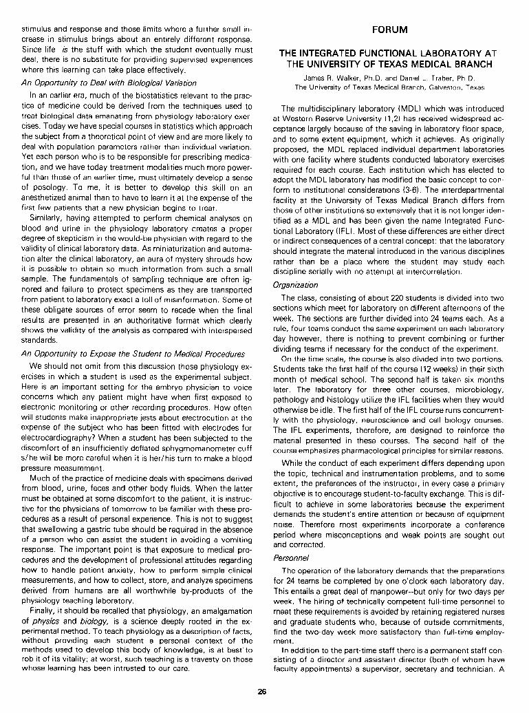
stimulus and response and those limits where a further small in- crease in stimulus brings about an entirely different response. Since life is the stuff with which the student eventually must deal, there is no substitute for providing supervised experiences where this learning can take place effectively.
An Opportunity to Deal with Biological Variation
In an earlier era, much of the biostatistics relevant to the prac- tice of medicine could be derived from the techniques used to treat biological data emanating from physiology laboratory exer- cises. Today we have special courses in statistics which approach the subject from a theoretical point of view and are more likely to deal with population parameters rather than individual variation. Yet each person who is to be responsible for prescribing medica- tion, and we have today treatment modalities much more power- ful than those of an earlier time, must ultimately develop a sense of posology. To me, it is better to develop this skill on an anesthetized animal than to have to learn it at the expense of the first few patients that a new physician begins to treat.
Similarly, having attempted to perform chemical analyses on blood and urine in the physiology laboratory creates a proper degree of skepticism in the would-be physician with regard to the validity of clinical laboratory data. As miniaturization and automa- tion alter the clinical laboratory, an aura of mystery shrouds how it is possible to obtain so much information from such a small sample. The fundamentals of sampling technique are often ig- nored and failure to protect specimens as they are transported from patient to laboratory exact a toll of misinformation. Some of these obligate sources of error seem to recede when the final results are presented in an authoritative format which clearly shows the validity of the analysis as compared with interspersed standards.
An Opportunity to Expose the Student to Medical Procedures
We should not omit from this discussion those physiology ex- ercises in which a student is used as the experimental subject. Here is an important setting for the embryo physician to voice concerns which any patient might have when first exposed to electronic monitoring or other recording procedures. How often will students make inappropriate jests about electrocution at the expense of the subject who has been fitted with electrodes for electrocardiography? When a student has been subjected to the discomfort of an insufficiently deflated sphygmomanometer cuff s/he will be more careful when it is her/his turn to make a blood pressure measurement.
Much of the practice of medicine deals with specimens derived from blood, urine, feces and other body fluids. When the latter must be obtained at some discomfort to the patient, it is instruc- tive for the physicians of tomorrow to be familiar with these pro- cedures as a result of personal experience. This is not to suggest that swallowing a gastric tube should be required in the absence of a person who can assist the student in avoiding a vomiting response. The important point is that exposure to medical pro- cedures and the development of professional attitudes regarding how to handle patient anxiety, how to perform simple clinical measurements, and how to collect, store, and analyze specimens derived from humans are all worthwhile by-products of the physiology teaching laboratory.
Finally, it should be recalled that physiology, an amalgamation of physics and biology, is a science deeply rooted in the ex- perimental method. To teach physiology as a description of facts, without providing each student a personal context of the methods used to develop this body of knowledge, is at best-to rob it of its vitality; at worst, such teaching is a travesty on those whose learning has been intrusted to our care.
FORUM
THE INTEGRATED FUNCTIONAL LABORATORY AT THE UNIVERSITY OF TEXAS MEDICAL BRANCH
James R. Walker, Ph.D. and Daniel L. Traber, Ph.D. The University of Texas Medical Branch, Galveston, Texas
The multidisciplinary laboratory (MDL) which was introduced at Western Reserve University (1,2) has received widespread ac- ceptance largely because of the saving in laboratory floor space, and to some extent equipment, which it achieves. As originally proposed, the MDL replaced individual department laboratories with one facility where students conducted laboratory exercises required for each course. Each institution which has elected to adopt the MDL laboratory has modified the basic concept to con- form to institutional considerations (3-6). The interdepartmental facility at the University of Texas Medical Branch differs from those of other institutions so extensively that it is not longer iden- tified as a MDL and has been given the name Integrated Func- tional Laboratory (IFL). Most of these differences are either direct or indirect consequences of a central concept: that the laboratory should integrate the material introduced in the various disciplines rather than be a place where the student may study each discipline serially with no attempt at intercorrelation.
Organization
The class, consisting of about 220 students is divided into two sections which meet for laboratory on different afternoons of the week. The sections are further divided into 24 teams each. As a rule, four teams conduct the same experiment on each laboratory day however, there is nothing to prevent combining or further dividing teams if necessary for the conduct of the experiment.
On the time scale, the course is also divided into two portions. Students take the first half of the course (12 weeks) in their sixth month of medical school. The second half is taken six months later. The laboratory for three other courses, microbiology, pathology and histology utilize the IFL facilities when they would otherwise be idle. The first half of the IFL course runs concurrent- ly with the physiology, neuroscience and cell biology courses. The IFL experiments, therefore, are designed to reinforce the material presented in these courses. The second half of the course emphasizes pharmacological principles for similar reasons.
While the conduct of each experiment differs depending upon the topic, technical and instrumentation problems, and to some extent, the preferences of the instructor, in every case a primary objective is to encourage student-to-faculty exchange. This is dif- ficult to achieve in some laboratories because the experiment demands the student’s entire attention or because of equipment noise. Therefore most experiments incorporate a conference period where misconceptions and weak points are sought out and corrected.
Personnel
The operation of the laboratory demands that the preparations for 24 teams be completed by one o’clock each laboratory day. This entails a great deal of manpower--but only for two days per week. The hiring of technically competent full-time personnel to meet these requirements is avoided by retaining registered nurses and graduate students who, because of outside commitments, find the two-day week more satisfactory than full-time employ- ment.
In addition to the part-time staff there is a permanent staff con- sisting of a director and assistant director (both of whom have faculty appointments) a supervisor, secretary and technician. A
26
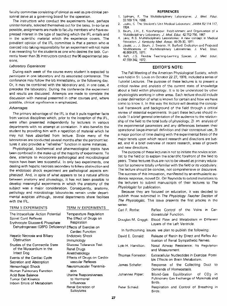
faculty committee consisting of clinical as well as pre-clinical per- sonnel serve as a governing board for the operation.
The instructors who conduct the experiments have, perhaps unintentionally, selected themselves out for the tasks. In-so-far as possible, assignments are made to faculty members who have ex- pressed interest in the type of teaching which the IFL entails and in the scientific material which the experience covers. The prevalent opinion of the IFL committee is that a person who is coerced into taking responsibility for an experiment will not make it as rewarding for the students as one who desires the task. Cur- rently fewer than 35 instructors conduct the 96 experimental ses- sions.
Labor-a tory Experiences
During each week of the course every student is expected to participate in one laboratory and its associated conference. The conference may follow the lab immediately, or the following day. Or, it may be combined with the laboratory and, in one case, it precedes the laboratory. During the conference the experiment and results are discussed. Attempts are made to correlate the discussion with material presented in other courses and, where possible, clinical significance is emphasized.
Advantages
The primary advantage of the IFL is that it pulls together facts from various disciplines which, prior to the inception of the IFL, were often presented independently by lecturers in various departments with no attempt at correlation. It also benefits the student by providing him with a repetition of material which he may not have absorbed from lecture. Since many of the laboratories are conducted several months after the pertinent lec- tures it also provides a “refresher” function in some instances.
Physiological, biochemical and pharmacological topics have been combined in the make-up of the majority of experiments, To date, attempts to incorporate pathological and microbiological topics have been less successful. In only two experiments, one dealing with hepatic necrosis secondary to biliary obstruction and the endotoxic shock experiment are pathological aspects em- phasized. And, in spite of what appears to be a natural affinity between anatomy and physiology, it has not been possible to develop meaningful experiments in which the anatomy of the subject was a major consideration. Consequently, anatomy, pathology and microbiology laboratories remain under depart- mental auspices although, several departments share facilities with the IFL.
TERM II EXPERIMENTS TERM IV EXPERIMENTS
The Intracellular Action Potential Temperature Regulation Spinal Cord Reflexes The Effect of Drugs on Erythrocyte Glucose-6 Phosphate Respiration
Dehydrogenase (GGPD Deficiency) Effects of Exercise on Cardiac Function
Hepatic Necrosis and Billiary Endotoxic Shock Obstruction
Studies of the Contractile State Immunology Glucose Tolerance Test
of the Myocardium in the Renal Drugs Intact Dog Anesthesiology
Events of the Cardiac Cycle Effects of Drugs on Cardio- Secretion and Absorption vascular Reflexes Hemorrhagic Shock Neuromuscular Transmis- Human Pulmonary Function sion Acid Base Balance Uterine Responsiveness Tumor Cell Kinetics to Hormonal Inborn Errors of Metabolism Influences
Renal Excretion of Salicylates
1.
2.
3.
4.
5.
6.
REFERENCES Spilman, E. The Multidisciplinary Laboratories. J. Med. Educ. 33: 169-174, 1958. Leake, C. The Student’s Unit Medical Laboratory. JAMA 82:114-l 17, 1924. Baum, J .H., E. Koushanpour. Establishment and Organization of a Multidisciplinary Laboratory. J. Med. Educ. 42:752-756, 1967. Levy, R .S. Multidisciplinary Laboratories: A new concept in Medical Education. J. Kentucky Med. Assoc. 69:25-30, 1971. Jaussi, J., J. Baum, J. Swaner, H. Burford. Evaluation and Proposed Modifications of Multidisciplinary Laboratories. J. Med. Educ. 46:869-875, 1971. Haft, J. S. Flexible Teaching-Learning Spaces. J. Med. Educ. 47339-342, 1972.
The Fall Meeting of the American Physiological Society, which was held in St. Louis on October 22-27, 1978, included a series of
EDITOR’S NOTE:
Tutorial Lectures. The purpose of these lectures is to present a critical review and analysis of the current state of knowledge about a field within physiology. It is to be understood by other physiologists working in other areas. Each lecture should include a clear delineation of what is known for certain and how we have come to know it. In this way the lecture will develop the concep- tual framework and background of the field through a critical review of essential experiments. In part these lectures should in- clude 1) a brief general orientation of the audience to the relation- ship of the field to the total body of physiology, 2) an analysis of the experimental parameters and any differences between their operational (experimental) definition and their conceptual use, 3) a major portion of time dealing with the experimental basis of the major tenets upon which recent research and directions are bas- ed, and 4) a brief overview of recent research, areas of growth and new directions.
David E. Donald:
Lyle H. Hamilton:
Thomas Forrester
James Schafer:
Johannes Piiper:
The purpose of these lectures is not to initiate the novice scien- tist to the field or to explain the scientific forefront of the field to peers. These lectures thus are not to be viewed as primary educa- tion for someone totally unfamiliar with the field or its vocabulary. The lecture should be concise, not comprehensive or discursive. The success of this innovation, manifested by an enthusiastic au- dience response, moved Dr. Orr Reynolds to extend invitations to the lecturers to submit manuscripts of their lectures to The Physiologist for publication.
Because they are focused on education, it was decided to publish those submitted in The physiology Teacher Section of The Physiologist. This issue presents the first articles in the series:
Carl F. Rothe: Reflex Control of the Veins in Car- diovascular Function.
Douglas M. Griggs: Blood Flow and Metabolism in Different Layers of the Left Ventricle.
In forthcoming issues we plan to publish the following:
Release of Renin by Direct and Reflex Ac- tivation of Renal Sympathetic Nerves.
Nasal Airway Resistance: Its Regulation and Measurement.
. . Extracellular Nucleotides in Exercise: Possi- ble Effects on Brain Metabolism.
Response of the Collecting Duct to Demands of Homeostasis.
Blood-Gas Equilibration of CO2 in Pulmonary Gas Exchange of Mammals and Birds.
Peter Scheid: Respiration and Control of Breathing in Birds.
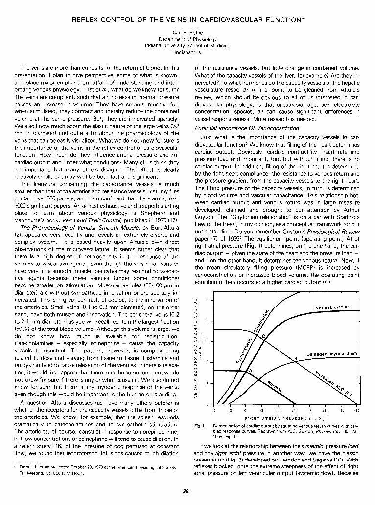
REFLEX CONTROL OF THE VEINS IN CARDIOVASCULAR FUNCTION*
Carl F. Rothe
Department of Physiology Indiana University School of Medicine
Indianapolis
The veins are more than conduits for the return of blood. In this presentation, I plan to give perspective, some of what is known, and place major emphasis on pitfalls of understanding and inter- preting venous physiology. First of a!, what do we know for sure? The veins are compliant, such that an increase in internal pressure causes an increase in volume. They have smooth muscle, for, when stimulated, they contract and thereby reduce the contained volume at the same pressure. But, they are innervated sparsely. We also know much about the elastic nature of the large veins (>2 mm in diameter) and quite a bit about the pharmacology of the veins that can be easily visualized. What we do not know for sure is the importance of the veins in the reflex control of cardiovascular function. How much do they influence arterial pressure and /or cardiac output and under what conditions? Many of us think they are important, but many others disagree. The effect is clearly relatively small, but may well be both fast and significant.
The literature concerning the capacitance vessels is much smaller than that of the arteries and resistance vessels. Yet, my files contain over 500 papers, and I am confident that there are at least 1000 significant papers. An almost exhaustive and a superb starting place to learn about venous physiology is Shepherd and Vanhoutte’s book, Veins and Their Control, published in 1975 (17).
The Pharmacology of Venular Smooth Muscle, by Burt Altura (21, appeared very recently and reveals an extremely diverse and complex system. It is based heavily upon Altura’s own direct observations of the microvasculature. It seems rather clear that there is a high degree of heterogeneity in the response of the venules to vasoactive agents. Even though the very small venules have very little smooth muscle, pericytes may respond to vasoac- tive agents because these venules (under some conditions) become smaller on stimulation. Muscular venules 00-100 pm in diameter) are without sympathetic innervation or are sparsely in- nervated. This is in great contrast, of course, to the innervation of the arterioles. Small veins (0.1 to 0.3 mm diameter), on the other hand, have both muscle and innervation. The peripheral veins (0.2 to 2.4 mm diameter), as you will recall, contain the largest fraction (60%) of the total blood volume. Although this volume is large, we do not know how much is available for redistribution. Catecholamines - especially epinephrine - cause the capacity vessels to constrict. The pattern, however, is complex being related to dose and varying from tissue to tissue. Histamine and bradykinin tend to cause relaxation of the venules. If there is relaxa- tion, it would then appear that there must be some tone, but we do not know for sure if there is any or what causes it. We also do not know for sure that there is any myogenic response of the veins, even though this would be important to the human on standing.
A question Altura discusses (as have many others before) is whether the receptors for the capacity vessels differ from those of the arterioles. We know, for example, that the spleen responds dramatically to catecholamines and to sympathetic stimulation. The arterioles, of course, constrict in response to norepinephrine, but low concentrations of epinephrine will tend to cause dilation. In a recent study (16) of the intestine of dog perfused at constant flow, we found that isoproterenol infusions caused much dilation
l Tutorial Lecture presented October 23, 1978 at the American Physiological Society
Fall Meeting, St. Louis, Missouri.
of the resistance vessels, but little change in contained volume. What of the capacity vessels of the liver, for example? Are they in- nervated? To what hormones do the capacity vessels of the hepatic vasculature respond? A final point to be gleaned from Altura’s review, which should be obvious to all of us interested in car- diovascular physiology, is that anesthesia, age, sex, electrol Yte concentration, species, all can cause significan t differences In vessel responsiveness. More research is needed.
Potential Importance Of Venocons tric tion
Just what is the importance of the capacity vessels in car- diovascular function? We kn ow that filling of the heart determines cardiac output. Obvious1 Yt cardiac contract .ility , heart rate and pressure load and important, too, but without filling, there is no cardiac output. In addition, filling of the right heart is determined by the right heart compliance, the resistance to venous return and the pressu re gradient from the capacity vessels to the right heart. The filling pressure of the capacity vessels, in turn , is determined by blood volume and vascular capacitance. This relationship bet- ween cardiac output and venous return was in large measure developed, clarified and brought to our attention by Arthur Guyton. The “Guytonian relationship” is on a par with Starling’s Law of the Heart, in my opinion, as a conceptual framework for our understanding. Do you remember Guyton’s Physiobgical Review paper (7) of 1955? The equilibrium point (operating point, A) of right atrial pressure (Fig. 1) determines, on the one hand, the car- diac output - given the state of the heart and the pressure load - and , on the other hand, it determines the venous return. Now, if the mean circulatory filling pressure (MCFP) is increased by venoconstriction or increased blood volume, the operating point equilibrium then occurs at a higher cardiac output (Cl.
a
0”
+2 +6 +8
RIGHT ATRIAL PRESSURE (mmHg)
Fig.1. Determination of cardiac output by equating venous return curves with car- diac response curves. Redrawn from A.C. Guyton, Physiol. Rev. 35:123, 1955, Fig. 5.
I f we look at the relationship between the systemic pressure load and the right atria/ pressure in another way, we have the classic presentation (Fig. 2) developed by Herndon and Sagawa (10). With reflexes blocked, note the extreme steepness of the effect of right atrial pressure on left ventricular output (systemic flow). Because
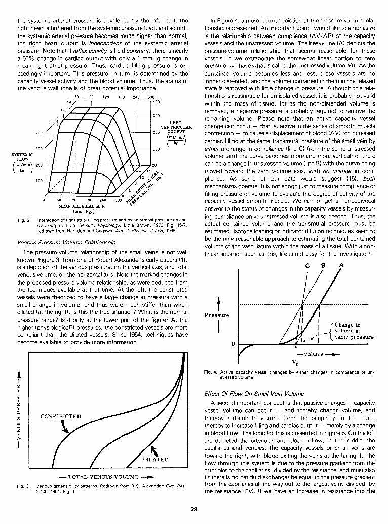
the systemic arterial pressure is developed by the left heart, the right heart is buffered from the systemic pressure load, and so until the systemic arterial pressure becomes much higher than normal, the right heart output is independent of the systemic arterial pressure. Note that if reflexactivity is held constant, there is nearly a 59% change in cardiac output with only a 1 mmHg change in mean right atrial pressure. Thus, cardiac filling pressure is ex- ceedingly important. This pressure, in turn, is determined by the capacity vessel activity and the blood volume. Thus, the status of the venous wall tone is of great potential importance.
16; 6,O 1;O l;O 2;O 3;0400
300
SYSTEMIC FLOW
ml/min ( > 200 kg
LEFT VENTRICULAR
OUTPUT
MEAN ARTERIAL B. P. (mm. Hg.)
Fig. 2. Interaction of right atrial filling pressure and mean arterial pressure on car- diac output. From Selkurt: Physiology, Little Brown, 1976, Fig. 15-7; redrawn from Herndon and Sagawa, Am. J. Physiol. 217:65, 1969.
Venous Pressure- Volume Relationship
The pressure-volume relationship of the small veins is not well known. Figure 3, from one of Robert Alexander’s early papers (l), is a depiction of the venous pressure, on the vertical axis, and total venous volume, on the horizontal axis. Note the marked changes in the proposed pressure-volume relationship, as were deduced from the techniques available at that time. At the left, the constricted vessels were theorized to have a large change in pressure with a small change in volume, and thus were much stiffer than when dilated (at the right). Is this the true situation? What is the normal pressure range? Is it only at the lower part of the figure? At the higher (physiological?) pressures, the constricted vessels are more compliant than the dilated vessels. Since 1954, techniques have become available to provide more information.
CONSTRICTED
-TOTAL VENOUS VOLUME e
Fig. 3. Venous distensibility patterns. Redrawn from R. S. Alexander: Circ. Res. 2405, 1954, Fig. 1.
In Figure 4, a more recent depiction of the pressure-volume rela- tionship is presented. An important point I would like to emphasize is the relationship between compliance (Ail/API of the capacity vessels and the unstressed volume. The heavy line (A) depicts the pressure-volume relationship that seems reasonable for these vessels. I f we extrapolate the somewhat linear portion to zero pressure, we have what is called the unstressed volume, Vu. As the contained volume becomes less and less, these vessels are no longer distended, and the volume contained in them in the relaxed state is removed with little change in pressure. Although this rela- tionship is reasonable for an isolated vessel, it is probably not valid within the mass of tissue, for as the non-distended volume is removed, a negative pressure is probably required to remove the remaining volume. Please note that an active capacity vessel change can occur - that is, active in the sense of smooth muscle contraction - to cause a displacement of blood (AV) for increased cardiac filling at the same transmural pressure of the small vein by either a change in compliance (line C) from the same unstressed volume (and the curve becomes more and more vertical) or there can be a change in unstressed volume (line B) with the curve being moved toward the zero volume axis, with no change in com- pliance. As some of our data would suggest (151, both mechanisms operate. It is not enogh just to measure compliance or filling pressure or volume to evaluate the degree of activity of the capacity vessel smooth muscle. We cannot get an unequivocal answer to the status of changes in the capacity vessels by measur- ing compliance only; unstressed volume is also needed. Thus, the actual contained volume and the transmural pressure must be estimated. Isotope loading or indicator dilution techniques seem to be the only reasonable approach to estimating the total contained volume of the vasculature within the mass of a tissue. With a non- linear situation such as this, life is not easy for the investigator!
t Pressure
0
C B A .
same pressure
t -Volume W
VU
Fig. 4. Active capacity vessel changes by either changes in compltance or un- stressed volume.
Effect Of Flow On Small Vein Volume
A second important concept is that passive changes in capacity vessel volume can occur - and thereby change volume, and thereby redistribute volume from the periphery to the heart, thereby to increase filling and cardiac output - merely by a change in blood flow. The logic for this is presented in Figure 5. On the left are depicted the arterioles and blood inflow; in the middle, the capillaries and venules; the capacity vessels or small veins are toward the right, with blood exiting the veins at the far right. The flow through this system is due to the pressure gradient from the arterioles to the capillaries, divided by the resistance, and must also (if there is no net fluid exchange) be equal to the pressure gradient from the capillaries all the way out to the largest veins divided by the resistance (Rv). I f we have an increase in resistance into the
29
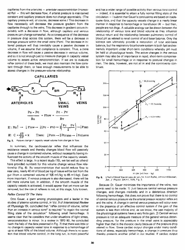
capillaries from the arterioles - arteriolar vasocons t&ion ( i ncreas- ed Ra) - this will decrease flow, if arterial pressure is maintained constant and capillary pressure does not change appreciably. (The capillary pressure will, of course, decrease some.) This decrease in flow necessarily will decrease the pressure gradient from the capillaries through to the veins. This decrease in gradient occurs in- evitably with a decrease in flow, although capillary and venous pressure can change somewhat. As a consequence of the decrease in pressure drop across this system, there will be a decrease in average pressure within the small veins (Psm). The decrease in in- ternal pressure will thus inevitably cause a passive decrease in volume, if we assume that compliance is constant. Thus, a mere decrease in flow will cause a passive decrease in venous volume, and so it is not enough just to measure changes in capacity vessel volume to assess active venoconstriction. If we are to evaluate reflex control of these beds, we must also maintain the flow cons- tant through them, or have enough measurements to be able to assess changes in the pressure-volume relationship.
CAPILLARIES
ARTERIOLES SMALL VEINS VEINS
Pa - PC PC - Pv = Flow =
Ra Rv
If: Rat - IFlow- I(Pc - Pv)- i(PC, PV)-IPsm
If: C&&k Then: CPsm - 1 Ptrans--c lVolume
Fig. 5. Passive change in capacity vessel volume from changes in flow.
In summary, the cardiovascular reflex that influences the resistance vessels and thereby changes blood flow will passively cause a change in contained volume, without necessarily having in- fluenced the activity of the smooth muscle of the capacity vessels.
This effect is large. In a recent study (16), we (as well as others) have provided numbers for this volume change versus flow rela- tionship (Fig. 6). By vasoconstriction that would reduce flow to near zero, nearly 40 ml of blood per kg of tissue will be lost from the gut (from a contained volume of 105 ml/kg to 65 ml/kg). Even more important, if venous pressure is also decreased, there will be still more volume lost. In addition, if the smooth muscle of the capacity vessels is activated, it would appear that yet more can be removed, but the role of reflexes is not, at this stage, fully known.
Reflex Control Of The Veins
Otto Gauer, a giant among physiologists and a leader in the studies of plasma volume control, in Vol. 9 of /nternationalReview of Physiology (51, concluded that “reflex control of volume, rather than an increase in venous tone” is required “to regain the normal filling state of the circulation” following small hemorrhage. It seems clear that he considers that under situations of light stress, venoconstriction is minor. In a review in 1970 (6), he concluded (with the help of the data presented in Fig. 7) that there was little or no change in capacity vessel tone in response to a hemorrhage of up to at least 10% of the blood volume. Although there is no ques- tion that blood volume control, in the long run, is more powerful
and has a wider range of possible activity than venous tone control - indeed, it is essential to attain a fully normal filling state of the circulation - I submit that Gauer’s conclusions are based on inade- quate data, and that the capacity vessels change in a nearly linear manner in response to hemorrhage or transfusion (4) - but then, people are not dogs. A valuable analogy can be drawn between the relationship of venous tone and blood volume as they influence venous return and the relationship between pulmonary control of blood pH as related to renal control of acid-base balance. Only the kidneys can ultimately provide a restoration of total acid-base balance, but the respiratory-bicarbonate system is both fast and ex- tremely important under short-term conditions whereby pH must be held at physiological levels. The active changes in the venous system may also be of importance in rapid, short-term compensa- tion for small hemorrhage or in response to postural changes in man. The data, however, are not all in and the controversy con- tinues.
100
80
60
0
I I I I I
200 400 600 800
FLOW (ml/min - kg)
1000
Fig. 6. Effect of blood flow on vascular volume. From Rothe, Johns and Bennett, Am. J. Physiol. 234:H7, 1978, Fig. 3.
Because Dr. Gauer minimizes the importance of the veins, two points need to be made: 1) Just because central venous pressure changes, and changes markedly and in a deleterious direction following hemorrhage, this does not prove that there is no control of central venous pressure via the arterial pressor receptor reflex arc and the veins. A change in central venous pressure will occur even in the presence of an effective control system if the gain of this system is less than infinite and, as we know, the vast majority of the physiological systems have a very finite gain. 2) Central venous pressure is not an adequate measure of the general venous disten- ding pressure, for the pressure in the small veins is appreciably higher than that in the thoracic cavity because of the pressure drop related to flow. Since cardiac output changes under many condi- tions of stress, especially hemorrhage, a change in pressure drop thereby presents another pitfall in our studies: If cardiac output
30

Another important finding of the Shoukas and Sagawa study (18) is that the compliance did not change appreciably in response to the changes in the carotid sinus pressures. They thus concluded that the change in the capacity vessel pressure-volume relationship was in terms of the unstressed volume. To repeat a previous con- clusion, vascular compliance does not provide a total measure of the status of the capacity vessels.
Vascular Tone Capacity Vessels
40
I l,ltl,l~1 -io -25 ISP, l 25 l 50 INTRA SINUS PRESSURE mmlig
100
50
0
‘\ Cardiac
.kL Receptors
-*\-
/ INTRA SINUS PRESSURE mmtig
-& 0
I I I I I
0 -10 -20 -30 -40 Fig. 0. Reflex changes in total vascular volume (at constant venous pressure) in
response to 25 mmHg changes in carotid sinus pressure (at various in- trasinus pressures). From Shoukas and Sagawa, Circ. Res. 33:22, 1973. % DECREASE OF BLOOD VOLUME
Fig. 7. Response of capacity vessel vascular tone to decreases in blood volume. Redrawn from Gauer, Henry and Behn, Ann. Rev. Physiol. 32547, 1970, Fig. 1.
In a recent mongraph on peripheral circulation (1 l), Paul Johnson estimated that denervation would cause the blood volume of tissue to increase about 20%, and with maximal stimulation, he felt that one could expect a 30% decrease in contained volume. Although reasonable, no reference was given for the data to sup- port these conclusions and so more research is needed.
In an attempt to assess the range of possible control of the pressure-volume relationship of the vasculature of the dog, we compared control pressure-volume changes 5 minutes after a volume change to results in which we had attempted to constrict the capacity vessels maximally with norepinephrine or block all ac- tivity with hexamethonium (4). From these data (Fig. 91, we con- cluded that the maximum range of smooth muscle control was of the order of 15 ml/kg body weight at a mean circulatory filling pressure of about 10 mmHg. The effect of ganglionic blockade is shown in Fig. 10 (bottom). It would appear, then, that the veins may have a larger potential range than is activated by the arterial pressor receptor system. Differences in the physiological state of the animal, especially as related to anesthesia, could be important.
changes, central venous pressure does not provide a direct and reliable measure of the pressure in the capacity vessels. In other forms of stress, there may be no change in central venous pressure if both the mean circulatory filling pressure and cardiac contractility change appropriately.
The study of Shoukas and Sagawa in 1973 (18) provided clear evidence that in the dog, the capacity vessel function changed in response to changes in carotid sinus pressure. In this study, the dogs were on a circulatory bypass pump to provide a constant car- diac output and, with these conditions, a change in the volume of the reservoir in which the venous outflow was collected before be- ing pumped back to the dog was a measure of the change in vascular volume. The data presented (Fig. 8) indicate that there was a change of reservoir volume that amounted to as much as 2 ml/kg as the sinus pressure changed 25 mmHg around the 130 mmHg maximum sensitivity point. The response was even larger, if the data were expressed as a change from the peak response sinus pressure (right hand panel of the figure). Here, the change was of the order of 3.6 ml/kg for a 25 mmHg change. The manner of presentation is for the sinus pressure changes, not a cumulative change. The total change in reservoir volume, as the sinus pressure was elevated from 75 to 200 mmHg, was 7.5 ml/kg. Although this is a rather small change (10%) of the total blood volume, these dogs were anesthetized, major surgery was involved, and so the reflex reserves may have been significantly reduced. Furthermore, the arterial pressure changes reduced the volume changes of the reservoir. In addition, the 7.5 ml/kg change was over a third of the distended volume. More important, however, the change amoun- ting to 10% of the blood volume may cause a change in cardiac output far larger than 10%. Indeed, assuming a total vascular com- pliance of 2 ml /kg* mmHg, the corresponding capacity vessel pressure change at constant volume would be about 3 mmHg, and this would be reflected to the thorax and right atrium if cardiac out- put did not change. Even a 1 mmHg pressure change at the right atrium could cause the cardiac output to increase 50%. We do not yet know the relative importance of these mechanisms, but they are not trivial.
I ’ I I I
I I I I I
i 25 1 1 ,CFNSTRICTED 1 _ __\ ,/-, / I
1 I
LUlY 1 nulJ (5 min.)
+
15
‘MC
Control Volume
A
I’ 1 ’ I ’ I ’ I ’ I’ I ’ I T -40 -30 -20 -10 0 10 20 30
CHANGE IN BLOOD VOLUME (ml/kg)
Fig. 9. Range of control of total vascular pressure-volume relationship in the dog. - (Pmc= Mean circulatory filling pressure. Time in minutes. 1 From Drees and Rothe, Circ. Res. 34360, 1974, Fig. 3.
31
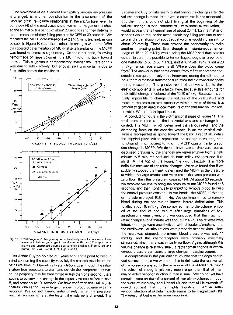
The movement of water across the capillary, as capillary pressure is changed, is another complication in the assessment of the vascular pressure-volume relationship at the microvessel level. In our study (4) of the total vasculature, we hemorrhaged or transfus- ed the animal over a period of about 20 seconds and then determin- ed the mean circulatory filling pressure (MCFP) at 30 seconds. We repeated the MCFP determinations at 2 and 5 minutes, and, as can be seen in Figure 10 (top) the relationship changes with time. With the repeated determination of MCFP after a transfusion, the MCFP was found to decrease significantly. On the other hand, following hemorrhage of large volumes, the MCFP returned back toward normal. This suggests a compensatory mechanism. Part of this was due to reflex activity, but another part was certainly due to fluid shifts across the capillaries.
15 - ‘MC -
(mmHg) 10 -
-40 -30 -20 -10 0 10 20 30
CHANGE IN BLOOD VOLUME (ml/kg)
A Hexamethonium
(mmHg) 15 -
-40 -30 -20 -10 0 10 20 30
CHANGE IN BLOOD VOLUME (ml/kg)
Fig. 10. (Top) Progressive changes in apparent total-body vasuclar pressure-volume relationship following changes in blood volume. (Bottom) Change in com- pliance and unstressed volume due to reflex blockade. From Drees and Rothe, Circ. Res. 34360, 1974, Figs. 2 and 4.
A concluding figure is the 3-dimensional maze of Figure 11. The total blood volume is on the horizontal axis and is change from control. The MCFP, which determines the venous return and the distending force on the capacity vessels, is on the vertical axis. Time is represented as going toward the back. First of all, notice the stippled plane which represents the change in volume, as a function of time, required to hold the MCFP constant after a sud- den change in MCFP. We do not have data at time zero, but as discussed previously, the changes are representative from a half- minute to 5 minutes and include both reflex changes and fluid shifts. At the top of the figure, the wild trajectory is a more definitive measure of the reflex changes. We have found that if we suddenly stopped the heart, determined the MCFP as the pressure at which the large arteries and veins are at the same pressure with zero flow, then this pressure increased (14). At about 20 seconds, we removed volume to bring the pressure to the MCFP found at 5 seconds, and then continually pumped to remove blood to keep the control pressure constant. In our hands, the MCFP of the dog on its side averaged 10.6 mmHg. We continually had to remove blood during the one-minute interval before defibrillation. This totalled about 15 ml/kg. We compared that to the volume remov- ed at the end of one minute after large quantities of hex- amethonium were given, and we concluded that the maximum reflex change at one minute was about 9 ml /kg. The reflexes were intact, the dogs were anesthetized with chloralose/urethane, and the cardiovascular stimulations were probably near maximal, since the heart was stopped, the arterial blood pressure was only 11 mmHg, and the chemoreceptors were probably maximally stimulated, since there was virtually no flow. Again, although this volume change is relatively small, a rather small change in central venous pressure can cause a large change in cardiac output.
As Arthur Guyton pointed out years ago (and a point to keep in A complication in this particular study was that the dogs had in- mind concerning the capacity vessels), the smooth muscles of the tact spleens, and so we were not able to delineate the relative role veins are slow in responding to stimulation. Even though the infor- of the spleen compared to the remainder of the vasculature. Since mation from receptors to brain and out via the sympathetic nerves the spleen of a dog is relatively much larger than that of man, to the periphery may be transmitted in less than one second, there maybe active venoconstriction in man is small. We do not yet have seems to be very little change in the capacity vessels before at least complete data on the reflex control of liver blood volume, although 5, and probably to 10, seconds.We have confirmed this (14). None- the work of Brooksby and Donald (3) and that of Hainsworth (8) theless, one cannot make large changes in blood volume within 7 would suggest that it is highly significant. Active reflex seconds. We do not know, unfortunately, what the pressure- venoconstriction of skeletal muscle seems to be insignificant (13). volume relationship is at the instant the volume is changed. The The intestinal bed may be more important.
Sagawa and Guyton labs seem to start timing the changes after the volume change is made, but it would seem this is not reasonable. But then, one should not start timing at the beginning of the volume change, either. Nonetheless, with some extrapolation, it would appear that a hemorrhage of about 20 ml/kg in a matter of seconds would reduce the mean circulatory filling pressure to near zero and a transfusion of about equal volume would increase it to about 20 mmHg. These data provide the opportunity to make another interesting point: Even though an instantaneous hemor- rhage of 15 to 20 ml/kg would bring the MCFP and thus cardiac output to zero, it is possible to hemorrhage a dog over a period of one half-hour to 50 to 60 ml/kg, and it survives. Why is not a 20 ml/kg hemorrhage always fatal? Where does the blood come from? The answer is that some comes from reflex venomotor con- striction, but quantitatively more important, during the half-hour to hour there is massive transfer of fluid from the extravascular space to the vasculature. The passive recoil of the veins due to their elastic components is not a factor here, because this accounts for their initial change in volume of the 1520 ml /kg. Because it is vir- tually impossible to change the volume of the vasculature and measure the pressure simultaneously within a mass of tissue, it is difficult to get an unequivocal measure of the pressure-volume rela- tionship. We are technique limited.
32
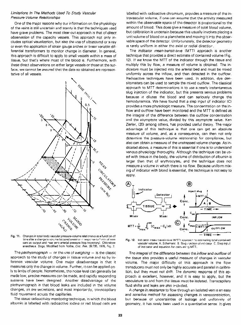
Limitations In The Methods Used To Study Vascular Pressure- Volume Relationships
One of the major reasons why our information on the physiology of the veins is still uncertain and scanty is that the techniques used have grave problems. The most clear-cut approach is that of direct observation of the capacity vessels. This approach not only in- cludes optical visualization, but also the use of ultrasound or x-ray or even the application of strain gauge arches or linear variable dif- ferential transformers to monitor change in diameter. In general, this approach is difficult to apply to small vessels within a mass of tissue, but that’s where most of the blood is. Furthermore, with these direct observations on either large vessels or those at the sur- face, we cannot be assured that the data so obtained are represen- tative of all vessels.
5 z
Fig. 11. Changes in total-body vascular pressure-volume relationship as a function of time after a change in volume (stippled areas) or in response to 1 min of zero cardiac output and near zero arterial pressure (top trajectory). Chloralose anesthesia. Dogs. Modified from Rothe, Circ. Res. 39:705, 1976, Fig. 2.
Theplethysmograph - or the use of weighing - is the classic approach to the study of changes in tissue volume and so by in- ference vascular volume. One major disadvantage is that it measures only the change in volume. Further, it can be applied on- ly to limbs of people. Nonetheless, the noise level can generally be made low, precise measures can be made, and rapidly responding systems have been designed. Another disadvantage of the plethysmograph is that blood leaks are included in the volume changes, as are secretions, and most importantly, transcapillary fluid movement across the capillaries.
The tissue radioactivity monitoring technique, in which the blood albumin is labelled with radioactive iodine or red blood cells are
labelled with radioactive chromium, provides a measure of the in- travascular volume, if one can assume that the activity measured within the observable space of the detector is proportional to the amount of blood. This does give a measure of total blood volume, but calibration is uncertain because this usually involves placing a unit volume of blood on a planchette and moving it into the obser- vable space of the detector. Unfortunately, the detector geometry is rarely uniform in either the axial or radial direction.
The indicator mean-transit-t/me (MTT) approach is another technique that provides a direct estimate of contained volume (Fig. 12). I f we know the MTT of the indicator through the tissue and multiply this by flow, a measure of volume is obtained. The in- dicators must be injected into the arterial bed and must be mixed uniformly across the inflow, and then detected in the outflow. Radioactive techniques have been used. In addition, dye den- sitometers can be used to sample the mixed outflow. The classical approach to MTT determinations is to use a nearly instantaneous slug injection of the indicator, but this presents serious problems because it dilutes the blood and can seriously change the hemodynamics. We have found that a step input of indicator (C) provides a more physiologic measure. The concentration on the in- flow and outflow have been monitored and the MTT is defined as the integral of the difference between the outflow concentration and the asymptote value, divided by this asymptote value. Ken Zierler, (20) among others, has provided useful theory. The major advantage of this technique is that one can get an absolute measure of volume, and, as a consequence, can then not only determine the pressure-volume relationship for compliance, but also can obtain a measure of the unstressed volume change. As in- dicated above, a measure of this is essential if one is to understand venous physiology thoroughly. Although the technique can be us- ed with tissue in the body, the volume of distribution of albumin is larger than that of ervthrocytes, and the technique does not . measure a volume in which there is no flow. Because uniform mix- ing of indicator with blood is essential, the technique is not easy to apply.
0 B
Inject slug I
Mixer Miier
- TIME -)-
1 %o Cont. INFLOW
MTT+ s max
a~Cmax-c,, dt
- - - TIME e
Fig. 12. Indicator mean-transit-time (MTT) approach to estimating total contained vascular volume. A. Schematic. B. Slug injection of indicator. C. Step input of indicator and equation for calculating MTT.
The integral of the difference between the inflow and outflow of the tissue also provides a useful measure of changes in vascular volume. The major difficulty ot this approach is the flow transducers must not only be highly accurate and parallel in calibra- tion, but they must not drift. The dynamic response of this ap- proach is excellent, however, and it is easy to apply, but the vasculature to and from the tissue must be isolated. Transcapillary fluid shifts and leaks are also included.
A change in resistance to flow through an isolated vein is an easy and sensitive method for assessing changes in venoconstriction, but because of uncertainties of leakage and uniformity of geometry, it has rarely been used in a quantitative sense. It gives
33
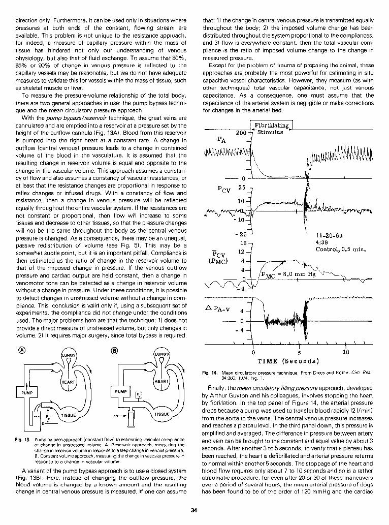
direction only. Furthermore, it can be used only in situations where pressures at both ends of the constant, flowing stream are available. This problem is not unique to the resistance approach, for indeed, a measure of capillary pressure within the mass of tissue has hindered not only our understanding of venous physiology, but also that of fluid exchange. To assume that 80%, 85% or 90% of change in venous pressure is reflected to the capillary vessels may be reasonable, but we do not have adequate measures to validate this for vessels within the mass of tissue, such as skeletal muscle or liver.
To measure the pressure-volume relationship of the total body, there are two general approaches in use: the pump bypass techni- que and the mean circulatory pressure approach.
With the pump bypasdresewoir technique, the great veins are cannulated and are emptied into a reservoir at a pressure set by the height of the outflow cannula (Fig. 13A). Blood from this reservoir is pumped into the right heart at a constant rate. A change in outflow (central venous) pressure leads to a change in contained volume of the blood in the vasculature. It is assumed that the resulting change in reservoir volume is equal and opposite to the change in the vascular volume. This approach assumes a constan- cy of flow and also assumes a constancy of vascular resistances, or at least that the resistance changes are proportional in response to reflex changes or infused drugs. With a constancy of flow and resistance, then a change in venous pressure will be reflected equally throughout the entire vascular system. If the resistances are not constant or proportional, then flow will increase to some tissues and decrease to other tissues, so that the pressure changes will not be the same throughout the body as the central venous pressure is changed. As a consequence, there may be an unequal, passive redistribution of volume (see Fig. 5). This may be a somewhat subtle point, but it is an important pitfall. Compliance is then estimated as the ratio of change in the reservoir volume to that of the imposed change in pressure. If the venous outflow pressure and cardiac output are held constant, then a change in venomotor tone can be detected as a change in reservoir volume without a change in pressure. Under these conditions, it is possible to detect changes in unstressed volume without a change in com- pliance. This conclusion is valid only if, using a subsequent set of experiments, the compliance did not change under the conditions used. The major problems here are that the technique: 1) does not provide a direct measure of unstressed volume, but only changes in volume. 2) It requires major surgery, since total bypass is required.
0 B
Fig. 13. Pump by-pass approach (constant flow) to estimating vascular compliance or change in unstressed volume. A. Reservoir approach, measuring the change in reservoir volume in response to a step change in venous pressure. 6. Constant volume approach, measuring the change in vascular pressure in response to a change in vascular volume.
A variant of the pump bypass approach is to use a closed system (Fig. 138). Here, instead of changing the outflow pressure, the blood volume is changed by a known amount and the resulting change in central venous pressure is measured. I f one can assume
that: 1) the change in central venous pressure is transmitted equally throughout the body; 2) the imposed volume change has been distributed throughout the system proportional to the compliances, and 3) flow is everywhere constant, then the total vascular com- pliance is the ratio of imposed volume change to the change in measured pressure.
Except for the problem of trauma of preparing the animal, these approaches are probably the most powerful for estimating in situ capacitive vessel characteristics. However, they measure (as with other techniques) total vascular capacitance, not just venous capacitance. As a consequence, one must assume that the capacitance of the arterial system is negligible or make corrections for changes in the arterial bed.
200
0 \ 1
pcv 25q
- 25 \
11-20-69 167 4:39
%v 12d Control, 0.5 min.
\ \ \ \ \ \ \ \ \ \ \ \ \ \ I I I 0 5 10
TIME (Seconds)
Fig. 14. Mean ci rculatory 34360, 1974, Fig
pressure technique. . 1.
D rees and Rothe, Circ. Res.
Finally, the mean ckculatory fillingpressure approach, developed by Arthur Guyton and his colleagues, involves stopping the heart by fibrillation. In the top panel of Figure 14, the arterial pressure drops because a pump was used to transfer blood rapidly (2 Vmin) from the aorta to the veins. The central venous pressure increases and reaches a plateau level. In the third panel down, this pressure is amplified and averaged. The difference in pressure between artery and vein can be brought to the constant and equal value by about 3 seconds. After another 3 to 5 seconds, to verify that a plateau has been reached, the heart is defibrillated and arterial pressure returns to normal within another 5 seconds. The stoppage of the heart and blood flow requires only about 7 to 10 seconds and so is a rather atraumatic procedure, for even after 20 or 30 of these maneuvers over a period of several hours, the mean arterial pressure of dogs has been found to be of the order of 120 mmHg and the cardiac
34
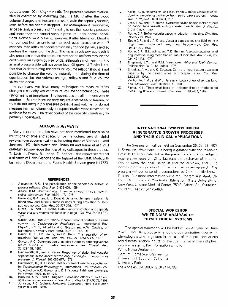
outputs over 100 ml /kg l min (15). The pressure-volume relation- ship is estimated by assuming that the MCFFj after the blood volume change, is at the same pressure as in the capacity vessels, even before the heart is stopped. The assumption is reasonable, since certainly a pressure of 11 mmHg is less than capillary pressure and more than the central venous pressure under normal condi- tions. Some error is present, however. I f after fibrillation, blood is not pumped from artery to vein to reach equal pressures within 7 seconds, then reflex venoconstriction may change the value and so confuse the meaning of the data. The mean circulatory approach is also uncertain in that the pressures may not be uniform through the cardiovascular system by 5 seconds, although a slight error on the arterial pressure side will not be serious. Of graver difficulty is the fact that in determining the pressure-volume relationship, it is not possible to change the volume instantly and, during the time of equilibration for the volume change, reflexes and fluid volume shifts can occur.
In summary, we have many techniques to measure reflex changes in capacity vessel pressure-volume characteristics. These rely on many assumptions. The techniques are all - in one way or another - faulted because they require anesthesia or trauma, or they do not adequately measure pressure and volume, or do not measure them simultaneously, or representative vessels may not be available for study. The reflex control of the capacity vessels is only partially understood.
ACKNOWLEDGEMENTS
Many important studies have not been mentioned because of limitations of time and space. Since the lecture, several helpful reviews have become available, including those of Vanhoutte and Janssens (191, Hainsworth and Linden (9) and Karim et al (12). I gratefully acknowledge the help of my colleagues in these studies: T. Lesh, J. Drees, B. Johns, T. Bennett; the expert secretarial assistance of Helen Glancy and the support of the IUMC Medical II- lustrations Department and Public Health Service grant HL7723.
1.
2.
3.
4.
5.
6.
7.
8.
9.
10.
11.
REFERENCES Alexander, R.S. The participation of the venomotor system in pressor reflexes. Circ. Res. 2:405-409, 1954. Altura, B.M. Pharmacology of venular smooth muscle: new in- sights. Microvasc. Res. 16:91-l 17, 1978. Brooksby, G.A., and D.E. Donald. Dynamic changes in splanchnic blood flow and blood volume in dogs during activation of sym- pathetic nerves. Circ. Res. 29:227-238, 1971. Drees, J.A., and C. F. Rothe. Reflex venoconstriction and capacity vessel pressure-volume relationships in dogs. Circ. Res. 34:360-373, 1974. Gauer, O.H., and J.P. Henry. Neurohormonal control of plasma volume. In: Cardiovascular Physiology //, International Rev. Physiol., Vol. 9, edited by A.C. Guyton and A.W. Cowley, Jr. Baltimore: University Park Press, 1976. P. 145-190. Gauer, O.H., J.P. Henry, and C. Behn. The regulation of ex- tracellular fluid volume. Ann. Rev. Physiol. 32547-595, 1970. Guyton, A.C. Determination of cardiac output by equating venous return curves with cardiac response curves. Physiol. Rev. 35:123-129, 1955. Hainsworth, R., and F. Karim. Responses of abdominal vascular capacitance in the anaesthetized dog to changes in carotid sinus pressure. J. Physiol. 262:659-677, 1976. Hainsworth, R., R. J. Linden. Reflex control of vascular capacitance. In: Cardiovascular Physiology ///, International Rev. Physiol., Vol. 18, edited by A.C. Guyton and D.B. Young. Baltimore: University Park Press, 1979. p. 67-124. Herndon, C.W., and K. Sagawa. Combined effects of aortic and right atrial pressures on aortic flow. Am. J. Physiol. 217:65-72, 1969. Johnson, P.C. (editor). Peripheral Circulation. New York: John Wiley & Sons, 1978.
12.
13.
14.
15.
16.
17.
18.
19.
20.
Karim, F., R. Hainsworth, and R.P. Pandey. Reflex response of ab- dominal vascular capacitance from aortic baroreceptors in dogs. Am. J. Physiol. H488-H493, 1978. Lesh, T.A., and C.F. Rothe. Sympathetic and hemodynamic effects on capacitance vessels in dog skeletal muscle. Am. J. Physiol. 217:819-827, 1969. Rothe, C.F. Reflex vascular capacity reduction in the dog. Circ. Res. 39:705-710, 1976. Rothe C.F., and J.A. Drees. Vascular capacitance and fluid shifts in dogs during prolonged hemorrhagic hypotension. Circ. Res. 38:347-356, 1976. Rothe, C.F., B. L. Johns, and T.D. Bennett. Vascular capacitance of dog intestine using mean transit time of indicator. Am. J. Physiol. 234:H7-H13, 1978. Shepherd, J.T., and P.M. Vanhoutte. Veins and Their Control. Philadelphia: W. B. Saunders, 1975. Shoukas, A.A., and K. Sagawa. Control of total systemic vascular capacity by the carotid sinus baroreceptor reflex. Circ. Res. 33:22-33, 1973. Vanhoutte, P.M., and W.J. Janssens. Local control of venous func- tion. Microvasc. Res. 16:196-214, 1978. Zierler, K. L. Theoretical basis of indicator-dilution methods for measuring flow and volume. Circ. Res. 10:393-407, 1962.
INTERNATIONAL SYMPOSIUM ON REGENERATIVE GROWTH PROCESSES AND THEIR CLINICAL APPLICATIONS
The Symposium will be held on September 26, 27, 28, 1979
in Syracuse, New York. It is being organized with the following
aims: 1) To accurately define the present state of knowledge in
regenerative research, 2) to facilitate the exchange of informa-
tion between the basic scientist and the clinician, and 3) to
identify promising areas of future interdisciplinary research. The
program will comprise of presentations by 25 nationally known
Faculty. For more information write to: Program Assistant, Of-
fice of Graduate and Continuing Education, State University of
New York, Upstate Medical Center, 750 E. Adams St., Syracuse,
NY 13210. Tel: (315) 473-4607
SPECIAL WORKSHOP WHITE NOISE ANALYSIS OF PHYSIOLOGICAL SYSTEMS
The special workshop will be held in Los Angeles on June
25-29, 1979. Its purpose is a lecture-demonstration course for
physiologists and engineers in the use of multiple, continuous
and discrete random inputs for the quantitative analysis of phys-
iological systems. For information write to:
White Noise Workshop
Dept. of Biomedical Engineering
University of Southern California
University Park
Los Angeles, CA 90007 (213) 741-6708
35
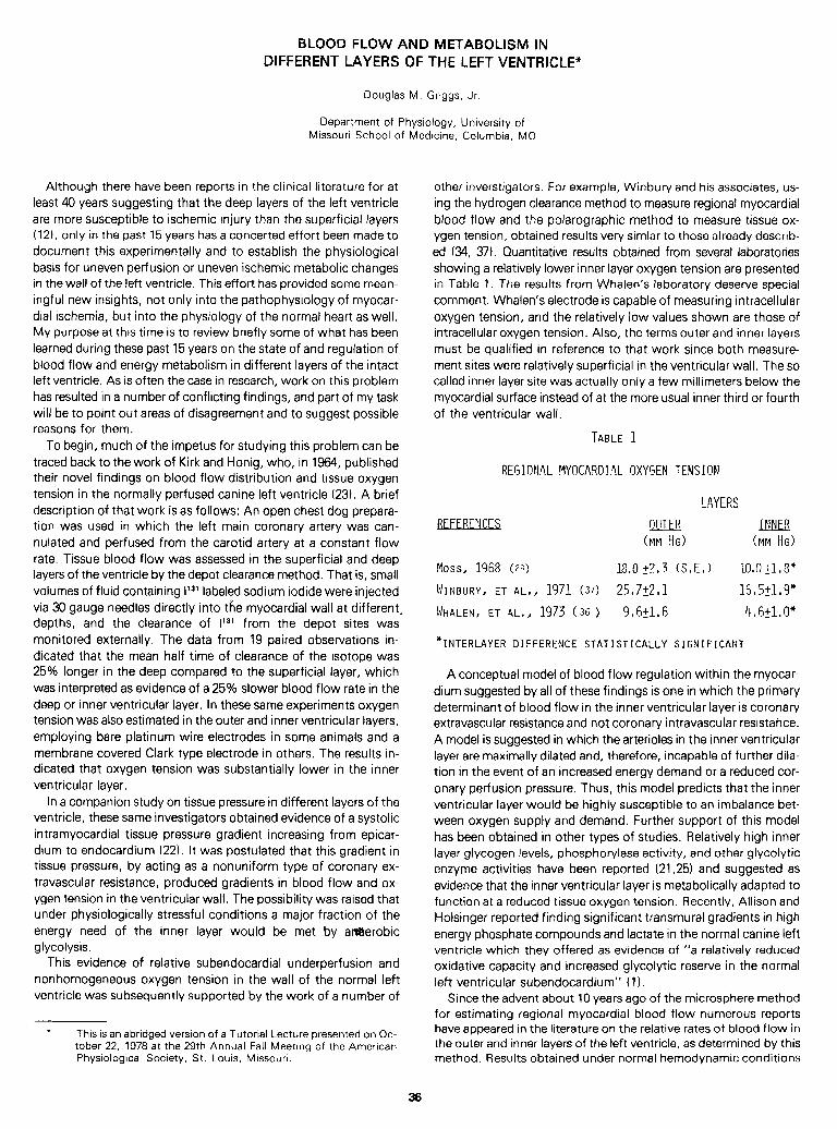
BLOOD FLOW AND METABOLISM IN DIFFERENT LAYERS OF THE LEFT VENTRICLE+
Douglas M. Griggs, Jr.
Department of Physiology, University of Missouri School of Medicine, Columbia, MO
Although there have been reports in the clinical literature for at least 40 years suggesting that the deep layers of the left ventricle are more susceptible to ischemic injury than the superficial layers (121, only in the past 15 years has a concerted effort been made to document this experimentally and to establish the physiological basis for uneven perfusion or uneven ischemic metabolic changes in the wall of the left ventricle. This effort has provided some mean- ingful new insights, not only into the pathophysiology of myocar- dial ischemia, but into the physiology of the normal heart as well. My purpose at this time is to review briefly some of what has been learned during these past 15 years on the state of and regulation of blood flow and energy metabolism in different layers of the intact left ventricle. As is often the case in research, work on this problem has resulted in a number of conflicting findings, and part of my task will be to point out areas of disagreement and to suggest possible reasons for them.
To begin, much of the impetus for studying this problem can be traced back to the work of Kirk and Honig, who, in 1964, published their novel findings on blood flow distribution and tissue oxygen tension in the normally perfused canine left ventricle (23). A brief description of that work is as follows: An open chest dog prepara- tion was used in which the left main coronary artery was can- nulated and perfused from the carotid artery at a constant flow rate. Tissue blood flow was assessed in the superficial and deep layers of the ventricle by the depot clearance method. That is, small volumes of fluid containing I 131 labeled sodium iodide were injected via 30 gauge needles directly into the myocardial wall at different. depths, and the clearance of V31 from the depot sites was monitored externally. The data from 19 paired observations in- dicated that the mean half time of clearance of the isotope was 25% longer in the deep compared to the superficial layer, which was interpreted as evidence of a 25% slower blood flow rate in the deep or inner ventricular layer. In these same experiments oxygen tension was also estimated in the outer and inner ventricular layers, employing bare platinum wire electrodes in some animals and a membrane covered Clark type electrode in others. The results in- dicated that oxygen tension was substantially lower in the inner ventricular layer.
In a companion study on tissue pressure in different layers of the ventricle, these same investigators obtained evidence of a systolic intramyocardial tissue pressure gradient increasing from epicar- dium to endocardium (22). It was postulated that this gradient in tissue pressure, by acting as a nonuniform type of coronary ex- travascular resistance, produced gradients in blood flow and ox- ygen tension in the ventricular wall. The possibility was raised that under physiologically stressful conditions a major fraction of the energy need of the inner layer would be met by amerobic glycolysis.
This evidence of relative subendocardial underperfusion and nonhomogeneous oxygen tension in the wall of the normal left ventricle was subsequently supported by the work of a number of
0 This is an abridged version of a Tutorial Lecture presented on Oc- tober 22, 1978 at the 29th Annual Fall Meeting of the American Physiological Society, St. Louis, Missouri.
36
other inverstigators. For example, Winbury and his associates, us- ing the hydrogen clearance method to measure regional myocardial blood flow and the polarographic method to measure tissue ox- ygen tension, obtained results very simlar to those already describ- ed (34, 37). Quantitative results obtained from several laboratories showing a relatively lower inner layer oxygen tension are presented in Table 1. The results from Whalen’s laboratory deserve special comment. Whalen’s electrode is capable of measuring intracellular oxygen tension, and the relatively low values shown are those of intracellular oxygen tension. Also, the terms outer and inner layers must be qualified in reference to that work since both measure- ment sites were relatively superficial in the ventricular wall. The so called inner layer site was actually only a few millimeters below the myocardial surface instead of at the more usual inner third or fourth of the ventricular wall.
TABLE 1
REGIONAL flYOCARDIAL 0XYrJEM TENSION
LAYERS
REFERF,?]CES OUTER J MNER (MM !-id (MM HG)
Moss, 1968 (29) 18,0+?,3 KEJ lO,O~l#3"
WINBURY, ET AL,, 1971 (37) 25,7+2,1 16,5+1,9*
'WHALEN, ET AL,, 1973 (36 > 9,6+1,6 4,6+1,0*
*INTERLAYER DIFFERENCE STATISTICALLY SIGNIFICANT
A conceptual model of blood flow regulation within the myocar- dium suggested by all of these findings is one in which the primary determinant of blood flow in the inner ventricular layer is coronary extravascular resistance and not coronary intravascular resistance. A model is suggested in which the arterioles in the inner ventricular layer are maximally dilated and, therefore, incapable of further dila- tion in the event of an increased energy demand or a reduced cor- onary perfusion pressure. Thus, this model predicts that the inner ventricular layer would be highly susceptible to an imbalance bet- ween oxygen supply and demand. Further support of this model has been obtained in other types of studies. Relatively high inner layer glycogen levels, phosphorylase activity, and other glycolytic enzyme activities have been reported (21,25) and suggested as evidence that the inner ventricular layer is metabolically adapted to function at a reduced tissue oxygen tension. Recently, Allison and Holsinger reported finding significant transmural gradients in high energy phosphate compounds and lactate in the normal canine left ventricle which they offered as evidence of “a relatively reduced oxidative capacity and increased glycolytic reserve in the normal left ventricular subendocardium” (1).
Since the advent about 10 years ago of the microsphere method for estimating regional myocardial blood flow numerous reports have appeared in the literature on the relative rates of blood flow in the outer and inner layers of the left ventricle, as determined by this method. Results obtained under normal hemodynamic conditions
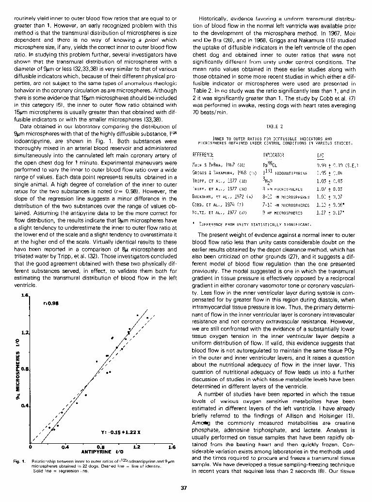
routinely yield inner to outer blood flow ratios that are equal to or greater than 1. However, an early recognized problem with this method is that the transmural distribution of microspheres is size dependent and there is no way of knowing a priori which microsphere size, if any, yields the correct inner to outer blood flow ratio. In studying this problem further, several investigators have shown that the transmural distribution of microspheres with a diameter of 9pm or less (3233,381 is very similar to that of various diffusible indicators which, because of their different physical pro- perties, are not subject to the same types of anomalous rheologic behavior in the coronary circulation as are microspheres. Although there is some evidence that 15pm microspheres should be included in this category (5), the inner to outer flow ratio obtained with 15pm microspheres is usually greater than that obtained with dif- fusible indicators or with the smaller microspheres (33,381.
Data obtained in our laboratory comparing the distribution of 9pm microspheres with that of the highly diffusible substance, IQ6 iodoantipyrine, are shown in Fig. 1. Both substances were thoroughly mixed in an arterial blood reservoir and administered simultaneously into the cannulated left main coronary artery of the open chest dog for 1 minute. Experimental maneuvers were performed to vary the inner to outer blood flow ratio over a wide range of values. Each data point represents results obtained in a single animal. A high degree of correlation of the inner to outer ratios for the two substances is noted (r= 0.98). However, the slope of the regression line suggests a minor difference in the distribution of the two substances over the range of values ob- tained. Assuming the antipyrine data to be the more correct for flow distribution, the results indicate that 9pm microspheres have a slight tendency to underestimate the inner to outer flow ratio at the lower end of the scale and a slight tendency to overestimate it at the higher end of the scale. Virtually identical results to these have been reported in a comparison of 8~ microspheres and tritiated water by Tripp, et al. (32). Those investigators concluded that the good agreement obtained with these two physically dif- ferent substances served, in effect, to validate them both for estimating the transmural distribution of blood flow in the left ventricle.
1.6,
r: 0.98
1.2
p
/ Y: -0.1s+1.22x / /
4 1 1 . I 1
0 0.4 0.8 1.2 1.6 ANTIPYRINE l/O
Fig. 1. Relationship between inner to outer ratios of 11*5iodoantipyrine and 9 pm microspheres obtained in 22 dogs. Dashed line = line of identity.
Solid line = regression line.
Historically, evidence favoring a uniform transmural distribu- tion of blood flow in the normal left ventricle was available prior to the development of the microsphere method. In 1967, Moir and De Bra (28), and in 1968, Griggs and Nakamura (15) studied the uptake of diffusible indicators in the left ventricle of the open chest dog and obtained inner to outer ratios that were not significantly different from unity under control conditions. The mean ratio values obtained in these earlier studies along with those obtained in some more recent studies in which either a dif- fusible indicator or microspheres were used are presented in Table 2. In no study was the ratio significantly less than 1, and in 2 it was significantly greater than 1. The study by Cobb et al. (7) was performed in awake, resting dogs with heart rates averaging 70 beats/min.
TABLE 2
INNER TO OUTER RATIOS FOR DIFFUSIBLE INDICATORS AND MICROSPHERES OBTAINED UNDER CONTROL CONDITIONS IN VARIOUS STUDIES,
REFEREWE INDICATOR I/O
MOIR S DEBRA, 1967 (28) !?B%L 034 t !I,10 KEJ
GRIGGS 8 ~AKAMURA, 1968 (15) $31 IODOANTIPYRINE 1035 + 0,04
TRIPP, ET AL,, 1977 (32) 3H20 l/l3 2 oar33
TRIPP, ET AL,, 1977 (32) 8 IJM MICROSPHERES 1,37 -f to3
BUCKBERG, ET AL,, 1972 (6) 8-10 I-rM MICROSPHERES 1,Ol 2 x37
COBB, ET AL,, 1974 (7) 7-10 r-IM MICROSPHERES 1013 + X06" HOLTZ, ET AL,, 1977 (19) 9 PM MICROSPHERES lo27 + O,17*
* DIFFERENCE FROM UNITY STATISTICALLY SIGNIFICANT,
The present weight of evidence against a normal inner to outer blood flow ratio less than unity casts considerable doubt on the earlier results obtained by the depot clearance method, which has also been criticized on other grounds (27), and it suggests a dif- ferent model of blood flow regulation than the one presented previously. The model suggested is one in which the transmural gradient in tissue pressure is effectively opposed by a reciprocal gradient in either coronary vasomotor tone or coronary vasculari- ty. Less flow in the inner ventricular layer during systole is com- pensated for by greater flow in this region during diastole, when intramyocardial tissue pressure is low. Thus, the primary determi- nant of flow in the inner ventricular layer is coronary intravascular resistance and not coronary extravascular resistance. However, we are still confronted with the evidence of a substantially lower tissue oxygen tension in the inner ventricular layer despite a uniform distribution of flow. If valid, this evidence suggests that b.lood flow is not autoregulated to maintain the same tissue PO2 in the outer and inner ventricular layers, and it raises a question about the nutritional adequacy of flow in the inner layer. This question of nutritional adequacy of flow leads us into a further discussion of studies in which tissue metabolite levels have been determined in different layers of the ventricle.
A number of studies have been reported in which the tissue levels of various oxygen sensitive metabolites have been estimated in different layers of the left ventricle. I have already briefly referred to the findings of Allison and Holsinger (1). Amarrg the commonly measured metabolities are creatine phosphate, adenosine triphosphate, and lactate. Analysis is usually performed on tissue samples that have been rapidly ob- tained from the beating heart and then quickly frozen. Con- siderable variation exists among laboratories in the methods used and the times required to procure and freeze a transmural tissue sample. We have developed a tissue sampling-freezing technique in recent years that requires less than 2 seconds (8). Our tissue
37
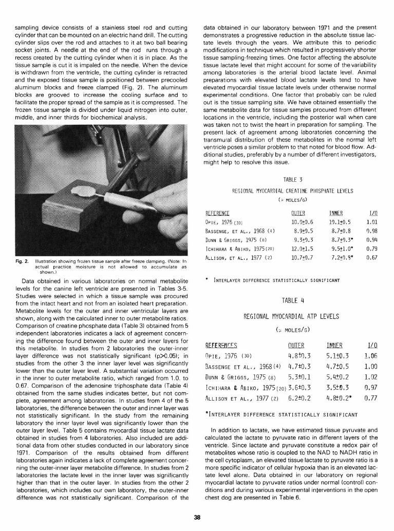
sampling device consists of a stainless steel rod and cutting cylinder that can be mounted on an electric hand drill. The cutting cylinder slips over the rod and attaches to it at two ball bearing socket joints. A needle at the end of the rod runs through a recess created by the cutting cylinder when it is in place. As the tissue sample is cut it is impaled on the needle. When the device is withdrawn from the ventricle, the cutting cylinder is retracted and the exposed tissue sample is positioned between precooled aluminum blocks and freeze clamped (Fig. 21. The aluminum blocks are grooved to increase the cooling surface and to facilitate the proper spread of the sample as it is compressed. The frozen tissue sample is divided under liquid nitrogen into outer, middle, and inner thirds for biochemical analysis.
Fig. 2. Illustration showing frozen tissue sample after freeze clamplng. (Note: In actual practice moisture is not allowed to accumulate as
shown.1
Data obtained in various laboratories on normal metabolite levels for the canine left ventricle* are presented in Tables 3-5. Studies were selected in which a tissue sample was procured from the intact heart and not from an isolated heart preparation. Metabolite levels for the outer and inner ventricular layers are shown, along with the calculated inner to outer metabolite ratios. Comparison of creatine phosphate data (Table 3) obtained from 5 independent laboratories indicates a lack of agreement concern- ing the difference found between the outer and inner layers for this metabolite. In studies from 2 laboratories the outer-inner layer difference was not statistically significant (p>O.O5); in studies from the other 3 the inner layer level was significantly lower than the outer layer level. A substantial variation occurred in the inner to outer metabolite ratio, which ranged from 1.0. to 0.67. Comparison of the adenosine triphosphate data (Table 4) obtained from the same studies indicates better, but not com- plete, agreement among laboratories. In studies from 4 of the 5 laboratories, the difference between the outer and inner layer was not statistically significant. In the study from the remaining laboratory the inner layer level was significantly lower than the outer layer level. Table 5 contains myocardial tissue lactate data obtained in studies from 4 laboratories. Also included are addi- tional data from other studies conducted in our laboratory since 1971. Comparison of the results obtained from different laboratories again indicates a lack of complete agreement concer- ning the outer-inner layer metabolite difference. In studies from 2 laboratories the lactate level in the inner layer was significantly higher than that in the outer layer. In studies from the other 2 laboratories, which includes our own laboratory, the outer-inner difference was not statistically significant. Comparison of the
data obtained in our laboratory between 1971 and the present demonstrates a progressive reduction in the absolute tissue lac- tate levels through the years. We attribute this to periodic modifications in technique which resulted in progressively shorter tissue sampling-freezing times. One factor affecting the absolute tissue lactate level that might account for some of the variability among laboratories is the arterial blood lactate level. Animal preparations with elevated blood lactate levels tend to have elevated myocardial tissue lactate levels under otherwise normal experimental conditions. One factor that probably can be ruled out is the tissue sampling site. We have obtained essentially the same metabolite data for tissue samples procured from different locations in the ventricle, including the posterior wall when care was taken not to twist the heart in preparation for sampling. The present lack of agreement among laboratories concerning the transmural distribution of these metabolites in the normal left ventricle poses a similar problem to that noted for blood flow. Ad- ditional studies, preferably by a number of different investigators, might help to resolve this issue.
TABLE 3
REGIO”lAL MYOCARBI4L CREATINE PHOSPYATE LEVELS
(U MOLES/G)
RFFFRF!\ICF @JIB IHIJER I/o
OPIE, 1976 (30) lO.O+O.G l!l.1+0.5 1.01
BASSENGE, ET AL., 1968 (4) 5.9fl.5 8.7+0.8 0.98
DUNN & GRIGGS, 1975 (8) 9.3t3.3 8.7yI.3' 0.94 ICHIHARA R ABIKO, 1975(201 12.0+1,5 9.5+1,0* 0.79 ALLISON, ET AL., 1977 (2) 10.7+0,7 7,2+7,3* 0.67
* INTERLAYER DIFFERENCE STATISTICALLY SIGNIFICANT
TABLE 4
REGIONAL MYOCARDIAL ATP LEVELS
(;I MOLES/G)
REFEREKES nUTER DL!Eg I/o
?PIE, 1376 (3'3) 4,8+r),3 5,1?0,3 1,06
QASSENGE ET AL., 1968(4) 4,7-10,3 4,7+0,5 1,oo
DUYN 2 GRIGGS, 1975 (8) 5,3+0,1 5,4+0,2 1.02
ICHII-IARA & ABIKO, 1975(20)3.6+0,3 3,5?0,3 on97
ALLISON ET AL., 1977 (2) 6,2-c0,2 4,8?0,2* on77
*INTERLAYER DIFFERENCE STATISTICALLY SIGNIFICANT
In addition to lactate, we have estimated tissue pyruvate and calculated the lactate to pyruvate ratio in different layers of the ventricle. Since lactate and pyruvate constitute a redox pair of metabolites whose ratio is coupled to the NAD to NADH ratio in the cell cytoplasm, an elevated tissue lactate to pyruvate ratio is a more specific indicator of cellular hypoxia than is an elevated lac- tate level alone. Data obtained in our laboratory on regional myocardial lactate to pyruvate ratios under normal (control) con- ditions and during various experimental interventions in the open chest dog are presented in Table 6.
3%
TABLE 5 beta adrenergic stimulation. Moderate aortic stenosis, achieved by tightening a ligature placed around the aorta proximal to the left coronary ostium, was without effect, but when this interven- tion was combined with beta adrenergic stim,ulation a significant interlayer difference in the lactate to pyruvate ratio was observed. Finally, moderate aortic insufficiency, produced by displacing the aortic valve leaflets with a special intravascular instrument, was without effect, but when severe aortic insufficiency was produc- ed a significant interlayer different in the lactate to pyruvate ratio was again noted.
The absence of a significant interlayer difference in the lactate to pyruvate ratio under control and moderately altered ex- perimental conditions has been interpreted by us as strong evidence against the existence of a metabolically important ox- ygen tension gradient in the normal left ventricle. The findings suggest that transmural blood flow is regulated in a manner that assures a nutritionally adequate blood supply to all layers of the ventricle under both normal and physiologically stressful condi- tions. This contention is reinforced by recent evidence that pac- ing the heart at 200 (10) or even 250 bpm (1) has no effect on the levels of creatine phosphate, adenosine triphosphate, or lactate in different layers of the ventricle.
The significant interlayer difference in the lactate to pyruvate ratio observed under the more severely altered experimental con- ditions clearly demonstrates that the inner ventricular layer is more susceptible than the outer layer to an imbalance between oxygen supply and demand when the autoregulatory capacity of the coronary circulation is exceeded. These positive results con- firm, in a completely independent manner, biophysical evidence of a nonuniform transmural force in the rhythmically contracting left ventricle (3, 22). It has been postulated that this nonuniform force consists of several components or stresses (26) which can cause not only uneven perfusion of the ventricle but uneven energy utilization as well.
Tangible evidence of uneven energy utilization in the forcibly contracting left ventricle has been provided from several sources. Results obtained by us (8) indicate that lactate production by the totally ischemic, pressure generating ventricle is uneven and greatest in the inner layer, whereas that by the totally ischemic, fibrillating or empty beating ventricle (9) is essentially equal in all layers. Evidence that myocardial oxygen consumption may be greater in the inner than the outer layer has been reported by two independent laboratories (19, 35). In both cases regional myocar- dial blood flow was estimated by the microsphere method and regional oxygen saturation in small arteries and veins (or capillaries) was determined in frozen tissue sections using a microspectrophotometric technique. When tissue oxygen con- sumption was calculated using the Fick equation significantly higher values were obtained in the inner than in the outer layer. The mean inner to outer ratios for oxygen consumption obtained in these two studies were 1.57 (19) and 1.27 (35).
The evidence that energy utilization may be greater in the inner layer provides additional insight into the mechanism for its greater susceptibility to ischemic injury and also raises the in- teresting possibility that the inner layer may be specially adapted to meet a constantly greater energy need. Specific vascular and cellular adaptations may exist in the inner layer to facilitate greater oxygen transport and chemical energy production. Studies in our laboratory have revealed a small but significantly higher myoglobin content in the inner layer compared to the outer layer (18), and certain transmural differences in mitochondrial membrane fatty acid saturation (31) and mitochondrial respiratory function (24) which could represent adaptations to an uneven energy need.
REGIONAL MYOCARDIAL LACTATE LEVELS
h' MOLES/G)
FREtiCES OllTER Jrn ul OPIE, 1976 (30) 1,80~0126 2,63+0,68 L44 ~CHIHARA & ABIKO, 1975 (20) 2,24~0,21 3,18~0,19* 1,42
ALLISON, ET AL,, 1977 (24 0,99tOJl 1,67+0,03* la69
GRIGGS, ET AL,, 1971 (17) 0,9%0J6 1,07+0,13 1809
GRIGGS, ET AL,, 1973 (14) 0,84+0,10 U39?0,16 1136
GRIGGs, ET AL,, 1974 (13) 0,55*0,04 0,06+0,09 1,09
DUNN 2 GRIGGS, 1975 ( 8) 3,36?0,06 0,35?9,04 0,97 DUNN, ET AL,, 1973 (II) 0,3:3~0,02 0,3SO,O2 1109
* IM-ER~AYER DIFFERENCE STATISTICALLY SIGNIFICANT
Table 6
Effects of Various Experimental Interventions
Myocardial Lactate to Pyruvate Ratios on Regional
Layer Intervention -
outer inner
9.821.0 9.3kO.8 I Control Conditions ( 17 )
II Moderate Coronary Constriction ( 17 ) 9.l-cl.9 7.3-el.6
III Severe Coronary Constriction ( 16 ) 39.2f7.6 113.727.2*
IV Beta Adrenergic Stimulation ( 17 ) 7.8k1.4 7.1-el .l
V Moderate Co ronary Constriction Plus Beta Adrene rgic Stimulation ( 17 ) 25.6f6.4 36.9?7.2*
VI Moderate Aortic Stenosis ( 14 ) 13.8k1.7 14.5k3.1
VII Moderate Ao rtic Stenosis Plus Beta Adrene rgic Stimulation ( 14 ) 2O.Ok3.1 45.5*10.3*
VIII Moderate Aortic Insufficiency ( 13 ) 10.812.1 11 .6_+1 .9
IX Severe Aortic Insufficiency ( 13 ) 14.2t4.7 38.7+10.8*
* interlayer difference statistically significant
Under control experimental conditions the outer-inner layer dif- ference was not statistically significant. Similar results were ob- tained in animals subjected to moderate coronary constriction. This intervention was produced by cannulating the left main cor- onary artery and constricting the cannula tubing enough to reduce coronary artery pressure (-38%) but not mean coronary blood flow. Different results were obtained when the coronary constriction was severe enough to reduce mean coronary blood flow (-50%). Under these frankly ischemic conditions the lactate to pyruvate ratio was elevated in both the outer and inner layers and a significant interlayer difference was present due to a greater elevation in the inner layer. Beta adrenergic stimulation, produc- ed by an intravenous infusion of isoproterenol, was without ef- fect on the lactate to pyruvate ratio in either layer despite marked chronotropic and iontropic responses of the heart. On the other hand, when this intervention was combined with moderate cor- onary constriction an elevated lactate to pyruvate ratio was noted in both layers and a significant interlayer difference was observ- ed, again due to a greater elevation in the inner layer. This same pattern was found in the case of moderate aortic stenosis and
39
1.
2.
3.
4.
5.
6.
8.
9.
10.
11.
12.
13.
14.
15.
16.
17.
18.
19.
20.
21.
22.
23.
24.
REFERENCES 25.
Allison, T.B., and J.W. Holsinger, Jr. Transmural metabolic gradients in the normal dogs left ventricle: effect of right atrial pacing. Am. J. Physiol. 233:H217-H221, 1977 or Am. J. Physiol.: Heart Circ. Physiol. 2: H217-H221, 1977. Allison, T.B., C.A. Ramey, and J.W. Holsinger, Jr. Transmural gradients of left ventricular tissue metabolites after circumflex artery ligation in dogs. J. Mol. Cell. Cardio. 9:837-852, 1977. Armour, J .A., and W.C. Randall. Canine left ventricular in- tramyocardial pressures. Am. J. Physiol. 220: 1833-1839, 1971. Bassenge, E., A. Schott, and P. Walter. Effect of coronary underperfusion on the energy metabolism in different layers of cardiac muscle. Proc. 5th Europ. Cong. Cardiol. :189-195, 1968. Becker, L. R. Ferreira, and M. Thomas. Comparison of 86Rb and microsphere estimates of left ventricular blood flow distribution. J. of Nut. Med. 15:969-973, 1974. Buckberg, G.D., D.E. Fixler, J.P. Archie, and J.I.E. Hoffman. Experimental subendocardial ischemia in dogs with normal cor- onary arteries. Circ. Res. 30:67-81, 1972. Cobb, F.R., R.J. Bathe, J.C. Greenfield, Jr. Regional myocar- dial blood flow in awake dogs. J. C/in. lnvest. 53:1618-1625, 1974. Dunn, R. B., and D. M. Griggs, Jr. Transmural gradients in ven- tricular tissue metabolites produced by stopping coronary blood flow in the dog. Circ. Res. 37:438-445, 1975. Dunn, R.B., K.M. Hickey, and D.M. Griggs, Jr. Effect of loading conditions on transmural lactate gradient in the ischemic left ventricle. Physiologist 18:200, 1975 (abst) Dunn, R.B., K.M. McDonough, and D.M. Griggs, Jr. Cardioac- celeration by atrial pacing and transmural metabolite levels in the canine left ventricle. Proc. Sot. Exper. Biol. and Med. 158:230-234, 1978. Dunn, R.B., K.M. McDonough, and D.M. Griggs, Jr. High energy phosphate stores and lactate levels in different layers of the canine left ventricle during reactive hyperemia. Circ. Res. In press. 1979. Friedberg, C.K., and H. Horn. Acute myocardial infarction not due to coronary occlusion. JAMA 112:1675-1679, 1939. Griggs, D.M., Jr. and C.C. Chen. Coronary hemodynamics and regional myocardial metabolism in experimental aortic insuffi- ciency. J. C/in. lnvest. 53: 1599-I 606, 1974. Griggs, D.M., Jr., C.C. Chen, and V.V. Tchokoev. Subendocar- dial anaerobic metabolism in experimental aortic stenosis. AM. J. Ph ysiol. 224: 607-6 12, 1973. Griggs, D.M., Jr., and Y. Nakamura. Effect of coronary con- striction on myocardial distribution of iodoantipiprine 13’ I. Am. J. Physiol. 215: 1082-1088, 1968. Griggs, D.M., Jr., V.V. Tchokoev, and C.C. Chen. Transmural differences in ventricular tissue substrate levels due to coronary constriction. Am. J. Physiol. 222:705-709, 1972. Griggs, D.M., Jr., V.V. Tchokoev, and J.W. DeClue. Effect of beta-adrenergic receptor stimulation on regional myocardial metabolism: Importance of coronary vessel patency. Am. Heart J. 82:492-502, 1971. Hickey, K.M., R.B. Dunn, and D.M. Griggs, Jr. Transmural dif- ferences in cellular constituents of the normal canine myocar- dium. Physiologist 18:247, 1975. (Abst) . Holtz, J., W.A. Grunewald, R. Manz, W.V. Restorff, and E. Bassenge. lntracapillary hemoglobin oxygen saturation and ox- ygen consumption in different layers of the left ventricular myocardium. Pfluger Archiv. 370:253-258, 1977. Ichihara, K., and Y. Abiko. Difference between endocardial and epicardial utilization of glycogen in the ischemic heart. Am. J. Physiol. 229: 1585-l 589, 1975. Jedeikin, L.A. Regional distribution of glycogen and phosphorylase in the ventricles of the heart. Circ. Res. 14:202-211, 1964. Kirk, E.S., and C. R. Honig. An experimental and theoretical analysis of myocardial tissue pressure. Am. J. Physiol. 207:361-367, 1964. Kirk, E.S., and C. R. Honig. Nonuniform distribution of blood flow and gradients of oxygen tension within the heart. Am. J. Physiol. 207:661-668, 1964. Long, J.W., Jr., A.P. Martin, D.M. Griggs, Jr., R.B. Dunn, and M. L. Vorbeck. Transmural mitochondrial respiration of canine left ventricular tissue. Fed. Proc. 37:230, 1978. (Abst).
26.
27.
28.
29.
30.
31.
32.
33.
34.
35.
36.
37.
38.
Lundsgaard-Hansen, P., C. Meyer, and H. Riedwyl. Transmural gradients of glycolytic enzyme activities in left ventricular myocardium. Pfluger Archiv. 297:89-106, 1967. Mirsky, I. Left ventricular stresses in the intact human heart. Biophys. J. 9: 189-208, 1969. Moir, T.W. Brief Reviews: Subendocardial distribution cf cor- onary blood flow and the effect of antianginal drugs. Circ. Res. 30:621-627, 1972. Moir, T.W. and D.W. DeBra. Effect of left ventricular hyperten- sion, ischemia, and vasoactive drugs on the myocardial distribu- tion of coronary flow. Circ. Res. 21:65-74, 1967. Moss, A. J. Intramyocardial oxygen tension. Cardiovas. Res. 3:314-318, 1968. Opie, L.H. Effects of regional ischemia on metabolism of glucose and fatty acids. Circ. Res. 38: (Suppl I), 52-68, 1976. Platner, W.S., D.M. Griggs, Jr., R.B. Dunn, K.M. McDonough and M. B. Abas. Transmural mitochondrial fatty acids of canine and porcine left ventricular tissue. Fed. Proc. 37:781, 1978. (Abst). Tripp, M.R., M.W. Meyer, S. Einzig, J.J. Leonard, C.R. Swayze, and I. J. Fox. Simultaneous regional myocardial blood flows by tritiated water and microspheres. Am. J. Physiol. 232: H 173-H 190, 1977 or Am. J. Physiol.: Heart Circ. Physiol. l:H173-H190, 1977. Utley, J., E.L. Carlson, J.I.E. Hoffman, H.M. Martinez, and G.D. Buckberg. Total and regional myocardial blood flow measurements with 25~~ 15~~ 9p, and filtered 1-10~ diameter microspheres and antipyrine in dogs and sheep. Circ. Res. 34:391-405, 1974. Weiss, H.R. Control of myocardial oxygenation - Effect of atrial pacing. Microvas. Res. 8:362-376, 1974. Weiss, H.R., J.A. Neubauer, J.A. Lipp and A.K. Sinha. Quan- titative determination of regional oxygen consumption in the dog heart. Circ. Res. 42:394-401, 1978. Whalen, W.J., P. Nair, and D. Buerk. Oxygen tension in the beating cat heart in situ. In Oxygen Supply. Eds. Kessler, M., et. al. University Park Press, Baltimore. pp. 199-201, 1973. Winbury, M.M., B.B. Howe, and H.R. Weiss. Effect of nitroglycerin and dipyridamole on epicardial and endocardial ox- ygen tension - further evidence for redistribution of myocardial blood flow. Pharmacol. Exp. Ther. 176:184-199, 1971. Yipintsoi, T., W.A. Dobbs, Jr., P.D. Scanlon, T.J. Knopp, and J. B. Bassingthwaighte. Regional distribution of diffusible tracers and carbonized microspheres in the left ventricle of isolated dog hearts. Circ. Res. 23:573-587, 1973.
40
BOOK REVIEW
Introduction To Physiology: Volume 4. Hugh Davson and M. B. Segal. Grune & Stratton, New York, 1978. 621 pp., tables, illus. $24.50
Davson and Segal, distinguished scholars from England, pre- sent a complete up-to-date account of how the body controls posture and locomotion. In a well organized fashion, they broaden our knowledge of basic muscle and nervous mechanisms covered in earlier volumes. Hundreds of salient references on some 2000 aspects of neurophysiology as well as many excellent illustrations provide a solid basis for study. Figure explanations under the photographs and drawings tell a delightful story in- dependent of the main text. The reader will enjoy seeing “How the horse runs and how two elephants flee from a hedgehog”. The great depth and experience of the authors, as they meander through broad facets of anatomy and physiology, makes us aware of the years required to compile and write a book of this caliber. Many classical studies show us how we arrived at our pre- sent status in physiology without our being bogged down in details that obscure the basic progress. The distinction between general, mammalian and clinical physiology disappear as sub- jects move from physics and chemistry to clinical syndromes. Complex aspects of neurophysiology integrate into a whole organism that has a strong basis in anatomy.
The book’s exhaustive nature forces readers to be serious students of physiology. They must be fond of the subject and have a good grasp of anatomy to fathom the heavy material presented. The title, Volume 4: “Introduction to Physiology” misleads us because of the book’s advanced character. When the “going gets tough” one can “browse and skip” for best results. The defining of difficult terms reveal concern for the reader but long sentences one after another, weak verbs and many preposi- tions create poor reading. In my opinion, 90 word sentences become hard to handle. One encounters repeating of critical areas such as the role servo-action plays in correcting movements already centrally planned. The limitations, exceptions and oppor- tunities in physiology receive much attention which would appeal to medical, dental and graduate students. I do not recommend this text for beginning nurses, pre-medical students or paramedical students. A few topics include: Patterns of behavior; muscle activation; sensory apparatus; spindle and tendon-organ control; brain control; neural organization of locomotion.
The reader learns how basic experimental information con- tributes to a wide spectrum of applied physiology. Experimental findings demonstrate how motor cortex damage produces spasticity which has important clinical implications. Motor defects may impair the executing of tasks seen in growth and mentally retarded children. Many examples show the physiological relation- ship of body parts. In steering a car, for instance, the authors mention that the corrective feedback come from the eyes. If your car moves a certain distance off-course, a wheel turn makes the required adjustment. Sophisticated animal models were presented for students to study clinical situations. These educa- tional and research models pin point the necessity of laboratory animal preparations in teaching physiology. Davson and Segal’s new outlook should bear much fruit in many fields for many years to come. Students will have a common ground to launch an at- tack upon the physiological unknown.
’ CAREERS 8 Ill
ANIMAL BIOLOGY
I NEW BROCHURE ISHED MARCH 1979
REPARED BY THE CATION COMMITTEE SOCIETY OF ZOOLOG ISTS
PRICES
1 COPY . . . . . . . FREE 2-49 COPIES . . . . 35$ EACH 50-99 COPIES . . . . 30$ EACH 100 OR MORE COPIES . 25$ EACH
ORDER FROM
AMERICAN SOCIETY OF ZOOLOGISTS BOX 2739
CALIFORNIA LUTHERAN COLLEGE THOUSAND OAKS, CA. 91360
ASZ
Robert H. Davis, Ph.D. Pennsylvania College of
Podiatric Medicine Philadelphia, Pennsylvania 19107
41
INTRODUCTION
This is the fourth annual collection of abstracts of educational material presented by the Educational Materials Review Board of the American Physiological Society under the direction of the Education Committee. Board members have submitted abstracts of review artricles, papers textbooks, books, manuals, handbooks and symposia which they have found valuable in teaching physiology. Selection of items is wholly at the discretion of members and where more than one member chooses to abstract the same material, each abstract is presented. We hope you continue to find this collection useful in teaching physiology.
The previous three collections constituted the entire issue of The Physiology Teacher. This year the abstracts are supplementing a regular issue of The Physiology Teacher Sec- tion of The Physiologist.
M. C. Shelesnyak Executive Editor The Physiology Teacher
CATEGORY PAGE
CELL PHYSIOLOGY 43
CIRCULATION 43
DEVELOPMENT AND AGING 43
ENDOCRINOLOGY AND METABOLISM 43
EVIRONMENTAL AND EXERCISE PHYSIOLOGY 44
GASTROINTESTINAL PHYSIOLOGY 44
NEUROBIOLOGY 45
RENAL AND ELECTROLYTE PHYSIOLOGY 45
RESPIRATION 48
TECHNOLOGY IN TEACHING PHYSIOLOGY 48
APPENDIX - Author Index 50
42

CELL PHYSIOLOGY ENDOCRINOLOGY AND METABOLISM
PHYSIOLOGY OF MEMBRANE DISORDERS. Edited by T.E. Andreoli, J.F. Hoffman. and D.D. Fanestil. Plenum Publishina Corp.. New York. 1978 ,
This textbook is written primarily for thedphysician or biomedical scientist who has interests that bridge the basic research labora- tory and a thorough understanding of pathophysiology of disease processes. Undoubtedly, it will be a valuable source book for phy- sicians in training and research trainees in physiology and membrane transport, particularly those motivated to understand the basis for many perturbations in disease at the level of cell membrane. Al- though the editors were eclectic in selecting included topics, this eclecticism turns out to be one major advantage of the book in that only topics of interest or controversy which have been influenced by recent scientific investigation are included. The clinical dis- orders are reduced to membrane phenomena which give the reader the most fundamental and contemporary understanding of these processes available. General topics included are cell membrane structure, problems in membrane biology, ccl 1 and epithel ial transport, and disorders of membrane transport; particular emphasis is given to normal and abnormal renal transport processes. Also included ‘are selected topics in gastrointestinal absorption, excitable tissue function, cardiac conduction, as well as more isolated discussions of problems such as cell volume regulation and the periodic para- lyses. In spite of the cell or membrane biology orientation of this text, each topic is presented in a section of reasonable length which is not burdensome to the reader. Similarly, the sections are nicely focused and understandable. I highly recommend perusal of this text by physiology teachers who undoubtedly will find infor- mation of immediate value for the classroom. David W. Ploth
CIRCULATION
THE MECHANICS OF THE CIRCULATION. Caro C G , : .,.T. J. Pedley_, R. C. Schroter, and W. A. Seed. Oxford UniversityxT?jxford, 1978.
This volume was written to provide a sinqle text devoted to the mechanics of the entire circulation suitable for advanced students of cardiovascular ohysioloay. Beqinninq with a presentation of basic laws of mechanics and their applications to deformation and flow, the authors proceed to describe the circulation in those terms. The heart, arteries, veins, microcirculation and blood are individually considered. Cell and fluid movement, heart valve motion, flow in collapsible vessels, transcapillary exchange and the pulmonary circulation are included. The mathematical treatment could be described as "intermediate" - more than many physiology graduate students can fully appreciate without some review of undergraduate mathematics and physics and less than many physicists miaht like. It is, nonetheless, probably optimum for the intended readership. If used as a research reference by investiqators, they may be disappointed by the lack of a complete bibliography covering specific points. Rather than such a bibliography, many, but not all, chapters have brief lists of further reading, usually of a aeneral nature. However, to have cited everything as would be done in a monograph, would have been a Herculean task and not within the expectations for a textbook.
T. C. Lloyd
DEVELPMENT AND AGING
LIVER AND AGEING. Platt, D. (editor). Fourth International Giessener Symposium on Experimental Gerontology, 260 pp. F.K. Shattauer, Stuttgart-New York, 1977.
Papers presented at this symposium covered changes occurring in the liver during senescence as deduced mainly from experimental studies on animals and, in particular, on the rat. However, a few clinical studies are also included. The changes in the liver with age reviewed include the following: (1) ultrastructural changes; (2) molecular changes in enzymes, chromatin proteins, DNA, and
microsomes; (3) protein synthesis changes in the whole tissue, in isolated hepatocytes, and at the subcellular level; (4) biochemical changes in connective tissue: (5) functional changes in the trans- fer of organic anions from the plasma into bile, in bile acid secre tion and bile flow, in the phagocytosis of senescent erythrocytes, and in the modulation of immunologic responses. This book will be especially useful for those who wish an up-to-date general review of age-dependent changes occurring in the liver at all levels of its organization from the molecular to the whole organ.
D.E. Buetow
STRUCTURE AND FUNCTION OF THE GONADOTROPINS. McKerns, K.W., ed. Plenum Press, New York, 1978.
This 25-chapter volume reports the proceedings of a symoosium reviewing and previewing the "state of the art" with regard to these most complex of the molecules known to have hormonal activity. Nine of the chapters deal with the primary and conformational structures of human chorionic gonadotrophin and/or the several pituitary glyco- proteins (both mammalian and teleost), with the conformational requirements for binding to a ligand, and with the subunit mechanism of glycoprotein hormone assembly. This is juxtaposed with findings on the biosynthesis of the non-glycoprotein reproductive hormone, prolactin, which involves manufacture and subsequent cleavage of a larger prohormone molecule. The majority of the papers deal with the cell biology of protein hormone action, including CAMP-mediated effects and some new evidence that macromolecular hormones actually can enter their target cells in vivo and/or act directly on isolated subcellular constituents in vitro.The publication faithfully preserves the original flavor of the discussions, many of which addressed the idea that the mechanisms of action of peptide and steroid hormones may not differ as markedly as popular belief, and much of current teaching, state. Consequently, the volume should serve not only as a guide to the rapidly expanding literature in several converging fields but also as a spectacle of the processes through which important conceptual redirections are, sometimes painfully, achieved.
J.M. Nolin and E.M. Bogdanove
PIONEERS IN NEUROENDOCRINOLOGY II. Meites, J., B.T. Donovan and S.M. McCann, eds. Plenum Press, New York, 1978.
A second collection of autobiographical vignettes by senior inves- tigators whose research, in the judgment of the editors, helped to establish neuroendocrinology as a major subdiscipline. The first volume, which appeared in 1975, was a valuable historical account, although it unfortunately came too late to include contributions by some of the very first "pioneers", including G.W. Harris. This vol- ume, which collects the reminiscences of a second generation of senescents (including the editors), presents equally fascinating in- sights into the personalities and motivations of the men (no women appear among the 24 self-portraitists) whose work helped advance the vision of the first "pioneers" to a point where (p. 239) "...pio- neering in neuroendocrinology ceased and became the harvesting of a new... science." Although there are some significdnt gaps in this volume also, the book manages to present a highly readable picture of an era in which mutual recognition and interactions (both pleasant and otherwise) among a score or so of coeval young investigators had results important enough to warrant - for two of the contributors - the coveted recognition of the Nobel Prize. To the credit of the editors who (sometimes repressing themselves painfully) allowed the authors almost total poetic license, these essays should help succeeding generations of students and investigators to understand how the circumstances in which their scientific forebears found themselves led to the events which culminated in discovery. Phrased differently, it adds flesh to the skeleton accounts present in the more formal scientific literature. E.M. Bogdanove
PIONEERS IN NEUROENDOCRINOLOGY 11, Meites, J., Donovan, B.T., and S.M. McCann, 422 pgs., Plenum Pub., Co., New York, N.Y., 1978.
This second volume of Pioneers in Neuroendocrinology contains autobiographical and research accounts from many of the leading and Tnostlv still active investigators in the field: Barraclo;q!l, Beach, Bem,,Bopdanove, Brodish, Cross, D'Angelo, Donovan, Ehdroczi, Etkin Flerko, Ganong, Greer, Guillemin, Halasz, Martini, McCann, Meites, Reichlin, Saffran, Schally, Schreiber, de!Jied, and Zuckerman. Each chapter is preceded by a photograph and biographical sketch of the author, and reveals much about his personality, goals, ideas, inspirations, triumphs, disappointments, research approaches, sources of financial support, views of colleagues, etc. Guillemin, McCann, Meites, Saffran, Schally and Schreiber each describe their early attempts to isolate 'releasing factors' from the hypothalamus, and Schally and Guillemin tell how they finally succeeded in deter- mining the structures of TRH, LHRH and somatostatin in 1969-1973 which led to Nobel Prizes in 1977. Also included are insights into the role of hypothalamic biogenic amines and other neurotransmitters on pituitary function; negative and positive feedback of target gland hormones on the pituitary and hypothalamus; neuroendocrine control of reproduction; CNS bioelectrical correlates to endocrine functions; effects of hormnes on behavior; localization of hypo- thalamic factors in the brain and elsewhere; relation of the hypo- thalams to mammary and pituitary tumors, and to problems in puberty and aging; and many other topics on current research in neuroendo- crinology. This book will be of particular interest to students and investigators in the fields of physiology, endocrinology, pharmacology and medicine.
43
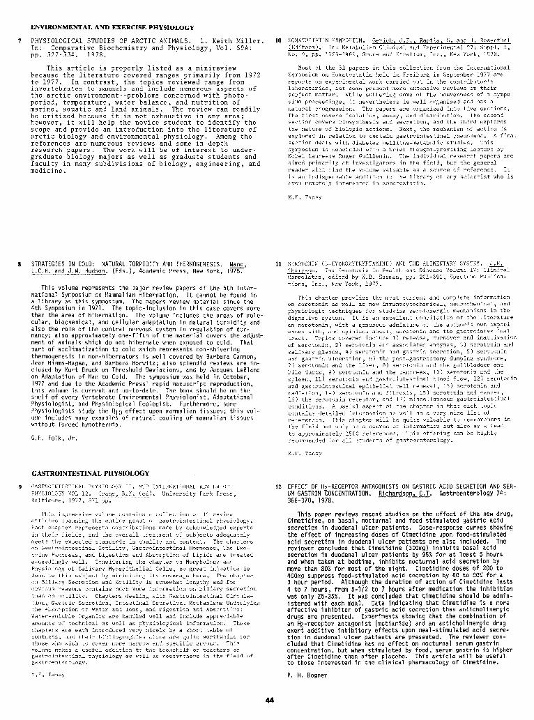
ENVIRONMENTAL AND EXERCISE PHYSIOLOGY
1 PHYSIOLOGICAL STUDIES OF ARCTIC ANIMALS. L. Keith Miller. In: Comparative Biochemistry and Physiology, Vol. 59A: PP. 327-334. 1978.
This article is properly listed as a minireview because the literature covered ranges primarily from 1972 to 1977. In contrast , the topics reviewed range from invertebrates to mammals and include numerous aspects of the arctic environment period,
--problems concerned with photo- temperature, water balance, and nutrition of
marine, aquatic and land animals. The review can readily be critized because it is not exhaustive in any area; however, it will help the novice student to identify the scope and provide an introduction into the literature of arctic biology and environmental physiology. Among the references are numerous reviews and some in-depth research papers. The work will be of interest to under- graduate biology majors as well as graduate students and faculty in many subdivisions of biology, engineering, and medicine.
8 STRATEGIES IN COLD: NATURAL TORPIDITY AND THERMOGENESIS. Wana, L.C.H. and J.W. Hudson, (Eds.), Academic Press, New York, 1978.
This volume represents the major review papers of the 5th Inter- national Symposium on Mammalian Hibernation. It cannot be found in a library as this symposium. The papers review material since the 4th Symposium in 1971. The topic-inclusion in this case covers more than the area of hibernation. The volume includes the areas of mole- cular, biochemical, and cellular adaptation in natural torpidity and also the role of the central nervous system in regulation of dor- mancy; also approximately one-fifth of the material covers the adjust- ment of animals which do not hibernate when exposed to cold. That part of acclimatization to cold which represents non-shivering thermogenesis in non-hibernators is well covered by Barbara Cannon, Jean Himms-Hagen, and Barbara Horwitz; also splendid reviews are in- cluded by Kurt Bruck on Threshold Deviations, and by Jacques LeBlanc on Adaptation of Man to Cold. The symposium was held in October, 1977 and due to the Academic Press' rapid manuscript reproduction, this volume is current and up-to-date. The book should be on the shelf of every Vertebrate Environmental Physiologist, Adaptational Physiologist, and Physiological Ecologist. Furthermore, some Physiologists study the 010 effect upon mammalian tissues; this vol- ume includes many examples of natural cooling of mammalian tissues without forced hypothermia.
G.E. Folk, Jr.
10 SOMATOSTATIN SYMPOSIUM. Gerich, J.E., Raptis, S. and J. Rosenthal (Editors). In: Metabolism Clinical and Experimental 27: Suppl. 1, No. 9, pp. 1129-1469, Grune and Strattom, Inc., New York, 1978.
Most of the 61 papers in this collection from the International Symposium on Somatostatin held in Freiburg in September 1977 are reports on experimental work carried out in the contributor's laboratories, but some present more extensive reviews on their subject matter. While suffering some of the unevenness of a sympo- sium proceedings, it nevertheless is well organized and has a natural progression. The papers are organized into five sections. The first covers isolation, assay, and distribution. The second section covers biosynthesis and secretion, and the third explores the nature of biologic actions. Next, the mechanism of action is explored in relation to certain gastrointestinal phenomena. A final section deals with diabetes mellitus-metabolic studies. This symposium is concluded with a brief thought-provoking lecture by Nobel laureate Roger Guillemin. The individual research papers are aimed primarily at investigators in the field, but the general reader will find the volume valuable as a source of reference. It is an indispensable addition to the library of any scientist who is even remotely interested in somatostatin.
M.F. Tansy
11 SEROTONIN (5-HYDROXYTRYPTAMINE) AND THE ALIMENTARY SYSTEM. J.H. Thompson. In: Serotonin in Health and Disease Volume IV: Clinical Correlates, edited by W.B. Cssman, pp. 201-392, Spectrum Publica- tions, Inc., New York, 1977.
This chapter provides the most current and complete information on serotonin as well as new immunocytochemical, neurochemical, and physiologic techniques for studying serotonergic mechanisms in the digestive system. It is an excellent compilation of the literature on serotonin, with a generous admixture of the author's own experi- ences with, and opinions about, serotonin and the gastrointestinal tract. Topics covered include 1) release, turnover and inactivation of serotonin, 2) serotonin and associated enzymes, 3) serotonin and salivary glands, 4) serotonin and gastric secretion, 5) serotonin and gastric ulceration, 6) the post-gastrectomy dumping syndrome, 7) serotonin and the liver, 8) serotonin and the gallbladder and bile ducts, 9) serotonin and the pancreas, 10) serotonin and the spleen, 11) serotonin and gastrointestinal blood flow, 12) serotonin and gastrointestinal epithelial cell renewal, 13) serotonin and radiation, 14) serotonin and fibrosis, 15) serotonin and cancer, 16) the serotonin receptor, and 17) miscellaneous gastrointestinal conditions. A useful aspect of the chapter is that each topic contains detailed information as well as a very nice list of references. This chapter will be quite valuable to researchers in the field not only as a source of information but also as a lead to approximately 1500 references. This offering can be highly recommended for all students of gastroenterology.
M.F. Tansy
GASTROINTESTINAL PHYSIOLOGY
9 GASTROINTESTINAL PHYSIOLOGY II. MTP INTERNATIONAL REVIEW OF PHYSIOLOGY VOL 12. Crane, R.K. ted). University Park Press, Baltimore, 1977, 371 pp.
This impressive volume contains a collection of 11 review articles spanning the entire gamut of gastrointestinal physiology. Each chapter represents contributions made by acknowledged experts in their fields, and the overall treatment of subjects adequately meets the expected standards in quality and content. The chapters on Gastrointestinal Motility, Gastrointestinal Hormones, The Exo- crine Pancreas, and Digestion and Absorption of Lipids are treated exceedingly well. Considering the chapter on Morphology and Physiology of Salivary Myoepithelial Cells, no great injustice is done to this subject by minimizing its coverage here. The chapter on Biliary Secretion and Motility is somewhat lengthy and for obvious reasons contains much more information on biliary secretion than on motility. Chapters dealing with Gastrointestinal Circula- tion, Gastric Secretion, Intestinal Secretion, Mechanisms Underlying the Absorption of Water and Ions, and Digestion and Absorption: Water-soluble Organics are handled well and include appreciable amounts of technical as well as physiological information. These chapters are each introduced very nicely by a short table of contents, and their bibliographies alone are quite worthwhile for those who wish to cover more narrow and specific ground. This volume makes a useful addition to the bookshelf of teachers of gastrointestinal physiology as well as researchers in the field of gastroenterology.
12 EFFECT OF H -RECEPTOR ANTAGONISTS ON GASTRIC ACID SECRETION AND SER- UM GASTRIN EONCENTRATION. Richardson, C.T. Gastroenterology 74: 366-370, 1978.
This paper reviews recent studies on the effect of the new drug, Cimetldine, on basal, nocturnal and food stimulated gastric acid secretion in duodenal ulcer patients. Dose-response curves showing the effect of increasing doses of Cimetidine upon food-stimulated acid secretion in duodenal ulcer patients are also included. The reviewer concludes that Cimetidine (300mg) inhibits basal acid secretion in duodenal ulcer patients by 95% for at least 5 hours and when taken at bedtime, inhibits nocturnal acid secretion by more than 80% for most of the night. Cimetidine doses of 200 to 400mg suppress food-stimulated acid secretion by 60 to 80% for a 3 hour period. Although the duration of action of Cimetidine lasts 4 to 7 hours, from 5-l/2 to 7 hours after medication the inhibition was only E-35%. It was concluded that Cimetidine should be admin- istered with each meal. Data indicating that Cimetidine is a more effective inhibitor of gastric acid secretion than anticholinergic drugs are presented. Experiments showing that the combination of an Hz-receptor antagonist (metiamide) and an anticholinergic drug exert additive inhibitory effects upon meal-stimulated acid secre- tion in duodenal ulcer patients are presented. The reviewer con- cluded that Cimetidine has no effect on nocturnal serum gastrin concentration, but when stimulated by food, serum gastrin is higher after Cimetidine than after placebo. This article will be useful to those interested in the clinical pharmacology of Cimetidine.
M.F. Tansy P. H. Bogner

13 HEALING OF DUODENAL ULCER WITH AN ANTACID REGIMEN. Peterson, W.L., R. Sturdevant, H. Franki, C. Richardson, J. Isenberg, 3. Elashoff, J. Sones, R. Gross, R. McCallum and J. Fordtran. N. Engl. J. Med. 297:341-345, 1977.
A double-blind clinical trial was carried out on 74 patients with endoscopically proven duodenal ulcer at 3 medical centers for 4 weeks. Patients were randomly allocated to either an inert placebo or a large dose liquid antacid regimen: 30ml (144mE
9 ) of Mg-Al or Al
hydroxide 1 and 3 hours after meals and at bedtime 7 doses per day= 1008mEq antacid). The ulcers healed completely in 28 of 36 antacid treated patients (78%) as compared with 17 of the 38 placebo-treated patients (45%). It was found that freedom from ulcer symptoms (i.e. frequency and severity of pain, night pain, vomiting, ability to work) was a poor predictor of ulcer healing in the final week of treatment: 33% of patients who were symptom-free had unhealed ulcers while 48% of those with healed ulcers continued to have ulcer symp- toms. Placebo-treated patients who smoked were less likely to heal than nonsmokers, but smoking had no effect on healing in the antacid group. This evidence that antacids promote the healing of duodenal ulcers contrasts with earlier studies. The authors suggest that the ineffectiveness of antacids in previous studies might be due to much smaller doses used or choice of antacid (ca carbonate induces re- bound acid hypersecretion) or to less accurate methods of measuring ulcer healing. This study should be of interest to G.I. physiolo- gists who like to present clinically relevant material in lectures and to all of us who have duodenal ulcers!
P. H. Bogner
NEUROBIOLOGY
14 FUNDAMENTALS OF SENSORY PHYSIOLOGY. Edited by R.F. Schmidt. pp 286. Springer Verlag. New York., l'm
This small interesting textbook is a companion volume to Fundamentals of Neurophysiology by the same author to which frequent reference is made, The book is designed for readers with minimal previous training in the sciences. Topics discussed include general principles of sensory phenomena, including psychophysics, basic neurophysiologi- cal processes in sensation, somatovisceral sensations with special reference to mechanoception, proprioception and thermoception as well as the different varieties of pain. Additional chapters are included on vision, hearing, equi- librium, taste, olfaction, hunger and thirst. Methods of measurement and applications of sensory physiology are dis cussed with respect to information theory, conditioning and behavior. The treatment of sensory physiology is reas- onably comprehensive, given the level of treatment and the intended audience. Enough anatomical description is pro- vided so that the reader is adequately oriented. The con- tributing authors, all faculty members of various German universities, take pains to define terms clearly, utilize a standardized form of illustration in two colors for em- phasis based on figures taken from the literature in large part and provide a number of useful study questions with answers in the back. The writing is simple, direct and lu- cid, a tribute to the translator, Dr. M.A. Biederman- Th- Orson. L.M.N. Bach
15 FUNDAMENTALS OF NEUROPHYSIOLOGY. Edited by R.F. Schmidt. pp 339. Second Edition. Springer Verlag, NesrkT78,
This excellent little text about neurophysiology has been revised and enlarged from the previous edition and sensory physiology has been extracted to provide the sub- ject of a companion volume. The topics discussed here in- clude basic structural features of the peripheral and central nervous system, including the neurone. Chapters are presented on excitation, transmission, reflexes, mus- cle contraction, motor systems, regulation of motor func- tion, autonomic nervous system and a new chapter on integ- rative function, the EEG, sleep-wake processes, speech and consciousness and learning. Terms are defined concise ly as they are introduced. Clearly understood diagrams are appropriately introduced, largely drawn from the lit- erature and suggested further readings are included. Stu- dy questions (and answers) are provided for the neophyte for whom this book is intended. The writing is straight- forward and the material is easy to understand, a trib- ute to the authors, all member6 of various German uni- versities and the translator, Dr. M.A. Biedrman-Thorson. The editor has skillfully provided an easy transition between chapter6 and a continuity of thought and level of presentation. This book is highly recommended for undergraduate students. L.M.N. Bach
HUNGER-BASIC MECHANISMS AND CLINICAL IMPLICATIONS. Edited by D. Novin, W. Wyricka and G.A. Bray. pp 494. Raven Press, New York, 19'16 0
This monograph consists of the proceeding6 of a con- ference on the subject held at the Brain Research Insti- tute, UCLA in January, 1975. The general subject areas include neurochemistry and neuroanatomy of hunger, the role of nutrients and energy metabolism in the control of food intake, developmental and motivational properties of hunger, set-point theory and relationship of weight regulation to food intake, ing (patterning,
short term regulation of feed- visceral and peripheral mechanisms), hun-
ger and obesity and concludes with an excellent summary by G.J. Mogenson. The sum effect of these articles is to up-date our understanding of the variety of processes by which food intake is regulated and to break away rather Completely from the simple ventromedial nucleus-lateral hypothalamic notion of reciprocal regulation. Some of the high-lights include a comparison of alpha-receptor and beta-receptor catecholamlne effects, 5HT and dopamine influences, the more general aspects of lateral hypothal- amic lesion effect6 and amygdalar influences, an excell- ent discourse on homeostatic mechanisms by D.A. Booth and colleagues, the idea of the lateral hypothalamus as a weight set-point mechanism, the androgen brain and the male pattern of food intake, the effects of the lipostat. L.M.N. Bach
RENAL AND ELECTROLYTE PHYSIOLOGY
RENAL CONCENTRATING AND DILUTING MECHANISMS. Kokko, J. P. Hospital Practice. Vol. 14, No. 2: 110-116, 1979.
This review discusses the renal countercurrent mechanisms for producing an osmotically concentrated or dilute urine. The newer, passive equilibration model (Kokko-Rector model) is preferred over other models which require active salt transport out of the thin ascending limb of Henle's loop. The main features of the passive equilibration model are presented. These include (1) active chloride reabsorption by the thick ascending limb of Henle's loop, (2) delivery of a concentrated urea solution into the inner medulla via the papillary collecting ducts and pelvic urine, (3) concen- tration of NaCl along the descending thin limb by water abstraction, (4) passive reabsorption of NaCl out of the thin ascending limb of Henle's loop, and (5) control of the water permeability of the collecting ducts by ADH. The article provides some historical background, and summarizes some of the critical studies which support this model. The important role of urea is highlighted. The article is nicely illustrated. Although the passive equili- bration model is ingenious, readers ought to be cautioned that there is still considerable disagreement as to whether this model is appropriate to all mammalian species. This review presents a straight-forward account of some of the newer ideas on urinary concentration and dilution, and is recommended for medical students and physicians.
G. A. Tanner
DISORDERS OF POTASSIUM BALANCE. Cohen, J. J. Hospital Practice, Vol. 14, No. 1: 119-128, 1979.
This review discusses potassium balance in health and disease. The factors influencing the distribution of potassium between cell water and extracellular fluid are discussed first. Data on the distribution of potassium within the body are summarized. The important role of the kidneys in regulating body potassium is briefly discussed. The major part of this review concerns hypokalemia and hyperkalemia: causes, clinical manifestations, and clinical management. The physiological bases for altered function and for proper treatment are emphasized. This readable review is especially recommended for medical students and physicians.
G. A. Tanner
45
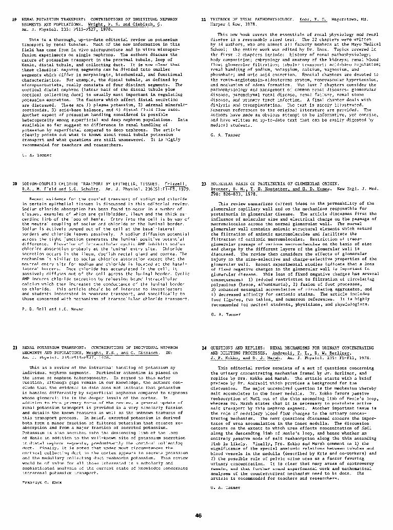
RENAL POTASSIUM TRANSPORT: CONTRIBUTIONS OF INDIVIDUAL NEPHRON SEGMENTS AND POPULATIONS. Wright, F. S. and Giebisch, G. Am. J. Physiol. 235: F515-F527, 1978.
This is a thorough, up-to-date editorial review on potassium transport by renal tubules. Much of the new information in this field has come from in vivo micropuncture and in vitro microper- fusion experiments on single nephrons. The authors discuss the nature of potassium transport in the proximal tubule, loop of Henle, distal tubule, and collecting duct. It is now clear that these classical nephron segments can be divided into smaller segments which differ in morphologic, biochemical, and functional characteristics. For example, the distal tubule, as defined by micropuncturists, really consists of four distinct segments. The cortical distal nephron (latter half of the distal tubule plus cortical collecting duct) is usually most important in regulating potassium excretion. The factors which affect distal secretion are discussed. These are 1) plasma potassium, 2) adrenal mineralo- corticoids, 3) acid-base balance, and 4) distal fluid flow rate. Another aspect of potassium handling considered is possible heterogeneity among superficial and deep nephron populations. Data available so far suggest no difference in renal handling of potassium by superficial compared to deep nephrons. The article clearly points out what is known about renal tubule potassium transport and what questions are still unanswered. It is highly recommended for teachers and researchers.
G. A. Tanner
20 SODIUM-COUPLED CHLORIDE TRANSPORT BY EPITHELIAL TISSUES. Frizzell. R.A., M. Field and S.G. Schultz. Am. J. Physiol. 236(5):Fl-F8, 197’9.
Recent evidence for the coupled transport of sodium and chloride in certain epithelial tissues is discussed in this editorial review. Sodium chloride absorption has been found to occur in a number of tissues, examples of which are gallbladder, ileum and the thick as- cending limb of the loop of Henle. Entry into the cell is by way of the neutral coupling of sodium and chloride at the luminal border. Sodium is actively pumped out of the cell at the basal-lateral borders and chloride leaves passively. A sodium diffusion potential across the tight junction generates the luminal positive potential difference. Elevation of intracellular cyclic AMP inhibits sodium chloride absorption probably at the luminal entry site. Chloride secretion occurs in the ileum, dogfish rectal gland and cornea. The mechanism is similar to sodium chloride absorption except that the neutral entry site for sodium and chloride is located at the basal- lateral borders. Once chloride has accumulated in the cell, it passively diffuses out of the cell across the luminal border. Cyclic AMP induces chloride secretion by releasing bound intracellular calcium which then increases the conductance of the luminal border to chloride. This article should be of interest to investigators and students interested in membrane transport, and specifically to those concerned with mechanisms of transcellular chloride transport.
22
23
TEXTBOOK OF RENAL PATHOPHYSIOLOGY. Knox, F. G. Hagerstown, Md. Harper 6 Row, 1978.
This new book covers the essentials of renal physiology and renal disease in a reasonably sized text. The 22 chapters were written by 18 authors, who are almost all faculty members at the Mayo Medical School; the entire work was edited by Dr. Knox. Topics covered in the first 12 chapters include: history of renal pathophysiology; body composition; embryology and anatomy of the kidneys; renal blood flow; glomerular filtration; tubular transport; acid-base regulation; renal handling of sodium, potassium, calcium, magnesium, and phosphate; and uric acid excretion. Special chapters are devoted to the renin-angiotensin-aldosterone system, renovascular hypertension, and mechanism of edema formation. The last 7 chapters consider the pathophysiology and management of common renal diseases- glomerular disease, parenchymal renal disease, renal failure, renal stone disease, and urinary tract infection. A final chapter deals with dialysis and transplantation. The text is nicely illustrated. Numerous references to the original literature are provided. The authors have made an obvious attempt to be informative, yet concise, and have written an up-to-date text that can be easily digested by medical students.
G. A. Tanner
MOLECULAR BASIS OF PROTEINURIA OF GLOMERULAR ORIGIN. Brenner, B. M., T. H. Hostetter, and H. D. Humes. New Engl. J. Med. 298: 826-833, 1978.
This review summarizes current ideas on the permeability of the glomerular capillary wall and on the mechanisms responsible for proteinuria in glomerular disease. The article discusses first the influence of molecular size and electrical charge on the passage of macromolecules across the normal glomerular wall. The normal glomerular wall contains anionic structural elements which retard the filtration of anionic macromolecules and facilitate the filtration of cationic macromolecules. Restriction of trans- glomerular passage of various macromolecules on the basis of size and charge by the different layers of the glomerular wall is discussed. The review then considers the effects of glomerular injury on the size-selective and charge-selective properties of the glomerular wall. Recent experimental studies indicate that a loss of fixed negative charges in the glomerular wall is important in glomerular disease. This loss of fixed negative charges has several consequences: 1) reduced restriction to filtration of circulating polyanions (hence, albuminuria), 2) fusion of foot processes, 3) enhanced mesangial accumulation of circulating aggregates, and 4) decreased affinity for cationic stains. The article includes four figures, two tables, and numerous references. It is highly recommended for medical students, physicians, and physiologists.
P. D. Bell and L.G. Navar G. A. Tanner
21 RENAL POTASSIUM TRANSPORT: CONTRIBUTIONS OF INDIVIDUAL NEPHRON SEGMENTS AND POPULATIONS, Wright, F.S., and G. Giebisch. IN: Am. J. Physiol. 235:F515-F527, 1978.
This is a review of the intrarenal handling of potassium by individual nephron segments. Particular attention is placed on the issue of nephron heterogeneity. In regard to this major question, although gaps remain in our knowledge, the authors con- clude that the evidence to date does not indicate that potassium is handled differently by superficial nephrons compared to nephrons whose glomeruli lie in the deeper levels of the cortex. In addition to this primary focus of the review, a general update of renal potassium transport is provided in a very scholarly fashion and details the known features as well as the unknown features of this transport process. In brief, excreted potassium is derived both from a minor fraction of filtered potassium that escapes re- absorption and from a major fraction of secreted potassium. Potassium is also secreted into-the descending limb of the loop of Henle in addition to the well-known site of potassium secretion in distal nephron segments, predominantly the cortical collecting duct. Finally, it is noted that under most circumstances the cortical collecting duct in the cortex appears to secrete potassium and the medullary collecting duct reabsorbs potassium. This review would be of value for all those interested in a scholarly and sophisticated analysis of the current state of knowledge concerning intrarenal potassium transport.
24 QUESTIONS AND REPLIES: RENAL MECHANISMS FOR URINARY CONCENTRATING AND DILUTING PROCESSES. Andreoli, T. E., R. W. Berliner, J. P. Kokko, and D. J. Marsh. Am. J. Physiol. 235: Fl-Fll, 1978.
This editorial review consists of a set of questions concerning the urinary concentrating mechanism framed by Dr. Berliner, and replies by Drs. Kokko and Marsh. The article starts with a brief preface by Dr. Andreoli which provides a background for the discussion. The major unresolved question is the mechanism whereby salt accumulates in the inner medulla. Dr. Kokko favors passive reabsorption of NaCl out of the thin ascending limb of Henle's loop, whereas Dr. Marsh states that it is necessary to postulate active salt transport by this nephron segment. Another important issue is the role of medullary blood flow changes in the urinary concen- trating mechanism. The next questions discussed concern the impor- tance of urea accumulation in the inner medulla. The discussion centers on the extent to which urea effects concentration of NaCl along the descending limb of Henle's loop, and hence whether an entirely passive mode of salt reabsorption along the thin ascending limb is likely. Finally, Drs. Kokko and Marsh comment on 1) the significance of the special anatomic relations between tubules and blood vessels in the medulla (described by Kriz and co-workers) and 2) the possible role of pelvic urine urea as a factor favoring urinary concentration. It is clear that many areas of controversy remain, and that further sound experimental work and mathematical analyses of the countercurrent mechanism need to be done. The article is recommended for teachers and researchers.
Franklyn G. Knox G. A. Tanner
25 THE CONTROL OF SODIUM EXCRETION. DeWardener, H.F. Am. J. Physiol. 235(3): F163-F173, 1978.
26
27
This review discusses the means by which the kidney controls sod- ium excretion. The author discusses tubuloglomerular feedback and autoregulation as a means of controlling filtrate production. The literature pertaining to control of proximal tubular reabsorption during blood volume expansion is reviewed; changes in Starling forces which has been considered a major factor in explaining glomerular tubular balance and the constancy of fractional reabsorp- tion in the proximal tubule is discussed as well as alternative explanations such as changes in hydrostatic pressure and composition of the filtrate; changes in filtrate composition may alter reabsorp- tion by controlling the motility of the brush border of the proximal tubule. Reabsorption rate in the loop of Henle and distal tubule is directly related to the rate of Na + delivery while in the collecting duct reabsorption of Na+ is controlled by aldosterone, prostoglan- dins, bradykinin, angiotensin and extrarenal natriuretic hormones. Certainly the exquisite fine control of Na+ excretion resides within the collecting duct yet as the author points out the precise under- standing of the control of the reabsorptive processes in the col- lecting duct are yet to be uncovered. The review should be of in- terest to medical and graduate student interested in a succinct review of the various tubular phenomena and their controls that are responsible for regulating Na+ excretion and ultimately volume reg- ulation.
H.M. Randall
28 THE DYNAMIC KIDNEY (Part One). Bennett, C. M., and R. J. Glassock. Available from Lilly Educational Resources Library, 565 Fifth Avenue, New York, N.Y. 10017.
This new film runs for 30 min and reviews the basic structure and function of the human kidney. The importance of the kidneys in regulating the composition and volume of the body fluids is empha- sized. Topics considered include: (1) kidney blood supply and structure of the nephron, (2) structure of the glomerular capillary wall, (3) glomerular hemodynamics, and (4) formation of the urine. The latter includes discussion of reabsorption of solutes and water in the proximal tubule, reabsorption of filtered bicarbonate, distal acidification, countercurrent multiplication, role of antidiuretic hormone, and the renin-angiotensin-aldosterone system. Relatively new subjects are considered, such as the relation between glomerular filtration rate and blood flow, role of gegative charges in the glomerular capillary wall in restricting the passage of plasma pro- teins, the paracellular pathway in proximal reabsorption, and the importance of the distal nephron in &, Na+, and K'+ excretion. The film is up-to-date, nicely animated, and covers a lot of ground. It is recommended as supplementary material for first year medical and graduate students.
G. A. Tanner
RENAL EFFECTS OF HEAD-OUT WATER IMMERSION IN MAN: IMPLICATIONS FOR 29 THE DYNAMIC KIDNEY (Part Two). Bennett, C. M., and R. J. Glassock. AN UNDERSTANDING OF VOLUME HOMEOSTASIS. Epstein, M. Physiol. Rev. Available from Lilly Educational Resources Library, 565 Fifth 58: 529-581, 1978. Avenue, New York, N.Y. 10017.
The subject of this review is volume homeostasis and how it can be altered by changes in intrathoracic blood volume. Head-out water immersion can be used as a long duration stimulus to alter intrathor- acic blood volume. After identifying the quantitative relationships between immersion and body position, on intrathoracic blood volume and cardiac output, the author discusses possible mechanisms which may be responsible for the relationships. The effects of immersion on the renal responses to increases in central blood volume in the hydrated and fluid-restricted subjects was discussed including diure- sis and natriuresis. Identification of and corroborating evidence for the controlling mechanisms which regulate the renal responses were also considered including: ADH, renal prostaglandins, sympathetic nervous system, aldosterone, the renin-angiotensin system and natri- uretic hormones as they influence salt and water output. Because the immersion model is a non-invasive technique which induces changes in blood volume, it can serve as a model for the study of abnormal volume regulation in patients with decompensated cirrhosis, hyperten- sion and aldosteronism. This review can be of value to both medical and graduate students and faculty who wish a better understanding of volume homeostatic mechanisms in normal and certain pathological disorders in man.
H.M. Randall
COMPARTMENTALIZATION OF PROSTAGLANDINS AND PROSTACYCLIN WITHIN THE KIDNEY: IMPLICATIONS FOR RENAL FUNCTION. McGiff, J.C. and P. Y-K
Federation Proc. Yong. 38: 89-93, 1979.
This review is concerned with prostaglandins synthesized in the kidney and their roles in effecting renal hemodynamics and renal functions. To place within a working framework the various func- tional effects of the PGs the authors discuss the synthesis and me- tabolism of PGs in terms of their localization with the kidney; cyclooxygenase which catalyzes the breakdown of arachidonic acid to endoperoxides is present within interstitial cells of the medulla, the lining cells of the collecting ducts and within the walls of vascular tissue. From the interstitial cells PGE2 can enter the interstitial fluid and effect both tubular function and renal hemo- dynamics. PGs produced in the collecting ducts can alter the ef- fects of ADH as well as Na+ transport. PGs, particularly the newly discovered prostacyclin (PG12) is produced in vascular tissue and can modulate renin release and effect renal hemodynamics which can in turn alter salt and water output. The authors also intergrate the relationships of the kinins with PGs in distal tubule and col- lecting duct in their role of promoting a natriuresis. It would appear that the prostaglandin system is most important in the hemo- dynamically stressed kidney thereby aiding in its perfusion. This review of the renal prostaglandins should be of particular interest to investigators who are studyin, 0 isolated perfused organs and for medical and graduate students who wish to better understand the controls of renal hemodynamics and tubular function that are respon- sible for volume homeostasis. H.M. Randall
30
This film describes tests commonly used in the clinical evalua- tion of renal function. Three types of tests are described: tests which assess plasma flow rate, tests for assessing glomerular filtration rate (GFR), and tests of tubular function. The renal clearance concept is explained first. Then the measurement of GFR using inulin or creatinine is discussed. The limited value of plasma creatinine and blood urea nitrogen in assessing GFR is pre- sented. The measurement of renal plasma flow using PAH is described next. The measurement of urinary concentrating and diluting ability and the assessment of renal acidifying ability are illustrated. Finally, urinalysis, using reagent strips and microscopic examina- tion of the urine sediment, is considered briefly. The film is nicely animated and presents the latest methods, such as the radio- isotopic evaluation of renal blood flow. The application of renal function tests in a clinical setting is illustrated, and for this reason the film will appeal to first and second year medical students. Obviously, a 30 minute film cannot fully describe the bases of all of these tests, but it may serve as a practical supp- lement to renal physiology lectures.
G. A. Tanner
THE PATHOPHYSIOLOGY OF SODIUM BALANCE. Levy, M. Hospital Practice, Vol. 13, No. 11: 95-106, 1978.
This article discusses the regulation of sodium balance under normal and abnormal circumstances. The importance of sodium in determining the volume of the extracellular fluid (ECF) and the importance of the kidneys in regulating this volume are emphasized. Afferent mechanisms ("volume receptors") which apprise the kidneys of a change in some aspect of ECF volume are considered. The efferent mechanisms whereby renal sodium excretion is altered are discussed. Situations in which the body is depleted of sodium are listed, and the compensatory mechanisms which aid in the conser- vation of sodium are discussed. Finally, situations in which there is sodium retention and associated generalized edema are discussed in detail. Current views are presented on the mechanisms involved in salt and water retention in congestive heart failure, hepatic cirrhosis, and nephrotic syndrome. Some principles applicable to the therapy of edema states are considered. The article treats a complex subject in a clear manner, and includes several helpful diagrams. Because of the clinical importance of the subject, this review will be especially valuable to medical students and physicians.
G. A. Tanner
47
31 THE NEPHRON IN TRANSPORT OF SODIUM, AMINO ACIDS, AND GLUCOSE. Burg, M. B. Hospital Practice, Vol. 13, No. 10: 99-109, 1978.
This article discusses the mechanisms that effect the transport of sodium, amino acids, glucose, water, and other substances in the mammalian nephron. Most of the information presented was obtained by microperfusion of single nephrons in vitro or by in vivo micro- puncture in experimental animals. The article discusses transport and permeability properties of each nephron segment. Events in the proximal tubule, thin limb, thick ascending limb, distal convoluted tubule, and collecting segments are discussed in turn. Finally, there is a brief discussion of the control of sodium excretion. The author clearly emphasizes the complexity of this control, and notes that we still do not fully understand how events at each nephron level combine to regulate sodium balance. The article is up-to- date, and is presented in a readable, interesting fashion. It is recommended for medical and graduate students, teachers, and physicians.
G. A. Tanner
32 PRINCIPLES OF ION AND WATER TRANSPORT IN THE KIDNEY. Hays, R. M. Hospital Practice, Vol. 13, No. 9: 79-88, 1978.
This article discusses current concepts of membrane transport in a clear, nonmathematical fashion. Transport phenomena in the kidney and in a variety of non-renal cells and tissues (e.g. red cells, gills of marine teleosts, toad urinary bladder) are considered. The model of plasma membrane structure formulated by Singer and co-workers is presented first. Then the importance of ion transport in all cells is discussed. Next, various modes of transport of materials are considered: simple diffusion, facili- tated diffusion, and active transport (both primary and secondary). The importance of Na-K-ATPase and of ATP in active transport is considered. Coupling processes in membrane transport are discussed Such processes are illustrated by the dependence of glucose and amino acid transport on the sodium gradient across the luminal cell membrane in intestine and proximal renal tubule, and by coupling between Na+ and Cl- transport across various epithelia. The role of the tight junction and intercellular pathway in salt and water transport across epithelia is discussed. Finally, the mechanism of
water transport across vasopressin-sensitive epithelia is considered. Recent experiments suggest that transport pathways for water are independent of those for urea and other solutes in such epithelia. The article is illustrated with several cell transport models. It is recommended for undergraduate biology majors, medical and graduate students, and teachers and physicians.
G. A. Tanner
33 THE STRUCTURAL AND MOLECULAR BASIS OF GLOMERULAR FILTRATION. alam. M. A., and H. G. Rennke. Circ. Res. 43: 337-347,
1978.
This brief review discusses the structure of the glomerular capillary wall in relation to its function as a barrier restricting the passage of large molecules. Results from ultrastructural studies in which the permeability properties of the glomerular filtration membrane were examined with tracer macromolecules are highlighted. The ultrastructure of the glomerular capillary wall is described and illustrated. The importance of fixed negative charges in the cell coats covering endothelial and epithelial cells and in the glomerular basement membrane is emphasized. These negative charges impede the passage of negatively charged macromolecules. Electrical charge, molecular size and shape, and glomerular blood flow and filtration rate are considered as factors that affect the transglomerular passage of molecules. The filtering membrane is viewed as a hydrated gel containing a network of fibers and fibrils which bear a net negative charge. The bulk of restriction of macromolecules is said to take place at the level of the endo- thelium and glomerular basement membrane. Molecules that penetrate the filter beyond the lamina densa of the basement membrane are impeded by the slit diaphragms and epithelial cell glycocalyx. The article concludes with a short discussion of structural factors that may influence glomerular permeability to water and small solutes. This article reviews important new advances in our understanding of glomerular permeability. It is recommended to teachers and researchers. G. A. Tanner
34 FUNCTIONAL ANATOMY OF THE KIDNEY. Tisher, C. C. Hospital Practice, Vol. 13, No. 5: 53-65, 1978.
This article is the first of a series dealing with renal function in health and disease. It focuses primarily on structure of the nephron, and emphasizes correlations between structure and function. The newer knowledge of nephron structure gained from transmission and scanning electron microscopy is featured. The structure of the following is discussed: renal corpuscle (glomer- ulus plus Bowman's capsule), proximal tubule, thin limb, thick ascending limb, distal convoluted tubule, collecting duct system, juxtaglomerular apparatus, and renal interstitial cells. It is now clear that the traditional anatomic divisions of the nephron can be further subdivided into distinct segments - e.g. the proximal tubule consists of Sl, S2, and S3 segments, which differ in cell structure and function. The article includes 25 figures, mostly electron micrographs. This article is recommended for anyone wishing to update his knowledge of renal anatomy.
G. A. Tanner
RESPIRATION
THE RESPIRATORY MUSCLES: MECHANICS, CONTROL AND PATHOPHYSIOLOGY. Derenne, J-P., P. T. Macklem, and C. Roussos. American Review Of kesplratory 6 Disease. 118: 119 - 133, 373 - 390, 581 - 601, 1978.
This review of the respiratory muscles is presented in three parts which separately consider mechanics, control and patho- physiology. It analyzes "the actions of the various inspiratory muscles and how they are coordinated to inflate the lung and displace the chest wall, the mechanical properties of these muscles, their control, how they are recruited with increasing ventilation and how they are influenced by disease". Although written for clinicians interested in physiology, these reviews (especially the first two) are excellent overviews of newer ideas suitable for use in a graduate course in respiratory physiology, for which it is often difficult to find instructional material on this topic. The bibliography includes 443 references. Part I focuses on chest wall mechanics, including a brief synopsis of the intrinsic properties of the respiratory muscles. Part II (control) reviews both central and peripheral control of breathing without covering in depth the central organization or the transduction aspects of physical and chemical signals. Part III considers disorders of breathing present with both pulmonary and non-pulmonary illnesses and briefly covers evaluation of respiratory muscle function.
T. C. Lloyd
TECHNOLOGY IN TEACHING PHYSIOLOGY
REVIEW OF HUMAN PHYSIOLOGY. H.F. Winter and M.L. Shourd. pp 563. W.B. Saunders, Philadelphia, lm .
This study guide is intended for use with Guyton@s Textbook of Medical Physiology, Fifth Edition. The authors follow the text chapter by chapter, providing cognitive objectives for each specific topic area. These are followed by content review statements which are de- signed as multiple choice "fill-i@ paragraphs which, when completed by the student, will provide a review of the major concepts in physiology as self-tested by the reader. The authors have resorted to code letters for each fill-in statement which may be useful for the student when his attention is limited to the paragraph under study. However the same letters are used repeatedly but with different meanings for successive paragraphs which may introduce an element of confusion. Most medical students may prefer a test question scheme based on the National Board question format.
L.M.N. Bach
37 INFORMATION TECHNOLOGY IN HEALTH SCIENCE EDUCATION. Edward C. Del- editor, Plenum Press, New York, 1978, 608 pp.
This comprehensive monograph produced by fifty-two contributors is an assessment of the potential for successful implementation of computer assisted instruction (CAI) from the perspective of the leaders in the field who are making it work. All aspects of health science education are covered including basic science, medical, dental, allied health, post-graduate, and even patient education. A great variety of hardware approaches is detailed from small "stand alone" machines to international networks. In particular, experience with the Experimental CA1 Network sponsored by the Lister Hill Center of the National Library of Medicine is reviewed and excerpts from a previously published evaluation appear as an appendix. Many of the contributors to this volume have been involved, in one way or another, with this ambitious project whose outgrowth, the Health Education Network, Inc., still remains as a viable alternative for accessing CA1 programs. The chapters were written in the middle of the decade, hence the new developments of the last three years have not been included. The advent of microprocessor based technology at a fraction of the cost of even the relatively less expensive minicomputers, serves to reinforce the main theme of the book: computerized medical information systems have been successfully developed in the past; presently, significant improvements are being made in these systems; and in the future, they will become a permanent feature of the health science education scene.
C.S. Tidball
38 INDEX TO COMPUTER BASED LEARNING, 1978 EDITION. Warm. Anastasia C. Instructional Media Lab., Univ. of Wise.-Milwaukee, 1978, 1,234 pp.
The sixth edition of this catalog of computer-based educational materials represents a substantial improvement over previous versions in both scope and format. The 1976 Edition cataloged 1,837 individual programs; it consisted of a table of contents by subject, 609 pp. of program descriptions and 119 pp. of indices. This new edition catalogs 2,997 individual programs; it contains an enlarged table of contents, 1,012 pp. of program listings and 220 pp. of indices. The 1976 Edition was available either in computer printout, a bulky 11x17~4~~ volume , or in microfiche. The present offering consists of three soft-cover volumes which are well-printed and easy to use. An additional index by Program Category has been added. Within the main program descriptions, information is included under 24 characteristics. The addition of the characteristic 'Operating System' to this new edition is an indication of continuing efforts to improve the utility of the catalog. The data is obtained by questionnaire which is a possible source of incomplete information; however, as this compilation becomes more comprehensive and thus more authoritative, program developers who fail to respond will do so at their own loss. The indices show signs of improvement but still need more work; redundant index entries caused by differences in the way respondents submitted information should be eliminated. Nevertheless, this work is without doubt the best source of information on computer based learning available today.
C.S. Tidball
49