Embed Size (px)
Citation preview
Vol. 45, No. 5APPLIED AND ENVIRONMENTAL MICROBIOLOGY, May 1983, p. 1602-16130099-2240/83/051602-12$02.00/0
Volatile Fatty Acid Production by the Hindgut Microbiota ofXylophagous Termitest
DAVID A. ODELSON AND JOHN A. BREZNAK*
Department of Microbiology and Public Health, Michigan State University, East Lansing, Michigan 48824
Received 31 January 1983/Accepted 2 March 1983
Acetate dominated the extracellular pool of volatile fatty acids (VFAs) in thehindgut fluid of Reticulitermes flavipes, Zootermopsis angusticollis, and Incisi-termes schwarzi, where it occurred at concentrations of 57.9 to 80.6 mM andaccounted for 94 to 98 mol% of all VFAs. Small amounts of C3 to C5 VFAs werealso observed. Acetate was also the major VFA in hindgut homogenates ofSchedorhinotermes lamanianus, Prorhinotermes simplex, Coptotermes formo-sanus, and Nasutitermes corniger. Estimates of in situ acetogenesis by thehindgut microbiota of R. flavipes (20.2 to 43.3 nmol * termite-1 * h-1) revealedthat this activity could support 77 to 100% of the respiratory requirements of thetermite (51.6 to 63.6 nmol of 02 * termite-1 * h-1). This conclusion was buttressedby the demonstration of acetate in R. flavipes hemolymph (at 9.0 to 11.6 mM), butnot in feces, and by the ability of termite tissues to readily oxidize acetate to CO2.About 85% of the acetate produced by the hindgut microbiota was derived fromcellulose C; the remainder was derived from hemicellulose C. Selective removalof major groups of microbes from the hindgut of R. flavipes indicated thatprotozoa were primarily responsible for acetogenesis but that bacteria alsofunctioned in this capacity. H2 and CH4 were evolved by R. flavipes (usuallyabout 0.4 nmol * termite-1 * h-1), but these compounds represented a minor fateof electrons derived from wood dissimilation within R. flavipes. A working modelis proposed for symbiotic wood polysaccharide degradation in R. flavipes, and thepossible roles of individual gut microbes, including C02-reducing acetogenicbacteria, are discussed.
A classical example of nutritional symbiosis isthat which occurs between phylogenetically"lower" termites (families Masto-, Kalo-,Hodo-, and Rhinotermitidae) and their intestinalmicrobiota, an interaction that enables suchtermites to thrive by xylophagy. Our under-standing of this symbiosis has been presented inseveral reviews (8-10, 20, 23, 30, 38). To sum-marize, the carbon and energy nutrition of lowertermites is centered on wood polysaccharides(cellulose and hemicelluloses), which constituteabout 70o of the dry weight of wood and whichundergo up to 99% degradation on passagethrough the gut of the insect. Most of thisdegradation occurs in the hindgut, a region anal-ogous to an anaerobic fermentation chamber,and the hindgut microbiota appears to be thedriving force of dissimilatory activity. The hind-gut microbiota of lower termites includes a het-erogeneous population of bacteria, as well asunique genera and species of flagellate protozoa.
t Journal article 10752 from the Michigan Agricultural Ex-periment Station.
Although the overall hindgut fermentation prob-ably reflects a concerted interaction betweenprotozoa and bacteria, protozoa appear to domi-nate this activity inasmuch as they are abundantand are key agents of cellulose hydrolysis. Theirpresence in the gut is critical to the survival oflower termites.A scheme for symbiotic cellulose utilization in
lower termites was proposed many years ago byHungate (22), who studied cellulose fermenta-tion by crude suspensions of mixed protozoaobtained from Zootermopsis species. Accordingto his model, protozoa ferment wood celluloseto C02, H2, and acetate, and the acetate issubsequently absorbed from the hindgut andoxidized by the termites for energy. The validityof Hungate's model was recently buttressed byYamin, who found that axenic cultures of hind-gut protozoa also formed CO2, H2, and acetatefrom cellulose (56, 57) and that survival ofdefaunated Zootermopsis termites (i.e., withprotozoa removed) on a diet of cellulose couldbe achieved by refaunating the termites withaxenic cultures of Trichomitopsis termopsidis(58).
1602
on March 10, 2020 by guest
http://aem.asm
.org/D
ownloaded from
TERMITE HINDGUT FERMENTATION 1603
Although acetate appears to be an importantoxidizable substrate for termites, as well as animportant precursor for the biosynthesis of ter-mite fatty acids (4, 33), terpenes (45), and (alongwith propionate) cuticular hydrocarbons (5), lit-tle is known of the actual concentration ofacetate and other volatile fatty acids (VFAs) intermite hindgut fluid. Moreover, no direct mea-surements have been made of VFA productionand oxidation in situ. This is probably due to thesmall size of most termites, which hampers suchanalyses. Nevertheless, we felt such informationwas critical to a better understanding of thetermite hindgut ecosystem and the importanceof hindgut microbes to termite nutrition. Ac-cordingly, it was to these issues that the presentstudy was directed. Reticulitermes flavipes(Kollar), the common eastern subterranean ter-mite, was used for the major portion of thisstudy because (i) it is one of the most abundanttermites in the United States (54), (ii) specimenswere available locally, (iii) wood polysaccha-rides are extensively degraded in its gut (18), and(iv) it is a species around which we have devel-oped a substantial data base (8, 11, 12, 40-44,48, 49).
(Portions of this work were presented at the81st Annual Meeting of the American Societyfor Microbiology, 1-6 March 1981, Dallas, Tex.,and at the 16th Conference on Rumen Function,11-13 November 1981, Chicago, Ill.)
MATERIALS AND METHODS
Termites and rumen fluid. R. flavipes (Kollar) (Rhi-notermitidae) was collected from Janesville, Wis., andfrom Dansville and Spring Arbor, Mich. Termiteswere used immediately or were maintained in thelaboratory for periods up to a year by incubation incovered, polycarbonate boxes containing slices ofcommercial Douglas fir lumber interspersed withmoist brown paper towels (Scott Singlefold, no. 175).Incubation was at 22 to 26°C, and the paper towelswere periodically remoistened with distilled water.Zootermopsis angusticollis (Hagen) (Hodotermitidae)was obtained from Dahl Biological Supplies, Berkeley,Calif.; Coptotermes formosanus Shiraki (Rhinotermi-tidae) was kindly supplied by G. R. Esenther (ForestProducts Laboratory, U.S. Dept. of Agriculture, Mad-ison, Wis.); Prorhinotermes simplex (Hagen) andSchedorhinotermes lamanianus (Sjostedt) (Rhinoter-mitidae), Incisitermes schwarzi (Banks) (Kalotermiti-dae), and the "higher" termite Nasutitermes corniger(Motschulsky) (Termitidae) were kindly supplied byB. L. Bentley and G. D. Prestwich (State Universityof New York, Stony Brook). Worker termites (i.e.,externally undifferentiated larvae beyond the thirdinstar) were used for all experiments.Rumen fluid was obtained by aspiration from a
fistulated dairy cow and passed through three layers ofcheesecloth before use.Sampling of VFAs. A "Micro" method was used to
sample termite hindgut fluid for VFA analysis. Ter-
mites were first chilled to 4°C to immobilize them andthen degutted by using fine-tipped forceps (12), aprocedure which yielded the entire hindgut along witha short piece of attached midgut. Such preparationswere referred to as extracted guts. Extracted gutswere dipped into 2 mM potassium phosphate buffer(pH 7.6) to rinse off hemolymph, blotted with papertissue, and transferred to a slab of dental wax. Thebulbous, paunch region of the hindgut (12) was thenpierced with a dissecting needle, and the liquidyhindgut contents that issued from the puncture sitewere aspirated into a glass capillary tube (1-,ul capaci-ty; Dade Div. American Hospital Supply Corp., Mi-ami, Fla.). One capillary tube was used per termite,and all sampling was done as rapidly as possible at 2 to4°C. The height of the column of hindgut contentswithin each capillary tube was measured by using adissecting microscope equipped with an ocular mi-crometer. From this measurement the extracellularfluid volume was inferred (see below). The contents of2 to 31 capillary tubes were then pooled by quantita-tive transfer into a small polypropylene centrifuge tube(Brinkmann Instruments, Westbury, N.Y.) containing10 to 50 Ill of BIS (BIS is 2 mM potassium phosphatebuffer [pH 7.6] containing 1.0 mM a-methylbutyrate asan internal standard). This resulted in a 20- to 40-folddilution of hindgut fluid, but yielded volumes thatcould be more easily manipulated for further process-ing. Mixtures were then centrifuged at 13,000 x g for20 min at 4°C, and supernatant fluids were used forquantitation of VFAs by gas chromatography.To determine the extracellular concentration of
VFAs in hindgut fluid, it was first necessary to estab-lish a conversion factor for the fraction of extracellularfluid in a given volume of hindgut contents. To do this,we plugged capillary tubes containing various amountsof hindgut contents at the base with paraffin, cappedthem with a small piece of Parafilm M (American CanCo., Greenwich, Conn.), and centrifuged them at13,000 x g for 60 min with a hematocrit centrifuge.After 60 min, no further compaction of particulatematerial was observed. The height of the supernatantfluid was then measured as described above and takento be the extracellular fluid volume. For varioustermites, the extracellular fluid volume of hindgutcontents was (mean ± standard error of the mean): R.flavipes, 38.6 + 7.0o (n = 9); 1. schwarzi, 60.7 + 3.0%(n = 4); and Z. angusticollis, 63.2 ± 7.5% (n = 5).These conversion factors were used to infer the extra-cellular fluid volume during routine analyses. All capil-lary tubes used in this study were first cleaned byimmersion in Chromerge (Manostat, New York,N.Y.)-H2SO4 solution for 24 h, followed by rinsingwith tap and deionized water. The capillary tubes werethen oven dried and coated with Sigmacote siliconereagent (Sigma Chemical Co., St. Louis, Mo.). Capil-lary tubes were calibrated by using a 3H20 standard(specific activity, 2.% x 106 dpm/ml).
Termite hemolymph was obtained by gently piercingthe cuticle between the third and fourth abdominaltergites and aspirating the clear fluid into a capillarytube. After volume determination, hemolymph sam-ples from nine termites were pooled in BIS for subse-quent analysis.To sample termite feces, we placed 30 termites in a
tared polypropylene centrifuge tube in which theywere allowed to defecate for 24 h. The termites were
VOL. 45, 1983
on March 10, 2020 by guest
http://aem.asm
.org/D
ownloaded from
1604 ODELSON AND BREZNAK
then removed, and the centrifuge tube was reweighedto estimate the fecal mass, after which 50 ,ul of BISwas added to extract VFAs. Particulate material wasremoved by centrifugation, and the supernatant fluidwas used for analysis.To estimate the VFA content of different hindgut
regions, extracted guts were removed to a slab of dryice covered with a thin sheet of plastic, whereuponthey immediately froze. A razor blade was then usedto liberate the posterior portion (rectum) of eachhindgut. The bulbous anterior portion (paunch pluscolon) of each hindgut was then separated from themidgut by a razor slice just anterior to the entericvalve. Generally, 6 anterior or 30 posterior hindgutregions were pooled in 20 ,ul of BIS and homogenizedwith a glass rod. Homogenates were then centrifugedas described above, and supernatant fluids were usedfor analysis of VFAs.Rumen fluid was sampled by the Micro method
described above, or handled by a "Macro" method asfollows. A 200-ml quantity of fluid was clarified byZn(OH)2 precipitation (37), and 15-ml samples of theclarified liquor were subjected to steam distillation(37). Distillates were neutralized with NaOH andbrought to dryness by heating at 80°C. The dry sodiumsalts of VFAs were dissolved in 2.0 ml of water,acidified with H2SO4, and extracted into diethyl ether(40) for subsequent quantitation.
Analysis of VFAs. Quantitation of VFAs in termitepreparations, as well as in rumen fluid sampled by theMicro method, was done with a Varian model 2440 gaschromatograph equipped with an H2 flame ionizationdetector. Temperature settings were (°C): injector,175; column, 125; detector, 175. Flow rates were(ml/min): N2 carrier gas, 30; air, 300; H2, 30. The glasscolumn (183 by 0.2 cm) was coated with Sigmacoteand packed with Carbopack C impregnated with 0.3%Carbowax 20 M and 0.1% H3PO4 (Supelco, Inc.,Bellefonte, Pa.). Before use, the packed columns werepreconditioned with water or H3PO4 according to themanufacturer's recommendations (bulletin no. 751B;Supelco, Bellefonte, Pa.). Chromatograms were re-corded with a Hewlett-Packard model 3390A reportingintegrator, which was also used to calculate quantitiesof VFAs by reference to the internal ot-methylbutyratestandard. This procedure afforded excellent separationand quantitation of C2 to C5 VFAs in the rangeencountered with diluted hindgut contents (0.2 to 5.0mM; see Fig. 1).To estimate formate, we homogenized 150 extracted
guts in 1.0 ml of 50 mM potassium phosphate buffer(pH 7.6) and removed particulate material by centrifu-gation. The supernatant fluid was then acidified withH2SO4, extracted with diethyl ether, and analyzed bygas chromatography on a column of SP-1220 (Supelco,Inc., Bellefonte, Pa.) (40).VFA analysis of rumen fluid sampled by the Macro
method was done by using gas chromatographic condi-tions previously described (40).Mass spectra of acetate were determined by divert-
ing a portion of the compound separated by gaschromatography to a Finnigan model MS/GC massspectrometer operating at 6 x 10⁻² Pa and 70 eV.
Feeding experiments. Incubation vessels and condi-tions were similar to those described previously (44).However, when 14C-labeled substrates were fed totermites, vessels were modified to be gas tight andcontained a piece of tissue wetted with 0.5% H3PO4below the screen platform supporting the termites. For
such vessels, the headspace was periodically flushedwith air, and 14CO2 in the exit air was trapped inphenethylamine (44).
Douglas fir powder (DFP) was the main constituentof food tablets. To prepare DFP, sawdust was firstextracted with hot water (12), oven dried, and then ballmilled for 120 h. Particles small enough to passthrough a standard 180-pum sieve were then pooled asDFP and used for compaction into 100-mg food tab-lets. DFP was assumed to contain 47% cellulose and23% hemicelluloses (dry weight basis; reference 18).To determine the contribution of cellulose versus
hemicellulose carbon to acetogenesis in termite hind-guts, we amended DFP with 20% (wt/wt) [U-14C]cellu-lose or 10.5% (wt/wt) [U-14C]hemicellulose beforecompaction into tablet form. The specific activity ofcellulose or hemicellulose in such tablets was calculat-ed from the amount of radioactivity in weighed por-tions of tablets and the amount of specific polysaccha-ride in those portions. The former was determined bycomplete combustion of tablet material with an ele-mental analyzer (C. Erba model 1104; Sanda, Inc.,Philadelphia, Pa.), followed by measurement of radio-activity liberated as 14CO2 (44). The latter was estimat-ed from the amount of "4C-labeled polysaccharideincorporated into tablets and the amount of unlabeledcomponent present as part of the DFP material itself(see above). Termites which had fed on such tabletsfor 4 days were degutted, and 4 to 6 extracted gutswere placed in a small centrifuge tube containing 20 ,ulof BIS, quickly frozen in dry ice, and then homoge-nized with a glass rod while thawing. Preparationswere centrifuged as described above, and supernatantfluids were used for analysis of [14C]acetate. Thelegitimacy of this approach was based on the fact thatvirtually all of the acetate associated with extractedguts was present in the hindgut fluid (see below). Fordetermination of the specific activity of [14C]acetate,samples were first injected into a Varian model 3700gas chromatograph to quantitate the amount of acetatepresent. The glass column (183 by 0.4 cm) was packedwith Chromosorb W impregnated with 15% SP-1220and 1% H3PO4. Temperatures were (°C): injector, 200;column, 175; flame ionization detector, 200. Flowrates were (ml/min): N2 carrier gas, 30; air, 300; H2,30. "CO2, liberated from [14C]acetate in the flameionization tower, was then collected by bubbling thegas through 7.0 ml of ethanolamine-methanol, 3:4(vol/vol). The trapping efficiency of this system wasdetermined to be 88%. The specific activity of [14C]a-cetate was then calculated from the amount of acetateof acetate present and its radioactivity.The ability of termites to oxidize VFAs was evaluat-
ed by measuring 14CO2 evolution from termites feed-ing on 14C-labeled VFAs. DFP tablets used for suchexperiments were first moistened lightly with 2 mMpotassium phosphate buffer (pH 7.6) followed by 20 p.lof buffer containing (nmol): sodium [U-14C]acetate,29.2; sodium [1-14C]propionate, 179.0; or sodium [2-14C]butyrate, 142.0. Carrier-free radioactive com-pounds were used, and the amounts added were suchthat each food tablet contained 0.2 to 2.0 ,uCi. Todetermine the origin of 14CO2 evolution in such experi-ments, we subsequently removed termites from theincubation vessels, and intact specimens, as well asdegutted bodies and extracted guts, were reincubatedseparately in 5-ml stoppered serum vials containing afilter paper disk moistened with water. 14CO2 wascollected as previously described (42).
APPL. ENVIRON. MICROBIOL.
on March 10, 2020 by guest
http://aem.asm
.org/D
ownloaded from
VOL. 45, 1983
The ability of defaunated or normally faunated ter-mites to survive on a diet of acetate was evaluated byusing food tablets consisting of 2 M sodium acetateincorporated into a gel of 10% (wt/vol) agarose. Con-trol tablets consisted of agarose alone and agarosecontaining 2 M NaCl.
Acetogenic activity in situ. In situ rates of acetogene-sis by the termite hindgut microbiota were estimatedby a modification of the zero-time-rate method (24).Termites were introduced into an anaerobic glove box(Coy Manufacturing Co., Ann Arbor, Mich.) contain-ing an atmosphere of 90% N2-10% H2 and quicklydegutted. Groups of six extracted guts were incubatedat 23°C in centrifuge tubes containing 45 p.1 of 2 mMpotassium phosphate buffer (pH 7.6). The buffer solu-tion had been boiled and cooled under 100% N2 beforeuse. At zero time, and at periodic intervals thereafter,triplicate groups of guts were quick frozen in dry iceand processed for VFA analysis as described above,except that the cx-methylbutyrate standard was addedafter the homogenization step. This approach permit-ted the isolation of acetogenic activity from the respi-ratory activity of the host (via gut removal and anaero-bic incubation), and it also allowed estimation ofacetate present within extracted guts as well as thatexported to the incubation buffer.
Elimination of gut microbes. Defaunation was ac-complished by treating termites with 100% 02 at 172kPa (25 lb/in2) for 2 h at ambient temperature (14).Termites were then reincubated in isolation for 4 daysbefore use. Microscopic examination of gut contentsimmediately after hyperbaric 02 treatment revealedthat all protozoa were immobile; after 3 days, noprotozoa were present.
Bacteria were eliminated from hindguts by feedingtermites antibacterial drugs for 4 days, a treatmentwhich reduces specific heterotrophic populations 100-fold (44).
Respiratory gas exchange. Respiratory activity oftermites was measured at 23°C by conventional mano-metric methods (52) and was corrected for H2 and CH4emitted by the termites (see below). Generally, groupsof 50 to 80 termites were held in individual reactionvessels, and each determination was made in tripli-cate. The volume occupied by the termites in thereaction vessels was estimated separately by liquiddisplacement.CH4 and H2 emission by termites was measured by
incubating 30 to 40 termites in 5-ml stoppered serumvials containing air and periodically sampling the head-space gas. CH4 analysis was made by using gaschromatographic conditions similar to those used formeasuring C2H4 (40). H2 analysis was done as de-scribed previously by Uffen (51).Other procedures. '4CO2 evolution from [U-_4C]ace-
tate by termite homogenates was measured by using 5-ml stoppered serum vials as reaction vessels. General-ly, 10 extracted guts or degutted bodies werehomogenized in 0.9 ml of 10 mM potassium phosphatebuffer (pH 7.6) and dispensed into the serum vials, andreactions were initiated by the addition of 0.1 ml of 2.5puM [U-_4C]acetate (ca. 0.1 puCi). Reactions wereterminated by the addition of 0.1 ml of 1 N HCl, and14Co2 in the headspace was trapped as describedabove.Measurements of radioactivity and quench correc-
tions were made as previously described (49).Chemicals. All chemicals used were reagent grade
and purchased from commercial sources. Radioactive
TERMITE HINDGUT FERMENTATION 1605
z it 10 MS v
I MBcr A
0
uJ
U B MBtiBtV
0 4 8 12 16 20 24RETENTION TIME (min)
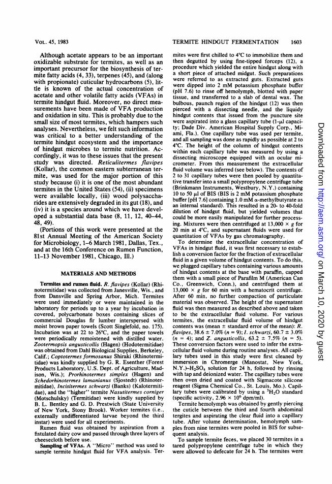
FIG. 1. Gas chromatograms of a standard mixtureof VFAs (4 mM each in BIS, top) and a diluted sampleof hindgut fluid from R. flavipes termites (bottom).Symbols: A, acetate; P, propionate; iB, isobutyrate;B, butyrate; MB, a-methylbutyrate (internal stan-dard); iV, isovalerate; V, valerate; and U, unknowncompounds.
chemicals were obtained from New England NuclearCorp., Boston, Mass., except for [U-14C]cellulose and[U-14C]hemicellulose, which were obtained from ICNPharmaceuticals, Inc., Irvine, Calif. The "4C-labeledcellulose and hemicellulose were repurified before useby the methods of Rapson (47) and Myhre and Smith(36), respectively.
RESULTSVFAs in termite hindguts and feces. Acetate
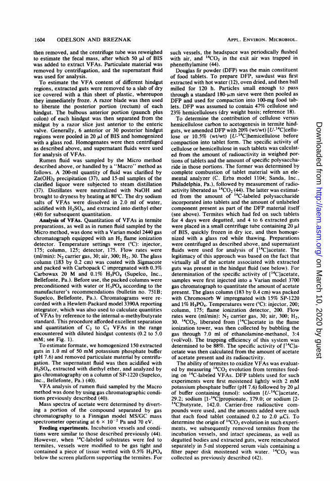
was the major VFA present in the hindgut fluidof R. flavipes. The compound was readily de-tected by gas chromatography (Fig. 1), and itsmass spectrum (Fig. 2) was virtually identical tothat of authentic acetic acid (16). The meanextracellular acetate pool size was 80.6 mM,which accounted for 94 mol% of all C, to C5VFAs (Table 1). Small amounts of propionateand butyrate were present, but their concentra-tion frequently fell below the limits of reliablequantitation. Consequently, the values for pro-pionate and butyrate reported in Table 1 wereonly from those analyses done on more concen-trated samples of hindgut fluid (i.e., pooled from>8 termites) which gave detector responseswithin quantifiable limits. Trace amounts of iso-butyrate, valerate, and isovalerate were some-times observed; however, formate was not de-tected. No significant differences in VFA
on March 10, 2020 by guest
http://aem.asm
.org/D
ownloaded from
1606 ODELSON AND BREZNAK
10
:0
0
0
._c
co
_
5
m/e
FIG. 2. Mass spectrum of acetic acid from hindgutfluid of R. flavipes termites.
content were observed between freshly collect-ed and laboratory-maintained R. flavipes, so thedata in Table 1 represent a pooled estimate fromall determinations. Limited experiments with Z.angusticollis and I. schwarzi revealed that ace-tate dominated the VFA pool in the hindgut fluidof these termites as well (Table 1).
Since the Micro method of sampling for VFAanalysis entailed manipulation of minuteamounts of hindgut fluid, a critical concern waspossible errors incurred through sample evapo-ration or VFA volatilization. However. whenbovine rumen fluid was used as a control, resultsobtained by the Micro sampling method com-pared favorably with those obtained by a Macromethod (Table 1). These in turn agreed well withpublished values for the VFA content of rumenfluid (24).As expected, acetate was the major VFA
present in homogenates of extracted guts of R.flavipes and occurred at a concentration of 18nmol per gut equivalent (Table 2). Since the
volume of a hindgut is about 0.7 ,u (48) andconsisted of 39% fluid (see above) containingacetate at 81 mM (Table 1), it could be calculat-ed that essentially all of the acetate in extractedguts was present in the hindgut fluid; littleexisted in intracellular pools of the hindgut mi-crobiota or gut tissue. When extracted guts weresectioned, the anterior region of the hindgut(i.e., paunch plus colon) was found to contain 19nmol of acetate per gut equivalent, whereas therectum contained only 0.4 nmol per gut equiva-lent (Table 2).
Acetate was also the major VFA in extractedguts of three other rhinotermitids examined andin N. corniger, and when dissections were per-formed, the compound occurred mainly in theanterior portion of the hindgut (Table 2). How-ever, when the acetate content of extracted gutswas normalized to body weight, a threefoldvariation was observed, ranging from 1.5nmol/mg (N. corniger) to 4.6 nmol/mg (R. fla-vipes) (Table 2).
In some analyses of hindgut fluid or extractedguts, gas chromatograms revealed minor peakswhich did not correspond to those of standardVFAs (Fig. 1, bottom). The compounds respon-sible for such peaks remain to be identified,although separate experiments indicated theywere not acetoacetate, P-hydroxybutyrate, lac-tate, or ethanol.Pooled feces, voided by 30 R. flavipes work-
ers over a 24-h period, contained only a traceamount of acetate which could not be accuratelyquantitated.These data indicated that: (i) acetate was
associated with, and probably produced in, thebulbous, microbe-packed anterior region of thehindgut; (ii) virtually all of the acetate present inthe gut was metabolized within R. flavipes andnot voided with feces; and (iii) the short segmentof midgut usually attached to extracted gutscontained little or no acetate.
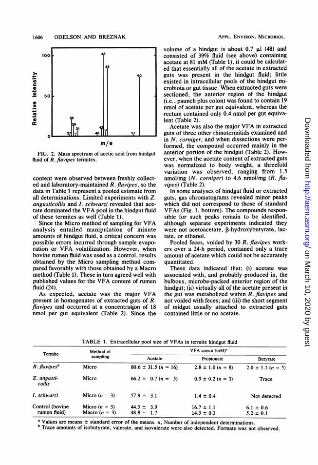
TABLE 1. Extracellular pool size of VFAs in termite hindgut fluid
Termite Method of VFA concn (mM)'sampling Acetate Propionate Butyrate
R.flavipesb Micro 80.6 ± 31.5 (n = 16) 2.8 ± 1.0 (n = 8) 2.0 + 1.1 (n = 5)
Z. angusti- Micro 66.2 ± 0.7 (n = 5) 0.9 ± 0.2 (n = 3) Trace
collisI. schwarzi Micro (n = 3) 57.9 ± 3.1 1.4 ± 0.4 Not detected
Control (bovine Micro (n = 3) 44.5 3.9 16.7 ± 1.1 6.1 ± 0.6
rumen fluid) Macro (n = 3) 48.8 1.7 14.3 ± 0.3 5.2 ± 0.1
a Values are means ± standard error of the means. n, Number of independent determinations.b Trace amounts of isobutyrate, valerate, and isovalerate were also detected. Formate was not observed.
10
~~~~~o ~ ~ 4
31 4
2 30 4 5
APPL. ENVIRON. MICROBIOL.
on March 10, 2020 by guest
http://aem.asm
.org/D
ownloaded from
TERMITE HINDGUT FERMENTATION 1607
TABLE 2. Acetate content of gut homogenates ofworker termites
Termite Acetate (nmol) per:cTerniite fresh wt Guepamro mg of
(mg) P Gut portion body wt
R.fla- 4.0 EG 11 18.3 ± 3.0 4.6 ± 0.8vipes AH 3 19.0 ± 2.6 4.8 ± 0.7
R 3 0.4 0.2 0.1 0.1
S. laman- 3.9 EG 2 7.6 2.0ianus AH 2 8.2 2.1
P. sim- 2.8 EG 2 6.5 2.3plex AH 3 5.8 ± 3.4 2.1 ± 1.2
C. formo- 3.0 EG 4 7.9 ± 0.6 2.6 ± 0.2sanus
N. cor- 2.2 EG 2 3.2 1.5nigera EG, Extracted gut; AH, anterior hindgut; R, rec-
tum.b n, Number of independent determinations.c Mean ± standard error of the mean, or mean of
two determinations as indicated.
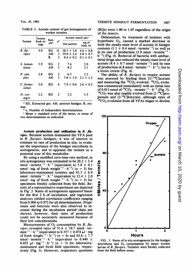
Acetate production and utilization in R. fla-vipes. Because acetate dominated the VFA poolin R. flavipes hindguts, it was of interest toestimate its rate of production in situ, to evalu-ate the importance of the hindgut microbiota toacetogenesis, and to appraise the termite as apotential user of this metabolite.By using a modified zero-time-rate method, in
situ acetogenesis was estimated to be 20.2 + 3.4nmol - termite-1 * h-1 (equivalent to 5.8 ± 0.1nmol - mg of fresh weight- . h-1) (n = 3) forlaboratory-maintained termites and 43.3 ± 6.9nmol - termite-1 * h-1 (equivalent to 12.4 ± 2.0nmol - mg of fresh weight-1 * h-1) (n = 3) forspecimens freshly collected from the field. Re-sults of a representative experiment are depictedin Fig. 3. Rates of acetogenesis appeared linearfor the first 2 h of incubation, and regressionanalyses yielded correlation coefficients rangingfrom 0.904 to 0.972 for all determinations. Propi-onate and butyrate were also observed to in-crease during the incubation period (data notshown); however, their rates of productioncould not be accurately measured because oftheir low concentrations.Measurements of 02 consumption by R. fla-
vipes revealed rates of 51.6 + 10.7 nmol - ter-mite-l h-1 (equivalent to 0.357 ± 0.074 ,ul - mgof fresh weight-' * h-1) (n = 6) and 63.6 ± 7.7nmol * termite-' * h-1 (equivalent to 0.440 ±0.053 ,ul - mg-' - h-1) (n = 3) for laboratory-maintained and fresh field specimens, respec-tively (Fig. 3). However, respiratory quotients
(RQs) were 1.00 to 1.05 regardless of the originof the insects.
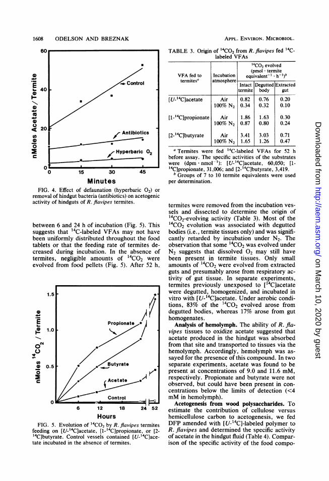
Defaunation, by treatment of termites withhyperbaric 02, caused a marked decrease inboth the steady-state level of acetate in hindgutcontents (1.1 + 0.4 nmol * termite-1) as well asin its rate of production (3.9 nmol * termite-1 -
h-1) (Fig. 4). Removal of bacteria with antibac-terial drugs also reduced the steady-state level ofacetate (8.4 + 0.7 nmol * termite-1) and its rateof production (6.8 nmol - termite* h-1), but toa lesser extent (Fig. 4).The ability of R. flavipes to respire acetate
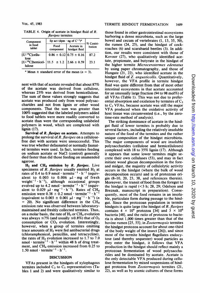
was assessed by feeding them [U-14C]acetateand measuring the 14Co2 evolved. 14Co2 evolu-tion commenced immediately with an initial rateof 0.013 nmol of 14CO2 * termite-1 - h-1 (Fig. 5).14CO2 was also readily evolved from [1-14C]pro-pionate and [2-14C]butyrate, although rates of?4C02 evolution from all VFAs began to decline
120
Oxygen
U
80 A tate
* 400 E *U
1 2Hours
FIG. 3. Rates of in situ acetogenesis by the hindgutmicrobiota and 02 consumption by intact workerlarvae of R. flavipes. Termites were freshly collectedfrom the field before assay.
VOL. 45, 1983
on March 10, 2020 by guest
http://aem.asm
.org/D
ownloaded from
1608 ODELSON AND BREZNAK
60
40)
40
20 csAntibioticsO4 -0
Hyperbaric 02
00 15 30 45
MinutesFIG. 4. Effect of defaunation (hyperbaric 02) or
removal of hindgut bacteria (antibiotics) on acetogenicactivity of hindguts of R. flavipes termites.
between 6 and 24 h of incubation (Fig. 5). Thissuggests that 14C-labeled VFAs may not havebeen uniformly distributed throughout the foodtablets or that the feeding rate of termites de-creased during incubation. In the absence oftermites, negligible amounts of 14CO2 wereevolved from food pellets (Fig. 5). After 52 h,
1.5
0~~~~~~~~
E Propionate*/
Btyrate
0) 0.5
*Acetate
Control
6 12 18 24 52
Hours
FIG. 5. Evolution of "4CO2 by R. flavipes termites
feeding on [U_14C]acetate, [1_14C]propionate, or [2-
14Cibutyrate. Control vessels contained [U_14C]ace-tate incubated in the absence of termites.
TABLE 3. Origin of 14CO2 from R. flavipes fed 14C-labeled VFAs
"4CO2 evolved(pmol * termite
VFA fed to Incubation equivalent-' *h-1).termiteSa atmosphere-
Intact Degutted Extractedtermite body gut
[U-14C]acetate Air 0.82 0.76 0.20100% N2 0.34 0.32 0.10
[1-14C]propionate Air 1.86 1.63 0.30100% N2 0.87 0.80 0.24
[2-14C]butyrate Air 3.41 3.03 0.71100% N2 1.65 1.26 0.47
a Termites were fed 14C-labeled VFAs for 52 hbefore assay. The specific activities of the substrateswere (dpm - nmol-'): [U-14C]acetate, 60,050; [1-4C]propionate, 31,006; and [2-14C]butyrate, 3,419.b Groups of 7 to 10 termite equivalents were used
per determination.
termites were removed from the incubation ves-sels and dissected to determine the origin of14CO2-evolving activity (Table 3). Most of the14Co2 evolution was associated with deguttedbodies (i.e., termite tissues only) and was signifi-cantly retarded by incubation under N2. Theobservation that some 14Co2 was evolved underN2 suggests that dissolved 02 may still havebeen present in termite tissues. Only smallamounts of 14CO2 were evolved from extractedguts and presumably arose from respiratory ac-tivity of gut tissue. In separate experiments,termites previously unexposed to [14C]acetatewere degutted, homogenized, and incubated invitro with [U-14C]acetate. Under aerobic condi-tions, 83% of the 14Co2 evolved arose fromdegutted bodies, whereas 17% arose from guthomogenates.
Analysis of hemolymph. The ability of R. fla-vipes tissues to oxidize acetate suggested thatacetate produced in the hindgut was absorbedfrom that site and transported to tissues via thehemolymph. Accordingly, hemolymph was as-sayed for the presence of this compound. In twoseparate experiments, acetate was found to bepresent at concentrations of 9.0 and 11.6 mM,respectively. Propionate and butyrate were notobserved, but could have been present in con-centrations below the limits of detection (<4mM in hemolymph).
Acetogenesis from wood polysaccharides. Toestimate the contribution of cellulose versushemicellulose carbon to acetogenesis, we fedDFP amended with [U-14C]-labeled polymer toR. flavipes and determined the specific activityof acetate in the hindgut fluid (Table 4). Compar-ison of the specific activity of the food compo-
APPL. ENVIRON. MICROBIOL.
on March 10, 2020 by guest
http://aem.asm
.org/D
ownloaded from
TERMITE HINDGUT FERMENTATION 1609
TABLE 4. Origin of acetate in hindgut fluid of R.flavipes termites
Component Sp act (dpm * ng ofC1C)ain food Food Acetate in butiontablet component hindgut fluid
[U-14C]cellu- 0.86 ± 0.12 0.75 ± 0.16 87.2lose
[U-14C]hemicel- 11.5 + 1.2 2.66 ± 0.59 23.1lulosea Mean ± standard error of the mean (n = 3).
nent with that of acetate revealed that about 87%of the acetate was derived from cellulose,whereas 23% was derived from hemicellulose.The sum of these values strongly suggests thatacetate was produced only from wood polysac-charides and not from lignin or other woodcomponents. That the sum was greater than100% suggested that the labeled polymers addedto food tablets were more readily converted toacetate than were the corresponding unlabeledpolymers in wood, which are complexed withlignin (17).
Survival of R. flavipes on acetate. Attempts toprolong the survival of R.flavipes on a cellulose-free diet with acetate were unsuccessful. Thiswas true whether defaunated or normally faunat-ed termites were used. In fact, termites feedingon sodium acetate or NaCl (control) generallydied faster than did those feeding on unamendedagarose.H2 and CH4 emission by R. flavipes. Live
specimens of R. flavipes usually emitted H2 atrates of 0.4 to 0.9 nmol * termite-1 * h-1 (equiv-alent to 0.003 to 0.006 RI.l - mg of freshweight-1 - h-1), although occasional groupsevolved up to 4.2 nmol * termite-1 * h-1 (equiv-alent to 0.029 ,ul * mg-'1 h-1). Rates of CH4emission were 0.38 0.2 nmol * termite-' * h-1(equivalent to 0.003 0.001 ,u * mg-1 - h-1) (n= 20). No significant difference in the CH4emission rate was observed between laboratory-maintained and freshly collected termites. Thus,on a molar basis, the rate of H2 or CH4 evolutionwas always c7% (and usually '0.8%) that of 02consumption or CO2 evolution. Interestingly,however, when a group of termites emittingtrace amounts of H2 were fed antibacterial drugs(chloramphenicol, penicillin, and tetracycline),the rates of H2 evolution increased to 1.7 to 7.0nmol * termite-1 * h-1 within 48 h of drug treat-ment, and CH4 emission increased from 0.25 to1.50 nmol - termite-1 * h-1.
DISCUSSIONVFAs present in the hindguts of xylophagous
termites included C2 to C5 representatives (Ta-bles 1 and 2) and were qualitatively similar to
those found in other gastrointestinal ecosystemsharboring a dense microbiota, such as the largebowel and cecum of vertebrates (1, 13, 35, 50),the rumen (24, 25), and the hindgut of cock-roaches (6) and scarabaeid beetles (3). In addi-tion, our results were consistent with those ofKovoor (27), who qualitatively identified ace-tate, propionate, and butyrate in the hindgut ofthe higher termite Microcerotermes edentatusby using paper chromatography, and those ofHungate (21, 22), who identified acetate in thehindgut fluid of Z. angusticollis. Quantitatively,however, the VFA profile in termite hindgutfluid was quite different from that of most otherintestinal ecosystems in that acetate accountedfor an unusually large fraction (94 to 98 mol%) ofall VFAs (Table 1). This was not due to prefer-ential absorption and oxidation by termites of C3to C5 VFAs, because acetate was still the majorVFA produced when the oxidative activity ofhost tissue was circumvented (i.e., by the zero-time-rate method of analysis).The striking dominance of acetate in the hind-
gut fluid of lower termites is probably due toseveral factors, including the relatively insolublenature of the food of the termites and the ratherunique composition of the hindgut microbiota.The major components of wood are insolublepolysaccharides (cellulose and hemicelluloses)complexed with 18 to 35% lignin (17). Althoughit appears that some lower termites might se-crete their own cellulases (53), and may in factinitiate wood glucan decomposition in the fore-and midgut, the majority of cellulolytic activityoccurs in the hindgut (where the bulk of wooddecomposition occurs) and is of protozoan ori-gin (8-10, 20, 23, 38, and references therein).Moreover, the transit offood from the foregut tothe hindgut is rapid (<3 h; 28, 29; Odelson andBreznak, manuscript in preparation). Conse-quently, most of the food remains in an insolu-ble, particulate form during passage to the hind-gut. Since the protozoan population in termitehindguts is quite large (the hindgut of R. flavipescontains 4 x 10⁴ protozoa [34] and 3 x 10⁶bacteria [48], and the ratio of protozoa to bacte-ria is about 1,000 times greater than that of thebovine rumen [25, 55]; in Zootermopsis termitesthe hindgut protozoa account for about one-thirdof the body weight of the insect [26]), and sincemost of the termite hindgut flagellates endocy-tose (and thereby sequester) wood particles asthey enter the hindgut, it follows that VFAproduction in the hindgut should reflect mainly aprotozoan fermentation of wood polysaccha-rides and be dominated by acetate. Acetate isthe only detectable VFA produced during cellu-lose fermentation by mixed suspensions of hind-gut protozoa from Zootermopsis termites (21,22), as well as by axenic cultures of these forms
VOL. 45, 1983
on March 10, 2020 by guest
http://aem.asm
.org/D
ownloaded from
1610 ODELSON AND BREZNAK
(56, 57), and a similar situation probably holdsfor cellulolytic protozoa from R. flavipes. Con-sistent with this interpretation is the drasticinhibition of acetogenesis in the hindgut of R.flavipes after defaunation (Fig. 4). Nevertheless,bacteria undoubtedly also produce acetate insitu. This inference is based on the moderatesuppression of hindgut acetogenesis after R.flavipes termites were fed antibacterial drugs(Fig. 4), as well as the recognized ability ofheterotrophic bacterial isolates to produce ace-
tate (and C, and C3 to C5 VFAs) in pure culture(42, 43, 48) and in two-species cocultures (49).However, it is impossible at this time to ascribethe exact quantitative contribution of protozoaor bacteria to acetogenesis in situ, because thesum of the acetogenic activity of defaunated R.flavipes and bacteria-free R. flavipes is signifi-cantly less than the acetogenic activity of con-trol termites (Fig. 4). Either one or both of thefollowing explanations for this observation are
possible. (i) The treatment used to remove pro-tozoa or bacteria (exposure of termites to hyper-baric 02 or to antibacterial drugs, respectively)has some deleterious effect on nontarget orga-nisms; or (ii) acetogenesis in normal R. flavipesinvolves a synergistic interaction between hind-gut protozoa and bacteria. Notwithstanding, itseems safe to conclude that protozoa dominateacetogenesis in R. flavipes hindguts, whereasbacteria are of secondary importance in thisparticular activity. Substrates for bacterial pro-duction of VFAs could include the small amountof soluble carbohydrate present in the wooditself (30), soluble intermediates secreted by theprotozoa (9, 56) or liberated from wood bytermite enzymes, or possibly CO2 and H2 (seebelow). True cellulolytic bacteria, i.e., bacteriacapable of degrading crystalline cellulose, ap-pear to be quantitatively insignificant in thehindgut of R. flavipes (48).
Results of our present studies with R. flavipes(family Rhinotermitidae) are consistent withHungate's (22) model for mutualistic celluloseutilization which was derived from his studieswith Zootermopsis species (family Hodotermiti-dae). First, protozoa appear to be primarilyresponsible for acetogenesis in the hindgut; sec-ond, rates of 02 consumption by R. flavipes (52to 64 nmol * termite-' * h-1) were approximate-ly twice that of hindgut acetogenesis (20 to 43nmol * termite-1 - h-1 for laboratory-main-tained and freshly collected termites, respective-ly). Since 2 mol of 02 is required for completeoxidation of acetate to 2CO2 and 2H20, it ap-peared that 77 to 100% of the energy require-ments of the termites could be met by oxidationof the acetate produced by the hindgut micro-biota. In support of this interpretation was thedemonstration of significant amounts of acetate
in R. flavipes hemolymph, as well as the abilityof termite tissues to readily respire acetate andother VFAs (Fig. 5; Table 3). We do not knowwhy the rates of acetogenesis in the hindguts offreshly collected R. flavipes were consistentlygreater than those of laboratory-maintainedspecimens. However, it seems likely that thefood on which the former were feeding beforeassay was more readily convertible to acetate(perhaps because it was partially decayed byfungi) than was the sound wood and paper towelmixture fed to laboratory specimens. Neverthe-less, the ability of both cellulose and hemicellu-lose to serve as substrates for acetogenesis bythe hindgut microbiota (Table 4) was in line withthe high digestibility of these compounds, butnot lignin, for R. flavipes (18).
Rates of 02 consumption by R. flavipes re-ported herein (0.357 to 0.440 ,ul * mg of freshweight-' * h-1) were similar to those of variousother termites, as summarized by Peakin andJosens (39), as well as that determined by La-Fage and Nutting (31) for Marginitermes hub-bardi. By contrast, present values were consid-erably lower than most of those determined forR. flavipes by Damaschke and Becker (summa-rized in reference 39). The reasons for thisdiscrepancy are not known. Rates of H2 andCH4 emissions by R. flavipes were also similarto those previously reported for various termitesincluding R. flavipes and R. tibialis (8, 15, 31,59). Although emission of such gases by termitesmight have a significant impact on our atmo-sphere globally (59), rates of H2 and CH4 emis-sion by R. flavipes were only about 0.7% that of02 consumption. Consequently, overall carbo-hydrate oxidation in R. flavipes closely approxi-mated the classical scheme: 100 (CH20) +10002 -- 100CO2 + 10OH20. Assuming an oxy-calorific equivalent of 5.05 mcal/,l of 02 con-sumed (39), our respirometric data indicate thatenergy flow through normally faunated, feedingworkers of R. flavipes would be 1.80 to 2.22mcal * mg-1 * h-1 at 230C.
If Hungate's model (22) for symbiotic cellu-lose degradation in lower termites is fundamen-tally valid, it must be amplified to accommodatethose termite species that evolve relatively littleH2 and CH4. For example, if symbiotic woodutilization in R. flavipes is envisioned to consistmainly of an anaerobic fermentation of glucan(nC6H1206) to acetate, C02, and H2 by protozoa(Table 5, reaction A), followed by termite oxida-tion of acetate (Table 5, reaction B), then anappreciable amount of reducing equivalents, asH2, should be evolved by termites (Table 5,reaction A + B). In fact, according to reaction A+ B, rates of H2 evolution should be equal tothat of 02 consumption and 66% that of CO2evolution, but they are almost always <1% of
APPL. ENVIRON. MICROBIOL.
on March 10, 2020 by guest
http://aem.asm
.org/D
ownloaded from
TERMITE HINDGUT FERMENTATION 1611
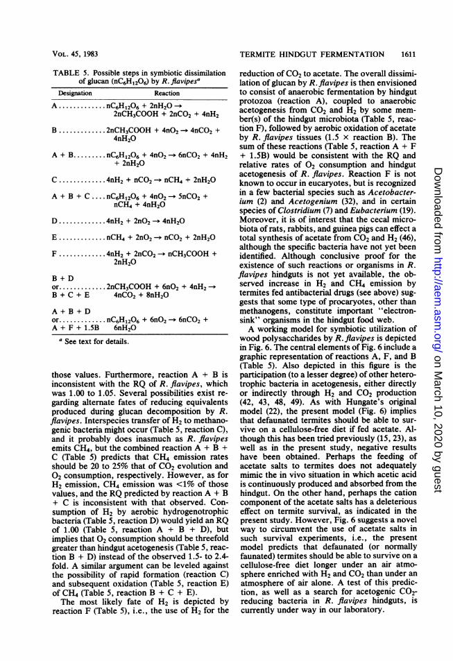
TABLE 5. Possible steps in symbiotic dissimilationof glucan (nC6Hl2O6) by R. flavipesa
Designation ReactionA .... .. nC6H1206 + 2nH20 -+
2nCH3COOH + 2nCO2 + 4nH2
B ...... 2nCH3COOH + 4nO2 -- 4nCO2 +4nH20
A + B...... nC6H1206 + 4nO2 -- 6nCO2 + 4nH2+ 2nH20
C ........ 4nH2 + nCO2 -* nCH4 + 2nH20
A + B + C ....nC6H1206+ 4nO2-5nCO2 +nCH4 + 4nH20
D .... .. 4nH2 + 2nO2 - 4nH20
E .... .. nCH4 + 2nO2 - nCO2 + 2nH20
F .... .. 4nH2 + 2nCO2 -. nCH3COOH +2nH20
B + Dor...... 2nCH3COOH + 6nO2 + 4nH2 -B + C + E 4nCO2 + 8nH20
A + B + Dor....... nC6H1206 + 6nO2 -+ 6nCO2 +A + F +1.5B 6nH20
a See text for details.
those values. Furthermore, reaction A + B isinconsistent with the RQ of R. flavipes, whichwas 1.00 to 1.05. Several possibilities exist re-garding alternate fates of reducing equivalentsproduced during glucan decomposition by R.flavipes. Interspecies transfer of H2 to methano-genic bacteria might occur (Table 5, reaction C),and it probably does inasmuch as R. flavipesemits CH4, but the combined reaction A + B +C (Table 5) predicts that CH4 emission ratesshould be 20 to 25% that of CO2 evolution and02 consumption, respectively. However, as forH2 emission, CH4 emission was <1% of thosevalues, and the RQ predicted by reaction A + B+ C is inconsistent with that observed. Con-sumption of H2 by aerobic hydrogenotrophicbacteria (Table 5, reaction D) would yield an RQof 1.00 (Table 5, reaction A + B + D), butimplies that 02 consumption should be threefoldgreater than hindgut acetogenesis (Table 5, reac-
tion B + D) instead of the observed 1.5- to 2.4-fold. A similar argument can be leveled againstthe possibility of rapid formation (reaction C)and subsequent oxidation (Table 5, reaction E)of CH4 (Table 5, reaction B + C + E).The most likely fate of H2 is depicted by
reaction F (Table 5), i.e., the use of H2 for the
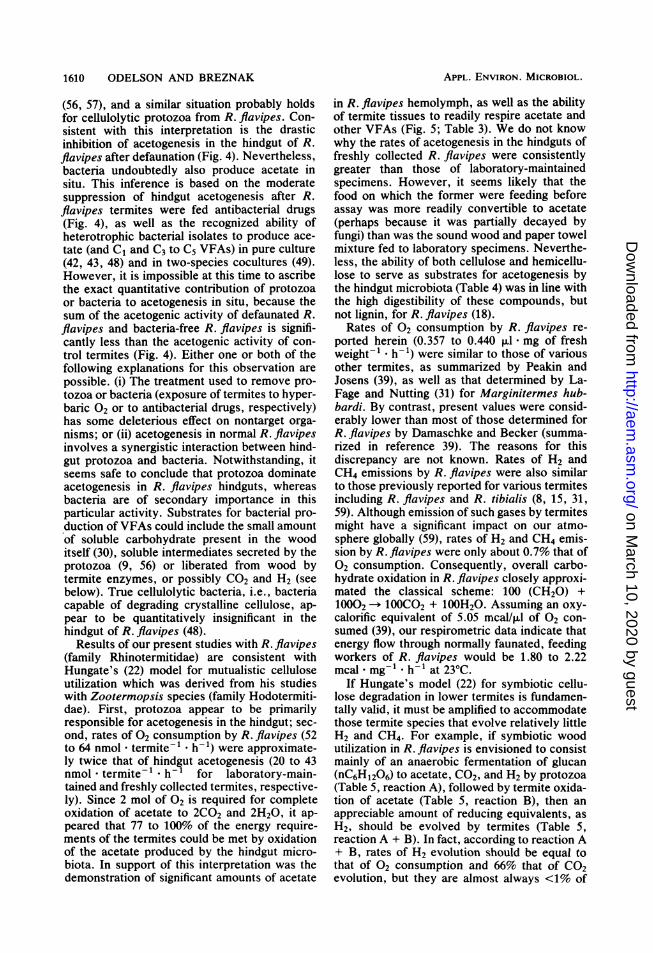
reduction of CO2 to acetate. The overall dissimi-lation of glucan by R. flavipes is then envisionedto consist of anaerobic fermentation by hindgutprotozoa (reaction A), coupled to anaerobicacetogenesis from CO2 and H2 by some mem-ber(s) of the hindgut microbiota (Table 5, reac-tion F), followed by aerobic oxidation of acetateby R. flavipes tissues (1.5 x reaction B). Thesum of these reactions (Table 5, reaction A + F+ 1.5B) would be consistent with the RQ andrelative rates of 02 consumption and hindgutacetogenesis of R. flavipes. Reaction F is notknown to occur in eucaryotes, but is recognizedin a few bacterial species such as Acetobacter-ium (2) and Acetogenium (32), and in certainspecies of Clostridium (7) and Eubacterium (19).Moreover, it is of interest that the cecal micro-biota of rats, rabbits, and guinea pigs can effect atotal synthesis of acetate from CO2 and H2 (46),although the specific bacteria have not yet beenidentified. Although conclusive proof for theexistence of such reactions or organisms in R.flavipes hindguts is not yet available, the ob-served increase in H2 and CH4 emission bytermites fed antibacterial drugs (see above) sug-gests that some type of procaryotes, other thanmethanogens, constitute important "electron-sink" organisms in the hindgut food web.A working model for symbiotic utilization of
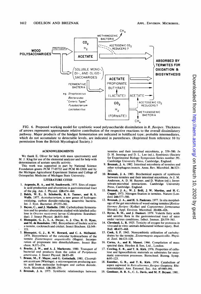
wood polysaccharides by R. flavipes is depictedin Fig. 6. The central elements of Fig. 6 include agraphic representation of reactions A, F, and B(Table 5). Also depicted in this figure is theparticipation (to a lesser degree) of other hetero-trophic bacteria in acetogenesis, either directlyor indirectly through H2 and CO2 production(42, 43, 48, 49). As with Hungate's originalmodel (22), the present model (Fig. 6) impliesthat defaunated termites should be able to sur-vive on a cellulose-free diet if fed acetate. Al-though this has been tried previously (15, 23), aswell as in the present study, negative resultshave been obtained. Perhaps the feeding ofacetate salts to termites does not adequatelymimic the in vivo situation in which acetic acidis continuously produced and absorbed from thehindgut. On the other hand, perhaps the cationcomponent of the acetate salts has a deleteriouseffect on termite survival, as indicated in thepresent study. However, Fig. 6 suggests a novelway to circumvent the use of acetate salts insuch survival experiments, i.e., the presentmodel predicts that defaunated (or normallyfaunated) termites should be able to survive on acellulose-free diet longer under an air atmo-sphere enriched with H2 and CO2 than under anatmosphere of air alone. A test of this predic-tion, as well as a search for acetogenic C02-reducing bacteria in R. flavipes hindguts, iscurrently under way in our laboratory.
VOL. 45, 1983
on March 10, 2020 by guest
http://aem.asm
.org/D
ownloaded from
1612 ODELSON AND BREZNAK
CH4METHANOGENIC
BACTERI
ACETOGENIC CO2PROTOZOAr- H2 --C-REURS
WOOD 2 -",REDUCERS ?
POLYSACCHARIDES ACETATE
(SOLUBLE MONO-:DI-, AND OLIGO-SACCHARIDES J ACETATE E :>
FERMENTIVE PROPIONATEBACTERIA BUTYRATE
eg. 5Sreplococcus (Bocterol'des (LACTATE) ACETATE -Enteric TypesFusobocter,'umLoc/oboci//us
CO2
H2
(FOR MATE)
ABSORBED BYTERMITES FOROXIDATION aBIOSYNTHESIS
ACETOGENIC CO2REDUCERS ?
METHANOGENICBACTERIA
CH4FIG. 6. Proposed working model for symbiotic wood polysaccharide dissimilation in R. flavipes. Thickness
of arrows represents approximate relative contribution of the respective reactions to the overall dissimilatorypathway. Major products of the hindgut fermentation are indicated in boldfaced type; probable intermediates,which do not accumulate to detectable levels, are indicated in parentheses. (Reprinted from reference 10 bypermission from the British Mycological Society.)
ACKNOWLEDGMENTS
We thank E. Oliver for help with mass spectrometry andM. J. Klug for use of the elemental analyzer and for help withdetermination of acetate specific activity.
This work was supported in part by National ScienceFoundation grants PCM 77-05732 and PCM 80-12026 and bythe Michigan Agricultural Experiment Station and College ofOsteopathic Medicine of Michigan State University.
LITERATURE CITED
1. Argenzio, R. A., and M. Southworth. 1975. Sites of organ-ic acid production and absorption in gastrointestinal tractof the pig. Am. J. Physiol. 228:454-460.
2. Balch, W. E., S. Schoberth, R. S. Tanner, and R. S.Wolfe. 1977. Acetobacterium, a new genus of hydrogen-oxidizing, carbon dioxide-reducing, anaerobic bacteria.Int. J. Syst. Bacteriol. 27:355-361.
3. Bayon, C., and J. Mathelin. 1980. Carbohydrate fermenta-tion and by-product absorption studied with labelled cellu-lose in Oryctes nasicornis larvae (Coleoptera: Scarabaei-dae). J. Insect Physiol. 26:833-840.
4. Blomquist, G. J., L. A. Dwyer, A. J. Chu, R. 0. Ryan,and M. de Renobales. 1982. Biosynthesis of linoleic acid ina termite, cockroach and cricket. Insect Biochem. 12:349-353.
5. Blomquist, G. J., R. W. Howard, and C. A. McDaniel.1979. Biosynthesis of the cuticular hydrocarbons of thetermite Zootermopsis angusticollis (Hagen). Incorpo-ration of propionate into dimethylalkanes. Insect Bio-chem. 9:371-374.
6. Bracke, J. W., and A. J. Markovetz. 1980. Transport ofbacterial end products from the colon of Periplanetaamericana. J. Insect Physiol. 26:85-89.
7. Braun, M., F. Mayer, and G. Gottschalk. 1981. Clostridi-um aceticum (Wieringa), a microorganism producing ace-tic acid from molecular hydrogen and carbon dioxide.Arch. Microbiol. 128:288-293.
8. Breznak, J. A. 1975. Symbiotic relationships between
termites and their intestinal microbiota, p. 559-580. InD. H. Jennings and D. L. Lee (ed.), Symbiosis (Societyfor Experimental Biology Symposium Series number 29).Cambridge University Press, Cambridge, England.
9. Breznak, J. A. 1982. Intestinal microbiota of termites andother xylophagous insects. Annu. Rev. Microbiol. 36:323-343.
10. Breznak, J. A. 1983. Biochemical aspects of symbiosisbetween termites and their intestinal microbiota. In J. M.Anderson, A. D. M. Rayner, and D. Walton (ed.), Inver-tebrate-microbial interactions. Cambridge UniversityPress, Cambridge, England.
11. Breznak, J. A., W. J. Brill, J. W. Mertins, and H. C.Coppel. 1973. Nitrogen fixation in termites. Nature (Lon-don) 244:577-580.
12. Breznak, J. A., and H. S. Pankratz. 1977. In situ morphol-ogy of the gut microbiota of wood-eating termites [Reticu-litermes flavipes (Kollar) and Coptotermes formosanusShiraki]. Appl. Environ. Microbiol. 33:406-426.
13. Byrne, B. M., and J. Dankert. 1979. Volatile fatty acidsand aerobic flora in the gastrointestinal tract of miceunder various conditions. Infect. Immun. 23:559-563.
14. Cleveland, L. R. 1925. Toxicity of oxygen for protozoa invivo and in vitro: animals defaunated without injury. Biol.Bull. 48:455-468.
15. Cook, S. F. 1943. Nonsymbiotic utilization of carbohy-drates by the termite, Zootermopsis angusticollis. Physi-ol. Zool. 16:123-128.
16. Cornu, A., and R. Massot. 1966. Compilation of massspectral data. Heyden & Son, Ltd., London.
17. Cowling, E. B., and T. K. Kirk. 1976. Properties of cellu-lose and lignocellulosic materials as substrates for enzy-matic conversion processes. Biotechnol. Bioeng. Symp.6:95-123.
18. Esenther, G. R., and T. K. Kirk. 1974. Catabolism ofaspen sapwood in Reticulitermesflavipes (Isoptera: Rhi-notermitidae). Ann. Entomol. Soc. Am. 67:989-991.
19. Genthner, B. R. S., C. L. Davis, and M. P. Bryant. 1981.
APPL. ENVIRON. MICROBIOL.
on March 10, 2020 by guest
http://aem.asm
.org/D
ownloaded from
TERMITE HINDGUT FERMENTATION 1613
Features of rumen and sewage sludge strains of Euibacter-ium limosum, a methanol- and H,-CO2-utilizing species.Appl. Environ. Microbiol. 42:12-19.
20. Honigberg, B. M. 1970. Protozoa associated with termitesand their role in digestion, p. 1-36. In K. Krishna andF. M. Weesner (ed.), Biology of termites. AcademicPress, Inc., New York.
21. Hungate, R. E. 1939. Experiments on the nutrition ofZootermopsis. III. The anaerobic carbohydrate dissimila-tion by the intestinal protozoa. Ecology 20:230-245.
22. Hungate, R. E. 1943. Quantitative analyses on the cellu-lose fermentation by termite protozoa. Ann. Entomol.Soc. Am. 36:730-739.
23. Hungate, R. E. 1946. The symbiotic utilization of cellu-lose. J. Elisha Mitchell Sci. Soc. 62:9-24.
24. Hungate, R. E. 1966. The rumen and its microbes. Aca-demic Press, Inc., New York.
25. Hungate, R. E. 1975. The rumen microbial ecosystem.Annu. Rev. Ecol. Syst. 6:39-66.
26. Katzin, L. I., and H. Kirby, Jr. 1939. The relative weightsof termites and their protozoa. J. Parasitol. 25:444-445.
27. Kovoor, J. 1967. Presence d'acides gras volatils dans lapanse d'un termite superieur (Microcerotermes edentatusWas., Amitermitinae). C.R. Acad. Sci. (Paris) 264:486-488.
28. Kovoor, J. 1967. Etude radiographique du transit intesti-nal chez un termite superieur. Experientia 23:820-821.
29^. Krishna, S. S., and N. B. Singh. 1968. Sugar-dye move-ment through the alimentary canal of Odontotermes obe-sus (Isoptera: Termitidae). Ann. Entomol. Soc. Am.61:230.
30. LaFage, J. P., and W. L. Nutting. 1978. Nutrient dynam-ics of termites, p. 165-232. In M. V. Brian (ed.), Produc-tion ecology of ants and termites. Cambridge UniversityPress, Cambridge, England.
31. LaFage, J. P., and W. L. Nutting. 1979. Respiratory gasexchange in the dry-wood termite, Marginitermes hub-bardi (Banks) (Isoptera: Kalotermitidae). Sociobiology4:257-267.
32. Leigh, J. A., F. Mayer, and R. S. Wolfe. 1981. Acetogeni-um kivui, a new thermophilic hydrogen-oxidizing, aceto-genic bacterium. Arch. Microbiol. 129:275-280.
33. Mauldin, J. K. 1982. Lipid synthesis from ['4C1-acetate bytwo subterranean termites, Reticulitermes flavipes andCoptotermes formosanus. Insect Biochem. 12:193-199.
34. Mauldin, J. K., and N. M. Rich. 1980. Effect of chlortetra-cycline and other antibiotics on protozoan numbers in theeastern subterranean termite. J. Econ. Entomol. 73:123-128.
35. McNeil, N. I., J. H. Cummings, and W. P. T. James.1978. Short chain fatty acid absorption by the human largeintestine. Gut 19:819-822.
36. Myhre, D. V., and F. Smith. 1960. Constitution of thehemicellulose of alfalfa (Medicago sativa). Hydrolysis ofhemicellulose and identification of neutral and acidiccomponents. J. Agric. Food Chem. 8:359-364.
37. Neish, A. C. 1952. Analytical methods for bacterial fer-mentations, report no. 46-8-3 (2nd revision). NationalResearch Council of Canada, Saskatoon, Saskatchewan.
38. O'Brien, R. W., and M. Slaytor. 1982. Role of microorga-nisms in the metabolism of termites. Aust. J. Biol. Sci.35:239-262.
39. Peakin, G. J., and G. Josens. 1978. Respiration and energyflow, p. 111-163. In M. V. Brian (ed.), Production ecolo-gy of ants and termites. Cambridge University Press,Cambridge, England.
40. Potrikus, C. J., and J. A. Breznak. 1977. Nitrogen-fixingEnterobacter agglomerans isolated from guts of wood-eating termites. Appi. Environ. Microbiol. 33:392-399.
41. Potrikus, C. J., and J. A. Breznak. 1980. Uric acid inwood-eating termites. Insect Biochem. 10:19-27.
42. Potrikus, C. J., and J. A. Breznak. 1980. Uric acid-de-grading bacteria in guts of termites [Reticulitermes fla-vipes (Kollar)]. Appl. Environ. Microbiol. 40:117-124.
43. Potrikus, C. J., and J. A. Breznak. 1980. Anaerobic deg-radation of uric acid by gut bacteria of termites. Appl.Environ. Microbiol. 40:125-132.
44. Potrikus, C. J., and J. A. Breznak. 1981. Gut bacteriarecycle uric acid nitrogen in termites: a strategy fornutrient conservation. Proc. Natl. Acad. Sci. U.S.A.78:4601-4605.
45. Prestwich, G. D., R. W. Jones, and M. S. Collins. 1981.Terpene biosynthesis by nasute termite soldiers (Isoptera:Nasutitermitinae). Insect Biochem. 11:331-336.
46. Prins, R. A., and A. Lankhorst. 1977. Synthesis of acetatefrom CO2 in the cecum of some rodents. FEMS Microbiol.Lett. 1:255-258.
47. Rapson, W. H. 1963. Cellulose from bleached wood pulp,p. 22-24. In R. L. Whistler (ed.), Methods in carbohy-drate chemistry, vol. 3. Academic Press, Inc., New York.
48. Schultz, J. E., and J. A. Breznak. 1978. Heterotrophicbacteria present in hindguts of wood-eating termites [Reti-culitermes flavipes (Kollar)]. Appl. Environ. Microbiol.35:930-936.
49. Schultz, J. E., and J. A. Breznak. 1979. Cross-feeding oflactate between Streptococcus lactis and Bacteroides sp.isolated from termite hindguts. Appl. Environ. Microbiol.37:1206-1210.
50. Sudo, S. Z., and G. E. Duke. 1980. Kinetics of absorptionof volatile fatty acids from the ceca of domestic turkeys.Comp. Biochem. Physiol. 67A:231-237.
51. Uffen, R. L. 1976. Anaerobic growth of a Rhodopseudo-monas species in the dark with carbon monoxide as solecarbon and energy substrate. Proc. Nati. Acad. Sci.U.S.A. 73:3298-3302.
52. Umbreit, W. W., R. H. Burris, and J. F. Stauffer. 1964.Manometric techniques. Burgess Publishing Co., Minne-apolis.
53. Veivers, P. C., A. M. Musca, R. W. O'Brien, and M.Slaytor. 1982. Digestive enzymes of the salivary glandsand gut of Mastotermes darviniensis. Insect Biochem.12:35-40.
54. Weesner, F. M. 1965. The termites of the United States, ahandbook. The National Pest Control Association. Eliza-beth, N.J.
55. Wolin, M. J. 1979. The rumen fermentation: a model formicrobial interactions in anaerobic ecosystems. Adv. Mi-crob. Ecol. 3:49-77.
56. Yamin, M. A. 1980. Cellulose metabolism by the termiteflagellate Trichomitopsis termopsidis. Appi. Environ. Mi-crobiol. 39:859-863.
57. Yamin, M. A. 1981. Cellulose metabolism by the flagellateTrichonympha from a termite is independent of endosym-biotic bacteria. Science 211:58-59.
58. Yamin, M. A., and W. Trager. 1979. Cellulolytic activityof an axenically-cultivated termite flagellate, Trichomitop-sis termopsidis. J. Gen. Microbiol. 113:417-420.
59. Zimmerman, P. R., J. P. Greenberg, S. 0. Wandiga, andP. J. Crutzen. 1982. Termites: a potentially large source ofatmospheric methane, carbon dioxide, and molecular hy-drogen. Science 218:563-565.
VOL. 45, 1983
on March 10, 2020 by guest
http://aem.asm
.org/D
ownloaded from