Embed Size (px)
Citation preview
Volar capitate dislocation: a case report
Christina M. Ward
Published online: 25 September 2013# American Association for Hand Surgery 2013
Abstract We present a rare injury involving a volar disloca-tion of the distal capitate with secondary median nervecompression. The injury was not recognized on theinitial radiographs resulting in a delayed diagnosis andtreatment. A satisfactory outcome was achieved follow-ing open reduction and internal fixation via dorsal andpalmar approaches. This case illustrates the importanceof careful review of radiographs for evidence of intercarpalinjuries.
Introduction
Carpal–metacarpal (CMC) joint dislocations are uncommoninjuries that usually result from high-energy trauma. Mostoften, the metacarpal base dislocates dorsal to the carpus,while the normal intercarpal relationships are maintained [5].Multiple CMC joint dislocations have been reported in anumber of cases. Although the injury can be identified onplain radiographs, 15 of 21 CMC dislocations were missed oninitial presentation in one series [3], while diagnosis wasdelayed in 4 of 20 patients in another series [5].
We present an unusual case of volar dislocation of the distalend of the capitate at the capitate–metacarpal joint that wasmissed during the initial emergency room evaluation. Thisinjury involved both disruption of the capitate–metacarpaljoint and the intercarpal ligaments of the distal carpal row,and was associated with a minimally displaced fracture of thetrapezoid. To our knowledge, this injury pattern has not beenpreviously reported.
Case Report
A 55-year-old right-hand-dominant male fell one story from aladder while shingling a roof. He landed on his outstretchedleft hand resulting in hand pain, swelling, and numbness andparesthesias that were worst in his thumb and index finger. Hepresented to an emergency room the day of the injury andposteroanterior (PA) and lateral radiographs of the left wristwere obtained (Fig. 1a, b). The radiologist identified a “possiblecapitate fracture.” The patient received a removable wrist splintand was discharged without seeing an orthopedic surgeon orhand surgeon. Nine days later, he saw his primary care physi-cian who recommended a wrist CT due to the severity of hiswrist pain and swelling and referred the patient to see anorthopedic surgeon. The patient was not a native Englishspeaker and had no health insurance and thus had difficultylocating an orthopedic surgeon willing to see him. The patientpresented to our clinic 17 days after the injury with continuedsevere pain, swelling, finger stiffness, and numbness.
On exam, the patient had a swollen hand and wrist. He hadminimal active motion of the digits but, when encouraged,could fire all finger flexors and extensors. He had significantlydiminished sensation in a median nerve distribution, with two-point discrimination >15 mm in his thumb, index, and longfingers. The hand was well perfused and his skin was intact.
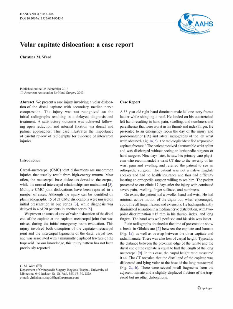
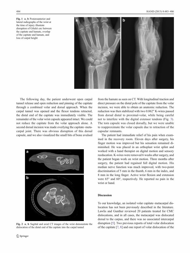
Plain radiographs obtained at the time of presentation showa break in Gilula's arc [2] between the capitate and hamate(Fig. 1a), as well as overlap between the ulnar capitate andradial hamate. There was also loss of carpal height. Typically,the distance between the proximal edge of the lunate and thedistal end of the capitate is equal to half the length of the longmetacarpal [9]. In this case, the carpal height ratio measured0.44. The CT revealed that the distal end of the capitate wasdislocated and lying volar to the base of the long metacarpal(Fig. 2a, b). There were several small fragments from theadjacent hamate and a slightly displaced fracture of the trap-ezoid but no other dislocations.
C. M. Ward (*)Department of Orthopaedic Surgery, Regions Hospital, University ofMinnesota, 640 Jackson St., St. Paul, MN 55130, USAe-mail: [email protected]
HAND (2013) 8:483–486DOI 10.1007/s11552-013-9545-2
The following day, the patient underwent open carpaltunnel release and open reduction and pinning of the capitatethrough a combined volar and dorsal approach. When thecarpal tunnel was opened and the flexor tendons retracted,the distal end of the capitate was immediately visible. Theremainder of the volar wrist capsule appeared intact. We couldnot reduce the capitate from the volar approach alone. Asecond dorsal incision was made overlying the capitate–meta-carpal joint. There was obvious disruption of this dorsalcapsule, and we also visualized the small bits of bone avulsed
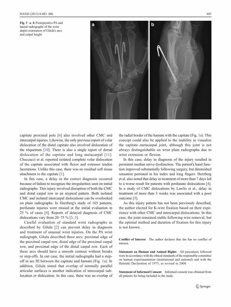
from the hamate as seen on CT. With longitudinal traction anddirect pressure on the distal pole of the capitate from the volarincision, we were able to obtain an anatomic reduction. Thereduction was then stabilized with two 0.062″ K-wires passedfrom dorsal distal to proximal-volar, while being carefulnot to interfere with the digital extensor tendons (Fig. 3).The torn capsule was closed dorsally, but we were unableto reapproximate the volar capsule due to retraction of thecapsular remnants.
The patient had immediate relief of his pain when exam-ined in the recovery room. Eleven days after surgery, hisfinger motion was improved but his sensation remained di-minished. He was placed in an orthoplast wrist splint andworked with a hand therapist on digital motion and sensoryreeducation. K-wires were removed 6 weeks after surgery, andthe patient began work on wrist motion. Three months aftersurgery, the patient had regained full digital motion. Hismedian nerve function was much improved, with two-pointdiscrimination of 5 mm in the thumb, 6 mm in the index, and8 mm in the long finger. Active wrist flexion and extensionwere 65° and 60°, respectively. He reported no pain in thewrist or hand.
Discussion
To our knowledge, an isolated volar capitate–metacarpal dis-location has not been previously described in the literature.Lawlis and Gunther reviewed 20 patients treated for CMCdislocations, and in all cases, the metacarpal was dislocateddorsal to the carpus, and there was no associated intercarpaldisruption [5]. Two previous reports of total volar dislocationof the capitate [7, 8] and one report of volar dislocation of the
Fig. 1 a , b Posteroanterior andlateral radiographs of the wrist atthe time of injury illustratedisruption of Gilula's arc betweenthe capitate and hamate, overlapof the capitate and hamate, andloss of carpal height
Fig. 2 a , b Sagittal and axial CT images of the wrist demonstrate thedislocation of the distal end of the capitate into the carpal tunnel
484 HAND (2013) 8:483–486
capitate proximal pole [6] also involved other CMC andintercarpal injuries. Likewise, the only previous report of volardislocation of the distal capitate also involved dislocation ofthe triquetrum [10]. There is also a single report of dorsaldislocation of the capitate and long metacarpal [11].Checcucci et al. reported isolated complete volar dislocationof the capitate associated with flexor and extensor tendonlacerations. Unlike this case, there was no residual soft tissueattachment to the capitate [1].
In this case, a delay in the correct diagnosis occurredbecause of failure to recognize the irregularities seen on initialradiographs. This injury involved disruption of both the CMCand distal carpal row in an atypical pattern. Both isolatedCMC and isolated intercarpal dislocations can be overlookedon plain radiographs. In Herzberg's study of 165 patients,perilunate injuries were missed at the initial evaluation in25 % of cases [4]. Reports of delayed diagnosis of CMCdislocations vary from 20–75 % [3, 5].
Careful evaluation of standard wrist radiographs asdescribed by Gilula [2] can prevent delay in diagnosisand treatment of unusual wrist injuries. On the PA wristradiograph, Gilula described three arcs: proximal edge ofthe proximal carpal row, distal edge of the proximal carpalrow, and proximal edge of the distal carpal row. Each ofthese arcs should have a smooth contour without breaksor step-offs. In our case, the initial radiographs had a step-off in arc III between the capitate and hamate (Fig. 1a). Inaddition, Gilula stated that overlap of normally parallelarticular surfaces is another indication of intercarpal sub-luxation or dislocation. In this case, there was an overlap of
the radial border of the hamate with the capitate (Fig. 1a). Thisconcept could also be applied to the inability to visualizethe capitate–metacarpal joint, although this joint is notalways distinguishable on wrist plain radiographs due towrist extension or flexion.
In this case, delay in diagnosis of the injury resulted inpersistent median nerve dysfunction. The patient's hand func-tion improved substantially following surgery, but diminishedsensation persisted in his index and long fingers. Herzberget al. also noted that delay in treatment of more than 7 days ledto a worse result for patients with perilunate dislocations [4].In a study of CMC dislocations by Lawlis et al., delay intreatment of more than 3 weeks was associated with a pooroutcome [5].
As this injury pattern has not been previously described,the author elected for K-wire fixation based on their expe-rience with other CMC and intercarpal dislocations. In thiscase, the joint remained stable following wire removal, butthe optimal method and duration of fixation for this injuryis not known.
Conflict of Interest The author declares that she has no conflict ofinterest.
Statement on Human and Animal Rights All procedures followedwere in accordance with the ethical standards of the responsible committeeon human experimentation (institutional and national) and with theHelsinki Declaration of 1975, as revised in 2008.
Statement of Informed Consent Informed consent was obtained fromall patients for being included in the study.
Fig. 3 a , b Postoperative PA andlateral radiographs of the wristdepict restoration of Gilula's arcsand carpal height
HAND (2013) 8:483–486 485

References
1. Checcucci G, Bigazzi P, ZucchiniM, CerusoM. Isolated complete volardislocation of the capitate: a case report. Hand Surg. 2011;16:353–6.
2. Gilula LA. Carpal injuries: analytic approach and case exercises. AmJ Roentgenol. 1979;133:503–17.
3. Henderson JJ, Arafa MAM. Carpometacarpal dislocation: an easilymissed diagnosis. J Bone Joint Surg Br. 1987;69:212–4.
4. Herzberg G, Comtet JJ, Linscheid RL, Amadio PC, Cooney WP,Stalder J. Perilunate dislocations and fracture-dislocations: a multi-center study. J Hand Surg [Am]. 1993;18:768–79.
5. Lawlis JF, Gunther SF. Carpometacarpal dislocations: long-termfollow-up. J Bone Joint Surg Am. 1991;73:52–9.
6. Lee JH, Ehara S, Furumachi K. Volar dislocation of the capitate.Radiat Med. 1999;17:363–4.
7. Lowrey DG, Moss SH, Wolff TW. Volar dislocation of the capitate. JBone Joint Surg Am. 1984;66:611–3.
8. Ruijters R, Kortmann J. A case of translunate luxation of the carpus.Acta Orthop Scand. 1988;59:461–3.
9. Schuind FA, Linscheid RL, An KN, Chao EY. A normal data base ofposteroanterior roentgenographic measurements of the wrist. J BoneJoint Surg Am. 1992;74:1418–29.
10. Uhl RL, Wickline A. Volar dislocation of the distal capitate: a reportof a case. Hand Surg. 1996;1:177–80.
11. Walker RH, Pradhan R. Dorsal dislocation of the capitate. J HandSurg (Br). 2000;25:403–5.
486 HAND (2013) 8:483–486