Embed Size (px)
Citation preview
ISSN 1348-8945
JAPANESE SOCIETY OFTROPICAL MEDICINE
Tropical Medicineand Health
Vol.34 No.04 DECEMBER 2006
ISSN 1348-8945
Trop.Med.Health
Tropical Medicineand Health
Vol.34No.04
DECEMBER 2006
Tro
pic
al M
ed
icin
e a
nd
He
alth
Vo
l.34
N
o.0
4 D
EC
EM
BER
2006
JA
PA
NESE S
OC
IETY
OF T
RO
PIC
AL M
ED
ICIN
E
日本熱帯医学会表紙�-� CMYK 稲 平島 宮副 宮副
【CONTENTS】Reviews
Emerging diseases in Indonesia: control and challenges
Kandun IN 141
Roles of Multi-Country Networking in Prevention and Control of Emerging and Re-emerging
Infections
Sawanpanyalert P 149
Exploring Fresh Collaborative Initiatives for Combating Infectious Diseases in the Philippines
Elio-Villa LP 153
Original articles
A new species of Simulium (Nevermannia) from the Ogasawara (Bonin) Islands,
Japan (Diptera: Simuliidae)
Takaoka H and Saito K 155
Uvulectomy and other traditional healing practices:
Traditional healers’ perceptions and practices in a Congolese refugee camp in Tanzania
Kunii O, Tanaka Y, Lewis A and Wakai S 159
Population Polymorphism of Trypanosoma cruzi in Latin America indicated by
Proteome analysis and by in vitro amastigote proliferation
Mu J, Sone T, Yanagi T, Tada I, Kikuchi M and Hirayama K 167
背は3mm(決定:宮副)
ISSN 1348-8945
JAPANESE SOCIETY OFTROPICAL MEDICINE
Tropical Medicineand Health
Vol.34 No.04 DECEMBER 2006
ISSN 1348-8945
Trop.Med.Health
Tropical Medicineand Health
Vol.34No.04
DECEMBER 2006
Tro
pic
al M
ed
icin
e a
nd
He
alth
Vo
l.34
N
o.0
4 D
EC
EM
BER
2006
JA
PA
NESE S
OC
IETY
OF T
RO
PIC
AL M
ED
ICIN
E
日本熱帯医学会表紙�-� CMYK 稲 平島 宮副 宮副
【CONTENTS】Reviews
Emerging diseases in Indonesia: control and challenges
Kandun IN 141
Roles of Multi-Country Networking in Prevention and Control of Emerging and Re-emerging
Infections
Sawanpanyalert P 149
Exploring Fresh Collaborative Initiatives for Combating Infectious Diseases in the Philippines
Elio-Villa LP 153
Original articles
A new species of Simulium (Nevermannia) from the Ogasawara (Bonin) Islands,
Japan (Diptera: Simuliidae)
Takaoka H and Saito K 155
Uvulectomy and other traditional healing practices:
Traditional healers’ perceptions and practices in a Congolese refugee camp in Tanzania
Kunii O, Tanaka Y, Lewis A and Wakai S 159
Population Polymorphism of Trypanosoma cruzi in Latin America indicated by
Proteome analysis and by in vitro amastigote proliferation
Mu J, Sone T, Yanagi T, Tada I, Kikuchi M and Hirayama K 167
背は3mm(決定:宮副)

EMERGING DISEASES IN INDONESIA:CONTROL AND CHALLENGES
I NYOMAN KANDUN
Recieved 31, October, 2006
ABSTRACT: Infectious diseases remain an important cause of morbidity and mortality in Indonesia. The reduc-tion, elimination, and eradication of infectious diseases have been the subject of numerous meetings and publichealth initiatives for decades. The malaria, yaws and other communicable disease eradication programs of earlieryears, although unsuccessful, contributed greatly to an understanding of the difficulties faced in trying to achievegoal of disease control. The reemergence of old infectious diseases, along with the emergence of new diseases suchas SARS, Avian Influenza and the development of antimicrobial resistance, pose significant challenges to publichealth.
BACKGROUND
The control, elimination and eradication of human dis-eases have been the subject of numerous public health inter-ventions and discussions for decades. The eradication ofsmallpox was declared on May 8, 1980 at the 33rd WorldHealth Assembly, and was followed soon after by polio-myelitis and other eradicable diseases. Although many dis-eases eradication program in the past were unsuccessful, thelessons learnt contributed to an understanding about thecomplexities and difficulties faced in trying to achieve theultimate goal of disease control. An understanding of thenatural history of a disease, including multiple causationand biological, sociopolitical and economic issues shedslight on the public health interventions available in dealingwith communicable diseases and their containment.
Indonesia stretches from west to east, with an area of1.9 million km2and a population of 230 million. In develop-ing countries like Indonesia, health resources have alwaysbeen limited, and decisions as to the most preferable andcost effective intervention programs have to be targeted topriority diseases. Disease control can be defined as the re-duction of diseases in a defined geographical area as a resultof deliberate control efforts. This definition should be fur-ther quantified to indicate the level of disease reduction tobe achieved. Disease elimination and eradication are the ul-timate goals of any public health intervention starting fromdisease control. Communicable diseases are still seriouspublic health problems, killing and causing suffering formillions of people in Indonesia, especially the most vulner-able groups, i.e. the poor, women and children. Communi-
cable diseases exert a negative effect on development andplace a burden on the economy of the individual and thecountry as a result of the huge costs of treatment and con-trol.
Technical solutions combined with strategies to mobi-lize all levels of society from high level decisions-makers tocommunities and families will ensure the effective controland prevention of communicable diseases. The following isa brief description of re-emerging diseases, newly emergingdiseases, and the challenges encountered in their control.
DENGUE HEMORRHAGIC FEVERAn epidemic of dengue fever / dengue hemorrhagic fe-
ver (DF/DHF) started in the Southeast Asia region after theSecond World War. The first case of DF/DHF in Indonesiawas reported in 1968 from Jakarta and Surabaya. Since thenthe frequency and magnitude of DF/DHF outbreaks have in-creased dramatically. As the principal mosquito vector,Aedes aegypti and the viruses (D1, D2, D3, D4)that cause DF/DHF, expanded their geographical and age distribution na-tionally. Figure 1 presents the distribution of DHF by prov-ince while Figure 2 presents the seasonal variation of DHFcases and deaths in 2004-2005 and a comparison of DHFcases in 2005 and 2006.
The epidemiologic trends in recent decades demon-strate that the prevention and control of dengue virus trans-mission have failed. There is no vaccine available for den-gue viruses nor effective mosquito control programs dealingwith breeding places. Emphasis has been placed on diseasesurveillance and immediate response using space sprays tar-geting adult mosquitoes in the affected focal areas. Now
Tropical Medicine and Health Vol. 34 No. 4, 2006, pp. 141-147Copyright� 2006 by The Japanese Society of Tropical Medicine
Disease Control and Environmental Health, Ministry of Health, Jakarta, Indonesia
141

strategies are being revised so as to focus on community in-volvement in the elimination of breeding places and to im-prove partnership and professionalism among programmanagers at all levels.
A decentralized integrated approach that targets larvalmosquitoes is being implemented for effective Aedes ae-gypti control. Along with high-level political commitment,community involvement is an important prerequisite forvector control.
TUBERCULOSIS (TB)Tuberculosis (TB) is a bacterial disease caused by My-
cobacterium tuberculosis transmitted primarily by airbornedroplets. Infection occurs when susceptible persons inhaleinfected droplets produces by coughs and sneezes of per-sons with active lung TB. TB causes suffering for millionsof people, particularly the poor, women, children, and HIV/AIDS patients. More than half a million new TB cases areestimated to occur every year, with 300-400 TB deathsdaily. To cope with this problem, the DOTS strategy hasbeen implemented since 1998 and aims to achieve a casedetection rate (CDR) of 70% and cure rate or success rate(SR) of 85% by the end of 2006. Figure 3 and 4 show thedate on CDR and SR from 1997 to 2005.
Figure 1. INCIDENCE OF DENGUE HEMORRHAGIC FEVER (DHF)BY PROVINCE IN 2005
a. Trends of Dengue Hemorrhagic Fever Cases & Deaths in2004-2005
b. NUMBER OF DENGUE HEMORRHAGIC FEVERCASE IN 2005-2006 (UP TO 19 June 2006)
Figure 2.
142

TB is diagnosed mainly if acid-fast- bacilli (AFB) arefound in the sputum, body fluids or tissue in combinationwith clinical symptoms. In special cases, chest radiographand PPD skin test abnormalities are also taken into consid-eration The principal challenges in TB control include ob-taining and continuing the political commitment of decisionmakers, and international funding for the support of TBcontrol efforts. Multiple drug resistance is also a challengethat will have to be addressed in the near future.
MALARIAMost malaria eradication programs in the past have
been unsuccessful. Figure 5 shows the malaria endemicityin 2005 and figure 6 the malaria situation in Indonesia from1989 to 2005.
Deforestation, mining, active rapid population migra-
Figure 3. Number of Cases Detected 1987-2005 in the TBCControl Program
Figure 4. CDR and SR of AFB Positive Cases in Indonesia1997-2005 Figure 6. MALARIA SITUATION IN INDONESIA 1989-
2005
Figure 5. MALARIA ENDEMICITY DISTRIBUTION IN 2005
143
tion and other development activities have contributed tothe resurgence of malaria in Indonesia. The roll back ma-laria strategy, which was implemented as GEBRAK Malariain 2000, focuses on partnership. Malaria control strategyprimarily focuses on vector and disease surveillance, earlydiagnosis and prompt treatment, integrated vector manage-ment and community participation.
The principal challenges to malaria control programsare effective coordination, long-term sustainability of vectorcontrol efforts, population migration and environmentalchanges. Anti-malaria drug resistance and insecticide resis-tance are also primary challenges for the future.
POLIOPolio eradication has been targeted for the year 2008.
The polio eradication campaign is a good example of multi-partner leadership in that partners come from a broad arrayof organizations such as Rotary International, WHOUNICEF, CDC-Atlanta, USAID, AUSAID, and WorldBank.
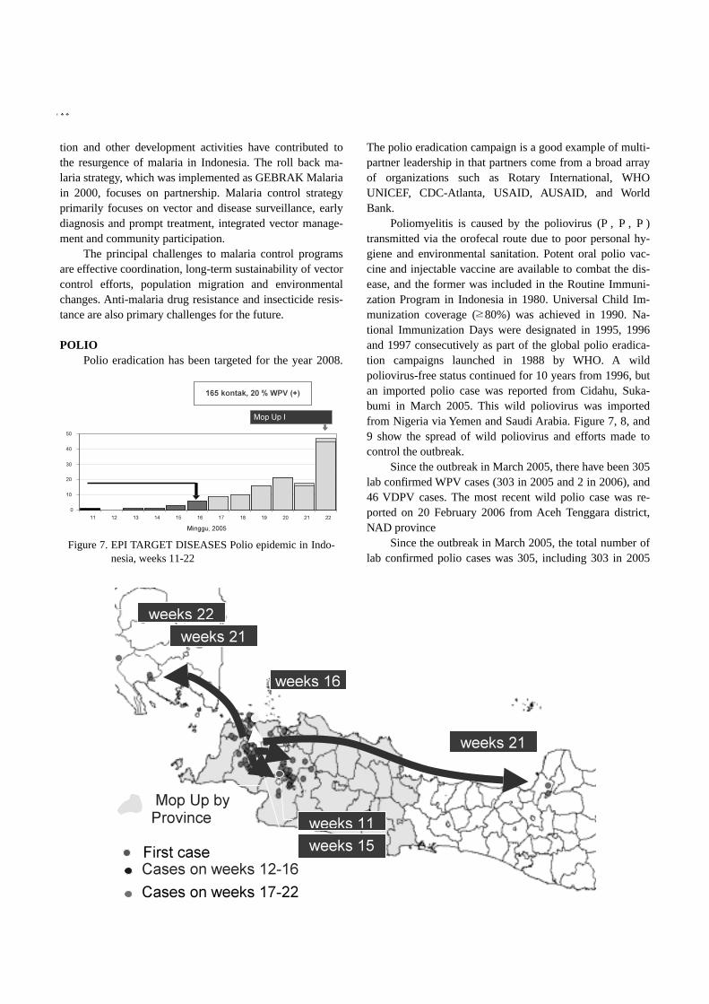
Poliomyelitis is caused by the poliovirus (P1, P2, P3)transmitted via the orofecal route due to poor personal hy-giene and environmental sanitation. Potent oral polio vac-cine and injectable vaccine are available to combat the dis-ease, and the former was included in the Routine Immuni-zation Program in Indonesia in 1980. Universal Child Im-munization coverage (�80%) was achieved in 1990. Na-tional Immunization Days were designated in 1995, 1996and 1997 consecutively as part of the global polio eradica-tion campaigns launched in 1988 by WHO. A wildpoliovirus-free status continued for 10 years from 1996, butan imported polio case was reported from Cidahu, Suka-bumi in March 2005. This wild poliovirus was importedfrom Nigeria via Yemen and Saudi Arabia. Figure 7, 8, and9 show the spread of wild poliovirus and efforts made tocontrol the outbreak.
Since the outbreak in March 2005, there have been 305lab confirmed WPV cases (303 in 2005 and 2 in 2006), and46 VDPV cases. The most recent wild polio case was re-ported on 20 February 2006 from Aceh Tenggara district,NAD province
Since the outbreak in March 2005, the total number oflab confirmed polio cases was 305, including 303 in 2005
Figure 7. EPI TARGET DISEASES Polio epidemic in Indo-nesia, weeks 11-22
144

and 2 cases in 2006, and 46 VDPV (vaccine derived poliovirus) cases. After various efforts including mopping up,National Immunization Days (NID) and Sub NID, the lastcase was reported in 20 February 2006 from SoutheastAceh. A future challenge to EPI is the sustainability of theroutine immunization, including both managerial and finan-cial sustainability. A long-term plan is being developed toaddress future challenges.
HIV/AIDSThe Human Immunodeficiency Virus (HIV) causes ac-
quired immunodeficiency syndrome (AIDS). The first HIV/AIDS case in Indonesia was reported in the late 80’s fromBali, after which the disease spread rapidly to 32 out of the33 provinces of Indonesia. The cumulative number of HIV
Figure 8. Wild Poliovirus Cases in Indonesia, March 2005- present
Figure 9. Wild Polio Cases by Week of Onset, Indonesia 2005-2006
Figure 10. CUMULATIVE AIDS CASES UP TO THE ENDOF SEPTEMBER 2006
145

infection and AIDS cases reported up to September 2006 is4617 and 6987, respectively. Figure 10 shows the trend ofAIDS cases from 1997 to 2006 and figure 11 shows the sexdistribution. The AIDS cases were predominantly male(82%), with females accounting for only 16% and unknownfor 2%. Risk factors by mode of transmission are shown infigure 12. Intense and rapid spread of HIV was documentedamong injecting drug users (IDU) (52%), followed by het-erosexuals (37.2%). The cumulative number of AIDS casesis expected to reach 93,968 to 130,000 by the year 2010. InPapua, pregnant women and newborn babies were infectedby HIV. The main strategy to combat HIV infection in-cludes efforts to prevent new infection, to promote compre-hensive care, and to increase coverage of HIV infection
through partnership. The principal challenges to HIV/AIDScontrol programs are intersector coordination, behaviorchanges, and human resources.
SARS (Severe Acute Respiratory Syndrome)During the SARS outbreak in 2003, there were only 7
suspected and 2 probable cases admitted to hospitals in In-donesia. No confirmed case was reported. Strategies and ac-tions taken during the SARS outbreak included the screen-ing of incoming passengers from affected countries withthermo scanners, establishment and capacity building of 35referral hospitals and 45 port health offices, public aware-ness and regional networking (ASEAN +3).
AVIAN INFLUENZAOutbreaks of H5N1 infection in the poultry population
were first reported in 2003. Human cases were first reportedin July 2005. To date, 32 of 33 provinces of Indonesia haveexperienced H5N1 infection among the poultry population,while H5N1 infection in humans has been reported fromonly 9 provinces. Figure 13 shows the distribution of avianinfluenza outbreaks in animals and humans.
Strategies to control H5N1 transmission include theprevention of H5N1 transmission in the poultry population,surveillance, bio-security, case management, public aware-ness/risk communication, research and development. Themain challenges to H5N1 control are coordination and re-sources.
NEGLECTED DISEASESLeprosy, lymphatic filariasis, yaws, rabies, and Japa-
nese encephalitis are considered to be neglected diseases.Very little attention has been paid to these diseases, and in-sufficient resources have been allocated for their control.Fortunately, international agencies and the internationalcommunity are interested in leprosy and lymphatic filariasisand as a result we aim to eliminate leprosy and lymphaticfilariasis by 2020. An integrated approach is being imple-mented to cope with the limited resources available.
Figure 11. SEX DISTRIBUTION OF AIDS CASESUP TO THE END OF SEPTEMBER 2006
Figure 12. RISK FACTOR OF AIDS CASES BY MODEOF TRANSMISSION UP TO SEPTEMBER2006
146
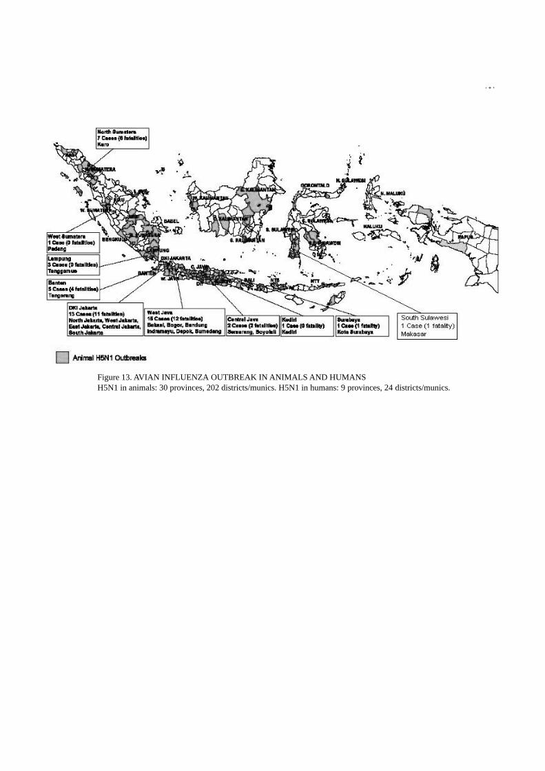
Figure 13. AVIAN INFLUENZA OUTBREAK IN ANIMALS AND HUMANSH5N1 in animals: 30 provinces, 202 districts/munics. H5N1 in humans: 9 provinces, 24 districts/munics.
147
ROLES OF MULTI-COUNTRY NETWORKING IN PREVENTION ANDCONTROL OF EMERGING AND RE-EMERGING INFECTIONS
Pathom SawanpanyalertRecieved 13, December, 2006
ABSTRACT: The emergence of new and the re-emergence of old infectious diseases reverses our previous beliefthat communicable diseases have been brought under human control. The development of antibiotic-resistant bacte-rial and fungal infections and the apparent lack of vaccines for many infectious diseases remind us of how vulner-able we are. Infections with SARS coronavirus, Nipah virus, and, more recently, H5N1 influenza virus in humansare just a few reality checks and more are expected. The problems of these emerging and re-emerging infections(ERI) give us several warnings. First, there are a number of pathogens, most of which are viral, that pose potentialrisks to human health and we know quite little about them. Second , many of these ERI are zoonotic. Effective con-trol of zoonoses needs involvement and collaboration from the non-health sector. Third , since these ERI can easilyspread across geopolitical boundaries, countries with a good public health infrastructure are not risk-free andshould not be complacent. Surveillance and response efforts cannot be limited within one national boundary.Fourth , aside from the fundamental tools in disease control that we have, e.g. basic sanitation, personal hygiene,isolation and quarantine, and the newly-revised International Health Regulations, we have little other choices, e.g.drugs and vaccines. This limitation suggests that we could eventually be defenseless. Fifth , recent occurrences ofcertain ERI in some countries have demonstrated that damages caused in non-health terms, e.g. economic losses,can be significant. As a consequence, ERI may be dealt with as an economic problem while the human and healthdimensions are ignored.
There are a number of guiding principles that we may have to adopt. First, no country can or is allowed tofight ERI alone, no matter how well-developed its economic condition and public health infrastructure. However, itshould be noted that ERI is a national health security problem and that national sovereignty must be recognizedwhen addressing the ERI issues. Second , ERI problems need a lot of non-health partners, e.g. business and agricul-ture. Third , we need to improve our capacity to do surveillance and to respond. Surveillance without response ispointless. Fourth, we need specific tools to help tackle ERI.
Networking is a mechanism through which countries can work together to fight ERI. A number of forums ex-ist to address the problems of ERI, e.g. meetings of WHO and other UN agencies including FAO/OIE, ASEAN+3,and ACMECS. In addition, several bilateral frameworks are in place to address ERI. However, it is important tonote the particular nature of ERI and follow the above-mentioned guiding principles to avoid failure.
INTRODUCTION
Emerging and re-emerging infections, by definition,are new threats to human beings, although many of thepathogens causing the infections may have been with us fora long time without our due attention. In the course of hu-man development, we are constantly challenged by threats,including those from infectious diseases. Discovery of ef-fective antibiotics and the rapid progress of virology mayinstill a complacency that infectious diseases are under ourcomplete control and we do not have to be wary of commu-nicable diseases. However, the emergence of HIV/AIDS
epidemic some twenty years ago and the recent outbreaks ofSARS coronavirus, Nipah virus, and H5N1 influenza virusin humans reverse this belief. The threat is further aggra-vated by long-recognized-but-largely-ignored problems ofantimicrobial resistance not only among bacteria but alsoviruses, fungi and even parasites. In other words, not onlyare we unable to control existing enemies, we are facedwith more and stronger enemies. In addition, our most ef-fective tools, e.g. antimicrobials, are becoming less effective.
There are several lessons to be leaned from our recentencounters with emerging and re-emerging infections (ERI).
First, the list of emerging and re-emerging infections is
Tropical Medicine and Health Vol. 34 No. 4, 2006, pp. 149-152Copyright� 2006 by The Japanese Society of Tropical Medicine
National Institute of HealthDepartment of Medical SciencesMinistry of Public HealthThailand
149

long and growing. If we visit the first issue of the journal“Emerging Infectious Diseases or EID” published in 1980,we can see the introductory paper by David Satcher of theUS Centers for Disease Control and Prevention on ERI, asshown in Figure 1[1]. Although the paper is now over 20years old, the list of EID in the paper was already long and,I am sure, if the list is revised, it will grow. Most of the dis-eases or syndromes on the list and the ones we have facedrecently are quite strange to us. That means we know rela-tively little about them. Our relative ignorance of ERI is themajor drawback in our fight against them.
Second, the origins of many recent ERI can be tracedback to animal reservoirs or carriers or cases, e.g. civet catsin SARS, pigs in Nipah viral infection, and birds in H5N1infection. Many ERI exist in animals long before they are
transmitted to humans. Increased contact between humanbeings and animals increases the chance of infections jump-ing onto humans. Such includes raising domesticated orfarm animals, contacting migratory animals such as birds,and hunting and eating exotic animals. Human medicinedoes not prepare doctors or public health professionals tohandle diseases of animal origin. Therefore, there is astrong and urgent need for better and closer coordinationbetween the veterinary sector and human sector.
Third, the readily transmissible nature of most ERIputs all countries at risk. SARS is a particularly contagiousdisease. Nipah encephalitis spreads rapidly among pigs andcould potentially do so in humans if effective control in ani-mals is not put in place rapidly. Migratory birds help spreadH5N1 infections to all corners of the globe. The rate of the
Figure 1: What emerging and re-emerging infections (ERI) to expect?
Figure 2: Global meat prices as a consequence of HPAI outbreaks (Source: FAO)
150
spread is virtually independent of geopolitical boundariesand economic, social and public health development status,although countries with better-developed public health sys-tems and infrastructures may be able to control the diseasesmore effectively and efficiently. This is a reminder that ERIcannot be fought in one country alone.
Fourth, most ERI not only take lives but also result inreduced or lost livelihoods of human beings. SARS reducedglobal air travel. Nipah viral infection resulted in the cullingof a large number of pig reservoirs and cases. H5N1 dis-eases among poultry and in humans resulted in the massiveculling of domesticated chickens and ducks. There is evi-dence that the livelihood of agricultural workers has beenaffected and the price of chicken meat (as the direct impact)and of other meats such as pork and beef (as a compensa-tory mechanism) increased significantly as a result of theculling, as shown in Figure 2. Since direct and indirect eco-nomic losses from ERIoutbreaks are enormous, many peo-ple and organizations are concerned that economic issuesmay eclipse problems related to the health of the people.
Fifth, we do not have specific interventions for mostERI. Most ERI are viral, and there are no specific antiviralsnor vaccines for the infections. Although efforts are under-way to develop a vaccine for SARS, it is not yet available.We have specific vaccines for circulating strains of humaninfluenza viruses, e.g. type A (H1, H3), type B, but specificvaccines need to be developed annually to match circulatingstrains. Furthermore, although we believe that vaccines pro-duced ion the same principle as seasonal influenza vaccineswill be effective against H5N1 and other pandemic influ-enza strains, we are yet to come to terms with how to makean adequate amount of effective vaccines for a pandemic.
Mechanisms available to combat ERI
Given the above characteristics of ERI, it is almost im-possible to discuss the prevention and control of ERI in anisolated single-country context. Most prevention and con-trol efforts involve other countries. International collabora-tion is a much-welcomed term in current public health prac-tices. Multi-national mechanisms are currently available atthe global level, e.g. World Health Organization (WHO;http://www.who.int/csr/en/), World Organization for AnimalHealth (OIE; http://www.oie.int/eng/en_index.htm), andFood and Agriculture Organization (FAO; http://www.fao.org/ag/againfo/subjects/en/health/diseases_cards/special_avian.html). A number of regional economic forums alsoaddress the issue of ERI, e.g. Asia-Pacific Economic Coop-eration (APEC; http://www.apec.org), Association of South-east Asian Nations (ASEAN; http://www.aseansec.org), andAyeyawady-Chaophraya-Mekong Economic CooperationStrategy (ACMECS;: http://www.geis.fhp.osd.mil/about-GEIS.asp). In addition, there are a number of bilateral col-laborations that specifically address ERI-related issues, e.g.those organized by the US Centers for Disease Control andPrevention and the US Department of Defense.
Guiding principles for dealing with ERI
The operation of international collaborations on ERIrequires a few common principles. The following principlesare proposed as overarching principles that could guide in-ternational collaboration, especially in fighting ERI.
First, no country can or is allowed to fight ERI alone,no matter how well-developed its economic condition orpublic health infrastructure. The rapid transmissibilityacross geopolitical boundaries demands that we all work to-gether. No country is immune and no one can be compla-cent. It is therefore a wise strategy for wealthy countries, e.g. countries in North America and Western Europe, to come
Figure 3: International (global and regional) forums for emerging and re-emerging infections
151

to Asia to fight H5N1 at the forefront. However, care shouldbe taken to avoid infringement on national sovereignty. Adelicate balance has to be maintained.
Second, ERI problems call for a lot of non-health part-ners, e.g. business and agriculture, and effective control isnot possible without involvement of these partners. It is en-couraging to see, at the global revel, a close working rela-tionship between human health and animal health sectors infighting H5N1 diseases. However, this working relationshipmay not be seen at the country and sub-country levels. Lackof such close collaboration at all levels could easily fail us.Therefore, mechanism(s) must develop to forge and fostersuch collaboration in the fight against ERI.
Third, we need to improve our capacity to conduct sur-veillance and to respond. The importance of surveillancecannot be stressed enough. However, we should not forgetthat the definition of surveillance is “information for action”.This means that surveillance without response is pointlessand may be useless. At the same time, response without sur-veillance is blind and can be really harmful.
To respond to ERI more effectively, we need specifictools to tackle ERI. Although basic health sanitation, e.g.hand wash, cough etiquette, and other improvements ingeneral hygiene, is effective in reducing human-to-humantransmission of ERI, we need more effective interventions.Development of vaccines and new drugs are tedious andtime-and resource-consuming, but we need to invest in them.If “we”, the private vaccine and drug manufacturers, are notready, then “we”, the governments, need to kick in. The rolethat governments can play ranges from providing sufficientsupplies of drugs and vaccines (from available channels) tosupport for research, development and production of drugsand vaccines. Unfortunately, most governments do not pos-sess the capacity nor carry the leverage to enhance research,development and production of the capacity necessary.Most of the capacity to produce drugs and vaccines belongsto multi-national private companies. In order for a countryat risk of ERI epidemics to be self-sufficient in the vaccinesand drugs needed to combat ERI, the government may needto promote technology transfer from private vaccine-anddrug-manufacturing companies. This technology transfermay require the cooperation of international organizations,such as the World Health Organization.
REFERENCES
1.Satcher D. Emerging Infections: Getting Ahead of theCurve. Emerg Infect Dis [serial online] 1995 Jan-Mar[cited2006 Dec 10] :1.Available from: URL: ftp://ftp.cdc.gov/pub/EID/vol 1 no 1/adobe/satcher.vol 1no1. pdf
152
EXPLORING FRESH COLLABORATIVE INITIATIVESFOR COMBATING INFECTIOUS DISEASES IN THE PHILIPPINES
LUNINGNING P. ELIO-VILLARecieved 27, October, 2006
Eight of the 10 leading causes of morbidity in the Phil-ippines are infectious in nature, including pneumonia, diar-rhea, bronchitis, influenza, tuberculosis, malaria, chickenpox and measles. Pneumonia and tuberculosis continue tocause a significant number of deaths across the country andpersist to be among the 10 leading causes of mortality. Thediminishing burden of communicable diseases as majorcauses of death may be attributed to improved health tech-nology and health care delivery systems. Appropriate strate-gies and technologies include immunization, improved sani-tation and personal hygiene, better nutrition, early treatmentand steady supply of antibiotics made available at the com-munity or at first level health facilities. However, the persis-tence of communicable diseases as major causes of morbid-ity points to the fact that much still needs to be done to re-duce the occurrence of illness through preventive and pro-motive health measures.
A 20-year (1981-2000) trend analysis reveals a generaldecline in number of cases, and number of deaths fromcommunicable diseases of public health importance, whichinclude, among others, tuberculosis, malaria, schistosomia-sis, tetanus, diphtheria, pertussis, measles, rabies, diarrhealdiseases and pneumonia.
Although tuberculosis remains to be a major publichealth concern in recent years, effective case finding, dis-ease management with DOTS strategy, and partnership withthe private sector have led to significant improvements inthe prevention and control of the disease.
Successful nationwide immunization campaigns un-dertaken over the years have resulted in the eradication ofpoliomyelitis and a continuous decline in the incidence ofmeasles, tetanus, diphtheria and pertussis. I mproved sanita-tion, better nutrition and increased awareness of the com-
munity and appropriate management with oral rehydrationresulted in the reduction of deaths from diarrhea. Reductionof mortality from pneumonia, especially among children,may be attributed to early recognition of the public andearly diagnosis and treatment with appropriate antibiotics.
HIV/AIDS is “slow and low” in the Philippines, whichmeans that transmission is slow and prevalence is low.However, it threatens to grow into a major epidemic in thecountry.
Although malaria is no longer a leading cause of death,it has remained among the leading causes of morbidity inthe country, especially in rural areas. The fatal conse-quences and cyclical occurrence of outbreaks of dengueevery three to five years is a public health concern. Effortsto eliminate filariasis are hindered by limited resources forannual mass treatment in endemic areas. Although theprevalence is declining, it is a source of concern that forsome endemic infectious diseases such as schistosomiasis,areas previously identified as non-endemic areas have beenfound to have new cases.
Emerging infectious diseases, i.e. newly identified orpreviously unknown infections including re-emerging IDs,cause serious public health problems if not contained asclose as possible to their source. The inherent unpredictabil-ity of a variety of previously known infections and un-known diseases can limit the responsiveness of even themost organized health system.
In 2003, the sudden and unexpected emergence ofSARS presented an opportunity to initiate measures tostrengthen local and international surveillance for emergingand re-emerging infections as well as to strengthen quaran-tine and isolation measures, build capacity in laboratory di-
Tropical Medicine and Health Vol. 34 No. 4, 2006, pp. 153-154Copyright� 2006 by The Japanese Society of Tropical Medicine
Medical Specialist�Program Manager for Emerging Infectious DiseasesNational Center for Disease Prevention and ControlDepartment of HealthPhilippines
153

agnosis and clinical management, strengthen structure, sys-tems and procedures for triaging, infection control, surveil-lance and epidemiologic investigation, hospital referral, ad-vocacy and risk communication.
Avian influenza (AI) or bird flu due to the highlypathogenic influenza virus, H5N1, is a grave threat to hu-manity. The unpredictability of the influenza virus and theserious possible consequences of a pandemic warrant con-stant vigilance and good planning in order to reduce the im-pact of a pandemic.
The unexpected and unusual increase in meningococ-cal disease, with meningococcemia as a predominant form,in the Cordillera Autonomous Region led to the death of atleast 50% of cases in the early stage of this occurrence,causing public anxiety and seriously affecting the economy,particularly in Baguio City. There is a need to develop theregional capability for laboratory diagnosis and to conductresearch studies on population carriage and serogroups pre-dominant in the Philippines.
Hepatitis C is an emerging infection in the Philippineswith high prevalence among IV drug users studied. There isa need for more epidemiological data among contacts ofpositive cases to determine the extent of transmission and tomonitor and avert the progression of the disease.
Leptospirosis increases during the rainy season as theaftermath of flooding but cases are also recognized in re-gions with vast farmlands. There is a need to establish alaboratory-based surveillance for leptospirosis, to determinethe burden of disease in the Philippines, and to identify thefactors for susceptibility and severity and the circulating se-rovars causing various degrees of severity of disease.
Tuberculosis has been identified as a leading cause ofCNS infections. Other etiologies: varicella, herpes zoster,Japanese encephalitis. There is a need for a continuinglaboratory-based surveillance of meningitis and encephalitisas identification of a specific etiology will serve as a basisfor specific treatment and vaccination programs.
The current data on Japanese encephalitis may be justthe tip of an iceberg. The national and regional incidenceand burden of disease of Japanese encephalitis and the re-gional distribution among its animal hosts need to be deter-mined as a basis for national policy and programs.
Despite the continuing decline in mortality from pneu-monia, more efforts can be done to strengthen measures for
its prevention and control. There is a need to strengthenlaboratory capabilities to conduct etiologic identificationand antimicrobial resistance surveillance for appropriatemanagement of cases. Infection control in the health caresetting also needs to be strengthened.
The following areas for possible technical cooperationmay be considered: strengthening systems for early recogni-tion of disease and response of local units, establishment ofreal-time surveillance and information systems, building di-agnostic and management capacities for infectious diseasesin designated national and sub-national facilities, epidemi-ological studies on emerging infectious diseases such asmeningococcal disease, melioidosis, Legionella, Hepatitis C,etiologies of CNS, diarrhea and pneumonia, burden of dis-ease as basis for public health programs for influenza, JE,leptospirosis, developing well-trained and well-equipped re-sponse teams from the national to the local level, prepared-ness assessment through simulation and other similar exer-cises, operational studies: evaluation of preparedness ofLGUs, of hospitals, public awareness on various EIDs.
154
A NEW SPECIES OF SIMULIUM (NEVERMANNIA) FROM THEOGASAWARA (BONIN) ISLANDS, JAPAN (DIPTERA: SIMULIIDAE)
HIROYUKI TAKAOKA1 and KATSUMI SAITO2
Accepted 5, December, 2006
ABSTRACT: Simulium (Nevermannia) satakei sp. nov. is described on the basis of the pupa and mature larvaecollected from the Ogasawara (Bonin) Islands in Japan. This new species, tentatively (due to lack of the adultstage) assigned to the vernum species-group of the subgenus Nevermannia , is characterized in the pupa by four gillfilaments lacking transverse ridges, and in the larva by a small, M-shaped postgenal cleft, antenna without hyalinebands, and simple rectal papilla. The morphological differences among this new species and the two known species,S. (N.) uemotoi from Japan and S. (N.) karzhantacum from Uzbekistan and Turkmenistan, are noted. This is thesecond species of the family Simuliidae from the Islands.Key words: Nevermannia , Simuliidae, Simulium , black fly, Ogasawara Islands, new species
Until now, Simulium (Nevermannia) bonninense(Shiraki), a member of the vernum species-group of thesubgenus Nevermannia Enderlein, was the only species ofthe family Simuliidae so far recorded from the Ogasawara(Bonin) Islands located in the Pacific Ocean, ca. 1,000 kmsouth-southeast of Tokyo (Shiraki, 1935; Stone, 1964; Saitoet al., 1974; Takaoka et al., 1999). Recently, a pupa and afew larvae of an unknown species were collected togetherwith some pupae and larvae of S. (N.) bonninense from asmall stream in Hahajima, one of the Ogasawara Islands.
This is described here as a new species and is tenta-tively (due to lack of the adult stage) assigned to the vernumspecies-group within the subgenus Nevermannia .
The terms for morphological features used here followthose of Takaoka (2003). Holotype and paratype specimensof the new species are deposited at the Department of Infec-tious Disease Control, Faculty of Medicine, Oita University,Oita, Japan.
Simulium (Nevermannia) satakei sp. nov.
DESCRIPTION. Pupa. Body length 2.0 mm. Head . In-tegument (Fig. 1A) yellowish, moderately covered withsmall tubercles; antennal sheath (Fig. 1B) sparsely coveredwith small tubercles; frons with 2 medium-long slendertrichomes (Fig. 1C) on each side; face with 1 long stouttrichome (Fig. 1D) on each side, which is 1.3―1.7 times aslong as those of frons. Thorax . Integument yellowish, mod-erately covered with small tubercles, and on each side with
3 long stout simple trichomes (Fig. 1E) mediodorsally, 2long simple trichomes (1 somewhat shorter and more slen-der than the other) (Fig. 1F) anterolaterally, 1 medium-longsomewhat stout simple trichome (Fig. 1G) posterolaterally,and 3 short slender simple trichomes (Fig. 1H) [though 1additional medium-long slender trichome (Fig. 1I) was pre-sent on the right side] ventrolaterally. Gill (Fig. 1J) with 4slender thread-like filaments arranged in dorsal and ventralpairs arising from short common basal stalk; stalk of ventralpair slightly shorter than common basal stalk but slightlylonger than the stalk of dorsal pair; dorsalmost filament andventralmost one basally diverged vertically at a right anglewhen viewed laterally; all filaments subequal in thickness toone another; lengths of all filaments not measurable due toloss of apical portion except dorsal filament of dorsal pair(2.7 mm long) and ventral filament of ventral pair (2.1 mmlong) of right gill; all filaments light yellow, gradually ta-pered toward apex, furnished with annular furrows but lack-ing ridges (Fig. 1K), and densely covered with minute tu-bercles on outer surface. Abdomen . Dorsally, all segmentsweakly sclerotized and pale yellow; segments 1 and 2sparsely or moderately covered with small tubercles (Fig. 1L); segment 1 with 1 medium-long simple slender seta (Fig.1M) on each side; segment 2 with 1 medium-long simpleslender seta and 5 short dark spinous setae (Fig. 1N) oneach side; segments 3 and 4, each with 4 dark stout hooksand 1 short spinous seta on each side; segments 5―9 eachwith spine-combs and comb-like groups of minute spineslying transversely along anterior margin (Fig. 1O) on each
Tropical Medicine and Health Vol. 34 No. 4, 2006, pp. 155-158Copyright� 2006 by The Japanese Society of Tropical Medicine
1Department of Infectious Disease Control, Faculty of Medicine, Oita University, Hasama, Yufu City, Oita, 879-5593 Japan2Laboratory of Medical Zoology, College of Environmental Health, Azabu University, 1-17-71, Fuchinobe, Sagamihara City, Kanagawa, 229-8501 Japan
155
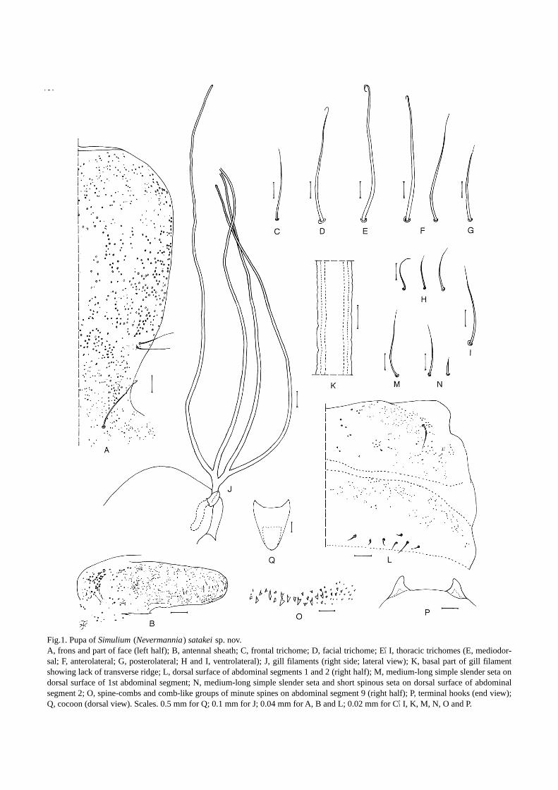
Fig.1. Pupa of Simulium (Nevermannia) satakei sp. nov.A, frons and part of face (left half); B, antennal sheath; C, frontal trichome; D, facial trichome; E―I, thoracic trichomes (E, mediodor-sal; F, anterolateral; G, posterolateral; H and I, ventrolateral); J, gill filaments (right side; lateral view); K, basal part of gill filamentshowing lack of transverse ridge; L, dorsal surface of abdominal segments 1 and 2 (right half); M, medium-long simple slender seta ondorsal surface of 1st abdominal segment; N, medium-long simple slender seta and short spinous seta on dorsal surface of abdominalsegment 2; O, spine-combs and comb-like groups of minute spines on abdominal segment 9 (right half); P, terminal hooks (end view);Q, cocoon (dorsal view). Scales. 0.5 mm for Q; 0.1 mm for J; 0.04 mm for A, B and L; 0.02 mm for C―I, K, M, N, O and P.
156

side; segment 9 with pair of cone-shaped terminal hooks(Fig. 1P). Ventrally, segments 3―8 nearly transparent andsegment 9 weakly sclerotized and pale yellow; segment 3with 3 short simple setae on each side; segment 4 with 1simple dark hooklet (slightly shorter and smaller than thoseon segments 5―7) and 3 short simple setae on each side;segment 5 with 2 bifid dark hooks and a few short simplesetae on each side; segments 6 and 7 each with 1 bifid darkinner hook and 1 simple or bifid dark outer hook, and a fewshort simple setae on each side; segments 4―8 with comb-like groups of minute spines. Segment 9 with short simpleseta on each lateral side. Cocoon (Fig. 1Q). Simple, wall-pocket-shaped, moderately woven, with anterior marginsomewhat thickly woven, and extending ventrolaterally;floor woven on posterior 2/5; individual threads visible; 2.5mm long by 2.0 mm wide.
Mature larva. Body length 3.5―4.0 mm. Body colorcreamy yellow. Cephalic apotome yellow; head spots allpositive and medium brown. Lateral surface of head capsuleyellowish except eye-spot region yellowish-white, with eye-brow light brown; 2 large and1small spots just before pos-terior margin, as well as 2 small spots below eye-spot re-
gion all positive and medium brown. Ventral surface of headcapsule yellow; 1 elongate spot on each side of postgenalcleft positive and medium brown. Cervical sclerites com-posed of 2 rod-like small pieces, not fused to occiput,widely separated from each other. Antenna composed of 3segments and apical sensillum, much longer than stem oflabral fan; proportional lengths of 1st, 2nd, and 3rd seg-ments 1.00: 0.62―0.78: 0.83―0.93. Labral fan with about 34rays. Mandible (Fig. 2A) with 1st comb-tooth longest; 2ndand 3rd comb-teeth subequal in length to each other; man-dibular serrations composed of 2 teeth (1 large and 1 small);large tooth at a right angle to mandible on apical side; 2―4supernumerary serrations present. Hypostoma (Fig. 2B)with a row of 9 apical teeth, the median tooth and cornerteeth being most prominent, and median tooth of 3 interme-diate teeth on each side smallest; lateral margins with welldeveloped teeth; 3 or 4 hypostomal bristles in a row, subpar-allel to, or slightly diverging from, lateral margin on eachside. Postgenal cleft (Fig. 2C,D) small, M-shaped, 0.41―0.64 times as long as postgenal bridge. Thoracic and ab-dominal cuticle almost bare except dorsal surface of a fewposterior segments sparsely to moderately covered with col-
Fig.2. Mature larva of Simulium (Nevermannia) satakei sp. nov.A, mandible; B, hypostoma; C and D, ventral surfaces of head capsules showing post-genal clefts of different sizes and shapes. Scales. 0.05 mm for C and D; 0.02 mm for B;0.01 mm for A.
157
orless minute setae and areas on both sides of anal scleritemoderately covered with colorless short setae. Rectal scalesabsent. Rectal papilla simple, without secondary lobules.Anal sclerite X-shaped, anterior arms 0.8 times as long asposterior ones; accessory sclerite absent. Ventral papillaepresent ventrolaterally. Posterior circlet of hooks with about64 rows of up to 15 hooks per row.
Female and Male. Unknown.
TYPE SPECIMENS. Holotype pupa with its associated co-coon, collected from a small shaded stream (width about 10cm) slowly flowing in a forest, located on the right side ofChibusa Dam, Hahajima, Ogasawara Islands, Tokyo, Japan,18.VI.2005, by K. Satake. Paratypes: 2 mature larvae and 1immature larva, same locality and data as those of the holo-type.
ETYMOLOGY. The species name satakei honors Dr. K.Satake, who collected this new species.
REMARKS. This new species is tentatively assigned to thevernum species-group of the subgenus Nevermannia byhaving the four gill filaments per side in the pupal stage, theantennae without any transverse hyaline bands, the mandi-ble with supernumerary serrations, the main tooth of themandibular serrations at a right angle on the apical side tothe mandible, the hypostoma with serrated lateral margins,and the ventral papillae well developed in the larval stage.
The larva of this new species is very similar to that ofS . (N .) uemotoi of the vernum species-group from Japan(Sato et al., 2004): it shares several characteristics includingthe small, M-shaped postgenal cleft and the simple rectalpapilla. There are some differences, however, in the relativelength of the three segments of the larval antennae (1.00:0.62―0.78: 0.83―0.93 versus 1.0: 1.2―1.4: 0.8―1.0) betweenthe two species. On the other hand, the pupa of this newspecies is easily distinguished from that of S . (N .) uemotoiby the following characteristics (those of S . (N .) uemotoiare shown in parentheses): frontal trichomes in two pairs (inthree pairs), antennal sheath sparsely covered with small tu-bercles (bare), transverse ridges on the gill filaments absent(present), dorsal surface of the abdominal segments 1 and 2sparsely covered with minute tubercles (densely and neatlycovered with minute tubercles), and spine-combs on the ab-dominal segment 9 distinct (absent or indistinct if any).
This new species is also similar to S . (N .) kar-zhantacum (Rubtsov, 1956) from Uzbekistan and Turk-menistan, which has a similarly shaped larval postgenalcleft, but differs in the pupal stage from the latter species bylacking the transverse ridges on the surface of the gill fila-ments.
Simulium (Nevermannia) satakei sp. nov. representsthe second species recorded from the Ogasawara Islands. Itshould be noted that this new species is not closely relatedto S . (N .) bonninense, which to date had been the only spe-cies prevalent in the islands, because there are distinct dif-ferences in the relative length of the first and second seg-ments of the larval antennae and in the shape of the larvalpostgenal cleft as well as in the presence or absence ofspine-combs on the dorsal surface of the fifth and ninth seg-ments of the pupal abdomen between the two species.
ACKNOWLEDGEMENTS
We are grateful to Dr. K. Satake, National Institute forEnvironmental Studies, Tsukuba, Japan, for kindly provid-ing his black fly specimens for our examination.
REFERENCES
Rubtsov, I.A. 1956. Blackflies (fam. Simuliidae) [Moshki (sem.Simuliidae)]. Fauna of the USSR. 859pp., New Series No.64,Insects, Diptera 6 (6). Akademii Nauk SSSR, Leningrad [=St.Petersburg], Russia. In Russian. [English translation: 1990.Blackflies (Simuliidae). 1,042 pp., 2nd Ed. Fauna of the USSR.Diptera, 6 (6). E.J. Brill, Leiden].Saito, K., Hori, E. and Ogata, K. 1974. Simuliidae of OgasawaraIslands. Jpn. J. Sanit. Zool. , 24: 338 (Japanese abstract only).Sato, H., Takaoka, H. and Fukuda, M. 2004. A new species ofSimulium (Nevermannia) (Diptera: Simuliidae) from Japan.Med. Entomol. Zool. , 55: 201―210.Shiraki, T. 1935. Simuliidae of the Japanese Empire. Mem. Fac.Sci. & Agr. Taihoku Imp. Univ. , 16: 1―90.Stone, A. 1964. Diptera: Simuliidae. Insects of Micronesia , 12:629―635.Takaoka, H. 2003. The Black Flies (Diptera: Simuliidae) of Su-lawesi, Maluku and Irian Jaya. xxii+581pp., Kyushu UniversityPress, Fukuoka.Takaoka, H., Saito, K. and Suzuki, H. 1999. Simulium (Never-mannia) bonninense from the Ogasawara (Bonin) Islands, Japan(Diptera: Simuliidae): taxonomic assignment to the vernum-group and descriptions of male, pupa and mature larva. Jpn. J.Trop. Med. Hyg. , 27: 189―194.
158

UVULECTOMY AND OTHER TRADITIONAL HEALING PRACTICES:TRADITIONAL HEALERS’ PERCEPTIONS AND PRACTICES
IN A CONGOLESE REFUGEE CAMP IN TANZANIA
OSAMU KUNII1, YASUO TANAKA2, ALYSON LEWIS3, SUSUMU WAKAI2
Accepted 17, December, 2006
ABSTRACT: Little is studied about traditional healers’ perceptions toward and practice of uvulectomy, which isknown as a traditional surgical practice mainly in Africa and which sometimes results in severe complications. Thisstudy aimed to clarify the perceptions toward and practice of uvulectomy and the other traditional healing practicesof traditional healers in a Congolese refugee camp in Tanzania. Interviews were conducted with 149 traditionalhealers, comprised of 59 registered, 68 non-registered and 22 faith healers.
A total of 1.7% of the registered healers and 8.8% of the non-registered healers had ever conducted uvulec-tomy on children (a median of 2 months to a median of 3 years of age) and had received cash or domestic fowlsequivalent to US$1-3 per operation. Although over 80% of the respondents believed traditional treatments to bemore effective than modern medicine, less than 20% considered uvulectomy beneficial and in fact about 40% con-sidered it to be harmful. The respondents raised cough, vomiting, appetite loss and other symptoms as an indicationfor uvulectomy, and death, bleeding, throat pain and other symptoms as harmful effects associated with uvulec-tomy. In this camp, the healers also performed other surgical procedures, such as male and female circumcision,tattoos and scarification.
In conclusion, only a limited number of the traditional healers believed that uvulectomy is beneficial and per-formed it on infants and young children, and these were mainly non-registered healers who had relatively little col-laboration with modern health professionals. In refugee settings where modern health professionals might not befamiliar with traditional healing, it is considered crucial to assess the risks of ongoing traditional practices and tostrive to achieve more strategic communication between modern and traditional health providers.Keywords: traditional healing, healers, uvulectomy, perception, Congolese refugees, Tanzania
INTRODUCTION
The persisting conflicts in the Great Lakes region ofAfrica have caused the flow of a large number of refugeesinto the United Republic of Tanzania. Tanzania has main-tained an open-door policy since its independence, and, as aresult, it hosted approximately 520,000 refugees includingmore than 370,000 from Burundi and about 140,000 fromCongo-Kinshasa, 3,500 from Somalia, and 2,700 fromRwanda by the end of 2002 [1].
In response to the extremely poor health status of theserefugees, modern health care services have been providedintensively in refugee camps, when possible, by skilled ortrained refugees and persons from the host community andaid organizations. However, there are still many refugeecommunities where traditional healing practices arestrongly preferred.
While modern health intervention has contributed
greatly to the improvement of the heath status in manycamps, efforts to reduce infant mortality have stagnated insome camps [2]. Although no survey was conducted there,modern health providers in these camps suspected a tradi-tional surgical practice called ‘uvulectomy’ to be one of thecauses. Uvulectomy is a procedure in which the uvula issevered. It is a traditional healing practice used mainly inAfrica, sometimes leading to serious complications [3, 4, 5].However, no study has shed light on the perceptions of tra-ditional healers as to its beneficial and adverse effects or itsactual practice, especially in refugee settings. In the refugeecamps, moreover, little was known about perceptions to-ward or practices of traditional healing methods other thanuvulectomy.
This study aimed to determine the traditional healers’perceptions and their implementation of traditional healingpractices, with special reference to uvulectomy.
Tropical Medicine and Health Vol. 34 No. 4, 2006, pp. 159-166Copyright� 2006 by The Japanese Society of Tropical Medicine
1Institute of Tropical Medicine, Nagasaki University, Nagasaki, Japan2Department of International Community Health, Graduate School of Medicine, The University of Tokyo, Tokyo, Japan3International Federation of Red Cross and Red Crescent Societies, Geneva, Switzerland
159
PARTICIPANTS AND METHODS
Study areaWe selected Lugufu Camp, one of the biggest refugee
camps in Kigoma Province, western Tanzania. It accommo-dated about 50,000 refugees from Congo-Kinshasa at thetime of our study, and it continued to grow by an average of1,000 refugees per month due to persistent armed conflicts,political instability, and deteriorating humanitarian condi-tions in that country. The camp was in the post-emergencyphase with a crude mortality rate of 0.65 deaths/10,000 per-sons/day and an under-five mortality rate of 1.88/10,000persons/day in 2000, thus indicating that the relief programshad successfully kept the health situation under control. [6]
ParticipantsSince there was no reliable document or registration
method to identify traditional healers in the study site, wegathered information through preliminary interviews withmodern health professionals and community health workersin the camp prior to the study. The results indicated thatthere were three types of healers, namely, registered tradi-tional healers, non-registered traditional healers and faithhealers. In the local language, the three types of healer arecalled ‘MFUMU’ or ‘MTEE’, ‘MLAKO’, and ‘BAYUM-BE’ or ’MAHA WA ASA’O’, respectively The ‘MFUMU’or ‘MTEE’ was registered with the Ministry of Health inCongo-Kinshasa and was certified to examine and treat pa-tients, supposedly with herbs and other medicinal subjects.The ‘MLAKO’ provided traditional healing practices with-out certification. The ‘BAYUMBE’ or ’MAHA WA ASA’O’was a faith healer, also referred to as a ‘prayer leader’,‘elder prayer’, ‘father/mother of prayer’ or ‘a director ofprayer’, who usually organized religious gatherings andprovided healing mainly through prayers.
Study PreparationPermission to carry out the study was obtained from
the UNHCR and Tanzania Red Cross Society, with the sup-port from the International Federation of Red Cross andRed Crescent Societies, organizations that took responsibil-ity for health and other related activities in the camp. Wemade a list of traditional healers that had been identified inthe camp by Congolese community health workers, and in-vited all of them to participate in our study. Informed con-sent was obtained, and finally all the healers in the list, 149individuals in total, agreed to participate.
Eighteen Congolese refugees engaged in communityhealth services speaking and writing English and Swahiliwere recruited and trained as interviewers for the study. Wealso recruited and trained Tanzanian health personnel com-
petent in speaking and writing both English and Swahili asstudy supervisors. The authors were responsible for thetraining of both interviewers and supervisors and checkedand confirmed the quality of supervisions. The study wasconducted from May to July 2001.
QuestionnaireSemi-structured interviews were designed to explore
traditional healers’ perceptions toward and use of uvulec-tomy and other traditional healing practices, and their atti-tude toward modern health care services. Background infor-mation gathered from each respondent included religion,years of education, and the person from whom the healingpractices were learned. Special efforts were made to obtain,in the respondent’s own words, of the healing practices andtheir indications, and the perceived benefits and harm ofuvulectomy. The respondents who used herbal medicinewere further asked about the type of herbal medicine, theirpreparation, usage and indications.
To quantify the perceived benefits and harm of tradi-tional practices, the interview also included the followingquestions: “In comparison to modern medicine, how effec-tive do you think your traditional treatments are?” with fourchoices “All of my treatments are more effective than mod-ern medicine”, “Some of my treatments are more effectivethan modern medicine”, “None of my treatments are moreeffective than modern medicine” and “I don’t know”; “Towhat extent do you trust modern doctors?” with fourchoices “very much”, “somewhat”, “not at all”, and “I don’tknow.”; “To what extent do you think uvulectomy is benefi-cial or effective?” with four choices “very much”, “some-what”, “not at all”, and “I don’t know”; “To what extent doyou think uvulectomy is harmful or results in a negative ef-fect?” with four choices “very much”, “somewhat”, “not atall”, and “I don’t know”. The interview also involved somequestions on the practice of uvulectomy such as the age ofthe patients and the amount of money or gifts received fromthe patients undergoing the uvulectomy procedure.
AnalysisThe chi-square test was used to compare categorical
variables using the SPSS statistical software package ver-sion 10.0 for Windows. The descriptive data and their inter-pretations were anonymously examined for specific mean-ings and clustered into meaningful groups. To quantify indi-cations for healing practices and harms of uvulectomy, onecommon term was labeled in each group. We also usedqualitative data by selecting typical expressions in eachgroup to complement or interpret the results of analyses ofquantitative data.
160
RESULTS
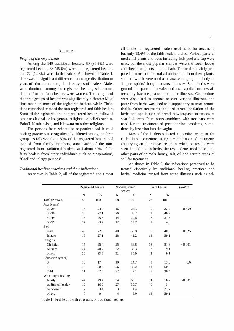
Profile of the respondentsAmong the 149 traditional healers, 59 (39.6%) were
registered healers, 68 (45.6%) were non-registered healers,and 22 (14.8%) were faith healers. As shown in Table 1,there was no significant difference in the age distribution oryears of education among the three types of healers. Maleswere dominant among the registered healers, while morethan half of the faith healers were women. The religion ofthe three groups of healers was significantly different: Mus-lims made up most of the registered healers, while Chris-tians comprised most of the non-registered and faith healers.Some of the registered and non-registered healers followedother traditional or indigenous religions or beliefs such asBaha’i, Kimbansiste, and Kitawara orthodox religions.
The persons from whom the respondent had learnedhealing practices also significantly differed among the threegroups as follows: about 80% of the registered healers hadlearned from family members, about 40% of the non-registered from traditional healers, and about 60% of thefaith healers from other individuals such as ‘inspiration’,‘God’ and ‘clergy persons’.
Traditional healing practices and their indicationsAs shown in Table 2, all of the registered and almost
all of the non-registered healers used herbs for treatment,but only 13.6% of the faith healers did so. Various parts ofmedicinal plants and trees including fruit peel and sap wereused, but the most popular choices were the roots, leavesand flowers of plants and tree bark. The healers mainly pre-pared concoctions for oral administration from these plants,some of which were used as a laxative to purge the body of‘impure spirits’ thought to cause illnesses. Some herbs wereground into paste or powder and then applied to sites af-fected by fractures, cancer and other illnesses. Concoctionswere also used as enemas to cure various illnesses, andpaste from herbs was used as a suppository to treat hemor-rhoids. Other treatments included steam inhalation of theherbs and application of herbal powder/paste to tattoos orscarified areas. Plant roots combined with tree bark wereused for the treatment of post-abortion problems, some-times by insertion into the vagina.
Most of the healers selected a specific treatment foreach illness, sometimes using a combination of treatmentsand trying an alternative treatment when no results wereseen. In addition to herbs, the respondents used bones andother parts of animals, honey, salt, oil and certain types ofsoil for treatment.
As shown in Table 3, the indications perceived to betreated effectively by traditional healing practices andherbal medicine ranged from acute illnesses such as cel-
Registered healers Non-registeredhealers
Faith healers p-value
N % N % N %
Total (N=149) 59 100 68 100 22 100Age (years)
20-29 14 23.7 16 23.5 5 22.7 0.45930-39 16 27.1 26 38.2 9 40.940-49 15 25.5 14 20.6 7 31.850-59 14 23.7 12 17.7 1 4.6
Sexmale 43 72.9 40 58.8 9 40.9 0.025female 16 27.1 28 41.2 13 59.1
ReligionChristian 15 25.4 25 36.8 18 81.8 <0.001Muslim 24 40.7 22 32.3 2 9.1others 20 33.9 21 30.9 2 9.1
Education (years)0 10 17 10 14.7 3 13.6 0.61-6 18 30.5 26 38.2 11 507-14 31 52.5 32 47.1 8 36.4
Who taught healingfamily 47 79.7 34 50 4 18.2 <0.001traditional healer 10 16.9 27 39.7 0 0by oneself 2 3.4 3 4.4 5 22.7others 0 0 4 5.9 13 59.1
Table 1. Profile of the three groups of traditional healers
161

lulites, diarrhea and wounds, to chronic illnesses such ascancer and diabetes. The registered and non-registered heal-ers treated similar indications, but the faith healers alsotreated other indications related to mental problems and ste-rility.
The exorcism of evil spirits was practiced by 30.5% ofthe registered and 25.0% of the non-registered healers, butnot by the faith healers. As shown in Table 3, the registeredand non-registered healers exorcised evil spirits as a treat-
ment for what they called ‘madness’ or ‘impure spirits’, ‘in-visible’ illnesses, sterility, and so forth, in addition to exam-ining such illnesses. The healers who answered ‘impurespirits’ explained that they caused not only mental but alsophysical problems.
Prayer was the main treatment procedure, and for somethe only treatment procedure, among the faith healers, whoexplained that their healing prayers were different from theexorcising of evil spirits.
Registered healers Nonregistered healers Faith healers p-valueN (=59) % N (=68) % N (=22) %
Use herbsyes 59 100 67 98.5 3 13.6 <0.001no 0 0 1 1.5 19 86.4
Exorcise evil spiritsyes 18 30.5 17 25 0 0 0.015no 41 69.5 51 75 22 100
Ever performed uvulectomyyes 1 1.7 6 8.8 0 0 0.088no 58 98.3 62 91.2 22 100
Effectiveness of traditional healingcompared to modern medicine
All are more effective 3 5.1 8 11.8 1 4.5 0.028Some are more effective 47 79.6 41 60.3 15 68.2None is more effective 6 10.2 4 5.9 0 0Don’t know 3 5.1 15 22 6 27.3
How beneficial is uvulectomyVery much 1 1.7 2 3 0 0 0.009Somewhat 4 6.8 12 17.6 4 18.2Not at all 14 23.7 13 19.1 4 18.2Don’t know 40 67.8 41 60.3 14 63.6
Do you know the benefits of uvulectomyyes 14 23.7 18 26.5 2 9.1 0.235no 45 76.3 50 73.5 20 90.9
How harmful is uvulectomyVery much 16 27.1 12 17.6 3 13.6 0.013Somewhat 2 3.4 15 22.1 7 31.8Not at all 0 0 2 2.9 0 0Don’t know 41 69.5 39 57.4 12 54.6
Do you know the harmful effects ofuvulectomy
yes 13 22 25 36.8 10 45.5 0.074no 46 78 43 63.2 12 54.5
Trust modern doctorsVery much 35 59.3 41 60.3 17 77.3 0.302Somewhat 24 40.7 23 33.8 5 22.7Not at all 0 0 1 1.5 0 0Don’t know 0 0 3 4.4 0 0
Cooperate with modern doctorsVery much 36 61 32 47.1 14 63.6 0.344Somewhat 21 35.6 27 39.7 5 22.7Not at all 1 1.7 5 7.4 2 9.1Don’t know 1 1.7 4 5.9 1 4.6
Table 2. Traditional healers’ practices and perceptions of healing and uvulectomy and attitudes toward modern doctors
162

The practice of uvulectomyUvulectomy is called ‘ELEMI’ in Kibembe, the refu-
gees’ local language. Only 7 (4.8%) of the healers had everconducted uvulectomy: 1 (1.7%) of the registered healers, 6(8.8%) of the non-registered and none of the faith healers; 1(1.7%) of the 58 Christians, 2 (4.2%) of 48 Muslims and 4(9.3%) of the 43 other religious beliefs. The above sevenhealers had performed uvulectomy repeatedly, for a total of47 cases: one registered healer had treated 2 cases and thesix non-registered healers had conducted an average of 7cases each (range: 2-12cases).
The recipients of uvulectomy were mostly infants andchildren, starting from a median of 2 months of age (range:1week-4months) up to a median of 3 years (range: 2-5years). Five of the healers received 1,375 Tanzanian shil-lings (TZS) in cash (equivalent to US$1.5 as of May 2001)on average (range: TZS 1,000-2,000; US$1.1-2.2) peruvulectomy, while the other two healers were given a hen(equivalent to TZS 1,400; US$1.6) and a duck (equivalentto TZS 2,000; US$2.2) per uvulectomy.
Perceived effects of traditional healing and attitudes towardmodern medicine
Over 80% of the traditional healers believed that ‘all orsome of the traditional treatments were more effective thanmodern medicine’. However, 11.8% of non-registered heal-ers believed that ‘all the traditional treatments were moreeffective than modern medicine’, while 10.2% of the regis-tered healers believed that ‘none of the traditional treat-ments were more effective than modern medicine’.
Over 98% of all respondents trusted modern doctors‘very much’ or ‘somewhat’. In practice, 96.6% of the regis-tered healers cooperated with modern doctors either ‘verymuch’ or ‘somewhat’. However, more than 10% of the non-registered and the faith healers did not cooperate with mod-ern doctors at all or did not know to what extent they coop-erated.
Perceived effects of uvulectomyMore than half of the respondents did not know
whether uvulectomy was effective or harmful. In all three
Registered healers (n) Non-registered healers (n) Faith healers (n)
Top five indicationstreated effectivelyby traditional healing
fracture 19 fracture 10 madness 7diabetes 17 diabetes 8 sterility 7epilepsy 10 blisters 7 epilepsy 5cancer 9 cellulites 6 impure spirits 2hemorrhoid 8 diarrhea 6 mental trouble 2
Top five indicationsfor herbs diabetes 22 cellulites 12 sterility 3
fracture 19 sterility 11 abortion 1cancer 16 diarrhea 11 impure spirits 1epilepsy 12 fracture 10 mental trouble 1cellulites 9 wound 9 paralysis 1
Top five indicationsfor exorcising impure spirits 8 madness 6
madness 4 examination 6examination 3 impure spirits 5sterility 2 cellulites 1invisible illness 2 epilepsy 1
Indications foruvulectomy cough 2 cough 5 cough 3
vomiting 1 vomiting 4 appetite loss 1appetite loss 1 appetite loss 2 throat pain 1baby’s crying 1 throat pain 2 fever 1throat pain 1 baby’s crying 1death 1 fever 1
Harmful effectsof uvulectomy death 8 bleeding 15 bleeding 7
bleeding 7 death 11 death 4throat pain/swelling 2 throat pain/swelling 10 throat pain/swelling 3nerve injury 1 cough 3 appetite loss 1
appetite loss 1
Table 3. Indications for healing practices and the harmful effects of uvulectomy as perceived by traditional healers
163

groups of healers, those who thought uvulectomy ‘veryharmful’ were greater in number than those who thought it‘very effective’.
Those who considered uvulectomy ‘very effective’ or‘somewhat effective’ were more likely to be non-registeredhealers than the others, while those who considered uvulec-tomy ‘very harmful’ or ‘somewhat harmful’ were morelikely to be faith healers.
None of those who considered uvulectomy ‘very effec-tive’ cooperated ‘very much’ with modern doctors, while25.6% of those who considered uvulectomy ‘very harmful’cooperated ‘very much’ with modern doctors.
As shown in Table 3, the three groups of healers gavesimilar answers regarding the indications for uvulectomy,such as cough, appetite loss and sore throat. They describedthe indications as follows: “Uvulectomy is effective forcough, vomiting, and throat dryness that stops the passageof air or oxygen.” “If uvulectomy is done, patients can theneat food, because a uvula prevents food from passingthrough throat.” “Uvulectomy can help babies when theyhave a cough, sore throat or high fever and when they can’teat.” “If uvulectomy is not done in time, a patient will diebecause of blocked respiration and swelling of the throat.”
Several responses were observed regarding the harmfuleffects of uvulectomy among the three groups of healers.The healers pointed out bleeding, death, sore throat, appe-tite loss, nerve injury, and responded as follows: “I haveseen somebody die from cutting the uvula because the pa-tient’s nerve was cut and throat swelling occurred.” “Blooddischarge can cause death.” “Throat inflammation andblood discharge can cause death.” “A patient may get thin-ner, vomit frequently and develop a sore throat.”
Thirteen respondents responded that uvulectomy washarmful only if inappropriately done. “Uvulectomy can bedangerous if the person who does it is a charlatan. The pa-tient may die.” “Uvulectomy is safe if it is done by an expe-rienced nurse in the dispensary.” “When done poorly, thehealer may cut the tonsils and the patient may bleed todeath.” “When uvulectomy is done poorly, it causes a sorethroat and the patient is not able to eat.” “If one doesn’tknow how to cut properly, the procedure will hurt the throatand the patient cannot eat.”
Six of the seven healers who had performed uvulec-tomy in the past responded that the procedure was ‘some-what effective’ and also ‘somewhat harmful’, and respondedas follows: “If one doesn’t know how to cut the uvula, thepatient may develop a sore throat. But I have done it in theCongo and had no problems.” “A person may develop asore throat after uvulectomy. It is dangerous if one doesn’tknow how to cut it properly. It is particularly dangerous forbabies. I performed uvulectomy in the Congo, but here I do
not have the proper instruments.” “If it is done by someonewho doesn’t know how to do it, it can cause serious prob-lems.” “A sore throat and bleeding occur after uvulectomy,but there is a type of root which I can use as a medicine tostop such problems.” “One must have sufficient experiencebut I think there is no problem for adults.” “Uvulectomy af-fects the throat. If the healer doesn’t know how to cut, it cancause respiratory problems.” The one remaining healer whohad performed uvulectomy said, “Uvulectomy is not goodat all. Actually it is very harmful. It causes bleeding anddeath.”
Other surgical practicesSome surgical procedures other than uvulectomy were
performed by the healers in the camp, such as male and fe-male circumcision, tattoo, scarification and hemorrhoidec-tomy.
The female external genitalia, especially the clitoris,and the male hemorrhoid were both called ‘EHANYA’ inthe local language. They were described as follows:“Women with EHAYA cannot get pregnant. So traditionalbirth attendants cut the EHANYA of women who want toget pregnant.” “EHANYA is cut away with a razor blade orknife. After the operation, traditional medicine is applied tothe site.” “It is usually effective, but some people say it isnot good because many recipients get sexual diseases.”“About one in 10 women receive this procedure and the re-sults are good. But sometimes it causes severe bleeding andinfection from sexual diseases and HIV/AIDS. “
Male circumcision is called “BOTENDE”. “The pre-puce of the penis is removed with a machete because in ourtribe you are considered to be a child if you still have it.”“Most men receive BOTENDE and it is necessary to win re-spect as an adult or grown-up.” “It is difficult to do. If youperform it improperly, you will kill many people.”
The herbal application used to treat scarified areas ofskin is called “EMBE”. “If someone is suffering from localpain, the skin in the painful region is cut with a razor bladeor knife and then the resin of a tree is applied to the bleed-ing site.” “It cures diseases. But it can also cause excessivebleeding and scaring.” “If one razor blade is used on manypatients, it can also cause the transmission of HIV.”
DISCUSSIONS
Uvulectomy was widely performed in African nationssuch as Tanzania [7-9], Ethiopia [10-12], Sudan [13], Nige-ria [14, 15, 16], Morocco [17], Cameroon [18], Niger [19],and in Middle Eastern countries including the South Sinai[20, 21] and Saudi Arabia [22]. Although it was reported asa common practice [5, 12, 23], its prevalence varied among
164

the tribes, regions and countries [11, 19, 20].This study demonstrated that various types of tradi-
tional healing practices were being conducted for differentillnesses in the refugee camp and that most healers wereconfident with their effectiveness. Among the healers in thiscamp, however, uvulectomy was not a common practice andonly a limited number of registered and non-registered heal-ers had performed it. Uvulectomy was performed on infantsand young children by the age of five in this camp, a findingconsistent with previous reports [16, 18, 24, 25]. However,in some reports the operation was performed on newbornssoon after birth [3, 17].
In our preliminary interview, modern health profes-sionals in the study site suggested that most of the tradi-tional healers believe in the beneficial effects of uvulectomy.However, our results showed that many healers did notknow the effect of uvulectomy and that those who thoughtit harmful outnumbered those who thought it beneficial.
Indications for and adverse effects of uvulectomyraised by the healers in our study were similar to those de-scribed in other studies [17, 26, 27, 28]. Moreover, previousstudies reported that uvulectomy was performed for its pro-phylactic and/or curative effect on abdominal pain, insom-nia [3] and chronic diarrhea [22]. Other studies reportedbronchopneumonia, tetanus, meningitis, sepsis, dehydration,edema of the glottis and cellulites of the neck as complica-tions of uvulectomy diagnosed by modern professionals [3,10, 13, 29-33].
However, our study indicated that a limited number ofhealers had repeatedly conducted uvulectomy and that mostof them believed it to be effective, although some of themknew its complications. In this camp, special huts were builtwithin modern health compounds for traditional healers topractice their procedures and to collaborate with modernhealth professionals. However, our preliminary interviewswith modern health professionals revealed that not many ofthe traditional healers had used the huts or engaged in anyreal collaboration. Our findings showed that the non-registered healers had cooperated less with modern healthpersonnel and performed uvulectomy more frequently thanthe other healers.
In addition to uvulectomy, healers performed tradi-tional surgical practices such as female genital circumcisionand scarification, which had potential complications [34,35]. However, as was the case in this study site, these riskysurgical practices were not regularly surveyed or assessed inrefugee camps.
Our study has some limitations. It could not be gener-alized to other refugee camps because perceptions and prac-tices of traditional healing might differ according to countryof origin, ethnicity and culture among the displaced popula-
tions. Semi-structured interviews might not be enough tocapture all the details of healers’ perceptions, beliefs andpractices, although this study attempted to gather qualitativeinformation to complement quantitative data.
Access to modern health services has improved, buttraditional healing may continue to be a popular or alterna-tive among those who are accustomed to receiving suchpractices. Especially in emergency or post-emergency situ-ations, modern health professionals might not be well ac-quainted with the cultures and traditions of refugees or dis-placed persons who have moved from different places.When severe complications resulting from traditional prac-tices are identified or expected, interventions could includenot only directly discouraging such practices but also reduc-ing high-risk procedures, for example by urging people toavoid shared instruments and promoting early contact withor referral to modern health providers.
It might seem easy to just recommend collaboration,but in reality it is difficult to promote real collaboration be-tween modern and traditional health providers. The firststep could be precise situation analysis, preferably with theparticipation of the healers themselves and community peo-ple. The second step could be active dialogue and closecommunication of facts and analysis based on mutual re-spect.
CONCLUSION
In a Congolese refugee camp in Tanzania, a smallnumber of traditional healers considered uvulectomy tohave beneficial effects on cough, vomiting and other condi-tions, and in practice had performed uvulectomy on infantsand young children. However, most of the healers who hadconducted uvulectomy were non-registered healers who en-gaged in relatively little collaboration with modern doctors.An increased child mortality risk due to the adverse effectsof uvulectomy was suspected. It is recommended, therefore,that the health authorities of the camp identify the healerswho performed this procedure and strive to achieve morestrategic communication and collaboration between modernand traditional health practitioners.
ACKNOWLEDGEMENTS
This study was funded by the Foundation for Develop-ment of the Community. We wish to thank Dr. T. Sugishita,Dr. T. Ojima and Mr. A. Kaseda for their kind assistance,Mr. H.S. Kateranya, Mr. Kibasa, Ms. S.M. Luta, and theHealth Information Team members in Lugufu Camp of Tan-zania Red Cross Society for kindly assisting us in the study,and all the traditional healers for participating in the study.
165

REFERENCES
1.UNHCR. The 2002 Global Report: United Republic ofTanzania. Geneva; 2003.
2.International Federation of Red Cross and Red CrescentSocieties. Annual Report: Tanzania. Tanzania; 2002.
3.Adekeye EO, Kwamin F, Ord RA. Serious complicationsassociated with uvulectomy performed by a ”native doctor”.Trop Doct 1984;14:160-1.
4.Nalin DR. Death of child submitted to uvulectomy for diar-rhoea. Lancet 1985; 1:643.
5.Eregie CO. Uvulectomy as an epidemiological factor inneonatal tetanus mortality: -observations from a cluster sur-vey. West Afr J Med 1994; 13:56-8.
6.UNHCR. Handbook for emergencies, 2nd ed. Geneva;2000.7.Manni JJ. Uvulectomy, a traditional surgical procedure in
Tanzania. Ann Trop Med Parasitol 1984; 78:49-53.8.Haddock DR, Chiduo AD. Uvulectomy in coastal Tanzania.
Cent Afr J Med 1965; 11:331-4.9.Wind J. Cross-cultural and anthropobiological reflections
on African uvulectomy. Lancet 1984; 2:1267-8.10.Asefa M, Hewison J, Drewett R. Traditional nutritional and
surgical practices and their effects on the growth of infantsin south-west Ethiopia. Paediatr Perinat Epidemiol 1998;12:182-98.
11.Dagnew MB, Damena M. Traditional child health practicesin communities in north-west Ethiopia. Trop Doct 1990;20:40-1.
12.Hodes R. Cross-cultural medicine and diverse health be-liefs. Ethiopians abroad. West J Med 1997; 166:29-36.
13.Miles SH, Ololo H. Traditional surgeons in sub-SaharanAfrica: images from south Sudan. Int J STD AIDS 2003;14:505-8.
14.Alabi EM. Cultural practices in Nigeria. Newsl Inter AfrComm Tradit Pract Affect Health Women Child 1990:6-7.
15.Ijaduola GT. Uvulectomy in Nigeria. J Laryngol Otol 1981;95:1127-33.
16.Oyelami OA. Traditional uvulectomy among preschoolchildren in the far north eastern Nigeria. J Trop Pediatr1993; 39:314-5.
17.Apffel CUvulectomy, Ethnic Mutilation of ProphylacticSurgery? An Oriental Tale. JAMA 1965; 193:164-5.
18.Einterz EM, Einterz RM, Bates ME. Traditional uvulec-tomy in northern Cameroon. Lancet 1994; 343:1644.
19.Prual A, Gamatie Y, Djakounda M, Huguet D. Traditionaluvulectomy in Niger: a public health problem? Soc SciMed 1994; 39:1077-82.
20.Nathan H, Hershkovitz I, Arensburg B, Kobyliansky Y,Goldschmidt-Nathan M. Mutilation of the uvula amongBedouins of the South Sinai. Isr J Med Sci 1982; 18:774-8.
21.Beverley D, Henderson C. A cross-sectional survey of thegrowth and nutrition of the Bedouin of the South SinaiPeninsula. Ann Trop Paediatr 2003; 23:209-14.
22.Abdullah MA. Traditional practices and other socio-cultural factors affecting the health of children in SaudiArabia. Ann Trop Paediatr 1993; 13:227-32.
23.Lowe KR. Severe anemia following uvulectomy in Kenya.
Mil Med. 2004; 169:712.24.Bonnlander BH. Uvulectomy. JAMA 1980; 243:515.25.Hunter L. Uvulectomy--the making of a ritual. S Afr Med J
1995; 85:901-2.26.Johnston NL, Riordan PJ. Tooth follicle extirpation and
uvulectomy. Aust Dent J. 2005; 50:267-72.27.Hartley BE. Uvulectomy to prevent throat infections. J La-
ryngol Otol 1994; 108:921.28.Lascaratos J, Assimakopoulos D. Surgery on the larynx and
pharynx in Byzantium (AD 324-1453): early scientific de-scriptions of these operations. Otolaryngol Head Neck Surg2000; 122:579-83.
29.Ijaduola GT. Hazards of traditional uvulectomy in Nigeria.East Afr Med J 1982; 59:771-4.
30.Asha Bai PV. Uvulectomy. Lancet 1985; 1:225.31.Olu Ibekwe A. Complications of the ’treatment’ of tonsillar
infection by traditional healers in Nigeria. J Laryngol Otol1983; 97:845-9.
32.Hawke M, Kwok P. Acute inflammatory edema of theuvula (uvulitis) as a cause of respiratory distress: a case re-port. J Otolaryngol 1987; 16:188-90.
33.Katz S. Uvulectomy: A common ethnosurgical procedurein Africa. Med Anthropol Q 1989; 3:62-9.
34.Bardia A, Williamson EE, Bauer BA. Scarring moxibustionand religious scarification resulting in hepatitis C and hepa-tocellular carcinoma. Lancet. 2006; 367:1790.
35.WHO study group on female genital mutilation and obstet-ric outcome; Banks E, Meirik O, Farley T, Akande O,Bathija H, Ali M. Female genital mutilation and obstetricoutcome: WHO collaborative prospective study in six Afri-can countries. Lancet. 2006; 367:1835-41.
166

Population Polymorphism of Trypanosoma cruzi in Latin America indicated byProteome analysis and by in vitro amastigote proliferation
JIAN-BING MU1**, TOSHIO SONE1, TETSUO YANAGI2, ISAO TADA3,MIHOKO KIKUCHI4, and KENJI HIRAYAMA1,4*
Accepted 26, December, 2006
Abstract: Nineteen stocks of Trypanosoma cruzi originating from several endemic countries for Chagas’ disease inCentral and South America were subjected to two-dimensional protein electrophoresis analysis. The presence orabsence of a total of 492 polypeptide spots among 19 gel profiles was determined. The stocks were classified intothree major distinctive groups derived from (I) Central America and the northern part of South America; (IIa) Cen-tral America and the northern part of South America; and (IIb) central and southern parts of South America, whichshowed perfect concordance with the previously reported classification based on isozyme and DNA sequenceanalyses. Late log phase of each epimastigote was inoculated to human cell lines WI-38 and Hs 224.T originatingfrom the lung and muscle, respectively, and the number of trypomastigotes released was counted. The number oftrypomastigotes from T . cruzi in group I released from the two cell lines was significantly higher than that in groupIII (p<0.05). The findings suggested that the phenetic distance appearing within the T . cruzi may, to some extent,be associated with the intracellular growth of T . cruzi, one of the characteristic features of growth found in the spe-cies.keywords: Trypanosoma cruzi; Two-dimensional gel electrophoresis; in vitro amastigote proliferation, geneticpolymorphism, Latin America
INTRODUCTION
Chagas’ disease, caused by infection of the flagellateprotozoan Trypanosoma cruzi , is an important health prob-lem in Central and South America, since 16 to 18 millionpeople in the endemic area suffer from this disease and anadditional 90 million are exposed to the risk of acquiringthe infection [1]. The illness may remain an asymptomaticinfection throughout life or develop into overwhelmingacute myocarditis in infants or cardiomyopathy in patientsin all endemic area and/or digestive forms predominantly inthe southern part of South America [2]. Although the fac-tors influencing this variable clinical course have not beenelucidated, it has been suggested that the association ofclinical symptoms and the severity of the disease in certaingeographic regions may be related to genetic factors in thehuman population and the variability within T . cruzi spe-cies [2-9].
The extent of genetic variability within the species T .
cruzi was studied in terms of its isozyme patterns (zymode-mes) [2, 4-10], kinetoplast DNA restriction fragments (schi-zodems) [11], karyotypes [12, 13], and random amplifica-tion of polymorphic DNA (RAPD) [8, 14, 15], leading to aconclusion that T . cruzi exhibits by nature a high geneticvariability. Recently, population-genetic approaches haveshown that T . cruzi presents a typical clonal populationstructure and consists of at least two major lineages that arevery ancient events [15-17], while analyses of the popula-tion of T . cruzi isolated from humans and insect vectors inmajor endemic areas of Central and South America by zy-modeme [16, 18, 19] and polymorphism of several genes[20, 21] suggest the existence of three lineages.
In the present study, the proteins extracted from 19stocks of T . cruzi isolated in Guatemala, Ecuador, Peru,Brazil, Paraguay, and Chile were analyzed by two-dimensional polyacrylamide gel electrophoresis (2D-PAGE).The number of shared and unshared polypeptide spots onthe profiles among the stocks indicated how many lineages
Tropical Medicine and Health Vol. 34 No. 4, 2006, pp. 167-174Copyright� 2006 by The Japanese Society of Tropical Medicine
1Department of Medical Zoology, Saitama Medical School, Iruma, Saitama 350-0495, Japan2Department of Protozoology, Institute of Tropical Medicine, Nagasaki University, Sakamoto, Nagasaki 852-8523, Japan3Department of Parasitology, Faculty of Medicine, Kyushu University, Fukuoka, Fukuoka 812-8582, Japan4Department of Immunogenetics, Institute of Tropical Medicine, Nagasaki University Sakamoto, Nagasaki 852-8523, Japan*Corresponding author: Kenji Hirayama, Department of Immunogenetics, Institute of Tropical Medicine, Nagasaki University, Sakamoto,Nagasaki 852-8523, [email protected]
**Present address Jian-bing Mu, National Institute of Allergy and Infectious Diseases Bethesda, MD 20892-8132, USA
167
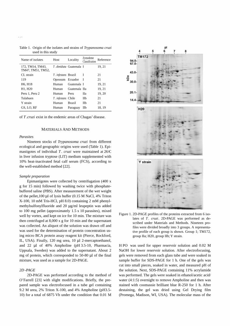
of T .cruzi exist in the endemic areas of Chagas’ disease.
MATERIALS AND METHODS
ParasitesNineteen stocks of Trypanosoma cruzi from different
ecological and geographic origins were used (Table 1). Epi-mastigotes of individual T . cruzi were maintained at 26°Cin liver infusion tryptose (LIT) medium supplemented with10% heat-inactivated fetal calf serum (FCS), according tothe well-established method [22].
Sample preparationEpimastigotes were collected by centrifugation (400 x
g for 15 min) followed by washing twice with phosphate-buffered saline (PBS). After measurement of the wet weightof the pellet,100 µl of lysis buffer (0.15 M NaCl, 4% TritonX-100, 10 mM Tris-HCl, pH 8.0) containing 2 mM phenyl-methylsulfonylfluoride and 20 µg/ml leupeptin was addedto 100 mg pellet (approximately 1.5 x 10⁹parasites), mixedwell by vortex, and kept on ice for 10 min. The mixture wasthen centrifuged at 8,000 x g for 10 min and the supernatantwas collected. An aliquot of the solution was drawn off andwas used for the determination of protein concentration us-ing micro BCA protein assay reagent kit (Pierce, Rockford,IL, USA). Finally, 120 mg urea, 10 µl 2-mercaptoethanol,and 22 µl of 40% Ampholine (pH 3.5-10, Pharmacia,Uppsala, Sweden) was added to the supernatant. About 2mg of protein, which corresponded to 50-80 µl of the finalmixture, was used as a sample for 2D-PAGE.
2D -PAGE2D-PAGE was performed according to the method of
O’Farrell [23] with slight modifications. Briefly, the pre-pared sample was electrofocused in a tube gel containing9.2 M urea, 2% Triton X-100, and 4% Ampholine (pH3.5-10) for a total of 6875 Vh under the condition that 0.01 M
H3PO4 was used for upper reservoir solution and 0.02 MNaOH for lower reservoir solution. After electrofocusing,gels were removed from each glass tube and were soaked insample buffer for SDS-PAGE for 1 h. One of the gels wascut into small pieces, soaked in water, and measured pH ofthe solution. Next, SDS-PAGE containing 11% acrylamidewas performed. The gels were soaked in ethanol/acetic acid/water (4:1:5) overnight to remove Ampholine and then wasstained with coomassie brilliant blue R-250 for 1 h. Afterdestaining, the gel was dried using Gel Drying film(Promega, Madison, WI, USA). The molecular mass of the
Table 1. Origin of the isolates and strains of Trypanosoma cruziused in this study
Name of isolates Host Locality ZymodemeClassification Reference
172, TM14, TM43,TM47, TM51, TM52,
T . dimidiata Guatemala I 19, 21
CL strain T . infestans Brazil I 21
119 Opossum Ecuador I 21
H6, H18 Human Guatemala I 19, 21
H1, H20 Human Guatemala IIa 19, 21
Peru 1, Peru 2 Human Peru IIa 19, 20
Tulahuen T . infestans Chile IIb 21
Y strain Human Brazil IIb 21
GS, LO, RF Human Paraguay IIb 18, 19
Figure 1. 2D-PAGE profiles of the proteins extracted from 6 iso-lates of T . cruzi. 2D-PAGE was performed as de-scribed under Materials and Methods. Nineteen pro-files were divided broadly into 3 groups. A representa-tive profile of each group is shown. Group 1; TM172,group IIa; H20, group IIb; Y strain.
168
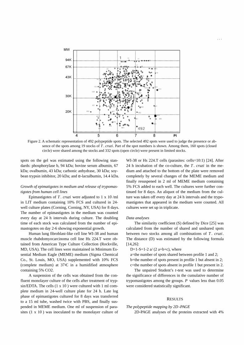
spots on the gel was estimated using the following stan-dards: phosphorylase b, 94 kDa; bovine serum albumin, 67kDa; ovalbumin, 43 kDa; carbonic anhydrase, 30 kDa; soy-bean trypsin inhibitor, 20 kDa; and α-lactalbumin, 14.4 kDa.
Growth of epimatigotes in medium and release of trypomas-tigotes from human cell lines
Epimastigotes of T . cruzi were adjusted to 1 x 10⁵/mlin LIT medium containing 10% FCS and cultured in 24-well culture plates (Corning, Corning, NY, USA) for 8 days.The number of epimastigotes in the medium was countedevery day at 24 h intervals during culture. The doublingtime of each stock was calculated from the number of epi-mastogotes on day 2-6 showing exponential growth.
Human lung fibroblast-like cell line WI-38 and humanmuscle rhabdomyocarcinoma cell line Hs 224.T were ob-tained from American Type Culture Collection (Rockville,MD, USA). The cell lines were maintained in Minimum Es-sential Medium Eagle (MEME) medium (Sigma ChemicalCo., St. Louis, MO, USA) supplemented with 10% FCS(complete medium) at 37°C in a humidified atmospherecontaining 5% CO2.
A suspension of the cells was obtained from the con-fluent monolayer culture of the cells after treatment of tryp-sin/EDTA. The cells (1 x 10⁵) were cultured with 1 ml com-plete medium in 24-well culture plate for 24 h. Late logphase of epimastigotes cultured for 8 days was transferredto a 15 ml tube, washed twice with PBS, and finally sus-pended in MEME medium. One ml of suspension of para-sites (1 x 10⁶) was inoculated to the monolayer culture of
WI-38 or Hs 224.T cells (parasites: cells=10:1) [24]. After24 h incubation of the co-culture, the T . cruzi in the me-dium and attached to the bottom of the plate were removedcompletely by several changes of the MEME medium andfinally resuspened in 2 ml of MEME medium containing5% FCS added to each well. The cultures were further con-tinued for 8 days. An aliquot of the medium from the cul-ture was taken off every day at 24 h intervals and the trypo-mastigotes that appeared in the medium were counted. Allcultures were set up in triplicate.
Data analysesThe similarity coefficient (S) defined by Dice [25] was
calculated from the number of shared and unshared spotsbetween two stocks among all combinations of T . cruzi .The distance (D) was estimated by the following formula[14,26]:
D=1-S=1-2 a/ (2 a+b+c), wherea=the number of spots shared between profile 1 and 2;b=the number of spots present in profile 1 but absent in 2;c=the number of spots absent in profile 1 but present in 2.
The unpaired Student’s t-test was used to determinethe significance of differences in the cumulative number oftrypomastigotes among the groups. P values less than 0.05were considered statistically significant.
RESULTS
The polypeptide mapping by 2D -PAGE2D-PAGE analyses of the proteins extracted with 4%
Figure 2. A schematic representation of 492 polypeptide spots. The selected 492 spots were used to judge the presence or ab-sence of the spots among 19 stocks of T . cruzi. Part of the spot numbers is shown. Among them, 160 spots (closedcircle) were shared among the stocks and 332 spots (open circle) were present in limited stocks.
169
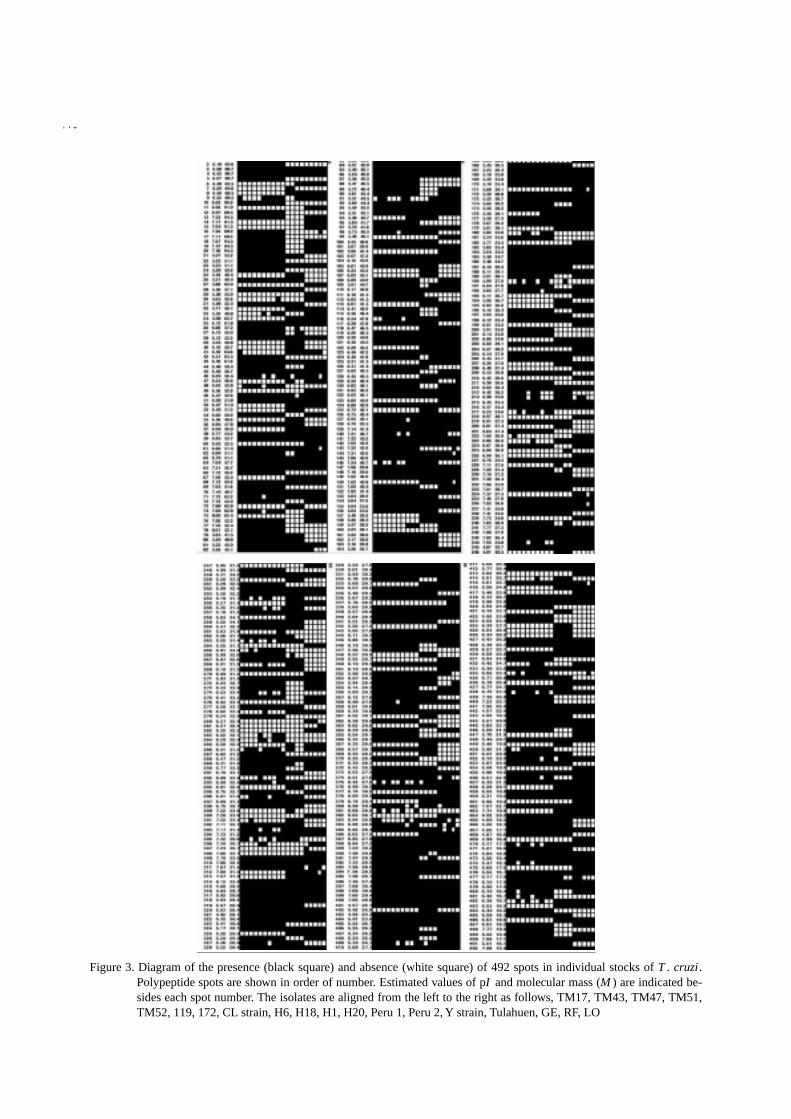
Figure 3. Diagram of the presence (black square) and absence (white square) of 492 spots in individual stocks of T . cruzi.Polypeptide spots are shown in order of number. Estimated values of pI and molecular mass (M ) are indicated be-sides each spot number. The isolates are aligned from the left to the right as follows, TM17, TM43, TM47, TM51,TM52, 119, 172, CL strain, H6, H18, H1, H20, Peru 1, Peru 2, Y strain, Tulahuen, GE, RF, LO
170
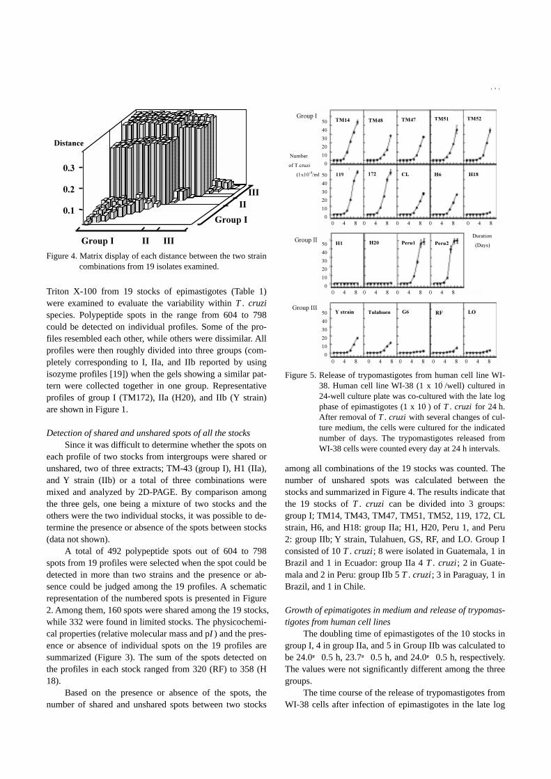
Triton X-100 from 19 stocks of epimastigotes (Table 1)were examined to evaluate the variability within T . cruzispecies. Polypeptide spots in the range from 604 to 798could be detected on individual profiles. Some of the pro-files resembled each other, while others were dissimilar. Allprofiles were then roughly divided into three groups (com-pletely corresponding to I, IIa, and IIb reported by usingisozyme profiles [19]) when the gels showing a similar pat-tern were collected together in one group. Representativeprofiles of group I (TM172), IIa (H20), and IIb (Y strain)are shown in Figure 1.
Detection of shared and unshared spots of all the stocksSince it was difficult to determine whether the spots on
each profile of two stocks from intergroups were shared orunshared, two of three extracts; TM-43 (group I), H1 (IIa),and Y strain (IIb) or a total of three combinations weremixed and analyzed by 2D-PAGE. By comparison amongthe three gels, one being a mixture of two stocks and theothers were the two individual stocks, it was possible to de-termine the presence or absence of the spots between stocks(data not shown).
A total of 492 polypeptide spots out of 604 to 798spots from 19 profiles were selected when the spot could bedetected in more than two strains and the presence or ab-sence could be judged among the 19 profiles. A schematicrepresentation of the numbered spots is presented in Figure2. Among them, 160 spots were shared among the 19 stocks,while 332 were found in limited stocks. The physicochemi-cal properties (relative molecular mass and pI ) and the pres-ence or absence of individual spots on the 19 profiles aresummarized (Figure 3). The sum of the spots detected onthe profiles in each stock ranged from 320 (RF) to 358 (H18).
Based on the presence or absence of the spots, thenumber of shared and unshared spots between two stocks
among all combinations of the 19 stocks was counted. Thenumber of unshared spots was calculated between thestocks and summarized in Figure 4. The results indicate thatthe 19 stocks of T . cruzi can be divided into 3 groups:group I; TM14, TM43, TM47, TM51, TM52, 119, 172, CLstrain, H6, and H18: group IIa; H1, H20, Peru 1, and Peru2: group IIb; Y strain, Tulahuen, GS, RF, and LO. Group Iconsisted of 10 T . cruzi; 8 were isolated in Guatemala, 1 inBrazil and 1 in Ecuador: group IIa 4 T . cruzi; 2 in Guate-mala and 2 in Peru: group IIb 5 T . cruzi; 3 in Paraguay, 1 inBrazil, and 1 in Chile.
Growth of epimatigotes in medium and release of trypomas-tigotes from human cell lines
The doubling time of epimastigotes of the 10 stocks ingroup I, 4 in group IIa, and 5 in Group IIb was calculated tobe 24.0±0.5 h, 23.7±0.5 h, and 24.0±0.5 h, respectively.The values were not significantly different among the threegroups.
The time course of the release of trypomastigotes fromWI-38 cells after infection of epimastigotes in the late log
Figure 4. Matrix display of each distance between the two straincombinations from 19 isolates examined.
Figure 5. Release of trypomastigotes from human cell line WI-38. Human cell line WI-38 (1 x 10⁵/well) cultured in24-well culture plate was co-cultured with the late logphase of epimastigotes (1 x 10⁶) of T . cruzi for 24 h.After removal of T . cruzi with several changes of cul-ture medium, the cells were cultured for the indicatednumber of days. The trypomastigotes released fromWI-38 cells were counted every day at 24 h intervals.
171
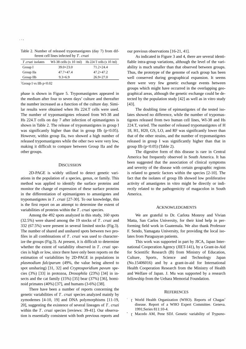
phase is shown in Figure 5. Trypomastigotes appeared inthe medium after four to seven days’ culture and thereafterthe number increased as a function of the culture day. Simi-lar results were obtained when Hs 224.T cells were used.The number of trypomastigotes released from WI-38 andHs 224.T cells on day 7 after infection of epimastigotes isshown in Table 2. The release of trypomastigotes in group Iwas significantly higher than that in group IIb (p<0.05).However, within group IIa, two showed a high number ofreleased trypomastigotes while the other two were very low,making it difficult to compare between Group IIa and theother groups.
DISCUSSION
2D-PAGE is widely utilized to detect genetic vari-ations in the population of a species, genus, or family. Thismethod was applied to identify the surface proteins andmonitor the change of expression of these surface proteinsin the differentiation of epimastigotes to amastigotes andtrypomastigotes in T . cruzi [27-30]. To our knowledge, thisis the first report on an attempt to determine the extent ofvariabilities of proteins within the T . cruzi species.
Among the 492 spots analyzed in this study, 160 spots(32.5%) were shared among the 19 stocks of T . cruzi and332 (67.5%) were present in several limited stocks (Fig.3).The number of shared and unshared spots between two pro-files in all combinations of T . cruzi was used to character-ize the groups (Fig.3). At present, it is difficult to determinewhether the extent of variability observed in T . cruzi spe-cies is high or low, since there have only been reports on theestimation of variabilities by 2D-PAGE in populations inplasmodium falciparum (49%, the value being altered tospot unsharing) [31, 32] and Cryptosporidium pavum spe-cies (3%) [33] in protozoa, Drosophila (23%) [34] in in-sects and the cat family (15%) [35] bear (37%) [36], homi-noid primates (40%) [37], and humans (3-6%) [38].
There have been a number of reports concerning thegenetic variabilities of T . cruzi species analyzed mainly byzymodemes [4-10, 19] and DNA polymorphisms [11-19,20], suggesting the existence of several lineages of T . cruziwithin the T . cruzi species [reviews: 39-41]. Our observa-tion is essentially consistent with both previous reports and
our previous observations [16-21, 41].As indicated in Figure 3 and 4, there are several identi-
fiable intra-group variations, although the level of the vari-ability is much smaller than that observed between groups.Thus, the prototype of the genome of each group has beenwell conserved during geographical expansion. It seemsthere were very few genetic exchange events betweengroups which might have occurred in the overlapping geo-graphical areas, although the genetic exchange could be de-tected by the population study [42] as well as in vitro study[43].
The doubling time of epimastigotes of the tested iso-lates showed no difference, while the number of trypomas-tigotes released from two human cell lines, WI-38 and Hs224.T, varied. The number of released trypomastigotes of H18, H1, H20, GS, LO, and RF was significantly lower thanthat of the other strains, and the number of trypomastigotesreleased in group I was significantly higher than that ingroup IIb (p<0.05) (Table 2).
The digestive form of this disease is rare in CentralAmerica but frequently observed in South America. It hasbeen suggested that the association of clinical symptomsand severity of the disease with certain geographic regionsis related to genetic factors within the species [2-10]. Thefact that the isolates of group IIb showed low proliferativeactivity of amastigotes in vitro might be directly or indi-rectly related to the pathogenicity of magacolon in SouthAmerica.
ACKNOWLEDGMENTS
We are grateful to Dr. Carlota Monroy and VivianMatta, San Carlos University, for their kind help in per-forming field work in Guatemala. We also thank ProfessorF. Sendo, Yamagata University, for providing the local iso-lates from Paraguayan patients.
This work was supported in part by JICA, Japan Inter-national Cooperation Agency (JIET-141), by a Grant-in-Aidfor Scientific Research (B) from Ministry of Education,Culture , Sports , Science and Technology Japan(No.15406016) and by a grant-in-aid for InternationalHealth Cooperation Research from the Ministry of Healthand Welfare of Japan. J. Mu was supported by a researchfellowship from the Uehara Memorial Foundation.
REFERENCES
1.World Health Organization (WHO). Reports of Chagas’disease. Report of a WHO Expert Committee. Geneva.1991;Series 811:10-4.
2.Macedo AM, Pene SDJ. Genetic variability of Trypano-
Table 2. Number of released trypomastigotes (day 7) from dif-ferent cell lines infected by T . cruzi
T .cruzi isolates WI-38 cells (x 104/ml) Hs 224 T cells (x 104/ml)
Group I 39.0+23.0* 71.2+24.4*
Group IIa 47.7+47.4 47.2+47.2
Group IIb 9.3+6.9* 26.9+27.0*
*Group I vs IIb p<0.02
172
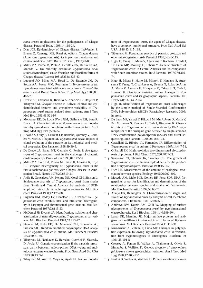
soma cruzi: implications for the pathogenesis of Chagasdisease. Parasitol Today 1998;14:119-24.
3.Dias JCP. Epidemiology of Chagas disease. In:Wendel S,Brener Z, Camargo ME, Rassi A, editors. Chagas disease(American trypanosomiasis): its impact on transfusion andclinical medicine. ISBT Brazil’92:Brazil, 1992;49-80.
4.Miles MA, Poroa M, Prata A, Cedillos RA, De Souza AA,Macedo V. Do radically dissimilar Trypanosoma cruzistrains (zymodemes) cause Vezuelan and Brazilian forms ofChagas’ disease? Lancet 1981;8234:1336-40.
5.Luquetti AO, Miles MA, Rossi L, De Rezende JM, DeSouza AA, Povoa MM, Rodrigues I. Trypanosoma cruzi:zymodemes associated with acute and chronic Chagas’ dis-ease in cetral Brazil. Trans R Soc Trop Med Hyg 1986;80:462-70.
6.Brenie SF, Carrasco R, Revollo S, Aparicio G, Desjeux P,Tibayrenc M. Chagas’ disease in Bolivia: clinical and epi-demiological features and zymodeme variability of Try-panosoma cruzi strains isolated from patients. Am J TropMed Hyg 1989;41:521-9?
7.Montamat EE, De Lucia D’oro GM, Gallerano RH, Sosa R,Blanco A. Characterization of Trypanosoma cruzi popula-tions by zymodemes: correlation with clinical picture. Am JTrop Med Hyg 1996;55:625-8.
8.Revollo S, Oury B, Laurent J-P, Barnabé, Quesney V, Carri-ère V, Noël S, Tibayrenc M. Trypanosoma cruzi: impact ofclonal evolution of the parasite on its biological and medi-cal properties. Exp Parasitol 1998;89:30-9.
9.De Diego JA, Palau MT, Gamallo C, Penin P. Are geno-types of Trypanosoma cruzi in the challenge of chagasiccardiomyopathy? Parasitol Res 1998;84:147-52.
10.Miles MA, Souza A, Povoa M, Shaw JJ, Lainson R, ToyePJ. Isozymic heterogeneity of Trypanosoma cruzi in thefirst autochthonous patients with Chagas’ disease in Ama-zonian Brazil. Nature 1978;272:819-21.
11.Avila H, Goncalves AM, Nehme NS, Morel CM, Simson L.Schizodeme analysis of Trypanosoma cruzi from stocksfrom South and Central America by analysis of PCR-amplified minicircle variable region sequences. Mol Bio-chem Parasitol 1990;42:175-88.
12.Engman DM, Reddy LV, Donelson JE, Kirchhoff LV. Try-panosoma cruzi exhibits inter- and intra-strain heterogene-ity in karyotype and chromosomal gene location. Mol Bio-chem Parasitol 1987;22:115-23.
13.McDaniel JP, Dvorak JA. Identification, isolation and char-acterization of naturally-occurring Trypanosoma cruzi vari-ants. Mol Biochem Parasitol 1993;57:213-22.
14.Steindel M, Neto ED, De Menezes CLP, Romanha AJ,Simson AJG. Random amplified polymorphic DNA analy-sis of Trypanosoma cruzi strains. Mol Biochem Parasitol1993;60:71-80.
15.Tibayrenc M, Neubauer K, Barnabé, Guerrini F, SkareckyD, Ayala FJ. Genetic charactrization if six parasitic proto-zoa: parity between random-primer DNA typing and mul-tulocus enzyme electrophoresis. Proc Natal Aced Sic USA1993;90:1335-9.
16.Tibayrenc M, Ward P, Moya A, Ayala FJ. Natural popula-
tions of Trypanosoma cruzi, the agent of Chagas disease,have a complex multiclonal structure. Proc Natl Acad SciUSA 1986;83:115-119.
17.Tibayrenc M. Population genetics of parasitic protozoa andother microorganisms. Adv Parasitol 1995;36:48-115.
18.Higo, H, Yanagi T, Matta V, Agatsuma T, Kanbara H, Tada I,De Leon MP, Monroy C, Tabaru Y. Genetic structure ofTrypanosoma cruzi in Central America and its comparisonwith South American strains. Int J Parasitol 1997;27:1369-74.
19.Higo H, Miura S, Horio M, Mimori T, Hamano S, Agat-suma T, Yanagi T, Cruz-Reyes A, Uyema N, Rojas de AriasA, Matta V, Akahara H, Hirayama K, Takeuchi T, Tada I,Himeno K. Genotypic variation among lineages of Try-panosoma cruzi and its geographic aspects. Parasitol Int.Dec;53(4):337-44, 2004
20.Higo H, Identification of Trypanosoma cruzi sublineagesby the simple method of Single-Stranded ConformationDNA Polymorphism (SSCP). Parasitology Research, 2006;in press.
21.De Leon MP, Yanagi T, Kikuchi M, Mu J, Ayau O, Matta V,Paz M, Juarez S, Kanbara H, Tada I, Hirayama K. Charac-terization of Trypanosoma cruzi populations by DNA poly-morphism of the cruzipain gene detected by single-strandedDNA conformation polymorphism (SSCP) and direct se-quencing. Int J Parasitol 1998;28:1867-74.
22.Castellani O, Ribeiro LV, Fernandes JF. Differentiation ofTrypanosoma cruzi in culture. J Protozoon 1967;14:447-51.
23.O’Farrell PH. High resolution two-dimensional electropho-resis of proteins. J Biol Chem. 1975;250:4007-21.
24.Sanderson CJ, Thomas JA, Twomey CE. The growth ofTrypanosoma cruzi in human diploid cells for the produc-tion of trypomastigotes. Parasitol 1980;153-62.
25.Dice LR. Measurement of the amount of ecological asso-ciation between species. Ecology 1945;26:297-302.
26.Macedo AM, Melo MN, Gomes RF, Pena SDJ. DNA fin-gerprints: a tool for identification and determination of therelationship between species and strains of Leishmania .Mol Biochem Parasitol 1992;53:63-70.
27.Araujo FG, Remington JS. Characterization of stages andstrains of Trypanosoma cruzi by analysis of cell membranecomponents. J Immunol 1981;127:855-9.
28.Andrews NW, Katzin AM, Colli W. Mapping of surfaceglycoproteins of Trypanosoma cruzi by two-dimensionalelectrophoresis. Eur J Biochem 1984;140:599-604.
29.Lanar DE, Manning JE. Major surface proteins and anti-gens on the different in vivo and in vitro forms of Trypano-soma cruzi. Mol Biochem Parasitol 1984;11:119-31.
30.Ruiz-Ruano A, Villalta F, Lima MF. Changes in polypep-tide expression following Trypanosoma cruzi differentia-tion from trypomastigotes to amastigotes. Biochem Int1991;25:101-8.
31.Creasey A, Fenton B, Walker A, Thaithong S, Olivia S,Mutambu S, Walliker D. Genetic diversity of plasmodiumfalciparum shows geographical variation. Am J Trop MedHyg 1990;42:403-13?
32.Fenton B, Walker A, Walliker D. Protein variation in clones
173

of plasmodium falciparum detected by two dimensionalelectrophoresis. Mol Biochem Parasitol 1985;16:173-83.
33.Mead JR, Humphreys RC, Sammons DW, Sterling CR.Identification of isolate-specific sporozoite proteins ofCryptosporidium parvum by two-dimensional gel electro-phoresis. Info Immune 1990;58:2071-5.
34.Choudhary M, Coulthart MB, Singh RS. A comprehensivestudy of genic variation in natural populations of Droso-phila melanogaster . VI. Patterns and process of genic di-vergence between D. melanogaster and its sibling species,Drosophila simulans. Genetics 1992;130:843-53.
35.Slattery JP, Johnson WE, Goldman D, O’Brien SJ. Phylo-genetic reconstruction of South American felids defined byprotein electrophoresis. J Mol Evil 1994;39:296-305.
36.Goldman D, Geri PR. Molecular genetic-distance estimatesamong theursidae as indicated by one- and two-dimensional protein electrophoresis. Evolution 1989;43:282-95.
37.Goldman D, Giri PR, O’Brien SJ. A molecular phylogenyof the hominoid primates as indicated by two-dimensionalprotein electrophoresis. Proc Natl Acad Sci USA 1987;84:3307-11.
38.Goldman D, O’Brien SJ, Lucas-Derse S, Dean M. Linkagemapping of human polymorphic proteins identified by two-dimensional electrophoresis. Genomics 1991;11:875-84.
39.Macedo AM, Pena SDJ. Genetic variability of Trypano-soma cruzi: implications for the pathogenesis of Chagasdisease. Parasitol Today 1998;14:119-124.
40.Souto RP, Zingales B, Fernandes O, Macedo AM, Camp-bell DA. Trypanosoma cruzi: how many relevant phyloge-netic subdivisions are there? Reply. Parasitol Today 1998;14:207.
41.Barnabe C, Brisse S, Tibayrenc M. Population structureand genetic typing of Trypanosoma cruzi, the agent ofChagas disease: a multilocus enzyme electrophoresis ap-proach. Parasitology 2000;120:513-526
42.Machado CA, Ayala FJ. Nucleotide sequences provide evi-dence of genetic exchange among distantly related lineagesof Trypanosoma cruzi. Proc Natl Acad Sci U S A. 2001 Jun19;98(13):7396-401.
43.Gaunt MW, Yeo M, Frame IA, Stothard JR, Carrasco HJ,Taylor MC, Mena SS, Veazey P, Miles GA, Acosta N, deArias AR, Miles MA. Mechanism of genetic exchange inAmerican trypanosomes. Nature 2003;421:936-9.
174