Embed Size (px)
Citation preview

ISSN 1286-0107
Vol 19 • No.4 • 2012 • p161-204
Endovenous thermal ablation for varicose veins: . . . . . PAGE 163strengths and weaknesses
Renate R. van den BOS (Rotterdam, The Netherlands)
Venous embryology: the key to understanding . . . . . PAGE 170anomalous venous conditions
Byung-Boong LEE (Washington D.C., USA)
The “C0s” patient: worldwide results . . . . . PAGE 182from the Vein Consult Program
Jean-Jérôme GUEX et al. (Nice, France)
Sclerotherapy in the patient with diabetes: . . . . . PAGE 193indications and results
Francesco FERRARA, Giovanni FERRARA (Naples, Italy)

Indexed in EMBASE, PASCAL,Index Copernicus, and Scopus.
Phlebolymphology
AIMS AND SCOPE
H. Partsch, MDProfessor of Dermatology, Emeritus Head of the Dermatological Department of the Wilhelminen HospitalBaumeistergasse 85, A 1160 Vienna, Austria
C. Allegra, MDHead, Dept of AngiologyHospital S. Giovanni, Via S. Giovanni Laterano, 155, 00184, Rome, Italy
P. Coleridge Smith, DM, FRCSConsultant Surgeon & Reader in SurgeryThames Valley Nuffield Hospital, Wexham Park Hall, Wexham Street, Wexham, Bucks, SL3 6NB, UK
M. De Maeseneer, MDDepartment of Dermatology,Erasmus Medical Centre, BP 2040, 3000 CA Rotterdam, Netherlands
A. Jawien, MD, PhDDepartment of SurgeryLudwik Rydygier University Medical School, Ujejskiego 75, 85-168 Bydgoszcz, Poland
G. W. Schmid Schönbein, MS, PhDProfessor of Bioengineering and MedicineThe Whitaker Institute for Biomedical Engineering, University of California San Diego, 9500 Gilman Drive, La Jolla, CA 92093-0412, USA
EDITORIAL MANAGER
F. Pitsch, PharmDServier International
EDITORIAL BOARD
EDITOR IN CHIEF
Phlebolymphology is an internationalscientific journal entirely devoted tovenous and lymphatic diseases.
The aim of Phlebolymphology is to pro-vide doctors with updated information onphlebology and lymphology written bywell-known international specialists.
Phlebolymphology is scientifically sup-ported by a prestigious editorial board.
Phlebolymphology has been pub lishedfour times per year since 1994, and,thanks to its high scientific level, wasincluded in several databases.
Phlebolymphology comprises an edito-rial, articles on phlebology and lympho-logy, reviews, news, and a congresscalendar.
CORRESPONDENCE
Editor in ChiefHugo PARTSCH, MDBaumeistergasse 851160 Vienna, AustriaTel: +43 431 485 5853 Fax: +43 431 480 0304E-mail: [email protected]
Editorial ManagerFrançoise PITSCH, PharmDServier International50, rue Carnot92284 Suresnes Cedex, FranceTel: +33 (1) 55 72 68 96 Fax: +33 (1) 55 72 56 86E-mail: [email protected]
PublisherLes Laboratoires Servier50, rue Carnot92284 Suresnes Cedex, FranceTel: +33 (1) 55 72 60 00 Fax: +33 (1) 55 72 68 88
© 2012 Les Laboratoires Servier - All rights reserved throughout the world and in all languages. No part of this publicationmay be reproduced, transmitted, or stored in any form or by any means either mechanicalor electronic, including photocopying, recording,or through an information storage and retrievalsystem, without the written permission of the copyright holder. Opinions expressed do notnecessarily reflect the views of the publisher, editors, or editorial board. The authors, editors,and publisher cannot be held responsible forerrors or for any consequences arising from theuse of the information contained in this journal.
ISSN 1286-0107

Phlebolymphology. Vol 19. No. 4. 2012 161
CONTENTS
EDITORIAL
Hugo PARTSCH (Vienna, Austria) Page 162
PHLEBOLOGYEndovenous thermal ablation for varicose veins: Page 163
strengths and weaknessesRenate R. van den BOS (Rotterdam, The Netherlands)
Venous embryology: the key to understanding Page 170
anomalous venous conditionsByung-Boong LEE (Washington D.C., USA)
The “C0s” patient: worldwide results Page 182
from the Vein Consult ProgramJean-Jérôme GUEX et al. (Nice, France)
Sclerotherapy in the patient with diabetes: Page 193
indications and resultsFrancesco FERRARA, Giovanni FERRARA (Naples, Italy)
CONGRESS
Congress and conference calendar Page 200
162 Phlebolymphology. Vol 19. No. 4. 2012
EDITORIAL
Dear Readers,
This issue of Phlebolymphology offers an exciting insight into someactual and very relevant fields of phlebology.
Renate van den Bos and Marianne de Maeseneer, both workingat the Dermatological University clinic in Rotterdam and outstandingexperts in the field, discuss the strengths and weaknesses of theendovenous thermal ablation of varicose veins. They point out thatduplex-guided endovenous procedures have widely replaced classicalstripping and that the newly developed steam ablation procedure hasa promising future, being safer, faster, easier, and cheaper thanendovenous laser and radiofrequency ablation (see also the article byRené Milleret: Obliteration of Varicose Veins with Superheated Steam.Phlebolymphology. 2011;19:174-187).
In an extensive review article, Professor Byung-Boong Lee, GeorgeWashington University School of Medicine, shows that knowledge ofembryology may be key to understanding vascular pathologies, forinstance for the differentiation between truncular and extratruncularforms of vascular malformations. His article contains beautifulillustrations of clinical conditions: radiological, MRI, and nuclearmedicine pictures and schematic drawings, which are helpful tounderstand this very complex topic.
Jean-Jérôme Guex from Nice, together with a group of prominentcoauthors working on the Vein Consult Program, a worldwideepidemiological survey (see the article by Francoise Pitsch in the last issueof Phlebolymphology; 2012;19:132-137), has analyzed a large groupof symptomatic C0 patients, a cohort of patients that is well-known toevery phlebologist. Out of a total of 91 545 subjects, 19.7% did notshow any visible or palpable signs of venous disease—this is thedefinition of the C0 class according to the CEAP classification—butpresented with subjective symptoms. Most of the 14% of patients whounderwent a duplex scan examination had superficial reflux, and 18%had deep reflux. The authors suggest that an “inflammatory hypothesis”may be the cause of the symptoms, which include a reduced quality oflife, and propose that venoactive drugs should be used for treatment.
Based on their extraordinary experience with sclerotherapy using Sigg’stechnique, Francesco and Giovanni Ferrara from Naples haveshowed, in a case series of 60 patients, that diabetes mellitus with goodglycemic control (HbA1c<6.5%) is not a contraindication for thistechnique, but rather gives equally good results and no morecomplications than in nondiabetics.
Have a great read!
Hugo PartschEditor in Chief
Phlebolymphology. Vol 19. No. 4. 2012 163
PHLEBOLOGY
Endovenous thermal ablation for varicoseveins: strengths and weaknesses
Keywords:
endovenous ablation treatments, saphenousinsufficiency, thermal ablation, varicose veins
Phlebolymphology. 2012;19(4):163-169.
ABSTRACTEndovenous ablation is a frequently used method for treating varicose veins.Endovenous laser ablation is the most frequently used technique, followed byradiofrequency ablation. Endovenous thermal treatments heat the vein,leading to thrombotic occlusion and finally fibrosis of the vein wall.Endovenous steam ablation is a new technique that has not yet beenextensively studied. In this article, the procedures, strengths, and weaknessesof the currently available endovenous thermal ablation treatments arediscussed.
INTRODUCTIONEndovenous treatment is currently one of the most frequently used methodsfor treating varicose veins. Varicose veins are manifestations of chronicvenous disease (CVD), which may lead to serious complications. CVD is acommon medical condition. The prevalence of varicose veins is estimated torange from 2%-40%.1-4 The prevalence of venous leg ulcers, the end-stage ofCVD, is much lower. It is very difficult, if not impossible, to predict whichpatients with varicose veins will develop a leg ulcer. Nevertheless, it has beenestimated that about half of venous leg ulcers are the result of superficialvenous insufficiency.5 The cost of treating leg ulcers is very high; thetreatment of varicose veins, which may reduce the incidence of leg ulcers by50%, is therefore likely to be cost-effective.
Treatment for varicose veins can roughly be divided into four categories:compression therapy, surgical treatment, sclerotherapy, and endovenousthermal ablation. Surgical ligation of the junction with or without strippinghas been the standard of care in the treatment of insufficient great and smallsaphenous veins for more than 100 years.
In the last decade, endovenous thermal ablation (EVTA) procedures havebecome the most frequently used therapy for saphenous varicose veins,especially in countries where reimbursement of the procedure has beenintroduced. Such minimally invasive techniques meet the demand forcosmetically superior, less invasive and more successful treatment modalities.Only introduced 10 years ago, these techniques have radically changed the
Renate R. van den BOS,Marianne M.G. de MAESENEERDepartment of Dermatology, Erasmus MC Rotterdam, The Netherlands
164 Phlebolymphology. Vol 19. No. 4. 2012
Renate R. van den BOS, Marianne M.G. de MAESENEERPHLEBOLOGY
treatment of varicose veins.6 The EVTA techniquescurrently available are: endovenous laser ablation(EVLA), radiofrequency ablation (RFA), and endovenoussteam ablation. The advantage of EVTA is that it isminimally invasive and can easily be performed underlocal tumescent anesthesia, without the need for spinalor general anesthesia. Moreover, according to a meta-analysis of the different treatment techniques forvaricose veins, recurrence rates are lower after EVTAthan after classic surgery.7
The first EVTA procedures were performed by RFA withthe VNUS® Closure Plus system.8 EVLA was developedsoon after and soon became the most frequently usedEVTA method around the world. In the last few years,two new RFA systems have been introduced: VNUSClosure Fast (segmental RFA) and radiofrequencyinduced thermotherapy (RFITT). The latest thermalablation technique uses steam at a temperature of 120°C.In the following paragraphs, the different EVTAtechniques will be described and their strengths andweaknesses explored.
ENDOVENOUS LASER ABLATION
ProcedureEVLA can be performed under local tumescentanesthesia in an outpatient setting. Venous access isobtained by puncturing with a 16F or 18F needle orcannula under ultrasound guidance. Most commonly,the insufficient great saphenous vein (GSV) is entered atknee level and the small saphenous vein (SSV) at mid-calf. After entrance to the vein has been established, aguide wire is passed through the needle into the vein upto the level of the junction with the deep venous system.If the vein is too tortuous, is small in diameter (due tospasm) and has large side branches, or containsthrombotic or sclerotic segments (after superficial veinthrombosis or prior treatment), advancing the wire canbe difficult and caution is indicated because of theincreased risk of perforation and embolic events. Afterchecking the position of the guide wire with ultrasound,the needle is removed, and a small cutaneous incision of3 mm is made. An introducer sheath is placed over theguide wire and is positioned a few centimeters below thejunction. Subsequently, the laser fiber (diameter rangesfrom 200 to 600 μm) is introduced after removing theguide wire. In some laser sets, there is no guide wire andthe sheath is directly introduced through a cannula. In
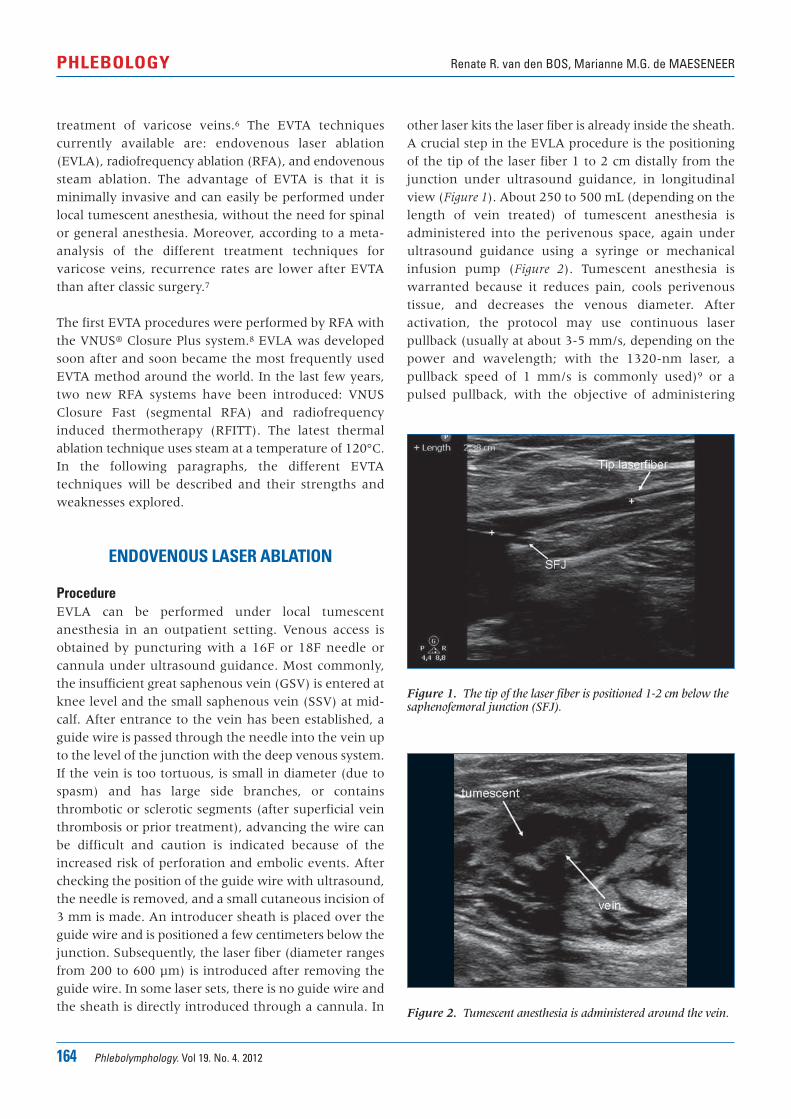
other laser kits the laser fiber is already inside the sheath.A crucial step in the EVLA procedure is the positioningof the tip of the laser fiber 1 to 2 cm distally from thejunction under ultrasound guidance, in longitudinalview (Figure 1). About 250 to 500 mL (depending on thelength of vein treated) of tumescent anesthesia isadministered into the perivenous space, again underultrasound guidance using a syringe or mechanicalinfusion pump (Figure 2). Tumescent anesthesia iswarranted because it reduces pain, cools perivenoustissue, and decreases the venous diameter. Afteractivation, the protocol may use continuous laserpullback (usually at about 3-5 mm/s, depending on thepower and wavelength; with the 1320-nm laser, apullback speed of 1 mm/s is commonly used)9 or apulsed pullback, with the objective of administering
Figure 1. The tip of the laser fiber is positioned 1-2 cm below thesaphenofemoral junction (SFJ).
Figure 2. Tumescent anesthesia is administered around the vein.
Phlebolymphology. Vol 19. No. 4. 2012 165
Endovenous thermal ablation PHLEBOLOGY
about 30 to 60 J/cm. Compressive bandages or medicalelastic stockings (20-30 mm Hg at the ankle) areindicated for 1 week after treatment.
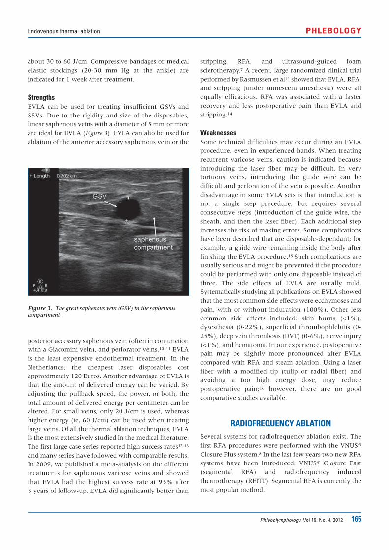
StrengthsEVLA can be used for treating insufficient GSVs andSSVs. Due to the rigidity and size of the disposables,linear saphenous veins with a diameter of 5 mm or moreare ideal for EVLA (Figure 3). EVLA can also be used forablation of the anterior accessory saphenous vein or the
stripping, RFA, and ultrasound-guided foamsclerotherapy.7 A recent, large randomized clinical trialperformed by Rasmussen et al14 showed that EVLA, RFA,and stripping (under tumescent anesthesia) were allequally efficacious. RFA was associated with a fasterrecovery and less postoperative pain than EVLA andstripping.14
WeaknessesSome technical difficulties may occur during an EVLAprocedure, even in experienced hands. When treatingrecurrent varicose veins, caution is indicated becauseintroducing the laser fiber may be difficult. In verytortuous veins, introducing the guide wire can bedifficult and perforation of the vein is possible. Anotherdisadvantage in some EVLA sets is that introduction isnot a single step procedure, but requires severalconsecutive steps (introduction of the guide wire, thesheath, and then the laser fiber). Each additional stepincreases the risk of making errors. Some complicationshave been described that are disposable-dependant; forexample, a guide wire remaining inside the body afterfinishing the EVLA procedure.15 Such complications areusually serious and might be prevented if the procedurecould be performed with only one disposable instead ofthree. The side effects of EVLA are usually mild.Systematically studying all publications on EVLA showedthat the most common side effects were ecchymoses andpain, with or without induration (100%). Other lesscommon side effects included: skin burns (<1%),dysesthesia (0-22%), superficial thrombophlebitis (0-25%), deep vein thrombosis (DVT) (0-6%), nerve injury(<1%), and hematoma. In our experience, postoperativepain may be slightly more pronounced after EVLAcompared with RFA and steam ablation. Using a laserfiber with a modified tip (tulip or radial fiber) andavoiding a too high energy dose, may reducepostoperative pain;16 however, there are no goodcomparative studies available.
RADIOFREQUENCY ABLATIONSeveral systems for radiofrequency ablation exist. Thefirst RFA procedures were performed with the VNUS®
Closure Plus system.8 In the last few years two new RFAsystems have been introduced: VNUS® Closure Fast(segmental RFA) and radiofrequency inducedthermotherapy (RFITT). Segmental RFA is currently themost popular method.
Figure 3. The great saphenous vein (GSV) in the saphenouscompartment.
posterior accessory saphenous vein (often in conjunctionwith a Giacomini vein), and perforator veins.10-11 EVLAis the least expensive endothermal treatment. In theNetherlands, the cheapest laser disposables costapproximately 120 Euros. Another advantage of EVLA isthat the amount of delivered energy can be varied. Byadjusting the pullback speed, the power, or both, thetotal amount of delivered energy per centimeter can bealtered. For small veins, only 20 J/cm is used, whereashigher energy (ie, 60 J/cm) can be used when treatinglarge veins. Of all the thermal ablation techniques, EVLAis the most extensively studied in the medical literature.The first large case series reported high success rates12-13
and many series have followed with comparable results.In 2009, we published a meta-analysis on the differenttreatments for saphenous varicose veins and showedthat EVLA had the highest success rate at 93% after 5 years of follow-up. EVLA did significantly better than
166 Phlebolymphology. Vol 19. No. 4. 2012
Renate R. van den BOS, Marianne M.G. de MAESENEERPHLEBOLOGY
ProcedureAccess to the GSV is obtained with a 16-gauge needleunder ultrasound guidance, typically at or below kneelevel or at the most distal point of reflux. The SSV isusually punctured at mid calf. The Closure catheter(VNUS Medical Technologies, Inc, Sunnyvale, California)is positioned 2 cm distally from the junction underlongitudinal ultrasound visualization. With the ClosurePlus system, a cuff or bandage can be used to express theblood from the vein. The small electrodes at the end ofthe “umbrella” catheter have direct contact with thevenous wall and emit high radiofrequency energy(regulated by power, impedance, and time) that isgenerated by a radiofrequency generator (VNUS MedicalTechnologies, Inc). The radiofrequency heats local tissueup to 85°C to 90°C at the site of direct contact, with theheat conducted to deeper tissue planes, causing collagenshrinkage, denudation of endothelium, and obliterationof the venous lumen.17 The catheter pullback speed is 3 cm/min (total pullback time is 20 min on average forthe GSV between the saphenofemoral junction and kneelevel, but can be faster at higher temperatures).18
Segmental RFA (Closure Fast) has a 7 cm therapeuticdistal segment that heats to 120°C.19 This technique ismuch faster than the Closure Plus technique and can beperformed under local tumescent anesthesia in anoutpatient setting. Similar to EVLA, perivenoustumescent anesthesia is applied to optimize surfacecontact and to decrease pain and risk of dysesthesia.20
According to the methodology described in the firstreport on segmental RFA, external compression providedby the ultrasound probe and manual compression isrecommended during the treatment to enhance contactof the catheter with the vein wall.21 The first 7 cm of veinis treated with two heat cycles (20 s each). The catheteris then repositioned to the adjacent segment guided byshaft markers in 6.5-cm steps to allow a 5 mm overlap ofheated vein segments. Total treatment time is muchshorter with segmental RFA than with the Closure Plussystem and usually takes only 2 to 3 min. Compressivebandages or medical elastic compression stockings areindicated for 1 week after treatment.
StrengthsSince 2000, several published case series have shownthat RFA can be successfully used to treat saphenousvaricose veins.8, 22-25 The first long-term, large, single-center case series reported that RFA was effective inabout 90% of 140 limbs after 2 years.20 A separate study
reported success rates of 83%-88% after 5-year follow-up.26 Our meta-analysis showed that RFA (using ClosurePlus) had a success rate of 88%, which was lower thanthe success rate of EVLA.6
Segmental RFA was not included in the analysis, as atthe time no studies were available. However, there arenow a few publications on segmental RFA withpromising results. The first case series of 252 treatedGSVs reported an occlusion rate of 99.6%,21 and twoother trials demonstrated success rates >90%.27,28 Themain advantage of segmental RFA is probably that itresults in less postoperative pain than EVLA. This isthought to be related to the lower maximal temperaturethat is reached during RFA, and the absence of vein wallperforations.27 A further advantage of segmental RFA isthat it is a standardized procedure and introduction ofthe catheter is performed in one step. This may lowerthe risk of disposable-related complications.
WeaknessesOn the one hand, standardization of a procedure is anadvantage. On the other hand, it may not be possible totreat certain ‘special’ cases. With segmental RFA it isimpossible to treat veins with a length smaller than 7 cm,although this may change with the recent introductionof a new catheter with a 3 cm heating segment. Incertain cases (ie, patients with side branches, or smalltortuous parts of varicose veins), it may also be desirableto change the energy delivery, but with segmental RFAit is not possible to treat veins at other than the presettemperature. As a result of the relatively lowtemperature that is reached during segmental RFA, theworking mechanism is collagen denaturation andshrinkage of the vein wall.17 This differs from EVLA inwhich carbonization and more rigorous destruction ofthe vein wall is also reported.29 The long-termeffectiveness of segmental RFA has not yet been studiedand will only become clear after a randomized studycomparing EVLA and segmental RFA with long-termfollow-up has been conducted.
STEAM ABLATION
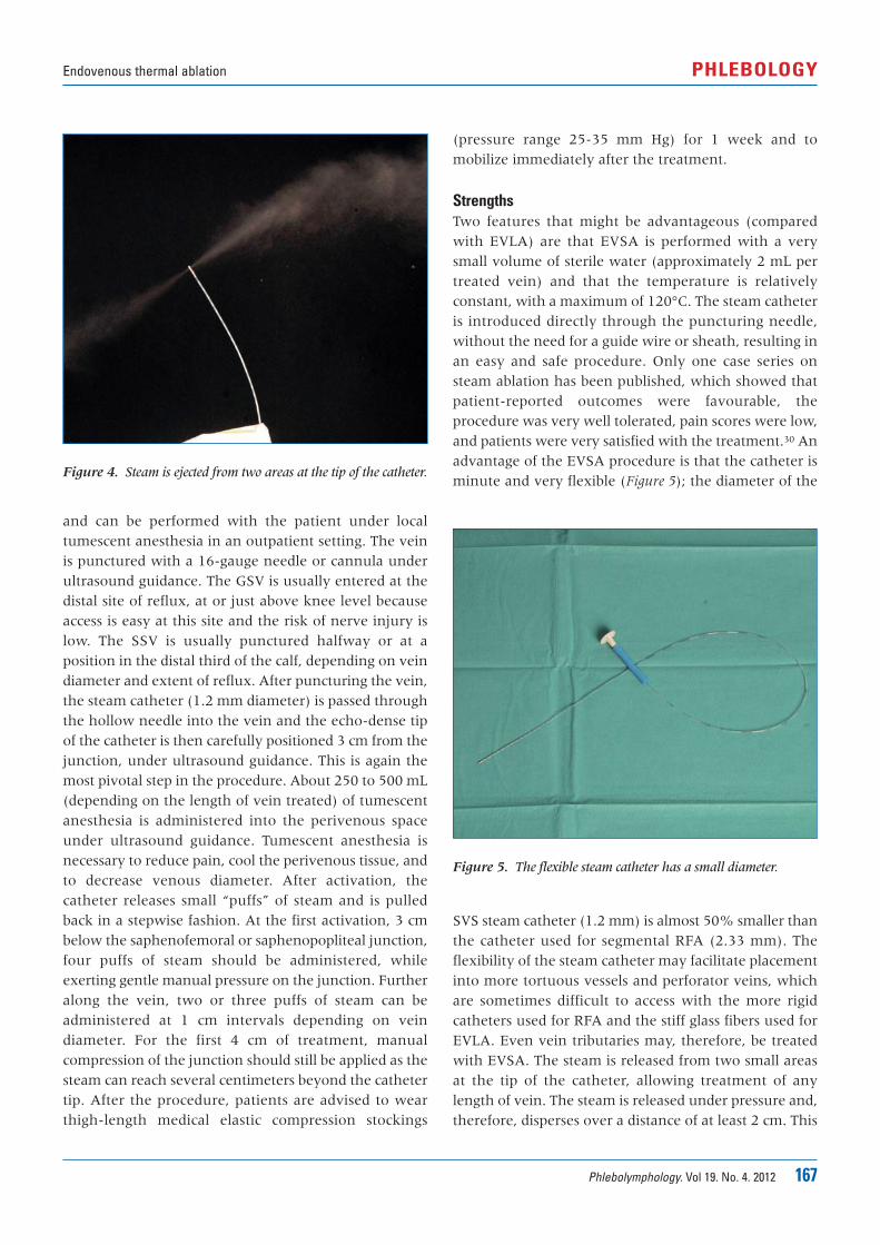
ProcedureEndovenous steam ablation (EVSA) is a new method ofthermal vein ablation that works by heating the venousstructure with steam to a maximum temperature of120°C (Figure 4). The procedure is very similar to EVLA
Phlebolymphology. Vol 19. No. 4. 2012 167
Endovenous thermal ablation PHLEBOLOGY
and can be performed with the patient under localtumescent anesthesia in an outpatient setting. The veinis punctured with a 16-gauge needle or cannula underultrasound guidance. The GSV is usually entered at thedistal site of reflux, at or just above knee level becauseaccess is easy at this site and the risk of nerve injury islow. The SSV is usually punctured halfway or at aposition in the distal third of the calf, depending on veindiameter and extent of reflux. After puncturing the vein,the steam catheter (1.2 mm diameter) is passed throughthe hollow needle into the vein and the echo-dense tipof the catheter is then carefully positioned 3 cm from thejunction, under ultrasound guidance. This is again themost pivotal step in the procedure. About 250 to 500 mL(depending on the length of vein treated) of tumescentanesthesia is administered into the perivenous spaceunder ultrasound guidance. Tumescent anesthesia isnecessary to reduce pain, cool the perivenous tissue, andto decrease venous diameter. After activation, thecatheter releases small “puffs” of steam and is pulledback in a stepwise fashion. At the first activation, 3 cmbelow the saphenofemoral or saphenopopliteal junction,four puffs of steam should be administered, whileexerting gentle manual pressure on the junction. Furtheralong the vein, two or three puffs of steam can beadministered at 1 cm intervals depending on veindiameter. For the first 4 cm of treatment, manualcompression of the junction should still be applied as thesteam can reach several centimeters beyond the cathetertip. After the procedure, patients are advised to wearthigh-length medical elastic compression stockings
(pressure range 25-35 mm Hg) for 1 week and tomobilize immediately after the treatment.
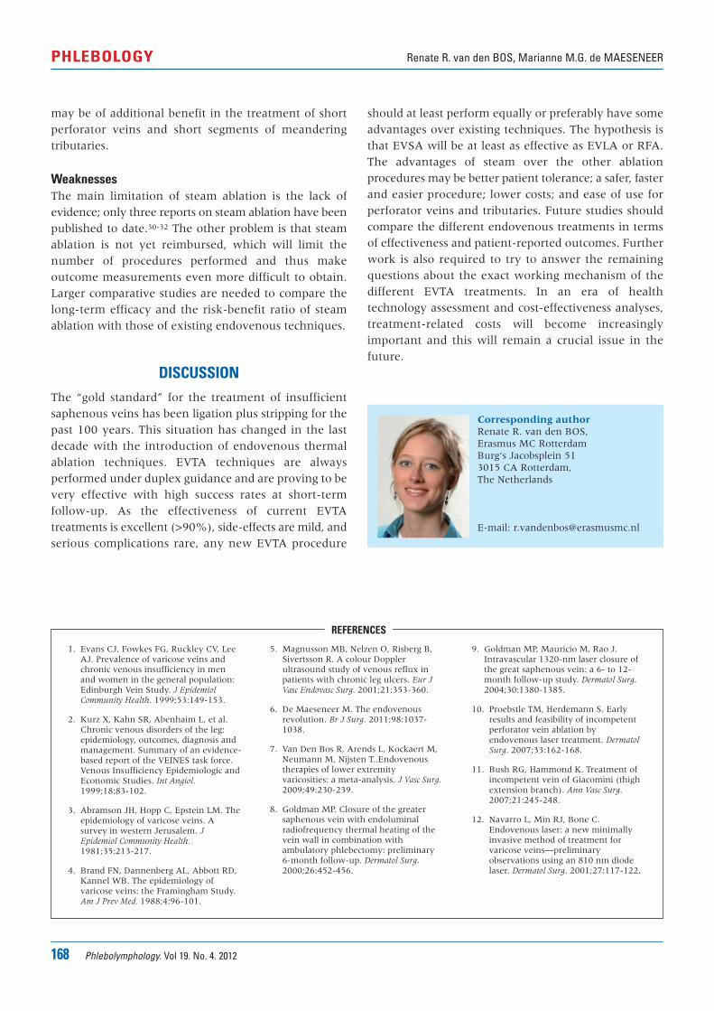
StrengthsTwo features that might be advantageous (comparedwith EVLA) are that EVSA is performed with a verysmall volume of sterile water (approximately 2 mL pertreated vein) and that the temperature is relativelyconstant, with a maximum of 120°C. The steam catheteris introduced directly through the puncturing needle,without the need for a guide wire or sheath, resulting inan easy and safe procedure. Only one case series onsteam ablation has been published, which showed thatpatient-reported outcomes were favourable, theprocedure was very well tolerated, pain scores were low,and patients were very satisfied with the treatment.30 Anadvantage of the EVSA procedure is that the catheter isminute and very flexible (Figure 5); the diameter of the
Figure 4. Steam is ejected from two areas at the tip of the catheter.
Figure 5. The flexible steam catheter has a small diameter.
SVS steam catheter (1.2 mm) is almost 50% smaller thanthe catheter used for segmental RFA (2.33 mm). Theflexibility of the steam catheter may facilitate placementinto more tortuous vessels and perforator veins, whichare sometimes difficult to access with the more rigidcatheters used for RFA and the stiff glass fibers used forEVLA. Even vein tributaries may, therefore, be treatedwith EVSA. The steam is released from two small areasat the tip of the catheter, allowing treatment of anylength of vein. The steam is released under pressure and,therefore, disperses over a distance of at least 2 cm. This
168 Phlebolymphology. Vol 19. No. 4. 2012
Renate R. van den BOS, Marianne M.G. de MAESENEERPHLEBOLOGY
should at least perform equally or preferably have someadvantages over existing techniques. The hypothesis isthat EVSA will be at least as effective as EVLA or RFA.The advantages of steam over the other ablationprocedures may be better patient tolerance; a safer, fasterand easier procedure; lower costs; and ease of use forperforator veins and tributaries. Future studies shouldcompare the different endovenous treatments in termsof effectiveness and patient-reported outcomes. Furtherwork is also required to try to answer the remainingquestions about the exact working mechanism of thedifferent EVTA treatments. In an era of healthtechnology assessment and cost-effectiveness analyses,treatment-related costs will become increasinglyimportant and this will remain a crucial issue in thefuture.
may be of additional benefit in the treatment of shortperforator veins and short segments of meanderingtributaries.
WeaknessesThe main limitation of steam ablation is the lack ofevidence; only three reports on steam ablation have beenpublished to date.30-32 The other problem is that steamablation is not yet reimbursed, which will limit thenumber of procedures performed and thus makeoutcome measurements even more difficult to obtain.Larger comparative studies are needed to compare thelong-term efficacy and the risk-benefit ratio of steamablation with those of existing endovenous techniques.
DISCUSSIONThe “gold standard” for the treatment of insufficientsaphenous veins has been ligation plus stripping for thepast 100 years. This situation has changed in the lastdecade with the introduction of endovenous thermalablation techniques. EVTA techniques are alwaysperformed under duplex guidance and are proving to bevery effective with high success rates at short-termfollow-up. As the effectiveness of current EVTAtreatments is excellent (>90%), side-effects are mild, andserious complications rare, any new EVTA procedure
Corresponding authorRenate R. van den BOS, Erasmus MC RotterdamBurg‘s Jacobsplein 513015 CA Rotterdam, The Netherlands
E-mail: [email protected]
REFERENCES
1. Evans CJ, Fowkes FG, Ruckley CV, LeeAJ. Prevalence of varicose veins andchronic venous insufficiency in menand women in the general population:Edinburgh Vein Study. J EpidemiolCommunity Health. 1999;53:149-153.
2. Kurz X, Kahn SR, Abenhaim L, et al.Chronic venous disorders of the leg:epidemiology, outcomes, diagnosis andmanagement. Summary of an evidence-based report of the VEINES task force.Venous Insufficiency Epidemiologic andEconomic Studies. Int Angiol.1999;18:83-102.
3. Abramson JH, Hopp C, Epstein LM. Theepidemiology of varicose veins. Asurvey in western Jerusalem. JEpidemiol Community Health.1981;35:213-217.
4. Brand FN, Dannenberg AL, Abbott RD,Kannel WB. The epidemiology ofvaricose veins: the Framingham Study.Am J Prev Med. 1988;4:96-101.
5. Magnusson MB, Nelzen O, Risberg B,Sivertsson R. A colour Dopplerultrasound study of venous reflux inpatients with chronic leg ulcers. Eur JVasc Endovasc Surg. 2001;21:353-360.
6. De Maeseneer M. The endovenousrevolution. Br J Surg. 2011;98:1037-1038.
7. Van Den Bos R, Arends L, Kockaert M,Neumann M, Nijsten T..Endovenoustherapies of lower extremityvaricosities: a meta-analysis. J Vasc Surg.2009;49:230-239.
8. Goldman MP. Closure of the greatersaphenous vein with endoluminalradiofrequency thermal heating of thevein wall in combination withambulatory phlebectomy: preliminary6-month follow-up. Dermatol Surg.2000;26:452-456.
9. Goldman MP, Mauricio M, Rao J.Intravascular 1320-nm laser closure ofthe great saphenous vein: a 6- to 12-month follow-up study. Dermatol Surg.2004;30:1380-1385.
10. Proebstle TM, Herdemann S. Earlyresults and feasibility of incompetentperforator vein ablation byendovenous laser treatment. DermatolSurg. 2007;33:162-168.
11. Bush RG, Hammond K. Treatment ofincompetent vein of Giacomini (thighextension branch). Ann Vasc Surg.2007;21:245-248.
12. Navarro L, Min RJ, Bone C.Endovenous laser: a new minimallyinvasive method of treatment forvaricose veins—preliminaryobservations using an 810 nm diodelaser. Dermatol Surg. 2001;27:117-122.
Phlebolymphology. Vol 19. No. 4. 2012 169
Endovenous thermal ablation PHLEBOLOGY
REFERENCES
13. Min RJ, Zimmet SE, Isaacs MN,Forrestal MD. Endovenous lasertreatment of the incompetent greatersaphenous vein. J Vasc Interv Radiol.2001;12:1167-1171.
14. Rasmussen LH, Lawaetz M, Bjoern L,Vennits B, Blemings A, Eklof B.Randomized clinical trial comparingendovenous laser ablation,radiofrequency ablation, foamsclerotherapy and surgical stripping forgreat saphenous varicose veins. Br J Surg. 2011;98:1079-1087.
15. Kichari JR, Salomonsz R, Postema RR.[Chronic pain due to a retainedguidewire following endovascular lasertherapy for varicose veins]. [Article inDutch]. Ned Tijdschr Geneeskd.2008;152:1387-1390.
16. Doganci S, Demirkilic U. Comparisonof 980 nm laser and bare-tip fibre with1470 nm laser and radial fibre in thetreatment of great saphenous veinvaricosities: a prospective randomisedclinical trial. Eur J Vasc Endovasc Surg.2010;40:254-259.
17. Schmedt CG, Sroka R, Steckmeier S, etal. Investigation on radiofrequencyand laser (980 nm) effects afterendoluminal treatment of saphenousvein insufficiency in an ex-vivo model.Eur J Vasc Endovasc Surg. 2006;32:318-325.
18. Zikorus AW, Mirizzi MS. Evaluation ofsetpoint temperature and pullbackspeed on vein adventitial temperatureduring endovenous radiofrequencyenergy delivery in an in-vitro model.Vasc Endovascular Surg. 2004;38:167-174.
19. VNUS website. http://www.vnus.com(last accessed 4 August 2008).
20. Weiss RA, Weiss MA. Controlledradiofrequency endovenous occlusionusing a unique radiofrequencycatheter under duplex guidance toeliminate saphenous varicose veinreflux: a 2-year follow-up. DermatolSurg. 2002;28:38-42.
21. Proebstle TM, Vago B, Alm J, GöckeritzO, Lebard C, Pichot O. Treatment ofthe incompetent great saphenous veinby endovenous radiofrequencypowered segmental thermal ablation:first clinical experience. J Vasc Surg.2008;47:151-156.
22. Sybrandy JE, Wittens CH. Initialexperiences in endovenous treatmentof saphenous vein reflux. J Vasc Surg.2002;36:1207-1212.
23. Goldman MP, Amiry S. Closure of thegreater saphenous vein withendoluminal radiofrequency thermalheating of the vein wall incombination with ambulatoryphlebectomy: 50 patients with morethan 6-month follow-up. DermatolSurg. 2002;28:29-31.
24. Manfrini S, Gasbarro V, Danielsson G,et al. Endovenous management ofsaphenous vein reflux. EndovenousReflux Management Study Group. J Vasc Surg. 2000;32:330-342.
25. Chandler JG, Pichot O, Sessa C,Schuller-Petrovic S, Osse FJ, Bergan JJ.Defining the role of extendedsaphenofemoral junction ligation: aprospective comparative study. J VascSurg. 2000;32:941-953.
26. Merchant RF, Pichot O; Closure StudyGroup. Long-term outcomes ofendovenous radiofrequencyobliteration of saphenous reflux as atreatment for superficial venousinsufficiency. J Vasc Surg. 2005;42:502-509.
27. Shepherd AC, Gohel MS, Lim CS,Hamish M, Davies AH. Pain following980-nm endovenous laser ablation andsegmental radiofrequency ablation forvaricose veins: a prospectiveobservational study. Vasc EndovascularSurg. 2010;44:212-216.
28. Proebstle TM, Alm J, Gockeritz O, etal. Three-year European follow-up ofendovenous radiofrequency-poweredsegmental thermal ablation of thegreat saphenous vein with or withouttreatment of calf varicosities. J VascSurg. 2011;54:146-152.
29. Weiss RA. Comparison of endovenousradiofrequency versus 810 nm diodelaser occlusion of large veins in ananimal model. Dermatol Surg.2002;28:56-61.
30. van den Bos RR, Milleret R, NeumannM, Nijsten T. Proof-of-principle studyof steam ablation as novel thermaltherapy for saphenous varicose veins. J Vasc Surg. 2010;53:181-186.
31. Milleret R, Mehier H, Llopinet A, et al.Oblitération veineuse par vapeur àhaute température. Phlebologie.2008;61:223-226.
32. van Ruijven PW, van den Bos RR,Alazard LM, van der Geld CW, NijstenT. Temperature measurements fordose-finding in steam ablation. J VascSurg. 2011;53:1454-1456.
PHLEBOLOGY
170 Phlebolymphology. Vol 19. No. 4. 2012
PHLEBOLOGY
Venous embryology: the key to understanding anomalousvenous conditions
Byung-Boong LEE
Professor of Surgery and Director, Center forLymphedema and Vascular Malformations,George Washington University School ofMedicine, Washington DC, USA
Keywords:
chronic venous hypertension, embryology, extratruncular stenosis, truncular, venous malformation
Phlebolymphology. 2012;19(4):170-181.
ABSTRACT Venous embryology can explain many of the defects resulting in venousanomalies in later life, yet is often overlooked. Venous malformations arevascular malformations that only affect the venous system. They are classifiedinto two different types depending on the embryological stage when thedefective development occurs. Venous malformations originating during theearly stage of embryogenesis are termed extratruncular, while thoseoriginating during the late stage of embryogenesis are classified as truncular.A defect at any point in the complex development stages of the evolution andinvolution of multiple paired embryonic veins can result in variousconditions of defective venous trunkTherefore, truncular lesions in generalare associated with more serious hemodynamic consequences thanextratruncular lesions due to their direct involvement with the truncalvenous system.
This review provides a detailed overview of venous embryology and anumber of truncular venous malformations to illustrate how a thoroughknowledge of this subject can aid in their diagnosis and treatment.
INTRODUCTION A thorough understanding of vascular system anatomy is a prerequisite forall vascular specialists. However, a knowledge of venous embryology isseldom acquired even though all mature and named vessels originate fromtheir precursor, embryonic vessels, and vascular anomalies are closely linkedto them.
Vascular anomalies are relatively rare and difficult to understand andinterpret. Yet, venous embryology is one of the most neglected areas of basicscience in medicine despite its critical ability to explain the many obscureconditions related to anomalous anatomy (eg, membranous occlusion ofsuprahepatic inferior vena cava as a cause of primary Budd-ChiariSyndrome).1,2
Phlebolymphology. Vol 19. No. 4. 2012 171
Embryological basis of venous anomalies PHLEBOLOGY
Such venous anomalies are a result of the defectivedevelopment of embryonic veins during the vasculartrunk formation period in the later stage of embryonicdevelopment.3,4 A benign narrowing (stenosis) of thejugular-azygos vein system is a good example of howdefective development can cause a unique condition, inthis case chronic cerebrospinal venous insufficiency(CCSVI).5,6
A basic knowledge of vascular embryology and inparticular, the evolutional and involutional developmentof the venous system involved in the maturation of thetruncal vein, is essential for the recognition andinterpretation of a number of venous anomalies.7,8
DEFINITION When the embryo starts to grow at an exponential ratein the early stage of embryogenesis, rapid growth andexpansion of the embryonic vessels must follow suit tofulfill their critical role as the channels to supply essentialnutrient requirements. A defect at any point in thecomplex development stages of evolution and involutionof multiple paired embryonic veins can result incongenital vascular malformations (CVM).9,10 Theprevalence of defective development in the vascularstructure of the newborn is in the range of 1% to 3%.
As CVMs are birth defects that arise during the variousstages of development of the vascular system,11,12 theycan involve one or more components: artery, vein,lymphatics, and/or capillary vessels. Venous
malformations are vascular malformations that onlyaffect the venous system.13,14 They may exist alone as anindependent lesion or combined with other CVMs aslymphatic malformations,15,16 arteriovenousmalformations,17,18 and/or capillary malformations.19,20
The clinical behavior of the malformation is solelydependent on the embryonic stage at which thedevelopmental arrest/defect occurs.
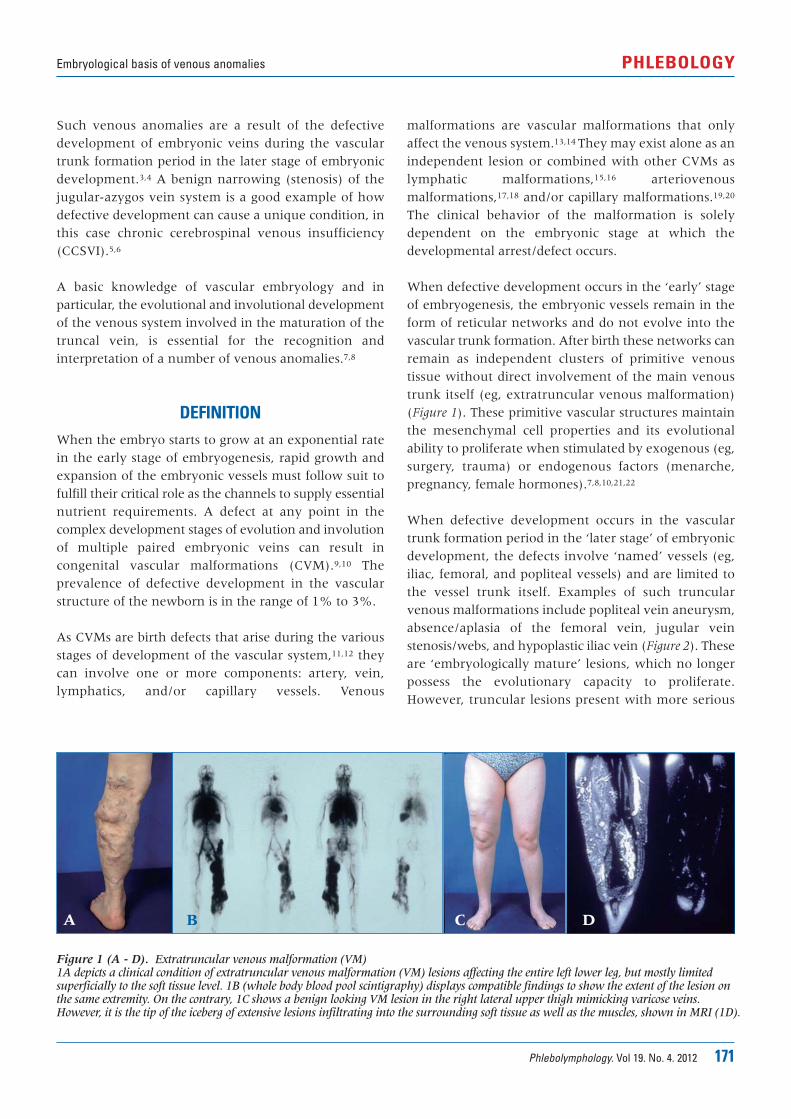
When defective development occurs in the ‘early’ stageof embryogenesis, the embryonic vessels remain in theform of reticular networks and do not evolve into thevascular trunk formation. After birth these networks canremain as independent clusters of primitive venoustissue without direct involvement of the main venoustrunk itself (eg, extratruncular venous malformation)(Figure 1). These primitive vascular structures maintainthe mesenchymal cell properties and its evolutionalability to proliferate when stimulated by exogenous (eg,surgery, trauma) or endogenous factors (menarche,pregnancy, female hormones).7,8,10,21,22
When defective development occurs in the vasculartrunk formation period in the ‘later stage’ of embryonicdevelopment, the defects involve ‘named’ vessels (eg,iliac, femoral, and popliteal vessels) and are limited tothe vessel trunk itself. Examples of such truncularvenous malformations include popliteal vein aneurysm,absence/aplasia of the femoral vein, jugular veinstenosis/webs, and hypoplastic iliac vein (Figure 2). Theseare ‘embryologically mature’ lesions, which no longerpossess the evolutionary capacity to proliferate.However, truncular lesions present with more serious
Figure 1 (A - D). Extratruncular venous malformation (VM)1A depicts a clinical condition of extratruncular venous malformation (VM) lesions affecting the entire left lower leg, but mostly limitedsuperficially to the soft tissue level. 1B (whole body blood pool scintigraphy) displays compatible findings to show the extent of the lesion onthe same extremity. On the contrary, 1C shows a benign looking VM lesion in the right lateral upper thigh mimicking varicose veins.However, it is the tip of the iceberg of extensive lesions infiltrating into the surrounding soft tissue as well as the muscles, shown in MRI (1D).
A B C D

172 Phlebolymphology. Vol 19. No. 4. 2012
Byung-Boong LEEPHLEBOLOGY
hemodynamic consequences in general compared withextratruncular lesions due to their direct involvementwith the truncal venous system (eg, avalvulosis,marginal veins, popliteal vein aneurysm, inferior venacava stenosis/occlusion).23,24
Based on the above definitions, the modified HamburgClassification separates venous malformations into twodifferent types: extratruncular and truncular, dependingon the embryological stage when the defectivedevelopment occurred (Table I).25,26 Venousmalformations originating from the ‘early’ stage ofembryogenesis are classified as extratruncular togetherwith all other types of vascular malformation from thesame ‘early’ stage (eg, lymphangioma). Venousmalformations originating from the ‘late’ stage ofembryogenesis are classified as truncular.
As all truncular lesions involve the already formed,established venous trunk to varying degrees, theypresent as either hypoplastic or hyperplasticvessels/lesions causing obstruction or dilatation (eg,internal jugular vein aneurysm, iliac vein stenosis),depending on the defect.27,28 It should be noted thatintraluminal defects within the vein (eg, vein webs ormembrane) can result in similar conditions of stenosisor obstruction (Figure 3).29,30
Figure 3 (A and B). Truncular venous malformation (VM)3A shows angiographic findings for a truncular VM lesioninvolving a segmental stenosis of the left iliac vein. This benignlooking condition precipitated a severe chronic venous insufficiencyto the affected lower extremity. 3B shows another form of truncular VM involving an aneurysmaldilatation of the right internal jugular vein (Courtesy of ProfessorsP. Zamboni and R. Galeotti for 3B).
Figure 2 (A and B). Truncular venous malformation (VM)2A demonstrates angiographic findings of a truncular VM lesionconsisting of an aneurysmal dilatation of the right popliteal vein;this truncular lesion is the outcome of developmental arrest duringthe vascular trunk formation period in the ‘later stage’ ofembryonic development.2B also presents angiographic findings of another type of truncularVM lesion this time a stenotic condition involving the right internaljugular vein trunk along its junction with the superior vena cava(Courtesy of Professors P Zamboni and R.Galeotti for 2B).
Table I. The modified Hamburg classification of congenitalvascular malformations.
* Based on the predominant vascular structure in themalformation.
** Based on anatomy and developmental arrest at the differentstages of embryonic life: extratruncular form from earlier stages;truncular form from late stages.
Primary classification*
Arterial malformations
Venous malformations
Arteriovenous malformations
Lymphatic malformations
Capillary malformations
Combined vascular malformations
Anatomical/embryological subclassification**
Extratruncular forms• Diffuse, infiltrating • Limited, localized
Truncular forms• Obstruction or narrowing
- Aplasia; Hypoplasia; Hyperplasia- Obstruction due to atresia or membranous
occlusion- Stenosis due to coarctation, spur, or membrane
• Dilatation- Localized (aneurysm)- Diffuse (ectasia)

Phlebolymphology. Vol 19. No. 4. 2012 173
Embryological basis of venous anomalies PHLEBOLOGY
Less frequently, truncular venous malformations maypresent as a persistent fetal remnant vein that has failedto involute or regress normally. This unique condition,which involves the lower extremity venous system, isknown as «marginal/sciatic/lateral embryonic veins»31,32
and represents the venous malformation component ofKlippel-Trenaunay Syndrome (Figure 4).3,4,21,22
As a consequence of their direct involvement with thevenous system, the chronic venous congestion andhypertension due to venous reflux or occlusion causedby truncular venous malformations result in more tissueand organ damage than extratruncal lesions.Membranous, focal, or segmental lesions can causesuprahepatic stenosis of the inferior vena cava along theproximal terminal segment, a condition known asprimary Budd-Chiari syndrome. This has a profoundhemodynamic impact, not only on the lower extremitieswhere it causes chronic venous hypertension, but alsoon the liver where it results in severe portalhypertension due to hepatic venous outlet obstruction.This congenital/developmental anomaly most frequentlyinvolves Asian and African races (Figure 5).33,34
The cerebrospinal venous circulation is not exempt fromtruncular venous malformations. Cerebrospinal venousmalformations carry the potential risk of long-termchronic venous hypertension to the brain resulting invarious clinical conditions/illnesses such as CCSVI.35,36
An example of CCSVI, internal jugular vein valveincompetence (IJVVI), has been postulated to be thecause of transient global amnesia.37,38 IJVVI is diagnosedwhen retrograde jugular vein flow is detected by
extracranial duplex ultrasound during Valsalvamaneuver. It is believed that IJVVI may producetransient mesiotemporal ischemia by venous congestion.This mechanism requires a patent venous pathway from
Figure 4 (A and B). Truncular venous malformation:marginal/lateral embryonic vein4A depicts a clinical condition of the marginal/lateral embryonicvein along the lateral aspect of the left lower extremity. This uniquevein structure is a persistent fetal remnant vessel following thefailure of normal involution/regression and its 'avalvulosis' causessevere venous reflux. Marginal vein remains are a hallmark ofKlippel-Trenaunay syndrome, representing its venousmalformation component.4B presents angiographic findings of this marginal vein, which isthe only remaining major venous drainage route with a lack ofnormal development of the deep venous system. Surgical excision tocontrol venous hypertension is therefore contraindicated.
Figure 5 (A - D). Suprahepatic inferior vena cava (IVC) occlusive lesion: primary Budd-Chiari syndromeA common cause of suprahepatic IVC occlusion is focal stenosis (shown in 5A and 5B) and segmental stenosis (5C), although membranousobstruction by the web is the most common cause among Asians (5D). These are relatively simple congenital VM, which develop during thelate vessel trunk formation stage. However, they have a profound hemodynamic impact on the liver with portal hypertension due to hepaticvenous outlet obstruction in addition to chronic venous insufficiency affecting the lower extremities.
A B C D
174 Phlebolymphology. Vol 19. No. 4. 2012
Byung-Boong LEEPHLEBOLOGY
the affected internal jugular vein through the transversesinus, confluence, straight sinus, and vein of Galen intothe basal vein of Rosenthal and into the internal cerebralveins.
There are now also data supporting a role for CCVI inthe development of multiple sclerosis as reported in theInternational Union of Phlebology Consensus on VenousMalformations - 2009.39 It is hypothesized that truncularvenous malformations causing stenosis along theinternal jugular, innominate, superior vena cava, andazygos vein system, may contribute to the developmentor exacerbation of multiple sclerosis.40,41
DEVELOPMENT OF THE PRIMITIVE VENOUSSYSTEM
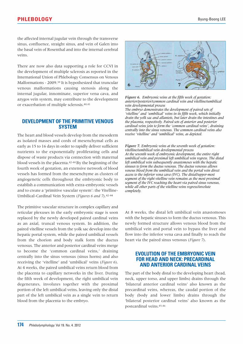
The heart and blood vessels develop from the mesodermas isolated masses and cords of mesenchymal cells asearly as 15 to 16 days in order to rapidly deliver sufficientnutrients to the exponentially proliferating cells anddispose of waste products via connection with maternalblood vessels in the placenta.42-44 By the beginning of thefourth week of gestation, an extensive network of bloodvessels has formed from the mesenchyme as clusters ofangiogenetic cells throughout the embryonic body toestablish a communication with extra-embryonic vesselsand to create a ‘primitive vascular system’: the Vitelline-Umbilical-Cardinal Vein System (Figures 6 and 7).42-44
The primitive vascular structure in complex capillary andreticular plexuses in the early embryonic stage is soonreplaced by the newly developed paired cardinal veinsas an axial, truncal venous system. In addition, thepaired vitelline vessels from the yolk sac develop into thehepatic portal system, while the paired umbilical vesselsfrom the chorion and body stalk form the ductusvenosus. The anterior and posterior cardinal veins mergeto become the ‘common cardinal veins,’ drainingcentrally into the sinus venosus (sinus horns) and alsoreceiving the ‘vitelline’ and ‘umbilical’ veins (Figure 6).At 4 weeks, the paired umbilical veins return blood fromthe placenta to capillary networks in the liver. Duringthe fifth week of development, the right umbilical veindegenerates, involutes together with the proximalportion of the left umbilical veins, leaving only the distalpart of the left umbilical vein as a single vein to returnblood from the placenta to the embryo.
At 8 weeks, the distal left umbilical vein anastomoseswith the hepatic sinuses to form the ductus venosus. Thisnewly formed structure allows venous blood from theumbilical vein and portal vein to bypass the liver andflow into the inferior vena cava and finally to reach theheart via the paired sinus venosus (Figure 7).
EVOLUTION OF THE EMBRYONIC VEIN FOR HEAD AND NECK: PRECARDINAL
AND ANTERIOR CARDINAL VEINS The part of the body distal to the developing heart (head,neck, upper torso, and upper limbs) drains through the‘bilateral anterior cardinal veins’ also known as theprecardinal veins, whereas, the caudal portion of thebody (body and lower limbs) drains through the‘bilateral posterior cardinal veins’ also known as thepostcardinal veins.45,46
Figure 6. Embryonic veins at the fifth week of gestation:anterior/posterior/common cardinal vein and vitelline/umbilicalvein developmental process The embryo demonstrates the development of paired sets of‘vitelline’ and ‘umbilical’ veins in its fifth week, which initiallydrain the yolk sac and allantois, but later drain the intestines andthe placenta, respectively. Paired sets of anterior and posteriorcardinal veins join to form the ‘common cardinal veins’, drainingcentrally into the sinus venosus. The common cardinal veins alsoreceive ‘vitelline’ and ‘umbilical’ veins, as depicted.
Figure 7. Embryonic veins at the seventh week of gestation:vitelline/umbilical vein developmental processAt the seventh week of embryonic development, the entire rightumbilical vein and proximal left umbilical vein regress. The distalleft umbilical vein subsequently anastomoses with the hepaticsinuses to form the ductus venosus. The ductus venosus allowsvenous blood from the umbilical vein and the portal vein directaccess to the inferior vena cava (IVC). The distal/upper-mostsegment of the right vitelline vein remains as the most proximalsegment of the IVC reaching the heart via paired sinus venosus,while all other parts of the vitelline veins regress/involutecompletely.
6 7
Phlebolymphology. Vol 19. No. 4. 2012 175
Embryological basis of venous anomalies PHLEBOLOGY
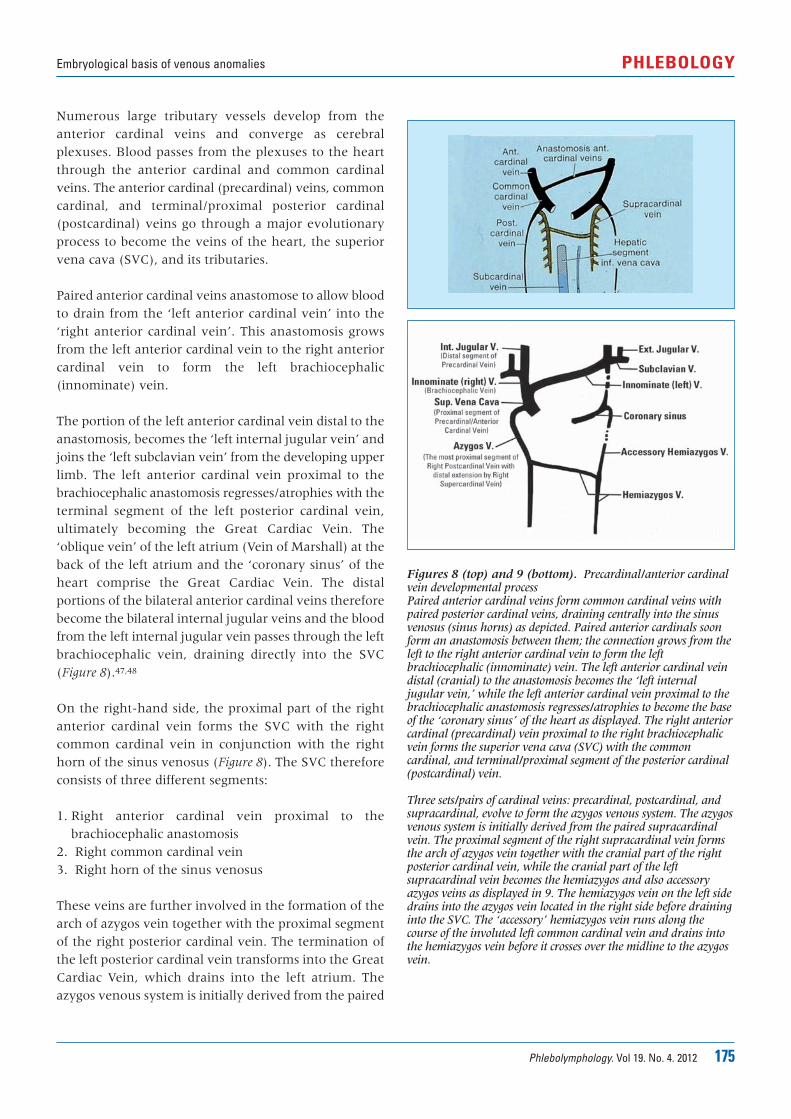
Numerous large tributary vessels develop from theanterior cardinal veins and converge as cerebralplexuses. Blood passes from the plexuses to the heartthrough the anterior cardinal and common cardinalveins. The anterior cardinal (precardinal) veins, commoncardinal, and terminal/proximal posterior cardinal(postcardinal) veins go through a major evolutionaryprocess to become the veins of the heart, the superiorvena cava (SVC), and its tributaries.
Paired anterior cardinal veins anastomose to allow bloodto drain from the ‘left anterior cardinal vein’ into the‘right anterior cardinal vein’. This anastomosis growsfrom the left anterior cardinal vein to the right anteriorcardinal vein to form the left brachiocephalic(innominate) vein.
The portion of the left anterior cardinal vein distal to theanastomosis, becomes the ‘left internal jugular vein’ andjoins the ‘left subclavian vein’ from the developing upperlimb. The left anterior cardinal vein proximal to thebrachiocephalic anastomosis regresses/atrophies with theterminal segment of the left posterior cardinal vein,ultimately becoming the Great Cardiac Vein. The‘oblique vein’ of the left atrium (Vein of Marshall) at theback of the left atrium and the ‘coronary sinus’ of theheart comprise the Great Cardiac Vein. The distalportions of the bilateral anterior cardinal veins thereforebecome the bilateral internal jugular veins and the bloodfrom the left internal jugular vein passes through the leftbrachiocephalic vein, draining directly into the SVC(Figure 8).47,48
On the right-hand side, the proximal part of the rightanterior cardinal vein forms the SVC with the rightcommon cardinal vein in conjunction with the righthorn of the sinus venosus (Figure 8). The SVC thereforeconsists of three different segments:
1. Right anterior cardinal vein proximal to thebrachiocephalic anastomosis
2. Right common cardinal vein3. Right horn of the sinus venosus
These veins are further involved in the formation of thearch of azygos vein together with the proximal segmentof the right posterior cardinal vein. The termination ofthe left posterior cardinal vein transforms into the GreatCardiac Vein, which drains into the left atrium. Theazygos venous system is initially derived from the paired
Figures 8 (top) and 9 (bottom). Precardinal/anterior cardinalvein developmental processPaired anterior cardinal veins form common cardinal veins withpaired posterior cardinal veins, draining centrally into the sinusvenosus (sinus horns) as depicted. Paired anterior cardinals soonform an anastomosis between them; the connection grows from theleft to the right anterior cardinal vein to form the leftbrachiocephalic (innominate) vein. The left anterior cardinal veindistal (cranial) to the anastomosis becomes the ‘left internaljugular vein,’ while the left anterior cardinal vein proximal to thebrachiocephalic anastomosis regresses/atrophies to become the baseof the ‘coronary sinus’ of the heart as displayed. The right anteriorcardinal (precardinal) vein proximal to the right brachiocephalicvein forms the superior vena cava (SVC) with the commoncardinal, and terminal/proximal segment of the posterior cardinal(postcardinal) vein.
Three sets/pairs of cardinal veins: precardinal, postcardinal, andsupracardinal, evolve to form the azygos venous system. The azygosvenous system is initially derived from the paired supracardinalvein. The proximal segment of the right supracardinal vein formsthe arch of azygos vein together with the cranial part of the rightposterior cardinal vein, while the cranial part of the leftsupracardinal vein becomes the hemiazygos and also accessoryazygos veins as displayed in 9. The hemiazygos vein on the left sidedrains into the azygos vein located in the right side before draininginto the SVC. The ‘accessory’ hemiazygos vein runs along thecourse of the involuted left common cardinal vein and drains intothe hemiazygos vein before it crosses over the midline to the azygosvein.

176 Phlebolymphology. Vol 19. No. 4. 2012
Byung-Boong LEEPHLEBOLOGY
supracardinal venous systems, one of three cardinalveins that drain the caudal portion of the body togetherwith the postcardinal (posterior cardinal) veins.49,50
The right supracardinal vein remains as the ‘azygos vein’together with the distal portion of the right posteriorcardinal vein to form the arch of azygos vein. The leftsupracardinal vein becomes the hemiazygos vein andaccessory azygos vein. The hemiazygos vein on the leftdrains into the azygos vein located on the right side andsubsequently into the SVC. The ‘accessory’ hemiazygosvein, which runs along the course of the involuted leftcommon cardinal vein, drains into the hemiazygos veinbefore it crosses the midline to flow into the azygos vein(Figure 9).
ANOMALOUS DEVELOPMENT OF THESUPERIOR VENA CAVA
Due to the complex nature of the various stages ofevolution and involution of multiple paired embryonicveins, several anomalous conditions associated with theSVC can develop. These may affect the commoncardinals, anterior and posterior cardinals, and primitivejugular veins. The likelihood of development anomaliesassociated with the SVC is relatively high due to theinvolvement of three different embryonic vein segments.
For example, a left-sided SVC may develop from‘persistent’ left anterior and left common cardinalveins,51,52 and is often associated with the absence of theright SVC.53,54 In this condition, the right brachiocephalicvein crosses the midline to join a vertical leftbrachiocephalic vein, thus forming a left SVC. As aconsequence of this developmental defect of thecommon cardinal vein, the persistent left SVC can beassociated with the presence of two azygos veins. Whena left SVC is present, the anatomy of the azygos veinsmay be reversed; the hemiazygos vein (the remnant ofthe proximal part of the left posterior cardinal vein)located on the left, will drain directly into the left-sidedSVC, in the way that a normal azygos vein (the remnantof the proximal part of the right posterior cardinal vein)would drain into the SVC on the right side. Thisanomalous condition is the result of a developmentalarrest/defect during the late stage of embryogenesis. Theleft SVC is grouped with other similar truncular venousmalformations (eg, double SVC, internal jugular veinstenosis/aneurysm).
A double SVC is another well known vein anomaly thatoccurs as a result of failure of degeneration/involution ofthe left anterior cardinal vein proximal to thebrachiocephalic anastomosis.55,56 The double SVC isfurther subgrouped based on combined anomalous veins.
EVOLUTION OF EMBRYONIC VEINS OF THETORSO: POSTCARDINAL, SUBCARDINAL AND
SUPRACARDINAL VEINSThe posterior cardinal (postcardinal) veins are the firstpair of embryonic veins to arise that drain the caudalbody. They soon become integrated and taken over bythe newly developing subcardinal and supracardinalveins.57-59 The shift of the systemic venous return to theright atrium in early embryonic life initiates the radicalremodeling of these cardinal (embryonic) venoussystems. The postcardinal, subcardinal, andsupracardinal veins go through extensive evolution aswell as involution for complex remodeling to form theinferior vena cava (IVC), which drains the trunk andlower extremities (Figure 10).60,61
Figure 10 (A-C). Developmental process for the inferior venacava involving postcardinal, supracardinal, and subcardinal veins Three pairs of the post-/sub-/supracardinal veins go throughextensive evolution and involution to form the inferior vena cava(IVC) as well as hepatic veins, together with the bilateral vitellineand umbilical veins. The role of postcardinal (posterior cardinal)veins, the first pair of embryological veins for venous drainage ofthe caudal body, is soon taken over by developing pairs ofsubcardinal and supracardinal veins, to form the IVC as shown.
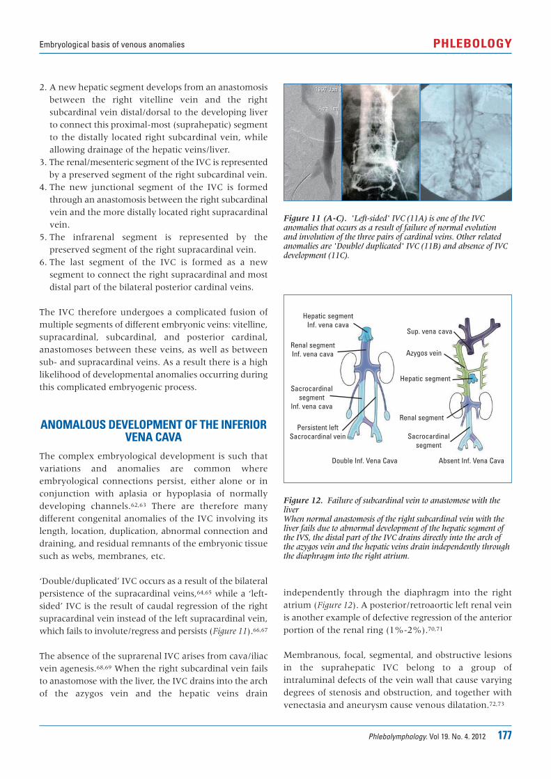
The IVC is formed in a complicated series ofdevelopmental stages from the following embryonicstructures (Figure 11):
1. Suprahepatic - the most proximal segment of theIVC develops from the persistent proximal portionof the right vitelline vein, which is the precursor ofthe common hepatic vein.
Phlebolymphology. Vol 19. No. 4. 2012 177
Embryological basis of venous anomalies PHLEBOLOGY
2. A new hepatic segment develops from an anastomosisbetween the right vitelline vein and the rightsubcardinal vein distal/dorsal to the developing liverto connect this proximal-most (suprahepatic) segmentto the distally located right subcardinal vein, whileallowing drainage of the hepatic veins/liver.
3. The renal/mesenteric segment of the IVC is representedby a preserved segment of the right subcardinal vein.
4. The new junctional segment of the IVC is formedthrough an anastomosis between the right subcardinalvein and the more distally located right supracardinalvein.
5. The infrarenal segment is represented by thepreserved segment of the right supracardinal vein.
6. The last segment of the IVC is formed as a newsegment to connect the right supracardinal and mostdistal part of the bilateral posterior cardinal veins.
The IVC therefore undergoes a complicated fusion ofmultiple segments of different embryonic veins: vitelline,supracardinal, subcardinal, and posterior cardinal,anastomoses between these veins, as well as betweensub- and supracardinal veins. As a result there is a highlikelihood of developmental anomalies occurring duringthis complicated embryogenic process.
ANOMALOUS DEVELOPMENT OF THE INFERIORVENA CAVA
The complex embryological development is such thatvariations and anomalies are common whereembryological connections persist, either alone or inconjunction with aplasia or hypoplasia of normallydeveloping channels.62,63 There are therefore manydifferent congenital anomalies of the IVC involving itslength, location, duplication, abnormal connection anddraining, and residual remnants of the embryonic tissuesuch as webs, membranes, etc.
‘Double/duplicated’ IVC occurs as a result of the bilateralpersistence of the supracardinal veins,64,65 while a ‘left-sided’ IVC is the result of caudal regression of the rightsupracardinal vein instead of the left supracardinal vein,which fails to involute/regress and persists (Figure 11).66,67
The absence of the suprarenal IVC arises from cava/iliacvein agenesis.68,69 When the right subcardinal vein failsto anastomose with the liver, the IVC drains into the archof the azygos vein and the hepatic veins drain
independently through the diaphragm into the rightatrium (Figure 12). A posterior/retroaortic left renal veinis another example of defective regression of the anteriorportion of the renal ring (1%-2%).70,71
Membranous, focal, segmental, and obstructive lesionsin the suprahepatic IVC belong to a group ofintraluminal defects of the vein wall that cause varyingdegrees of stenosis and obstruction, and together withvenectasia and aneurysm cause venous dilatation.72,73
Figure 11 (A-C). 'Left-sided' IVC (11A) is one of the IVCanomalies that occurs as a result of failure of normal evolutionand involution of the three pairs of cardinal veins. Other relatedanomalies are 'Double/ duplicated' IVC (11B) and absence of IVCdevelopment (11C).
Figure 12. Failure of subcardinal vein to anastomose with theliverWhen normal anastomosis of the right subcardinal vein with theliver fails due to abnormal development of the hepatic segment ofthe IVS, the distal part of the IVC drains directly into the arch ofthe azygos vein and the hepatic veins drain independently throughthe diaphragm into the right atrium.
Absent Inf. Vena CavaDouble Inf. Vena Cava
Sup. vena cava
Azygos vein
Hepatic segment
Renal segment
Sacrocardinalsegment
Hepatic segmentInf. vena cava
Renal segmentInf. vena cava
Sacrocardinalsegment
Inf. vena cava
Persistent leftSacrocardinal vein
178 Phlebolymphology. Vol 19. No. 4. 2012
Byung-Boong LEEPHLEBOLOGY
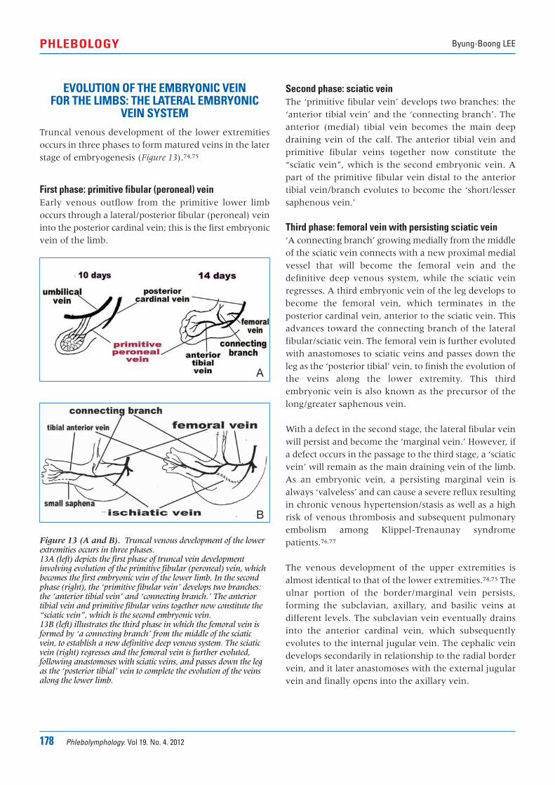
Second phase: sciatic veinThe ‘primitive fibular vein’ develops two branches: the‘anterior tibial vein’ and the ‘connecting branch’. Theanterior (medial) tibial vein becomes the main deepdraining vein of the calf. The anterior tibial vein andprimitive fibular veins together now constitute the“sciatic vein”, which is the second embryonic vein. Apart of the primitive fibular vein distal to the anteriortibial vein/branch evolutes to become the ‘short/lessersaphenous vein.’
Third phase: femoral vein with persisting sciatic vein‘A connecting branch’ growing medially from the middleof the sciatic vein connects with a new proximal medialvessel that will become the femoral vein and thedefinitive deep venous system, while the sciatic veinregresses. A third embryonic vein of the leg develops tobecome the femoral vein, which terminates in theposterior cardinal vein, anterior to the sciatic vein. Thisadvances toward the connecting branch of the lateralfibular/sciatic vein. The femoral vein is further evolutedwith anastomoses to sciatic veins and passes down theleg as the ‘posterior tibial’ vein, to finish the evolution ofthe veins along the lower extremity. This thirdembryonic vein is also known as the precursor of thelong/greater saphenous vein.
With a defect in the second stage, the lateral fibular veinwill persist and become the ‘marginal vein.’ However, ifa defect occurs in the passage to the third stage, a ‘sciaticvein’ will remain as the main draining vein of the limb.As an embryonic vein, a persisting marginal vein isalways ‘valveless’ and can cause a severe reflux resultingin chronic venous hypertension/stasis as well as a highrisk of venous thrombosis and subsequent pulmonaryembolism among Klippel-Trenaunay syndromepatients.76,77
The venous development of the upper extremities isalmost identical to that of the lower extremities.74,75 Theulnar portion of the border/marginal vein persists,forming the subclavian, axillary, and basilic veins atdifferent levels. The subclavian vein eventually drainsinto the anterior cardinal vein, which subsequentlyevolutes to the internal jugular vein. The cephalic veindevelops secondarily in relationship to the radial bordervein, and it later anastomoses with the external jugularvein and finally opens into the axillary vein.
Figure 13 (A and B). Truncal venous development of the lowerextremities occurs in three phases. 13A (left) depicts the first phase of truncal vein developmentinvolving evolution of the primitive fibular (peroneal) vein, whichbecomes the first embryonic vein of the lower limb. In the secondphase (right), the ‘primitive fibular vein’ develops two branches:the ‘anterior tibial vein’ and ‘connecting branch.’ The anteriortibial vein and primitive fibular veins together now constitute the“sciatic vein”, which is the second embryonic vein. 13B (left) illustrates the third phase in which the femoral vein isformed by ‘a connecting branch’ from the middle of the sciaticvein, to establish a new definitive deep venous system. The sciaticvein (right) regresses and the femoral vein is further evoluted,following anastomoses with sciatic veins, and passes down the legas the ‘posterior tibial’ vein to complete the evolution of the veinsalong the lower limb.
EVOLUTION OF THE EMBRYONIC VEIN FOR THE LIMBS: THE LATERAL EMBRYONIC
VEIN SYSTEMTruncal venous development of the lower extremitiesoccurs in three phases to form matured veins in the laterstage of embryogenesis (Figure 13).74,75
First phase: primitive fibular (peroneal) vein Early venous outflow from the primitive lower limboccurs through a lateral/posterior fibular (peroneal) veininto the posterior cardinal vein; this is the first embryonicvein of the limb.
A
B

Phlebolymphology. Vol 19. No. 4. 2012 179
Embryological basis of venous anomalies PHLEBOLOGY
Corresponding authorLee BB, MD, PhD, FACS, Professor of Surgery and Director,Center for Lymphedema and VascularMalformations, George Washington University School of Medicine, Washington DC, USA
E-mail: [email protected]
REFERENCES
1. Lee BB, Villavicencio L, Kim YW, et al.Primary Budd-Chiari syndrome:outcome of endovascular managementfor suprahepatic venous obstruction. J Vasc Surg. 2006;43:101-108.
2. Romagnoli R, Bertolani M, Saviano M,Pantusa M, Modena MG, Benassi A.Developmental interruption of theintra-hepatic segment of the inferiorvena cava with azygos-hemiazygoscontinuation. Eur J Radiol. 1984;4:244-247.
3. Lee BB, Laredo J, Lee TS, Huh S,Neville R. Terminology andclassification of congenital vascularmalformations. Phlebology.2007;22:249-252.
4. Lee BB, Villavicencio L. Generalconsiderations. Congenital vascularmalformations. Arteriovenousanomalies. In: Cronenwett JL,Johnston KW, eds. Rutherford’sVascular Surgery. 7th Edition.Philadelphia, PA, USA: SaundersElsevier;2010:1046-1064.
5. Zamboni P, Galeotti R, Menegatti E, etal. Chronic cerebrospinal venousinsufficiency in patients with multiplesclerosis J Neurol Neurosurg Psychiatry.2009;80:392-399.
6. Lee BB, Laredo J, Neville R.Embryological background of truncularvenous malformation in theextracranial venous pathways as thecause of chronic cerebrospinal venousinsufficiency. Int Angiol. 2010;29:95-108.
7. Leu HJ. Pathoanatomy of congenitalvascular malformations. In: Belov S,Loose DA, Weber J, eds. VascularMalformations. Reinbek, Germany:Einhorn-Presse Verlag;1989;16:37-46.
8. Woolard HH. The development of theprincipal arterial stems in the forelimbof the pig. Contrib Embryol.1922;14:139-154.
9. Lee BB. New approaches to thetreatment of congenital vascularmalformations (CVMs) – single centerexperiences – (Editorial Review). Eur JVasc Endovasc Surg. 2005;30:184-197.
10. Bastide G, Lefebvre D. Anatomy andorganogenesis and vascularmalformations. In: Belov St, Loose DA,Weber J, eds. Vascular Malformations.Reinbek: Einhorn-Presse VerlagGmbH;1989:20-22.
11. Lee BB, Bergan JJ. Advancedmanagement of congenital vascularmalformations: a multidisciplinaryapproach. Cardiovasc Surg.2002;10:523-533.
12. Lee BB, Laredo J, Deaton DH, et al.Arteriovenous malformations:evaluation and treatment. In: GloviczkiP, ed. Handbook of Venous Disorders.Guidelines of the American Venous Forum.3rd Edition. London, UK: A HodderArnold Ltd;2009.
13. Lee BB, Do YS, Byun HS, Choo IW,Kim DI, Huh SH. Advancedmanagement of venous malformationwith ethanol sclerotherapy: mid-termresults. J Vasc Surg. 2003;37:533-538.
14. Lee BB. Current concept of venousmalformation (VM). Phlebolymphology.2003;43:197-203.
15. Lee BB, Kim YW, Seo JM, et al.Current concepts in lymphaticmalformation (LM). J Vasc EndovascSurg. 2005;39:67-81.
16. Lee BB, Villavicencio JL. Primarylymphedema and lymphaticmalformation: are they the two sidesof the same coin? Eur J Vasc EndovascSurg. 2010;39:646-653.
17. Lee BB, Lardeo J, Neville R. Arterio-venous malformation: how much dowe know? Phlebology. 2009;24:193-200.
18. Lee BB, Do YS, Yakes W, Kim DI,Mattassi R, Hyon WS. Management ofarterial-venous shuntingmalformations (AVM) by surgery andembolosclerotherapy. Amultidisciplinary approach. J Vasc Surg.2004;39:590-600.
19. Goldman MP, Fitzpatrick RE, Ruiz-Esparza J. Treatment of port-winestains (capillary malformation) withthe flashlamp-pumped pulsed dyelaser. J Pediatr. 1993;122:71-77.
20. Berwald C, Salazard B, Bardot J,Casanova D, Magalon G. Port winestains or capillary malformations:surgical treatment. Ann Chir PlastEsthet. 2006;51:369-372.
21. Lee BB, Laredo J, Lee SJ, Huh SH, JoeJH, Neville R. Congenital vascularmalformations: general diagnosticprinciples. Phlebology. 2007;22:253-257.
22. Lee BB, Laredo J, Kim YW, Neville R.Congenital vascular malformations:general treatment principles.Phlebology. 2007;22:258-263.
23. Lee BB. Changing concept on vascularmalformation: no longer enigma. AnnVasc Dis. 2008;1:11-19.
24. Lee BB. Mastery of vascular andendovascular surgery. In: Zelenock,Huber, Messina, Lumsden, Moneta(eds). Chapter 76. Arteriovenousmalformation. Philadelphia: Lippincott,Williams and Wilkinspublishers;2006:597-607.
25. Belov S. Classification, terminology,and nosology of congenital vasculardefects. In: Belov S, Loose DA, WeberJ, eds. Vascular Malformations. Reinbek,Germany: Einhorn-Presse;1989:25-30.
26. Belov ST. Anatomopathologicalclassification of congenital vasculardefects. Sem Vasc Surg. 1993;6:219-224.
27. Zamboni P, Cossu A, Carpanese L,Simonetti G, Massarelli G, Liboni A.The so-called venous aneurysms.Phlebology. 1990;5:45-50.
28. Vaket L, Poppelier G, Vermeire P. Surun cas d’anomalie combinée de laveine cave supérieure et du systèmeazygos. Acta Anat. 1958 ;32:235-239.
29. Croquet V, Aube C, Pilette C, et al.Syndrome due to membranousobstruction of the inferior vena cava ofcongenital origin. Ten-year follow-upafter radiologic treatment. GastroenterolClin Biol. 1999;23:259-263.
30. Rao KS, Gupta BK, Banerjee A,Srivastava KK. Chronic Budd-Chiarisyndrome due to congenitalmembranous obstruction of theinferior vena cava: clinical experience.Aust N Z J Surg. 1989;59:335-338.
180 Phlebolymphology. Vol 19. No. 4. 2012
Byung-Boong LEEPHLEBOLOGY
REFERENCES
31. Kim YW, Lee BB, Cho JH, Do YS, KimDI, Kim ES. Haemodynamic andclinical assessment of lateral marginalvein excision in patients with apredominantly venous malformationof the lower extremity. Eur J VascEndovasc Surg. 2007;33:122-127.
32. Mattassi R. Approach to marginal vein:current issue. Phlebology. 2007;22:283-286.
33. Lee BB, Laredo J, Deaton D, et al.Endovascular management of Budd-Chiari Syndrome – suprahepaticinferior vena cava occlusive disease. In:Heuser RR, Henry M, eds. Textbook ofPeripheral Vascular Interventions. Secondedition. Section XII. Chapter 83.London, UK: Informa Healthcare,Informa UK Ltd;2008:725-731
34. Zamboni P, Pisano L, Mari C, GaleottiR, Feo C, Liboni A. Membranousobstruction of the inferior vena cavaand Budd-Chiari syndrome. Report ofa case. J Cardiovasc Surg. 1996 (Torino);37:583-587.
35. Abe T, Singer RJ, Marks MP, NorbashAM, Crowley RS, Steinberg GK.Coexistence of occult vascularmalformations and developmentalvenous anomalies in the centralnervous system: MR evaluation. Am J Neuroradiol. 1998;19:51-57.
36. Schaller B. Physiology of cerebralvenous blood flow: from experimentaldata in animals to normal function inhumans. Brain Res Rev. 2004;46:243-260.
37. Schreiber SJ, Doepp F, Klingebiel R,Valdueza JM. Internal jugular veinvalve incompetence and intracranialvenous anatomy in transient globalamnesia. J Neurol Neurosurg Psychiatry.2005;76:509-513.
38. Akkawi NM, Agosti C, Rozzini L,Anzola GP, Padovani A. Transientglobal amnesia and disturbance ofvenous flow patterns. Lancet.2001;357:957.
39. Lee BB, Bergan J. Gloviczki P, et al;International Union of Phlebology(IUP). Diagnosis and treatment ofvenous malformations - ConsensusDocument of the International Unionof Phlebology (IUP)-2009. Int Angiol.2009;28:434-451.
40. Nedelmann M, Kaps M, Mueller-ForellW. Venous obstruction and jugularvalve insufficiency in idiopathicintracranial hypertension. J Neurol.2009;256:964-969.
41. Leriche H, Aubin ML, Aboulker J.Cavo-spinal phlebography inmyelopathies. Stenoses of internaljugular and azygos veins, venouscompressions and thrombosis. ActaRadiol Suppl. 1976;347:415-417.
42. Langman J. Medical Embryology. 5th ed.Baltimore, MD: Williams andWilkins;1985:212–217.
43. Warwick R, Williams P. Gray’s Anatomy.37th ed. Edinburgh, London,Melbourne, New York: ChurchillLivingstone;1989:326-327.
44. Hamilton WJ, Mossman HW. Hamilton,Boyd & Mossman’s Human Embryology.4th ed. Cambridge: Heffer;1972:261.
45. Collins P. Embryology anddevelopment. In: Williams PL,Bannister LH, Berry MM, et al (eds).Gray’s Anatomy: The Anatomical Basis ofMedicine and Surgery. 38th ed.Edinburgh: ChurchillLivingston;1995:327.
46. Padget DH. The development of thecranial venous system in man, fromthe viewpoint of comparative anatomy.Contrib Embryol Carneg Inst Washington.1957;36:79-140.
47. Beattie J. The importance of anomaliesof the superior vena cava in man.Canad Med Assoc J. 1931;25:281-284.
48. FitzGerald DP. The study ofdevelopmental abnormalities as an aidto that of human embryology, basedon observations on a persistent leftsuperior vena cava. Dublin J Med Sci.1909;14-18.
49. Keyes DC, Keyes HC. A case ofpersistent left superior vena cava withreversed azygos system. Anat Rec.1925;31:23-26.
50. Nandy K, Blair CB, Jr. Double superiorvena cavae with completely pairedazygos veins. Anat Rec. 1965;15:1-9.
51. Huffmire AP, Bower GC. A case ofpersistence of the left superior venacava in an aged adult. Anat Rec. 1919-20;17:127-129.
52. Basu BN. Persistent «left superior venacava,» «left duct of Cuvier» and lefthorn of the sinus venosus. J Anat.1932;66:628-270.
53. Greenfield WS. Persistence of the leftvena cava superior, with absence ofright. Trans Pathol Soc Lond.1876;27:120-124.
54. Atwell WJ, Zoltowski P. A case of a leftsuperior vena cava without acorresponding vessel on the right side.Anat Rec. 1938;70:525-532.
55. Howden R. Case of double superiorvena cava with left -sided arrangementof the azygos vein. J Anat Physiol.1887;21:72-75.
56. Gruber W. Duplicität der vena cavasuperior, mit vorkommen zweir nenaeazygae und einer sufficienten valvulaan der mündung der vena azygossinistra. Arch Pathol Anat Physiol KlinMed. 1880;81:462-465.
57. Krizan Z, Herman O, Dzidrov V.Teilweiser fortbestand dessupracardinalsystems neben dernormalen vena cava inferior beimmenschen. Acta Anat. 1958;34:312-325.
58. Lewis FT. The development of venacava inferior. Am J Anat. 1902;1.
59. Bailey FR, Miller AM. Development ofthe vascular system. In: Textbook ofEmbryology. 2nd edition. New York:William Woon andCompany;1911:222-291.
60. Nemec J, Heifetz S. Persistence of leftsupracardinal vein in an adult patientwith heart-hand syndrome and cardiacpacemaker. Congenit Heart Dis.2008;3:219-222.
61. McClure CFW, Butler EG. Thedevelopment of the vena cava inferiorin man. Am J Anat. 1925:35:331-383.
62. Cornillie P, Van Den Broeck W,Simoens P. Origin of the infrarenal partof the caudal vena cava in the pig. AnatHistol Embryol. 2008;37:387-393.
63. Nemec J, Heifetz S. Persistence of leftsupracardinal vein in an adult patientwith heart-hand syndrome and cardiacpacemaker. Congenit Heart Dis.2008;3:219-222.
64. Hashmi ZA, Smaroff GG. Dual inferiorvena cava: two inferior vena cavafilters. Ann Thorac Surg. 2007;84:661-663.
65. Esposito S, Mansueto G, Amodio F, etal. Duplication of the vena cavainferior with a continuation into thevena azygos. A report of a rare case.Minerva Chir. 1999;54:261-265.
66. Munechika H, Cohan RH, Baker ME,Cooper CJ, Dunnick NR. Hemiazygoscontinuation of a left inferior venacava: CT appearance. J Comput AssistTomogr. 1988;12:328-330.
67. Honma S, Tokiyoshi A, Kawai K,Koizumi M, Kodama K. Left inferiorvena cava with regressed right inferiorvena cava. Anat Sci Int. 2008;83:173-178.
68. Gil RJ, Pérez AM, Arias JB, Pascual FB,Romero ES. Agenesis of the inferiorvena cava associated with lowerextremities and pelvic venousthrombosis. J Vasc Surg. 2006;44:1114-1116.
69. Romagnoli R, Bertolani M, Saviano M,Pantusa M, Modena MG, Benassi A.Developmental interruption of theintra-hepatic segment of the inferiorvena cava with azygos-hemiazygoscontinuation. Eur J Radiol. 1984;4:244-247.
Phlebolymphology. Vol 19. No. 4. 2012 181
Embryological basis of venous anomalies PHLEBOLOGY
REFERENCES
70. Trigaux JP, Vandroogenbroek S, DeWispelaere JF, Lacrosse M, Jamart J.Congenital anomalies of the inferiorvena cava and left renal vein:evaluation with spiral CT. J Vasc IntervRadiol. 1998;9:339-345.
71. Royal SA, Callen PW. CT evaluation ofanomalies of the inferior vena cavaand left renal vein. AJR Am JRoentgenol. 1979;132:759-763.
72. Walden R, Hiss J, Morag B, Adar R.Congenital membranous obstruction ofthe inferior vena cava. Isr J Med Sci.1978;14:342-346.
73. Wang ZG, Zhu Y, Wang SH, et al.Recognition and management ofBudd-Chiari syndrome: report of onehundred cases. J Vasc Surg.1989;10:149-156.
74. Lewis FT. The development of theveins in the limbs of rabbit embryo.Am J Anat. 1905;5:1-120.
75. Wyman J. On symmetry andhomology in. limbs. Proc Boston Soc NatHist. 1867;11:246-278.
76. Gloviczki P, Stanson AW, Stickler GB,et al. Trenaunay syndrome: the risksand benefits of vascular interventions.Surgery. 1991;110:469-479.
77. Noel AA, Gloviczki P, Cherry KJ Jr,Rooke TW, Stanson AW, Driscoll DJ etal. Surgical treatment of venousmalformations in Klippel-Trenaunaysyndrome. J Vasc Surg. 2000;32:840-847.
182 Phlebolymphology. Vol 19. No. 4. 2012
PHLEBOLOGY
The “C0s” patient: worldwide resultsfrom the Vein Consult Program
Keywords:
classification, chronic venous disorders, cross-sectional studies, epidemiology, prevalence,signs, symptoms.
Jean-Jérôme GUEX1, Eberhard RABE2, Sanchez Ignacio ESCOTTO3,José-Román ESCUDERO4,Angelo SCUDERI5,Hendro Sudjono YUWONO6, and the Vein Consult Programcoordinators7
1. 32, Boulevard Dubouchage, Nice, France2. Department of Dermatology, University of
Bonn, Germany3. National Autonomous University of Mexico,
Mexico City, Mexico4. Hospital de la Santa Creu i Sant Pau,
Barcelona, Spain5. Instituto Sorocabano de Pesquisa de
Molestias Circulatorios (INSPEMOC), São Paulo, Brazil
6. Hasan Sadikin General Hospital, Bandung,Indonesia
7. National coordinators of the Vein ConsultProgram (VCP): Alberti T, Venezuela;Andercou A, Giurcaneanu C, and Puskas A,Romania; Dinh Thi Thu H and Le Nu Thi HoaH, Vietnam; Escotto Sanchez I, Mexico;Escudero JR, Spain; Guex JJ, France; HoyosAS, Colombia; Kazim Y and Swidan A,United Arab Emirates; Kecelj Leskovec Nand Planinsek Rucigaj T, Slovenia; Larisa C,Ukraine; Liptak P, Slovakia; Matyas L,Hungary; Paocharoen V, Thailand;Pargalava N, Georgia; Radak D andMaksimovic Z, Serbia; Saveliev V, Russia;Scuderi A, Brazil; Yuwono HS, Indonesia
Phlebolymphology. 2012;19(4):182-192.
ABSTRACTThe Vein Consult Program is an international, observational, prospectivesurvey that aims to collect global epidemiological data on chronic venousdisorder (CVD)-related symptoms and signs based on the Clinical, Etiological,Anatomical, and Pathophysiological (CEAP) classification, assess the impactCVD may have on an individual’s daily activities, and identify CVDmanagement practices worldwide.
The survey was organized within the framework of ordinary consultationswith general practitioners (GPs) trained in the use of the CEAP classification.Screening for CVD was performed by enrolling all consecutive outpatientsover 18 years of age, whatever the reason for their consultation. Patients’data were recorded and patients were classified according to the CEAP, fromstage C0s to C6. GPs were able to refer screened patients with CVD to avenous specialist for further ultrasound investigation.
A total of 6232 GPs participated in the program and 91545 subjects wereanalysed, of whom 15290 (19.7%) were assigned to category C0s. Thepercentage of the global survey population eligible for a venous specialistconsultation was 22.2%, but C0s subjects comprised only 4.1% (n=634). Inthe total survey population only 43% of eligible patients visited a specialist,but surprisingly most C0s patients eligible for referral did visit a specialist.Among the C0s patients a duplex scan investigation was performed in 14%.The majority were found to have reflux, which was mostly superficial, butan appreciable percentage (18%) had deep reflux.
C0s individuals were younger with a lower body mass index compared withthe C1 to C6 subjects (48.6 versus 55.5 years; and 25.76 versus 27.24 kg/m2,respectively), and had fewer CVD risk factors.
Of all screened women, 18.5% were at the C0s stage versus 22.4% ofscreened men. Whatever the age group, men were more likely to be assignedto the C0s class of the CEAP than women, except in the 18-34 age bracket.
Although the quality of life of C0s subjects was impaired, they were often notconsidered to have CVD by GPs (25.5% in C0s versus 71% in the total survey
Phlebolymphology. Vol 19. No. 4. 2012 183
The C0s patient PHLEBOLOGY
population), and were poorly treated (13% receivedlifestyle advice).
It is not known whether C0s subjects deserve moreinvestigation and treatment as longitudinal studiesproviding information on the possible progression ofdisease in such patients are lacking. Nevertheless, giventhe inflammatory nature of CVD, we can speculate thattreatments able to hamper inflammation would at leastrelieve C0s symptoms.
INTRODUCTIONThe grading of chronic venous disorders (CVD) wassimplified and standardized by the introduction of theClinical, Etiological, Anatomical, and Pathophysiological(CEAP) classification system.1 The CEAP classificationcategorizes limbs into seven classes from C0 to C6. Eachclinical class is further characterized by a subscript (S) ifthe categorized limb is symptomatic, or a subscript (A) ifthe limb is asymptomatic. C0s patients are defined asthose presenting with one or more CVD-relatedsymptoms, but showing no clinical signs of the diseaseduring a physical examination, and generally noabnormalities on a duplex scan. In a revised CEAPclassification published in 2004, the C0s patient profilewas introduced.2 Patients in this category are describedas C0s,En,An,Pn as no etiology, anatomical localisationor pathological cause can be found. Symptoms associatedwith CVD and with the C0s stage were further defined as“complaints related to venous disease, which mayinclude tingling, aching, burning, pain, muscle cramps,swelling, sensations of throbbing or heaviness, itchingskin, restless legs, leg-tiredness and/or fatigue. Althoughnot pathognomonic, these may be suggestive of chronicvenous disease, particularly if they are exacerbated byheat or dependency in the day’s course, and relievedwith leg rest and/or elevation.”3
Despite the acknowledgment that symptoms play animportant part in CVD, little is known about theirprevalence, particularly in the C0s patient. The aim ofthe present analysis was to determine the prevalence andclinical profile of C0s patients worldwide, how suchpatients are managed by general practitioners (GPs) indifferent geographical areas, the rate of spontaneousconsultations due to C0s, and finally the impact ofsymptoms alone on patients’ quality of life. This wasachieved through the Vein Consult Program (VCP), an
international, observational, prospective survey carriedout as an initiative of the Union Internationale de
Phlébologie to raise awareness of CVD among patients,healthcare professionals, and health authorities.
MATERIALS AND METHODSThe VCP was conducted within the framework ofordinary GP consultations over a very short period oftime (less than 2 weeks) to gather nationallyrepresentative data on the prevalence of CVD-relatedsymptoms, signs, and CEAP classes. The design was across-sectional, two-part survey. In the first part of thesurvey, GPs recruited consecutive patients aged 18 yearsand over attending the practice for a routineconsultation. Practices were distributed throughout thecountry and included both urban and rural locations.The only patients excluded were those attending foremergency visits. Informed consent was obtained fromeach participant.
The initial survey was conducted as a face-to-faceinterview. The 23-item questionnaire consisted of foursections and determined the individuals’ demographics;CVD risk factors; any patient-reported symptomssuggestive of CVD such as leg pain, heat sensation, anditching; and any visible signs of CVD. Signs, includingtelangiectasies, varicose veins, edema, trophic changes,and/or ulcers, were reported by patients themselves, andconfirmed by the physician during a physicalexamination. Patients were then assigned to anappropriate clinical category (C) according to the CEAPclassification when any type of venous pathology(classification C1–C6) was seen in at least one leg. Theresults were recorded by physicians in a case report form.The grade was determined by considering only thehighest single reported sign. Physicians were requestedto report whether patients complained about their legsspontaneously. All patients with a physician diagnosis ofCVD were invited to complete a 14-question form of theChronIc Venous Insufficiency Questionnaire (CIVIQ-14).4
Physicians could refer any individuals with a diagnosis ofCVD to a venous specialist, using their normalprocedure. The aim of the second part of the survey wasto confirm the initial physician diagnosis of CVD. Thespecialists completed an additional questionnaire toestablish the patient’s history of CVD and CVD riskfactors. They also investigated the CVD status of the
184 Phlebolymphology. Vol 19. No. 4. 2012
JJ. GUEX et al.PHLEBOLOGY
patient using Doppler or duplex scanning anddetermined whether treatment was required. Thespecialists were unaware of the physicians’ initialdiagnosis.
STATISTICAL ANALYSESUnless otherwise specified, qualitative and quantitativevariables were respectively described as number(percentage) and mean ± standard deviation.Percentages were calculated out of the total number ofvalid answers, excluding missing values. In case ofmissing values, the number of valid answers waspresented. For cross tables, statistical tests wereperformed to compare means for quantitative variablesusing the Student t-test or analysis of variance for onefactor, and frequencies for qualitative variables using theChi² test. An � level of 0.001 was used, and a P-valueequal to or lower than 0.001 indicated a statisticallysignificant difference.
For the venous specialist questionnaire, the prevalenceof CVD was estimated as the number of patients with aDoppler abnormality (primary CVD with or withoutother symptoms) out of the total number of patients whoreceived a Doppler examination.
For CIVIQ-14 (ChronIc Venous InsufficiencyQuestionnaire with 14 questions), a global index score(GIS) was calculated with values ranging from 0 to 100.In this analysis, we adopted the following scoringconvention: GIS 0 equals worst score and GIS 100 equalsbest score.
RESULTSThe VCP was conducted between October 2009 and July2011 in five geographical zones: Western Europe (Franceand Spain; 36 004 subjects), Central and Eastern Europe(Georgia, Hungary, Romania, Russia, Serbia, SlovakRepublic, Slovenia, Ukraine; 32 225 subjects), LatinAmerica (Brazil, Colombia, Mexico, Venezuela; 12 686subjects), the Middle East (Pakistan, United ArabEmirates; 3518 subjects), and the Far East (Indonesia,Singapore, Thailand, Vietnam; 7112 subjects) totalling20 countries.
Of the people who agreed to be screened, 91 545 hadfiles suitable for analysis. The mean age of globalparticipants was 50.6 ± 16.9 years, 68% were female,
and the mean body mass index (BMI) was 26.17 ± 5.07.The profile of the global population of the survey hasalready been published elsewhere,5 and the distributionof patients among the CEAP clinical classes was asfollows: 16 901 (21.7%) in C1, 13 888 (17.9%) in C2,and 18 863 (24.3%) in C3 to C6 (chronic venousinsufficiency patients), totalizing 46 451 patients. Thenumber of subjects complaining solely of symptoms, theso-called C0s patients, was 15 290 (19.7%), indicatingthat almost 20% of the survey population had CEAPgrade C0s. Only 12 774 (16.4%) individuals had nosymptoms or signs of CVD and thus were exempt fromleg problems (C0a).
Clinical profile of the C0s patient and comparison with thesurveyed population presenting with clinical signs (C1 toC6 of the CEAP)As shown in Table I, screened subjects were younger inthe C0s than the C1 to C6 population: 48.3 ± 16.8 yearsversus 55.5 ± 15.3 years (P<0.0001), less often female(65.9% in C0s versus 77.4%; P<0.0001), with a lowerBMI, although the latter did not reach statisticalsignificance (25.76 ± 4.85 versus 27.24 ± 5.31; P=NS).Overall, the screened population was overweight (26.17± 5.07).
Frequency of CVD symptoms (Table I)Participants described their symptoms in response to thequestions set by the physician. The most frequentcomplaints in the C1-C6 population of the survey were‘heavy legs,’ reported by 80%; ‘pain in the legs,’ reportedby 74%; ‘sensation of swelling,’ reported by 63%, and‘night cramps,’ reported by 48%. The average numberof symptoms per patient was 3.1 ± 1.0. Symptomdistribution and ranking was respected compared withthe global population (not shown).5 In the C0spopulation, despite symptoms being reported lessfrequently, the symptom distribution and ranking weresimilar to those of symptomatic subjects in the C1 to C6population of the survey, except that night crampsranked higher. The mean number of reported symptomswas lower in the C0s population compared with the C1-C6 population (2.1 versus 3.1; P<0.0001). Subjects werealso asked what exacerbated their symptoms. The mostfrequent response, reported by 68.7% of symptomaticC1 to C6 participants, was that their complaints ‘increaseat the end of the day.’ Subjects in C1 to C6 classes alsoreported that their complaints: ‘increase with prolongedstanding’ (55.3%), ‘increase during the night’ (45.1%),‘increase in the summer’ (39.0%), ‘worsen during
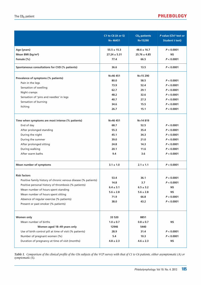
Phlebolymphology. Vol 19. No. 4. 2012 185
The C0s patient PHLEBOLOGY
C1 to C6 (A or S)
N= 46451
C0s patients
N=15290
P value (Chi2 test or
Student t test)
Age (years)
Mean BMI (kg/m²)
Female (%)
55.5 ± 15.3
27.24 ± 5.31
77.4
48.6 ± 16.7
25.76 ± 4.85
66.5
P < 0.0001
NS
P < 0.0001
Spontaneous consultations for CVD (% patients) 36.6 13.5 P < 0.0001
Prevalence of symptoms (% patients)
Pain in the legs
Sensation of swelling
Night cramps
Sensation of ‘pins and needles’ in legs
Sensation of burning
Itching
N=46 451
80.0
73.9
62.7
48.2
40.7
34.6
26.7
N=15 290
58.5
52.4
29.1
32.6
27.3
15.5
15.1
P < 0.0001
P < 0.0001
P < 0.0001
P < 0.0001
P < 0.0001
P < 0.0001
P < 0.0001
Time when symptoms are most intense (% patients)
End of day
After prolonged standing
During the night
During the summer
After prolonged sitting
During walking
After warm baths
N=46 451
68.7
55.3
45.1
39.0
24.8
20.1
9.4
N=14 819
52.5
35.4
34.3
21.0
14.3
11.6
3.6
P < 0.0001
P < 0.0001
P < 0.0001
P < 0.0001
P < 0.0001
P < 0.0001
P < 0.0001
Mean number of symptoms 3.1 ± 1.0 2.1 ± 1.1 P < 0.0001
Risk factors
Positive family history of chronic venous disease (% patients)
Positive personal history of thrombosis (% patients)
Mean number of hours spent standing
Mean number of hours spent sitting
Absence of regular exercise (% patients)
Present or past smoker (% patients)
53.4
14.8
6.4 ± 3.1
5.6 ± 2.8
71.9
38.0
36.1
3.7
6.5 ± 3.2
5.6 ± 2.8
66.8
43.2
P < 0.0001
P < 0.0001
NS
NS
P < 0.0001
P < 0.0001
Women only
Mean number of births
Women aged 18−49 years only
Use of birth control pill at time of visit (% patients)
Number of pregnant women (%)
Duration of pregnancy at time of visit (months)
33 520
1.0 ± 0.7
12946
28.9
5.4
4.8 ± 2.3
8851
0.8 ± 0.7
5440
31.4
10.3
4.6 ± 2.3
NS
P < 0.0001
P < 0.0001
NS
Table I. Comparison of the clinical profile of the C0s subjects of the VCP survey with that of C1 to C6 patients, either asymptomatic (A) orsymptomatic (S).
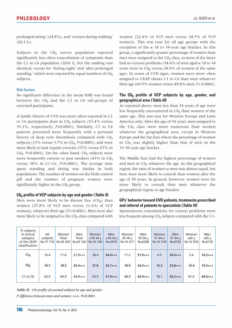
186 Phlebolymphology. Vol 19. No. 4. 2012
JJ. GUEX et al.PHLEBOLOGY
prolonged sitting’ (24.8%), and ‘worsen during walking’(20.1%).
Subjects in the C0s survey population reportedsignificantly less often exacerbation of symptoms thanthe C1 to C6 population (Table I), but the ranking wasidentical, except for ‘during night’ and ‘after prolongedstanding,’ which were reported by equal numbers of C0ssubjects.
Risk factorsNo significant difference in the mean BMI was foundbetween the C0s and the C1 to C6 sub-groups ofscreened participants.
A family history of CVD was more often reported in C1to C6 participants than in C0s subjects (53.4% versus35.3%, respectively, P<0.0001). Similarly, C1 to C6patients presented more frequently with a personalhistory of deep vein thrombosis compared with C0ssubjects (15% versus 3.7% in C0s, P<0.0001), and weremore likely to lack regular exercise (72% versus 67% inC0s; P<0.0001). On the other hand, C0s subjects weremore frequently current or past smokers (43% in C0sversus 38% in C1-C6, P<0.0001). The average timespent standing and sitting was similar in bothpopulations. The number of women on the birth controlpill and the number of pregnant women weresignificantly higher in the C0s group.
C0s profile of VCP subjects by age and gender (Table II)Men were more likely to be disease free (C0a) thanwomen (27.8% of VCP men versus 11.6% of VCPwomen), whatever their age (P< 0.0001). Men were alsomore likely to be assigned to the C0s class compared with
women (22.4% of VCP men versus 18.5% of VCPwomen). This was true for all age groups with theexception of the � 18 to 34-year age bracket. In thisgroup, a significantly greater percentage of women thanmen were assigned to the C0s class, as most of the latterhad no venous problems (54.4% of men aged � 18 to 34years were in C0a versus 28.4% of women of the sameage). In terms of CVD signs, women were more oftenassigned to CEAP classes C1 to C6 than men whatevertheir age (69.9% women versus 49.8% men; P< 0.0001).
The C0s profile of VCP subjects by age, gender, andgeographical area (Table III)As reported above, men less than 34 years of age wereless frequently encountered in C0s than women of thesame age. This was true for Western Europe and LatinAmerica only. After the age of 34 years, men assigned tothe C0s class were more numerous than womenwhatever the geographical area, except in WesternEurope and the Far East where the percentage of womenin C0s was slightly higher than that of men in the 35-50-year age bracket.
The Middle East had the highest percentage of womenand men in C0s whatever the age. In this geographicalregion, the ratio of women to men was almost equal, butmen were more likely to consult than women after theage of 50 years. In general, however, women were farmore likely to consult than men whatever thegeographical region or age bracket.
GPs’ behavior toward CVD patients, treatments prescribedand referral of patients to specialists (Table IV)Spontaneous consultations for venous problems wereless frequent among C0s subjects compared with the C1-
% subjects in clinicalcategory
of the CEAP classification:
Allsubjects
N=77 716
WomenTotal
N=54 343
MenTotal
N=23 162
Women�18-34 yN=10 160
Men�18-34 yN=3970
Women35-50 y
N=15 371
Men35-50 yN=6200
Women51-64 y
N=15 518
Men 51-64 yN=6743
Women�65 y
N=12 976
Men �65 y
N=6135
C0a 16.4 11.6 27.8+++ 28.4 54.4+++ 11.2 31.9+++ 6.5 20.0+++ 5.6 16.2+++
C0s 19.7 18.5 22.4+++ 27.8 23.7+++ 19.9 24.3+++ 15.3 22.8+++ 13.4 19.3+++
C1 to C6 63.9 69.9 49.8+++ 43.9 21.9+++ 68.9 43.9+++ 78.1 56.2+++ 81.0 64.6+++
Table II. C0s profile of screened subjects by age and gender.
P difference between men and women: +++, P<0.0001

Phlebolymphology. Vol 19. No. 4. 2012 187
The C0s patient PHLEBOLOGY
Table III. C0s profile of the screened subjects by age, gender, and geographical area.
Table IV. Treatment of C0s subjects in the Vein Consult Program. Comparison with the global population of the survey and with C2 andC6 patients.
C0s in the globalpopulation
Western EuropeCentral and
Eastern EuropeLatin America Middle East Far East
Mean age(years)
50.6 + 16.9 52.5 + 17.2 53.3 + 16.0 43.5 + 15.9 38.9 + 13.3 47.2 + 16.4
% women 68.4 66.1 70.8 73.2 51.3 69.2
% C0s 19.7 18.1 19.2 19.2 43.4 19.6
% C1 to C6 63.9 59.8 67.9 68.6 41.8 67.4
Women-Global 5433 (100%) 20 498 21 351 (100%) 7675 (100%) 1537 (100%) 3282 (100%)
C0s (%) 18.5 18.1 17.6 17.7 40.5 18.8
Men-Global 23 162 (100%) 9405 (100%) 8356 (100%) 2672 (100%) 1247 (100%) 1482
C0s (%) 22.4 18.1 23.4 23.7 47.3 21.3
Women �18-34 10 160 (100%) 3325 (100%) 2929 (100%) 2437 (100%) 623 (100%) 846 (100%)
y C0s (%) 27.8 24.6 28.9 27.9 47.4 21.4
Men �18-34 y 3970 (100%) 1393 (100%) 1074 (100%) 880 (100%) 401 (100%) 222 (100%)
C0s (%) 23.7 11.1 30.7 20 56.1 25.2
Women 35-50 y 15 371 (100%) 5657 (100%) 5447 (100%) 2553 (100%) 651 (100%) 1063 (100%)
C0s (%) 19.9 20.5 20.0 14.8 37.6 17.7
Men 35-50 y 6200 (100%) 2373 (100%) 2032 (100%) 893 (100%) 519 (100%) 383 (100%)
C0s (%) 24.3 18.6 27.6 24.2 43.5 17
Women 51-64 y 15 518 (100%) 5486 (100%) 7216 (100%) 1753 (100%) 202 (100%) 901 (100%)
C0s (%) 15.3 17.0 14.1 11.7 30.7 18.2
Men 51-64 y 6743 (100%) 2658 (100%) 2862 (100%) 525 (100%) 247 (100%) 451 (100%)
C0s (%) 22.8 21.4 20.9 28.8 41.7 25.3
Women >65 y 12 976 (100%) 5978 5561 (100%) 940 (100%) 54 (100%) 443 (100%)
C0s (%) 13.4 13.2 13.6 9.8 35.2 17.6
Men >65 y 6135 (100%) 2955 2321 369 (100.0%) 73 (100%) 417 (100%)
C0s (%) 19.3 17.9 19.5 24.4 41.1 18.9
Globalpopulationof survey
C0sN=15 290
C2N=13 888
C6N=535
% of spontaneous consultations for chronic venous disease (N=64 942) 22.9 13.5 36.5 73.8
Considered as CVD patients by GPs (N=64 942) 71 25.5 89.2 93.8
Type of treatment% patients with (N= 91 545):
Lifestyle advice 51.6 13.1 90.2 85.7
Venoactive drug 39.7 7.9 88.8 64.3
Compression therapy 24.4 1.4 44.9 35.7
Anticoagulant therapy 7.7 0.2 6.5 35.7
Sclerotherapy/endovenous ablation/open surgery 5.9 0.0 4.2 21.4

188 Phlebolymphology. Vol 19. No. 4. 2012
JJ. GUEX et al.PHLEBOLOGY
C6 population (13.5% versus 36.6%, respectively,P=0.0001, Table I), and with the global population of thesurvey (13.5% versus 22.9%, respectively, Table IV). Thefrequency of spontaneous consultation increased withCEAP class (36.5% of C2 and 73.8% of C6 subjectsconsulted spontaneously for venous leg problems).
GPs considered 71% of the total survey population tohave CVD. Of the 15 290 patients in C0s, only 3902(25.5%), ie, one-quarter were considered to have CVDby GPs, which is far less than in the global surveypopulation. In the C2 class, 89.2% were considered tohave CVD by GPs, and in C6, 93.8% were considered tohave CVD.
The percentage of C0s patients considered to have CVDwas highest in Western Europe (almost 38%) and theFar East (56%). In contrast, C2 patients were less likelyto be considered as having CVD in these two regions.Patients with ulcers were under-recognized in the FarEast (69.8% were considered to have CVD by GPs,versus 93.8% for the global survey population).
C0s patients were poorly treated in general: only 13%received lifestyle advice and 8% were prescribed avenoactive drug. In contrast, lifestyle advice and drugswere administered to the majority of patients in theglobal population as well as those in C2 and C6. It isnoteworthy that venoactive drugs were prescribed twice
as frequently as compression therapy for C2 and C6stages.
Quality of life score in C0s patients and relation to CEAPprofile (Table V)The GIS for the total survey population was 73.07. TheGIS of C0s patients was lower (82.73) than that ofhealthy subjects (90.92), indicating that their quality oflife was slightly worse. Quality of life decreased withincreasing CEAP class (73.62 in C2 and 51.96 in C6), anddrastically worsened with the mean number ofpresenting symptoms.
Characteristics of reflux according to CEAP profile –Population referred to specialists (Table VI)GPs referred 22.2% of the global population to venousspecialists. This included only 4.1% of C0s patients,36.7% of C2 patients and 60.2% of C6 patients. It isinteresting to note that a relatively low proportion of C2and C6 patients eligible for referral actually visited avenous specialist (37% and 54%, respectively) comparedwith 89% of C0s patients.
Among the 565 C0s patients who visited a specialist, only78 right legs and 69 left legs were investigated witheither Doppler (45.4%) or duplex scan (54.6%). MostC0s legs had superficial reflux (72% and 67% in theright and left legs, respectively), 11% had perforatorreflux, but an appreciable percentage had deep reflux
CEAP classification N Global Index Score
Survey population 34557 73.07 ± 20.18
Healthy subjects (C0a) 2074 90.92 ± 14.32
C0s 2170 82.73 ± 16.43
C2 7823 73.62 ± 18.37
C6 380 51.96 ± 26.39
P-value <0.0001
Number of venous symptoms
0 2074 90.92 ± 14.32
1 2929 85.74 ± 15.39
2 4807 80.18 ± 16.20
3 6073 75.24 ± 16.40
>3 14222 63.64 ± 19.97
P-value <0.0001
Table V. Quality of life score according to the CEAP profile and the number of venous symptoms.
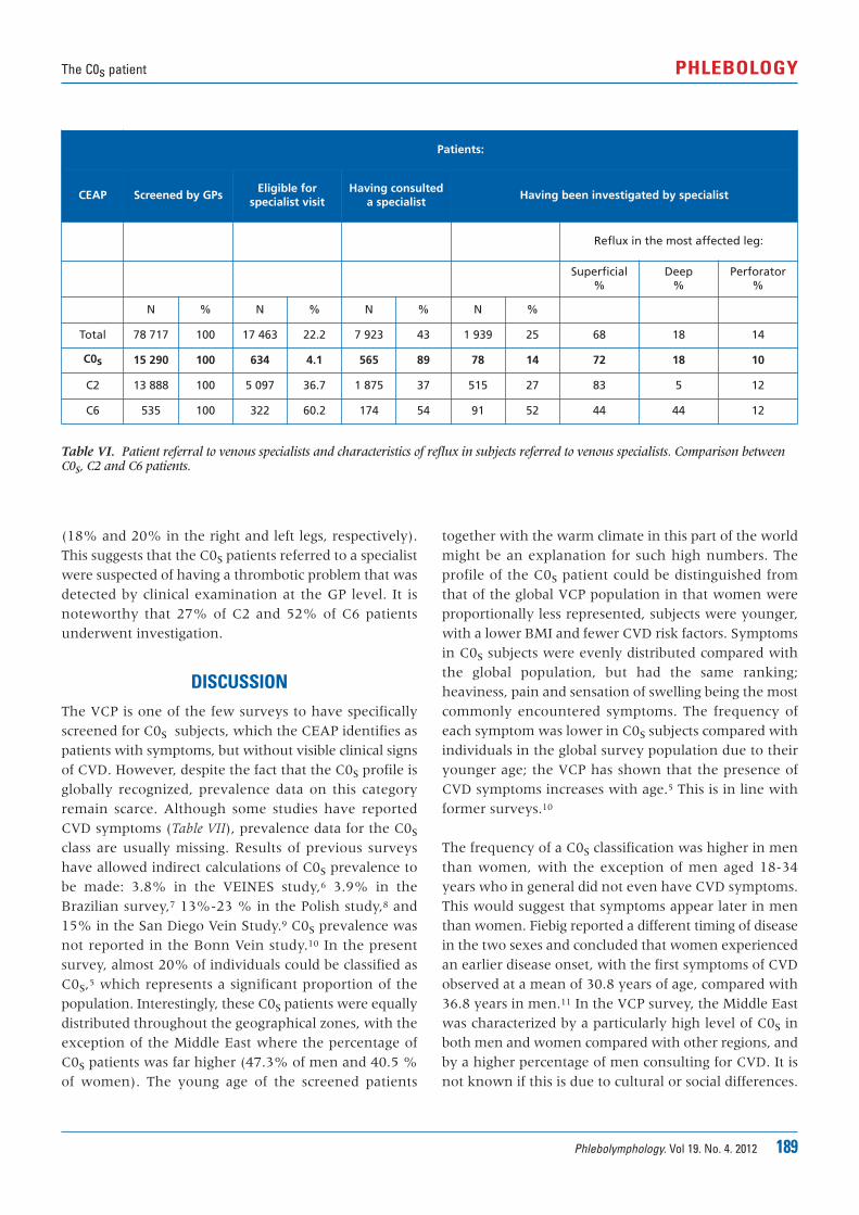
Phlebolymphology. Vol 19. No. 4. 2012 189
The C0s patient PHLEBOLOGY
(18% and 20% in the right and left legs, respectively).This suggests that the C0s patients referred to a specialistwere suspected of having a thrombotic problem that wasdetected by clinical examination at the GP level. It isnoteworthy that 27% of C2 and 52% of C6 patientsunderwent investigation.
DISCUSSIONThe VCP is one of the few surveys to have specificallyscreened for C0s subjects, which the CEAP identifies aspatients with symptoms, but without visible clinical signsof CVD. However, despite the fact that the C0s profile isglobally recognized, prevalence data on this categoryremain scarce. Although some studies have reportedCVD symptoms (Table VII), prevalence data for the C0sclass are usually missing. Results of previous surveyshave allowed indirect calculations of C0s prevalence tobe made: 3.8% in the VEINES study,6 3.9% in theBrazilian survey,7 13%-23 % in the Polish study,8 and15% in the San Diego Vein Study.9 C0s prevalence wasnot reported in the Bonn Vein study.10 In the presentsurvey, almost 20% of individuals could be classified asC0s,5 which represents a significant proportion of thepopulation. Interestingly, these C0s patients were equallydistributed throughout the geographical zones, with theexception of the Middle East where the percentage ofC0s patients was far higher (47.3% of men and 40.5 %of women). The young age of the screened patients
together with the warm climate in this part of the worldmight be an explanation for such high numbers. Theprofile of the C0s patient could be distinguished fromthat of the global VCP population in that women wereproportionally less represented, subjects were younger,with a lower BMI and fewer CVD risk factors. Symptomsin C0s subjects were evenly distributed compared withthe global population, but had the same ranking;heaviness, pain and sensation of swelling being the mostcommonly encountered symptoms. The frequency ofeach symptom was lower in C0s subjects compared withindividuals in the global survey population due to theiryounger age; the VCP has shown that the presence ofCVD symptoms increases with age.5 This is in line withformer surveys.10
The frequency of a C0s classification was higher in menthan women, with the exception of men aged 18-34years who in general did not even have CVD symptoms.This would suggest that symptoms appear later in menthan women. Fiebig reported a different timing of diseasein the two sexes and concluded that women experiencedan earlier disease onset, with the first symptoms of CVDobserved at a mean of 30.8 years of age, compared with36.8 years in men.11 In the VCP survey, the Middle Eastwas characterized by a particularly high level of C0s inboth men and women compared with other regions, andby a higher percentage of men consulting for CVD. It isnot known if this is due to cultural or social differences.
Table VI. Patient referral to venous specialists and characteristics of reflux in subjects referred to venous specialists. Comparison betweenC0s, C2 and C6 patients.
Patients:
CEAP Screened by GPsEligible for
specialist visitHaving consulted
a specialistHaving been investigated by specialist
Reflux in the most affected leg:
Superficial%
Deep %
Perforator%
N % N % N % N %
Total 78 717 100 17 463 22.2 7 923 43 1 939 25 68 18 14
C0s 15 290 100 634 4.1 565 89 78 14 72 18 10
C2 13 888 100 5 097 36.7 1 875 37 515 27 83 5 12
C6 535 100 322 60.2 174 54 91 52 44 44 12
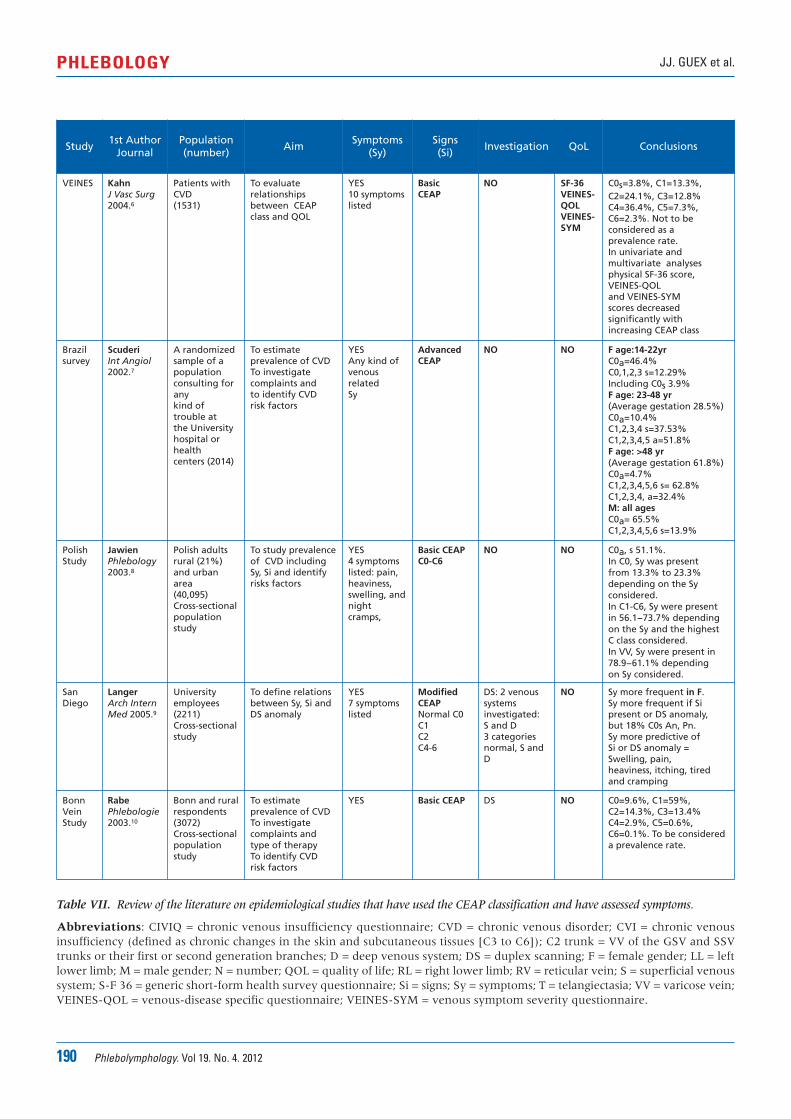
190 Phlebolymphology. Vol 19. No. 4. 2012
JJ. GUEX et al.PHLEBOLOGY
Study1st Author
JournalPopulation(number)
AimSymptoms
(Sy)Signs(Si)
Investigation QoL Conclusions
VEINES KahnJ Vasc Surg2004.6
Patients withCVD(1531)
To evaluaterelationshipsbetween CEAPclass and QOL
YES10 symptomslisted
Basic CEAP
NO SF-36VEINES-QOLVEINES-SYM
C0s=3.8%, C1=13.3%, C2=24.1%, C3=12.8%C4=36.4%, C5=7.3%,C6=2.3%. Not to be considered as a prevalence rate.In univariate and multivariate analysesphysical SF-36 score, VEINES-QOLand VEINES-SYMscores decreasedsignificantly with increasing CEAP class
Brazilsurvey
ScuderiInt Angiol2002.7
A randomizedsample of a population consulting forany kind oftrouble atthe Universityhospital orhealthcenters (2014)
To estimateprevalence of CVDTo investigate complaints andto identify CVDrisk factors
YESAny kind of venousrelatedSy
Advanced CEAP
NO NO F age:14-22yrC0a=46.4%C0,1,2,3 s=12.29% Including C0s 3.9%F age: 23-48 yr(Average gestation 28.5%)C0a=10.4%C1,2,3,4 s=37.53%C1,2,3,4,5 a=51.8%F age: >48 yr(Average gestation 61.8%)C0a=4.7%C1,2,3,4,5,6 s= 62.8%C1,2,3,4, a=32.4%M: all agesC0a= 65.5%C1,2,3,4,5,6 s=13.9%
PolishStudy
JawienPhlebology2003.8
Polish adultsrural (21%)and urbanarea(40,095) Cross-sectionalpopulationstudy
To study prevalenceof CVD includingSy, Si and identifyrisks factors
YES4 symptomslisted: pain, heaviness, swelling, andnightcramps,
Basic CEAPC0-C6
NO NO C0a, s 51.1%.In C0, Sy was present from 13.3% to 23.3% depending on the Syconsidered.In C1-C6, Sy were present in 56.1−73.7% depending on the Sy and the highestC class considered.In VV, Sy were present in 78.9−61.1% depending on Sy considered.
SanDiego
LangerArch InternMed 2005.9
Universityemployees(2211)Cross-sectionalstudy
To define relationsbetween Sy, Si andDS anomaly
YES7 symptomslisted
ModifiedCEAPNormal C0C1C2C4-6
DS: 2 venoussystemsinvestigated: S and D3 categoriesnormal, S andD
NO Sy more frequent in F.Sy more frequent if Si present or DS anomaly,but 18% C0s An, Pn.Sy more predictive ofSi or DS anomaly =Swelling, pain, heaviness, itching, tiredand cramping
BonnVein Study
RabePhlebologie2003.10
Bonn and ruralrespondents(3072)Cross-sectionalpopulationstudy
To estimateprevalence of CVDTo investigate complaints and type of therapyTo identify CVDrisk factors
YES Basic CEAP DS NO C0=9.6%, C1=59%, C2=14.3%, C3=13.4%C4=2.9%, C5=0.6%,C6=0.1%. To be considered a prevalence rate.
Table VII. Review of the literature on epidemiological studies that have used the CEAP classification and have assessed symptoms.
Abbreviations: CIVIQ = chronic venous insufficiency questionnaire; CVD = chronic venous disorder; CVI = chronic venousinsufficiency (defined as chronic changes in the skin and subcutaneous tissues [C3 to C6]); C2 trunk = VV of the GSV and SSVtrunks or their first or second generation branches; D = deep venous system; DS = duplex scanning; F = female gender; LL = leftlower limb; M = male gender; N = number; QOL = quality of life; RL = right lower limb; RV = reticular vein; S = superficial venoussystem; S-F 36 = generic short-form health survey questionnaire; Si = signs; Sy = symptoms; T = telangiectasia; VV = varicose vein;VEINES-QOL = venous-disease specific questionnaire; VEINES-SYM = venous symptom severity questionnaire.
Phlebolymphology. Vol 19. No. 4. 2012 191
The C0s patient PHLEBOLOGY
The present analysis showed a relationship betweenimpaired quality of life and increasing CEAP class, as wellas with increasing number of symptoms, a finding thathas also been demonstrated in other studies.6,12
Although they may be experiencing a poorer quality oflife, C0s patients were hesitant to consult GPs, and evenwhen they did so they received little treatment.Furthermore, only 13% received any lifestyle advice. It is important to note the lower-than-expectedprescription of compression therapy in this survey, evenat severe CVD stages, highlighting the need for patientand GP education.
The results of this survey suggest that C0s subjects arevery frequently encountered in general practice,particularly in Western Europe and the Far East, the tworegions where individuals were also more likely to beconsidered as to be suffering from CVD. However, only4% of C0s subjects were referred to a specialist (referralwas almost nonexistent in the Far East, and it isnoteworthy that an appreciable proportion of C0sindividuals who did consult a venous specialist weresuffering with secondary venous disease.
The present analysis provides a snapshot of C0s subjectsin ordinary consultation. A limitation is that this was nota longitudinal study and therefore the future of theseC0s individuals and their CVD progression is not known.It remains to be determined whether such individualsdeserve more in-depth investigation and care because ofthe possible progression of their symptoms to moresevere stages of CVD. In the San Diego Vein Study, upto 15.5% subjects complaining of leg ache had nofunctional anomaly, and up to 14.2% of subjects withpain had no visible signs of CVD.9 As a result, leg painwas considered a non-specific CVD symptom by theauthors of this study, in which case the C0s subjectwould be denied a diagnosis of CVD.
Our understanding of the mechanisms at work in CVD-related pain is gaining momentum. A considerable bodyof evidence now shows that inflammatory reactions viathe release of various mediators are involved in diseaseprogression and might explain the origins of venouspain. Nerve structures known as C-nociceptors that areeasily activated by inflammatory mediators have beenfound in the media of varicose veins13 and are alsolocated in perivenous connective tissue.14 Suchnociceptors are thought to be activated in themicrocirculation, where contact between nerve endings,
the arteriole, the vein, and the capillary is probably muchcloser than at a macrovascular level. Microcirculatoryparameters have been studied and vary with increasingseverity of CVD.15,16 We are presently just able to assessmacrocirculatory parameters, but these are not usuallyabnormal at the C0s stage, and there are currently noinstrument sensitive enough at our disposal to assess themicrocirculation at stages as early as C0s. Without ameans of assessment, we can only speculate that venouspain at the C0s stage might be the result of a disturbedmicrocirculation, but this remains to be documented.
Early work by Andreozzi brought attention to C0ssubjects, who he referred to as hypotonic phlebopathicpatients because of normal Duplex investigations, butdisturbed parameters with photo- and strain-gaugeplethysmography, leading him to suggest ahyperdistensibilty of the venous wall.17 More recently, astudy of casts from amputated legs has revealed thatvalvular incompetence can exist in the microvalves outto the third generation ‘boundary’of tributaries from thegreat saphenous vein (GSV) from stages C0 and C1, eventhough no truncular reflux could be observed in theGSV.18 Such findings lend support to a role for earlydegenerative processes in the microvasculature, longbefore the macrocirculation is targeted.
CONCLUSION Little is known about the future of subjects with C0s, agroup that has until recently been poorly studied, but aninflammatory hypothesis to explain the nature of thereported symptoms is plausible. Given the highprevalence of such subjects in the population, whateverthe geographical region, it would be beneficial to knowwhich C0s patients deserve investigation and treatment.In the meantime, treatment that could delay and evenhamper the inflammatory progression of the diseasemight prove useful. It has been shown that thesymptoms of C0s individuals are greatly improved withconservative treatments, in particular, by venoactivedrugs such as micronized purified flavonoid fraction(Daflon 500 mg*).19
*Also registered as: Alvenor®, Ardium®, Arvenum® 500, Capiven®,
Detralex®, Elatec®, Flebotropin®, Variton®, Venitol®.
192 Phlebolymphology. Vol 19. No. 4. 2012
JJ. GUEX et al.PHLEBOLOGY
REFERENCES
1. Beebe HG, Bergan JJ, Bergqvist D, etal. Classification and grading of chronicvenous disease in the lower limbs. Aconsensus state ment. Eur J VascEndovasc Surg. 1996;12:487-491.
2. Eklof B, Rutherford RB, Bergan JJ, andthe American Venous ForumInternational Ad Hoc Committee forRevision of the CEAP Classification.Revision of the CEAP classification forchronic venous disorders: consensusstatement. J Vasc Surg. 2004;40:1248-1252.
3. Eklof B, Perrin M, Delis K, Gloviczki P;American Venous Forum; EuropeanVenous Forum; International Union ofPhlebology; American College ofPhlebology; International Union ofAngiology. Updated terminology ofchronic venous disorders: the VeinTerm Transatlantic InterdisciplinaryConsensus Document. J Vasc Surg.2009;49:498-501.
4. Launois R, Lemoine JG, Lozano FS,Mansilha A. Construction andinternational validation of CIVIQ-14 (ashort form of CIVIQ-20), a newquestionnaire with a stable factorialstructure. Qual of Life Res.2011;21:1051-1058.
5. Rabe E, Guex JJ, Puskas A, Scuderi A,Fernandez Quesada F; VCPCoordinators. Epidemiology of chronicvenous disorders in geographicallydiverse populations: results from theVein Consult Program. Int Angiol.2012;3:105-115.
6. Kahn SR, M’lan CE, Lamping DL, KurzX, Bérard A, Abenhaim LA; VEINESStudy Group. Relationship betweenclinical classification of chronic venousdisease and patient-reported quality oflife: results from an internationalcohort study. J Vasc Surg. 2004;39:823-828.
7. Scuderi A, Raskin B, Assal FA, et al.The incidence of venous disease inBrazil based on the CEAP classification.Int Angiol. 2002;21:316-321.
8. Jawien A, Grzela T, Ochwat A.Prevalence of chronic venousinsufficiency in men and women:cross-sectional study in 40 095patients. Phlebology. 2003;18:110-122.
9. Langer RD, Ho E, Denenberg JO,Fronek A, Allison M, Criqui MH.Relationship between symptoms andvenous disease. The San Diegopopulation study. Arch Int Med.2005;165:1420-1424.
10. Rabe E, Pannier-Fischer F, Bromen K,et al. Bonner Ve nenstudie derDeutschen Gesellschaft fürPhlebologie. Phlebologie. 2003; 32:1-14.[German]
11. Fiebig A, Krusche P, Wolf A, et al.Heritability of chronic venous disease.Hum Genet. 2010;127:669-674.
12. Jantet G and the RELIEF Study Group.Chronic venous insufficiency:worldwide results of the RELIEF study.Reflux assEssment and quaLity of lIfeimprovEment with micronizedFlavonoids. Angiology. 2002;53:245-256.
13. Vital A, Carles D, Serise JM, BoisseauMR. Evidence for unmyelinated Cfibers and inflammatory cells inhuman varicose saphenous veins. Int JAngiol. 2010;19:e73-e77.
14. Danziger N. [Pathophysiology of painin venous disease]. J Mal Vasc.2007;32:1-7. Review. French.
15. Howlader MH, Smith PD. Correlationof severity of chronic venous diseasewith capillary morphology assessed bycapillary microscopy. J Vasc Surg.2006;43:563-569.
16. Virgini-Magalhaes CE, Porto CL,Fernandes FF, Dorigo DM, Bottino DA,Bouskela E. Use of microcirculatoryparameters to evaluate chronic venousinsufficiency. J Vasc Surg.2006;43:1037-1044.
17. Andreozzi GM, Signorelli S, Di Pino L,et al. Varicose symptoms withoutvaricose veins: the hypotonicphlebopathy, epidemiology andpathophysiology. The Acireale project.Minerva Cardioangiol. 2000;48:277-285.
18. Vincent JR, Jones GT, Hill GB, van RijAM. Failure of microvenous valves insmall superficial veins is a key to theskin changes of venous insufficiency. JVasc Surg. 2011;54(6)suppl:62S-69S.
19. Perrin M, Ramelet AA.Pharmacological treatment of primarychronic venous disease: rationale,results and unanswered questions. EurJ Vasc Endovasc Surg. 2011;41:117-125.
Phlebolymphology. Vol 19. No. 4. 2012 193
PHLEBOLOGY
Sclerotherapy in the patient with diabetes: indications and results
Francesco FERRARA andGiovanni FERRARA
Studio Flebologico Ferrara, Acerra, Naples,Italy
Keywords:chronic venous disease, compression, diabetes,duplex scan, sclerotherapy, varicose veins.
Phlebolymphology. 2012;19(4):193-198.
ABSTRACTGoal: To assess the effects of combined compression and sclerotherapytreatment on varicose veins in patients with diabetes.
Materials and methods: We evaluated 60 lower limbs with varicose veinsfrom people with diabetes who underwent one session of sclerotherapyaccording to Sigg’s method. Varices were associated with reflux along thelength of the great saphenous vein in 47 limbs, in the small saphenous veinin 8 limbs, and with nonsaphenous reflux in 5 limbs. Efficacy was determinedby clinical and duplex scan examinations performed at 6, 8, and 12 months,and at 2, and 4 years.
Results: A total of 7 clinical failures (12%) and 14 duplex scan failures (23%)were observed. No major complications such as arterial injection, severeallergic reaction or deep vein thrombosis were reported.
Conclusions: Successful sclerotherapy for varicose veins can be performedindependently of a history of diabetes, with a success rate similar to thatobserved in patients without diabetes, provided the patient has good glycemiccontrol (HbA1c <6.5%).
INTRODUCTIONVaricose veins of the legs are a common condition caused by venousinsufficiency as a result of valve reflux.1 Most varices are located in the greatsaphenous vein (GSV) and are associated with symptoms such as legheaviness, fatigue, or throbbing pain.2 People with diabetes may beparticularly prone to varicose veins as they generally present with a numberof common risk factors for chronic venous disease (CVD) including poorcirculation, overweight, hypertension as well as a propensity for endothelialdysfunction. Indeed, a number of studies in the literature have shown thatCVD and diabetes mellitus frequently coexist.3-6 Furthermore, diabetes isassociated with more severe signs in 50% of patients.7 The medical rationalefor treating varicose veins in patients with diabetes is the same as those
194 Phlebolymphology. Vol 19. No. 4. 2012
Francesco FERRARA, Giovanni FERRARAPHLEBOLOGY
without diabetes: to reduce symptoms, improve qualityof life, and prevent disease progression. Earlyintervention reduces the risk of ongoing vein- and skin-related damage secondary to chronic venoushypertension. These changes progressively worsen withtime, and earlier treatment is preferable to a wait-and-see approach.
Traditionally, treatment of varicose veins involved aninvasive procedure of groin to ankle stripping andligation of the GSV. More recently, ultrasound-guidedfoam sclerotherapy has gained acceptance as an effectiveand safe alternative to surgery to treat varicose veins.1
Sclerotherapy is defined as the targeted elimination ofsmall vessels, varicose veins, and vascular anomalies bythe injection of a sclerosant (chemical irritant) into theveins with a small needle.8 The sclerosant causesinflammation, thrombosis, and finally fibrosis of thevein, which is transformed into a fibrous cord thatcannot be recanalized. Sclerotherapy is a simple, cost-effective, efficacious, and esthetically acceptable modalityfor both therapeutic and esthetic purposes.
There are few studies in the literature discussingsclerotherapy for varicose veins in patients with diabetesand the few that do exist are contradictory with somestating that diabetes is a contraindication forsclerotherapy8 and others stating that it is not.9 Mostcontraindications and/or special precautions relating tosclerotherapy relate to factors that either directlyincrease the risk of deep vein thrombosis or indirectlyinterfere with posttreatment compression andmobilisation.9
Sclerotherapy should only be performed in people withchronic diseases if their condition is well controlled, anddiabetes is no exception.10 The standard measure of long-term glycemic control is glycosylated hemoglobin(HbA1c). In a person without diabetes, normal HbA1clevels fluctuate between 4% and 6%. A person withdiabetes is considered to have good glycemic control iftheir HbA1c level is below about 6.5%, although thismay not always be achievable particularly in olderpatients or in those in whom hypoglycemia must beavoided.11 When levels rise to above 7% the individualis not considered to have good control. However, in theabsence of a true contraindication,8 several precautionsshould be observed before performing sclerotherapy inan individual with diabetes.
Before the procedureBefore the procedure, a complete clinical examinationand duplex scan investigation should be performed todetect any associated arterial disease. To avoidprolonging inflammation in the vein wall, any existingedema and soft tissue changes associated with CVDshould first be reduced by the application of a short-stretch bandage.
Choice of sclerosing agentConcerning the choice of sclerosing agent, foam is notcontraindicated, but agents containing ahyperconcentration of glucose should be avoided. Foamhas been shown to be superior to liquid sclerotherapy inthe GSV in terms of clinical and hemodynamicoutcomes, with several advantages over traditional liquidsclerotherapy.12 When delivered as foam, detergentsclerosant is not diluted by blood, instead displacing it toallow direct contact of the sclerosant with theendothelium.13 This allows for the use of a smaller doseof sclerosant, which is important as the vein walls ofpatients with diabetes are very sensitive to these agents.Second, should extravasation occur, foam is much bettertolerated than extravasated liquid. Third, foam can bereadily visualized by ultrasound, which increases theaccuracy with which individual varicose veins can betreated.
Puncture procedureDue to the impaired immune response in individualswith diabetes, they are particularly susceptible toinfection. Care must therefore be taken to ensure thatthe materials and puncture site are sterile. Wheninjecting the sclerosing agent, the bevel of the needleshould be placed into the lumen of the vein as patientswith diabetic neuropathy have reduced sensitivity topain and if the needle is not properly positionedextravasation of sclerosant might go unnoticed.
Compression therapyProlonged compression therapy starting immediatelyafter the procedure and lasting at least 30 days isimperative in all patients to achieve optimal results withsclerotherapy, but particularly in patients with diabetesbecause of their endothelial pathology.14 Compressionminimizes the blood reentering the injected area andthrombus formation, increases fibrinolytic activity andtherefore reduces scar tissue formation, decreases theincidence of postsclerotherapy hyperpigmentation, andimproves venous blood flow.
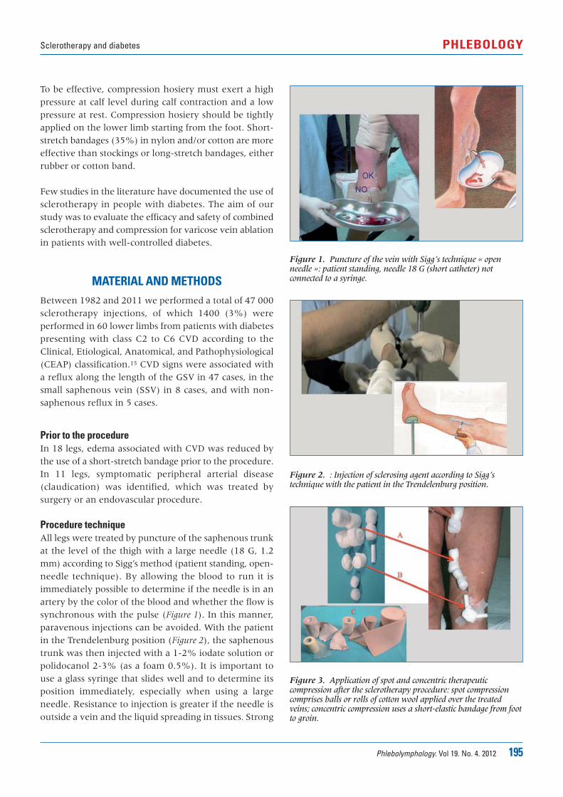
Phlebolymphology. Vol 19. No. 4. 2012 195
Sclerotherapy and diabetes PHLEBOLOGY
To be effective, compression hosiery must exert a highpressure at calf level during calf contraction and a lowpressure at rest. Compression hosiery should be tightlyapplied on the lower limb starting from the foot. Short-stretch bandages (35%) in nylon and/or cotton are moreeffective than stockings or long-stretch bandages, eitherrubber or cotton band.
Few studies in the literature have documented the use ofsclerotherapy in people with diabetes. The aim of ourstudy was to evaluate the efficacy and safety of combinedsclerotherapy and compression for varicose vein ablationin patients with well-controlled diabetes.
MATERIAL AND METHODSBetween 1982 and 2011 we performed a total of 47 000sclerotherapy injections, of which 1400 (3%) wereperformed in 60 lower limbs from patients with diabetespresenting with class C2 to C6 CVD according to theClinical, Etiological, Anatomical, and Pathophysiological(CEAP) classification.15 CVD signs were associated witha reflux along the length of the GSV in 47 cases, in thesmall saphenous vein (SSV) in 8 cases, and with non-saphenous reflux in 5 cases.
Prior to the procedureIn 18 legs, edema associated with CVD was reduced bythe use of a short-stretch bandage prior to the procedure.In 11 legs, symptomatic peripheral arterial disease(claudication) was identified, which was treated bysurgery or an endovascular procedure.
Procedure techniqueAll legs were treated by puncture of the saphenous trunkat the level of the thigh with a large needle (18 G, 1.2mm) according to Sigg’s method (patient standing, open-needle technique). By allowing the blood to run it isimmediately possible to determine if the needle is in anartery by the color of the blood and whether the flow issynchronous with the pulse (Figure 1). In this manner,paravenous injections can be avoided. With the patientin the Trendelenburg position (Figure 2), the saphenoustrunk was then injected with a 1-2% iodate solution orpolidocanol 2-3% (as a foam 0.5%). It is important touse a glass syringe that slides well and to determine itsposition immediately, especially when using a largeneedle. Resistance to injection is greater if the needle isoutside a vein and the liquid spreading in tissues. Strong
Figure 1. Puncture of the vein with Sigg’s technique « openneedle »: patient standing, needle 18 G (short catheter) notconnected to a syringe.
Figure 2. : Injection of sclerosing agent according to Sigg’stechnique with the patient in the Trendelenburg position.
Figure 3. Application of spot and concentric therapeuticcompression after the sclerotherapy procedure: spot compressioncomprises balls or rolls of cotton wool applied over the treatedveins; concentric compression uses a short-elastic bandage from footto groin.
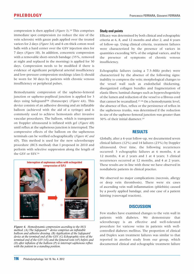
196 Phlebolymphology. Vol 19. No. 4. 2012
Francesco FERRARA, Giovanni FERRARAPHLEBOLOGY
compression is then applied (Figure 3).10 This comprisesimmediate spot compression (to reduce the size of thevein sclerosis) with gauze pads applied over the treatedvarices for 2 days (Figure 3A) and 4-cm thick cotton woolballs with a hard center over the GSV injection sites for7 days (Figure 3B). In addition, concentric compressionwith a removable short-stretch bandage (35%, removedat night and replaced in the morning) is applied for 30days. Compression needs to be modified if there isevidence of significant peripheral arterial insufficiencyand low-pressure compression stockings (class I) shouldbe worn for 30 days by patients with chronic venousinsufficiency or peripheral pulses.
Hemodynamic compression of the sapheno-femoraljunction or sapheno-popliteal junction is applied for 3days using Safeguard™ (Datascope) (Figure 4A). Thisdevice consists of an adhesive dressing and an inflatableballoon (achieved with the aid of a syringe) and iscommonly used to achieve hemostasis after invasivevascular procedures. The balloon, which is transparenton Doppler ultrasound is inflated with gel (Figure 4B)until reflux at the saphenous junction is interrupted. Thecompressive effects of the balloon on the saphenousterminals can be verified echographically (Figure 4C and
4D). This method is used for the new sclerotherapyprocedure (HCS method) that I proposed in 2010 andperform with selective suppression along the length ofthe GSV or SSV.16
Study end pointsEfficacy was determined by both clinical and echographiccriteria at 6, 8, and 12 months and after 2, and 4 yearsof follow-up. Using clinical criteria, treatment failureswere characterized by the presence of varices inquantities exceeding 50% of the original varices, and bythe presence of symptoms of chronic venousinsufficiency.
Echographic failures (using a 7.5-MHz probe) werecharacterized by the absence of the following signs:inability to compress the vein; morphological changes tothe vessel wall such as endothelial thickening,disorganized collagen bundles and fragmentation ofelastic fibers; luminal changes such as hyperechogenicityof the lumen and reduction of the vessel to a fibrous cordthat cannot be recanalized.17,18 On a hemodynamic level,the absence of flux, reflux or the persistence of reflux inthe saphenous trunks, was determined if the reductionin size of the sapheno-femoral junction was greater than30% of their initial diameter.19
RESULTSGlobally, after a 4-year follow-up, we documented sevenclinical failures (12%) and 14 failures (23%) by Dopplerultrasound. Over time, the following recurrencesoccurred: 3 echographic failures at 6 months, 6 at 12 months, 4 at 2 years and 1 at 4 years; 3 clinicalrecurrences occurred at 12 months, and 4 at 2 years.These results are in line with those we have observed innondiabetic patients in clinical practice.
We observed no major complications (necrosis, allergyor deep vein thrombosis). There were six casesof ascending vein wall inflammation (phlebitis) causedby a poorly applied bandage, and one case of a patientfainting (vasovagal reaction).
DISCUSSIONFew studies have examined changes to the vein wall inpatients with diabetes. We demonstrate thatsclerotherapy is an effective and well-toleratedprocedure for varicose veins in patients with well-controlled diabetes mellitus. The proportion of clinicaland Duplex scan treatment failures was similar to thatreported in another study from our group, whichdocumented clinical and echographic treatment failure
Figure 4. Hemodynamic compression according to the HCSmethod. (A) The Safeguard� device comprises an inflatableballoon and adhesive dressing. (B) Application of the Safeguarddevice at the terminal end of the GSV. (C) Echographic view of theterminal end of the GSV (vS) and the femoral vein (vF) before and(D) after inflation of the balloon (P) to interrupt saphenous refluxwith the patient in a standing position.

Phlebolymphology. Vol 19. No. 4. 2012 197
Sclerotherapy and diabetes PHLEBOLOGY
rates of 12% and 23%, respectively, in 1500 cases of GSVreflux treated with the Sigg method over the course of15 years.18,19
We have previously evaluated the results ofsclerotherapy in different pathophysiological contexts(Table I).20 Diabetes is associated with vascular sensitivity,as proposed by Lanza et al who suggested the existenceof a “diabetic phlebosclerosis”.21 Endothelial dysfunctionin patients with diabetes, as a result of chronicallyelevated blood glucose levels, may influence severalmediators secreted by the endothelium. For example,there is an augmentation of levels of von Willebrandfactor (vWF), a glycoprotein synthesized and stored inthe vascular endothelium, increased levels of which maycontribute to an increased risk of thrombosis, and adecrease in production of prostacyclin, which isassociated with platelet hyperaggregability. For thisreason, when performing sclerotherapy in people withdiabetes, concentrations of sclerosing agent must bereduced by half (whether using foam or liquid) andsuitable compression must be applied.
In our center, compression is provided by a short-stretchbandage (35%), which only prevents arterial perfusionin the presence of a significant arterial obstruction, witha systolic pressure of less than 70 mm Hg at the ankle.However, the latter is a hypothetical patient and wewould not propose sclerotherapy or surgical treatmentin a person with significant symptoms of peripheralarterial disease. In reality, it is the patient withclaudication and varices that occasionally presents for
phlebological consultation (Table II). This patient is acandidate for sclerotherapy when the claudication hasbeen resolved. We should highlight that in thesepatients, the re-establishment of the arterial pathway bycatheter or surgery should be attempted. However,sclerotherapy may then pose a problem, as it demandscompression associated with ambulation. A test bandageworn during the day for 1 week, without injectionsshould be conducted as a feasibility assessment. A similarapproach should be followed in case of suspected arterialdisease. A nonelastic bandage may be tolerated inpatients with a suspected subclinical arteriopathy.10 Inthese patients, a compression stocking deliveringcompression greater than 20 mm Hg should be avoided,especially in the absence of good systolic blood pressure.
Sclerotherapy, when performed appropriately, is anefficacious and safe technique, but complications canoccur. The most severe include cutaneous necrosis, as aresult of extravasation of sclerosant into perivasculartissues or direct injection into an arteriole, allergicreactions to sclerosants, and deep vein thrombosis. Noneof these adverse events were observed in our study. Thepersistence of significant reflux into a vein that has beentreated with a sclerosing agent can lead to phlebitis andthis was documented in six cases as a result of poorlyapplied compression. We observed one vasovagalreaction, which resolved spontaneously.
Sclerotherapy is an important tool for preventing theprogression of CVD to more severe stages. In a prospective,7-year follow-up of patients with both superficial and
Table I. Sclerotherapy in patients with arteriopathy – a decisional tree.
Table II. Three different treatment responses for varicose veins inpatients with general pathophysiological conditions.
Sclero sensitization
Hepatitis C, hepatitis B
Estrogen-plus-progesterone pills
Sclerosing agents
Sclero sensitivity
Endocrine diseases and diabetes
Collagen and rhumatological disorders
Metabolic syndrome
Pregnancy and menopause
Obesity and low body weight (anorexia)
Thrombophilia
Sclero resistance
Embryologic factors
Constitutional factors
Genetic factors
No
No
Yes
Yes
Sclerosis Trial bandageClaudication?
Restorearterial
pathway
Stage III-IVStage I-IIClaudication?
Obstructivearteriopathy
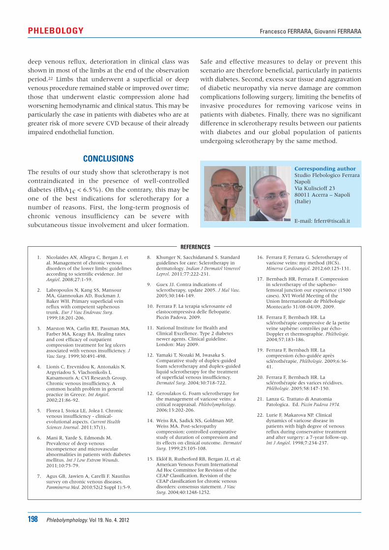
198 Phlebolymphology. Vol 19. No. 4. 2012
Francesco FERRARA, Giovanni FERRARAPHLEBOLOGY
Safe and effective measures to delay or prevent thisscenario are therefore beneficial, particularly in patientswith diabetes. Second, excess scar tissue and aggravationof diabetic neuropathy via nerve damage are commoncomplications following surgery, limiting the benefits ofinvasive procedures for removing varicose veins inpatients with diabetes. Finally, there was no significantdifference in sclerotherapy results between our patientswith diabetes and our global population of patientsundergoing sclerotherapy by the same method.
deep venous reflux, deterioration in clinical class wasshown in most of the limbs at the end of the observationperiod.22 Limbs that underwent a superficial or deepvenous procedure remained stable or improved over time;those that underwent elastic compression alone hadworsening hemodynamic and clinical status. This may beparticularly the case in patients with diabetes who are atgreater risk of more severe CVD because of their alreadyimpaired endothelial function.
CONCLUSIONSThe results of our study show that sclerotherapy is notcontraindicated in the presence of well-controlleddiabetes (HbA1c < 6.5%). On the contrary, this may beone of the best indications for sclerotherapy for anumber of reasons. First, the long-term prognosis ofchronic venous insufficiency can be severe withsubcutaneous tissue involvement and ulcer formation.
Corresponding authorStudio Flebologico FerraraNapoliVia Kuliscioff 2380011 Acerra – Napoli(Italie)
E-mail: [email protected]
REFERENCES
1. Nicolaides AN, Allegra C, Bergan J, etal. Management of chronic venousdisorders of the lower limbs: guidelinesaccording to scientific evidence. IntAngiol. 2008;27:1-59.
2. Labropoulos N, Kang SS, MansourMA, Giannoukas AD, Buckman J,Baker WH. Primary superficial veinreflux with competent saphenoustrunk. Eur J Vasc Endovasc Surg.1999;18:201-206.
3. Marston WA, Carlin RE, Passman MA,Farber MA, Keagy BA. Healing ratesand cost efficacy of outpatientcompression treatment for leg ulcersassociated with venous insufficiency. JVasc Surg. 1999;30:491-498.
4. Lionis C, Erevnidou K, Antonakis N,Argyriadou S, Vlachonikolis I,Katsamouris A; CVI Research Group.Chronic venous insufficiency. Acommon health problem in generalpractice in Greece. Int Angiol.2002;21:86-92.
5. Florea I, Stoica LE, Jolea I. Chronicvenous insufficiency - clinical-evolutional aspects. Current HealthSciences Journal. 2011;37(1).
6. Mani R, Yarde S, Edmonds M.Prevalence of deep venousincompetence and microvascularabnormalities in patients with diabetesmellitus. Int J Low Extrem Wounds.2011;10:75-79.
7. Agus GB, Jawien A, Carelli F. Nautilussurvey on chronic venous diseases.Panminerva Med. 2010;52(2 Suppl 1):5-9.
8. Khunger N, Sacchidanand S. Standardguidelines for care: Sclerotherapy indermatology. Indian J Dermatol VenereolLeprol. 2011;77:222-231.
9. Guex JJ. Contra indications ofsclerotherapy, update 2005. J Mal Vasc.2005;30:144-149.
10. Ferrara F. La terapia sclerosante edelastocompressiva delle flebopatie.Piccin Padova. 2009.
11. National Institute for Health andClinical Excellence. Type 2 diabetesnewer agents. Clinical guideline.London: May 2009.
12. Yamaki T, Nozaki M, Iwasaka S.Comparative study of duplex-guidedfoam sclerotherapy and duplex-guidedliquid sclerotherapy for the treatmentof superficial venous insufficiency.Dermatol Surg. 2004;30:718-722.
12. Geroulakos G. Foam sclerotherapy forthe management of varicose veins: acritical reappraisal. Phlebolymphology.2006;13:202-206.
14. Weiss RA, Sadick NS, Goldman MP,Weiss MA. Post-scleropathycompression: controlled comparativestudy of duration of compression andits effects on clinical outcome. DermatolSurg. 1999;25:105-108.
15. Eklöf B, Rutherford RB, Bergan JJ, et al;American Venous Forum InternationalAd Hoc Committee for Revision of theCEAP Classification. Revision of theCEAP classification for chronic venousdisorders: consensus statement. J VascSurg. 2004;40:1248-1252.
16. Ferrara F, Ferrara G. Sclerotherapy ofvaricose veins: my method (HCS).Minerva Cardioangiol. 2012;60:125-131.
17. Bernbach HR, Ferrara F. Compressionin sclerotherapy of the sapheno-femoral junction our experience (1500cases). XVI World Meeting of theUnion Internationale de PhlébologieMontecarlo 31/08-04/09, 2009.
18. Ferrara F, Bernbach HR. Lasclérothérapie compressive de la petiteveine saphène: contrôles par écho-Doppler et thermographie. Phlébologie.2004;57:183-186.
19. Ferrara F, Bernbach HR. Lacompression écho-guidée aprèssclérothérapie, Phlébologie. 2009;6:36-41.
20. Ferrara F, Bernbach HR. Lasclérothérapie des varices récidives.Phlébologie. 2005;58:147-150.
21. Lanza G. Trattato di AnatomiaPatologica. Ed. Piccin Padova 1974.
22. Lurie F, Makarova NP. Clinicaldynamics of varicose disease inpatients with high degree of venousreflux during conservative treatmentand after surgery: a 7-year follow-up.Int J Angiol. 1998;7:234-237.

Phlebolymphology. Vol 19. No. 4. 2012 199
LYMPHOLOGY
AIM AND SCOPE
Phlebolymphology is a quarterly peer-reviewed publication that aims toprovide clinicians with updated information on every aspect of the venousand lymphatic disorders: epidemiology, pathophysiology, diagnosis,management, and basic science. Articles are usually in the form of reviewarticles on timely topics with a broad update of recent developments andtheir clinical applications.
GENERAL INSTRUCTIONSArticles should discuss a topic of current interest, outline currentknowledge of the subject treated, give personal views and also analyzethe different opinions regarding the topic discussed, and be up to date onthe latest literature data. The text should be 3000-5000 words, not including references, tables,figures. Illustrations are strongly encouraged. All texts should besubmitted in English.Submission: Manuscripts may be submitted by e-mail1, double-spaced, 8 to 16 typed. All pages should be numbered. All corresponding authorsshould supply a portrait photograph for inclusion at the end of the article.This may be sent by e-mail, provided the resolution of the file is at least 300dpi.Title page: The title page should include a title, the full names of all theauthors, the highest academic degrees of all authors (in country-of-originlanguage), affiliations (names of department[s] and institution[s] at thetime the work was done), a short running title (no more than 50 letters andspaces), 5 to 10 keywords, the corresponding author’s complete mailingaddress, telephone, fax, and e-mail., and acknowledgments.Abstract: A 150-word abstract should be provided for all articles. Theeditorial department will edit abstracts that are too short or too long. Text:. Abbreviations should be used sparingly and expanded at firstmention. The style of titles and subtitles should be consistent throughoutthe text. The editorial department reserves the right to add, modify, ordelete headings if necessary. Phlebolymphology uses SI units and genericnames of drugs.
REFERENCES
Citation in text: All references should be cited in the text and numberedconsecutively using superscript Arabic numerals. Reference list: Presentation of the references should be based on theUniform Requirements for Manuscripts Submitted to Biomedical Journals.Ann Intern Med. 1997;126:36-47 (“Vancouver style”). The author-datesystem of citation is not acceptable. “In press” references should beavoided. In the bibliography, titles of journals should be abbreviatedaccording to Index Medicus. All authors should be listed for up to sixauthors; if there are more, only the first three should be listed, followed by“et al.” Where necessary, references will be styled by the editorialdepartment to Phlebolymphology copyediting requirements. Authors beartotal responsibility for the accuracy and completeness of all referencesand for correct text citation.
Examples of style for references
Journal article: Sessa C, Perrin M, Porcu P, et al. Popliteal venousaneurysms. A two-center experience with 21 cases and review of theliterature. Int J Angiol. 2000;9:164-170.Article in a supplement: Sansilvestri-Morel P, Rupin A, Badier-CommanderC, et al. Chronic venous insufficiency: dysregulation of collagen synthesis.Angiology. 2003;(suppl 1):S13-S18.Chapter in a book: Coleridge Smith PD. The drug treatment of chronicvenous insufficiency and venous ulceration. In: Gloviczki P, Yao JST, eds.Handbook of Venous Disorders: Guidelines of the American Venous Forum.2nd ed. London, UK: Arnold; 2001:309-321.Web-based material: Nicolaides AN. Investigation of chronic venousinsufficiency: a consensus statement. American Heart Association, 2000.Available at: http://www.circulationaha.org. Accessed October 17, 2005.Presentation at a conference: Jantet G. Epidemiological results of theRELIEF study across different continents. Paper presented at: 15th WorldCongress of the Union Internationale de Phlébologie; October 2-7, 2005;Rio de Janeiro, Brazil.
FIGURES AND TABLESFigures should be of good quality or professionally prepared, with theproper orientation indicated when necessary (eg, “top” or “left”), and beidentified by Arabic numerals, eg, Figure 2. Tables should be identified byroman numerals. Provide each table and figure on a separate sheet.Legends must be provided with all illustrations, including expansion of allabbreviations used (even if they are already defined in the text). All figuresand tables should be numbered and cited in the text.
PHOTOGRAPHIC ILLUSTRATIONS Illustrations in color are encouraged.
EDITORIAL ASSESSMENT AND PROCESSINGEditorial processing: All manuscripts are copyedited according to theguidelines of the latest edition of the American Medical AssociationManual of Style (Baltimore, Md: Williams & Wilkins); the spelling used isAmerican (reference dictionaries: latest editions of Merriam-Webster’sCollegiate Dictionary and Stedman’s Medical Dictionary). Proofs: Page proofs will be sent to the corresponding author for approvalin PDF format by e-mail. Authors who wish to receive a hard copy of theirproofs should contact the editorial offices upon receipt of the proofs by e-mail. Author corrections should be returned within 72 hours by e-mail orfax.2 If this deadline is not met, the editorial department will assume thatthe author accepts the proofs as they stand. Authors are responsible forall statements made in their work, including changes made by the editorialdepartment and authorized by the author.
COPYRIGHTTransfer of copyright: Copyright of articles will be transferred to thepublisher of Phlebolymphology. The Copyright Transfer Agreement mustbe signed by all authors and returned to the publisher. Permissions: The author should inform the editorial office if any of thefigures, tables or illustrations are reproduced from elsewhere. Forreproduction of copyrighted work, the editorial office will obtainauthorization from the publisher concerned. Requests for permission toreproduce material published in Phlebolymphology should be sent directlyto the editorial office.1,2
Instructions for authors
1. Françoise Pitsch 2. E-mail: [email protected]

DATES CONGRESS COUNTRY CITY
7-9 December2012
3RD LISBON INTERNATIONAL FORUM ON VASCULAR DISEASES Portugal Lisbon
8 December 2012 ANNUAL MEETING OF THE AUSTRIAN SOCIETY OF PHLEBOLOGY Austria Vienna
17-19 Januay2013
CONTROVERSIES AND UPDATES IN VASCULAR SURGERY France Paris
24-26 January2013
ANNUAL CONFERENCE OF VENOUS ASSOCIATION OF INDIAVAICON 2013
India Goa
2-3 February 2013 3E CONTROVERSIAS EN CIRURGIA VASCULAR E ENDOVASCULAR Brazil São Paulo
19-22 February2013
XXI SLOVAK ANGIOLOGY CONGRESS WITH INTERNATIONAL PARTICIPATION
SlovakiaTatranskaLomnica,
High Tatras
28 February - 2 March 2013
XXXVIII. ANGIOLOGICAL DAYS 2013 WITH INTERNATIONAL PARTICIPATION
Czech Republic Prague
Mid March 2013 XXXIII VENOUS MEETING OF PORTUGUESE SURGICAL SOCIETY Portugal Lisbon
14-16 March 201346ÈME CONGRES DU COLLEGE FRANÇAIS DE PATHOLOGIE
VASCULAIREFrance Paris
18-19 April 2013UKRAINIAN NATIONAL CONGRESS "ANGIOLOGY AND VASCULAR
SURGERY"Ukraine Kiev
Congress and conference calendar
CONGRESS
200 Phlebolymphology. Vol 19. No. 4. 2012

PROGRAMME DIRECTOR CONTACT WEB SITE
Prof. Dr. Fernandes andFernandes
Faculdade de Medicina de Lisboa:[email protected]
Instituto Cardiovascular de Lisboa:[email protected]
www.icvl.pt
Prof. Dr. WolfgangSalmhofer
IFC Ilona fuchs CongressTel.: +43 1 602 25 48
E-mail: [email protected]
Jean-Pierre Becquemin
divine [id] agency - 17 rue VentureF-13001 Marseille
Tel.: + 33 (0) 4 91 57 19 60E-mail: [email protected]
www.cacvs.org
Dr. Shoaib Padariachairman organising
comittee
E-mail: [email protected].: +91 9820023683
www.vaicon2013.com
Dr. Adnan Nasser www.sabcv.com.br
Prof. MUDr. Viera Štvrtinová, CSc.
Slovak angiological societyE-mail: [email protected] /
Czech Angiological SocietyAmca s.r.o. Vysehradska 320/49,
128 00 Praha 2www.angiology.cz
Prof. Dr. Carlos PereiraAlves
Portuguese Surgical SocietyE-mail: [email protected]
www.spcir.com
Jean-Claude Wautrecht
Nex & coM Medical Events - 159 rue de SillyF-92100 Boulogne Billancourt
Tel.: +33 (0) 1 46 43 33 00E-mail: [email protected]
www.cfpv.fr
A. A. Shalimov NationalInstitute of Surgery and
Transplantology NationalAcademy Of MedicalScience Of Ukraine
“Voldservice group”Tel.: (044) 520-27-27, 209-08-59
www.vasc.com.ua.
CONGRESS
Phlebolymphology. Vol 19. No. 4. 2012 201
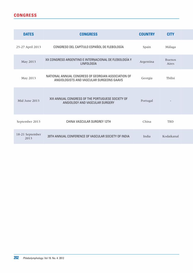
202 Phlebolymphology. Vol 19. No. 4. 2012
CONGRESS
DATES CONGRESS COUNTRY CITY
25-27 April 2013 CONGRESO DEL CAPÍTULO ESPAÑOL DE FLEBOLOGÍA Spain Málaga
May 2013XX CONGRESO ARGENTINO E INTERNACIONAL DE FLEBOLOGÍA Y
LINFOLOGÍAArgentina
BuenosAires
May 2013NATIONAL ANNUAL CONGRESS OF GEORGIAN ASSOCIATION OF
ANGIOLOGISTS AND VASCULAR SURGEONS GAAVSGeorgia Tbilisi
Mid June 2013XIII ANNUAL CONGRESS OF THE PORTUGUESE SOCIETY OF
ANGIOLOGY AND VASCULAR SURGERYPortugal -
September 2013 CHINA VASCULAR SURGREY 12TH China TBD
18-21 September2013
20TH ANNUAL CONFERENCE OF VASCULAR SOCIETY OF INDIA India Kodaikanal
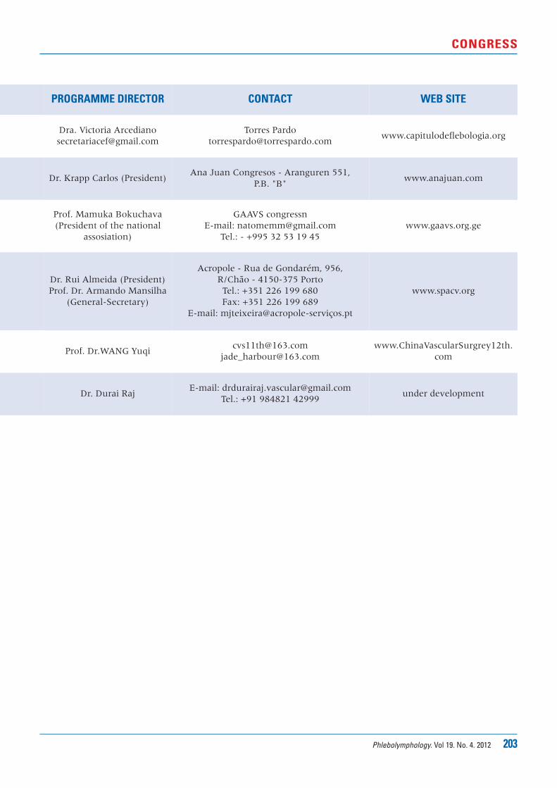
Phlebolymphology. Vol 19. No. 4. 2012 203
CONGRESS
PROGRAMME DIRECTOR CONTACT WEB SITE
Dra. Victoria [email protected]
Torres [email protected]
www.capitulodeflebologia.org
Dr. Krapp Carlos (President)Ana Juan Congresos - Aranguren 551,
P.B. "B"www.anajuan.com
Prof. Mamuka Bokuchava(President of the national
assosiation)
GAAVS congressnE-mail: [email protected]
Tel.: - +995 32 53 19 45www.gaavs.org.ge
Dr. Rui Almeida (President)Prof. Dr. Armando Mansilha
(General-Secretary)
Acropole - Rua de Gondarém, 956, R/Chão - 4150-375 PortoTel.: +351 226 199 680Fax: +351 226 199 689
E-mail: mjteixeira@acropole-serviços.pt
www.spacv.org
Prof. Dr.WANG Yuqi [email protected]
com
Dr. Durai RajE-mail: [email protected]
Tel.: +91 984821 42999under development
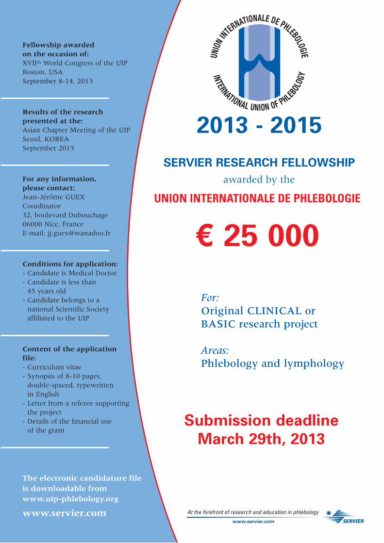
Fellowship awarded on the occasion of:XVIIth World Congress of the UIPBoston, USASeptember 8-14, 2013
Results of the researchpresented at the:Asian Chapter Meeting of the UIPSeoul, KOREASeptember 2015
For any information,please contact:Jean-Jérôme GUEXCoordinator32, boulevard Dubouchage06000 Nice, FranceE-mail: [email protected]
Conditions for application:- Candidate is Medical Doctor- Candidate is less than
45 years old- Candidate belongs to a
national Scientific Society affiliated to the UIP
Content of the application file:- Curriculum vitae- Synopsis of 8-10 pages,
double-spaced, typewritten in English
- Letter from a referee supporting the project
- Details of the financial use of the grant
SERVIER RESEARCH FELLOWSHIP
awarded by the
2013 - 2015
UNION INTERNATIONALE DE PHLEBOLOGIE
€ 25 000
Submission deadline
March 29th, 2013
For: Original CLINICAL orBASIC research project
Areas:Phlebology and lymphology
The electronic candidature fileis downloadable fromwww.uip-phlebology.org
www.servier.com At the forefront of research and education in phlebology
www.servier.com
13 D
N 1
016
BACorrespondent:
Servier International - 50, rue Carnot - 92284 Suresnes Cedex - France
Website: www.servier.com


![[KMS] State of New York Revised EVTA-2 (July 2017 ... · Web viewElectronic Value Transfer Administrator Form EVTA-2, Key Merchant Services (KMS) Work Order Contract PS65792 (updated](https://img.pdfslide.us/doc/110x75/5acb8ac17f8b9a93268b5e04/kms-state-of-new-york-revised-evta-2-july-2017-viewelectronic-value-transfer.jpg)