Embed Size (px)
Citation preview

9
Beń-Skowronek I. i inni: Two mechanisms of damage to thyrocytes in Hashimoto’s thyroiditis
Vol. 11/2012 Nr 3(40)
Endokrynologia PediatrycznaPediatric Endocrinology
9
Introduction. The development of the Hashimoto’s thyroiditis is the result of the damage to thyrocytes, apoptosis, and autoimmune cytotoxic action of lymphocytes. The aim of the study is to present ultrastructural changes in thyroid cells in the course of damage in Hashimoto’s thyroiditis. Patients and methods. The study involved 40 children: 20 children with Hashimoto’s thyroiditis and 20 children as a control group. Specimens for ultrastructural investigations were obtained during thyroidectomy. Ultrathin sections were contrasted with uranyl acetate and lead citrate and examined under the EM 900 Zeiss Germany Electron Microscope. Results. In the control group, the thyroid follicular cells were cuboidal or cylindrical and were lying on a thin basement membrane. Varied degrees of apoptosis were observed in the Hashimoto’s thyroiditis patients. The basement membrane of the follicles was thickened by deposition of numerous collagen fibers. The thyrocytes had irregular cell nuclei and an increased number of mitochondria in the basal pole. Different stages of thyrocyte necrosis were visible at the sites of contact between lymphocytes or plasma cells and thyrocytes. The nuclei of dying thyroid cells were deformed, the cisterns of the endoplasmic reticulum were swollen, and the cell membrane disrupted. Conclusion: Ultrastructural examinations of thyroid sections sampled from patients with Hashimoto’s thyroiditis suggest the following stages of process thyrocyte damage: thickening of the basement membrane caused by collagen deposition, thyrocyte apoptosis, stimulation of lymphocytes and plasma cells to cytotoxic reactions, and necrosis of thyrocytes damaged by the cytotoxic reaction. Pediatr. Endocrinol. 11/2012;3(40):9-16.
Wstęp. Rozwój zapalenia tarczycy typu Hashimoto jest skutkiem uszkodzenia tyreocytów, apoptozy i autoimmu-nologicznej reakcji cytotoksycznej limfocytów. Celem badań jest prezentacja zmian ultrastrukturalnych w komór-kach tarczycy przebiegu zapalenia tarczycy typu Hashimoto. Pacjenci i metody. Badania obejmowały 40 dzieci: 20 pacjentów z zapaleniem tarczycy typu Hashimoto i 20 dzieci stanowiących grupę kontrolną. Fragmenty tarczycy do badań ultrastrukturalnych były pobierane w czasie strumektomii. Skrawki ultracienkie były kontrastowane octanem uranylu i cytrynianem ołowiu oraz badane w transmisyjnym mikroskopie elektronowym EM 900 Zeiss Germany
ABSTRACT/STRESZCZENIE
Two mechanisms of damage to thyrocytes in Hashimoto’s thyroiditis
Dwa mechanizmy uszkodzenia tyreocytów w zapaleniu tarczycy typu Hashimoto
1Iwona Beń-Skowronek, 2Roman Ciechanek, 1Leszek Szewczyk, 3Elżbieta Korobowicz
1Dept. Paediatric Endocrinology and Diabetology, Medical University of Lublin, Poland2Division of Surgery of Specialist Voivodship Hospital in Lublin3Dept. Pathomorphology, Medical University of Lublin
Corresponding author: Iwona Ben-Skowronek I., Medical University, Dept. Paediatric Endocrinology and Diabetology, ul. Chodzki 2, 20-093 Lublin, Poland, e-mail:[email protected], tel. +48 81 7185 440, fax +48 81 7185 120
Key words: Hashimoto’s thyroiditis, thyrocyte, apoptosis, necrosisSłowa kluczowe: zapalenie tarczycy typu Hashimoto, tyreocyty, apoptoza, nekroza
10
Praca oryginalna Endokrynol. Ped., 11/2012;3(40):9-16
11
Beń-Skowronek I. i inni: Two mechanisms of damage to thyrocytes in Hashimoto’s thyroiditis
Electron Microscope. Wyniki. W grupie kontrolnej komórki pęcherzykowe tarczycy były sześcienne lub walcowate i leżały na cienkiej błonie podstawnej. U pacjentów z zapaleniem tarczycy typu Hashimoto obserwowano różne fazy apoptozy. Błona podstawna pęcherzyków była pogrubiała ze względu na odkładanie licznych włókien kolagenowych. W tyreocytach obserwowano nieregularne jądra komórkowe i liczne mitochondria w biegunie podstawnym. W miej-scach kontaktu tyreocytów z limfocytami lub plazmocytami obserwowano różne fazy nekrozy. Jądra komórkowe tych komórek tarczycy były zdeformowane, cysterny siateczki endoplazmatycznej rozdęte, a błona komórkowa uszko-dzona. Wniosek. Badania ultrastrukturalne skrawków tarczycy pobranych od pacjentów z zapaleniem tarczycy typu Hashimoto sugerują następujące fazy uszkodzenia tyreocytów: pogrubienie błony podstawnej spowodowane odkłada-niem się kolagenu, apoptozę tyreocytów, stymulację limfocytów i komórek plazmatycznych do reakcji cytotoksycz-nej, obumieranie tyreocytów uszkodzonych w wyniku tej reakcji. Endokrynol. Ped. 11/2012;3(40):9-16.
release their contents into the environment. Cells that undergo rapid necrosis in vitro do not have suf-ficient time or energy to activate apoptotic machin-ery and will not express apoptotic markers [6].
Morphological examination by electron micro-scopy is considered a gold standard technique to demonstrate the classical features of apoptosis and necrosis [7].
The aim of the study is to present ultrastructural changes in thyroid cells in the course of damage in Hashimoto’s thyroiditis.
Patients and methods
PatientsA group of children and adolescents was chosen
for the study to exclude the impact of aging proc-esses and other diseases connected with age: circu-latory disorders, arterial sclerosis, and drug use.
The study involved 40 children: 20 children with Hashimoto’s thyroiditis and 20 children as a control group. The children were treated at the Department of Pediatric Endocrinology and Diabetology in Lu-blin and at the Pediatric Department in Rzeszow in the years 1994–2007 and operated on at the Surgery Department of the Voivodship Hospital in Lublin and of the Voivoidship Hospital in Rzeszów.
Control groupThe control group consisted of 20 children aged
6-19. The specimens were taken during a surgical resection of thyroglossal cysts and during surgery of parathyroid glands. These were fragments of routinely sampled tissue specimens for standard pathologic investigations. All the children were euthyroid (Tab. I).
The patient qualification procedureAll parents and patients signed an informed con-
sent before these investigations.
Introduction
The development of the autoimmune thyroid disease – Hashimoto’s thyroiditis – is the result of the damage to thyrocytes, apoptosis, and the cytotoxic action of lymphocytes. The Nomencla-ture Committee on Cell Death (NCCD) proposes unified criteria for the definition of cell death and of its different morphologies, while formulating several caveats against the misuse of words and concepts that slow down progress in the area of cell death research [1]. Cell death can be classified according to its morphological appearance (which may be apoptotic, necrotic, autophagic, or associ-ated with mitosis), enzymological criteria (with and without the involvement of nucleases or of distinct classes of proteases, such as caspases, calpains, cathepsins and transglutaminases), functional as-pects (programmed or accidental, physiological or pathological) or immunological characteristics (im-munogenic or non-immunogenic) [2].
Apoptosis or programmed cell death is an active process of self-destruction that requires the activa-tion of a genetic program that may lead to changes in cell morphology [3]. Apoptosis is involved in the homeostasis of follicular cells of the thyroid gland as well as in the destructive mechanisms of autoim-mune thyroiditis. The process of apoptosis is con-trolled by a diverse range of cell signals: extracel-lularly or intracellularly. Extracellular signals may include toxins, hormones, growth factors, nitric oxide or cytokines that must either cross the plasma membrane or transduce to effect a response [4,5].
Necrosis is the premature death of cells in living tissue. Necrosis is caused by factors external to the cell or tissue, such as infection, toxins, or trauma. This is in contrast to apoptosis, which is a naturally occurring cause of cellular death. Cells undergoing necrosis typically exhibit rapid swelling, lose mem-brane integrity, and shut down metabolism, and they

10
Praca oryginalna Endokrynol. Ped., 11/2012;3(40):9-16
11
Beń-Skowronek I. i inni: Two mechanisms of damage to thyrocytes in Hashimoto’s thyroiditis
All the patients received physical examination to assess the goiter and clinical signs and symptoms of thyroid disorders. The TSH (Thyroid-stimulat-ing hormone), fT4 (free thyroxin) and TT3 (total triiodothyronine) hormones were assayed by MEIA (Abbott Kit, Langford, Ireland). The levels of TSH receptor antibodies were measured by RIA (TRAb assay BRAHMS Diagnostica GmbH, Berlin, Ger-many). The thyroperoxidase (TPO normal ranges 0-34 IU/l)) and thyroglobulin (TG normal ranges 0-115 IU/l) antibodies were assayed by LIA (Lumitest BRAHMS Diagnostica GmbH, Berlin, Germany).
Hashimoto’s thyroiditis was recognized in pa-tients with parenchymal or nodular goiter in the phase of euthyreosis or hypothyreosis, rarely in hyperthyreosis (Hashitoxicosis). In ultrasonogra-phy, a non-homogenous structure of the thyroid was observed. The levels of TPOAb and TGAb were increased, but the levels of the TRAb were in normal ranges. Mononuclear lymphatic infiltrations in the thyroid parenchyma were detected during a histopathological examination and Hashimoto’s thyroiditis was diagnosed. Before surgery, these patients were usually treated with l-thyroxin 25-100 µg/day; they were operated on for a large-sized goiter which exerted pressure on other neck struc-tures (Tab. I).
Ultrastructural investigations
Specimens for ultrastructural investigations were obtained during thyroidectomy from every patient. Small segments of thyroid were cut into 0,5mm³ pieces and fixed in 4% glutaraldehyde in 0,1 M cacodylate buffer, pH 7.4 for 24 h in 4ºC, post fixed in 2% OsO4 in the same buffer for 1h at room temperature, dehydrated in a graded series (up to
100%) of ethanol and embedded in 812 Epon. They were then polymerized at 60ºC. Epon blocks were cut with an RMC MT-7 ultramicrotome, USA. Ten serial slides were analyzed from every specimen. Ultrathin sections were contrasted with uranyl ac-etate and lead citrate and examined under the EM 900 Zeiss Germany Electron Microscope.
The investigation was accepted by the local Ethi-cal Committee at the Medical University of Lublin.
Results
In the control group, the thyroid follicular cells were cuboidal or cylindrical and they were lying on a thin basement membrane adjacent to the fenes-trated membrane of capillaries. The basement mem-brane was made of loosely arranged connective tissue fibers and protein substances. The basal pole of the thyrocytes contained mitochondria, cisterns of rough and smooth endoplasmic reticulum, and a round, one-celled nucleus filled with a big amount of euchromatin, and a small amount of heterochro-matin with a prominent nucleolus. Numerous mito-chondria, cisterns of rough and smooth endoplasmic reticulum, Golgi apparatus, secretory vesicles, and colloidal vesicles were visible in the apical pole as well; numerous microvilli were found on the lumi-nal surface. Junctions of the zonula occludens type were present between the thyrocytes (Fig. 1).
Varied degrees of destructive changes were ob-served in the all Hashimoto’s thyroiditis patients. The basement membrane of the follicles was thick-ened at the site of normal thyrocytes, which was caused by deposition of numerous collagen fibres. The thyrocytes present at those sites had irregular cell nuclei and an increased number of mitochon-dria in the basal pole (Fig. 2).
Table I. Characteristics of the examined patientsTabela I. Charakterystyka badanych pacjentów
Number of patients
Age[years]
TSHmIU/L
fT4ng/dl
TPOAbIU/L
TGAbIU/L
TRAb IU/L
Pathomorphological diagnosis (hematoxilin-eosin staining)
Hashimoto’s thyroiditis 20 8-19 0.600-
98.8000.1-2.3
132-9856
128-14567
0-0.99 Hashimoto’s thyroiditis
Control group 20 6-19 0.290-3.900
0.9-2.1 0-30 16-56 0-
0,56 Healthy thyroid
Normal ranges 0.270-4.200
0.8-2.3 <34 <115 <1

12
Praca oryginalna Endokrynol. Ped., 11/2012;3(40):9-16
13
Beń-Skowronek I. i inni: Two mechanisms of damage to thyrocytes in Hashimoto’s thyroiditis
Deep recesses in the nuclear membrane (kary-orrhexis) were visible in other thyroid follicles surrounded by the thickened basement membrane. A part of the thyrocytes exhibited typical features of apoptosis: chromatin condensation, karyorrhexis, mitochondrial swelling, and condensation of other elements of the cytoplasm. Retraction of microvilli, reduction in the cellular volume (pyknosis), chro-matin condensation, nuclear fragmentation, clas-sically little or no ultrastructural modifications of cytoplasmic organelles, plasma membrane blebbing (although its integrity was maintained until the final stages of the process) were observed in thyrocytes surrounded by the thickened basement membrane. Some follicles displayed a thinned thyrocyte layer, which formed the epithelium (Fig. 3).
Fibrotic processes in the basement membrane lead not only to the typical apoptotic process. The follicles in contact with lymphoid cells infiltrating
Fig. 1. Normal thyrocytes from the control group. A round nucleus with a big amount of euchromatin and nucleolus. Numerous mitochondria and cisterns of rough endoplasmic reticulum in the basal pool. Numerous vesicles with colloid, Golgi systems, cistern of the rough and smooth endoplasmic reticulum, and secretory vesicles in the apical pool. Numerous microvilli on the surface of the cells. TEM Magn. 15 000xRyc. 1. Prawidłowe komórki tarczycy z grupy kontrolnej. Okrągłe jądro komórkowe z dużą ilością euchromatyny i ją-derkiem. Liczne mitochondria i cysterny szorstkiej siateczki endoplazmatycznej na biegunie podstawnym. Liczne pęche-rzyki koloidu, układ Golgiego, cysterny szorstkiej siateczki endoplazmatycznej pęcherzyki sekrecyjne na biegunie api-kalnym. TEM Pow. 15 000x
Fig. 2. The collagen fibers deposit in the basal membrane of the thyroid follicle, The basal pool of thyrocyte normal mitochondria and the rough endoplasmic reticulum. Deformation of the nucleus. TEM Magn. 15 000xRyc. 2. Złogi włókien kolagenowych w błonie podstawnej pęcherzyka tarczycy. Prawidłowy biegun podstawny tyreocytu – mitochondria i szorstka siateczka śródplazmatyczna. Deformacja jądra komórkowego. TEM Pow. 15 000x
the thyroid glands exhibited damage to the thyro-cyte cell membrane.
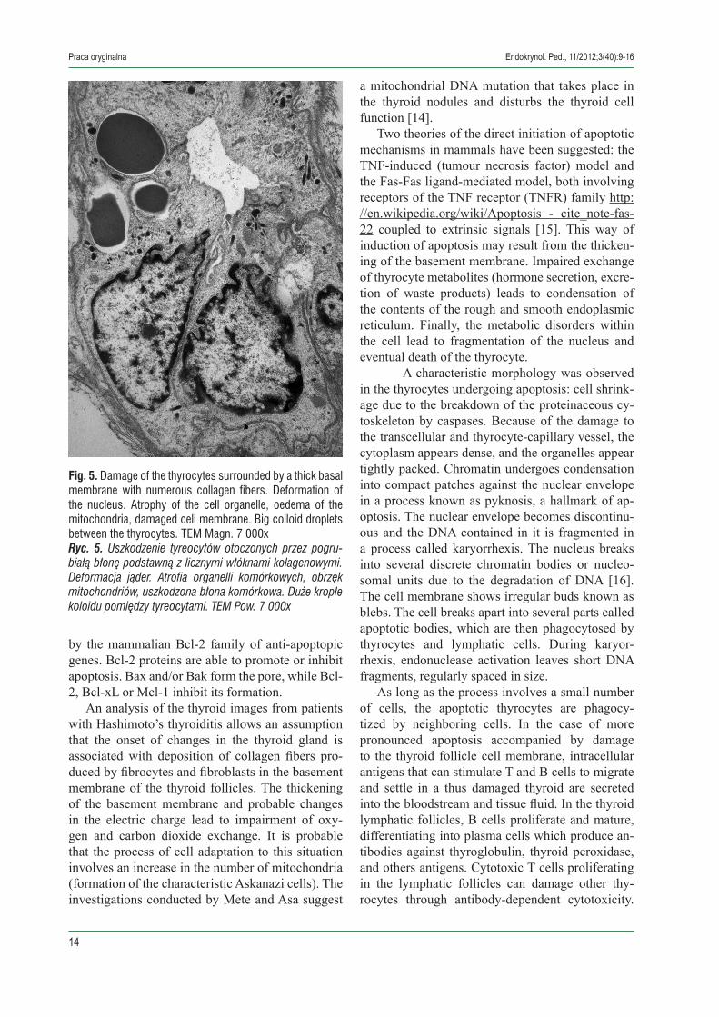
The thyroid gland of patients with Hashimoto’s thyroiditis contained numerous cells with the T-cell ultrastructure – a pyknotic nucleus and a small amount of cytoplasm, as well as plasma cells with a characteristic nucleus and numerous concentri-cally arranged cisterns of the rough cytoplasmic reticulum (Fig. 4). Various stages of thyrocyte necrosis were visible at the sites of contact between lymphocytes or plasma cells and thyrocytes. The nuclei of dying thyroid cells were deformed, the cisterns of the endoplasmic reticulum were swol-len, and the cell membrane disrupted (Fig. 4, 5). Debris of thyrocyte cytoplasm or nuclei was visible in many sites.
12
Praca oryginalna Endokrynol. Ped., 11/2012;3(40):9-16
13
Beń-Skowronek I. i inni: Two mechanisms of damage to thyrocytes in Hashimoto’s thyroiditis
Discussion
The term ‘apoptosis’ should be applied exclu-sively to cell death events that manifest several among these morphological features [8]. Several proteins are involved in apoptosis, but two main methods of regulation have been identified: target-ing mitochondria functionality, or directly transduc-ing the signal via adaptor proteins to the apoptotic mechanisms. Another extrinsic pathway for initia-tion identified in several toxin studies is an increase in calcium concentration within a cell caused by drug activity, which also can cause apoptosis via calcium binding protease calpain [9,10].
Apoptotic proteins that target mitochondria affect them in different ways. They may cause mitochondrial swelling through the formation of membrane pores, or they may increase the perme-ability of the mitochondrial membrane and cause
Fig. 3. Apoptosis of the thyrocytes. Deformation and pykno-sis of the nucleus. Oedema of the mitochondria. Destruction of microvilli, rough endoplasmic reticulum, and secretory vesicles. TEM Magn. 7 000xRyc. 3. Apoptoza tyreocytów. Deformacja i pyknoza jadra komórkowego. Obrzęk mitochondriów. Destrukcja mikroko-smków, szorstkiej siateczki śródplazmatycznej i pęcherzy-ków wydzielniczych. TEM Pow. 7 000x
Fig. 4. Necrosis of the thyrocytes as a result of autoimmune reaction with plasma cells and lymphocyt in Hashimoto’s thyroiditis. Deformation and destruction of the nuclus. Light cytoplasm with oedema of the endoplasmic reticulum and mitochondria. Damage of the cellular membrane – a thick basal membrane. TEM Magn. 7 000xRyc. 4. Nekroza tyreocytu jako rezultat reakcji autoimmuno-logicznej z komórką plazmatyczną i limfocytem w zapaleniu tarczycy Hashimoto. Deformacja i destrukcja jądra komór-kowego. Jasna cytoplazma, obrzęk siateczki śródplazma-tycznej i mitochondriów. Uszkodzenie błony komórkowej – pogrubienie błony podstawnej. TEM Pow. 15 000x
apoptotic effectors to leak out [11]. There is also a growing body of evidence indicating that nitric oxide is able to induce apoptosis by helping to dissipate the membrane potential of mitochondria and therefore make it more permeable [5].http://en.wikipedia.org/wiki/Apoptosis - cite_note-NO-12 A research done in 1999 exhibits how NO can both initiate and inhibit apoptosis due to the cellular vari-ables [5]. Mitochondrial proteins known as SMACs (small mitochondria-derived activator of caspases) are released into the cytosol following an increase in permeability. SMAC binds to inhibitor of apopto-sis proteins and deactivates them, preventing these proteins from arresting the apoptotic process and therefore allowing apoptosis to proceed. Inhibitor of apoptosis proteins also normally suppresses the activity of a group of cysteine proteases called caspases, which carry out the degradation of the cell, therefore the actual degradation enzymes can be seen to be indirectly regulated by mitochondrial permeability [12]. Cytochrome c is also released from mitochondria due to formation of a channel, the mitochondrial apoptosis-induced channel, in the outer mitochondrial membrane [13]. This process is regulated by various proteins, such as those encoded
14
Praca oryginalna Endokrynol. Ped., 11/2012;3(40):9-16
15
Beń-Skowronek I. i inni: Two mechanisms of damage to thyrocytes in Hashimoto’s thyroiditis
by the mammalian Bcl-2 family of anti-apoptopic genes. Bcl-2 proteins are able to promote or inhibit apoptosis. Bax and/or Bak form the pore, while Bcl-2, Bcl-xL or Mcl-1 inhibit its formation.
An analysis of the thyroid images from patients with Hashimoto’s thyroiditis allows an assumption that the onset of changes in the thyroid gland is associated with deposition of collagen fibers pro-duced by fibrocytes and fibroblasts in the basement membrane of the thyroid follicles. The thickening of the basement membrane and probable changes in the electric charge lead to impairment of oxy-gen and carbon dioxide exchange. It is probable that the process of cell adaptation to this situation involves an increase in the number of mitochondria (formation of the characteristic Askanazi cells). The investigations conducted by Mete and Asa suggest
a mitochondrial DNA mutation that takes place in the thyroid nodules and disturbs the thyroid cell function [14].
Two theories of the direct initiation of apoptotic mechanisms in mammals have been suggested: the TNF-induced (tumour necrosis factor) model and the Fas-Fas ligand-mediated model, both involving receptors of the TNF receptor (TNFR) family http://en.wikipedia.org/wiki/Apoptosis - cite_note-fas-22 coupled to extrinsic signals [15]. This way of induction of apoptosis may result from the thicken-ing of the basement membrane. Impaired exchange of thyrocyte metabolites (hormone secretion, excre-tion of waste products) leads to condensation of the contents of the rough and smooth endoplasmic reticulum. Finally, the metabolic disorders within the cell lead to fragmentation of the nucleus and eventual death of the thyrocyte.
A characteristic morphology was observed in the thyrocytes undergoing apoptosis: cell shrink-age due to the breakdown of the proteinaceous cy-toskeleton by caspases. Because of the damage to the transcellular and thyrocyte-capillary vessel, the cytoplasm appears dense, and the organelles appear tightly packed. Chromatin undergoes condensation into compact patches against the nuclear envelope in a process known as pyknosis, a hallmark of ap-optosis. The nuclear envelope becomes discontinu-ous and the DNA contained in it is fragmented in a process called karyorrhexis. The nucleus breaks into several discrete chromatin bodies or nucleo-somal units due to the degradation of DNA [16]. The cell membrane shows irregular buds known as blebs. The cell breaks apart into several parts called apoptotic bodies, which are then phagocytosed by thyrocytes and lymphatic cells. During karyor-rhexis, endonuclease activation leaves short DNA fragments, regularly spaced in size.
As long as the process involves a small number of cells, the apoptotic thyrocytes are phagocy-tized by neighboring cells. In the case of more pronounced apoptosis accompanied by damage to the thyroid follicle cell membrane, intracellular antigens that can stimulate T and B cells to migrate and settle in a thus damaged thyroid are secreted into the bloodstream and tissue fluid. In the thyroid lymphatic follicles, B cells proliferate and mature, differentiating into plasma cells which produce an-tibodies against thyroglobulin, thyroid peroxidase, and others antigens. Cytotoxic T cells proliferating in the lymphatic follicles can damage other thy-rocytes through antibody-dependent cytotoxicity.
Fig. 5. Damage of the thyrocytes surrounded by a thick basal membrane with numerous collagen fibers. Deformation of the nucleus. Atrophy of the cell organelle, oedema of the mitochondria, damaged cell membrane. Big colloid droplets between the thyrocytes. TEM Magn. 7 000xRyc. 5. Uszkodzenie tyreocytów otoczonych przez pogru-białą błonę podstawną z licznymi włóknami kolagenowymi. Deformacja jąder. Atrofia organelli komórkowych, obrzęk mitochondriów, uszkodzona błona komórkowa. Duże krople koloidu pomiędzy tyreocytami. TEM Pow. 7 000x
14
Praca oryginalna Endokrynol. Ped., 11/2012;3(40):9-16
15
Beń-Skowronek I. i inni: Two mechanisms of damage to thyrocytes in Hashimoto’s thyroiditis
REFERENCES/PIŚMIENNICTWO
[1] Kroemer G., El-Deiry W.S. et al.: Classification of cell death: recommendations of the Nomenclature Committee on Cell Death. Cell Death Differ, 2005:12(Suppl 2), 1463-1467.
[2] Melino G.: The Sirens’ song. Nature, 2001:412, 23.[3] Promega Corporation (2007) Protocols and Applications Guide. Apoptosis.[4] Popov S.G., Villasmil R., Bernardi J.: Lethal toxin of Bacillus anthracis causes apoptosis of macrophages. Biochem. Biophys. Res.
Commun., 2002:293 (1), 349-55. [5] Brüne B.: Nitric oxide: NO apoptosis or turning it ON? Cell Death Differ, 2003:10 (8), 864-869. [6] Promega Corporation (2006) Protocols and Applications Guide. Cell Viability.[7] Mirakian R., Nye K., Palazzo F.F. et al.: Methods for detecting apoptosis in thyroid diseases. J. Immunol. Methods, 2002:265,
161-175.[8] Kerr J.F., Wyllie A.H., Currie A.R.: Apoptosis: a basic biological phenomenon with wide-ranging implications in tissue kinetics. Br.
J. Cancer, 1972:26, 239-257.[9] Nimmrich V., Reymann K.G., Strassburger M. et al.: Inhibition of calpain prevents NMDA-induced cell death and beta-amyloid-in-
duced synaptic dysfunction in hippocampal slice cultures. Br. J. Pharmacol., 2010:159 (7), 1523-1531. [10] Liu J., Liu M.C., Wang K.K.: Calpain in the CNS: from synaptic function to neurotoxicity. Sci Signal., 2008:8, 1-14.[11] Cotran; Kumar, Collins Robbins Pathologic Basis of Disease. Philadelphia 1998: W.B Saunders Company. ISBN 0-7216-7335-X[12] Fesik S.W., Shi Y.: Controlling the caspases. Science, 2001:294 (5546), 1477-1478.[13] Dejean L.M., Martinez-Caballero S., Kinnally K.W.: Is MAC the knife that cuts cytochrome c from mitochondria during apoptosis?
Cell Death and Differentiation, 2006:13 (8), 1387-1395.[14] Mete O., Asa S.L.: Oncocytes, oxyphils, hurthle and askanazy cells: morphological and molecular features of ancocytic thyroid
nodules. Endocrine Pathology, 2010:21, 16-24.[15] Wajant H.: The Fas signaling pathway: more than a paradigm. Science, 2002:296 (5573), 1635-1636. [16] Nagata S.: Apoptotic DNA fragmentation. Exp. Cell Res., 2000:256 (1), 12-18. [17] Festjens N., Vanden Berghe T., Vandenabeele P.: Necrosis, a well-orchestrated form of cell demise: signalling cascades, important
mediators and concomitant immune response. Biochim Biophys Acta, 2006:1757, 1371-1387.
Necrotic cell death or necrosis is morphologically characterized by a gain in cell volume (oncosis), swelling of organelles, plasma membrane rupture, and subsequent loss of intracellular contents. For a long time, necrosis has been considered merely as an accidental uncontrolled form of cell death, but evidence is accumulating that the execution of necrotic cell death may be finely regulated by a set of signal transduction pathways and catabolic mechanisms [17]. In the second reaction stage, thy-rocytes undergo necrosis at the sites of the greatest damage to the thyroid, and the thyrocyte debris present in the gland enhances the already ongoing autoimmune response.
Conclusion
Ultrastructural examinations of thyroid sections sampled from patients with Hashimoto’s thyroiditis suggest the following stages of the process of thy-rocyte damage: • thickening of the basement membrane caused by
collagen deposition, • thyrocyte apoptosis, • stimulation of lymphocytes and plasma cells to
cytotoxic reactions, • necrosis of thyrocytes damaged by the cytotoxic
reaction.