Embed Size (px)
Citation preview
VNG Pro User’s Manual BETA DRAFT 270712

1
The VNG Goggles
The VNG goggles hold the cameras that are used to record the eye images. The cameras use infrared light (IR), which is not visible to the naked eye. The IR illumination enables sessions to be performed in complete darkness. The mirrors simultaneously direct IR illumination toward the pupil and reflects an image of the eye back to the video camera. The mirror reflects short wave infrared (~700-800 nm) and passes visible wavelengths.
The VNG goggles can function in both free field of view and light occluding mode. The front cover plate of the mask is magnetically fixed and can easily be removed.
Before using the VNG goggles please consider the following:
• The goggles and cameras contain sensitive electronic and optical devices. Do not expose the goggles or cameras to mechanical shock.
• VNG goggles are not to be used around explosive gases
Patient-stimulus relative position and head angles record and tracking
VNG Pro has two different kinds of sensors to monitor test conditions:
- Ultrasound sensor to get 3D static patient position relative to visual stimulator: It is consisting on ultrasound receptor positioned on to visual stimulator and an ultrasound emitter, located on goggles. System measures the patient head position relative to the center of visual stimulator.
- Accelerometers to get tilt of patient head: Three Axis Low-g micromachined Accelerometer is used as tilt sensor (measuring gravidity vector projection at each axis and convert it in angles).
These sensors are just intended to help trained doctor to positioning patient in right place and with right head angles for each type of test and keeping a record of these parameters. Never are intended to use these sensor’s data into test analysis just to monitor test conditions. Depending of test type host software indicates to hardware witch sensor monitor, for example if an optokinetic test will be performed patient needs to use visual stimulator, so system active ultrasound sensors to monitor patient relative position respect stimulator to achieve desired stimulus.
2
System Description
System requirement
• PC
Intel core2 duo 2 GHz 2GB RAM
Video card with 128MB dedicated memory
Hard Disk 250GB
USB Port 2.0
• Supported Operating Systems
Windows XP SP2 Operating System
Windows Vista Business SP1
Windows Vista Ultimate with or without SP1
Windows 7 (32 & 64 bit)
DirectX and Windows Media distribution

3
Patient Consideration
Vision: Patients must have adequate vision to follow targets for the oculomotor portion.
Physical status: If the patient has back or neck injuries, consideration should be given for some positional testing (head hanging) and the Dix-Hallpike maneuver to avoid further complications.
To screen for vertebrobasilar insufficiency, the clinician may want to assess the patient prior to head hanging or Dix-Hallpike maneuvers. This may include having the patient engage in mental tasking (e.g., counting, reciting multiplication tables) while gradually tilting the head back and then holding. Change in cognitive status or reports of lightheadedness may be significant. This screening method is especially important for older patients.
Status of the outer and middle ear: This should be evaluated prior to caloric assessment. Presence of drainage in the outer ear canal precludes the use of water irrigation; it may also affect air caloric stimulation because moisture will change the calibrated temperature, thus limiting interpretation. Pressure equalization tubes or perforation of the tympanic membrane precludes the use of water calorics. If unilateral, large perforations limit interpretation of air calorics. Large perforations can increase stimulation with cool air above calibrated expectation and can exhibit a cooling effect for warm air because moisture of the middle ear mucosa is evaporated. Excessive cerumen must be removed prior to any vestibular stimulation. Middle ear fluid affects stimulation of the vestibular system with air and water.
Medications: Many medications can affect test results. With physician approval, patients should discontinue all medications, unless contraindicated, for 24-72 hours prior to testing. Any medications taken should be clearly noted on the test results. Alcohol ingestion can affect ENG test results for 72 hours post-ingestion; results are unpredictable because alcohol can be an agonist or antagonist.
Patient preparation: Inform patients that VNG may cause dizziness, nausea, or both. Patients should be advised to limit food intake prior to examination and arrange for transportation after the examination, which usually takes 1-1.5 hours.
For many parts of the test, mental tasking is necessary to prevent central suppression of responses. If the examiner does not speak the patient’s language or the patient is hearing-impaired, an interpreter may be necessary to assist in giving instructions, explanations, and mental tasking

4
VNG pro Considerations
This algorithm accuracy depends on several factors:
The eye image quality • An accurate eye detection procedure (training during the installation)
• The patient’s co-operation ( blink, keeping eyes open, wearing no eye makeup, others)
• The calibration process
Recorded eye angles and patient sensors will be processed according type of test. Software collects on real time this data:
- Right/Left eye images (eye position/velocity) - Stimulator target position - Patient position (Warning Position) - Head angles sensors ( Warning Movement/ Angles) -
Warnings: 1) Relative subject position to the visual stimulation to achieve desired stimulus.
2) Head angles
3) Head tilt:

5
Warning State on recording
Ok
Wrong
Warning State Analysis
Green (excellent)
Yellow (regular)
Red (poor)
Calibration:
This is not a true calibration to determine the amplitude of the eye movements in degrees but more an allowance for a correction factor due to the individual anatomical differences of the orbit of the eye
These corrections are especially important in the evidence that eye movements are compared to a target stimulus from a known position (speed) are compared with a target stimulus of a known position (velocity). In other test calibration is not essential. However, if the mask has been repositioned or removed since the initial calibration was carried out the procedure must be repeated.
Calibration recommended:
• Optokinetic tests • Smooth pursuit test • Saccade test
6
• Gaze test • AHR
During the calibration, the patient is requested to fixate upon points that are onto the stimulus projector (monitor). The resulting eye movements will be processed by the Vng Pro vestibular system.

7
Setting Calibration The calibration parameters can be set for each test. To do so:
1. Open Setup protocols. 2. Create New protocols 3. Add test, 4. Add calibration set:
See: Setup software protocols

8
VNG PRO “STANDARD” USE
The parameters that defined on the protocols procedure cannot be changed once the test has been performed. The advantage of such software architecture is that it ensures that the conditions under which the measurement is performed can be reproduced at any time, even the patient position or movements head. In general, all windows have a bottom toolbar where the controls that allow forward the Next, Back, return to Home and Go to a particular window and a toolbar that allow perform actions on the window itself. In some cases VNG PRO advances to the next window automatically upon the occurrence of the event expected. This action can be scheduled. (See: Software setup: Tab Acquisitions)
The VNG Pro user interface is available in English
New patient: To Create Patient File (required): enter First name, last name, #chart, birth of date (DOB), gender and “more” data (optional).
Naming session, as you can have multiple sessions for the same patient.
Select the test protocol to perform (see: Software Setup Protocols)
Opening an existing file Press Load button and select the patient on the list and open the File.
Opening an existing session For the patient press Load session button and select the session on the list and open the File.
Starting a test
When you have completed patient data and the session can begin the study. VNG PRO will instruct you step by step in a simple and safe way to perform the tests programmed
Dizziness handicap Inventory The dizziness handicap inventory (DHI) is an optional 25-item questionnaire, which was designed to measure the self-perceived disability or handicap caused by symptoms of dizziness or imbalance. The individual questions are designed to address the impact of the symptoms on the physical, emotional, and functional aspects of daily activities, and there are three possible answers, "yes," "sometimes," and "no," to each question. The maximum total score (indicating maximum handicap) is 100. There are seven questions that comprise the physical subscale (maximum score of 28) and nine questions each on the functional and emotional subscale (maximum scores of 36). For ease of comparison the subscale scores are converted to percentage scores. (See appendix Dizziness Handicap Inventory)·

9
Data acquisition windows operations Display on real time eyes position (or velocity) graphically, and the target line whose plot is comparation criteria. The graphical outputs which are frequently employed to evaluate results The eye position [º] (or speed [º/sec]) is plotted against time, whose description is the following • Plot Horizontal position vs. time on a Cartesian (arithmetic coordinate) scale. Green Line • Plot Vertical position vs. time on a Cartesian (arithmetic coordinate) scale. Blue line • Target position - Red line Below the window are the test warnings and also are displayed the eyes video. When the subject is ready, press the Start button to record the test Tests have a configurable time limit, and they will stop automatically. However, you can stop a test manually at any time pressing Next or Go to buttons.
For each test, recorded data and video are saved. If restart the test done, it is overwriting data recorded
Analysis Windows

10
Toolbar bottom:
Home: Return Home window Back: return previous window Go to: select another Window Start: Recording video eyes and warning state Next: forward window
Test review windows operations Show all tests done and allows select each of them to re-play records, video and the warnings and view their analysis

11
Starting the test
To create a session:
1- Enter a patient data required or load a patient file stored. For include another data press More
2- Create a new session
Once complete the patient data Press Start Session
Patient option:
More Demographics Data: address, Phone, etc
Anamnesis: Medical release/ Anamnesis

12
Session Options
Report: View report preview
Analysis window
New Session
Open a session stored
Auto session name
Add a new item , in the list
Toolbar bottom:
Shut down: Close VNG pro Setup: Setup hardware and software Support: Request technical support remote (if it is available) or by email Start session
New patient File
Load a patient File stored

13
Custom Protocol:
Shows sequence of the windows to follow. The list includes patient preparation, calibration and test. All are included in the protocol you selected. (See Software Setup Protocol)
You can remove some items unchecking them. These changes do not be saved
View detail Test allows look more data Test (Frequency, direction, Time, etc)
Toolbar bottom:
Home: Return Home window Back: return previous window Go to: select another Window Next: forward window

14
Dizziness handicap inventory
It is and optional questionnaire. There are three possible answers, "yes," "sometimes," and "no", to
each questio
n
Press the answer button or type the letters Y (yes), S (Sometime) or N (No).
Dizziness Handicap Inventory Military, have four choices. But it work the same way
Next question is automatic (checked) or
Manually
For ease of comparison the subscale (emotional, functional, physical) scores are converted to percentage scores
Toolbar bottom:

15
Back: return previous window Go to: select another Window Next: forward window

16
Position
The flat panel TV is used, it should be centered in front of the patient’s eyes, and because of the reduced distance it becomes extremely important that the distance between the patient’s eyes and the screen is kept within close and precise limits. For various flat panels TV sizes the following distances between patient’s eyes and screen can be setting (See: Software Setup Advanced : Tab Screen)
As the patients distance to the screen is a key part of the geometry to produce the visual stimulation in degrees, it should only be changed on purpose. But do not worry about to return the chair to its original position in case it has been moved, VNG is controlling it all the time
Toolbar bottom:
Home: Return Home window Back: return previous window Go to: select another Window Next: forward window

17
Goggles
The Video Goggles shall be put in such a way that it fits the patient’s face the best possible. Both elastic strips shall be firmly fixed to the patient’s head. To center the image on eye displayed move the mirrors slowly but don’t touch it.
It is advisable that the environment where the study is performed is dark or it is possible the goggles are occluded, in order to avoid fixing due to light filtration in the case of caloric tests or lack of concentration in the case of visual stimulation tests
Toolbar bottom:
Home: Return Home window Back: return previous window Go to: select another Window Next: forward window

18
Angles
Toolbar bottom:
Home: Return Home window Back: return previous window Go to: select another Window Next: forward window
19
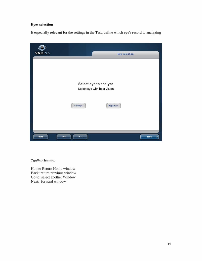
Eyes selection
It especially relevant for the settings in the Test, define which eye's record to analyzing
Toolbar bottom:
Home: Return Home window Back: return previous window Go to: select another Window Next: forward window

20
Calibration
On protocols setting are determine what stimulus type you want to use for calibration (Center, right, left, down and up) If you check the Perform Calibration option, a calibration will be performed before test recording is started Request the patient to fixate upon points that are projected onto a monitor in a pre-defined pattern for about 5 second while the calibration proceeds. When it is finish the test screen opens automatically
Toolbar bottom:
Back: return previous window Go to: select another Window Next: forward window

21
Data acquisitions
Spontaneous Admistration: Spontaneous nystagmus denotes movement of the eyes without a cognitive, visual or vestibular stimulus. The tests are conducted with vision occluded, and with the eyes on primary position.
Warnings:
• Patient position
• Head Movements
• Head angles
Gaze:
Admistration: For gaze testing, the patient is instructed to look straight ahead and then to fixate on a target to the right, left, up, and down..
Warnings:
• Patient position
• Head Movements
• Head angles
Saccades Admistration: For saccadic testing, one may place dots on the Monitor at specified distances from each other and then instruct the patient to look back and forth between the dots, keeping the head fixed.
Warnings:
• Patient position
• Head Movements
• Head angles

22
Smooth pursuit:
Admistration: The patient is instructed to follow a sinusoidal moving target with his or her eyes only.
Warnings:
• Patient position
• Head Movements
• Head angles
OKN Admistration: For optkinetic testing, the patient tracks multiple stimuli. Stimuli are moved at a rate programmed in each direction. (Clockwise and counterclockwise)
Warnings:
• Patient position
• Head Movements
• Head angles
Active head rotation (AHR) This test records the movements of the eyes and head. This requires that you have placed the headphones.
Admistration: The patient must move his head to the right or left when you hear a click in the respective ear
Warnings
• Patient position
Supine
Administration: The examiner places the patient in each position and evaluates him or her for a minimum of 20-30 seconds. Mental tasking is used to keep the patient from suppressing nystagmus. Visual suppression must also be avoided by the use of infrared goggles or with the patient's eyes closed with electrodes. Some standard positions used include the following:
• Supine Head 20-30º • Supine, head right 60º

23
• Supine, head left 60º • Supine Head 20-30º (Primary position)
Warnings
• Patient position
Dix Hallpike
Dix-Hallpike is performed over a stretcher that allows lie down and sit the patient quickly
Admistration: The Dix-Hallpike maneuver is performed by turning a patient’s head to the right or left and then briskly assisting him or her to a supine position with the head hanging to the right or left. The patient is left in this position for a brief period while eye movements are observed. Finally, the patient is returned to a sitting position.
The testing is conducted whit fixation suppression
This test records the patient position and plotting themself
Show
• Patient Head
Calorics
Admistration: The patient is placed in a reclining position with his or her head at a 30° angle. This position orients the lateral semicircular canals in the most vertical plane. Careful otoscopic examination allows the stimulus to be directed appropriately and at an equivalent depth in each ear canal.
Protocol for caloric stimulation, alternating binaural bithermal caloric stimulation includes the following conditions:
24
Right ear cool (RC)
Left ear cool (LC)
Left ear warm (LW)
Right ear warm (RW)
During each stimulation, the cool or warm stimulus is delivered for a preset time that is determined by the type of stimulus and normative data. The countdown timer show the warm or cold stimulation time and recovery times
The testing is conducted whit fixation suppression
Important: It should explore the ears (outer and middle) before proceeding with the caloric test (See Patient Considerations: Status of the outer and middle ear)
Warnings
• Head Movements
• Head position = 30º

25
Test Review:
On this screen you can see list evidence in buttons form. The active buttons are those corresponding to the tests done. Pressing these buttons you can access to review it.
Toolbar bottom
Exit : quit the window

26
Analysis The test will be opened and test data will be displayed in the test analysis window
On Textbox down select the current record to display. To view Horizontal or vertical channel alone, click on it.
Zoom out/ Zoom in Time scale
Zoom out/Zoom in amplitude Scale
Create record print windows

27
Edit Nystagmus manually. When reviewing an editable test, you can click on edit Icon to open the Edit mode and display the Edit toolbar.The Edit icon appears example from Caloric test (See Edit Nystagmus)
Tab: graphical analysis. Graphs that display the measuring process over time are shown in the bottom of the window. Those graphs that show analyzed data (e.g., the Gain in the smooth pursuit test). The abnormal threshold is the dotted gray area (See: ABNORMALITY THRESHOLD)
The software displays all test information in a single display screen. This allows the user to simultaneously view the data collection along the analysis displays. In each measurement diagram, you can maximize and minimize the display of the measured data by using the zoom option
Tab: Plot the head trajectory in each Cartesians dimensions ( x, y, z)
Tab: warning and eyes display
Toolbar bottom:
Back : return previous window
Play: Replay the test done ( record, warnings and videos)
28
General
In this section you will find the information needed to understand the data displays and analysis options
Spontaneous
Spontaneous nystagmus: This may indicate either central or peripheral pathology. The presence of nystagmus with eyes open is always diagnostically significant.
• Nystagmus Count • SPV(º/sec) is average slow phase velocity
Gaze
These nystagmus tests document and measure the inability of the eyes to maintain a static position. Nystagmus and slow phase velocity are evaluated for each position
• Nystagmus Count • SPV(º/sec) is average slow phase velocity
Saccades: Saccadic test results are influenced by patient cooperation and visual acuity. Velocity, latency, and accuracy should all be taken into consideration when interpreting saccades.
• Velocity (º/sec) is approximately proportional to saccadic amplitude for sizes 5º and 20º. After amplitude reaches 20º, saccadic velocity undergoes a soft saturation with respect to further increase in amplitude. Velocity= asymptote( 1-e Amplitude/15) The usual upper limit for saccadic velocity is about 750º/sec and the lower limit is set at 350º/sec
• Latency (msec) is calculated from difference in time between target displacement and the onset of the first saccade toward the new target position
• Accuracy (%) keeping in mind that the goal of a saccadic eye movement is to fixate visually both quickly and accurately on a new object. The eye movement should be equal in amplitude to the distance between the former object of interest and the new target.
29
Smooth pursuit The smooth pursuit system is responsible for following targets within the visual field.
Care should be taken in interpreting smooth pursuit test results in geriatric and pediatric patients. Tracking is also affected by attention and patient cooperation. Gain, Phase, and THD should all be taken into consideration when interpreting smooth pursuit.
• Gain refers to the ratio of the eye velocity (non saccadic) to the target velocity.
• Phase refers to delay between the target and the tracking wave forms
• THD: Total harmonic distortion. It is refer a global measurement to tracking test in frequency and amplitude. If the peak is around the target frequency, better was tracking
OKN
For optokinetic testing, the patient tracks multiple stimuli. Gain and Asymmetry should all be taken into consideration when interpreting OKN
• Gain refers to the ratio of the eye velocity to the target velocity.
• Asymmetry refers to gain significantly worse in one direction than another
Active head rotation (AHR)
The vestibular autorotation test provides a measure of vestibular function during active head rotation. As such, it is a test of canal function during natural active movement, a common situation for patients. This test is performed recording the eyes and head movement. The gain, phase and THD of the vestibular ocular reflex are analyzed.
• Gain refers to the ratio of the eye velocity to the head velocity.
• Phase refers to delay between the head and the eyes movement wave forms, normalized to the period, and multiplied by 360º
• THD: Total harmonic distortion. It is refer a global measurement to tracking test in frequency and amplitude. If the peak is around the target frequency, better was tracking

30
Head shaking Nystagmus
The head-shake nystagmus (HSN) test is most useful in the assessment of vestibular disorders that produce asymmetries in vestibular function.
• Nystagmus Count • SPV(º/sec) is average slow phase velocity
Supine
These are VNG test to diagnose BPPV and other vestibular complaints. Parameters are calculated for horizontal channel all positions
• Nystagmus Count • Latency (sec) is calculated from the difference in time between head displacement and
the onset the first nystagmus • SPV(º/sec) is average slow phase velocity
Dix -Hallpike
These are very important as part of the standard VNG to diagnose BPPV and other vestibular complaints. Parameters are calculated for horizontal channel both side (DixHallpike Right -DixHallpike Left)
• Nystagmus Count • Latency (sec) is calculated from the difference in time between head displacement and
the onset the first nystagmus
• SPV(º/sec) is average slow phase velocity
Caloric
The caloric tests evaluate the viability of the peripheral end organs by stimulating them with warm and cool water or air. The resulting dizziness and nystagmus is taken as an index of the viability of the organ. The culmination phase area is automatically defined. Nystagmus that lie outside the area, does not contribute to the analysis
• Time /SPV: show the slow phase velocity evolution of the tests performed. To be able to appreciate the test symmetry, it is possible to generate an interpolating line that automatically fits.

31
• Freyss graph: show the frequency (Hz) and slow phase velocity (º/s) as well as absolute and directional preponderance data, right and left nystagmus beats, symmetry, frequency and slow phase velocity for each ear.
• Parameters: show the absolute and directional preponderance data, right and left nystagmus beats, symmetry, frequency and slow phase velocity for each ear.
- Unilateral weakness= (RE44 + RE30 )-( LE44 + LE30) x100 ……………………………(RE44 + RE30 + LE44 + LE30)
- Abs Prep = (RE44 * LE30 - RE30 * LE44)
(RE44 + RE30 + LE44 + LEI30)
- Dir Preponderances = (RE44 + LE30 )-( LE44 + RE30) x100 ………… (RE44 +RE30 + LE44 + LE30)
- Right Beat = RE44+LE30 - Left Beat = RE30+LE44 - Right Ear = RE44+RE30 - Left Ear = LE44+LE30

32
Report The VNG system gives you the multiples printing option
Settings
� Graphics: graphical analysis
� Medical release
� Analysis: To print the measure data of a test
� CPT: Current Procedural Terminology codes are numbers assigned to every task and service a medical practitioner may provide to a patient on USA. They are then used to determine the amount of reimbursement that a practitioner will receive. They ensure uniformity. The codes are developed, maintained and copyrighted by the AMA (American Medical Association.)
� DHI: dizziness Handicap inventory score
� Warning: warning state
� Records: eye position trace
33
� Only printing window: To print one or more specific sections of the records (See: Create record print windows)
� Print Nystagmus mark : on to printout eyes position trace show nystagmus mark
� Print current view: printout current page
Analysis: Goto Analysis window Toolbar bottom:
Print: Print report Exit: quit the report window
34
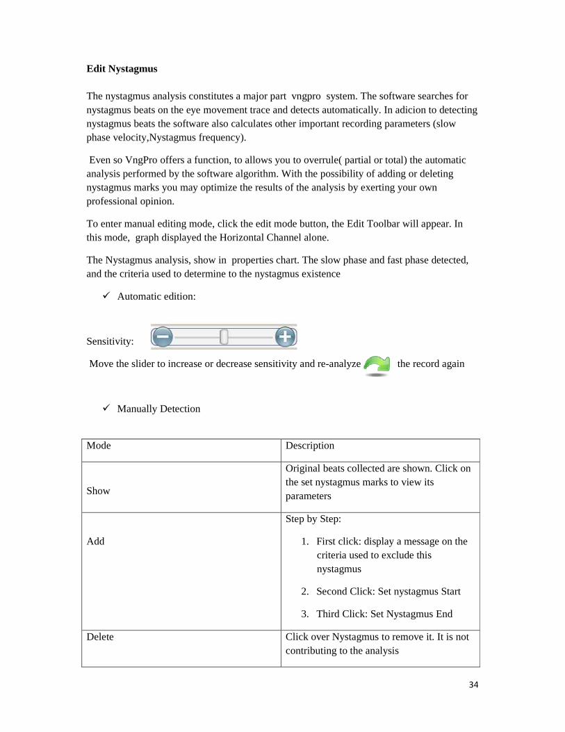
Edit Nystagmus The nystagmus analysis constitutes a major part vngpro system. The software searches for nystagmus beats on the eye movement trace and detects automatically. In adicion to detecting nystagmus beats the software also calculates other important recording parameters (slow phase velocity,Nystagmus frequency).
Even so VngPro offers a function, to allows you to overrule( partial or total) the automatic analysis performed by the software algorithm. With the possibility of adding or deleting nystagmus marks you may optimize the results of the analysis by exerting your own professional opinion.
To enter manual editing mode, click the edit mode button, the Edit Toolbar will appear. In this mode, graph displayed the Horizontal Channel alone.
The Nystagmus analysis, show in properties chart. The slow phase and fast phase detected, and the criteria used to determine to the nystagmus existence
� Automatic edition:
Sensitivity:
Move the slider to increase or decrease sensitivity and re-analyze the record again
� Manually Detection
Mode Description
Show
Original beats collected are shown. Click on the set nystagmus marks to view its parameters
Add
Step by Step:
1. First click: display a message on the criteria used to exclude this nystagmus
2. Second Click: Set nystagmus Start
3. Third Click: Set Nystagmus End
Delete Click over Nystagmus to remove it. It is not contributing to the analysis

35
Toolbar
Cancel: Nystagmus edition (or modification) will be disregarded
Ok: New edition will be saved
36
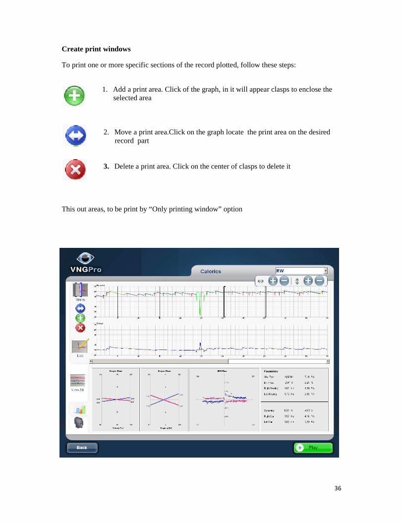
Create print windows
To print one or more specific sections of the record plotted, follow these steps:
1. Add a print area. Click of the graph, in it will appear clasps to enclose the
selected area
2. Move a print area.Click on the graph locate the print area on the desired record part
3. Delete a print area. Click on the center of clasps to delete it
This out areas, to be print by “Only printing window” option

37
Software Setup
Lists: Edit the list to be available in patient records: Doctor, Administrators and Clinics. Here you can add or remove items

38
Management database:
Delete patient file
Delete patient session
List Filter : Search a inside the patient list
By:
• Chart#
• Last name
• First name

39
40
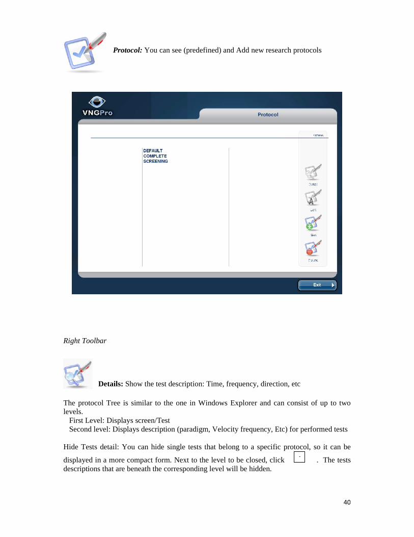
Protocol: You can see (predefined) and Add new research protocols
Right Toolbar
Details: Show the test description: Time, frequency, direction, etc
The protocol Tree is similar to the one in Windows Explorer and can consist of up to two levels. First Level: Displays screen/Test Second level: Displays description (paradigm, Velocity frequency, Etc) for performed tests Hide Tests detail: You can hide single tests that belong to a specific protocol, so it can be
displayed in a more compact form. Next to the level to be closed, click . The tests descriptions that are beneath the corresponding level will be hidden.
-

41
Show Tests detail: To show the hidden test, click beside the corresponding level. The tests description will be shown again.
E.g. Saccades 04 Hz (30): Random T
Test time: 309sec
Frequency 0.4 Hz
All tests are saved. The test disabled (unchecked) don't be included on the protocol sequence and should use the GO TO function, to open this window.
Edit: Allow modify protocols add/ Remove/Move test y/or calibration process.
Except the protocols predefined
Move up
Move Down
Delete
Enable/ Disable
+

42
New: To create a new protocol (windows sequence), type a name on the textbox and select the Test by type /Subtype:
You can select in the drop down list. It is showed below
� Posture: Hallpike, Phalpike, Posture � Visual Stimulation: Gaze, OKN, Saccades, Smooth pursuit � Caloric � At: AHR � Nystagmus: Spontaneous � Questionnaire: DHI, DHI military � Demonstration: Position, Angles, Goggles � Configuration: Eyes selection, Test selection
Calibration: � CUSTOM: Set your calibration criteria (vertical/ horizontal/ center) in the list order it
considers appropriate � AUTO (recommended): In this mode, the calibration algorithm will proceed
automatically through the whole calibration sequence by criteria. It´s set before visual tests and after in which test the patient's head has been moved (e.g. Posture Test) Type soft/ normal / hard perform the calibration more or less strict way.
Your Protocol is now fully programmed for VNG testing. Store it and you can start testing or calibration process

43
Delete: Erase the selected protocol

44
Advanced
Toolbar
Back: return previous window
Default: Reset all parameters
Save and Exit: Store all new parameters and coming back to previous window
Tab warnings:
Select the warning control: Soft /Normal/ Hard
Tab Screen: Installing the Projector / Monitor

45
To produce stimulation patterns of correct size, position and angle velocity, the required information about the geometry of the screen setup in your laboratory. These values will have to be updated whenever the projection screen geometry is changed. Complete parameters on inch [1'' (inch) = 2,54cm]
Tab Print Report:
Check / uncheck the items you are want print. It is possible change on Print report session

46
Tab Result: Time plot displays the history of eye position data. The time axis is always horizontal and directed to the right. Here you can choose the Time window (time axis) to be displayed or printed. The offset- time area relative to the whole area allocated for the chart is defined here and the scrollbar may be used to change the chart visibility. Not so in the printing, that will print only the defined time window. For example: OKN Recorded: 60 sec Time Viewer: 30 sec Time record Print: 30 sec
47
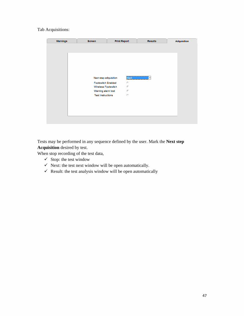
Tab Acquisitions:
Tests may be performed in any sequence defined by the user. Mark the Next step Acquisition desired by test. When stop recording of the test data,
� Stop: the test window � Next: the test next window will be open automatically. � Result: the test analysis window will be open automatically

48
Hardware
Exclusively to use technical support

49
ABNORMALITY THRESHOLD
These comments are generalities. Exceptions may occur
TEST ABNORMALITY SIGNIFICANCE
SPONTANEOUS Nystagmus: slow component, followed by fast component.
Bilateral gaze nystagmus: eyes open and looking to right or left.
2 Beats (horizontal) in 5 sec. with average velocity of >6 deg/sec.
CNS: nystagmus in all gaze directions or direction changing nystagmus in same gaze direction. SPONTANEOUS NYSTAGMUS: CNS (brain stem) PVS: direction-fixed, horizontal, or torsional nystagmus. Inhibited by fixation, follows Alexander’s law.* rule out drugs, lack of alertness
SMOOTH PURSUIT/ TRACKING Gain: eye velocity/target velocity
Phase: lag or lead of eye relative to target.
Asymmetry: right gain – left gain
Gain: Age 50 and under: <70% or >140% Age 60 – 69:
<65% or > 145% Age 70 – 79 :
<60% or > 150% Age 80 – 89 :
<55% or > 155% Age 90 and above : <50% or >160% Gain asymmetry: >30%
Low gain in one direction: CNS lesion. Marked saccadic pursuit:(stair step tracking) CNS lesion
SACCADE TEST
Velocity: peak eye velocity
Latency: reaction time in msec.
Accuracy: undershoots/overshoots
Latency: Age 50 and under : >260 msec Age 60- 69: >270 msec Age 70- 79: >280 msec Age 80– 89: >290msec Age 90 and above: 300 msec
Significant saccadic slowing, overshooting or undershooting: CNS lesion or ocular disorder Lower velocity of trailing eye: Internuclear ophthalmoplegia ( Brain stem)

50
Velocity: Age 50 and under: <240 deg/sec Age 60- 69: < 230º/sec Age 70-79: <220 º/sec Age 80-89: < 210 º/sec Age 90 and above: <200 º/sec Accuracy:<55%
OPTOKINETIC NYSTAGMUS Gain: eye velocity/ target velocity.
Gain < 60% is abnormal. Symmetry > 30% difference is abnormal. SPV < 6.0 is abnormal
Symmetrical low gain: impaired vision or likely CNS (parietal) lesion.
DIX-HALLPIKE >2 beats (horizontal) with average velocity >6.0 deg/sec.
1. delay onset of nystagmus
2. paroxysmal nystagmus
3. associated with vertigo
4. fatigable nystagmus
Unilateral: Usually peripheral undermost ear. Bilateral: peripheral (both ears) or CNS BPPV: both horizontal and vertical nystagmus (with possible rotation) observed.
CALORIC TEST Directional Preponderance: nystagmus beats stronger one way than another. Fixation Index: when nystagmus is at its peak, the ratio of velocity with no fixation to velocity with fixation.
Unilateral weakness: of more than 25% is abnormal. Bilateral weakness: sum of velocities of all 4 irrigations <20 º/sec Directional preponderance: >25% difference.
Caloric weakness: is a function of the labyrinth or VIII cranial nerve (vestibular portion). Unilateral: indicates disabled side. Bilateral: peripheral organs, acute unilateral lesion or perhaps CNS (cerebellar). Directional preponderance: is not localizing. High fixation index: is a prime indicator of CNS.

51
While Videonystagmography is the most widely used clinical laboratory test to assess vestibular function, remember that normal VNG test results do not necessarily mean that a patient has typical vestibular function. VNG abnormalities can be useful in the diagnosis and localization of site of lesion; however, many abnormalities are no localizing; therefore, the clinical history
and otologic examination of the patient are vital in formulating a diagnosis and treatment plan for a patient presenting with dizziness or vertigo.
.

52
Freyss Diagram
It was designed to perform the bilateral bi-thermal caloric test. Freyss butterfly or diagram shows the nystagmus intensity together with he results of absolute symmetry and preponderance.
In its most traditional form, Freyss butterfly represents the number of nystagmic flaps observed in a 30-second period. It also requires the stimulus corresponding to Hallpike rules: irrigate 125cc of water at 30 and 44° C during 30 seconds. The patient shall be in horizontal position with the head at 30 degrees.
In these conditions, it is possible to use the results of the normative data published by G. Freyss giving the symmetry normative limits (<15%) and a directional preponderance (12%). It is also possible to evaluate a hypo-sensitivity condition (<30 saccades) or hyper-sensitivity (>122 saccades) of a single ear, if you add up the saccades induced by both ears with hot and cold stimulus.
In general, the graph is centered in a point, the ordinate axis represents the nystagmus intensity and the abscissa axis is graduated in percentage. There are two limits: x = +100 and x = -100. Rightwards nystagmic flaps are positive while the leftwards ones are negative. The results obtained from the right ear are represented in line X = - 100 and the left ear ones in line x = +100. Both points corresponding to the intensity measure of a bilateral isotherm (cold or hot) are thus connected defining a straight line
and the equation may be represented as y = a x + b whose slope is positive for cold tests and negative for hot tests.
Once the four tests are completed, then the intersection point projections in X and Y of both straight lines represent the following two results:
In the vertical projection, the lateral symmetry value or channel paresis (X) is represented in percentage. If the nystagmus direction for each of the four tests is as expected, then you may use the traditional Jongkee’s formula:
=RE44 + RE30 )-( LE44 + LE30) x100 (RE44 - RE30 - LE44 + LE30)
In the horizontal projection, the absolute preponderance (Y) is calculated using the following formula
= (RE44 * LE30 - RE30 * LE44)
(RE44 - RE30 - LE44 + LEI30)
53
The hyper or hyposensitivity condition for each ear is assumed from segment length covered by the butterfly wing in the axis: x = - 100 for the right side, and x = + 100 for the left side.
Freyss diagram for slow phase speeds
The diagonal intersection coordinates represent the unilateral weakness calculated using Jongkee’s formula. The absolute directional preponderance is not shown as a percentage but as an absolute value, in degrees per second.
This format offers different advantages as the ability to compare the preponderance speed against the speed of any nystagmus that may be present. The relative directional preponderance is also shown in % since this format is more familiar.
.

54
Dizziness Handicap Inventory (Civil)
Yes Some…. No
(4) (3) (2)
P1. Does looking up increase your problem? ��� ��� ���
E2. Because of your dizziness do you feel frustrated?
��� ��� ���
F3. Because of your dizziness do you restrict your travel for business or recreation? ��� ��� ���
P4. Does walking down the aisle of a supermarket increase your dizziness? ��� ��� ���
F5. Because of your dizziness do you have difficult getting into or out bed? ��� ��� ���
F6. Does your dizziness significantly restrict your participation in social activities, such as going out to dinner, going to the movies, dancing, or going to parties?
��� ��� ���
F7 Because of your problem do you have difficulty reading? ��� ��� ���
P8. Does performing more ambitious activities like sport, dancing, or household chores such as sweeping or putting dishes away increase your dizziness?
��� ��� ���
E9 Because of your problem are you leave home without having someone accompany you? ��� ��� ���
E10. Because of your dizziness have you been embarrassed in front of others? ��� ��� ���
P11. Do quick movements of your head increase your dizziness? ��� ��� ���
F12. Because of your dizziness, do you avoid heights? ��� ��� ���
P13. Does turning over in bed increase your dizziness? ��� ��� ���

55
E14. Because of your dizziness is it difficult for you to do strenuous housework or yard work? ��� ��� ���
E15. Because of your dizziness are you afraid people may think you are intoxicated? ��� ��� ���
F16. Because of your dizziness, is it difficult for you to go for a walk by yourself? ��� ��� ���
P17. Does walking down a sidewalk increase your dizziness? ��� ��� ���
E18. Because of your dizziness, is it difficult for you to concentrate? ��� ��� ���
F19. Because of your dizziness is it difficult for you to walk around your house in the dark? ��� ��� ���
E20. Because of your dizziness are you afraid to stay home alone? ��� ��� ���
E21. Because of your dizziness do you feel handicapped? ��� ��� ���
E22. Has your dizziness placed stress on your relationships with members of your family or friends? ��� ��� ���
E23. Because of your dizziness, are you depressed? ��� ��� ���
F24. Does your dizziness interfere with your job or household responsibilities? ��� ��� ���
P25. Does bending over increase your dizziness? ��� ��� ���
Score:
Functional = …………Emotional=…………Physical=……………..Total Score=……….

56
Dizziness Handicap Inventory (Military)
All Most Some Never
(4) (3) (2) (0)
P1. Does looking up increase your problem? ��� ��� ��� ���
P 2. Does walking down the aisles of the commissary or PX without a cart increase your problem? ��� ��� ��� ���
P3. Does performing more ambitious activities like sports, dancing, or military common duties/tasks increase your problem?
��� ��� ��� ���
P4. Do quick head movements increase your problem? ��� ��� ��� ���
P5. Does turning over in bed increase your problem? ��� ��� ��� ���
P6. Does walking on uneven terrain increase your problem? ��� ��� ��� ���
P7 Does bending over increase your problem? ��� ��� ��� ���
F8. Because of your problem do you restrict your travel for duty or recreation? ��� ��� ��� ���
F9 Because of your problem do you have difficulty getting into or out of bed? ��� ��� ��� ���
F10. Does your problem significantly restrict your participation in social activities? ��� ��� ��� ���
F11. Because of your problem do you have difficulty reading? ��� ��� ��� ���
F12. Because of your problem, do you have someone accompany you when you leave quarters? ��� ��� ��� ���
F13. Because of your problem, is it difficult for you to take care of yourself (bathe, dress, prepare a meal)? ��� ��� ��� ���
F14. Because of your problem, is it difficult for you to walk around your quarters in the dark? ��� ��� ��� ���

57
F15. Because of your problem, do you avoid driving your vehicle during the daytime? ��� ��� ��� ���
F16. Because of your problem, is it difficult for you to go for a walk by yourself? ��� ��� ��� ���
F17. Because of your problem, is it difficult for you to walkup and down stairs? ��� ��� ��� ���
F18. Because of your problem, do you avoid driving your vehicle in the dark? ��� ��� ��� ���
F19. Does your problem interfere with your job or your military duties? ��� ��� ��� ���
F20. Because of your problem, is it difficult for you to concentrate? ��� ��� ��� ���
E21. Because of your problem, do you feel frustrated? ��� ��� ��� ���
E22. Because of your problem, are you afraid to stay home alone? ��� ��� ��� ���
E23. Because of your problem, are you afraid people think you are intoxicated? ��� ��� ��� ���
E24. Has your problem places stress on your relationships with members of your family or friends?
��� ��� ��� ���
E25. Because of your problem, are you depressed? ��� ��� ��� ���
Score:
Functional= …………Emotional=…………Physical=……………..Total Score=……….

58
Troubleshooting