Embed Size (px)
Citation preview
Case ReportVitamin K Deficiency Embryopathy fromHyperemesis Gravidarum
Andrew S. Lane,1 Jennifer L. Stallworth,2 Kacey Y. Eichelberger,3 and Kenneth F. Trofatter3
1Department of Obstetrics and Gynecology, University of South Carolina School of Medicine Greenville, Greenville Health System,890 W. Faris Road, Suite 470, Greenville, SC 29605, USA2Greenwood Genetic Center, 14 Edgewood Drive, Greenville, SC 29605, USA3Division of Maternal Fetal Medicine, Department of Obstetrics and Gynecology, University of South Carolina School of MedicineGreenville, Greenville Health System, 890 W. Faris Road, Suite 470, Greenville, SC 29605, USA
Correspondence should be addressed to Andrew S. Lane; [email protected]
Received 14 May 2015; Accepted 30 July 2015
Academic Editor: Line Bjørge
Copyright © 2015 Andrew S. Lane et al.This is an open access article distributed under the Creative CommonsAttribution License,which permits unrestricted use, distribution, and reproduction in any medium, provided the original work is properly cited.
A 21-year-old primigravida had a pregnancy complicated by hyperemesis gravidarum (HG) beginning at 7-week gestation. Despitemedical therapy, she lost 18% of her prepregnancy weight. Early ultrasound at 14 weeks demonstrated a flattened facial profilewith nasal hypoplasia (Binder phenotype) consistent with vitamin K deficiency from HG. She had a percutaneous endoscopicgastrojejunostomy tube placed for enteral feeding at 15-week gestation. At repeated anatomy ultrasound at 21-week gestation,delivery, and postnatal pediatric genetics exam, nasal hypoplasia was consistent with vitamin K deficiency embryopathy fromHG. Nausea and vomiting of pregnancy is a common condition. HG, the most severe form, has many maternal and fetal effects.Evaluation of vitamin K status could potentially prevent this rare and disfiguring embryopathy.
1. Introduction
Nausea and vomiting of pregnancy (NVP) is a commoncondition affecting 70–85% of pregnancies [1]. Hyperemesisgravidarum (HG) is a severe form of NVP most commonlydefined as persistent vomiting not related to other causes,ketonuria, and weight loss greater than 5% of prepregnancyweight [2]. HG affects 0.5–2% of pregnancies and is the mostcommon indication for hospitalization in the first trimesterof pregnancy [3]. We present the earliest diagnosed case ofvitamin K deficiency embryopathy due to HG. Our objectiveis to raise awareness of a rare fetal complication of HG.
2. Case Presentation
A 21-year-old African American primigravida experiencednausea and vomiting of pregnancy (NVP) since 7-weekgestation. She was diagnosed with hyperemesis gravidarum(HG) based on NVP, ketosis, and loss of greater than 5% ofprepregnancy weight. Her HG was resistant to intravenousfluids, ondansetron, promethazine, andmetoclopramide. She
was referred to ourmaternal-fetalmedicine department at 14-week gestation.
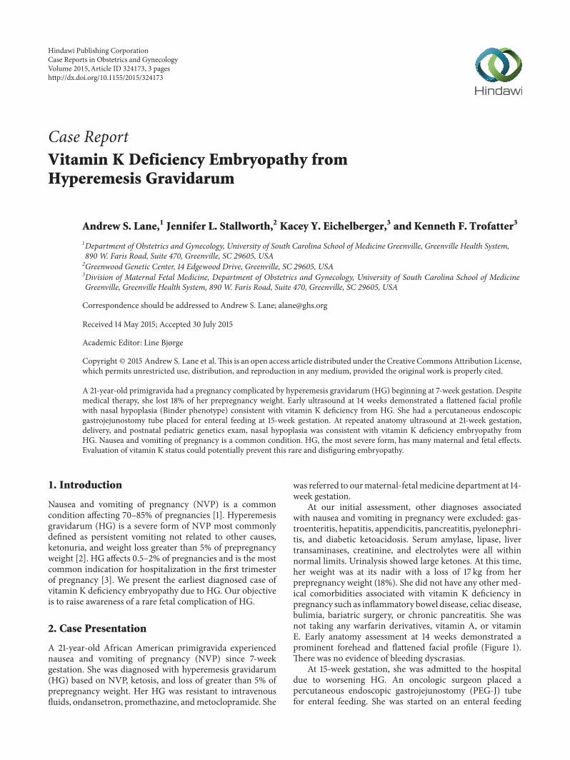
At our initial assessment, other diagnoses associatedwith nausea and vomiting in pregnancy were excluded: gas-troenteritis, hepatitis, appendicitis, pancreatitis, pyelonephri-tis, and diabetic ketoacidosis. Serum amylase, lipase, livertransaminases, creatinine, and electrolytes were all withinnormal limits. Urinalysis showed large ketones. At this time,her weight was at its nadir with a loss of 17 kg from herprepregnancy weight (18%). She did not have any other med-ical comorbidities associated with vitamin K deficiency inpregnancy such as inflammatory bowel disease, celiac disease,bulimia, bariatric surgery, or chronic pancreatitis. She wasnot taking any warfarin derivatives, vitamin A, or vitaminE. Early anatomy assessment at 14 weeks demonstrated aprominent forehead and flattened facial profile (Figure 1).There was no evidence of bleeding dyscrasias.
At 15-week gestation, she was admitted to the hospitaldue to worsening HG. An oncologic surgeon placed apercutaneous endoscopic gastrojejunostomy (PEG-J) tubefor enteral feeding. She was started on an enteral feeding
Hindawi Publishing CorporationCase Reports in Obstetrics and GynecologyVolume 2015, Article ID 324173, 3 pageshttp://dx.doi.org/10.1155/2015/324173
2 Case Reports in Obstetrics and Gynecology
Figure 1: Profile at 14-week gestation shows prominent forehead andnasal hypoplasia.
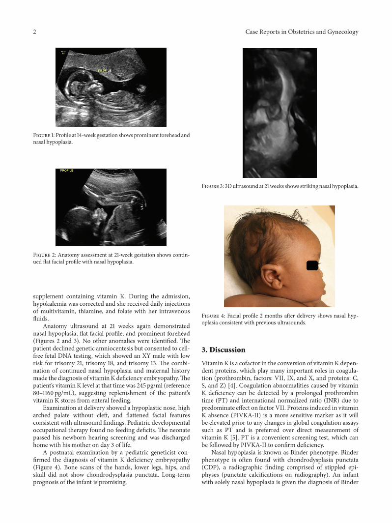
Figure 2: Anatomy assessment at 21-week gestation shows contin-ued flat facial profile with nasal hypoplasia.
supplement containing vitamin K. During the admission,hypokalemia was corrected and she received daily injectionsof multivitamin, thiamine, and folate with her intravenousfluids.
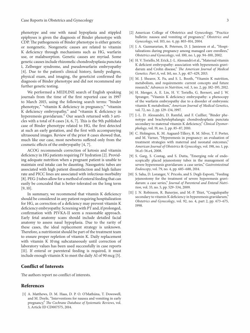
Anatomy ultrasound at 21 weeks again demonstratednasal hypoplasia, flat facial profile, and prominent forehead(Figures 2 and 3). No other anomalies were identified. Thepatient declined genetic amniocentesis but consented to cell-free fetal DNA testing, which showed an XY male with lowrisk for trisomy 21, trisomy 18, and trisomy 13. The combi-nation of continued nasal hypoplasia and maternal historymade the diagnosis of vitaminKdeficiency embryopathy.Thepatient’s vitamin K level at that time was 245 pg/ml (reference80–1160 pg/mL), suggesting replenishment of the patient’svitamin K stores from enteral feeding.
Examination at delivery showed a hypoplastic nose, higharched palate without cleft, and flattened facial featuresconsistent with ultrasound findings. Pediatric developmentaloccupational therapy found no feeding deficits. The neonatepassed his newborn hearing screening and was dischargedhome with his mother on day 3 of life.
A postnatal examination by a pediatric geneticist con-firmed the diagnosis of vitamin K deficiency embryopathy(Figure 4). Bone scans of the hands, lower legs, hips, andskull did not show chondrodysplasia punctata. Long-termprognosis of the infant is promising.
Figure 3: 3Dultrasound at 21weeks shows striking nasal hypoplasia.
Figure 4: Facial profile 2 months after delivery shows nasal hyp-oplasia consistent with previous ultrasounds.
3. Discussion
Vitamin K is a cofactor in the conversion of vitamin K depen-dent proteins, which play many important roles in coagula-tion (prothrombin, factors: VII, IX, and X, and proteins: C,S, and Z) [4]. Coagulation abnormalities caused by vitaminK deficiency can be detected by a prolonged prothrombintime (PT) and international normalized ratio (INR) due topredominate effect on factor VII. Proteins induced in vitaminK absence (PIVKA-II) is a more sensitive marker as it willbe elevated prior to any changes in global coagulation assayssuch as PT and is preferred over direct measurement ofvitamin K [5]. PT is a convenient screening test, which canbe followed by PIVKA-II to confirm deficiency.
Nasal hypoplasia is known as Binder phenotype. Binderphenotype is often found with chondrodysplasia punctata(CDP), a radiographic finding comprised of stippled epi-physes (punctate calcifications on radiography). An infantwith solely nasal hypoplasia is given the diagnosis of Binder
Case Reports in Obstetrics and Gynecology 3
phenotype and one with nasal hypoplasia and stippledepiphyses is given the diagnosis of Binder phenotype withCDP. The pathogenesis of Binder phenotype is either geneticor nongenetic. Nongenetic causes are related to vitaminK deficiency through mechanisms such as HG, warfarinuse, or malabsorption. Genetic causes are myriad. Somegenetic causes include rhizomelic chondrodysplasia punctata1, Zellweger syndrome, and pseudowarfarin embryopathy[4]. Due to the patient’s clinical history, family pedigree,physical exam, and imaging, the geneticist confirmed thediagnosis of Binder phenotype and did not recommend anyfurther genetic testing.
We performed a MEDLINE search of English speakingjournals from the time of the first reported case in 1997to March 2015, using the following search terms: “binderphenotype,” “vitamin K deficiency in pregnancy,” “vitaminK deficiency embryopathy,” and “vitamin K deficiency inhyperemesis gravidarum.” Our search returned with 3 arti-cles with a total of 8 cases [4, 6, 7]. This is the 9th publishedcase of Binder phenotype related to HG, the first detectedat such an early gestation, and the first with accompanyingultrasound images. Review of the prior 8 cases showed that,much like our case, most newborns suffered only from thecosmetic effects of the embryopathy [4, 7].
ACOG recommends correction of ketosis and vitamindeficiency in HG patients requiring IV hydration [2]. Provid-ing adequate nutrition when a pregnant patient is unable tomaintain oral intake can be daunting. Nasogastric tubes areassociated with high patient dissatisfaction and high failurerate and PICC lines are associated with infectious morbidity[8]. PEG-J tubes allow for amethod of enteral feeding that caneasily be concealed that is better-tolerated on the long term[9, 10].
In summary, we recommend that vitamin K deficiencyshould be considered in any patient requiring hospitalizationfor HG, as correction of a deficiency may prevent vitamin Kdeficiency embryopathy. Screeningwith PT and, if prolonged,confirmation with PIVKA-II seem a reasonable approach.Early fetal anatomy scans should include detailed facialanatomy to assess nasal hypoplasia. Due to the rarity ofthese cases, the ideal replacement strategy is unknown.Therefore, a nutritionist should be part of the treatment teamto ensure proper repletion of vitamin K. Daily replacementwith vitamin K 10mg subcutaneously until correction oflaboratory values has been used successfully in case reports[11]. If enteral or parenteral feeding is required, it mustinclude enough vitamin K to meet the daily AI of 90mcg [5].
Conflict of Interests
The authors report no conflict of interests.
References
[1] A. Matthews, D. M. Haas, D. P. O. O’Mathuna, T. Dowswell,and M. Doyle, “Interventions for nausea and vomiting in earlypregnancy,” The Cochrane Database of Systematic Reviews, vol.3, Article ID CD007575, 2014.
[2] American College of Obstetrics and Gynecology, “Practicebulletin: nausea and vomiting of pregnancy,” Obstetrics andGynecology, vol. 103, no. 4, pp. 803–814, 2004.
[3] J. A. Gazmararian, R. Petersen, D. J. Jamieson et al., “Hospi-talizations during pregnancy among managed care enrollees,”Obstetrics and Gynecology, vol. 100, no. 1, pp. 94–100, 2002.
[4] H.V. Toriello,M. Erick, J.-L. Alessandri et al., “Maternal vitaminK deficient embryopathy: association with hyperemesis gravi-darum and Crohn disease,” The American Journal of MedicalGenetics. Part A, vol. 161, no. 3, pp. 417–429, 2013.
[5] M. J. Shearer, X. Fu, and S. L. Booth, “Vitamin K nutrition,metabolism, and requirements: current concepts and futureresearch,” Advances in Nutrition, vol. 3, no. 2, pp. 182–195, 2012.
[6] H. Menger, A. E. Lin, H. V. Toriello, G. Bernert, and J. W.Spranger, “Vitamin K deficiency embryopathy: a phenocopyof the warfarin embryopathy due to a disorder of embryonicvitamin K metabolism,” American Journal of Medical Genetics,vol. 72, no. 2, pp. 129–134, 1997.
[7] J.-L. D. Alessandri, D. Ramful, and F. Cuillier, “Binder phe-notype and brachytelephalangic chondrodysplasia punctatasecondary to maternal vitamin K deficiency,” Clinical Dysmor-phology, vol. 19, no. 2, pp. 85–87, 2010.
[8] C. Holmgren, K. M. Aagaard-Tillery, R. M. Silver, T. F. Porter,and M. Varner, “Hyperemesis in pregnancy: an evaluation oftreatment strategies with maternal and neonatal outcomes,”American Journal of Obstetrics & Gynecology, vol. 198, no. 1, pp.56.e1–56.e4, 2008.
[9] S. Garg, S. Contag, and S. Dutta, “Emerging role of endo-scopically placed jejunostomy tubes in the management ofsevere hyperemesis gravidarum: a case series,” GastrointestinalEndoscopy, vol. 79, no. 4, pp. 685–688, 2014.
[10] S. Saha, D. Loranger, V. Pricolo, and S. Degli-Esposti, “Feedingjejunostomy for the treatment of severe hyperemesis gravi-darum: a case series,” Journal of Parenteral and Enteral Nutri-tion, vol. 33, no. 5, pp. 529–534, 2009.
[11] J. N. Robinson, R. Banerjee, and M.-P. Thiet, “Coagulopathysecondary to vitamin K deficiency in hyperemesis gravidarum,”Obstetrics and Gynecology, vol. 92, no. 4, part 2, pp. 673–675,1998.

Submit your manuscripts athttp://www.hindawi.com
Stem CellsInternational
Hindawi Publishing Corporationhttp://www.hindawi.com Volume 2014
Hindawi Publishing Corporationhttp://www.hindawi.com Volume 2014
MEDIATORSINFLAMMATION
of
Hindawi Publishing Corporationhttp://www.hindawi.com Volume 2014
Behavioural Neurology
EndocrinologyInternational Journal of
Hindawi Publishing Corporationhttp://www.hindawi.com Volume 2014
Hindawi Publishing Corporationhttp://www.hindawi.com Volume 2014
Disease Markers
Hindawi Publishing Corporationhttp://www.hindawi.com Volume 2014
BioMed Research International
OncologyJournal of
Hindawi Publishing Corporationhttp://www.hindawi.com Volume 2014
Hindawi Publishing Corporationhttp://www.hindawi.com Volume 2014
Oxidative Medicine and Cellular Longevity
Hindawi Publishing Corporationhttp://www.hindawi.com Volume 2014
PPAR Research
The Scientific World JournalHindawi Publishing Corporation http://www.hindawi.com Volume 2014
Immunology ResearchHindawi Publishing Corporationhttp://www.hindawi.com Volume 2014
Journal of
ObesityJournal of
Hindawi Publishing Corporationhttp://www.hindawi.com Volume 2014
Hindawi Publishing Corporationhttp://www.hindawi.com Volume 2014
Computational and Mathematical Methods in Medicine
OphthalmologyJournal of
Hindawi Publishing Corporationhttp://www.hindawi.com Volume 2014
Diabetes ResearchJournal of
Hindawi Publishing Corporationhttp://www.hindawi.com Volume 2014
Hindawi Publishing Corporationhttp://www.hindawi.com Volume 2014
Research and TreatmentAIDS
Hindawi Publishing Corporationhttp://www.hindawi.com Volume 2014
Gastroenterology Research and Practice
Hindawi Publishing Corporationhttp://www.hindawi.com Volume 2014
Parkinson’s Disease
Evidence-Based Complementary and Alternative Medicine
Volume 2014Hindawi Publishing Corporationhttp://www.hindawi.com


![[PPT]Hyperemesis Gravidarum - Philadelphia University …philadelphia.edu.jo/academics/aalrazek/uploads... · Web viewHyperemesis Gravidarum Learning objective Identify Hyperemesis](https://img.pdfslide.us/doc/110x75/5af587257f8b9a190c8e7497/ppthyperemesis-gravidarum-philadelphia-university-viewhyperemesis-gravidarum.jpg)