Embed Size (px)
Citation preview

Vitamin D receptor activation modulates
the allergic immune response
vorgelegt von
Diplom-Biochemiker
Björn Hartmann
Von der Fakultät III – Prozesswissenschaften
der Technischen Universität Berlin
zur Erlangung des akademischen Grades
Doktor der Naturwissenschaften
Dr. rer. nat.
genehmigte Dissertation
Promotionsausschuss:
Vorsitzender: Prof. Dr. rer. nat. Peter Neubauer
Gutachter: Prof. Dr. rer. nat. Roland Lauster
Gutachter: Prof. Dr. med. Margitta Worm
Gutachter: Prof. Dr. rer. nat. Leif-Alexander Garbe
Tag der wissenschaftlichen Aussprache: 22.02.2011
durchgeführt an der
Charité, Klinik für Dermatologie, Venerologie und Allergologie, Berlin
Berlin 2011
D 83

“A vitamin is a substance that makes you ill if you don’t eat it.”
(Albert Szent-Gyorgyi, Nobel Prize in Physiology or Medicine, 1937)
Meinen Eltern

Table of Contents I
1. LIST OF ABBREVIATIONS................................................................................................................. 1
1. ABSTRACT............................................................................................................................................ 8
2. ZUSAMMENFASSUNG ........................................................................................................................ 9
3. INTRODUCTION................................................................................................................................ 11
3.1. VITAMIN D AND PHYSIOLOGY ......................................................................................................... 11
3.2. VITAMIN D RECEPTOR AND SIGNALING ............................................................................................ 14
3.3. VITAMIN D AND THE IMMUNE SYSTEM ............................................................................................. 19
3.4. VITAMIN D IN ALLERGIC DISEASES AND ATOPIC DERMATITIS ............................................................ 21
3.4.1. Type I allergic immune response ............................................................................................ 21
3.4.2. Role of TH2 cells in the allergic immune response................................................................... 22
3.4.3. Mechanism of IgE class switch recombination........................................................................ 23
3.4.4. Vitamin D in the context of IgE and atopic dermatitis (AD)..................................................... 25
3.4.5. Atopic dermatitis (AD)........................................................................................................... 25
4. OBJECTIVES ...................................................................................................................................... 30
5. MATERIALS AND METHODS.......................................................................................................... 31
5.1. MATERIALS .................................................................................................................................... 31
5.1.1. Antibodies.............................................................................................................................. 31
5.1.2. Buffers and solutions.............................................................................................................. 32
5.1.3. Chemical and biological reagents .......................................................................................... 33
5.1.4. Labware ................................................................................................................................ 35
5.1.5. Technical equipment .............................................................................................................. 36
5.1.6. Software ................................................................................................................................ 37
5.2. METHODS....................................................................................................................................... 38
5.2.1. Cellular methods.................................................................................................................... 38
5.2.1.1. Human cell isolation......................................................................................................................... 38 5.2.1.2. Murine splenocyte isolation .............................................................................................................. 38 5.2.1.3. Cell culture conditions...................................................................................................................... 39
5.2.2. Animal work .......................................................................................................................... 39
5.2.2.1. Mouse model of type I allergy .......................................................................................................... 39 5.2.2.2. Mouse model of allergen-induced eczema ......................................................................................... 40 5.2.2.3. Assessment of AD symptoms ........................................................................................................... 41
5.2.3. Immunological methods ......................................................................................................... 42
5.2.3.1. Enzyme-linked immunosorbent assay (ELISA) ................................................................................. 42 5.2.3.2. Human immunoglobulin ELISA ....................................................................................................... 42 5.2.3.3. Murine immunoglobulin ELISA ....................................................................................................... 43 5.2.3.4. Enzyme-linked immunospot (ELISPOT) ........................................................................................... 43 5.2.3.5. Principles of flow cytometry............................................................................................................. 44 5.2.3.6. Flow cytometric analysis .................................................................................................................. 45 5.2.3.7. 5(6)-carboxyfluorescein diacetate N-succinimidyl ester (CFSE) dilution analysis ............................... 45 5.2.3.8. Flow cytometric analysis of STAT6 phosphorylation......................................................................... 45

Table of Contents II
5.2.3.9. Flow cytometric analysis of IκBα degradation................................................................................... 46
5.2.4. Immunohistochemistry ........................................................................................................... 46
5.2.4.1. Staining of CD4+ and CD8+ T cell infiltrates ..................................................................................... 46 5.2.4.2. Staining of Foxp3+ cells.................................................................................................................... 47 5.2.4.3. Histological Analyses....................................................................................................................... 47
5.2.5. Molecular biology methods .................................................................................................... 47
5.2.5.1. RNA isolation from murine skin ....................................................................................................... 47 5.2.5.2. RNA isolation from cultured cells..................................................................................................... 48 5.2.5.3. cDNA synthesis ............................................................................................................................... 48 5.2.5.4. Real-time PCR/quantitative PCR (qPCR).......................................................................................... 48
5.2.6. Statistical analyses................................................................................................................. 50
6. RESULTS ............................................................................................................................................. 51
6.1. THE VDR AGONIST MEDIATES VDR ACTIVATION IN B CELLS ........................................................... 51
6.2. VDR ACTIVATION BY THE VDR AGONIST INHIBITS IGE PRODUCTION IN B CELLS .............................. 52
6.3. STAT6 PHOSPHORYLATION AND IκBα DEGRADATION IS NOT MODULATED ....................................... 54
6.4. REDUCTION OF AICDA EXPRESSION BY CALCITRIOL AND THE VDR AGONIST ...................................... 56
6.5. REDUCTION OF THE CD19+ CD27HIGH CD38+
B CELL POPULATION BY ACTIVATED VDRS ................... 57
6.6. VDR ACTIVATION REDUCES THE IGE RESPONSE IN A TYPE I ALLERGY MOUSE MODEL WITHOUT
CALCEMIC SIDE EFFECTS.................................................................................................................. 59
6.7. VDR AGONIST TREATMENT AMELIORATES ALLERGEN-TRIGGERED SKIN ECZEMA IN MICE .................. 61
6.8. VDR ACTIVATION DOES NOT CHANGE THE NUMBERS OF INFILTRATING T CELLS IN LESIONAL SKIN ..... 63
6.9. INCREASED NUMBERS OF FOXP3+ CELLS IN SKIN LESIONS OF VDR AGONIST-TREATED MICE............... 63
6.10. MODULATION OF CYTOKINE AND CHEMOKINE EXPRESSION IN LESIONAL SKIN ................................... 65
6.11. VDR AGONIST TREATMENT INDUCES BARRIER AND ANTIMICROBIAL PEPTIDE GENE EXPRESSION ........ 66
7. DISCUSSION ....................................................................................................................................... 68
7.1. TARGETING THE VDR INHIBITS THE HUMORAL IMMUNE RESPONSE, PREFERENTIALLY IGE, IN VITRO AND
IN VIVO ........................................................................................................................................... 68
7.2. VDR ACTIVATION AMELIORATES ALLERGEN-INDUCED SKIN ECZEMA ................................................ 74
8. BIBLIOGRAPHY ................................................................................................................................ 80
9. DANKSAGUNG................................................................................................................................. 101
10. PUBLICATIONS................................................................................................................................ 102
List of Abbreviations 1
1. List of Abbreviations
1αOHase 25-hydroxyvitamin D3-1α-hydroxylase (CYP27B1)
7-DHC 7-dehydrocholesterol
A647 Alexa 647
aa amino acid (only with numbers)
AD atopic dermatitis
ADHR autosomal dominant hypophosphatemic rickets
AEC 3-amino-9-ethylcarbazole
AID activation-induced cytidine deaminase
alum aluminium hydroxide and magnesium hydroxide
AMP antimicrobial peptide
AP alkaline phosphatase
AP alternative pocket (regarding VDR)
AP-1 activator protein 1
APC allophycocyanin or antigen presenting cell
AU arbitrary units
BALB/c a mouse strain
BCL6 B cell lymphoma 6
BLIMP1 B lymphocyte-induced maturation protein 1
BCR B cell receptor
bio biotinylated
bp base pair (only with numbers)
BSA bovine serum albumin
CBP/p300 CREB-binding protein
CCL CC chemokine ligand
CCR CC chemokine receptor
CCS charcoal stripped fetal calf serum
CD cluster of differentiation
CD40L CD40 ligand
cDNA complementary DNA
CDK cyclin-dependent kinase
CE cornified envelope
CFSE 5(6)-carboxyfluorescein diacetate N-succinimidyl ester

List of Abbreviations 2
CH constant heavy chain
CK-II casein kinase II
CSR class switch recombination
CT/CP threshold cycle value/crossing point
CTACK cutaneous T cell-attracting chemokine (CCL27)
C-terminal carboxyl-terminal or COOH-terminal
CTLA-4 cytotoxic T-lymphocyte antigen 4 (CD152)
CYP24A1 25-hydroxyvitamin D3-24-hydroxylase
CYP27A1 vitamin D3-25-hydroxylase
d day(s); deoxy; distilled (as in dH2O)
DAPI 4',6'-diamidino-2-phenylindole
DBD DNA-binding domain
DBP vitamin D-binding protein
DC dendritic cell
DMF N,N-dimethylformamide
DMSO dimethylsulfoxide
DNA deoxyribonucleic acid
DNase deoxyribonuclease
DR direct repeat
ds double-stranded (as dsDNA)
DSB double-strand DNA breaks
e.c. epicutaneous
EDTA ethylenediaminetetraacetic acid
ELISA enzyme-linked immunosorbent assay
ELISPOT enzyme-linked immunospot
ER everted repeat (only with numbers); endoplasmic reticulum
ERK extracellular signal-regulated kinase
ERp57 endoplasmic reticulum protein of 57 kDa
FACS fluorescence-activated cell sorting
FCS fetal calf serum
FcεR receptor for IgE (FcεRI high affinity/ FcεRII (CD23) low affinity)
FGF23 fibroblast growth factor 23
FITC fluorescein isothiocyanate
FL- fluorescence

List of Abbreviations 3
FO follicular zone
FSC forward scatter channel
g acceleration of gravity
GM-CSF granulocyte macrophage colony stimulation factor
GP genomic pocket
GRp58 glucose regulated protein of 58 kDa/PDIA3
H helix (only with numbers)
h hour (only with numbers)
HBD human β-defensin
HDAC histone deacetylase
HE hematoxylin and eosin
HIGM2 Hyper-IgM syndrome, type 2
HPRT hypoxanthine-guanine phosphoribosyltransferase
HRP horseradish peroxidase
HSV herpes simplex virus
i.p. intraperitoneal
IDEC inflammatory dendritic epidermal cell
IFN interferon (e.g., IFN-γ)
Ig immunoglobulin (also IgA, IgD, IgE, IgG, IgM)
IL- interleukin (e.g., IL-4)
IP inverted repeat
IRF4 interferon-regulatory factor 4
ISC immunoglobulin secreting cell
IκB inhibitor of NF-κB
JAK or Jak Janus kinase
JN 1α,25-dihydroxy-lumisterol D3
JNK JUN amino-terminal kinase
kb kilobase (only with numbers)
Kd distribution coefficient; dissociation constant
LAGeSo State Office of Health and Social Affairs
LBD ligand-binding domain
LBP ligand-binding pocket
LC Langerhans cell
LU laboratory unit

List of Abbreviations 4
mAb monoclonal antibody
MACS magnetic-activated cell sorting
MAPK mitogen-activated protein kinase
MARRS membrane associated, rapid response, steroid-binding
MCP-1 monocyte chemotactic protein 1
MDC macrophage-derived chemokine (CCL22)
M-DC myeloid DC
MEK mitogen-activated protein kinase kinase
MEKK1 mitogen-activated protein kinase kinase kinase 1
MFI median fluorescence intensity
mg milligram (only with numbers)
MHC major histocompatibility complex
min minute (only with numbers)
ml milliliter (only with numbers)
MP milk powder
mRNA messenger RNA
MZ marginal zone
n number in study or group
n.d. not detectable
n.s. not significant
NBNT cells non-B non-T cells
NCoA nuclear receptor coactivator
NCoR nuclear receptor corepressor
NF nuclear factor
NFAT nuclear factor of activated T cells
NF-IL-6 nuclear factor of interleukin-6
NF-κB nuclear factor κB
NK cell natural killer cell
NKT cell natural killer T cell
NLS nuclear localization signal
NR nuclear receptor
N-terminus amino terminus or NH2-terminus
NTP nucleoside 5'-triphosphate
nVDRE negative VDRE

List of Abbreviations 5
OVA ovalbumin
P probability
PAX5 paired box protein 5
PBAF polybromo-associated BAF, chromatin-remodeling complexes of the SWI/SNF
family
PBMC peripheral blood mononuclear cell
PBS phosphate-buffered saline
PBS-T PBS with Tween20
PCR polymerase chain reaction
PE phycoerythrin
PerCP peridinin-chlorophyll proteins
PI propidium iodide
PKC protein kinase C
PL phospholipase
PLA2 phospholipase A2
PLC phospholipase C
PMTs photomultiplier tubes
pNPP para-nitrophenylphosphate
pSTAT phosphorylated signal transducer and activator of transcription
PTH parathyroid hormone
PTHrP parathyroid hormone-related protein
qPCR quantitative PCR
R receptor (e.g., IL-4R)
r recombinant, (e.g., rIL4)
RANKL receptor activator of NF-κB ligand (also known as CD254, OPGL and TRANCE)
RBC red blood cell
RNA ribonucleic acid
RNase ribonuclease
RT room temperature
RXR retinoid X receptor
s second (use only with numbers)
S. aureus Staphylococcus aureus
s.c. subcutaneous
SDLN skin-draining lymph nodes
List of Abbreviations 6
SEM standard error of the mean
SMRT silent mediator for retinoid and thyroid hormone receptor
Src tyrosine kinase
SRC-1 steroid receptor coactivator 1
SSC side scatter channel
STAT signal transducer and activator of transcription
SWI/SNF SWItch/Sucrose NonFermentable
Sε genomic epsilon switch region
TBS Tris-buffered saline
TBS-T TBS with Tween 20
TCR T cell receptor for Ag
TEWL transepidermal water loss
TGF transforming growth factor
TH cell T helper cell
TIO tumor-induced osteomalacia
TLR Toll-like receptor
TMB 3,3',5,5'-tetramethylbenzidine
TNF tumor necrosis factor (e.g., TNFα)
TNP trinitrophenyl
TRAF TNF-receptor-associated factor
Treg regulatory T cell
Tris tris(hydroxymethyl)aminomethane
TSLP thymic stromal lymphopoietin
UV ultraviolet
VDJ variable (V), diversity (D), and joining (J)
VDR vitamin D receptor
VDRE 1α,25-hydroxyvitamin D3 response element, vitamin D response element
VDS vitamin D sterol
VV vaccinia virus
wk week (only with numbers)
XLH X-linked hypophosphatemic rickets
XBP1 X-box-binding protein 1
ZK159222 (5Z,7E,22E)-(1S,3R,24R)-1,3,24-trihydroxy-26,27-cyclo-9,10-secocholesta
5,7,10(19),22-tetraene-25-carboxylic acid butyl ester

List of Abbreviations 7
ZK203278 (5Z,7E)-(1S,3R,24aS)-24a-(thiazol-2-yl)-24a-homo-9,10-secochola-5,7,10(19)-
triene-1,3,24a-triol
β-ME β-mercaptoethanol
εGLT epsilon germline transcript
µg microgram (only with numbers)
µl microliter (only with numbers)

Abstract 8
1. Abstract
Vitamin D, known as “the sunshine vitamin”, has become increasingly recognized as a
pluripotent autocrine and paracrine hormone to regulate biological functions beyond its
classical effects on bone and calcium homeostasis. Much of the growing interest in vitamin D
is provoked by new data on nonclassical immunomodulatory effects of calcitriol, the active
form of vitamin D, and by the growing worldwide trend to nutritional vitamin D
insufficiency. Epidemiological data indicate a significant association between vitamin D
deficiency and increased incidence or risk of several autoimmune and cardiovascular diseases
as well as cancer. Moreover, an important modulatory role of vitamin D in asthma and other
allergic disorders is now clearly recognized. Based on the critical role of vitamin D and the
vitamin D receptor (VDR) in the immune system and skin homeostasis VDR targeted
therapies have gained great interest over the last years.
Unfortunately, the effective therapeutic doses of calcitriol required for the treatment of most
disorders can have toxic side effects, especially hypercalcemia. This limitation of calcitriol
therapy has stimulated the development of dissociated vitamin D analogs that retain the
therapeutically important properties of the natural ligand, but have reduced calcemic activity.
This thesis aimed to investigate the impact of a novel low-calcemic VDR agonist on the
allergic immune response in vitro and in vivo. As recently reported for calcitriol, the low-
calcemic VDR agonist strongly inhibited the stimulated IgE response of human peripheral B
cells. IgG and to a lesser extent IgA were affected as well. The mechanisms leading to
reduced levels of immunoglobulins involved a reduction of immunoglobulin-secreting cells
and diminished gene expression of activation-induced deaminase (AID). Importantly, the
VDR agonist was well tolerated in vivo and impaired the humoral IgE response in a type I
allergy mouse model in a therapeutic setting (i.e. treatment subsequent to allergic
sensitization). Moreover, treatment with the VDR agonist improved the clinical symptoms in
a mouse model of allergic skin inflammation, mimicking atopic dermatitis (AD) in humans. In
this setting, an induction of Foxp3+ cells and increased expression of skin barrier genes,
known to play a crucial role in AD, likely contributed to the improvement of allergen-induced
dermatitis.
In conclusion, these data provide evidence that VDR activation by a low-calcemic agonist
modulates the humoral immune response including IgE in vitro and in vivo. Hence, VDR
targeting by a low-calcemic ligand may prove a promising strategy for the treatment of AD
and allergic diseases.

Zusammenfassung 9
2. Zusammenfassung
Vitamin D, auch bekannt als das „Sonnenschein-Vitamin“, ist ein pluripotentes, autokrin und
parakrin wirkendes Hormon, das neben den klassischen Funktionen im Knochen und der
Kalziumhomöostase, auch weitere biologische Prozesse beeinflussen kann. Das wachsenden
Interesse an Vitamin D wird unter anderem durch neue Erkenntnisse zu den sogenannten
nicht-klassischen, immunmodulatorischen Effekten von Calcitriol, der aktiven Form des
Vitamin D, und die weltweit zunehmende, nahrungsbedingte Vitamin D-Insuffizienz
verursacht. Epidemiologische Daten deuten darauf hin, dass es einen signifikanten
Zusammenhang zwischen der Vitamin D-Defizienz und der steigenden Inzidenz von
verschiedenen Autoimmun- und Krebserkrankungen sowie kardiovaskulären Erkrankungen
gibt. Darüber hinaus ist eine wichtige modulatorische Funktion von Vitamin D im Hinblick
auf die Entwicklung und Ausprägung von Asthma und anderen allergischen Erkrankungen
deutlich erkennbar.
Vitamin D und Vitamin D-Rezeptoren (VDR) haben in den letzten Jahren, aufgrund ihrer
wichtigen Funktion im Immunsystem und der Homöostase der Haut, eine wichtige Stellung
für therapeutische Ansätze erlangt. Jedoch können die therapeutisch wirksamen Dosen von
Calcitriol, die für die Behandlung der meisten Erkrankungen erforderlich wären, toxische
Nebenwirkungen, insbesondere Hyperkalzämie, verursachen. Die daraus resultierende
Limitierung einer Therapie mit Calcitriol hat die Entwicklung selektiver Vitamin D-Analoga,
die die therapeutisch entscheidenden Eigenschaften des natürlichen Liganden bei gleichzeitig
verminderter hyperkalzämischer Aktivität besitzen, vorangetrieben.
Diese Arbeit hatte das Ziel, den Einfluss eines neuen, niedrigkalzämischen VDR-Agonisten
auf die allergische Immunantwort in vitro und in vivo zu untersuchen. Wie bereits für
Calcitriol gezeigt wurde, führte auch der niedrigkalzämische VDR-Agonist zu einer
Hemmung der IgE-Immunantwort in humanen peripheren B-Zellen. Des Weiteren waren auch
die Isotypen IgG und, in geringerem Ausmaß, IgA beeinflusst. Als Mechanismen, die zur
Abnahme in der IgE-Produktion führten, konnten eine Verminderung der Anzahl
Immunglobulin-sezernierender Zellen sowie eine verringerte Genexpression der
aktivierungsinduzierten Cytidin-Deaminase (AID) nachgewiesen werden. Weitere Analysen
ergaben, dass der VDR-Agonist in vivo gut verträglich ist und auch hier die humorale IgE-
Immunantwort durch therapeutische Applikation in einem Typ I-Allergie Mausmodell
inhibierte. Darüber hinaus führte die therapeutische Anwendung des VDR-Agonisten in
einem allergischen Hautentzündungsmodell der Maus zu einer signifikanten Verbesserung der

Zusammenfassung 10
Ekzemausprägung. Dabei könnten der beobachtete Anstieg von Foxp3+-Zellen in der Haut
sowie die verstärkte Genexpression von Hautbarriere-Genen, beides wichtige Parameter in der
Pathogenese der atopischen Dermatitis, zur Verbesserung des Hautbildes beigetragen haben.
Zusammenfassend zeigen die hier dargestellten Ergebnisse, dass die Aktivierung des VDRs
durch niedrigkalzämische VDR-Agonisten die humorale Immunantwort, einschließlich IgE,
in vitro und in vivo reguliert. Die VDR-Aktivierung könnte darüber hinaus, durch Modulation
spezifischer Immunantworten und der positiven Wirkung auf die Barrierefunktion der Haut,
auch von Vorteil für die Behandlung allergisch bedingter Hauterkrankungen wie der
atopischen Dermatitis sein. Somit stellen niedrigkalzämische VDR-Agonisten einen
vielversprechenden Ansatz für neue immunmodulatorische Therapien dar.

Introduction 11
3. Introduction
3.1. Vitamin D and physiology
Vitamins are defined as small organic compounds derived from nutritional origin and required
in tiny amounts by an organism for diverse biochemical reactions. Although vitamin D3 can
be obtained by nutritional uptake or dietary supplements, photochemical synthesis following
exposure to sunlight in the skin serves as the major source of this secosteroid compound
which, under usual circumstances, contributes to more than 90% to the serum concentration of
vitamin D31. Therefore, by definition vitamin D3 is not a true vitamin2.
Solar ultraviolet B (UV-B) radiation (wavelength: 280 – 320 nm) penetrates throughout the
epidermis of skin and converts 7-dehydrocholesterol (7-DHC) in a photochemical reaction
with a maximum spectral effectiveness from 297 to 302 nm to the secosteroid pre-vitamin D3
(Figure 1)1, 3, 4. The epidermal layers with the highest capacity for conversion to pre-vitamin D3
are the stratum spinosum and basale3, 5. In a next step, pre-vitamin D3 is converted in a time-
and temperature-dependent nonenzymatic isomerization to the secosteroid vitamin D3
(cholecalciferol, calciol, calciferol) (Figure 1)1, 3. Experimental evidence indicates that about
50% of the pre-vitamin D3 can isomerize to vitamin D3 within 2.5 hours in the skin1. Thereby,
the effectiveness of cutaneous synthesis of vitamin D3 is determined by the content of 7-DHC
in the skin, mainly regulated by the activity of the 7-DHC-∆7-reductase6; the wavelength and
exposure doses of the UV-B radiation7-9; the solar zenith angle and time of day10; the skin
pigmentation and use of sunscreens11-13; the temperature3; and the age14. As UV-B exposure
higher than the suberythemogenic dose causes photoisomerization of pre-vitamin D3 to
inactive isomers, such as lumisterol, tachysterol, toxisterols, and 7-DHC15, and of vitamin D3
to suprasterols I, II and 5,6-trans-vitamin D39, the photochemical synthesis of pre-vitamin D3
and vitamin D3 in the skin is a self-limiting process to prevent vitamin D3 intoxication.
Vitamin D3 (cholecalciferol) itself is biologically inert and requires a stepwise activation by
several hydroxylation steps1. In the course of this process, vitamin D3 is preferentially
translocated into the circulation by binding to serum carrier proteins, in particular, vitamin D-
binding protein (DBP)16-18. Vitamin D3 from dietary sources, fortified foods and supplements
is incorporated into chylomicrons and absorbed into the lymphatic system19. Vitamin D3 is
transported by the abundant, multifunctional DBP, also known as Gc-globulin16, 17, to the liver
where it is enzymatically metabolized in a first step to 25-hydroxyvitamin D3 (25-
hydroxycholecalciferol, calcidiol) (Figure 1). This side chain hydroxylation is catalyzed by the
microsomal cytochrome P450-containing vitamin D3-25-hydroxylase CYP2R1, the major

Introduction 12
player in vivo, with the highest affinity and specificity for vitamin D3, and by the
mitochondrial cytochrome P450-containing CYP27A120, 21. Recently, additional microsomal
cytochrome P450 mixed function oxidases from different species, namely CYP2D11,
CYP3A4, CYP2D25, and CYP2J2/3, were found to be capable of 25-hydroxylation20-22.
Figure 1 Vitamin D3 synthesis, sequential activation and catabolism. Vitamin D3 is mainly
produced in the basal and suprabasal layers of the skin by the photolytic cleavage of 7-
dehydrocholesterol (7-DHC) to pre-vitamin D3 followed by a time (t)- and temperature (T)-
dependent isomerization to vitamin D3 (cholecalciferol). Vitamin D3 is mainly transported to the
liver via the serum vitamin D binding protein (DBP), where it is converted by the 25-hydroxylase
(CYP27A1) to 25-hydroxyvitamin D3 (25-hydroxycholecalciferol), the major circulating metabolite
of vitamin D3. The final activation step, 1α-hydroxylation, occurs primarily, but not exclusively, in
the kidney by the CYP27B1 (see text), forming 1α,25-dihydroxyvitamin D3 (calcitriol), the hormonal
and biologically active form of the vitamin. The inactivation of the active compound is carried out
by the 24-hydroxylase (CYP24A1), which catalyzes a series of oxidation steps resulting in side
chain cleavage, e.g. to calcitroic acid.
After the first hydroxylation step, 25-hydroxyvitamin D3 enters the circulation where it is
tightly bound to the DBP (Kd = 5 x 10⁻⁸ M)17. Caused by this tight binding and the high
plasma concentration of DBP (5 – 6 µM, 0.3 – 0.5 mg/ml) virtually all 25-hydroxyvitamin D3
in the circulating blood is bound to the DBP17. Only approximately 0.04% (equivalent to
12.4 ± 4.5 pM)1, 17 of this metabolite is found in free form resulting in a half-life of about

Introduction 13
15 days23. The serum concentration of 25-hydroxyvitamin D3, the major circulating form of
vitamin D3, is between 25 nM to 200 nM and determines plus 25-hydroxyvitamin D2 a
patient’s vitamin D status23-25. Currently, serum levels of less than 20 ng/ml (50 nM) are
considered to indicate vitamin D deficiency. Vitamin D insufficiency is now recognized as
a 25-hydroxyvitamin D3 serum concentration of 21 – 29 ng/ml. Levels of 30 ng/ml (75 nM) to
60 ng/ml (150 nM) are considered by many investigators as preferred range, while levels
much greater than 150 ng/ml (374 nM) are believed to be toxic, associated with
hypercalcemia, hypercalciuria and, often, hyperphosphatemia24, 25.
Subsequent to the transport to the kidneys, 25-hydroxyvitamin D3 bound to the DBP is
endocytosed in the proximal tubular epithelium via the endocytic receptor pathway
recognizing DBP by cubilin and megalin26-28. The endocytosed complexes are delivered to
lysosomes where DBP is degraded27 and 25-hydroxyvitamin D3 finally hydroxylated by the
25-hydroxyvitamin D3-1α-hydroxylase CYP27B1 (1αOHase) to the main biologically active
form 1α,25-dihydroxyvitamin D3 (1α,25-dihydroxycholecalciferol, calcitriol)29, 30 (Figure 1).
This reaction is tightly regulated by the parathyroid hormone (PTH), calcium, phosphate,
calcitonin, fibroblast growth factor 23 (FGF23), and calcitriol itself1, 20, 24, 31, 32. Calcitriol
binds DBP with an affinity of 2 x 10⁻⁷ M, and has a serum half-life of 10 – 20 hours23. Thus,
the calcitriol concentration in the circulation ranges from 75 pM to 200 pM whereas 0.4% of
the ligand are in the free form1, 16, 17.
Interestingly, numerous reports considerably evidence additional extrarenal sites of calcitriol
synthesis from 25-hydroxyvitamin D3 (summarized in Table 1) followed by autocrine,
intracrine and paracrine hormonal effects1, 33-37. More importantly, several immune cells
including, human monocytes and macrophages38-42, dermal and monocyte-derived dendritic
cells (DCs)43-45, B cells46, 47, and T cells45, 48 express CYP27B1 (1αOHase) which enables
them to locally hydroxylate 25-hydroxyvitamin D3 upon activation by immune stimuli
resulting in the autocrine production of calcitriol (Table 1).
A number of cell types, including epidermal keratinocytes, macrophages, DCs, prostate
epithelial cells, and osteoblasts, express both vitamin D3-metabolizing enzymes, vitamin D3-
25-hydroxylase (CYP27A1) and 25-hydroxyvitamin D3-1α-hydroxylase (CYP27B1)1, 45, 49-54.
Hence, these cells are capable to produce their own calcitriol from the precursor vitamin D3,
which was shown for human keratinocytes in vitro55-57 and in vivo
58. In contrast, dermal
fibroblasts only express the vitamin D3-25-hydroxylase (CYP27A1) and might play an
important role in supplying calcitriol precursors, e.g. vitamin D3 and 25-hydroxyvitamin D3
for keratinocytes and, probably, for the serum59.
Introduction 14
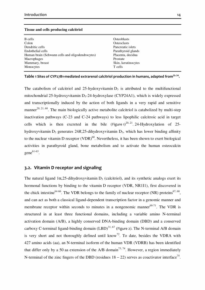
Tissue and cells producing calcitriol
B cells Osteoblasts Colon Osteoclasts Dendritic cells Pancreatic islets Endothelial cells Parathyroid glands Human brain (Schwann cells and oligodendrocytes) Placenta, decidua Macrophages Prostate Mammary, breast Skin, keratinocytes Monocytes T cells
Table 1 Sites of CYP27B1-mediated extrarenal calcitriol production in humans, adapted from33, 34.
The catabolism of calcitriol and 25-hydoxyvitamin D3 is attributed to the multifunctional
mitochondrial 25-hydroxyvitamin D3-24-hydroxylase (CYP24A1), which is widely expressed
and transcriptionally induced by the action of both ligands in a very rapid and sensitive
manner20, 21, 60. The main biologically active metabolite calcitriol is catabolized by multi-step
inactivation pathways (C-23 and C-24 pathways) to less lipophilic calcitroic acid in target
cells which is then excreted in the bile (Figure 1)20, 21. 24-Hydroxylation of 25-
hydroxyvitamin D3 generates 24R,25-dihydroxyvitamin D3, which has lower binding affinity
to the nuclear vitamin D receptor (VDR)60. Nevertheless, it has been shown to exert biological
activities in parathyroid gland, bone metabolism and to activate the human osteocalcin
gene61-63.
3.2. Vitamin D receptor and signaling
The natural ligand 1α,25-dihydroxyvitamin D3 (calcitriol), and its synthetic analogs exert its
hormonal functions by binding to the vitamin D receptor (VDR, NR1I1), first discovered in
the chick intestine64-66. The VDR belongs to the family of nuclear receptor (NR) proteins67, 68,
and can act as both a classical ligand-dependent transcription factor in a genomic manner and
membrane receptor within seconds to minutes in a nongenomic manner69-71. The VDR is
structured in at least three functional domains, including a variable amino N-terminal
activation domain (A/B), a highly conserved DNA-binding domain (DBD) and a conserved
carboxy C-terminal ligand-binding domain (LBD)31, 67 (Figure 2). The N-terminal A/B domain
is very short and not thoroughly defined until know72. To date, besides the VDRA with
427 amino acids (aa), an N-terminal isoform of the human VDR (VDRB) has been identified
that differ only by a 50 aa extension of the A/B domain73, 74. However, a region immediately
N-terminal of the zinc fingers of the DBD (residues 18 − 22) serves as coactivator interface75.
Introduction 15
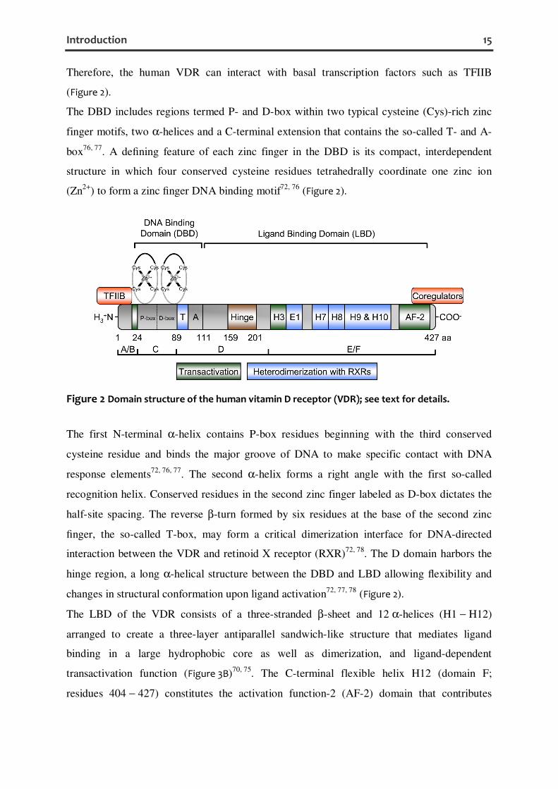
Therefore, the human VDR can interact with basal transcription factors such as TFIIB
(Figure 2).
The DBD includes regions termed P- and D-box within two typical cysteine (Cys)-rich zinc
finger motifs, two α-helices and a C-terminal extension that contains the so-called T- and A-
box76, 77. A defining feature of each zinc finger in the DBD is its compact, interdependent
structure in which four conserved cysteine residues tetrahedrally coordinate one zinc ion
(Zn2+) to form a zinc finger DNA binding motif72, 76 (Figure 2).
Figure 2 Domain structure of the human vitamin D receptor (VDR); see text for details.
The first N-terminal α-helix contains P-box residues beginning with the third conserved
cysteine residue and binds the major groove of DNA to make specific contact with DNA
response elements72, 76, 77. The second α-helix forms a right angle with the first so-called
recognition helix. Conserved residues in the second zinc finger labeled as D-box dictates the
half-site spacing. The reverse β-turn formed by six residues at the base of the second zinc
finger, the so-called T-box, may form a critical dimerization interface for DNA-directed
interaction between the VDR and retinoid X receptor (RXR)72, 78. The D domain harbors the
hinge region, a long α-helical structure between the DBD and LBD allowing flexibility and
changes in structural conformation upon ligand activation72, 77, 78 (Figure 2).
The LBD of the VDR consists of a three-stranded β-sheet and 12 α-helices (H1 − H12)
arranged to create a three-layer antiparallel sandwich-like structure that mediates ligand
binding in a large hydrophobic core as well as dimerization, and ligand-dependent
transactivation function (Figure 3B)70, 75. The C-terminal flexible helix H12 (domain F;
residues 404 − 427) constitutes the activation function-2 (AF-2) domain that contributes

Introduction 16
together with helix H3, H4 and H5 to the activation of transcription by forming high affinity
interfaces for nuclear coregulators such as coactivator (NCoA) molecules (Figure 2)70, 75.
Recently, Mitzwicki and colleagues proposed the so-called vitamin D sterol (VDS)-vitamin D
receptor (VDR) conformational ensemble model70, 71, 79. The VDS-VDR conformational
ensemble model is separated into three interconnected parts: the VDS conformational
ensemble (Figure 3A), the VDR two pocket model (Figure 3B), and the VDR (helix 12)
conformational ensemble (Figure 3C)71. The VDS-VDR conformational ensemble model
proposes that two overlapping, functionally distinct ligand-binding pockets (LBPs) exist in
the VDR, the genomic and alternative pocket (Figure 3B)70, 71.
Vitamin D3 or its active form calcitriol are unusually conformationally flexible containing
three flexible regions, the A-ring, seco-B ring, and side chain (Figure 3A)69, 70, 80. Therefore,
calcitriol exists in two principal conformations. These conformations result from 360° rotation
around the 6,7 carbon-carbon single bond of the seco-B ring resulting in the 6-s-cis-conformer
(the steroid-like shape) and the 6-s-trans-conformer (the extended shape) (Figure 3A)81. The
“bowl-shape” and “planar-shape” 6-s-trans-conformers of calcitriol can also form a stable
complex with the VDR71. The 6-s-cis-conformation is preferred for rapid nongenomic
biological responses, e.g. shown for the 6-s-cis-locked 1α,25-dihydroxy-lumisterol D3 (JN)
that binds in the alternative pocket (AP) (Figure 3B)70, 80, 82. The conformationally flexible
natural ligand calcitriol is a full agonist for both rapid and genomic responses80, 83. Recent
results from structure-function analyses suggest that the major physicochemical traits of a
potent and efficient VDR genomic agonist are: the ability to form hydrogen bonds (H-bonds)
with the VDR Ser237 and Arg274 residues; the ability to conform to a “bowl-shaped” molecular
geometry; and a molecular volume that is similar to that calculated for calcitriol.
Alternatively, for a efficient non-genomic agonist: the ability to form H-bonds with Ser237 and
Arg274; the ability to conform to a more linear, “planar-like” molecular geometry, and a
molecular volume that is ≤ to that calculated for calcitriol are major traits71.
VDR nuclear localization signals (NLS, residues 49 − 55, 79 − 105) direct the receptor mainly
into the nucleus84, 85. Upon ligand-binding to the LBD, VDR is stabilized by the
phosphorylation of Ser51 in the DBD by protein kinase C (PKC)86, and Ser208 in the hinge
region by casein kinase II (CK-II)87. As a transcription factor, the occupied VDR is tightly
associated with its heterodimeric partner, RXR – which has three main isoforms: α, β and
γ76, 88 – and binds to vitamin D responsive elements (VDREs) in the promoters of vitamin D-
regulated genes to up- or downregulate transcription31, 70.

Introduction 17
Figure 3 The vitamin D sterol (VDS)-vitamin D receptor (VDR) conformational ensemble model,
adapted from82, 89. (A) Conformations of 1α,25-dihydroxyvitamin D3. (B) Ribbon diagram from
molecular modeling of the VDR LBD. The more hydrophobic VDR genomic pocket (GP, blue
Connolly surface) is occupied by 6-s-trans-1α,25-dihydroxyvitamin D3 and 1α,25-dihydroxy-
lumisterol D3 (JN) is docked in the putative overlapping more hydrophilic VDR alternative pocket
(AP, ochre Connolly surface). The C-terminal end of helix 11 (H11) and H12 are colored orange and
H12 is shown in the closed, transcriptionally active conformation. H2 and the β-sheet are colored
yellow, whereas the purple ribbons indicate their final position after JN was docked in the A
pocket. Important amino acid residues are labelled. (C) Dynamic conformational heterogeneity of
the helix 12 (black ribbon, labeled H12) of the human VDR molecule in the absence of ligand
projected using NR x-ray data (see Protein Data Bank). In VDR-c1, helix 12 is in the closed, active
conformation. In the VDR-c2 conformational isomer, H12 is bound to the NCoA surface. It is
proposed that in VDR-c3, H12 is in the opened conformation, where the ligand binding pocket is
accessible to ligand (bottom, VDR-c3 structure) or is occupied by H11 residues (orange ribbon in
the top VDR-c3 structure), thereby exposing the C-terminal H11 Arg402 residue. Certainly, synthetic
or natural ligands can do the same70, 82, 89.

Introduction 18
VDREs composed of two binding sites arranged either as a direct repeat of two hexameric
half-elements with a spacer of commonly three nucleotides (DR3), inverted palindromes
interspaced by nine nucleotides (IP9) or an everted repeat of two half-elements with a spacer
of six nucleotides (ER6) motif31, 75. The consensus sequence considered for a VDRE half site
is (A/G)G(G/T)TCA, with RXR occupying the 5′ half-element and VDR the 3′ half-
element31, 75. Positive VDREs promote the binding of a coactivator complex to the ligated
VDR-RXR heterodimer that includes not only steroid receptor coactivator 1 (SRC-1), but also
CBP/p300, nuclear receptor coactivator-62 (NCoA-62) and a SWI/SNF chromatin remodeling
complex anchored by polybromo-associated BAF (PBAF) resulting in transcriptional
activation75. In contrast, negative VDREs (nVDRE), which closely resemble the consensus
sequence, bind either VDR-RXR heterodimers or VDR homodimers and mediate repression
of transcription by recruiting histone deacetylases (HDAC) and NR corepressors
(NCoR)/silent mediator for retinoid and thyroid hormone receptor (SMRT) corepressors31, 75.
Another group of nVDREs, composed of E-box type motifs with the sequence
5’-CATCTG-3’, was identified in the human CYP27B1, as well as in the human PTH and
PTHrP promoters32, 90, 91.
Besides genomic responses, a wide range of rapid, non-genomic responses have been shown
for the classical VDR, which can be associated with caveolae in the plasma membrane92, 93.
Caveolae, a subset of membrane (lipid) rafts, are flask-shaped membrane invaginations
containing scaffolding caveolin proteins, sphingolipids and cholesterol that serve as
interaction platforms for signaling components94. Thereby, calcitriol mediated rapid responses
include the modulation of several signaling molecules such as PKC, mitogen-activated protein
kinase (MAPK), phospholipase A2 (PLA2), phospholipase C (PLC), store-operated Ca2+
channels and Src kinases70, 95. More recently, another membrane receptor for calcitriol, which
is a distinct gene product from the classical nuclear VDR, was discovered. This 57 kDa
protein is named 1,25D3-MARRS (membrane associated, rapid response, steroid-binding),
and is identical to a previously cloned member of the thioredoxin family of proteins, ERp57
(endoplasmic reticulum protein of 57 kDa) or alternatively GRp58 (glucose regulated protein
of 58 kDa)/PDIA396-99. In addition, localization in the endoplasmic reticulum (ER) and in the
nucleus has been shown for MARRS (ERp57/PDIA3), which also contains a domain that can
bind to DNA99-101. Nevertheless, some of these non-genomic signals can modulate the
expression of genes, either through effects on the function of the VDR in the nucleus or
independently of VDR70.

Introduction 19
3.3. Vitamin D and the immune system
The VDR is expressed in a wide range of tissues and cells (Table 2) reflecting its capability to
influence about 3% of the humane genome34, 102.
Tissue and cell distribution of VDR
Adipose Monocytes/Macrophages Adrenal Muscle, cardiac Bone, osteoblasts Muscle, embryonic Brain, general Muscle, smooth Brain, amygdale Ovary Brain, hypothalamus Pancreas β-cell Brain, glial cells Parathyroid Breast Parotid Cartilage Pituitary Colon Placenta Dendritic cells Prostate Eggshell gland Retina Epididymus, seminiferous tubules Skin Gills (fish) Sperm Hair follicle Stomach Intestine Testis Kidney Thymus Liver Thyroid Lung Tonsils, dendritic cells Lymphocytes (B, T) Uterus Mast cells Yolk sac
Table 2 Sites of vitamin D receptor (VDR) expression, adapted from33, 34.
The classic physiological function of the hormonally active calcitriol is to maintain serum
calcium and phosphorus levels within the normal physiologic range to support most
metabolic functions, neuromuscular transmission, and bone mineralization19, 24. Inherited
vitamin D-resistant syndromes such as vitamin D-dependent rickets type 1 to 3, vitamin D or
calcium deficiency, X-linked hypophosphatemic rickets (XLH), autosomal dominant
hypophosphatemic rickets (ADHR) and tumor-induced osteomalacia (TIO) result in
hypocalcemia, hyperparathyroidism, hypophoshatemia, phosphaturia, rickets, and
osteomalacia19, 103. During the past decades, the knowledge of the vitamin D metabolism has
greatly evolved. The discovery of VDRs and vitamin D-activating enzymes in extrarenal
tissues and cell types, strongly indicating a more diverse role of vitamin D metabolism, and
the worsening, world-wide trend to vitamin D insufficiency or deficiency heightened the
interest in vitamin D physiology104, 105. Thereby, besides causing osteoporosis and muscle
weakness, vitamin D deficiency is also linked to an increased incidence of multiple
malignancies, metabolic and cardiovascular diseases, neurological, and immune disorders

Introduction 20
such as autoimmune diseases24, 103-105. The ubiquitous expression of VDR in multiple immune
cells such as dendritic cells45, 106, monocytes107, macrophages107, mast cells108, and activated T
and B cells45, 48, 109-111 (Table 2) led to its recognition as a central immunomodulator. Thus,
VDR signaling can modulate the innate and adaptive immunity.
It is known that activation of monocyte/macrophage toll-like receptor (TLR)2/1 stimulates
expression of the VDR and 25-hydroxyvitamin D3-1α-hydroxylase (CYP27B1) in an
interleukin-15 (IL-15)-dependent manner112. In addition, interferon γ (IFNγ) and CD14/TLR4
activation is also reported to induce CYP27B1 expression in human monocytes38. In this way,
25-hydroxyvitamin D3 is converted into the hormonally active form calcitriol in the
mitochondria of monocytes/macrophages41. Consequently, calcitriol binds to the VDR and
transcriptionally induces target genes such as the antimicrobial protein human cathelicidin
(CAMP/hCAP18/LL-37)113, 114. The antimicrobial protein human β-defensin 4 (DEFB4,
formerly HBD2) is also transcriptionally induced by calcitriol but requires co-stimulation by
activators of NF-κB, such as IL-1β115. Remarkably, the regulation of the camp (encoding
hCAP18) and defb4 (encoding β-defensin 4) genes by calcitriol appears to be not conserved
between human and mouse113, 116. Additionally, calcitriol can inhibit the expression of
inflammatory cytokines, including IL-1β, IL-6, TNFα, IL-8, and IL-12 in monocytes117-119.
Differentiation of monocytes, into either macrophages or immature and mature DCs, is
accompanied by a reciprocal organization of CYP27B1 and VDR expression ensuring that
mature antigen-presenting DCs are relatively insensitive to calcitriol, thereby shaping defined
T cell responses43, 107. VDR signaling prevents DC maturation as evidenced by a decreased
expression of DC markers (e.g. CD1a), major histocompatibility complex class II (MHC-II),
co-stimulatory molecules (CD40, CD80, and CD86), and other maturation induced surface
markers (e.g. CD83)120-123. Calcitriol also inhibits the production of the T helper 1 (TH1)
polarizing factor IL-12 and the TH17 polarizing factor IL-23 in DCs whereas the release of the
tolerogenic cytokine IL-10 and the chemokine CCL22, involved in the recruitment of CCR4-
expressing regulatory T cells (Tregs)124, 125, is enhanced120, 121. Together, VDR activation
promotes a tolerogenic phenotype and function in myeloid DCs (M-DCs)126 and seems to
foster the induction of CD4+ CD25+ Tregs127.
VDR activation by its natural ligand also directly modulates the function of adaptive immune
cells. It inhibits T cell proliferation128, the production of inflammatory TH1 cytokines such as
IL-2128-130 and IFNγ131, the TH17-derived cytokines IL-17 and IL-21132, and CD8 T cell-
mediated cytotoxicity133, whereas IL-10 expression can be induced132, 134 or inhibited48, 135 in
human T cells. Contradictory reports exist regarding TH2-skewing effects of VDR activation

Introduction 21
in murine CD4+ T cells, showing an induction of the TH2 cytokines IL-4136, IL-5, and IL-10 as
well as TH2-specific transcription factors GATA3 and c-Maf137 whereas others report an
inhibition of IL-4 and IFNγ with stable GATA3 and c-Maf expression in mice138. Whether or
not VDR triggering favors a shift toward TH1 versus TH2 dominance remains to be elucidated.
More interestingly, VDR activation by calcitriol alone or in combination with dexamethasone
has been shown to induce human Tregs expressing CTLA-4 and Foxp3132 and murine naive
CD4+ T cells to differentiate into IL-10-producing Tregs, even in the absence of antigen-
presenting cells (APCs)139. However, VDR triggering not only facilitates induction and
expansion of Tregs and enhance their suppressive activity, but can promote their recruitment to
sites of inflammation, too140-143.
In human B cells, an inhibitory effect of calcitriol on immunoglobulin E (IgE) synthesis in
vitro, at least in part mediated by diminished nuclear factor-κB (NF-κB) activation and
inhibition of the epsilon germline transcript (εGLT), has been proven144. Additionally, a
recent study has shown that the liganded VDR binds to the εGLT gene promoter and exhibits
transrepressive activity145. In contrast, another report showed that VDR activation by calcitriol
inhibits the proliferation, plasma cell differentiation and immunoglobulin secretion including
IgG and IgM, memory B cell generation and induces B cell apoptosis46. Despite these
suppressive functions of calcitriol on human B cells, it enhances IL-10 expression in activated
B cells47 and promotes expression of the skin-homing receptor CCR10 in terminally
differentiating B cells146. Likewise it has been described in activated T cells45.
3.4. Vitamin D in allergic diseases and atopic dermatitis
3.4.1. Type I allergic immune response
The term allergy can be used to refer an abnormal and harmful adaptive immune response
directed against various non-infectious environmental antigens (allergens)147. There are two
main types of allergen. The first type includes any non-infectious environmental protein that
induces IgE- and TH2 cell-mediated responses leading to anaphylaxis, allergic rhinitis (hay
fever), some food allergies and allergic asthma. The second type of allergen is a non-
infectious environmental molecule that can induce an adaptive immune response associated
with a local inflammation, where IgE is thought not to be important, e.g. allergic contact
dermatitis. Allergic diseases, also known as atopic disorders (from the Greek atopos, meaning
out of place, abnormal), are common and afflict roughly 25% of people in the developed
world.

Introduction 22
In case of IgE dependent hypersensitivity, a single allergen exposure produces an acute
reaction, which is known as an early-phase reaction or a type I immediate hypersensitivity
reaction. Such a reaction can occur locally (for example, acute asthma or rhinoconjunctivitis)
or systemically (anaphylaxis) within minutes of allergen exposure, followed by allergen-
induced crosslinking of IgE bound to FcεRI (high-affinity receptor for IgE) on mast cells and
basophils. The crosslinking results in the release of diverse preformed and newly synthesized
mediators, including cytokines, chemokines, histamine, heparin, serotonin and proteases,
causing vasodilation, increased vascular permeability with edema, and acute functional
changes in affected organs. These events also promote the local recruitment and activation of
leukocytes, contributing to the development of late-phase reactions. The late-phase reaction
typically develops 2 – 6 h later and peaks 6 – 9 h after allergen exposure. Usually it
completely resolves in 1 – 2 days. This type of reaction reflects the local recruitment and
activation of TH2 cells, eosinophils, basophils and other leukocytes, and persistent mediator
production by resident cells (such as mast cells) resulting in edema, pain, warmth and
erythema (redness) in the skin as well as airway narrowing and mucus hypersecretion in the
lungs. Persistent or repetitive exposure to allergen leads to a chronic allergic inflammation,
with presence of large numbers of innate and adaptive immune cells and alterations in the
affected tissue. There are many factors affecting the probability of developing allergic
diseases including genetic148, 149, epigenetic150, 151 and environmental components as proposed
in the hygiene hypothesis152, 153.
3.4.2. Role of TH2 cells in the allergic immune response
TH2 cells are indispensable for host immunity to extracellular parasites, such as helminths, but
are also responsible for the development of asthma and other allergic inflammatory
diseases154. The initiation of TH2 cell responses takes place in the tissue sites where allergens
or parasites are encountered. Activated APCs such as macrophages, Langerhans cells (LCs),
DCs and basophils migrate from tissues to the draining lymph nodes to stimulate proliferation
and differentiation of naïve CD4+ (TH0) into TH2 cells under the influence of various
cytokines such as thymic stromal lymphopoietin (TSLP), IL-4 and IL-25 (IL-17E) (Figure 4).
Also other cell types like activated lung and intestinal epithelial cells can initiate TH2 immune
responses by acting on basophils, DCs and/or non-B non-T cells (NBNT cells) by producing
TSLP, IL-25 and IL-33. Other immune cells, including natural killer (NK) cells, NKT cells,
γδ T cells, macrophages, B cells, eosinophils and mast cells, may also participate in the
initiation and amplification of TH2 immune responses by creating a TH2-biased cytokine
Introduction 23
environment. TH2 cell differentiation requires both, GATA-binding protein 3 (GATA3)
expression and activated signal transducer and activator of transcription 5 (STAT5)
activation; and is favored by low-strength T cell receptor (TCR) signaling. TH2 cells produce
various TH2 cell-associated cytokines, including IL-2, IL-4, IL-5, IL-9, IL-13 and IL-25. TH2
cells regulate B cell class switch recombination (CSR) to IgE through their production of IL-4
and IL-13155 (Figure 4). Both of these cytokines use the common γ-chain (γc)-related IL-4
receptor α-chain (IL-4Rα) to signal through three cytokine-receptor combinations. IL-4
signals through the type I receptor IL-4Rα-γc, and both IL-4 and IL-13 can signal through the
type II receptor IL-4Rα-IL-13Rα1156. Ligation of the IL-4R activates the Janus family
tyrosine kinase JAK1 (through IL-4Rα), JAK3, IRS1 and TYK1 (through γc) (Figure 4). IL-
13R triggering leads to the activation of JAK1 and TYK2. Subsequently, activated JAKs
phosphorylate tyrosine residues in the intracellular domains of IL-4Rα, which serve as
docking sites for STAT6 monomers (Figure 4). STAT6 gets tyrosine phosphorylated itself by
the receptor-associated JAK kinases, dimerizes and translocates to the nucleus, where it can
activate the transcription of target genes155, 157 (Figure 4). Additionally, IL-21 signals via the
IL-21R, which is structurally related to the IL-2Rβ associated with γc; and activates STAT3
subsequently stimulating the IgE production in human beings158, 159. Another important signal
to facilitate T cell-dependent class switch recombination to IgE is provided by the membrane-
bound trimeric CD40L (CD154) on TH2 cells (Figure 4). CD40-CD40L interaction in B cells
mainly allows the recruitment of tumour-necrosis factor-associated factors (TRAFs) 2, 3 and
6 that promote the activation of nuclear factor-κB (NF-κB), activator protein 1 (AP1)
(Figure 4), nuclear factor of interleukin-6 (NF-IL-6), MEKK1 [mitogen-activated protein
kinase kinase kinase/extracellular signal-regulated kinase (ERK)], JUN amino-terminal kinase
(JNK) and p38, resulting in proliferation, differentiation, isotype switching, cytokine
production, surface-molecule upregulation; protection from apoptosis; and promotion of
humoral memory155, 160.
3.4.3. Mechanism of IgE class switch recombination
STAT6 and NF-κB (p50/p65) are essential for the CSR to IgE as shown by STAT6-deficient
(STAT6−/−) mice that are severely impaired in IgE production and NF-κB p50−/− mice that
have a preferentially impaired IgE switching161-164. Thereby, both transcription factors can
bind to promoter regions of the epsilon germline transcript (εGLT) and aicda, encoding
activation-induced cytidine deaminase (AID)165-168 (Figure 4). Upon activation antibody class

Introduction 24
switching in mature B cells occurs by a unique type of intrachromosomal deletional
recombination within special G-rich tandem repeated DNA sequences [called switch, or S,
regions located upstream of each of the heavy chain constant (CH) region genes, except
Cδ]169, 170. Thus, the εGLT is transcribed as a sterile transcript, starting with the Iε exon,
which is located immediately upstream of Sε and proceeds with Sε and Cε. The Iε exon is
spliced to Cε yielding the human 1.7 kb εGLT168, 171. The sterile εGLT, which remains
hybridized to the template strand of the switch region DNA, forms the “R loop” structure
(RNA-DNA hybrid). During this process, AID is recruited by an unknown mechanism and
deaminates several cytosine bases to uracil bases in the donor and acceptor S regions, which
results in double-strand DNA breaks (DSBs) in both S regions by DNA-repair proteins and
CSR by an end-joining type of recombination. Joining can then occur between breaks
upstream of the Cε cluster of exons and downstream of the variable (V), diversity (D), and
joining (J) gene segments, giving rise to an intact IgE heavy-chain gene169, 172, 173. The
phenotype observed in patients with the autosomal recessive form of Hyper-IgM syndrome
(HIGM2) and in AID−/− mice demonstrates the absolute requirement for AID in several
crucial steps of B cell terminal differentiation necessary for efficient antibody
responses174, 175.
Figure 4 Classical interactions important for IgE class switch recombination, adapted from155.
Antigen presenting cells, such as dentritic cells, take up allergens, which are presented to T cells
by MHC class II. Subsequent activation of CD4+ T cells leads to the induction of TH2 cells,
expressing IL-4 and CD40L (CD154). CD40-mediated activation of B cells synergizes with IL-4 to
induce class switch recombination to IgE by enhancing the transcription of εGLT and aicda
(encoding AID), which results in rearrangement of the IgE locus and production of IgE antibodies.

Introduction 25
3.4.4. Vitamin D in the context of IgE and atopic dermatitis (AD)
IgE is a stringently regulated key molecule in type I hypersensitivity that is usually present in
very low concentrations, in the range of 1 − 400 ng/ml in non-atopic human170.
Epidemiological data suggest that vitamin D insufficiency, reflected by less than 30 ng/ml of
the circulating precursor 25-hydroxyvitamin D3, is associated with elevated IgE levels in
asthmatic children176. Hyppönen et al. reported a significant but nonlinear relationship
between serum 25-hydroxyvitamin D3 and IgE, suggesting the possibility of a threshold
effect177. Interestingly, type I sensitized VDR knockout mice have higher serum IgE
concentrations than wild type controls in an allergic asthma model178. Accordingly, allergen-
specific immunotherapy has been suggested to be more efficient upon calcitriol treatment179.
Additionally, reports show a beneficial association between higher maternal vitamin D intake
during pregnancy and protection from childhood asthma and allergic diseases in the
offspring180-182. In contrast, further studies have shown that the prevalence of atopic diseases
was equal or even higher in subjects who received vitamin D supplementation during
infancy183, 184. Another study by Oren et al. reported no significant associations between
vitamin D status and the prevalence of asthma or allergic rhinitis in an obese population185.
Nevertheless, these conflicting data indicate that there might be a complex relationship
between external supply, endogenous production, metabolism, signaling pathways on one
hand and the development of allergy on the other hand.
However, there is also a debate on the role of vitamin D metabolism and its potential
beneficial effects on atopic dermatitis (AD), a common skin disease that is often associated
with other atopic disorders. Thereby, a negative correlation between serum 25-
hydroxyvitamin D3 levels and severity of atopic dermatitis in children has been reported186. A
randomized controlled trial by Sidbury et al. showed a favorable impact of vitamin D
supplementation on winter-related AD187. Studies by Vähävihu et al. showed an improved
vitamin D balance along with an altered antimicrobial peptide (AMP) expression in skin
lesions followed by an improvement in AD after heliotherapy or narrowband UV-B
treatment188, 189.
3.4.5. Atopic dermatitis (AD)
AD is a chronic and relapsing eczematous skin inflammation associated with epidermal
barrier dysfunction, intense pruritis, and cutaneous hyperreactivity to environmental triggers
that frequently starts at infancy or early childhood (early-onset atopic dermatitis)190, 191.
Infants with AD have an increased tendency to develop asthma and allergic rhinitis later in

Introduction 26
life, a phenomenon known as the atopic march. Nevertheless, the disease can also start in
adults, called late-onset atopic dermatitis. Histopathology of AD skin lesions reveals intense
infiltrates of mononuclear cells in particular T cells in the dermis combined with intercellular
edema in the epidermis (spongiosis)190. The lifetime prevalence of AD is estimated to
15 ∼ 30% in children and 2 ∼ 10% in adults while the incidence of AD has increased by 2- to
3-fold during the past 3 decades in industrialized countries190, 192. AD is a multifactorial,
heterogenous disease with a variety of defects in the immune system, in antimicrobial defense
mechanisms and epidermal barrier integrity that collectively contribute to the risk and severity
of AD development193, 194. AD can be categorized into the extrinsic (allergic AD) and intrinsic
types (non-allergic AD)195-197. The classical extrinsic type of AD with high prevalence
(70 − 80% of AD patients) shows high total serum IgE levels and the presence of specific IgE
for environmental and food allergens. Intrinsic or non-allergic AD exhibits normal total IgE
values and the absence of specific IgE with an incidence of approximately 20 − 30% and
female predominance. Early-onset atopic dermatitis usually emerges in the absence of
detectable IgE-mediated allergic sensitization which often occurs several weeks or months
after the lesions appear190. Therefore, the non-IgE associated form may represent a
transitional phase of the IgE-associated form, at least in infancy192. The initial mechanisms
inducing skin inflammation in patients with AD are mostly unknown. They may involve
neuropeptide-induced, irritation-induced, or pruritus-induced scratching, which leads to the
release of pro-inflammatory cytokines from keratinocytes190. They also entail T cell-mediated
but IgE-independent reactions to allergens from pollens, house-dust-mite products, microbes,
and food. Subsequent studies have shown that TSLP, expressed in keratinocytes of AD
patients, can also promote pathogenesis of AD in the effector phase by acting on DCs or
directly on skin-infiltrating T cells to induce TH2 cytokine secretion198, 199. All these factors
provide signals that drive a TH2 polarization in the skin, the point of entry for atopic
sensitization. Inflammation in atopic dermatitis is a biphasic process190, 192, 196. The initial
acute eczematous skin lesions present clinically as intensely pruritic, erythematous papules
associated with excoriation and exudation. Thereby, Langerhans cells (LCs) in lesional skin
are activated by binding through allergens by means of specific IgE and the high affinity
receptor FcεRI in the IgE-associated form but not in the non-IgE-associated form of AD.
They produce monocyte chemotactic protein 1 (MCP-1), IL-16 and present allergen-derived
peptides to recruited T cells that induce a TH2 profile. The TH2 cytokines IL-4, IL-5, and IL-
13 predominate in the acute phase. Eosinophils are seen in the acute lesions but basophils and
neutrophils are rarely present. Mast cells are present in various stages of degranulation. After

Introduction 27
migration into the skin, the recruited monocytes differentiate into inflammatory dendritic
epidermal cells (IDECs) and produce the pro-inflammatory cytokines IL-1, IL-6, and TNFα as
well as IL-12 and IL-18, which contribute to the switch from TH2 to TH1/0 and thereby lead to
the chronic phase of the disease. A hyperplastic epidermis with elongation of the rete ridges,
prominent hyperkeratosis and minimal spongiosis characterizes chronic AD skin. In chronic
AD skin lesions, macrophage-dominated mononuclear cell infiltrates in the dermis are
detected. They display an increase of IFNγ and IL-12, as well as IL-5 and granulocyte
macrophage colony stimulation factor (GM-CSF), which are characteristic for TH1/0
dominance. The intrinsic type of AD is characterized by a lower expression of interleukin IL-
4, IL-5, and IL-13, and a higher expression of IFNγ. Treg cells, expressing the alpha chain of
the IL-2 receptor (CD25) and the transcription factor Foxp3, can directly suppress allergic
TH2 immune responses200, 201. The number of Treg cells is decreased in lesional skin of patients
with AD, suggesting that there might be a role for Treg cells in AD202.
The skin barrier, as one part of the first line defense, is perturbed in the extrinsic, but not
intrinsic type of AD193-195. Epidermal barrier dysfunction is a prerequisite for the penetration
of allergens and microbes and causes increased transepidermal water loss, one hallmark of
AD190. The epidermal barrier is formed by the cornified layer (stratum corneum) consisting of
terminally differentiated, dead, cornified, flattened cells (corneocytes) and the cornified
envelope (CE), which is mainly composed of the structural proteins, loricrin, involucrin,
filaggrin and small proline-rich proteins203. By cross-linking these and other proteins,
transglutaminases are likewise involved in the formation of the CE203, 204. Recently, loss-of-
function genetic variants of the filaggrin gene (FLG) have been reported to be strong
predisposing factors for extrinsic205-207, but not intrinsic AD206, 208. Other reports show a
reduced expression of antimicrobial peptides (AMPs), involucrin and loricrin in AD skin193.
Numerous functions of the skin, including formation of the permeability barrier, are regulated
by VDR signaling209. For instance, calcitriol has been reported to increase involucrin and
transglutaminase expression in keratinocytes210-212. Silencing of the VDR and two VDR
coactivators blocked keratinocyte differentiation as shown by decreased expression of
filaggrin213. VDR null mice exhibit a defect in epidermal differentiation as shown by reduced
levels of involucrin, filaggrin and loricrin, and loss of keratohyalin granules214. Additionally,
CYP27B1 (1αOHase) null animals, which are unable to produce the biologically active
calcitriol from its precursor 25-hydroxyvitamin D3, showed a reduction in levels of the
epidermal differentiation markers involucrin, filaggrin and loricrin, and displayed a markedly
delayed recovery of normal barrier function following disruption of the barrier215. Disruption

Introduction 28
of the permeability barrier, suppression of innate immune cells and the reduction in AMPs,
such as cathelicidin and human β-defensins HBD2, and HBD3 in AD skin leads to the
colonization and infection by Staphylococcus aureus (S. aureus), herpes simplex virus (HSV),
and vaccinia virus (VV) in AD patients193, 216-219. The most important symptom of AD is
persistent pruritus leading to itching induced by neuropeptides, proteases, kinins, and
cytokines190. Moreover, keratinocyte-derived chemokines, TSLP, and IL-31 secretion are
induced and augmented by S. aureus enterotoxins, leading to an increased inflammation in
atopic dermatitis and provoke the generation of enterotoxin-specific IgE. Therefore, IL-31 is a
major pruritogenic factor as shown by the overexpression of this cytokine and its receptor in
lesional skin220, 221. Besides IgE antibodies against food and aeroallergens, autoantibodies
against proteins from keratinocytes and endothelial cells such as manganese superoxide
dismutase and calcium-binding proteins were found in the serum of patients with AD,
correlating with disease severity222-225. Thus, AD seems to be at the frontier between allergy
and autoimmunity190.
Interestingly, a new unifying hypothesis, in which the natural history of atopic dermatitis has
three phases including gene-gene and gene-environment interactions, emerges (Figure 5)190.
Thereby, no classification distinguishing between an IgE-associated (extrinsic) and non-IgE-
associated (intrinsic) form of AD exists. In the initial phase, genetically determined
epidermal-barrier dysfunctions and the effect of environmental factors lead to non-atopic
dermatitis, the first manifestation of AD in early infancy. In the transition phase, IgE-
mediated sensitization to environmental allergens, influenced by genetic factors, is induced.
In the third phase, pruritus-induced scratching damages the skin, which release autoantigens
that can induce IgE autoantibodies in AD patients.

Introduction 29
Figure 5 Proposed model for the three phases of the natural history of atopic dermatitis,
adapted from190, see text for details.

Objectives 30
4. Objectives
Over the past years, the knowledge of the vitamin D endocrinology, including the pleiotropic
functions of vitamin D receptor (VDR) activation in the immune system increased steadily.
Recent epidemiologic data also indicated a role for the vitamin D metabolism in the allergic
immune response. Unfortunately, the clinical use of the natural VDR ligand calcitriol is
hampered by its toxic side effects, in particular hypercalcemia.
This work aimed to analyze the role of VDR activation in the allergic immune response in
vitro and in vivo by using the novel low-calcemic VDR agonist ZK203278.
The first part of the work intended to investigate the in vitro-efficacy of the VDR agonist to
activate the VDR and to modulate the stimulated IgE response in isolated human peripheral B
cells in comparison to the natural ligand (calcitriol) and a known VDR antagonist. Thereby,
mechanisms involved in the VDR-dependent modulation of human peripheral B cells were in
the focus of this work. Second, the effect of the low-calcemic VDR agonist on the humoral
IgE response in a mouse model of type I sensitization was studied in this work.
Finally, based on the crucial role of the VDR and VDR signaling in skin homeostasis and
epidermal barrier function, the work aimed to investigate the clinical efficiency of VDR
agonist treatment in a mouse model of allergen-induced eczema.

Materials and Methods 31
5. Materials and Methods
5.1. Materials
5.1.1. Antibodies
Antibody Clone Manufacturer
Anti-chicken egg albumin (OVA) OVA-14 Sigma-Aldrich
Anti-human CD14 MicroBeads Miltenyi Biotec
Anti-human CD14 PE MφP9 BD Pharmingen
Anti-human CD19 FITC 4G7 BD Pharmingen
Anti-human CD19 MicroBeads Miltenyi Biotec
Anti-human CD19 MultiSort Kit Miltenyi Biotec
Anti-human CD19 PE LT19 Miltenyi Biotec
Anti-human CD20 FITC 2H7 eBioscience
Anti-human CD23 APC EBVCS-5 BD Pharmingen
Anti-human CD27 APC M-T271 Miltenyi Biotec
Anti-human CD27 FITC LG.7F9 eBioscience
Anti-human CD27 MicroBeads Miltenyi Biotec
Anti-human CD27 PE LG.7F9 eBioscience
Anti-human CD3 APC SK7 BD Pharmingen
Anti-human CD38 FITC HIT2 BD Pharmingen
Anti-human CD38 PE-Cy5 HIT2 eBioscience
Anti-human CD38 PerCP-Cy5.5 HIT2 BD Pharmingen
Anti-human CD40 626 Santa Cruz Biotechnology
Anti-human CD40 G28.5 DRFZ
Anti-human CD69 FITC FN50 BD Pharmingen
Anti-human IgA G20-359 BD Pharmingen
Anti-human IgA, IgG, IgM Matched pairs Jackson ImmunoResearch
Anti-human IgE HP6061, HP6029b Southern Biotech
Anti-human IgG G18-145 BD Pharmingen
Anti-human IκBα Alexa Fluor 647 L35A5 Cell Signaling Technology Inc.
Anti-human pSTAT6 Alexa Fluor 647 pY641, 18 BD Pharmingen
Anti-human STAT6 PE 23 BD Pharmingen
Anti-mouse CD4 RM4-5 BD Pharmingen
Anti-mouse CD8 53-6.7 BD Pharmingen
Anti-mouse CD21/35 FITC 7G6 BD Pharmingen
Anti-mouse CD23 PE B3B4 BD Pharmingen

Materials and Methods 32
Antibody Clone Manufacturer
Anti-mouse CD19 APC 1D3 BD Pharmingen
Anti-mouse IgA, biotin Southern Biotech
Anti-mouse IgE R35-72 BD Pharmingen
Anti-mouse IgE, biotin EM95.3 DRFZ
Anti-mouse IgG1, biotin A85-1 BD Pharmingen
Anti-mouse IgG2a, biotin R19-15 BD Pharmingen
Anti-rat IgG, biotin BD Pharmingen
Bio-conjugated OVA DRFZ
Purified mouse anti-TNP IgE C38-2 BD Pharmingen
5.1.2. Buffers and solutions
Buffer/Solution Composition Manufacturer
50x Tris-acetate-EDTA (TAE) buffer Gibco, Invitrogen
1x Tris-EDTA (TE), pH 8.0 10 mM Tris
1 mM EDTA
AEC-DMF buffer (pH 5.0) 64 mM CH3COONa
45 mM CH3COOH
Blocking buffers 3% BSA/1x PBS
2% BSA/1x TBS
3% MP/1x PBS
Coating buffer, pH 9.6 34.8 mM NaHCO3
154 mM NaCl
15.1 mM Na2CO3
FACS buffer 1% BSA or FCS/1x PBS
Loading buffer, pH 8.0 1x TE
40% glycerine Sigma-Aldrich
0.25% bromphenol blue Sigma-Aldrich
MACS buffer 0.2% BSA/1x PBS
20 mM EDTA
Medium complete 500 ml Advanced RPMI 1640 Gibco, Invitrogen
25 ml CCS Biochrom AG
4 mM L-glutamine Biochrom AG
100 U/ml penicillin Biochrom AG
100 µg/ml streptomycin Biochrom AG

Materials and Methods 33
Buffer/Solution Composition Manufacturer
PBS, pH 7.4 137 mM NaCl
2.7 mM KCl
10 mM Na2HPO4
1.8 mM KH2PO4
Sodium carbonate buffer, pH 9.6 28.6 mM NaHCO3
15.1 mM Na2CO3
Substrate buffer, pH 9.8 1 M diethanolamine
0.5 mM MgCl2
TBS, pH 7.4 100 mM Tris-Base
154 mM NaCl
TMB buffer, pH 5.0 0.05 M Na2HPO4
0.02 M citric acid
5.1.3. Chemical and biological reagents
Reagent Specification Manufacturer
1α,25-dihydroxyvitamin D3 (calcitriol) 4 x 10⁻³ M in ethanol Fluka
3,3',5,5'-tetramethylbenzidine (TMB) Sigma-Aldrich
3-amino-9-ethylcarbazole 20 mg per tablet Sigma-Aldrich
4',6'-diamidino-2-phenylindole (DAPI)- Roth
dihydrochloride
Acetic acid, CH3COOH ≥ 99.7% Sigma-Aldrich
Advanced RPMI 1640 Invitrogen
Agarose LE Agarose Biozym Scientific GmbH
Albumin from chicken egg white (OVA) Grade V Sigma-Aldrich
Antibody diluent (Dako REALTM) DAKO Diagnostika
Avidin/biotin blocking Kit Vector
Bovine serum albumin (BSA) Fraction V, pH 7.0 Serva
5(6)-carboxyfluorescein diacetate Sigma-Aldrich
N-succinimidyl ester (CFSE)
Charcoal stripped FCS (CCS) S3113, heat-inactivated Biochrom AG
Citric acid, C6H8O7 ≥ 99% Merck
Detection System (Dako REALTM) K 5005, AP/RED DAKO Diagnostika
Diethanolamine, (HOCH2CH2)2NH ≥ 98% Sigma-Aldrich
Disodium carbonate, Na2CO3 Merck

Materials and Methods 34
Reagent Specification Manufacturer
Disodium hydrogen phosphate, Na2HPO4 Merck
Dimethylsulfoxide (DMSO) Hybri-Max™ Sigma-Aldrich
DNAse Macherey-Nagel
Dry milk powder (MP) blotting grade, powdered Carl ROTH®
Dulbecco´s phosphate-buffered saline (PBS), without Ca2+/Mg2+ PAA
Ethanol, CH3CH2OH absolute Merck
Ethidium bromide solution 10 mg/ml Gibco/Invitrogen
Ethylenediaminetetraacetic acid (EDTA) ≥ 99%, anhydrous Sigma-Aldrich
Extra Avidine Peroxidase Sigma-Aldrich
FACS Flow BD Pharmingen
FastStart DNA Master SYBR® Green Roche
Fetal calf serum (FCS) 0314G, heat-inactivated Biochrom AG
Ficoll sterile, d = 1.077 g/mL PAA
Goat serum normal DakoCytomation, X097 DAKO Diagnostika
Heparan sulfate Rotexmedica GmbH
Hydrochloric acid, HCl ≥ 37% Merck
Hydrogen peroxide, H2O2 ≥ 30% Merck
Imject® Alum (Al(OH)3, Mg(OH)2) 40 mg/ml Thermo Fisher Scientific
Isoflurane (Forane) ≥ 99.9% w/w Abott
Kaiser’s glycerol gelantine Merck
L-glutamine 200 mM Biochrom
Magnesium chloride, MgCl2*6H2O Merck
N,N-dimethylformamide (DMF) ≥ 99% Sigma-Aldrich
NucleoSpin® RNA II Kit Macherey-Nagel
Papanicolaou, Harris' Hematoxylin solution Merck
para-nitrophenylphosphate (pNPP) Sigma-Aldrich
Penicillin/streptomycin 10000 U/ml, µg/ml Biochrom AG
Potassium chloride, KCl Merck
Potassium dihydrogen phosphate, KH2PO4 Merck
Propidium iodide Sigma-Aldrich
ProTaqs Tris, pH 7.6 BIOCYC
Proteinase K Macherey-Nagel
Recombinant human interleukin (IL)-21 Immunotools
Recombinant human interleukin (IL)-4 Miltenyi Biotec
Red blood cell (RBC) lysis buffer eBioscience
RNeasy Mini Kit Quiagen

Materials and Methods 35
Reagent Specification Manufacturer
Sodium acetate, CH3COONa Merck
Sodium bicarbonate, NaHCO3 Merck
Sodium chloride, NaCl Merck
Streptavidin-alkaline phosphatase (AP) ZYMED
Streptavidin-horseradish peroxidase (HRP) R&D Systems®
Sulfuric acid, H2SO4 ≥ 96% Merck
TaqMan® Reverse Transcription Reagents Applied Biosystems
Toluidine blue Merck
Tris(hydroxymethyl)aminomethane (Tris-Base) Sigma-Aldrich
Tween20 Sigma-Aldrich
Quick-Load® 100 bp DNA Ladder New England BioLabs®
ZK159222 10-2 M in ethanol Bayer Schering Pharma AG
ZK203278 10-2 M in ethanol Bayer Schering Pharma AG
β-Mercaptoethanol 14.3 M, ≥ 98% Sigma-Aldrich
5.1.4. Labware
Labware Specification Manufacturer
12-, 24-, 48-, 96-well plates multiwell suspension culture Greiner Bio-One
autoMACS® columns Miltenyi Biotec
Biopsy punch, sterile 4 mm diameter Stiefel
Blade for Cryostat Feather
Cell strainers 40 µm, 100 µm BD Falcon
Cohesive bandage Peha-haft®, 6 cm width Hartmann
Coverslips Menzel-Gläser
Cryo-spray cryogenic spray Bio Optica
Disposable wet razor Wilkinson
ELISA plates Immuno 96 MicroWell™ Nunc
Solid Plates, MaxiSorp™
Filter paper discs 7.5 mm Epitest Ltd Oy
Finn chambers® Epitest Ltd Oy
Hypodermic needle, sterile Sterican Braun
LightCycler® capillaries 20 µl Roche
LS/LD columns Miltenyi Biotec
Microscope slide Superfrost™ Plus R. Langenbrinck
Mortar and pestle Carl ROTH®

Materials and Methods 36
Labware Specification Manufacturer
MultiScreenHTS IP hydrophobic PVDF, 0.45 µm Millipore
Petri dishes Greiner Bio-One
Pre-Separation Filters 30 µm Miltenyi Biotec
Shredder columns for tissue samples Quiagen
Syringes, sterile 1, 2, 5, 10, 20, 50 ml Braun
Tissue freezing medium Tissue-Tek® O.C.T™ Compound Sakura Finetek
Vinyl specimen mold Sakura Finetek Pipettes, reaction tubes, pipette tips and further standard labware were obtained from Eppendorf,
Sarstedt, BD Falcon and Kimberly-Clark.
5.1.5. Technical equipment
Equipment Model Manufacturer
Analytical balances Sartorius AG
CASY® Technology Cell Counter Casy 1, Model TT Roche Innovatis AG
Centrifuges Varifuge RF Heraeus Holding
Megafuge 1.0R Heraeus Holding
Clean bench HeraSafe Heraeus Holding
Cryostat Chryotome FSE Thermo Fisher Scientific
Digital camera EOS20D Canon
Electrical pipetting aid Pipetus Hirschmann Laborgeräte
ELISPOT Analyzer CTL-ImmunoSpot® S4 C.T.L Cellular Technology
Flow cytometer/FACS FACSCalibur™ BD Biosciences
FACSAria II cell sorter BD Biosciences
MACSQuant® Analyzer Miltenyi Biotec
Freezer (-20°C)/Fridge (4°C) TKF380 EUREKA
Freezer (-80°C) HeraFreeze Heraeus Holding
Gel chamber Bio-Rad Laboratories
Heating block Thermomixer 5436 Eppendorf
Hot plate nuova II Thermolyne
Incubator Heracell® Heraeus Holding
Magnetic cell sorter autoMACS™ Separator Miltenyi Biotec
Magnetic stirrer Magnetmix 2070 Hecht-Assistent
Microscope and camera AxioPlan 2 Carl Zeiss AG
AxioCam HRC Carl Zeiss AG

Materials and Methods 37
Equipment Model Manufacturer
pH electrode neoLab GmbH
pH meter MV 870 Digital Präcitronic
Pipetts 10 µl, 100 µl, 200 µl, 1000 µl Eppendorf, Brandt
Plate reader MRX Microplate Reader Dynex Technologies GmbH
Plate washer TECAN
Power supply Power Pac300 Bio-Rad Laboratories
Shaker IKA-Vibrax-VXR IKA® Werke GmbH & Co. KG
Spectrophotometer NanoDrop 1000 Thermo Fisher Scientific
Table top centrifuge 5417 C Eppendorf
Table top centrifuge 5417 R Eppendorf
Thermal cyclers LightCycler® 1.5 Roche
Px2 Thermo Electron Corporation
Ultrasonic bath Sonorex TK52 Bandelin
Vortex mixer Reax 2000 Heidolph
Water bath U3 Julabo
5.1.6. Software
Software Release Developer
AxioVision 4.6.3 Carl Zeiss AG
CASY®measure 1.5 Schärfe System
Excel 2003, 2007 Microsoft Corporation
FCS Express V3 De Novo Software
FlowJo 7.6.1 Tree Star, Inc.
Immunocapture 6.0 C.T.L Cellular Technology
ImmunoSpot v4.0.13 C.T.L Cellular Technology
LightCycler® Software Version 3 Roche
Prism 5.00 GraphPad
Revelation G3.2 Dynex Technologies GmbH

Materials and Methods 38
5.2. Methods
5.2.1. Cellular methods
All cell culture procedures were performed under a tissue culture hood with sterile
commodities and reagents.
5.2.1.1. Human cell isolation
Peripheral blood mononuclear cells (PBMC) were isolated from buffy coats of healthy donors
by density gradient separation with lymphocyte separation medium (d = 1.077 g/ml) for
20 min at 900 g, room temperature (RT). B cells were purified by magnetic-activated cell
sorting (MACS) using anti-CD19-coupled magnetic beads. Briefly, 1-2 x 10⁸ PBMC were
incubated with 300 µl MACS buffer, 100 µl autologous serum and 100 µl CD19 MicroBeads
for 12 min at 4°C. After washing, CD19 positive B cells were isolated by the MACS®
Column Technology according to the manufacture’s instructions or by double positive
selection with the autoMACS™ Separator. The enriched cell population contained more than
95% or 99% B cells as assessed by flow cytometry.
CD19+ CD27- B cells were purified by the CD19 MultiSort Kit followed by depletion of
CD27+ and CD14+ cells by CD27 and CD14 MicroBeads, according to the manufacture’s
protocols or by fluorescence-activated cell sorting (FACS) using the FACSAria. Sorted B cell
populations were > 95% enriched. Cell number of single cell suspensions was measured by
CASY®-Technology.
5.2.1.2. Murine splenocyte isolation
BALB/c mice were splenectomized with sterile surgical instruments and each spleen was kept
in 1x phosphate buffered saline (PBS) at 4°C, separately. Each spleen was placed on a sterile
100 µm nylon mesh of a cup-shaped cell strainer in a petri dish under sterile conditions. The
splenic capsule was gently mashed through the nylon mesh of the cell strainer with the rubber
end of a plunger from a 5 ml syringe into ice-cold 1x PBS/2 mM ethylenediaminetetraacetic
acid (EDTA). To rinse the clumps and elute any imbedded cells the procedure was repeated
until the capsule appeared white. To create a single cell suspension dispersed cells were
resuspended with chilled 1x PBS/2 mM EDTA and filtered through a second 40 µm cell
strainer into a 50 ml conical tube. After washing several times with 1x PBS/2 mM EDTA the
cell suspension was centrifuged for 10 min at 340 g, and 4°C. The supernatant was discarded
and the cell pellet was loosening by flicking the tube. 2.5 ml red blood cell (RBC) lysis buffer

Materials and Methods 39
was added and incubated for 5 min on ice. After stopping the reaction with 30 ml chilled
1x PBS/2 mM EDTA cells were centrifuged for 10 min at 340 g, and 4°C. After discarding
the supernatant the cell pellet was resuspended in 30 ml 1x PBS/2 mM EDTA and the cell
number was measured by CASY®-Technology.
5.2.1.3. Cell culture conditions
Cells (1 x 10⁶ cells/ml) were cultured in Advanced RPMI 1640 culture medium supplemented
with 4 mM L-glutamine, 50 U/ml penicillin, 50 µg/ml streptomycin and 5% heat inactivated
charcoal stripped fetal calf serum (CCS). Cells were stimulated with 1 µg/ml anti-CD40 (626)
and 10 ng/ml rhIL-4 or additionally with 50 ng/ml IL-21 in the presence or absence of the
synthetic VDR ligands ZK203278226 (hereinafter referred to as VDR agonist), ZK159222227
(hereinafter referred to as VDR antagonist) or calcitriol at concentrations ranging from 1 nM
to 100 nM. All cell cultures were carried out at 37°C and 5% CO2 in a humidified
atmosphere. VDR ligands were dissolved in 100% sterile filtered ethanol and stored at -20°C.
The vehicle control was performed in culture media containing the same volume of ethanol.
5.2.2. Animal work
5.2.2.1. Mouse model of type I allergy
To induce a profound IgE response in vivo mice were sensitized with ovalbumin (OVA) and a
mixture of aluminium hydroxide and magnesium hydroxide (alum)228. OVA is known as a
major allergen and is a 385 amino acid glycoprotein of approximately 45 kDa, which
constitutes about 54% of the egg white proteins231, 232. The carbohydrate moieties of OVA
account for about 3% of its molecular weight229. Alum was used as an adjuvant for the
adaptive immune reaction exerting its function by inducing uric acid and activating
inflammatory dendritic cells230.
Female, 10-week-old BALB/c mice were kept in a specific pathogen-free environment under
laboratory conditions at 21°C ± 1°C, humidity 55 ± 5% with chow and water provided ad
libitum. All experimental procedures were performed in compliance with protocols approved
by the local State Office of Health and Social Affairs (LAGeSo). The mice were sensitized
intraperitoneally (i.p.) using 10 µg OVA adsorbed to 1.5 mg alum in 100 µl 1x PBS on days
0, 14 and 21 (Figure 6). The VDR agonist ZK203278 (6 µg/kg/d in 100 µl PBS/0.03%
ethanol) or an equal volume of PBS/0.03% ethanol as vehicle was injected intraperitoneally
for two consecutive 5-day-periods (day 33-37 and 40-44) separated by a 2-day-break
(Figure 6). On day 47 mice were treated with the inhalational anesthesia isoflurane and

Materials and Methods 40
exsanguinated via the orbital sinus or plexus. Blood samples were collected in 1.5 ml reaction
tubes with 2 µl heparan sulfate. Samples were centrifuged at 6800 g, 4°C and serum was
aliquoted and stored at -80°C. Mice were sacrificed by cervical dislocation immediately after
blood collection. Using surgical scissors, an incision on the left side of each animal between
the last rib and the hip joint, was made. The spleen was grasped using sterilized medium
forceps and pulled through the incision in the peritoneal wall. While holding the spleen with
the medium forceps, the spleen was separated from connective tissue by using fine forceps or
scissors and kept in 1x PBS at 4°C. Calcium serum concentrations were determined from
pooled sera of each respective group on day 47 by the Charité, Institute of Laboratory
Medicine and Pathobiochemistry.
Figure 6 Experimental scheme of the mouse model of type I allergy and the treatment with the
VDR agonist. 10-week-old female BALB/c mice were sensitized 3 times intraperitoneally with
OVA/alum on day 0, 14 and 21. Mice were treated by intraperitoneal injections with either the VDR
agonist or PBS (0.03% ethanol) as described above.
5.2.2.2. Mouse model of allergen-induced eczema
For the induction of AD skin lesions, a modified epicutaneous (e.c.) allergen application patch
test, originally described by Wang G. et al., Spergel et al. and Wang L.F. et al., was
used231-233. Therefore, mice were sensitized intraperitoneally (i.p.) using 10 µg OVA adsorbed
to 1.5 mg alum in a total volume of 100 µl 1x PBS on day 0, 14 and 21 (Figure 7). Mice were
shaved on the ventral side with a wet razor (Figure 8). Afterwards, 100 µg OVA adsorbed to
1.5 mg alum in a total volume of 20 µl 1x PBS was applied to a sterile patch and secured to
the skin with an elastic cohesive bandage. The patch was placed for three 1-week-periods
(with patch renewal after 3 days) separated by a 2-week interval between each application
(Figure 7). The VDR agonist ZK203278 (6 µg/kg/d in PBS/0.03% ethanol) or an equal volume
of PBS/0.03% ethanol as vehicle was injected intraperitoneally for two consecutive 5-day-
periods separated by a 2-day-break after the first and second patch (Figure 7). On day 72 mice
were exsanguinated under isoflurane inhalational anesthesia and sacrificed by cervical

Materials and Methods 41
dislocation. Photographs of the patch regions were taken. Skin samples from lesional skin
were taken by biopsy punches (5 mm diameter) directly frozen into liquid nitrogen (RNA) or
embedded in O.C.T compound and subsequently carefully frozen into liquid nitrogen. The
frozen samples were stored at -80°C.
Figure 7 Experimental scheme of the mouse model of allergen-induced eczema and the
treatment with the VDR agonist. Mice were sensitized intraperitoneally (i.p.) with 10 µg
OVA/alum on days 0, 14, and 21 (dark grey arrows), and epicutaneously (e.c.) by patches with
100 µg OVA/alum (OVA-Patch) or PBS/alum (PBS-Patch) as a control, twice a week (gray arrows).
E.c. sensitization comprised three 1-week exposures. Treatment with VDR agonist or vehicle
started with removing the patch after the first week of exposure. 0.15 µg of VDR agonist
in 100 µl PBS/0.03% ethanol was applied intraperitoneally five times a week for two weeks and
was repeated after the second patching period. On day 71 mice were sacrificed and skin and blood
samples were collected.
Figure 8 Schematic representation
of the patching procedure. (a) Mice
were shaved on the ventral side with
a wet razor. (b and c) A sterile patch
(filter paper disc in a metal chamber)
was placed on the ventral side and
secured to the skin with an elastic
cohesive bandage.
5.2.2.3. Assessment of AD symptoms
Erythema, edema, excoriation, dryness and extension were used as typical hallmarks of
human AD to evaluate clinical severity of the induced eczema, as described234. Each
parameter was evaluated independently and blindly by six investigators. Severity for each
parameter was rated according to scores as follows: 0, no reaction observed; 1, mild
symptoms; 2, intermediate symptoms; 3, severe symptoms. The score values for each of these
factors were then summed up and the total score was taken as index of eczema severity.

Materials and Methods 42
5.2.3. Immunological methods
5.2.3.1. Enzyme-linked immunosorbent assay (ELISA)
ELISA is an immunoassay based on the immunochemical “sandwich” principle to quantitate
antigens or immunoglobulins in mostly cell-free media235. In the present study the ELISA
technique was used to analyze immunoglobulins released by B cells. Thereby, the solid phase
is coated with antibodies or antigen to capture the specific immunoglobulins released by the
cells. In the next step the bound analyte is incubated with biotinylated secondary or enzyme-
coupled antibodies. Biotinylated antibodies are detected by enzyme-conjugated streptavidin.
Horseradish peroxidase (HRP) or alkaline phosphatase (AP) is used to convert chromogenic
substrates. The resulting color is read by a spectrophotometer. The protein concentration in
the sample is calculated by means of standard curve.
5.2.3.2. Human immunoglobulin ELISA
Human B cells were cultured for 8 days and immunoglobulin concentrations were determined
in the cell free supernatants by ELISA. For IgE detection anti-human IgE (HP6061) diluted in
sodium carbonat buffer was coated on 96-well MaxiSorp plates overnight at 4°C. After
blocking with 2% bovine serum albumin (BSA)/Tris-buffered saline (TBS), samples and
standards were incubated for 2 h at RT. Wells were washed and subsequently biotinylated
anti-IgE (HP6029) diluted in 0.2 % BSA/TBS was used as detection antibody for 1.5 h at RT.
After the final washing step captured IgE was detected by the enzymatic activity of
streptavidin-AP and the colorimetric analysis of the cleaved para-nitrophenylphosphate
(pNPP) in substrate buffer at 405 nm in a microplate ELISA reader.
For the detection of human IgM, IgA and IgG matched antibody pairs were used. Briefly,
coating antibodies diluted in sodium carbonate buffer pH 9.6 were incubated over night at
4°C followed by blocking as described above. Wells were incubated with samples and serial
dilutions of standard for 2 h at RT and subsequently detected with AP-conjugated anti-Ig
antibody diluted in 0.2% BSA/TBS for 1.5 h at RT. Diluted human serum of an atopic
individual served as standard in which the respective Ig-levels were determined by the
Charité, Institute of Laboratory Medicine and Pathobiochemistry.

Materials and Methods 43
5.2.3.3. Murine immunoglobulin ELISA
To analyze total and OVA-specific murine immunoglobulins, MaxiSorp plates were coated
with the appropriate antibody in coating buffer pH 9.6 over night at 4°C (Table 3). After
washing with 1x PBS/0.05% Tween 20 (PBS-T) plates were blocked with either 3% milk
powder (MP)/1x PBS or 3% BSA/1x PBS (OVA-IgE) for 1.5 h at RT. Standards and samples
were diluted in 1% MP/1x PBS or in 1% BSA/1x PBS and applied in suitable concentrations
for 2 h at RT. Plates were washed with PBS-T. Subsequently, indicated biotin conjugated
detection antibodies or OVA (Table 3) diluted in 1% MP/1x PBS or in 1% BSA/1x PBS were
applied for 2 h at RT. After washing, samples were incubated with streptavidin conjugated
HRP for 30 min at RT. The colorimetric reaction was developed with 3,3',5,5'-
tetramethylbenzidine (TMB, 1 tablet dissolved in 10 ml of TMB buffer with 4 µl of 30%
hydrogen peroxide (H2O2)) and stopped with 1 M sulphuric acid resulting in a yellow color.
The plates were read spectrophotometrically at 450 nm (reference wavelength 600 nm).
Diluted mouse anti-trinitrophenyl (TNP) IgE κ isotype control (C38-2) was used for standard
curve calculation of total IgE. The concentrations of OVA-specific antibodies were estimated
by comparison to a standard curve prepared from pooled sera of OVA-sensitized mice. Data
are expressed as laboratory units per ml (LU/ml). For OVA-IgG1 OVA-14 was used as
standard.
Analyte Coating Detection
IgE anti-mouse IgE (R35-72) anti-mouse IgE bio
* (EM95.3)
OVA-IgE anti-mouse IgE (R35-72) ovalbumin (OVA) bio*
OVA-IgG1 ovalbumin (OVA) anti-mouse IgG1 bio* (A85-1)
OVA-IgG2a ovalbumin (OVA) anti-mouse IgG2a bio* (R19-15)
OVA-IgA ovalbumin (OVA) anti-mouse IgA bio*
* biotinylated
Table 3 Antibodies used for ELISA.
5.2.3.4. Enzyme-linked immunospot (ELISPOT)
The same immunochemical “sandwich” principle as for ELISA is shared by the ELISPOT
technique. The main differences between these two assays are first live cells are cultured
directly in ELISPOT plates and more importantly the frequency of secreting cells can be
determined by enumerating these cells236.

Materials and Methods 44
In this study the number of human antibody secreting cells was determined by ELISPOT. The
surface of 96-well MultiScreen filter plates (0.45 µm) was activated for 1 min by incubation
with 15 µl ethanol (υ = 100%). After washing with 1x PBS plates were coated with anti-
human IgE (HP6061) or goat anti-human IgA or IgG diluted in sodium carbonat buffer over
night at 4°C. Plates were washed with 1x PBS and blocked with 3% BSA/1x PBS for 2 h at
RT. Human peripheral B cells were cultured for 6 days as described above in the presence or
absence of VDR ligands. Subsequent to a washing step 300 µl of each cell suspension was
applied and diluted serially. Cells were incubated over night (IgE) or for 3 h (IgG, IgA) at
37°C and 5% CO2 in a humidified atmosphere. Afterwards, cells were removed and the plates
were washed 6x with PBS-T. Detection was performed using biotin conjugated mouse anti-
human IgE (HP6029), mouse anti-human IgA (G20-359) or mouse anti-human IgG (G18-
145) in 0.2% BSA/1x PBS for 2 h at RT. Plates were washed and streptavidin-HRP (1:3000)
was applied for 45 min at RT. The reaction was developed with a mixture of 1 tablet 3-amino-
9-ethylcarbazole dissolved in N,N-dimethylformamide (DMF) with 65 mM sodium acetate,
45 mM acetic acid (pH 5) and H2O2. Antibody secreting cells appeared as red spots and were
counted with the CTL ImmunoSpot® Analyzer.
5.2.3.5. Principles of flow cytometry
The unique feature of fluorescence-activated cell sorting (FACS) and flow cytometry is the
measurement of the individual light scattering and emitted fluorescence from single cells or
particles enabling detailed multi-parameter analysis on single cell level mostly in the size
range of 0.2 µm to 50 µm diameter. Thereby, cells tagged with fluorescent dyes are hydro-
dynamically focused to the center of a sheath flow and pass through one or more beams of
lasers as appropriate light excitation source. The emitted light is detected by separate
fluorescence (FL-) channels consisting either of silicon photodiodes or photomultiplier tubes
(PMTs) additionally controlled by optical filters.
The forward light scatter (FSC) intensity roughly equates to the cell size and can also be used
to distinguish between cellular debris and living cells. The side scatter channel (SSC)
provides information about the granular content within a cell. Analysis of cells with
fluorochrome conjugated antibodies specific to certain intracellular and surface antigens
provide additional information about the target cell population.
In the major application of flow cytometry a process called cell sorting or FACS analysis the
hydrodynamically focused sample in the fluid stream is charged according to their specific

Materials and Methods 45
scatter and fluorescence signal. The electrostatically charged particles are deflected by
passing through a strong electrostatic field and can be collected as defined populations.
5.2.3.6. Flow cytometric analysis
Cells were harvested, washed with 1% BSA in PBS and centrifuged for 10 min at 350 g, 4°C.
After removal of the supernatant cells were stained in 1% BSA in PBS for 15 min at 4°C in
the presence of the directly conjugated monoclonal antibodies (mAb) anti-human CD19 PE
(LT19), anti-human CD19 FITC (4G7) anti-human CD27 APC (M-T271), anti-
human CD38 FITC (HB7), anti-human CD23 APC (EBVCS-5), anti-human CD69 FITC
(FN50), anti-human CD14 FITC (MφP9), anti-human CD3 APC (SK7), anti-
mouse CD21/CD35 FITC (7G6), anti-mouse CD23 PE (B3B4), anti-mouse CD19 APC
(1D3). Stained cells were washed and data were collected immediately using a
FACSCalibur™ or MACSQuant® Analyzer. Dead cells were excluded using 1 µM propidium
iodide (PI) or 1 µg/ml 4',6'-diamidino-2-phenylindole (DAPI)-dihydrochloride. Data were
analyzed using FlowJo software.
5.2.3.7. 5(6)-carboxyfluorescein diacetate N-succinimidyl ester (CFSE) dilution
analysis
The proliferation of activated B cells was monitored at a single cell level by dilution of CFSE-
fluorescence. Freshly isolated B cells were washed and resuspended in PBS at a final
concentration of 107 cells/ml. 5 µM CFSE solved in dimethylsulfoxide (DMSO) was added
for 5 min at RT and the reaction was stopped by washing the cells with Advanced RPMI 1640
medium supplemented with 5% CCS. Cells were stimulated with IgE inducing conditions as
described above or additionally with 50 ng/ml IL-21 and various VDR ligands. Cell
proliferation was assessed after 5 days by flow cytometry and dead cells were excluded using
1 µM PI.
5.2.3.8. Flow cytometric analysis of STAT6 phosphorylation
Human peripheral B cells (1 x 10⁶/ml) were stimulated with 1 µg/ml anti-CD40 (G28.5) and
5 ng/ml rhIL-4 24 h to induce the VDR expression. After starving B cells in serum free
medium over night, an additional stimulation with 20 ng/ml rhIL-4 in the presence or absence
of 100 nM VDR agonist or calcitriol has been carried out. After 1 h stimulation cells and the
unstimulated control were harvested into the same volume prewarmed BD Phosflow Fix
Buffer I. After 10 min fixation at 37°C, B cells were permeabilized with 750 µl Phosflow
Materials and Methods 46
Perm Buffer III for 30 min on ice. After washing with 1% FCS/PBS for 10 min at 350 g, and
4°C, cells were stained with anti-human pSTAT6 A647 (pY641; 18); anti-human STAT6 PE
(23), anti-human CD27 PE (LG.7F9) and anti-human CD38 PE-Cy5 (HIT2) for 1 h at RT.
Cells were washed and immediately analyzed by flow cytometry.
5.2.3.9. Flow cytometric analysis of IκκκκBα degradation
CD19+ B cells (1 x 10⁶/ml) were stimulated with 1 µg/ml anti-CD40 (G28.5) and 5 ng/ml
rhIL-4 with or without 100 nM of VDR agonist or calcitriol for 24 h. After additional 1 h
preincubation in the absence or presence of the VDR agonist or calcitriol, a 50 min
stimulation step with 1 µg/ml anti-CD40 (G28.5) has been carried out. Subsequently,
stimulated and unstimulated cells were harvested directly into prewarmed
4% paraformaldehyde (PFA). After fixation for 10 min at 37°C, cells were washed and
centrifuged for 10 min at 350 g, and 4°C. B cells were incubated for 15 min with anti-human
CD20 FITC (2H7), anti-human CD27 PE (LG.7F9) and anti-human CD38 PerCP-Cy5.5
(HIT2) on ice. Subsequent to an additional washing step in 1% saponin/FACS buffer, cells
were stained intracellularly with anti-human IκBα A647 (L35A5) in 1% saponin/FACS buffer
for 30 min at 4°C. After the final washing step with 1% saponin/FACS buffer, cells were
analyzed by flow cytometry.
5.2.4. Immunohistochemistry
Tissue samples were cut into 5 µm cross-sections by a cryotome at -24°C to -28°C and
directly transferred on microscope slides. After drying on a hot plate, samples were stored at
-80°C until further preparation.
5.2.4.1. Staining of CD4+ and CD8
+ T cell infiltrates
Skin sections were blocked for 20 min with 5% normal goat serum in 1x TBS following the
avidin/biotin blocking kit. Rat anti-mouse CD4 (RM4-5) or CD8 (53-6.7), 1:30 diluted in
antibody diluent, was incubated for 1 h at RT. After washing in 1x TBS, biotin-conjugated
polyclonal goat anti-rat IgG, 1:200 diluted in antibody diluent, was incubated for 30 min at
RT. Negative controls were run in parallel omitting either the primary or the secondary
antibody. Signals were detected by Dako REAL™ Detection System AP/RED according to the
manufacture’s instructions and hematoxylin counter staining to stain nuclei (blue) and
eosinophilic structures (red).

Materials and Methods 47
5.2.4.2. Staining of Foxp3+ cells
Foxp3 staining was performed by Simone Spiekermann and Christoph Loddenkemper
(Charité Institute for Pathology, Campus Benjamin Franklin). Samples were additionally
costained with hematoxylin and eosin (HE). In brief, skin sections were deparaffinized and
subjected to a heat-induced epitope retrieval step before incubation with antibodies. Sections
were immersed in sodium citrate buffer solutions at pH 6.0 and heated in a high-pressure
cooker. The slides were rinsed in cool running water, washed in TBS (pH 7.4), blocked using
a commercial peroxidase-blocking reagent and incubated for 30 min with rat anti-mouse
Foxp3 (FJK-16s, dilution 1:100), followed by a secondary rabbit anti-rat antibody (1:200) and
the EnVision peroxidase kit against rabbit antibodies237.
5.2.4.3. Histological Analyses
Photographs from the stained samples were taken on the Axioplan light microscope at x 100
magnification. Analysis of the images was made by AxioVision software tools. In all cases,
positively stained cells were counted in defined areas of the dermis at a x 100 magnification
and normalized to 1 mm2.
5.2.5. Molecular biology methods
5.2.5.1. RNA isolation from murine skin
Frozen samples of lesional skin were homogenized by pounding with a mortar and pestle in
liquid nitrogen. Homogenized samples were transferred into RNase-free prechilled 15 ml
conical tubes. 500 µl lysis buffer (RA1, NucleoSpin® RNA II) containing 5 µl β-
mercaptoethanol (β-ME) was added to each sample following vigorous vortexing.
Homogenized samples were transferred to tissue shredder columns and centrifuged at
maximum speed for 3 min at RT. Proteins were digested by adding 100 µl proteinase K for
10 min at 55°C. For RNA extraction the RNeasy Mini Kit was used according to the
instructions for RNA extraction from tissue samples. After binding of RNA on the column
membrane an additional DNase digestion step for 15 min at RT was applied. Further steps of
RNA isolation were performed according to the manufacturer’s protocol. RNA was finally
eluted with 50 µl of RNase-free water. RNA concentration was measured at 260 nm with the
NanoDrop UV-Vis spectrophotometer. Additionally, RNA quality was checked by measuring
the 260/280 ratio and by running a 1% agarose gel with 5 µl of the eluate per lane. RNA
samples were stored at -80°C.

Materials and Methods 48
5.2.5.2. RNA isolation from cultured cells
A minimum of 2.5 x 10⁵ cultured cells was transferred from cell culture plates into 1.5 ml
reaction tubes and 1x PBS was added. After a centrifugation step for 10 min at 340 g, 4°C the
supernatant was aspirated and the cell pellet was resuspended in 350 µl lysis buffer (RA1,
NucleoSpin® RNA II) containing 3.5 µl β-ME. Lysed cells were stored at -20°C. RNA was
isolated by using the RNA isolation kit NucleoSpin® RNA II according to manufacturer’s
instructions. Finally, RNA was eluted by 30 µl RNase-free water. A second elution step was
performed with the same eluate.
5.2.5.3. cDNA synthesis
The cDNA synthesis was performed by a reverse transcriptase-PCR with TaqMan® Reverse
Transcription Reagents containing a recombinant Moloney Murine Leukemia Virus Reverse
Transcriptase, random hexamers and oligo d(T)16. Up to 1 µg RNA in a total volume of 7.7 µl
was mixed with 12.3 µl reaction mix (Table 4) and transcribed into cDNA. The reverse
transcription was performed in a thermal cycler with 10 min at 25°C for initial primer-RNA
template binding, 40 min at 48°C for reverse transcription, and 95°C for reverse transcriptase
inactivation. cDNA samples were stored at -20°C.
Component Volume per sample Final concentration
10x TaqMan RT Puffer 2.0 µl 1x
25 mM MgCl2 (or prediluted) 4.4 µl 3-6 mM
2 mM dNTPs Mixture (each) 4.0 µl 500 µM (each)
50 µM Random Hexamers 0.5 µl 1.25 µM
50 µM Oligo d(T)16 0.5 µl 1.25 µM
20 U/µl RNase Inhibitor 0.4 µl 0.4 U/µl
50 U/µl MultiScribe Reverse Transcriptase 0.5 µl 1.25 U/µl
Table 4 Reverse transcription reaction mix.
5.2.5.4. Real-time PCR/quantitative PCR (qPCR)
The fluorescence-based real-time polymerase chain reaction (real-time PCR/qPCR) has
become one of the most widely used methods for the quantification and characterization of
gene expression patterns and mRNA levels. For the assessment of gene expression levels
qPCR was performed with the LightCycler® FastStart DNA Master SYBR Green I. The real-
time detection of PCR product formation is monitored by measuring the increase in

Materials and Methods 49
fluorescence caused by the highly specific binding of SYBR Green fluorescence dye to
double-stranded (ds) DNA. The 10x SYBR Green PCR Buffer also includes a passive
reference dye as an internal reference to which the SYBR Green-dsDNA complex signal can
be normalized. The threshold cycle value (CT) or cycle number of crossing point (CP) is the
cycle at which a statistically significant increase of the normalized fluorescence intensity
above an arbitrarily placed threshold is first detected. Based on the CT values and the
efficiency of primers in the real-time PCR/qPCR reaction relative quantification methods
were applied.
Primers for the qPCR analysis were mostly designed by the Primer3 software238 according to
considerations for primer design described by Nolan et al.239. For qPCR reaction 2 µl of
prediluted cDNA (1:2 to 1:4) was added to 3 µl of reaction mix (Table 5) in a LightCycler®
capillary. Primer sequences for human and murine target gene analyses are shown in Table 6
and Table 7.
The target gene expression based on the CT value was normalized to the expression of
hypoxanthine-guanine phosphoribosyltransferase (Hprt, hprt) as a housekeeping gene.
Relative expression ratio was calculated by the efficiency correction method as
described240, 241.
Component Volume per sample Final concentration
10x FastStart DNA Master SYBR Green I 0.50 µl 1x
25 mM MgCl2 0.80 µl 3-5 mM
10 µM Primer, forward 0.25 µl 100-500 nM
10 µM Primer, reverse 0.25 µl 100-500 nM
H2O, PCR grade 1.20 µl
Table 5 Real-time PCR/qPCR reaction mix.
Gene Size Sequence Product size Efficiency
aicda 20 for: 5’-agaggcgtgacagtgctaca-3’ 93 bp 1.88 20 rev: 5’-atgtagcggaggaagagcaa-3’
cyp24a1 23 for: 5’-cgggtgtaccatttacaactcgg-3’ 317 bp 1.88 23 rev: 5’-ctcaacaggctcattgtctgtgg-3’
hprt 29 for: 5’-atcagactgaagagctattgtaatgacca-3’ 231 bp 2.00 23 rev: 5’-tggcttatatccaacacttcgtg-3’
vdr 20 for: 5’-acttgcatgaggaggagcat-3’ 199 bp 1.84 20 rev: 5’-aggtcggctagcttctggat-3’
Table 6 Human primers for real-time/qPCR analyses.

Materials and Methods 50
Gene Size Sequence Product size Efficiency
Ccl22 20 for: 5’-tggtgccaatgtggaagaca-3’ 101 bp 1.77 21 rev: 5’-ggcaggattttgaggtccaga-3’
Ccl27 20 for: 5’-atagacagccactcccaagc-3’ 101 bp 1.94 18 rev: 5’-ccaggtgaagcacgacag-3’
Defb2 21 for: 5’-cactccagctgttggaagttt-3’ 148 bp 1.94 20 rev: 5’-gcaacaggggttcttctctg-3’
Defb3 20 for: 5’-ctccacctgcagcttttagc-3’ 118 bp 2.09 20 rev: 5’-ggaactccacaactgccaat-3’
Flg 22 for: 5’-cactgagcaaagaagagctgaa-3’ 81 bp 1.70 20 rev: 5’-cgatgtcttggtcatctgga-3’
Foxp3242
21 for: 5’-cccaggaaagacagcaacctt-3’ 89 bp 2.00 21 rev: 5’-ttctcacaaccaggccacttg-3’
Hprt 20 for: 5’-cgtcgtgattagcgatgatg-3’ 221 bp 1.80 20 rev: 5’-aatccagcaggtcagcaaag-3’
Ifng 27 for: 5’-aactattttaactcaagtggcatagat-3’ 217 bp 1.95 21 rev: 5’-tgctgttgctgaagaaggtag-3’
Il4 19 for: 5’-gactctttcgggcttttcg-3’ 105 bp 1.97 21 rev: 5’-tgatgctctttaggctttcca-3’
Ivl 22 for: 5’-ctcctgtgagtttgtttggtct-3’ 156 bp 2.10 20 rev: 5’-ggatgtggagttggttgctt-3’
Lor 21 for: 5’-tccttccctcactcatcttcc-3’ 126 bp 1.86 21 rev: 5’-ctcctccaccagaggtctttc-3’
Tgm1 21 for: 5’-agacccaaggtcctcaatgtc-3’ 131 bp 1.89 21 rev: 5’-acttgggaaagctgtggactg-3’
Tnfa 20 for: 5’-ccaccacgctcttctgtcta-3’ 98 bp 2.28 21 rev: 5’-ggttgtctttgagatccatgc-3’
Tslp 20 for: 5’-agagaagccctcaatgacca-3’ 82 bp 2.00 20 rev: 5’-ggacttcttgtgccatttcc-3’
Table 7 Murine primers for real-time/qPCR analyses.
5.2.6. Statistical analyses
Statistical analyses were performed using GraphPad Prism. When a non-Gaussian distribution
of sample values was assumed non-parametrical two-tailed Wilcoxon matched pairs test or
Mann-Whitney U test for independent groups was applied. For normally distributed values a
two-tailed paired or unpaired t test was applied. Results are presented as scatter dot plots with
median values or column bar graphs with mean ± SEM. Statistical significance was assumed
at P < 0.05 for all tests.

Results 51
6. Results
6.1. The VDR agonist mediates VDR activation in B cells
First, the expression of the VDR and the activity of the VDR agonist were assessed by
investigating the effects on the expression of the vitamin D responsive gene cyp24a1 in
human naïve and memory B cells as defined by their expression of CD19 and CD27243.
Cyp24a1, a classical VDR responsive gene244, encodes the calcitriol inactivating enzyme 25-
OH-D3-24-hydoxylase20. In CD27- naïve B cells the VDR agonist induced cyp24a1 to a
similar extent as calcitriol after 24 h and 48 h; a slightly increased tendency of the cyp24a1
induction in CD27+ memory versus CD27- naïve B cells was observed for both VDR ligands
after 48 h (Figure 9A). At the same time, vdr expression by itself, induced by CD40/IL-4
stimulation, was not influenced either by the VDR agonist or by calcitriol (data not shown).
Figure 9 The VDR agonist activates the VDR in human B cells. A, Human naïve and memory B
cells were stimulated with anti-CD40 and IL-4 in the presence or absence of the VDR agonist or
calcitriol for 24 h and 48 h. The mRNA of the vitamin D responsive gene cyp24a1 was assessed by
quantitative PCR (n ≥ 4, n.d. = not detectable). B, Peripheral human CD19+ B cells were cultured
with anti-CD40 and IL-4 in the presence of increasing amounts of the VDR agonist. After 8 d of
culture, the expression of CD38 was analyzed by flow cytometry. The median fluorescence
intensity (MFI) was measured and is given in % of stimulated control (100%, n ≥ 3, P < 0.05 *,
P < 0.01 **).
Additionally, the induction of another well-known vitamin D-induced molecule, namely
CD3847, 245 was determined at the protein level. As shown in Figure 9B, the VDR agonist
induced expression of CD38 on the cell surface of anti-CD40/IL-4 stimulated CD19+ B cells
dose-dependently.

Results 52
6.2. VDR activation by the VDR agonist inhibits IgE production in B cells
After showing that human B cells express the VDR and respond to the VDR agonist, its
impact on B cell function was assessed. As we have previously shown that VDR targeting in
B cells modulates IgE production144, the VDR agonist in comparison to the natural ligand
calcitriol was studied. Indeed, the data shown in Figure 10 demonstrate that calcitriol but also
the VDR agonist inhibits the anti-CD40/IL-4-induced IgE production in human B cells in a
dose-dependent manner. A maximum of inhibition was reached with 70.1 ± 6.0%
(VDR agonist) and 59.9 ± 11.6% (calcitriol) (Figure 10A). In contrast, by addition of the VDR
antagonist no inhibition of IgE secretion was observed, suggesting a specific VDR-dependent
effect. To address the question whether VDR signaling alters IgE secretion per cell or inhibits
differentiation of IgE secreting cells, ELISPOT analyses were performed. VDR ligation by
the agonist strongly reduced IgE secreting cells by 68.1 ± 12.7% (Figure 10B). Furthermore, in
the presence of calcitriol the number of IgE secreting cells was reduced by 81.3 ± 5.8%
(Figure 10B). Taken together, these data show that VDR activation by the low-calcemic
agonist strongly inhibits IgE production and reduces the number of IgE secreting cells with a
comparable efficacy to calcitriol.
Figure 10 VDR activation by the VDR agonist inhibits IgE production in B cells. CD19+ B cells were
activated by CD40/IL-4 in the presence of various VDR ligands. A, IgE concentrations in the cell
free supernatants were determined by ELISA after 8 d of culture. The dashed line indicates the
mean stimulated IgE concentration. Mean values ± SEM are given (n ≥ 3). B, B cells were cultured
for 5 d as described before and analyzed after 5 d by ELISPOT assay. Data are shown as
mean ± SEM in % of the anti-CD40/IL-4 stimulated control (n = 6 − 7, P < 0.05 *, P < 0.01 **,
P < 0.001 ***).

Results 53
To investigate the effect of the VDR agonist on other Ig isotypes than IgE, ELISA and
ELISPOT analyses from the same samples were performed. The secretion of IgG was
inhibited upon VDR activation by the VDR agonist (64.0 ± 5.6%), whereas IgA was affected
to a lesser extent (27.6 ± 8.5%) (Figure 11A). These data were confirmed by ELISPOT analyses
showing a reduction of IgG secreting cells by 70.4 ± 6.5% whereas IgA secreting cells were
decreased by 26.6 ± 8.0% upon stimulation with the VDR agonist (Figure 11B and C). Calcitriol
diminished the IgG secreting cells by 62.2 ± 6.9% and IgA secreting cells by 40 ± 9.5%
(Figure 11B and C). Conversely, IgM secretion was also reduced but did not reach statistical
significance (Figure 11A).
Figure 11 Analysis of other isotypes than IgE. A, Peripheral CD19+ B cells were activated by anti-
CD40 and IL-4 in the presence of increasing amounts of the VDR agonist. Ig concentrations in the
cell free supernatants were determined by ELISA after 8 d of culture (n ≥ 3). Data are normalized
on the anti-CD40/IL-4 stimulated control indicated by the dotted line. It corresponds to a mean
concentration of IgG = 257.6 ± 94.4 ng/ml (○), IgM = 629.4 ± 271.4 ng/ml (����) and
IgA = 452.5 ± 98.4 ng/ml (�), respectively. B and C, Peripheral CD19+ B cells were activated as
described above in the presence or absence of 100 nM VDR agonist, calcitriol or VDR antagonist.
After 5 d of culture B cells were analyzed for IgG (B) and IgA (C) secreting B cells by ELISPOT
assay. Data are shown as mean ± SEM in % of the anti-CD40/IL-4 stimulated control (n ≥ 6,
P < 0.05 *, P < 0.01 **).
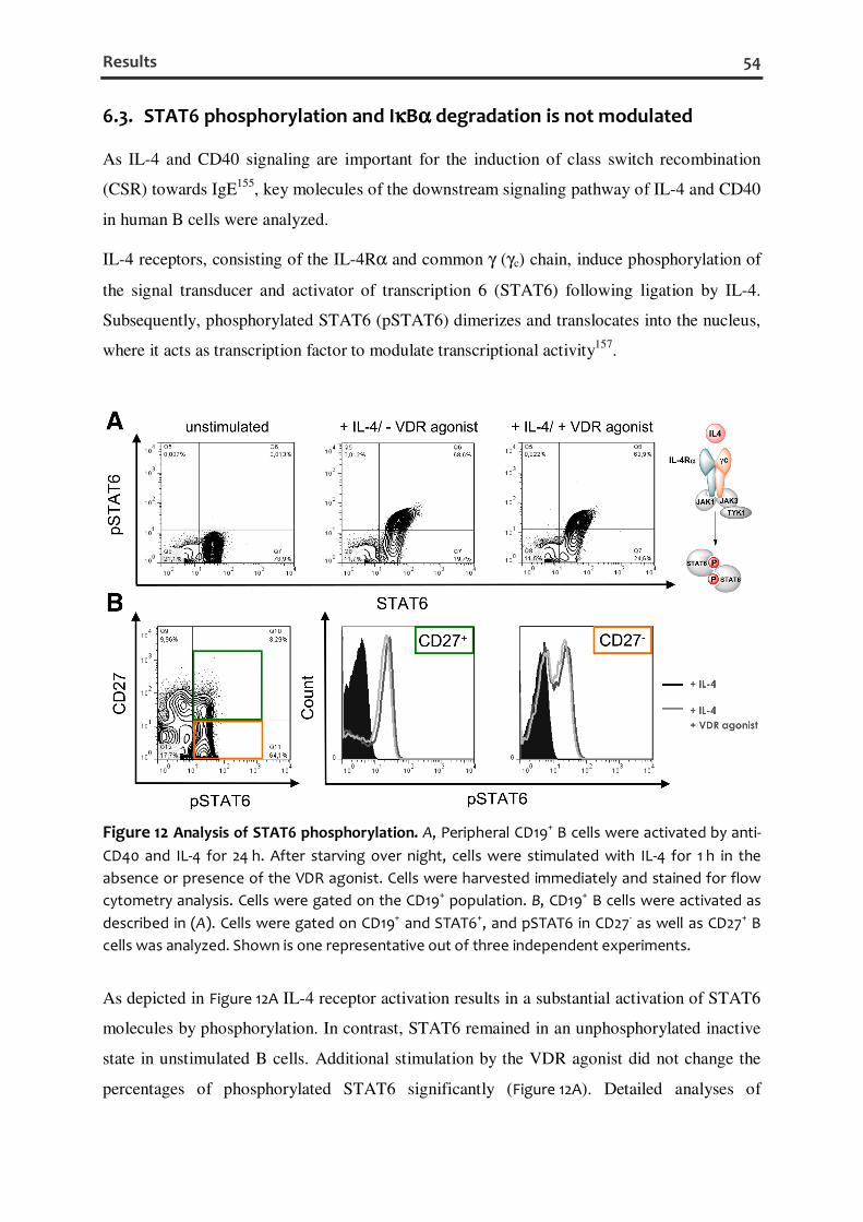
Results 54
6.3. STAT6 phosphorylation and IκκκκBαααα degradation is not modulated
As IL-4 and CD40 signaling are important for the induction of class switch recombination
(CSR) towards IgE155, key molecules of the downstream signaling pathway of IL-4 and CD40
in human B cells were analyzed.
IL-4 receptors, consisting of the IL-4Rα and common γ (γc) chain, induce phosphorylation of
the signal transducer and activator of transcription 6 (STAT6) following ligation by IL-4.
Subsequently, phosphorylated STAT6 (pSTAT6) dimerizes and translocates into the nucleus,
where it acts as transcription factor to modulate transcriptional activity157.
Figure 12 Analysis of STAT6 phosphorylation. A, Peripheral CD19+ B cells were activated by anti-
CD40 and IL-4 for 24 h. After starving over night, cells were stimulated with IL-4 for 1 h in the
absence or presence of the VDR agonist. Cells were harvested immediately and stained for flow
cytometry analysis. Cells were gated on the CD19+ population. B, CD19+ B cells were activated as
described in (A). Cells were gated on CD19+ and STAT6+, and pSTAT6 in CD27- as well as CD27+ B
cells was analyzed. Shown is one representative out of three independent experiments.
As depicted in Figure 12A IL-4 receptor activation results in a substantial activation of STAT6
molecules by phosphorylation. In contrast, STAT6 remained in an unphosphorylated inactive
state in unstimulated B cells. Additional stimulation by the VDR agonist did not change the
percentages of phosphorylated STAT6 significantly (Figure 12A). Detailed analyses of

Results 55
pSTAT6 in CD27- naïve and CD27+ memory B cells did not demonstrate any inhibitory effect
of the VDR agonist on STAT6 phosphorylation in this setting (Figure 12B).
In the next step, CD40-induced degradation of the dominant inhibitor of nuclear factor-κB
(NF-κB), IκBα, which sequesters the inactive p50-p65 NF-κB dimer in the cytoplasm246 was
determined by flow cytometry.
Figure 13 Analysis of IκκκκBαααα degradation. Peripheral CD19+ B cells were activated by anti-CD40 and
IL-4 for 24 h. After additional 60 min preincubation in the absence or presence of the VDR agonist,
cells were stimulated with 1 µg/ml anti-CD40 for 50 min. Cells were harvested immediately and
stained for flow cytometry analysis. Cells were gated on the CD19+ population. The expression of
IκBα in CD27- as well as CD27+ B cells was analyzed. Shown is one representative out of three
independent experiments.
After 24 h stimulation with anti-CD40/IL-4, almost all human CD27- and CD27+ B cells
expressed IκBα in the cytoplasm (Figure 13, upper left), suggesting an inactive state regarding
CD40 signaling. Upon additional stimulation with anti-CD40, cells were strongly activated as
shown by the reduction of IκBα (Figure 13, lower left). More importantly, no impact of the
VDR agonist on IκBα degradation was detected (Figure 13, lower right). Taken together, VDR
activation by the VDR agonist does not influence the downstream signaling of IL-4 and CD40
at the level of STAT6 phosphorylation and IκBα degradation.

Results 56
6.4. Reduction of aicda expression by calcitriol and the VDR agonist
Previously, it has been shown that calcitriol inhibits the expression of the epsilon germline
transcript (εGLT) by direct binding of the VDR on the εGLT promoter and subsequent
recruitment of transrepressing factors144, 145. To answer the question whether inhibition of
other subtypes than IgE by the VDR agonist is due to common mechanisms, which are also
involved in IgE class switch recombination, the expression of aicda was analyzed. Aicda
encodes the enzyme activation-induced cytidine deaminase (AID), which is essential for
somatic hypermutation and class switching towards IgE but also other subtypes155, 175.
The expression of aicda in anti-CD40/IL-4 stimulated CD19+ CD27- naïve B cells was
inhibited by 33.2 ± 3.0% after 24 h and by 39.6 ± 7.6% after 48 h of culture in the presence of
the VDR agonist (Figure 14A). Calcitriol also reduced the expression of aicda in CD19+ CD27-
B cells by 27.9 ± 6.6% after 48 h (Figure 14A). The aicda expression in CD19+ CD27+
memory B cells was inhibited by 29.1 ± 5.7% after 24 h and by 35.7 ± 3.8% after 48 h of
incubation with the VDR agonist and to a comparable extent (28.1 ± 8.3%) with calcitriol
after 48 h (Figure 14B). Together, both calcitriol and the low-calcemic VDR agonist interfere
with the switching process by reducing aicda expression.
Figure 14 Inhibition of aicda expression. A, Human naïve B cells (CD19+ CD27-) were stimulated
with anti-CD40 and IL-4 in the presence or absence of the VDR agonist or calcitriol for 24 h and
48 h. Gene expression of the activation-induced cytidine deaminase (aicda) was assessed by
quantitative PCR. B, Memory B cells (CD19+ CD27+) were analyzed as described in A. The relative
quantification was calculated and is given in % of the anti-CD40/IL-4 stimulated control. The
percentages are shown as mean ± SEM (n ≥ 6, P < 0.05, *).

Results 57
6.5. Reduction of the CD19+ CD27high CD38+ B cell population by activated
VDRs
Having found a suppressive effect on aicda expression, but even more potently, on the
production of distinctive Ig subclasses themselves, the question whether additional events
may also be targets of VDR dependent action was addressed.
Therefore, B cell proliferation was analyzed by CFSE dilution assays. As shown in Figure 15A,
little proliferation of B cells upon anti-CD40/IL-4 stimulation was observed. Application of
the VDR agonist resulted in a slightly reduced frequency of proliferating cells to 10.8 ± 1.0%
compared to 12.4 ± 1.2% (P > 0.05), while the percentages of PI-positive cells remained
stable (Figure 15B).
Figure 15 Impact of the VDR agonist on proliferation and cell viability. A and B, Cells were
labeled with CFSE and stimulated with anti-CD40 and IL-4 in the presence or absence of 100 nM
VDR agonist. After a 5-day-culture, cells were analyzed by flow cytometry and the frequency of
proliferated and PI+ cells was determined. The percentages of proliferated and PI-positive cells
were measured and are shown as mean ± SEM (n = 4).
As IL-21 is known to facilitate IgE production and B cell proliferation in humans158, 159, 247-249,
the effect of VDR activation under highly proliferative conditions through simultaneous
treatment with IL-21 and anti-CD40/IL-4 was delineated. As expected, B cell proliferation
was 4.6-fold more pronounced under these conditions compared to the stimulation with anti-
CD40/IL-4 (Figure 15), as determined by increased CFSE dilution corresponding to a higher
number of proliferating cells (Figure 16A and B). The most pronounced induction of
proliferation by IL-21 was observed in the naïve B cell compartment (Figure 16A). This may
result from the higher expression level of IL-21R on naive, compared to memory, B cells249.
Notably, in B cells treated with the VDR agonist, a slight, but not significant antiproliferative
effect of 16.8 ± 7.4% (100 nM) was observed under these conditions (Figure 16B). A minor,

Results 58
but also slight inhibitory effect (9.5 ± 8.0%) on B cell proliferation was observed for calcitriol
(Figure 16B).
Figure 16 VDR agonist signaling inhibits the generation of CD27high cells. A, Peripheral human
CD19+ B cells were cultured with anti-CD40/IL-4/IL-21 in the absence or presence of the VDR
agonist and calcitriol and analyzed after 5 d by flow cytometry. Shown is one representative
density plot. B, The percentages of proliferated CD19+ B cells are shown as mean ± SEM (n = 4). C,
The frequency of CD19+ CD27high B cells is shown as mean ± SEM (n = 7; P < 0.05, *).
Additionally, IL-21 is known to promote the differentiation of human B cells into
immunoglobulin-secreting cells (ISCs)248, 250 which are characterized as CD27high at their
transitional stage to ISCs250, 251. Interestingly, by stimulation with anti-CD40/IL-4 and IL-21,
a CD27high population comprising 6.1 ± 1.5% of CD19+ B cells at d 5, appeared
approximately after three division cycles (Figure 16C). Additional data show that if CD27high
cells were depleted by FACSsort before stimulation under equal conditions, a newly formed
CD27high subset was induced (data not shown). These data suggest an induced formation of
the CD27high cells by stimulation rather than a maintenance or expansion of preexisting cells.
More importantly, additional activation by the VDR agonist diminished the generation of
CD27high cells dose-dependently, with a maximum inhibition of 51.1 ± 6.3%. The natural
ligand calcitriol inhibited the CD27high population by 64.6 ± 3.3%.
In vitro generated human Ig-producing blasts or ISCs, however, are also characterized by the
surface expression of CD38252, 253. Therefore, CD19+ B cells were stimulated with anti-
CD40/IL-4 and IL-21 to induce CD27high CD38+ cells, and the impact of VDR activation was
examined.

Results 59
Figure 17 VDR agonist signaling reduces the frequency of CD27high CD38+ B cells while increasing
the expression of CD38. A, Human peripheral CD19+ B cells were stimulated with anti-CD40/IL-4
and IL-21 in the presence or absence of increasing amounts of the VDR agonist and calcitriol. After
a 5-day-culture, cells were analyzed by flow cytometry. B, Representative percentages of CD19+,
PI-, CD27high, CD38+ B cells are shown. C, The MFI of CD38, gated on CD19+, PI-, CD27high cells, was
determined. Shown is one representative out of 3 experiments. MFI = median fluorescence
intensity.
In consequence of stimulation with anti-CD40/IL-4 and IL-21, about 8.75% CD27high CD38+
cells were induced, shown by one representative experiment (Figure 17A and B). Upon addition
of the VDR agonist and calcitriol, this population was strongly reduced by 46.6% and 32.8%.
In contrast, surface expression of the VDR target gene CD3847, 245 on these cells was
increased upon treatment with the VDR agonist or calcitriol, as determined by a 2-fold
increase in the median fluorescence intensity (MFI) (Figure 17C). In summary, VDR activation
profoundly interferes with the generation of ISCs in vitro as shown by the reduced frequency
of CD27high CD38+ cells. These results correlate with the reduced Ig secretion and numbers of
Ig secreting cells determined by ELISA and ELISPOT.
6.6. VDR activation reduces the IgE response in a type I allergy mouse
model without calcemic side effects
Based on the potency to counteract the IgE production in vitro, the impact of the low-
calcemic VDR agonist on the allergic immune response in vivo was delineated. Mice were

Results 60
sensitized and treated as depicted in Figure 6. The low-calcemic nature of the VDR agonist
was confirmed by measurement of serum calcium concentrations on day 47.
Figure 18 The VDR agonist is well tolerated in vivo. A, Ca2+-levels in pooled sera from sensitized
and non-sensitized BALB/c mice on d 47, treated with the VDR agonist or vehicle control. B,
Weight gain over time of VDR agonist-treated mice compared to vehicle-treated control groups.
Data represent means ± SEM of 8 mice per group.
Both, the VDR agonist and PBS treated groups showed comparable serum Ca2+-levels,
confirming the low-calcemic character of the VDR agonist (Figure 18A). In addition, the mice
remained healthy and displayed a comparable development of weight gain throughout the
experiment (Figure 18B). As depicted in Figure 19 the therapeutic treatment regimen led to a
profound inhibition of the allergen-specific IgE response by 46.7% (P = 0.03; Figure 19A,
right). As expected, no allergen-specific Ig production was detected in non-sensitized control
mice (data not shown). Total IgE levels were also reduced by 35.5% (P = 0.05; Figure 19A,
left). Although the effect on the IgE level was most distinctive, an inhibition of the allergen-
specific IgG1 levels by 20.7% (P = 0.04; Figure 19B) was also observed. A similar tendency as
for IgG1 was found for IgG2a and IgA, but overall levels of these isotypes were barely
detectable and variable (data not shown). To investigate whether the suppressing effects on
IgE production were accompanied by alterations in the B cell compartment, the composition
of splenic B cells was assessed. As shown in Figure 19C, the B cell compartment consisted of
5.3 ± 0.2% marginal zone B cells, expressing high levels of CD21 but no CD23, and
80.9 ± 0.9% of follicular B cells, expressing CD21 and CD23. This pattern did not change
upon VDR activation, demonstrating that the suppression of IgE production is not the result of
overt changes in B cell subpopulations, further emphasizing the specificity of the Ig reducing
effect.

Results 61
Figure 19 The VDR agonist reduces total and allergen-specific IgE in a mouse model of type I
allergy. A and B, Total IgE (A, left), allergen-specific IgE (A, right) and IgG1 levels (B) in sera of
treated mice on d 47 were determined by ELISA. C, The splenic B cell compartment gated on living
CD19+ B cells was analyzed by flow cytometry. Follicular (FO, CD21mid, CD23+) and marginal zone
(MZ, CD21hi, CD23-) B cells were identified by different expression levels of CD21 and CD23. Bars in
the scatter plot represent medians (n ≥ 7), triangles represent single values (P < 0.05, *).
6.7. VDR agonist treatment ameliorates allergen-triggered skin eczema in
mice
Although vitamin D3 and its metabolites are known for their substantial impact on the
immune system39, 254 and the skin differentiation and function209, the effects of VDR ligands
on atopic dermatitis (AD) have not been studied in detail. Using the VDR agonist, which was
proven low-calcemic but nevertheless exhibiting strong immunomodulatory effects, the role
of VDR signaling in a defined model of allergen-driven dermatitis (Figure 7) was investigated.
In accordance with previous results234, a strong induction of the eczema by epicutaneous
application of OVA compared to PBS as reflected by the increased clinical skin score (median
OVA-patched mice: 10.0, range: 8.0 − 12.0; median PBS-patched mice: 4.25, range:
1.5 − 6.5; P = 0.0003) was observed (Figure 20A). The clinical skin score of 4.25, induced by

Results 62
the patch itself in the absence of the allergen (antigen), corresponds to an allergen-
independent, mild dermatitis.
Figure 20 The VDR agonist ameliorates allergen-triggered skin eczema in mice. A, Mice were
sensitized with OVA/alum and challenged with either OVA/alum (OVA-Patch) or PBS/alum (PBS-
Patch) locally, according to the scheme in Figure 7. B, Mice were sensitized with OVA/alum as
described above, followed by treatment with VDR agonist or vehicle control, according to the
method section (see also Figure 7). On day 71 mice were sacrificed and the severity of the eczema
was evaluated as described in methods. The index of severity (clinical skin score) is shown as the
median for each group (n ≥ 9; P < 0.05 *, P < 0.001 ***). One representative picture of each group
is shown.
To delineate the effect of the VDR agonist on the allergen-induced eczema, following results
are shown from OVA-sensitized mice treated with either the VDR agonist or a vehicle
control. VDR activation by the VDR agonist led to a clear improvement of all signs and
symptoms of the OVA-induced eczema, including erythema, edema, excoriation, dryness and
extension, as evidenced by a reduced clinical skin score (median VDR agonist-treated mice:
7.5, range: 4.0 − 10.5; median vehicle-treated mice: 10.0, range: 8.0 − 12.0; P = 0.0195)
(Figure 20B). Taken together, the data clearly show that the low-calcemic VDR agonist
significantly modulates the local immune response as shown by improvement of the allergen-
induced eczema.

Results 63
6.8. VDR activation does not change the numbers of infiltrating T cells in
lesional skin
Immunohistochemistry was employed to analyze the relative distribution of different T cell
subsets in lesional skin. As shown in Figure 21A, there was no significant difference between
VDR agonist-treated and control mice in terms of total CD4+ T cells. Nevertheless, a
tendency of higher numbers of CD4+ T cells (1.8-fold increase compared to vehicle-treated
group) was observed in the VDR agonist-treated group (median VDR agonist-treated mice:
612.8, range: 369.8 − 740.5; median vehicle-treated mice: 347.9, range: 128.0 − 764.5;
P = 0.1628). In line with previous results234, CD8+ T cells were strongly induced upon
epicutaneous sensitization with OVA (P = 0.0068; data not shown) homing into the dermis
and epidermis. There was a slight tendency of CD8+ T cells to decrease in the skin of VDR
agonist-treated mice, which did not reach statistical significance (Figure 21B).
Figure 21 CD4+ and CD8+ cells in skin lesions of OVA-sensitized and VDR ligand treated mice.
CD4+ (A) and CD8+ T cells (B) were stained and quantified in the dermis of lesional skin of VDR
agonist-treated and untreated mice. Numbers of cells per mm2 are shown as the median for each
group (n ≥ 9). One representative photograph of each group is shown; n.s.: not significant.
6.9. Increased numbers of Foxp3+ cells in skin lesions of VDR agonist-
treated mice
As VDR agonist treatment was associated with only minor changes in the skin infiltrating
CD4+ and CD8+ T cells, the question for other mechanisms and changes in defined cell types

Results 64
that might contribute to the amelioration of the dermatitis was asked. As Foxp3+ Treg cells are
known to inhibit both TH1 and TH2 cells by reducing their cytokine production255, and due to
its crucial role in the maintenance of immune homeostasis in normal skin256, the number of
Foxp3+ cells in lesional skin was analyzed.
Figure 22 The VDR agonist increases the number of Foxp3+ cells in lesional skin. A, Foxp3+ cells
were stained and quantified in the dermis of lesional skin of VDR agonist-treated and untreated
mice. Numbers of cells per mm2 are shown as the median for each group (n ≥ 8; P < 0.05 *). One
representative photograph of each group is shown (arrows indicate Foxp3+ cells). Expression
levels of Foxp3 (B), Il4 (C) and Ifng (D) in lesional skin of each group were assessed by quantitative
PCR. Shown is the relative expression of the target gene compared to the housekeeping gene
Hprt in arbitrary units (AU) (n ≥ 8; P < 0.05 *).
Indeed, there was a clear-cut increase in the number of Foxp3+ cells by 60.0% upon
therapeutic treatment with the VDR agonist (median VDR agonist-treated mice: 211.9, range:
126.0 − 232.3; median vehicle-treated mice: 132.0, range: 91.1 − 226.9; P = 0.026)
(Figure 22A) which might explain the 1.8-fold increase in infiltrating CD4+ T cells (Figure 21A).
This observation is also supported by a significantly 2.5-fold increased expression of Foxp3 in

Results 65
the skin of VDR agonist-treated mice (median VDR agonist-treated mice: 2.61, range:
1.17 − 4.22; median vehicle-treated mice: 0.94, range: 0.42 − 3.13; P = 0.0148) (Figure 22B).
To delineate possible functions of the Foxp3+ cells in the lesional skin the expression of the
TH1 and TH2 signature cytokines IFNγ and IL-4, known to be involved in skin
inflammation232, was examined. The VDR agonist strongly reduced Il4 expression in lesional
skin by 44.0% (median VDR agonist-treated mice: 0.54, range: 0.34 − 1.05; median vehicle-
treated mice: 0.97, range: 0.53 − 1.35; P = 0.0435) (Figure 22C). Additionally, Ifng expression
was reduced by 29.4%, but this did not reach statistical significance (median VDR agonist-
treated mice: 0.12, range: 0.05 − 0.24; median vehicle-treated mice: 0.17, range: 0.11 − 0.50;
P = 0.0749) (Figure 22D). The data show that the selective increase in Foxp3+ cells and Foxp3
expression may have contributed to reduce TH1 and TH2 cytokine expression in lesional skin
and subsequent improvement of the eczema.
6.10. Modulation of cytokine and chemokine expression in lesional skin
Having found only minor differences in CD8+ T cell numbers in lesional skin, whereas CD4+
T cells and Foxp3+ cells were increased, chemokines attracting skin-homing CD4+ T cells, in
particular CCL22 and CCL27257-260 were analyzed. Ccl22 expression (MDC, macrophage-
derived chemokine), encoding the ligand for the chemokine receptor CCR4, was 1.8-fold
increased in the VDR agonist-treated mice (median VDR agonist-treated mice: 0.28, range:
0.15 − 0.36; median vehicle-treated mice: 0.15, range: 0.07 − 0.39; P = 0.2523) but this did
not reach statistical significance (Figure 23A). The expression of Ccl27, the ligand for the skin-
homing receptor CCR10, was 2.1-fold increased in VDR agonist-treated mice compared to
vehicle-treated mice (median VDR agonist-treated mice: 0.75, range: 0.4 − 1.01; median
vehicle-treated mice: 0.37, range: 0.21 − 0.49; P = 0.0012) (Figure 23B).
As TSLP is known to promote TH2 cell responses198, 199 and is induced by topical VDR
activation261, its expression in lesion skin was assessed. A 2.4-fold induction in Tslp
expression was observed upon VDR agonist treatment (median VDR agonist-treated mice:
1.71, range: 1.13 − 2.54; median vehicle-treated mice: 0.71, range: 0.52 − 1.51; P = 0.0016)
(Figure 23C). In addition, the expression of the proinflammatory cytokine TNFα, involved in
AD as well as psoriasis190, 262, was analyzed in lesional skin of VDR agonist-treated and
vehicle-treated mice. Tnfa expression was 2.9-fold increased in skin samples of VDR agonist-
treated compared to control mice (median VDR agonist-treated mice: 0.004, range:

Results 66
0.001 − 0.008; median vehicle-treated mice: 0.001, range: 0.0002 − 0.004; P = 0.0079)
(Figure 23D).
Figure 23 Systemically applied VDR agonist modulates cytokine and chemokine expression in
lesional skin. Expression levels of cytokines and chemokines in lesional skin were analyzed by
quantitative PCR. The relative expression of CCL22 (Ccl22, A), CCL27 (Ccl27, B), TSLP (Tslp, C), and
TNFα (Tnfa, D) was measured in comparison to the housekeeping gene Hprt and is shown as the
median for each group (n ≥ 7; P < 0.01 **).
6.11. VDR agonist treatment induces barrier and antimicrobial peptide gene
expression
The VDR and its ligands are described to induce several genes important for skin barrier
function and integrity209. Disruption of the skin barrier is involved in the development of
allergic skin inflammation, such as AD194. Finally, the impact of the VDR agonist on barrier
and antimicrobial peptide gene expression in lesional skin was determined. Therapeutic
treatment with the VDR agonist robustly induced the expression of the skin barrier genes
loricrin (Lor), involucrin (Ivl), transglutaminase 1 (Tgm1) and filaggrin (Flg), and the
antimicrobial peptides β-defensin 2 (Defb2) and β-defensin 3 (Defb3) (Figure 24).

Results 67
Figure 24 Systemic VDR agonist application induces barrier and antimicrobial peptide genes in
lesional skin. Expression levels of skin barrier genes and antimicrobial peptides in lesional skin
were analyzed by quantitative PCR. The relative expression of loricrin (Lor, A), involucrin (Ivl, B),
transglutaminase 1 (Tgm1, C), filaggrin (Flg, D), β-defensin 2 (Defb2, E) and β-defensin 3 (Defb3, F)
was measured in comparison to the housekeeping gene Hprt and is shown as the median for each
group (n ≥ 8; P < 0.05 *, P < 0.001 ***).
Thereby, the highest induction in gene expression of 7.2-fold was examined for loricrin
(P = 0.0003) and transglutaminase 1 (5-fold, P < 0.0001) (Figure 24A and C). Filaggrin
(Figure 24D) and involucrin (Figure 24B) were 4.0-fold (P < 0.0001) and 4.5-fold (P < 0.0001)
induced by the VDR agonist in lesional skin. The expression of β-defensin 2 was 2.0-fold
(P = 0.0006, Figure 24E) and β-defensin 3 0.5-fold (P = 0.0464, Figure 24F) increased by the
VDR agonist compared to the vehicle control while the overall expression of these
antimicrobial peptides was barely detectable.
Taken together, these data show that VDR activation induces the skin barrier and
antimicrobial peptide gene expression in allergen-induced AD-like skin lesions.

Discussion 68
7. Discussion
7.1. Targeting the VDR inhibits the humoral immune response,
preferentially IgE, in vitro and in vivo
IgE antibodies, normally present at low levels in the plasma, are the key effector molecules of
type I allergic diseases155. Epidemiological data suggest that vitamin D insufficiency, as
defined by less than 30 ng/ml of the circulating precursor 25-hydroxyvitamin D3, is associated
with elevated IgE levels176. Other reports show a beneficial association between higher
maternal vitamin D intake during pregnancy and protection from childhood asthma and
allergic diseases in the offspring180-182. In addition, genetic variants at the vitamin D receptor
(VDR) locus but also at a number of genes involved in the vitamin D pathway have been
shown to be associated with asthma and atopy263, 264. In VDR knockout mice, total and
antigen-specific IgE serum levels are increased178. Taken together, these studies clearly
support a potential and probably beneficial role of VDR signaling in IgE dependent diseases.
Therefore, the VDR might be a promising target for the prevention and treatment of allergic
diseases.
The active metabolite of vitamin D, calcitriol, mediates anti-inflammatory functions on
adaptive immune responses including IgE synthesis39, 40, 144, 254, but its use is limited by
hypercalcemic side effects in pharmacological dosages resulting in significant toxicity when
systemically applied265, 266. Consequently, synthetic analogs with reduced hypercalcemic
activity were developed for systemic application. The actions of vitamin D analogs with
regard to selective immunomodulatory versus hypercalcemic effects are determined by their
interactions with the nuclear VDR, the serum vitamin D binding protein (DBP), the 25-
hydroxyvitamin D3-24-hydroxylase (CYP24A1) and perhaps other metabolizing enzymes, the
membrane receptor(s) (MARRS) that mediate the rapid actions of vitamin D compounds, and
intracellular vitamin D binding proteins that can facilitate both metabolism and VDR
activation267, 268.
The VDR agonist used in the present study has been recently shown to exert
immunomodulatory properties in vitro and in vivo226, 269
. The advantage of the VDR agonist in
comparison to calcitriol is its dissociated profile (immunomodulatory vs. calcemic)226. Thus,
the VDR agonist being immunomodulatory while not being hypercalcemic suggests a
potential therapeutic window226.

Discussion 69
In line with previous reports, the expression of the VDR in human peripheral B cells was
induced by stimulation with anti-CD40/IL-4144. More recent results show that the binding
affinity of the used VDR agonist to the VDR is in the same range as for calcitriol226.
The results of the present work show that the VDR agonist activates the VDR in naïve and
memory human B cells, reflected by cyp24a1 gene expression and the induction of CD38 on
the surface of CD19+ B cells, analogous to calcitriol110, 245.
To induce IgE synthesis in human peripheral B cells, monoclonal anti-CD40 antibodies and
recombinant human IL-4 were used270. Furthermore, these stimuli are known to induce the
switch to IgG in human B cells271, resulting in IgG production270, 272, 273. The present results
clearly demonstrate that the VDR agonist strongly inhibits the anti-CD40/IL-4 induced IgE
production dose-dependently, as shown by reduced secreted IgE and a diminished number of
IgE secreting B cells. The efficacy of the VDR agonist to inhibit the IgE response in vitro was
comparable to that of the natural ligand calcitriol which is in line with a previous report
showing the inhibitory effect of calcitriol on the IgE response in vitro144. In contrast, the use
of a carboxylic ester VDR antagonist, binding to the VDR with a potency similar to that of
calcitriol274, showed no impact on stimulated B cell responses. This observation is in
accordance with previous reports showing a selective stabilization of an antagonistic
conformation of the ligand-binding domain (LBD) of the VDR within VDR-RXR-VDRE
complexes by disturbing the helix 12 position275, which in turn inhibits the interaction of the
VDR with coactivator proteins and an induction of transactivation227, 276. In conclusion, these
data suggest that direct involvement of the liganded VDR and its downstream signaling events
are responsible for the observed inhibition of IgE production.
Nevertheless, besides IgE, IgG and to a lesser extent IgA were also reduced following VDR
activation. IgA which is considered rather protective in allergic diseases277, 278 was far less
affected in the context of VDR stimulation than IgE. A skewing effect on isotype switching
away from IgE towards IgA must have occurred. In fact, the IgA/IgE ratio increased upon
VDR activation from 1.0 to 2.2 in human B cells. In comparison to IgA, IgM was as strongly
induced as IgG by this kind of stimulation but lacked a statistically significant inhibition upon
treatment with the VDR agonist in the same experimental setup.
STAT6-mediated IL-4 and NF-κB-mediated CD40 signaling synergize to induce IgE
switching and production167, 171, 270. Therefore, critical downstream events were analyzed by
applying flow cytometry. The results indicate that the VDR agonist does not have an impact
on either STAT6 phosphorylation or IκBα degradation. These data were also confirmed by
the observation of the stable IL-4 and CD40-dependent expression of the surface markers

Discussion 70
CD23 (FcεRII) and CD69279-281 upon VDR agonist treatment in the initial phase of B cell
activation (data not shown).
Therefore, the question whether other downstream events of anti-CD40/IL-4 signaling,
necessary for class switch recombination and terminal differentiation, are impaired by the
VDR agonist was addressed. A previous report has shown that VDR activation by calcitriol
inhibits the expression of the εGLT144. These data were confirmed by a subsequent report
showing direct binding of the liganded VDR to the εGL promoter, exhibiting transrepressive
activity145. However, the assessment of the εGLT expression is only suitable to a limited
extent to evaluate IgE production caused by the fact that although IL-4 induces the
transcription of germline ε mRNA by CD40-stimulated human B cells, two-thirds of these B
cells fail to produce IgE282.
Thus, more general mechanisms involved in class switch recombination and differentiation
were analyzed. At the molecular level, an inhibition of aicda expression, encoding the
activation-induced cytidine deaminase (AID), which is indispensable for somatic
hypermutation and class switching towards IgE but also other subtypes155, 175, was observed in
both B cell subsets. AID deficiency causes the autosomal recessive form of the Hyper-IgM
syndrome (HIGM2) which is characterized by normal or elevated serum IgM levels in the
absence of IgG, IgA, and IgE174. It is likely that the diminished expression of aicda may
contribute to the decreased immunoglobulin production of all isotypes other than IgM and
IgD, as determined here.
As B cell proliferation is another key factor associated with isotype class switching as well as
immunoglobulin secretion253, 272 the impact of VDR activation on proliferation was
investigated in more detail. The proliferation of anti-CD40/IL-4 activated human B cells with
or without additional mitogenic IL-21 was altered only to a minor extent in the presence of
the VDR agonist, which did not reach statistical significance. Nevertheless, such slight
antiproliferative effects of the VDR agonist may contribute to the observed reduction in Ig
production as well. In fact, a strong link between differentiation of human B cells to IgE-
secreting cells and the degree of cell division has been described previously159. However, this
antiproliferative effect cannot fully explain the remarkable inhibition of immunoglobulin E
secretion by 70.1 ± 6.0% and the reduction of other isotypes like IgG and IgA.
Due to the strong reduction in the number of immunoglobulin-secreting cells (ISCs), the
differentiation of CD27high CD38+ B cells which define in vitro generated ISCs that express a
transcriptional profile comparable to plasma cells251, 253 was analyzed. In fact, diminished

Discussion 71
frequencies of CD27high CD38+ B cells were detected, although CD38 expression, a well-
known target of VDR activation47, 247, was increased on the CD27high subset, suggesting a
recent VDR activation in these cells. The clear reduction of this cell population may
contribute to the reduced number of Ig secreting cells resulting in a reduced Ig secretion of
activated B cells. Such reduction of plasmablasts is in accordance with previous data using
calcitriol46. Taken together, two potential mechanisms by which the VDR agonist may inhibit
the IgE response should be taken into consideration, first the interference with the switching
machinery and second a reduction of the differentiation into Ig secreting cells. Unravelling the
mechanistic details in their entire complexity and the determination of their relative
contribution upon VDR activation will require future experiments. First, the contribution of
AID expression to the inhibition of class switching to IgE and other subtypes should be
confirmed at the protein level, e.g., by western blotting or intracellular flow cytometry.
Furthermore, it has been shown that VDR signaling most likely plays a role in controlling
proliferation, supporting the potential role of calcitriol and analogs as preventative and
therapeutic anticancer agents95. Cheng and colleagues have shown that calcitriol can increase
the expression of p27, a cyclin-dependent kinase (CDK) inhibitor, while suppressing cyclin
D1, cyclin D2, cyclin T1, cyclin T2, CDK4, and CDK6 in human B cells46. Although different
stimuli were used in the present work, an effect on cell cycle progression by VDR agonist
signaling cannot be excluded. Therefore, the impact of the VDR agonist on the cell cycle
progression should be examined by flow cytometric cell cycle analysis and cell cycle-related
gene expression. In addition, the impact of VDR signaling on terminal differentiation of B
cells into plasma cells, as shown in the present work, should be determined in more detail by
gene expression analysis of the key transcription factors X-box-binding protein 1 (XBP1),
paired box protein 5 (PAX5), B cell lymphoma 6 (BCL6), B lymphocyte-induced maturation
protein 1 (BLIMP1), and interferon-regulatory factor 4 (IRF4). Recent data indicate that the
calcitriol-mediated inhibition of IgE production can only be achieved by activating VDRs at
the beginning of the B cell culture144. This suggests that there might also be a modulation of
early cellular responses, e.g. the activation of the mitogen-activated protein kinase (MAPK)-
extracellular signal-regulated kinase (ERK) cascade, which may engage in cross talk with the
classical VDR pathway to modulate gene expression or directly affect various signaling
molecules.

Discussion 72
Based on these in vitro observations, the immune modulation by the low-calcemic VDR
agonist in a murine model of type I sensitization was assessed. As IgE, normally present at
low levels in the plasma, is elevated in type I allergic diseases, BALB/c as an IgE high
responder mouse strain was used in this study. BALB/c mice are known as a TH2-biased
mouse strain. The susceptibility to develop a TH2-biased immune response is caused by
different gene loci, one of which is the MYC-induced nuclear antigen (Mina) locus on
chromosome 16283. Mina, which binds to the Il4 promoter in a NFAT-dependent fashion, is
expressed at lower levels in BALB/c mice, which leads to a higher IL-4 production
influencing the balance between TH1 and TH2.
The serum calcium concentration as well as the weight gain over time in VDR agonist-treated
mice remained stable. Thus, hypercalcemic or toxic side effects were excluded, clearly
supporting the advantage of this low-calcemic VDR agonist in pharmacological doses in
accordance with previous data226. In contrast, numerous previous studies have shown calcium
mobilizing side effects upon calcitriol treatment when it was used at immunomodulatory
dosages. For example, Osborn et al. observed hypercalcemia (grade I) as the most common
side effect of oral treatment with calcitriol284. They used a daily dose ranging from 0.5 µg to
1.5 µg calcitriol. Calculated to a normal average body weight of 70 kg, they used between
0.007 µg/kg/d and 0.02 µg/kg/d in their study. In the present study 6 µg/kg/d of the VDR
agonist was used. This is a 280-fold excess over the dose of calcitriol used by Osborn and
colleagues. Boehm et al. showed that a 5-day treatment of 5 µg/kg calcitriol induces
significant changes of the serum Ca2+-level in BALB/c mice285. As mentioned above, two 5-
day treatments with the VDR agonist (6 µg/kg/d) were performed in the present work. The
10% toxic dose (TD10, 10% loss of bodyweight) for calcitriol in SJL mice was determined as
0.1 µg/kg/d286. In addition, these authors showed that the maximal dose of calcitriol that could
be administered intraperitoneally during 7 consecutive days in NMRI mice, without inducing
hypercalcemia, was 0.1 µg/kg/d286. Thus, a 60-fold lower amount of the VDR ligand was used
here. Furthermore, other studies also show hypercalcemic effects by the administration of
calcitriol above 8 µg (0.11 µg/kg, s.c.)287. Using a model of rodent contact hypersensitivity
(CHS) other studies showed that a therapeutic efficacy of calcitriol occurs at 0.1 µg/kg/d only,
i.e. when calcium excretion is already enhanced226, 288. In contrast, the VDR agonist exerted
full therapeutic efficacy at doses up to 60 µg/kg/d, and even these high doses did not cause
hypercalciuria226. Thus, a 60-fold excess of the VDR agonist over the immunomodulatory
dose of calcitriol that causes hypercalcemic side effects was applied by using 6 µg/kg/d.

Discussion 73
In summary, these data evidently indicate the limitation of the systemic treatment with
calcitriol in vivo. Thus, the great benefit of the VDR agonist used in this study is its
dissociated character, clearly separating the calcemic from its immunomodulatory properties.
Importantly, a strong VDR agonist-mediated reduction of the antigen-specific IgE response
and to a lesser degree total serum IgE production was observed. Moreover, a reduction of the
antigen-specific IgG1 response was determined, although the extent of suppression was 2.3-
fold more pronounced on specific IgE than IgG1, suggesting a predominance for specific IgE.
IgG, however, can also induce the alternative pathway of systemic anaphylaxis in mice and
there is some evidence this may also occur in humans, at least when the antigen is available in
abundant quantities289. Besides anaphylaxis, IgG also plays a role in type II hypersensitivity
triggering a variety of autoimmune diseases such as blistering skin diseases like pemphigus
vulgaris or bullous pemphigoid, autoimmune hemolytic anemia, autoimmune-mediated
chronic urticaria and certain types of drug allergy290. Therefore, IgG may contribute to
allergic symptoms, at least under certain conditions as well. In addition, the molecular
mechanisms that govern murine IgE and IgG1 production are, in fact, quite similar228, 291.
Hence, a partially coordinated VDR agonist-induced change in the levels of these two
isotypes in vivo is not surprising – and it does in no way imply a generalized immune
suppression.
There are only a few reports that directly addressed the role of VDR signaling for allergic
immune responses in vivo, but these support the notion that VDR triggering possesses anti-
allergic effects. Accordingly, type I sensitized VDR knockout mice have higher serum IgE
concentrations than wild type controls in an allergic asthma model178, suggesting an important
role for VDR signaling in regulating IgE responses in vivo. Additionally, allergen-specific
immunotherapy has been suggested to be more efficient upon calcitriol treatment179.
However, so far no data are available on such a concept in human allergic patients.
In conclusion, these data provide evidence that VDR activation by a low-calcemic agonist
modulates the humoral immune response including IgE. Thus, targeting vitamin D receptors
by low-calcemic ligands may be a promising strategy for novel therapeutic approaches.
However, VDR expression and subsequent VDR-mediated immunomodulatory effects are not
limited to B cells, but also relevant in T cells, DCs and myeloid cells. Hence, to carefully
dissect the in vivo effects of VDR activation on the immune response it will be necessary to
use animal models in which VDR deficiency is restricted to T cells, B cells and myeloid cells.

Discussion 74
7.2. VDR activation ameliorates allergen-induced skin eczema
VDR signaling has been shown to be important not only in the immune system, but also in the
skin and in particular keratinocytes209, 254. Atopic dermatitis (AD) is a common chronic
inflammatory skin disease caused by a combination of epidermal barrier dysfunction and
immune dysregulation190, 194. Therefore, the effect of VDR activation by the low-calcemic
VDR agonist on allergen-induced eczema was delineated in a mouse model of OVA-induced
skin inflammation, mimicking AD in humans. Importantly, AD and psoriasis, another
inflammatory skin disease, are both skin disorders sharing many similarities. They are
common, chronic, inflammatory, and proliferative skin disorders in which both genetic and
environmental factors have an important pathogenetic role190, 192, 262, 292, 293. In both diseases T
cells might play an important role as well. Psoriasis is primarily characterized by TH1-
mediated responses but TH17 cells have also been implicated in the pathogenesis of psoriasis.
In AD, a biphasic T cell response, marked by an acute phase with a TH2-dominated milieu
and a chronic phase with TH1-mediated responses have been described190. Altough the
presence of TH17 cells in AD skin lesions is less prominent than in psoriasis, they might
contribute to skin inflammation in AD patients294, 295. An additional overlap in both disorders
is the impairment of the skin barrier formation193, 296. Moreover, topically applied calcitriol
and analogs are known to be successful therapeutic approaches in psoriasis267, 297. Thus, it is
likely that VDR activation by systemic application of low-calcemic agonists may also be of
potential benefit for the treatment of AD. To analyze potential modulatory effects on AD, the
immunmodulatory, low-calcemic VDR agonist was therapeutically applied in a mouse model
of allergen-induced eczema.
Indeed, there was a clear-cut improvement of the symptom severity of AD-like skin lesions,
indicated by a decreased clinical skin score. This improvement of the clinical skin score was
associated with a decreased transepidermal water loss (TEWL) (298 and own unpublished
data), one critical hallmark to determine barrier function. T cell receptor (TCR)αβ+ T cells are
critical for skin inflammation and TH2 responses in AD299. Furthermore, a role for epidermal
CD8+ T cells in the pathogenesis of atopic eczema has been suggested300, 301. CD8+ T cell-
depleted mice show no skin inflammation upon allergen exposure in a mouse model of
allergen-induced AD, indicating a crucial role of CD8+ cells in the development of AD302. In
accordance with previous results234, an increase in skin-infiltrating CD8+ T cells by
epicutaneous OVA challenge was observed (data not shown), which only slightly decreased
upon therapeutic VDR agonist treatment. However, these data suggest that the slightly

Discussion 75
decreased numbers of CD8+ T cells are most likely not the major mechanism resulting in the
VDR agonist-mediated improvement of the allergen-induced eczema in this mouse model.
Nevertheless, a contribution to the decreased skin inflammation by a direct or indirect
suppressive effect of VDR activation on CD8+ T cells cannot be completely excluded.
Dermal CD4+ T cell infiltration is known to be involved in skin lesion development196. In
contrast, the present data show a minor increase of infiltrating CD4+ T cells simultaneously
with a clear-cut improvement of the symptom severity upon VDR agonist treatment. CD4+ T
cells consist of several subsets of T helper (TH) cells modulating the immune response303, 304.
Natural CD4+ CD25+ Foxp3+ Treg cells, comprising 5 – 10% and 1 – 2% of the CD4+ T cell
pool in mice and humans142, and other subtypes of induced Treg cells are known to inhibit both
TH1 and TH2 cells by reducing their cytokine production or by direct interaction with effector
T cells255, 305, 306. Importantly, they also have a crucial role in the maintenance of immune
homeostasis in normal skin256. Thus, it is likely that the minor increase of CD4+ T cells is
reflected by the increased number of Foxp3+ Treg cells, observed in the lesional skin of VDR
agonist-treated mice.
Recent work has shown the importance of the forkhead family transcription factor Foxp3 for
the development and function of Treg cells. Scurfy mice, carrying a recessive mutation that
involves a 2-bp insertion in the Foxp3 gene307, develop an intense multiorgan inflammatory
response associated with allergic airway inflammation, a striking hyper-
immunoglobulinemia E, eosinophilia, and dysregulated TH1 and TH2 cytokine production as
well as scaly and inflamed skin resulting in eczema308. Moreover, the number of natural Tregs
is decreased in lesional skin of patients with AD or psoriasis309. Another study, using mixed
CCR4−/Foxp3− bone marrow chimeras, showed that mice lacking Foxp3+ T cells in the skin
spontaneously develop a cutaneous inflammatory disease with red, scaly skin, hyperkeratosis,
lymphocytic and eosinophilic infiltration, and elevated local levels of IL-4310. Thus, the
increased number of Foxp3+ cells upon VDR agonist treatment might contribute to the
amelioration of AD-like skin lesions by the reduction of Il4 and Ifng expression and/or
inhibiting CD8+ T cell priming and activation in the skin. Interestingly, VDR knockout mice
fail to increase Foxp3+ Tregs in skin-draining lymph nodes (SDLN) following UV-
irradiation142. Additionally, topical calcitriol or calcipotriol treatment was shown to enhance
the suppressive activity of Tregs in SDLN whereas calcipotriol caused the expansion of
antigen-specific Treg cells142, 143. Thereby, keratinocyte expression of receptor activator of NF-
κB ligand (RANKL; also known as CD254, OPGL and TRANCE) and its subsequent
interaction with RANK seem to be critically involved in the maintenance and/or peripheral
Discussion 76
expansion as well as function of CD4+ CD25+ Tregs by changing epidermal DC
function142, 311, 312. It has been shown that the RankL gene includes a unique VDRE sequence
mediating sensitivity to agonistic VDR activation313. Notably, there was a slight increase of
RankL expression in lesional skin of VDR agonist-treated mice (data not shown) which did
not reach statistical significance but it might be of importance in contributing to the increased
number of Foxp3+ cells in the skin. Another explanation for increased numbers of Foxp3+
cells in the lesional skin of VDR agonist-treated mice might be a change in the migratory
behavior of these cells, induced in the SDLN. Gene expression analyses from VDR agonist-
treated mice revealed an up-regulation of CCL22 (MDC, macrophage-derived chemokine)
and CCL27 (CTACK, cutaneous T cell-attracting chemokine) in lesional skin. CCL22,
expressed by activated T cells and professional APCs such as DCs and
monocytes/macrophages, is known to preferentially attract Treg cells expressing the
chemokine receptor CCR4124. The importance of CCR4 expression on Treg cells has been
demonstrated by CCR4−/− mice developing lymphocytic infiltration and severe inflammatory
disease in the skin and lungs, accompanied by peripheral lymphadenopathy and increased
differentiation of skin-tropic CD4+ Foxp3− T cells314. Recent reports have shown that the
exposure of myeloid DCs to calcitriol upregulated the production of CCL22 whereas
production of CCL17, the other CCR4 ligand, was reduced126, 315. Besides CCR4 and CCR8,
cutaneous T cells have been shown to preferentially express CCR10259. In addition to
cutaneous T cells, Treg subsets in tissues can also express CCR10316. Skin-specific CCL27, the
ligand for CCR10, is constitutively produced by keratinocytes but can also be induced upon
stimulation with TNFα and IL-1β and is thought to mediate the “epidermotropism”
(chemoattraction to the epidermis)259, 260, 317. A recent report from Sigmundsdottir et al. has
shown that calcitriol can induce CCR10 expression on activated human T cells whereas
calcitriol is capable of, but relatively inefficient at, inducing CCR10 on mouse T cells in
vitro45. Thus, the expression of CCL22 and CCL27 in VDR agonist-treated mice may
influence the migration of Treg but also Teff cells by the interaction with CCR4 and
CCR10318, 319.
TSLP, an IL-7-like type I cytokine of the IL-2 “cytokine family”, which was induced upon
VDR agonist treatment in lesional skin, is known to promote TH2 cell responses resulting in
the promotion of allergic skin inflammation198, 199. Blockade of TSLP by a neutralizing
antibody and TSLPR−/− mice have shown an inhibition in the development of allergic skin
inflammation in a mouse model of AD320. Inducible overexpression of TSLP on BALB/c
background leads to AD-like changes with a TH2 cell profile even in the absence of T cells

Discussion 77
when crossed with TCRβ−/− mice321. These data suggest that TSLP could amplify skin
inflammation, independently of T cells. Notably, in addition to a dramatic increase in TH2
CD4+ T cells expressing the cutaneous homing receptor CCR4, CCL17 but not CCL22 was
detectable in inflamed skin of TSLP transgenic mice321. This may support the idea of
preferential migration of suppressive Treg cells towards CCL22. However, several studies
support a potential role for TSLP in the direct promotion of Foxp3+ Treg cell development in
the thymus322-324 but the precise role of TSLP in the development of Treg cells, still remains
controversial.
However, Chambon et al. reported that topical calcitriol and MC903, another VDR agonist,
induce TSLP gene expression261. In addition, serum TSLP levels were increased after
treatment with topical MC903. The authors show that TSLP promoter regions contain putative
VDREs that may bind RXR/VDR heterodimers in mouse keratinocytes and trigger an AD-
like phenotype. In their model, TSLP production and skin inflammation induced by VDR
activation were T and B cell-independent supporting a direct effect on keratinocytes. Notably,
topical application of calcitriol at a dose of 4 nmol (1.7 µg) per ear in their study resulted in
mouse death within 7 days. Therefore, 0.25 nmol (0.1 µg) to 0.4 nmol (0.17 µg) of calcitriol
were applied. In contrast, in the present study 0.15 µg of the low-calcemic VDR agonist was
systemically applied. Thus, it is not likely that such a high local concentration was achieved
by the VDR agonist treatment resulting in AD-like skin inflammation by TSLP induction in
keratinocytes. Moreover, treatment for mild to moderate psoriasis with calcitriol 3 µg/g
ointment was considered as effective and safe with only mild local adverse effects in a small
number of patients297. These results suggest that there may also be other VDR agonist-
mediated compensating mechanisms which modulate various responses and processes
resulting in an improved clinical outcome.
A major hallmark of both AD and psoriasis is a change of the epidermal barrier
function193, 296. The epidermal barrier is ensured by the correct formation and function of the
cornified enevelope (CE), consisting of proteins such as filaggrin, involucrin, loricrin and
transglutaminases and a complex series of lipids203. Its crucial role in AD, however, is
underscored by a spontaneous mouse model of AD in which NC/Nga mice show skin barrier
abnormalities with increased TEWL and abnormal skin conductivity under conventional
conditions, but not under specific pathogen-free conditions, which might predispose these
mice to the development of AD-like skin lesions325. Furthermore, flaky tail mice (ft/ft) are
characterized by dry, flaky skin associated with increased TEWL, as observed in patients with
AD and filaggrin mutations326, 327. In addition, loss-of-function genetic variants of the

Discussion 78
filaggrin gene (FLG) have been reported to be strong predisposing factors for atopic
dermatitis205-207 whereas others showed a reduced expression of antimicrobial peptides,
involucrin and loricrin in AD skin193, 328. As numerous functions of the skin, including
formation of the permeability barrier, are regulated by VDR signaling209 the effect of the low-
calcemic VDR agonist on the regulation of skin barrier genes was assessed. Indeed, there was
a strong increase in the gene expression of the CE-forming proteins filaggrin, loricrin,
involucrin and transglutaminase I in skin lesions of VDR agonist-treated mice. Importantly,
there are evidences for the direct increase of involucrin and transglutaminase expression by
VDR activation in keratinocytes210-212. Silencing of the VDR and VDR coactivators led to
decreased filaggrin expression and subsequently decreased keratinocyte differentiation213.
Moreover, data from VDR−/− and CYP27B1−/− (1αOHase−/−) mice exhibit a defect in
epidermal differentiation as shown by reduced levels of involucrin, filaggrin, and
loricrin214, 215. In line with these data, VDR agonist treatment in a mouse model of allergen-
induced eczema may improve the clinical outcome by modulating the expression of skin
barrier genes. Whether these effects are mediated by a direct rather than indirect modulation
induced by activated VDRs needs to be elucidated. Notably, it is also likely that VDR agonist
treatment affects the cytokine milieu in the skin, as shown by reduced expression of Il4 and
Ifng which may contribute to the upregulation of genes forming the CE. This is in accordance
with recent results from Kim et al. showing that TH2 cytokines, such as IL-4, downregulate
the gene expression of loricrin and involucrin328. This was further supported by a report from
Sehra et al. demonstrating that expression of filaggrin and involucrin is increased in the
absence of endogenous IL-4329.
In contrast to psoriasis patients, demonstrating a marked upregulation of cathelicidin and
defensin expression in their skin lesions, AD skin lesions are associated with a decrease in the
expression of human β-defensin (HBD) 2 and 3, and LL-37216, 218. Expression of TH2
cytokines, such as IL-4 and IL-13, have been shown to downregulate AMP expression in vitro
whereas proinflammatory cytokines, such as TNFα, IFNγ, and IL-1β, can increase AMP
expression. Thus, the TH2 dominated cytokine milieu in AD might account for low AMP
levels in the skin of AD patients. In the present study, murine β-defensin 2 and 3 were shown
to be increased in skin lesions of VDR agonist-treated mice. Likewise, the lower expression
level of IL-4 and the increased expression of TNFα in VDR agonist-treated mice may have
contributed to the elevated expression of β-defensin 2 and 3. Interestingly, a recent report
from Hata et al. showed a significant induction of cathelicidin expression in lesional skin of
AD patients by oral vitamin D3 administration330, suggesting a local effect by systemic

Discussion 79
administration of vitamin D3. However, whether AMP expression in the present work is
directly induced by VDR activation or results from alterations of the local cytokine milieu
needs to be elucidated. Additionally, the impact of increased AMP expression on the severity
of allergen-induced eczema in specific pathogen-free mice remains to be clarified in detail.
Taken together, these data suggest that systemically applied low-calcemic VDR agonists may
contribute to the improvement of the clinical outcome in allergen-induced eczema. The
detailed mechanisms responsible for the benefical effect of VDR activation on allergen-
induced eczema in vivo are not completely clear. However, it is likely that a direct modulation
of immune cells (e.g. DCs, T cells, B cells) in draining lymph nodes, resulting in changes of
the cytokine milieu and/or induction of Treg cells, and/or a local modulation of immune cells
and keratinocytes, altering the local cytokine milieu and/or skin barrier function are involved.
In conclusion, the data from the present thesis support the crucial role of VDR signaling in the
modulation of immune responses, in particular with regard to B cells, but also other cells like
Tregs and keratinocytes. Accordingly, VDR activation by low-calcemic agonists may have a
promising potential for the therapeutic intervention of immune-mediated disorders.

Bibliography 80
8. Bibliography
1. Lehmann, B. and Meurer, M., Vitamin D metabolism. Dermatol Ther, 2010. 23(1): p. 2-12. 2. Baeke, F., Etten, E.v., Gysemans, C., et al., Vitamin D signaling in immune-mediated
disorders: Evolving insights and therapeutic opportunities. Mol Aspects Med, 2008. 29(6): p. 376-387.
3. Holick, M.F., MacLaughlin, J.A., Clark, M.B., et al., Photosynthesis of previtamin D3 in
human skin and the physiologic consequences. Science, 1980. 210(4466): p. 203-205. 4. Lehmann, B., Role of the vitamin D3 pathway in healthy and diseased skin - facts,
contradictions and hypotheses. Exp Dermatol, 2009. 18(2): p. 97-108. 5. Holick, M.F., The Cutaneous Photosynthesis of Previtamin D3: A Unique Photoendocrine
System. J Invest Dermatol, 1981. 77(1): p. 51-58. 6. Bonjour, J.P., Trechsel, U., Granzer, E., et al., The increase in skin 7-dehydrocholesterol
induced by an hypocholesterolemic agent is associated with elevated 25-hydroxyvitamin D3 plasma level. Pflugers Arch, 1987. 410(1): p. 165-168.
7. MacLaughlin, J.A., Anderson, R.R., and Holick, M.F., Spectral character of sunlight
modulates photosynthesis of previtamin D3 and its photoisomers in human skin. Science, 1982. 216(4549): p. 1001-1003.
8. Chen, T.C., Chimeh, F., Lu, Z., et al., Factors that influence the cutaneous synthesis and
dietary sources of vitamin D. Arch Biochem Biophys, 2007. 460(2): p. 213-217. 9. Webb, A.R., Decosta, B.R., and Holick, M.F., Sunlight Regulates the Cutaneous Production of
Vitamin D3 by Causing Its Photodegradation. J Clin Endocrinol Metab, 1989. 68(5): p. 882-887.
10. Webb, A.R., Kline, L., and Holick, M.F., Influence of Season and Latitude on the Cutaneous
Synthesis of Vitamin D3: Exposure to Winter Sunlight in Boston and Edmonton Will Not Promote Vitamin D3 Synthesis in Human Skin. J Clin Endocrinol Metab, 1988. 67(2): p. 373-378.
11. Clemens, T.L., Henderson, S.L., Adams, J.S., et al., Increased skin pigment reduces the
capacity of skin to synthesize vitamin D3. Lancet, 1982. 319(8263): p. 74-76. 12. Matsuoka, L.Y., Ide, L., Wortsman, J., et al., Sunscreens Suppress Cutaneous Vitamin D3
Synthesis. J Clin Endocrinol Metab, 1987. 64(6): p. 1165-1168. 13. Matsuoka, L.Y., Wortsman, J., Hanifan, N., et al., Chronic Sunscreen Use Decreases
Circulating Concentrations of 25-Hydroxyvitamin D: A Preliminary Study. Arch Dermatol, 1988. 124(12): p. 1802-1804.
14. MacLaughlin, J. and Holick, M.F., Aging decreases the capacity of human skin to produce
vitamin D3. J Clin Invest, 1985. 76(4): p. 1536-1538. 15. Holick, M.F., MacLaughlin, J.A., and Doppelt, S.H., Regulation of cutaneous previtamin D3
photosynthesis in man: skin pigment is not an essential regulator. Science, 1981. 211(4482): p. 590-593.

Bibliography 81
16. Cooke, N.E. and Haddad, J.G., Vitamin D Binding Protein (Gc-Globulin). Endocr Rev, 1989. 10(3): p. 294-307.
17. Haddad, J.G., Plasma vitamin D-binding protein (Gc-globulin): Multiple tasks. J Steroid
Biochem Mol Biol, 1995. 53(1-6): p. 579-582. 18. Mizwicki, M.T. and Norman, A.W., Two Key Proteins of the Vitamin D Endocrine System
Come Into Crystal Clear Focus: Comparison of the X-ray Structures of the Nuclear Receptor for 1α,25(OH)2 Vitamin D3, the Plasma Vitamin D Binding Protein, and Their Ligands. J
Bone Miner Res, 2003. 18(5): p. 795-806. 19. Holick, M.F., Resurrection of vitamin D deficiency and rickets. J Clin Invest, 2006. 116(8): p.
2062-2072. 20. Prosser, D.E. and Jones, G., Enzymes involved in the activation and inactivation of vitamin D.
Trends Biochem Sci, 2004. 29(12): p. 664-673. 21. Schuster, I., Cytochromes P450 are essential players in the vitamin D signaling system.
Biochim Biophys Acta, 2011. 1814(1): p. 186-199. 22. Ohyama, Y. and Yamasaki, T., Eight cytochrome P450s catalyze vitamin D metabolism.
Front Biosci, 2004. 9: p. 3007-3018. 23. Jones, G., Pharmacokinetics of vitamin D toxicity. Am J Clin Nutr, 2008. 88(2): p. 582S-586. 24. Holick, M.F., Vitamin D Deficiency. N Engl J Med, 2007. 357(3): p. 266-281. 25. Holick, M.F., Vitamin D Status: Measurement, Interpretation, and Clinical Application. Ann
Epidemiol, 2009. 19(2): p. 73-78. 26. Nykjaer, A., Dragun, D., Walther, D., et al., An Endocytic Pathway Essential for Renal
Uptake and Activation of the Steroid 25-(OH) Vitamin D3. Cell, 1999. 96(4): p. 507-515. 27. Nykjaer, A., Fyfe, J.C., Kozyraki, R., et al., Cubilin dysfunction causes abnormal metabolism
of the steroid hormone 25(OH) vitamin D3. Proc Natl Acad Sci U S A, 2001. 98(24): p. 13895-13900.
28. Christensen, E.I. and Birn, H., Megalin and cubilin: multifunctional endocytic receptors. Nat
Rev Mol Cell Biol, 2002. 3(4): p. 258-268. 29. Norman, A.W., Evidence for a new kidney-produced hormone, 1,25-dihydroxycholecalciferol,
the proposed biologically active form of vitamin D. Am J Clin Nutr, 1971. 24(11): p. 1346-1351.
30. Holick, M.F., Schnoes, H.K., DeLuca, H.F., et al., Isolation and identification of 1,25-
dihydroxycholecalciferol. A metabolite of vitamin D active in intestine. Biochemistry, 1971. 10(14): p. 2799-2804.
31. Bouillon, R., Carmeliet, G., Verlinden, L., et al., Vitamin D and Human Health: Lessons from
Vitamin D Receptor Null Mice. Endocr Rev, 2008. 29(6): p. 726-776. 32. Kim, M.-S., Fujiki, R., Kitagawa, H., et al., 1[alpha],25(OH)2D3-induced DNA methylation
suppresses the human CYP27B1 gene. Mol Cell Endocrinol, 2007. 265-266: p. 168-173. 33. Norman, A.W., From vitamin D to hormone D: fundamentals of the vitamin D endocrine
system essential for good health. Am J Clin Nutr, 2008. 88(2): p. 491S-499.

Bibliography 82
34. Norman, A.W. and Bouillon, R., Vitamin D nutritional policy needs a vision for the future. Exp Biol Med, 2010. 235(9): p. 1034-1045.
35. Kogawa, M., Findlay, D.M., Anderson, P.H., et al., Osteoclastic Metabolism of 25(OH)-
Vitamin D3: A Potential Mechanism for Optimization of Bone Resorption. Endocrinology, 2010. 151(10): p. 4613-4625.
36. Kogawa, M., Anderson, P.H., Findlay, D.M., et al., The metabolism of 25-(OH)vitamin D3 by
osteoclasts and their precursors regulates the differentiation of osteoclasts. J Steroid Biochem
Mol Biol, 2010. 121(1-2): p. 277-280. 37. Holick, M.F. and Bikle, D.D., Extrarenal Synthesis of 1,25-Dihydroxyvitamin D and Its
Health Implications, in Vitamin D, Bendich, A., Editor. 2010, Humana Press. p. 277-295. 38. Stoffels, K., Overbergh, L., Giulietti, A., et al., Immune Regulation of 25-Hydroxyvitamin-
D3-1α-Hydroxylase in Human Monocytes. J Bone Miner Res, 2006. 21(1): p. 37-47. 39. Hewison, M., Vitamin D and the intracrinology of innate immunity. Mol Cell Endocrinol,
2010. 321(2): p. 103-111. 40. Adams, J.S. and Hewison, M., Unexpected actions of vitamin D: new perspectives on the
regulation of innate and adaptive immunity. Nat Clin Pract Endocrinol Metab, 2008. 4(2): p. 80-90.
41. Liu, P.T., Stenger, S., Li, H., et al., Toll-Like Receptor Triggering of a Vitamin D-Mediated
Human Antimicrobial Response. Science, 2006. 311(5768): p. 1770-1773. 42. Hewison, M., Burke, F., Evans, K.N., et al., Extra-renal 25-hydroxyvitamin D3-1[alpha]-
hydroxylase in human health and disease. J Steroid Biochem Mol Biol, 2007. 103(3-5): p. 316-321.
43. Hewison, M., Freeman, L., Hughes, S.V., et al., Differential Regulation of Vitamin D
Receptor and Its Ligand in Human Monocyte-Derived Dendritic Cells. J Immunol, 2003. 170(11): p. 5382-5390.
44. Fritsche, J., Mondal, K., Ehrnsperger, A., et al., Regulation of 25-hydroxyvitamin D3-
1{alpha}-hydroxylase and production of 1{alpha},25-dihydroxyvitamin D3 by human dendritic cells. Blood, 2003. 102(9): p. 3314-3316.
45. Sigmundsdottir, H., Pan, J., Debes, G.F., et al., DCs metabolize sunlight-induced vitamin D3
to 'program' T cell attraction to the epidermal chemokine CCL27. Nat Immunol, 2007. 8(3): p. 285-293.
46. Chen, S., Sims, G.P., Chen, X.X., et al., Modulatory Effects of 1,25-Dihydroxyvitamin D3 on
Human B Cell Differentiation. J Immunol, 2007. 179(3): p. 1634-1647. 47. Heine, G., Niesner, U., Chang, H.D., et al., 1,25-dihydroxyvitamin D(3) promotes IL-10
production in human B cells. Eur J Immunol, 2008. 38(8): p. 2210-2218. 48. Baeke, F., Korf, H., Overbergh, L., et al., Human T lymphocytes are direct targets of 1,25-
dihydroxyvitamin D3 in the immune system. J Steroid Biochem Mol Biol, 2010. 121(1-2): p. 221-227.
49. Gottfried, E., Rehli, M., Hahn, J., et al., Monocyte-derived cells express CYP27A1 and
convert vitamin D3 into its active metabolite. Biochem Biophys Res Commun, 2006. 349(1): p. 209-213.

Bibliography 83
50. Tokar, E. and Webber, M., Chemoprevention of prostate cancer by cholecalciferol (vitamin D3): 25-hydroxylase (CYP27A1) in human prostate epithelial cells. Clin Exp Metastasis, 2005. 22(3): p. 265-273.
51. Lehmann, B. and Meurer, M., Extrarenal sites of calcitriol synthesis: the particular role of the
skin. Recent Results Cancer Res, 2003. 164: p. 135-145. 52. Bikle, D.D., Nemanic, M.K., Gee, E., et al., 1,25-Dihydroxyvitamin D3 production by human
keratinocytes. Kinetics and regulation. J Clin Invest, 1986. 78(2): p. 557-566. 53. Fu, G.K., Lin, D., Zhang, M.Y.H., et al., Cloning of Human 25-Hydroxyvitamin D-1{alpha}-
Hydroxylase and Mutations Causing Vitamin D-Dependent Rickets Type 1. Mol Endocrinol, 1997. 11(13): p. 1961-1970.
54. Flanagan, J.N., Young, M.V., Persons, K.S., et al., Vitamin D Metabolism in Human Prostate
Cells: Implications for Prostate Cancer Chemoprevention by Vitamin D. Anticancer Res, 2006. 26(4A): p. 2567-2572.
55. Lehmann, B., Knuschke, P., and Meurer, M., UVB-induced Conversion of 7-
Dehydrocholesterol to 1α,25-Dihydroxyvitamin D3 (Calcitriol) in the Human Keratinocyte Line HaCaT. Photochem Photobiol, 2000. 72(6): p. 803-809.
56. Lehmann, B., Genehr, T., Knuschke, P., et al., UVB-Induced Conversion of 7-
Dehydrocholesterol to 1α,25-Dihydroxyvitamin D3 in an In Vitro Human Skin Equivalent Model. J Invest Dermatol, 2001. 117(5): p. 1179-1185.
57. Schuessler, M., Astecker, N., Herzig, G., et al., Skin is an autonomous organ in synthesis,
two-step activation and degradation of vitamin D3: CYP27 in epidermis completes the set of essential vitamin D3-hydroxylases. Steroids, 2001. 66(3-5): p. 399-408.
58. Lehmann, B., Sauter, W., Knuschke, P., et al., Demonstration of UVB induced synthesis of
1α,25-dihydroxyvitamin D3 (calcitriol) in human skin by microdialysis. Arch Dermatol Res, 2003. 295(1): p. 24-28.
59. Vantieghem, K., De Haes, P., Bouillon, R., et al., Dermal fibroblasts pretreated with a sterol
∆7-reductase inhibitor produce 25-hydroxyvitamin D3 upon UVB irradiation. J Photochem
Photobiol B, 2006. 85(1): p. 72-78. 60. Lou, Y.-R., Molnár, F., Peräkylä, M., et al., 25-Hydroxyvitamin D3 is an agonistic vitamin D
receptor ligand. J Steroid Biochem Mol Biol, 2010. 118(3): p. 162-170. 61. Henry, H.L., Taylor, A.N., and Norman, A.W., Response of Chick Parathyroid Glands to the
Vitamin D Metabolites, 1,25-Dihydroxycholecalciferol and 24,25-Dihydroxycholecalciferol. J. Nutr., 1977. 107(10): p. 1918-1926.
62. Uchida, M., Ozono, K., and Pike, W.J., Activation of the human osteocalcin gene by 24R,25-
dihydroxyvitamin D3 occurs through the vitamin d receptor and the vitamin D-responsive element. J Bone Miner Res, 1994. 9(12): p. 1981-1987.
63. Norman, A.W., Okamura, W.H., Bishop, J.E., et al., Update on biological actions of
1[alpha],25(OH)2-vitamin D3 (rapid effects) and 24R,25(OH)2-vitamin D3. Mol Cell
Endocrinol, 2002. 197(1-2): p. 1-13. 64. Haussler, M.R. and Norman, A.W., Chromosomal receptor for a vitamin D metabolite. Proc
Natl Acad Sci U S A, 1969. 62(1): p. 155-162.

Bibliography 84
65. Lawson, D.E. and Wilson, P.W., Intranuclear localization and receptor proteins for 1,25-dihydroxycholecalciferol in chick intestine. Biochem J, 1974. 144(3): p. 573-583.
66. Brumbaugh, P.F. and Haussler, M.R., Specific binding of 1alpha,25-dihydroxycholecalciferol
to nuclear components of chick intestine. J Biol Chem, 1975. 250(4): p. 1588-1594. 67. Evans, R.M., The steroid and thyroid hormone receptor superfamily. Science, 1988.
240(4854): p. 889-895. 68. Mangelsdorf, D.J., Thummel, C., Beato, M., et al., The nuclear receptor superfamily: The
second decade. Cell, 1995. 83(6): p. 835-839. 69. Losel, R. and Wehling, M., Nongenomic actions of steroid hormones. Nat Rev Mol Cell Biol,
2003. 4(1): p. 46-55. 70. Mizwicki, M.T. and Norman, A.W., The Vitamin D Sterol-Vitamin D Receptor Ensemble
Model Offers Unique Insights into Both Genomic and Rapid-Response Signaling. Sci. Signal., 2009. 2(75): p. re4.
71. Mizwicki, M.T., Menegaz, D., Yaghmaei, S., et al., A molecular description of ligand binding
to the two overlapping binding pockets of the nuclear vitamin D receptor (VDR): Structure-function implications. J Steroid Biochem Mol Biol, 2010. 121(1-2): p. 98-105.
72. Issa, L.L., Leong, G.M., and Eisman, J.A., Molecular mechanism of vitamin D receptor
action. Inflamm Res, 1998. 47(12): p. 451-475. 73. Esteban, L.M., Fong, C., Amr, D., et al., Promoter-, cell-, and ligand-specific transactivation
responses of the VDRB1 isoform. Biochem Biophys Res Commun, 2005. 334(1): p. 9-15. 74. Sunn, K.L., Cock, T.A., Crofts, L.A., et al., Novel N-Terminal Variant of Human VDR. Mol
Endocrinol, 2001. 15(9): p. 1599-1609. 75. Haussler, M.R., Haussler, C.A., Bartik, L., et al., Vitamin D receptor: molecular signaling and
actions of nutritional ligands in disease prevention. Nutr Rev, 2008. 66: p. S98-S112. 76. Aranda, A. and Pascual, A., Nuclear Hormone Receptors and Gene Expression. Physiol Rev.,
2001. 81(3): p. 1269-1304. 77. Germain, P., Staels, B., Dacquet, C., et al., Overview of Nomenclature of Nuclear Receptors.
Pharmacol Rev, 2006. 58(4): p. 685-704. 78. Quack, M., Szafranski, K., Rouvinen, J., et al., The role of the T-box for the function of the
vitamin D receptor on different types of response elements. Nucleic Acids Res, 1998. 26(23): p. 5372-5378.
79. Mizwicki, M.T., Bishop, J.E., and Norman, A.W., Applications of the Vitamin D sterol-
Vitamin D receptor (VDR) conformational ensemble model. Steroids, 2005. 70(5-7): p. 464-471.
80. Norman, A.W., Okamura, W.H., Hammond, M.W., et al., Comparison of 6-s-cis- and 6-s-
trans-Locked Analogs of 1{alpha},25-Dihydroxyvitamin D3 Indicates That the 6-s-cis Conformation Is Preferred for Rapid Nongenomic Biological Responses and That Neither 6-s-cis- nor 6-s-trans-Locked Analogs Are Preferred for Genomic Biological Responses. Mol
Endocrinol, 1997. 11(10): p. 1518-1531.

Bibliography 85
81. Norman, A.W., Henry, H.L., Bishop, J.E., et al., Different shapes of the steroid hormone 1[alpha],25(OH)2-vitamin D3 act as agonists for two different receptors in the vitamin D endocrine system to mediate genomic and rapid responses. Steroids, 2001. 66(3-5): p. 147-158.
82. Mizwicki, M.T., Keidel, D., Bula, C.M., et al., Identification of an alternative ligand-binding
pocket in the nuclear vitamin D receptor and its functional importance in 1α,25(OH)2-vitamin D3 signaling. Proc Natl Acad Sci U S A, 2004. 101(35): p. 12876-12881.
83. Norman, A.W., Bishop, J.E., Bula, C.M., et al., Molecular tools for study of genomic and
rapid signal transduction responses initiated by 1[alpha],25(OH)2-vitamin D3. Steroids, 2002. 67(6): p. 457-466.
84. Hsieh, J.-C., Shimizu, Y., Minoshima, S., et al., Novel nuclear localization signal between the
two DNA-binding zinc fingers in the human vitamin D receptor. J Cell Biochem, 1998. 70(1): p. 94-109.
85. Michigami, T., Suga, A., Yamazaki, M., et al., Identification of Amino Acid Sequence in the
Hinge Region of Human Vitamin D Receptor That Transfers a Cytosolic Protein to the Nucleus. J Biol Chem, 1999. 274(47): p. 33531-33538.
86. Hsieh, J.C., Jurutka, P.W., Galligan, M.A., et al., Human vitamin D receptor is selectively
phosphorylated by protein kinase C on serine 51, a residue crucial to its trans-activation function. Proc Natl Acad Sci U S A, 1991. 88(20): p. 9315-9319.
87. Jurutka, P.W., Hsieh, J.C., MacDonald, P.N., et al., Phosphorylation of serine 208 in the
human vitamin D receptor. The predominant amino acid phosphorylated by casein kinase II, in vitro, and identification as a significant phosphorylation site in intact cells. J Biol Chem, 1993. 268(9): p. 6791-6799.
88. Desvergne, B. and Gerald, L., RXR: From Partnership to Leadership in Metabolic
Regulations, in Vitamins & Hormones. 2007, Academic Press. p. 1-32. 89. Mizwicki, M.T., Bula, C.M., Mahinthichaichan, P., et al., On the Mechanism Underlying
(23S)-25-Dehydro-1α(OH)-vitamin D3-26,23-lactone Antagonism of hVDRwt Gene Activation and Its Switch to a Superagonist. J Biol Chem, 2009. 284(52): p. 36292-36301.
90. Kim, M.-s., Fujiki, R., Murayama, A., et al., 1{alpha},25(OH)2D3-Induced Transrepression
by Vitamin D Receptor through E-Box-Type Elements in the Human Parathyroid Hormone Gene Promoter. Mol Endocrinol, 2007. 21(2): p. 334-342.
91. Kato, S., Kim, M.S., Yamaoka, K., et al., Mechanisms of transcriptional repression by
1,25(OH)2 vitamin D. Curr Opin Nephrol Hypertens, 2007. 16(4): p. 297-304. 92. Huhtakangas, J.A., Olivera, C.J., Bishop, J.E., et al., The Vitamin D Receptor Is Present in
Caveolae-Enriched Plasma Membranes and Binds 1{alpha},25(OH)2-Vitamin D3 in Vivo and in Vitro. Mol Endocrinol, 2004. 18(11): p. 2660-2671.
93. Buitrago, C. and Boland, R., Caveolae and caveolin-1 are implicated in 1α,25(OH)2-vitamin
D3-dependent modulation of Src, MAPK cascades and VDR localization in skeletal muscle cells. J Steroid Biochem Mol Biol, 2010. 121(1-2): p. 169-175.
94. Patel, H.H., Murray, F., and Insel, P.A., Caveolae as Organizers of Pharmacologically
Relevant Signal Transduction Molecules. Annu Rev Pharmacol Toxicol, 2008. 48(1): p. 359-391.

Bibliography 86
95. Deeb, K.K., Trump, D.L., and Johnson, C.S., Vitamin D signalling pathways in cancer: potential for anticancer therapeutics. Nat Rev Cancer, 2007. 7(9): p. 684-700.
96. Nemere, I., Safford, S.E., Rohe, B., et al., Identification and characterization of 1,25D3-
membrane-associated rapid response, steroid (1,25D3-MARRS) binding protein. J Steroid
Biochem Mol Biol, 2004. 89-90: p. 281-285. 97. Nemere, I., Farach-Carson, M.C., Rohe, B., et al., Ribozyme knockdown functionally links a
1,25(OH)2D3 membrane binding protein (1,25D3-MARRS) and phosphate uptake in intestinal cells. Proc Natl Acad Sci U S A, 2004. 101(19): p. 7392-7397.
98. Rohe, B., Safford, S.E., Nemere, I., et al., Identification and characterization of 1,25D3-
membrane-associated rapid response, steroid (1,25D3-MARRS)-binding protein in rat IEC-6 cells. Steroids, 2005. 70(5-7): p. 458-463.
99. Wu, W., Beilhartz, G., Roy, Y., et al., Nuclear translocation of the 1,25D3-MARRS
(membrane associated rapid response to steroids) receptor protein and NF[kappa]B in differentiating NB4 leukemia cells. Exp Cell Res, 2010. 316(7): p. 1101-1108.
100. Coppari, S., Altieri, F., Ferraro, A., et al., Nuclear localization and DNA interaction of protein
disulfide isomerase ERp57 in mammalian cells. J Cell Biochem, 2002. 85(2): p. 325-333. 101. Coe, H. and Michalak, M., ERp57, a multifunctional endoplasmic reticulum resident
oxidoreductase. Int J Biochem Cell Biol, 2010. 42(6): p. 796-799. 102. Bouillon, R., Bischoff-Ferrari, H., and Willett, W., Vitamin D and Health: Perspectives From
Mice and Man. J Bone Miner Res, 2008. 23(7): p. 974-979. 103. Verstuyf, A., Carmeliet, G., Bouillon, R., et al., Vitamin D: a pleiotropic hormone. Kidney Int,
2010. 78(2): p. 140-145. 104. Baeke, F., Takiishi, T., Korf, H., et al., Vitamin D: modulator of the immune system. Curr
Opin Pharmacol, 2010. 10(4): p. 482-496. 105. Adams, J.S. and Hewison, M., Update in Vitamin D. J Clin Endocrinol Metab, 2010. 95(2): p.
471-478. 106. Brennan, A., Katz, D.R., Nunn, J.D., et al., Dendritic cells from human tissues express
receptors for the immunoregulatory vitamin D3 metabolite, dihydroxycholecalciferol. Immunology, 1987. 61(4): p. 457-461.
107. Kreutz, M., Andreesen, R., Krause, S.W., et al., 1,25-dihydroxyvitamin D3 production and
vitamin D3 receptor expression are developmentally regulated during differentiation of human monocytes into macrophages. Blood, 1993. 82(4): p. 1300-1307.
108. Babina, M., Krautheim, M., Grützkau, A., et al., Human Leukemic (HMC-1) Mast Cells Are
Responsive to 1[alpha],25-Dihydroxyvitamin D3: Selective Promotion of ICAM-3 Expression and Constitutive Presence of Vitamin D3 Receptor. Biochem Biophys Res Commun, 2000. 273(3): p. 1104-1110.
109. Veldman, C.M., Cantorna, M.T., and DeLuca, H.F., Expression of 1,25-Dihydroxyvitamin D3
Receptor in the Immune System. Arch Biochem Biophys, 2000. 374(2): p. 334-338. 110. Morgan, J.W., Morgan, D.M., Lasky, S.R., et al., Requirements for induction of vitamin D-
mediated gene regulation in normal human B lymphocytes. J Immunol, 1996. 157(7): p. 2900-2908.

Bibliography 87
111. Provvedini, D.M., Tsoukas, C.D., Deftos, L.J., et al., 1,25-dihydroxyvitamin D3 receptors in human leukocytes. Science, 1983. 221(4616): p. 1181-1183.
112. Krutzik, S.R., Hewison, M., Liu, P.T., et al., IL-15 Links TLR2/1-Induced Macrophage
Differentiation to the Vitamin D-Dependent Antimicrobial Pathway. J Immunol, 2008. 181(10): p. 7115-7120.
113. Gombart, A.F., Borregaard, N., and Koeffler, H.P., Human cathelicidin antimicrobial peptide
(CAMP) gene is a direct target of the vitamin D receptor and is strongly up-regulated in myeloid cells by 1,25-dihydroxyvitamin D3. FASEB J, 2005. 19(9): p. 1067-1077.
114. Wang, T.-T., Nestel, F.P., Bourdeau, V., et al., Cutting Edge: 1,25-Dihydroxyvitamin D3 Is a
Direct Inducer of Antimicrobial Peptide Gene Expression. J Immunol, 2004. 173(5): p. 2909-2912.
115. Liu, P.T., Schenk, M., Walker, V.P., et al., Convergence of IL-1beta and VDR activation
pathways in human TLR2/1-induced antimicrobial responses. PLoS ONE, 2009. 4(6): p. e5810.
116. Wang, T.-T., Tavera-Mendoza, L.E., Laperriere, D., et al., Large-Scale in Silico and
Microarray-Based Identification of Direct 1,25-Dihydroxyvitamin D3 Target Genes. Mol
Endocrinol, 2005. 19(11): p. 2685-2695. 117. Almerighi, C., Sinistro, A., Cavazza, A., et al., 1[alpha],25-Dihydroxyvitamin D3 inhibits
CD40L-induced pro-inflammatory and immunomodulatory activity in Human Monocytes. Cytokine, 2009. 45(3): p. 190-197.
118. D'Ambrosio, D., Cippitelli, M., Cocciolo, M.G., et al., Inhibition of IL-12 production by 1,25-
dihydroxyvitamin D3. Involvement of NF-kappaB downregulation in transcriptional repression of the p40 gene. J Clin Invest, 1998. 101(1): p. 252-262.
119. Giulietti, A., van Etten, E., Overbergh, L., et al., Monocytes from type 2 diabetic patients have
a pro-inflammatory profile: 1,25-Dihydroxyvitamin D3 works as anti-inflammatory. Diabetes
Res Clin Pract, 2007. 77(1): p. 47-57. 120. Penna, G. and Adorini, L., 1α,25-Dihydroxyvitamin D3 Inhibits Differentiation, Maturation,
Activation, and Survival of Dendritic Cells Leading to Impaired Alloreactive T Cell Activation. J Immunol, 2000. 164(5): p. 2405-2411.
121. Pedersen, A.W., Holmstrom, K., Jensen, S.S., et al., Phenotypic and functional markers for
1alpha,25-dihydroxyvitamin D(3)-modified regulatory dendritic cells. Clin Exp Immunol, 2009. 157(1): p. 48-59.
122. Berer, A., Stöckl, J., Majdic, O., et al., 1,25-Dihydroxyvitamin D3 inhibits dendritic cell
differentiation and maturation in vitro. Exp Hematol, 2000. 28(5): p. 575-583. 123. Bartels, L.E., Hvas, C.L., Agnholt, J., et al., Human dendritic cell antigen presentation and
chemotaxis are inhibited by intrinsic 25-hydroxy vitamin D activation. Int Immunopharmacol, 2010. 10(8): p. 922-928.
124. Iellem, A., Mariani, M., Lang, R., et al., Unique Chemotactic Response Profile and Specific
Expression of Chemokine Receptors Ccr4 and Ccr8 by Cd4+Cd25+ Regulatory T Cells. J Exp
Med, 2001. 194(6): p. 847-854.

Bibliography 88
125. Hirahara, K., Liu, L., Clark, R.A., et al., The Majority of Human Peripheral Blood CD4+CD25highFoxp3+ Regulatory T Cells Bear Functional Skin-Homing Receptors. J
Immunol, 2006. 177(7): p. 4488-4494. 126. Penna, G., Amuchastegui, S., Giarratana, N., et al., 1,25-Dihydroxyvitamin D3 Selectively
Modulates Tolerogenic Properties in Myeloid but Not Plasmacytoid Dendritic Cells. J
Immunol, 2007. 178(1): p. 145-153. 127. Lombardi, G., Riffo-Vasquez, Y., Adorini, L., et al., Induction of Tolerogenic Dendritic Cells
by Vitamin D Receptor Agonists, in Dendritic Cells, Starke, K., Born, G.V.R., Eichelbaum, M., Ganten, D., Hofmann, F., Kobilka, B., Rosenthal, W., and Rubanyi, G., Editors. 2009, Springer Berlin Heidelberg. p. 251-273.
128. Rigby, W.F., Stacy, T., and Fanger, M.W., Inhibition of T lymphocyte mitogenesis by 1,25-
dihydroxyvitamin D3 (calcitriol). J Clin Invest, 1984. 74(4): p. 1451-1455. 129. Alroy, I., Towers, T.L., and Freedman, L.P., Transcriptional repression of the interleukin-2
gene by vitamin D3: direct inhibition of NFATp/AP-1 complex formation by a nuclear hormone receptor. Mol. Cell. Biol., 1995. 15(10): p. 5789-5799.
130. Lemire, J.M., Adams, J.S., Kermani-Arab, V., et al., 1,25-Dihydroxyvitamin D3 suppresses
human T helper/inducer lymphocyte activity in vitro. J Immunol, 1985. 134(5): p. 3032-3035. 131. Cippitelli, M. and Santoni, A., Vitamin D3: a transcriptional modulator of the interferon-γ
gene. Eur J Immunol, 1998. 28(10): p. 3017-3030. 132. Jeffery, L.E., Burke, F., Mura, M., et al., 1,25-Dihydroxyvitamin D3 and IL-2 Combine to
Inhibit T Cell Production of Inflammatory Cytokines and Promote Development of Regulatory T Cells Expressing CTLA-4 and FoxP3. J Immunol, 2009. 183(9): p. 5458-5467.
133. Meehan, M.A., Kerman, R.H., and Lemire, J.M., 1,25-Dihydroxyvitamin D3 enhances the
generation of nonspecific suppressor cells while inhibiting the induction of cytotoxic cells in a human MLR. Cell Immunol, 1992. 140(2): p. 400-409.
134. Urry, Z., Xystrakis, E., Richards, D.F., et al., Ligation of TLR9 induced on human IL-10-
secreting Tregs by 1alpha,25-dihydroxyvitamin D3 abrogates regulatory function. J Clin
Invest, 2009. 119(2): p. 387-398. 135. Matilainen, J.M., Räsänen, A., Gynther, P., et al., The genes encoding cytokines IL-2, IL-10
and IL-12B are primary 1[alpha],25(OH)2D3 target genes. J Steroid Biochem Mol Biol, 2010. 121(1-2): p. 142-145.
136. Mahon, B.D., Wittke, A., Weaver, V., et al., The targets of vitamin D depend on the
differentiation and activation status of CD4 positive T cells. J Cell Biochem, 2003. 89(5): p. 922-932.
137. Boonstra, A., Barrat, F.J., Crain, C., et al., 1{alpha},25-Dihydroxyvitamin D3 Has a Direct
Effect on Naive CD4+ T Cells to Enhance the Development of Th2 Cells. J Immunol, 2001. 167(9): p. 4974-4980.
138. Staeva-Vieira, T.P. and Freedman, L.P., 1,25-Dihydroxyvitamin D3 Inhibits IFN-{gamma}
and IL-4 Levels During In Vitro Polarization of Primary Murine CD4+ T Cells. J Immunol, 2002. 168(3): p. 1181-1189.

Bibliography 89
139. Barrat, F.J., Cua, D.J., Boonstra, A., et al., In Vitro Generation of Interleukin 10-producing Regulatory CD4+ T Cells Is Induced by Immunosuppressive Drugs and Inhibited by T Helper Type 1 (Th1)- and Th2-inducing Cytokines. J. Exp. Med., 2002. 195(5): p. 603-616.
140. Gregori, S., Casorati, M., Amuchastegui, S., et al., Regulatory T Cells Induced by 1α,25-
Dihydroxyvitamin D3 and Mycophenolate Mofetil Treatment Mediate Transplantation Tolerance. J Immunol, 2001. 167(4): p. 1945-1953.
141. Gregori, S., Giarratana, N., Smiroldo, S., et al., A 1α,25-Dihydroxyvitamin D3 Analog
Enhances Regulatory T-Cells and Arrests Autoimmune Diabetes in NOD Mice. Diabetes, 2002. 51(5): p. 1367-1374.
142. Ghoreishi, M., Bach, P., Obst, J., et al., Expansion of Antigen-Specific Regulatory T Cells
with the Topical Vitamin D Analog Calcipotriol. J Immunol, 2009. 182(10): p. 6071-6078. 143. Gorman, S., Kuritzky, L.A., Judge, M.A., et al., Topically applied 1,25-dihydroxyvitamin D3
enhances the suppressive activity of CD4+CD25+ cells in the draining lymph nodes. J
Immunol, 2007. 179(9): p. 6273-6283. 144. Heine, G., Anton, K., Henz, B.M., et al., 1alpha,25-dihydroxyvitamin D3 inhibits anti-CD40
plus IL-4-mediated IgE production in vitro. Eur J Immunol, 2002. 32(12): p. 3395-3404. 145. Milovanovic, M., Heine, G., Hallatschek, W., et al., Vitamin D receptor binds to the [epsilon]
germline gene promoter and exhibits transrepressive activity. J Allergy Clin Immunol, 2010. 126(5): p. 1016-1023.e1014.
146. Shirakawa, A.-K., Nagakubo, D., Hieshima, K., et al., 1,25-Dihydroxyvitamin D3 Induces
CCR10 Expression in Terminally Differentiating Human B Cells. J Immunol, 2008. 180(5): p. 2786-2795.
147. Galli, S.J., Tsai, M., and Piliponsky, A.M., The development of allergic inflammation. Nature,
2008. 454(7203): p. 445-454. 148. Cookson, W., The immunogenetics of asthma and eczema: a new focus on the epithelium. Nat
Rev Immunol, 2004. 4(12): p. 978-988. 149. Vercelli, D., Discovering susceptibility genes for asthma and allergy. Nat Rev Immunol, 2008.
8(3): p. 169-182. 150. Kuriakose, J.S. and Miller, R.L., Environmental epigenetics and allergic diseases: recent
advances. Clin Exp Allergy, 2010. 40(11): p. 1602-1610. 151. Martino, D.J. and Prescott, S.L., Silent mysteries: epigenetic paradigms could hold the key to
conquering the epidemic of allergy and immune disease. Allergy, 2010. 65(1): p. 7-15. 152. Okada, H., Kuhn, C., Feillet, H., et al., The ‘hygiene hypothesis’ for autoimmune and allergic
diseases: an update. Clin Exp Immunol, 2010. 160(1): p. 1-9. 153. Tesse, R., Pandey, R.C., and Kabesch, M., Genetic variations in toll-like receptor pathway
genes influence asthma and atopy. Allergy, 2010(DOI: 10.1111/j.1398-9995.2010.02489.x). 154. Paul, W.E. and Zhu, J., How are TH2-type immune responses initiated and amplified? Nat Rev
Immunol, 2010. 10(4): p. 225-235. 155. Geha, R.S., Jabara, H.H., and Brodeur, S.R., The regulation of immunoglobulin E class-switch
recombination. Nat Rev Immunol, 2003. 3(9): p. 721-732.

Bibliography 90
156. Minton, K., Allergy and Asthma: What 'drives' IL-4 versus IL-13 signalling? Nat Rev
Immunol, 2008. 8(3): p. 166-167. 157. Hebenstreit, D., Wirnsberger, G., Horejs-Hoeck, J., et al., Signaling mechanisms, interaction
partners, and target genes of STAT6. Cytokine Growth Factor Rev, 2006. 17(3): p. 173-188. 158. Kobayashi, S., Haruo, N., Sugane, K., et al., Interleukin-21 stimulates B-cell immunoglobulin
E synthesis in human beings concomitantly with activation-induced cytidine deaminase expression and differentiation into plasma cells. Hum Immunol, 2009. 70(1): p. 35-40.
159. Avery, D.T., Ma, C.S., Bryant, V.L., et al., STAT3 is required for IL-21-induced secretion of
IgE from human naive B cells. Blood, 2008. 112(5): p. 1784-1793. 160. Bishop, G.A., The multifaceted roles of TRAFs in the regulation of B-cell function. Nat Rev
Immunol, 2004. 4(10): p. 775-786. 161. Takeda, K., Tanaka, T., Shi, W., et al., Essential role of Stat6 in IL-4 signalling. Nature, 1996.
380(6575): p. 627-630. 162. Snapper, C.M., Zelazowski, P., Rosas, F.R., et al., B cells from p50/NF-kappa B knockout
mice have selective defects in proliferation, differentiation, germ-line CH transcription, and Ig class switching. J Immunol, 1996. 156(1): p. 183-191.
163. Sha, W.C., Liou, H.-C., Tuomanen, E.I., et al., Targeted disruption of the p50 subunit of NF-
[kappa]B leads to multifocal defects in immune responses. Cell, 1995. 80(2): p. 321-330. 164. Dedeoglu, F., Horwitz, B., Chaudhuri, J., et al., Induction of activation-induced cytidine
deaminase gene expression by IL-4 and CD40 ligation is dependent on STAT6 and NF{kappa}B. Int Immunol, 2004. 16(3): p. 395-404.
165. Messner, B., Stutz, A.M., Albrecht, B., et al., Cooperation of binding sites for STAT6 and NF
kappa B/rel in the IL-4- induced up-regulation of the human IgE germline promoter. J
Immunol, 1997. 159(7): p. 3330-3337. 166. Shen, C.-H. and Stavnezer, J., Interaction of Stat6 and NF-kappa B: Direct Association and
Synergistic Activation of Interleukin-4-Induced Transcription. Mol Cell Biol, 1998. 18(6): p. 3395-3404.
167. Iciek, L.A., Delphin, S.A., and Stavnezer, J., CD40 cross-linking induces Ig epsilon germline
transcripts in B cells via activation of NF-kappaB: synergy with IL-4 induction. J Immunol, 1997. 158(10): p. 4769-4779.
168. Gould, H.J., Beavil, R.L., and Vercelli, D., lgE isotype determination: {varepsilon}-germline
gene transcription, DNA recombination and B-cell differentiation. Br Med Bull, 2000. 56(4): p. 908-924.
169. Stavnezer, J., Guikema, J.E.J., and Schrader, C.E., Mechanism and Regulation of Class Switch
Recombination. Annu Rev Immunol, 2008. 26(1): p. 261-292. 170. Hellman, L., Regulation of IgE homeostasis, and the identification of potential targets for
therapeutic intervention. Biomed Pharmacother, 2007. 61(1): p. 34-49. 171. Gauchat, J.F., Lebman, D.A., Coffman, R.L., et al., Structure and expression of germline
epsilon transcripts in human B cells induced by interleukin 4 to switch to IgE production. J
Exp Med, 1990. 172(2): p. 463-473.

Bibliography 91
172. Oettgen, H.C., Regulation of the IgE isotype switch: new insights on cytokine signals and the functions of [var epsilon] germline transcripts. Curr Opin Immunol, 2000. 12(6): p. 618-623.
173. Chaudhuri, J. and Alt, F.W., Class-switch recombination: interplay of transcription, DNA
deamination and DNA repair. Nat Rev Immunol, 2004. 4(7): p. 541-552. 174. Revy, P., Muto, T., Levy, Y., et al., Activation-Induced Cytidine Deaminase (AID) Deficiency
Causes the Autosomal Recessive Form of the Hyper-IgM Syndrome (HIGM2). Cell, 2000. 102(5): p. 565-575.
175. Muramatsu, M., Kinoshita, K., Fagarasan, S., et al., Class Switch Recombination and
Hypermutation Require Activation-Induced Cytidine Deaminase (AID), a Potential RNA Editing Enzyme. Cell, 2000. 102(5): p. 553-563.
176. Brehm, J.M., Celedon, J.C., Soto-Quiros, M.E., et al., Serum Vitamin D Levels and Markers
of Severity of Childhood Asthma in Costa Rica. Am J Respir Crit Care Med, 2009. 179(9): p. 765-771.
177. Hypponen, E., Berry, D.J., Wjst, M., et al., Serum 25-hydroxyvitamin D and IgE - a
significant but nonlinear relationship. Allergy, 2009. 64(4): p. 613-620. 178. Wittke, A., Weaver, V., Mahon, B.D., et al., Vitamin D Receptor-Deficient Mice Fail to
Develop Experimental Allergic Asthma. J Immunol, 2004. 173(5): p. 3432-3436. 179. Taher, Y.A., van Esch, B.C.A.M., Hofman, G.A., et al., 1{alpha},25-Dihydroxyvitamin D3
Potentiates the Beneficial Effects of Allergen Immunotherapy in a Mouse Model of Allergic Asthma: Role for IL-10 and TGF-{beta}. J Immunol, 2008. 180(8): p. 5211-5221.
180. Erkkola, M., Kaila, M., Nwaru, B.I., et al., Maternal vitamin D intake during pregnancy is
inversely associated with asthma and allergic rhinitis in 5-year-old children. Clin Exp Allergy, 2009. 39(6): p. 875-882.
181. Camargo, C.A., Jr., Rifas-Shiman, S.L., Litonjua, A.A., et al., Maternal intake of vitamin D
during pregnancy and risk of recurrent wheeze in children at 3 y of age. Am J Clin Nutr, 2007. 85(3): p. 788-795.
182. Devereux, G., Litonjua, A.A., Turner, S.W., et al., Maternal vitamin D intake during
pregnancy and early childhood wheezing. Am J Clin Nutr, 2007. 85(3): p. 853-859. 183. Hyppönen, E., Sovio, U., Wjst, M., et al., Infant Vitamin D Supplementation and Allergic
Conditions in Adulthood: Northern Finland Birth Cohort 1966. Ann N Y Acad Sci, 2004. 1037(1): p. 84-95.
184. Kull, I., Bergström, A., Melén, E., et al., Early-life supplementation of vitamins A and D, in
water-soluble form or in peanut oil, and allergic diseases during childhood. J Allergy Clin
Immunol, 2006. 118(6): p. 1299-1304. 185. Oren, E., Banerji, A., and Camargo Jr, C.A., Vitamin D and atopic disorders in an obese
population screened for vitamin D deficiency. J Allergy Clin Immunol, 2008. 121(2): p. 533-534.
186. Peroni, D.G., Piacentini, G.L., Cametti, E., et al., Correlation between serum 25 (OH)-vitamin
D levels and severity of atopic dermatitis in children. Br J Dermatol, 2010: p. 10.1111/j.1365-2133.2010.10147.x.

Bibliography 92
187. Sidbury, R., Sullivan, A.F., Thadhani, R.I., et al., Randomized controlled trial of vitamin D supplementation for winter-related atopic dermatitis in Boston: a pilot study. Br J Dermatol, 2008. 159(1): p. 245-247.
188. Vähävihu, K., Ylianttila, L., Salmelin, R., et al., Heliotherapy improves vitamin D balance and
atopic dermatitis. Br J Dermatol, 2008. 158(6): p. 1323-1328. 189. Vähävihu, K., Ala-Houhala, M., Peric, M., et al., Narrowband ultraviolet B treatment
improves vitamin D balance and alters antimicrobial peptide expression in skin lesions of psoriasis and atopic dermatitis. Br J Dermatol, 2010. 163(2): p. 321-328.
190. Bieber, T., Atopic Dermatitis. N Engl J Med, 2008. 358(14): p. 1483-1494. 191. Leung, D.Y.M., Boguniewicz, M., Howell, M.D., et al., New insights into atopic dermatitis. J
Clin Invest, 2004. 113(5): p. 651-657. 192. Bieber, T., Atopic dermatitis. Ann Dermatol, 2010. 22(2): p. 125-137. 193. De Benedetto, A., Agnihothri, R., McGirt, L.Y., et al., Atopic Dermatitis: A Disease Caused
by Innate Immune Defects? J Invest Dermatol, 2009. 129(1): p. 14-30. 194. Cork, M.J., Danby, S.G., Vasilopoulos, Y., et al., Epidermal Barrier Dysfunction in Atopic
Dermatitis. J Invest Dermatol, 2009. 129(8): p. 1892-1908. 195. Tokura, Y., Extrinsic and intrinsic types of atopic dermatitis. J Dermatol Sci, 2010. 58(1): p.
1-7. 196. Oyoshi, M.K., He, R., Kumar, L., et al., Cellular and Molecular Mechanisms in Atopic
Dermatitis, in Adv Immunol. 2009, Academic Press. p. 135-226. 197. Novak, N. and Bieber, T., Allergic and nonallergic forms of atopic diseases. J Allergy Clin
Immunol, 2003. 112(2): p. 252-262. 198. Rui, H. and Raif, S.G., Thymic stromal lymphopoietin. Ann N Y Acad Sci, 2009. 1183(The
Year in Immunology 2): p. 13-24. 199. Ziegler, S.F. and Artis, D., Sensing the outside world: TSLP regulates barrier immunity. Nat
Immunol, 2010. 11(4): p. 289-293. 200. Ziegler, S.F., FOXP3: Of Mice and Men. Annu Rev Immunol, 2006. 24(1): p. 209-226. 201. Oscar, P., Görkem, Y., Ahmet, K.A., et al., Role of Treg in immune regulation of allergic
diseases. Eur J Immunol, 2010. 40(5): p. 1232-1240. 202. Verhagen, J., Akdis, M., Traidl-Hoffmann, C., et al., Absence of T-regulatory cell expression
and function in atopic dermatitis skin. Journal of Allergy and Clinical Immunology, 2006. 117(1): p. 176-183.
203. Candi, E., Schmidt, R., and Melino, G., The cornified envelope: a model of cell death in the
skin. Nat Rev Mol Cell Biol, 2005. 6(4): p. 328-340. 204. Hitomi, K., Transglutaminases in skin epidermis. Eur J Dermatol, 2005. 15(5): p. 313-319. 205. Palmer, C.N.A., Irvine, A.D., Terron-Kwiatkowski, A., et al., Common loss-of-function
variants of the epidermal barrier protein filaggrin are a major predisposing factor for atopic dermatitis. Nat Genet, 2006. 38(4): p. 441-446.

Bibliography 93
206. Weidinger, S., Illig, T., Baurecht, H., et al., Loss-of-function variations within the filaggrin gene predispose for atopic dermatitis with allergic sensitizations. J Allergy Clin Immunol, 2006. 118(1): p. 214-219.
207. O'Regan, G.M. and Irvine, A.D., The role of filaggrin loss-of-function mutations in atopic
dermatitis. Curr Opin Allergy Clin Immunol, 2008. 8(5): p. 406-410. 208. Weidinger, S., Rodriguez, E., Stahl, C., et al., Filaggrin Mutations Strongly Predispose to
Early-Onset and Extrinsic Atopic Dermatitis. J Invest Dermatol, 2006. 127(3): p. 724-726. 209. Bikle, D., Vitamin D and the skin. J Bone Miner Metab, 2010. 28(2): p. 117-130. 210. Su, M.J., Bikle, D.D., Mancianti, M.L., et al., 1,25-Dihydroxyvitamin D3 potentiates the
keratinocyte response to calcium. J Biol Chem, 1994. 269(20): p. 14723-14729. 211. Bikle, D.D., Ng, D., Tu, C.L., et al., Calcium- and vitamin D-regulated keratinocyte
differentiation. Mol Cell Endocrinol, 2001. 177(1-2): p. 161-171. 212. Bikle, D.D., Ng, D., Oda, Y., et al., The Vitamin D Response Element of the Involucrin Gene
Mediates its Regulation by 1,25-Dihydroxyvitamin D3. J Invest Dermatol, 2002. 119(5): p. 1109-1113.
213. Oda, Y., Ishikawa, M.H., Hawker, N.P., et al., Differential role of two VDR coactivators,
DRIP205 and SRC-3, in keratinocyte proliferation and differentiation. J Steroid Biochem Mol
Biol, 2007. 103(3-5): p. 776-780. 214. Xie, Z., Komuves, L., Yu, Q.-C., et al., Lack of the Vitamin D Receptor is Associated with
Reduced Epidermal Differentiation and Hair Follicle Growth. J Invest Dermatol, 2002. 118(1): p. 11-16.
215. Bikle, D.D., Chang, S., Crumrine, D., et al., 25 Hydroxyvitamin D 1 [alpha]-Hydroxylase Is
Required for Optimal Epidermal Differentiation and Permeability Barrier Homeostasis. J
Invest Dermatol, 2004. 122(4): p. 984-992. 216. Ong, P.Y., Ohtake, T., Brandt, C., et al., Endogenous Antimicrobial Peptides and Skin
Infections in Atopic Dermatitis. N Engl J Med, 2002. 347(15): p. 1151-1160. 217. Hata, T.R. and Gallo, R.L., Antimicrobial Peptides, Skin Infections, and Atopic Dermatitis.
Semin Cutan Med Surg, 2008. 27(2): p. 144-150. 218. Nomura, I., Goleva, E., Howell, M.D., et al., Cytokine Milieu of Atopic Dermatitis, as
Compared to Psoriasis, Skin Prevents Induction of Innate Immune Response Genes. J
Immunol, 2003. 171(6): p. 3262-3269. 219. McGirt, L.Y. and Beck, L.A., Innate immune defects in atopic dermatitis. J Allergy Clin
Immunol, 2006. 118(1): p. 202-208. 220. Sonkoly, E., Muller, A., Lauerma, A.I., et al., IL-31: A new link between T cells and pruritus
in atopic skin inflammation. J Allergy Clin Immunol, 2006. 117(2): p. 411-417. 221. Neis, M.M., Peters, B., Dreuw, A., et al., Enhanced expression levels of IL-31 correlate with
IL-4 and IL-13 in atopic and allergic contact dermatitis. J Allergy Clin Immunol, 2006. 118(4): p. 930-937.
222. Altrichter, S., Kriehuber, E., Moser, J., et al., Serum IgE Autoantibodies Target Keratinocytes
in Patients with Atopic Dermatitis. J Invest Dermatol, 2008. 128(9): p. 2232-2239.

Bibliography 94
223. Aichberger, K.J., Mittermann, I., Reininger, R., et al., Hom s 4, an IgE-Reactive Autoantigen Belonging to a New Subfamily of Calcium-Binding Proteins, Can Induce Th Cell Type 1-Mediated Autoreactivity. J Immunol, 2005. 175(2): p. 1286-1294.
224. Valenta, R., Natter, S., Seiberler, S., et al., Molecular characterization of an autoallergen, Hom
s 1, identified by serum IgE from atopic dermatitis patients. J Invest Dermatol, 1998. 111(6): p. 1178-1183.
225. Schmid-Grendelmeier, P., Flückiger, S., Disch, R., et al., IgE-mediated and T cell-mediated
autoimmunity against manganese superoxide dismutase in atopic dermatitis. J Allergy Clin
Immunol, 2005. 115(5): p. 1068-1075. 226. Zugel, U., Steinmeyer, A., May, E., et al., Immunomodulation by a novel, dissociated Vitamin
D analogue. Exp Dermatol, 2009. 18(7): p. 619-627. 227. Herdick, M., Steinmeyer, A., and Carlberg, C., Antagonistic Action of a 25-Carboxylic Ester
Analogue of 1α,25-Dihydroxyvitamin D3 Is Mediated by a Lack of Ligand-induced Vitamin D Receptor Interaction with Coactivators. J Biol Chem, 2000. 275(22): p. 16506-16512.
228. Beck, L. and Spiegelberg, H.L., The polyclonal and antigen-specific IgE and IgG subclass
response of mice injected with ovalbumin in alum or complete Freund's adjuvant. Cell
Immunol, 1989. 123(1): p. 1-8. 229. Mine, Y. and Yang, M., Epitope characterization of ovalbumin in BALB/c mice using
different entry routes. Biochim Biophys Acta, 2007. 1774(2): p. 200-212. 230. Kool, M., Soullie, T., van Nimwegen, M., et al., Alum adjuvant boosts adaptive immunity by
inducing uric acid and activating inflammatory dendritic cells. J. Exp. Med., 2008. 205(4): p. 869-882.
231. Wang, L.F., Lin, J.Y., Hsieh, K.H., et al., Epicutaneous exposure of protein antigen induces a
predominant Th2-like response with high IgE production in mice. J Immunol, 1996. 156(11): p. 4079-4082.
232. Spergel, J.M., Mizoguchi, E., Brewer, J.P., et al., Epicutaneous sensitization with protein
antigen induces localized allergic dermatitis and hyperresponsiveness to methacholine after single exposure to aerosolized antigen in mice. J Clin Invest, 1998. 101(8): p. 1614-1622.
233. Wang, G., Savinko, T., Wolff, H., et al., Repeated epicutaneous exposures to ovalbumin
progressively induce atopic dermatitis-like skin lesions in mice. Clin Exp Allergy, 2007. 37(1): p. 151-161.
234. Dahten, A., Koch, C., Ernst, D., et al., Systemic PPARgamma ligation inhibits allergic
immune response in the skin. J Invest Dermatol, 2008. 128(9): p. 2211-2218. 235. Walker, J.M. and Jordan, W., Antigen Measurement Using ELISA, in The Protein Protocols
Handbook. 2009, Humana Press. p. 1827-1833. 236. Kalyuzhny, A.E., Chemistry and Biology of the ELISPOT Assay, in Handbook of ELISPOT,
Walker, J.M., Editor. 2005, Humana Press. p. 15-31. 237. Heimesaat, M.M., Fischer, A., Siegmund, B., et al., Shift Towards Pro-inflammatory
Intestinal Bacteria Aggravates Acute Murine Colitis via Toll-like Receptors 2 and 4. PLoS
ONE, 2007. 2(7): p. e662.

Bibliography 95
238. Rozen, S. and Skaletsky, H.J., Primer3 on the WWW for general users and for biologist
programmers., in Krawetz S, Misener S (eds) Bioinformatics Methods and Protocols: Methods
in Molecular Biology. Humana Press, Totowa, NJ. 2000. p. 365-386. 239. Nolan, T., Hands, R.E., and Bustin, S.A., Quantification of mRNA using real-time RT-PCR.
Nat Protocols, 2006. 1(3): p. 1559-1582. 240. Schmittgen, T.D. and Livak, K.J., Analyzing real-time PCR data by the comparative CT
method. Nat Protocols, 2008. 3(6): p. 1101-1108. 241. Pfaffl, M.W., A new mathematical model for relative quantification in real-time RT-PCR.
Nucleic Acids Res, 2001. 29(9): p. e45. 242. Abboud, G., Staumont-Salle, D., Kanda, A., et al., Fc{epsilon}RI and Fc{gamma}RIII/CD16
Differentially Regulate Atopic Dermatitis in Mice. J Immunol, 2009. 182(10): p. 6517-6526. 243. Agematsu, K., Hokibara, S., Nagumo, H., et al., CD27: a memory B-cell marker. Immunol
Today, 2000. 21(5): p. 204-206. 244. Chen, K.S., Prahl, J.M., and DeLuca, H.F., Isolation and expression of human 1,25-
dihydroxyvitamin D3 24-hydroxylase cDNA. Proc Natl Acad Sci U S A, 1993. 90(10): p. 4543-4547.
245. Stoeckler, J.D., Stoeckler, H.A., Kouttab, N., et al., 1alpha,25-Dihydroxyvitamin D3
modulates CD38 expression on human lymphocytes. J Immunol, 1996. 157(11): p. 4908-4917. 246. Ghosh, S. and Hayden, M.S., New regulators of NF-[kappa]B in inflammation. Nat Rev
Immunol, 2008. 8(11): p. 837-848. 247. Parrish-Novak, J., Dillon, S.R., Nelson, A., et al., Interleukin 21 and its receptor are involved
in NK cell expansion and regulation of lymphocyte function. Nature, 2000. 408(6808): p. 57-63.
248. Avery, D.T., Deenick, E.K., Ma, C.S., et al., B cell-intrinsic signaling through IL-21 receptor
and STAT3 is required for establishing long-lived antibody responses in humans. J. Exp.
Med., 2010. 207(1): p. 155-171. 249. Good, K.L., Bryant, V.L., and Tangye, S.G., Kinetics of Human B Cell Behavior and
Amplification of Proliferative Responses following Stimulation with IL-21. J Immunol, 2006. 177(8): p. 5236-5247.
250. Ettinger, R., Sims, G.P., Fairhurst, A.-M., et al., IL-21 Induces Differentiation of Human
Naive and Memory B Cells into Antibody-Secreting Plasma Cells. J Immunol, 2005. 175(12): p. 7867-7879.
251. Avery, D.T., Ellyard, J.I., Mackay, F., et al., Increased Expression of CD27 on Activated
Human Memory B Cells Correlates with Their Commitment to the Plasma Cell Lineage. J
Immunol, 2005. 174(7): p. 4034-4042. 252. Arpin, C., Dechanet, J., Van Kooten, C., et al., Generation of memory B cells and plasma cells
in vitro. Science, 1995. 268(5211): p. 720-722. 253. Tangye, S.G., Avery, D.T., and Hodgkin, P.D., A Division-Linked Mechanism for the Rapid
Generation of Ig-Secreting Cells from Human Memory B Cells. J Immunol, 2003. 170(1): p. 261-269.

Bibliography 96
254. Mora, J.R., Iwata, M., and von Andrian, U.H., Vitamin effects on the immune system: vitamins A and D take centre stage. Nat Rev Immunol, 2008. 8(9): p. 685-698.
255. Ozdemir, C., Akdis, M., and Akdis, C.A., T regulatory cells and their counterparts: masters of
immune regulation. Clin Exp Allergy, 2009. 39(5): p. 626-639. 256. Dudda, J.C., Perdue, N., Bachtanian, E., et al., Foxp3+ regulatory T cells maintain immune
homeostasis in the skin. J Exp Med, 2008. 205(7): p. 1559-1565. 257. Wang, X., Fujita, M., Prado, R., et al., Visualizing CD4 T-cell migration into inflamed skin
and its inhibition by CCR4/CCR10 blockades using in vivo imaging model. Br J Dermatol, 2009. 162(3): p. 487-496.
258. Reiss, Y., Proudfoot, A.E., Power, C.A., et al., CC chemokine receptor (CCR)4 and the
CCR10 ligand cutaneous T cell-attracting chemokine (CTACK) in lymphocyte trafficking to inflamed skin. J Exp Med, 2001. 194(10): p. 1541-1547.
259. Homey, B., Wang, W., Soto, H., et al., Cutting Edge: The Orphan Chemokine Receptor G
Protein-Coupled Receptor-2 (GPR-2, CCR10) Binds the Skin-Associated Chemokine CCL27 (CTACK/ALP/ILC). J Immunol, 2000. 164(7): p. 3465-3470.
260. Homey, B., Alenius, H., Muller, A., et al., CCL27-CCR10 interactions regulate T cell-
mediated skin inflammation. Nat Med, 2002. 8(2): p. 157-165. 261. Li, M., Hener, P., Zhang, Z., et al., Topical vitamin D3 and low-calcemic analogs induce
thymic stromal lymphopoietin in mouse keratinocytes and trigger an atopic dermatitis. Proc
Natl Acad Sci U S A, 2006. 103(31): p. 11736-11741. 262. Nestle, F.O., Kaplan, D.H., and Barker, J., Psoriasis. N Engl J Med, 2009. 361(5): p. 496-509. 263. Poon, A.H., Laprise, C., Lemire, M., et al., Association of Vitamin D Receptor Genetic
Variants with Susceptibility to Asthma and Atopy. Am. J. Respir. Crit. Care Med., 2004. 170(9): p. 967-973.
264. Bosse, Y., Lemire, M., Poon, A.H., et al., Asthma and genes encoding components of the
vitamin D pathway. Respir Res, 2009. 10: p. 98. 265. van Etten, E. and Mathieu, C., Immunoregulation by 1,25-dihydroxyvitamin D3: Basic
concepts. J Steroid Biochem Mol Biol, 2005. 97(1-2): p. 93-101. 266. Smith, E.A., Frankenburg, E.P., Goldstein, S.A., et al., Effects of long-term administration of
vitamin D3 analogs to mice. J Endocrinol, 2000. 165(1): p. 163-172. 267. Brown, A.J. and Slatopolsky, E., Vitamin D analogs: Therapeutic applications and
mechanisms for selectivity. Mol Aspects Med, 2008. 29(6): p. 433-452. 268. Bouillon, R., Verlinden, L., Eelen, G., et al., Mechanisms for the selective action of Vitamin
D analogs. J Steroid Biochem Mol Biol, 2005. 97(1-2): p. 21-30. 269. Peric, M., Koglin, S., Dombrowski, Y., et al., Vitamin D analogs differentially control
antimicrobial peptide/"alarmin"expression in psoriasis. PLoS One, 2009. 4(7): p. e6340. 270. Jabara, H.H., Fu, S.M., Geha, R.S., et al., CD40 and IgE: synergism between anti-CD40
monoclonal antibody and interleukin 4 in the induction of IgE synthesis by highly purified human B cells. J Exp Med, 1990. 172(6): p. 1861-1864.

Bibliography 97
271. Fujieda, S., Zhang, K., and Saxon, A., IL-4 plus CD40 monoclonal antibody induces human B cells gamma subclass- specific isotype switch: switching to gamma 1, gamma 3, and gamma 4, but not gamma 2. J Immunol, 1995. 155(5): p. 2318-2328.
272. Tangye, S.G., Ferguson, A., Avery, D.T., et al., Isotype Switching by Human B Cells Is
Division-Associated and Regulated by Cytokines. J Immunol, 2002. 169(8): p. 4298-4306. 273. Fear, D.J., McCloskey, N., O'Connor, B., et al., Transcription of Ig Germline Genes in Single
Human B Cells and the Role of Cytokines in Isotype Determination. J Immunol, 2004. 173(7): p. 4529-4538.
274. Castillo, A.I., Sanchez-Martinez, R., Jimenez-Lara, A.M., et al., Characterization of Vitamin
D Receptor Ligands with Cell-Specific and Dissociated Activity. Mol Endocrinol, 2006. 20(12): p. 3093-3104.
275. Tocchini-Valentini, G., Rochel, N., Wurtz, J.-M., et al., Crystal Structures of the Vitamin D
Nuclear Receptor Liganded with the Vitamin D Side Chain Analogues Calcipotriol and Seocalcitol, Receptor Agonists of Clinical Importance. Insights into a Structural Basis for the Switching of Calcipotriol to a Receptor Antagonist by Further Side Chain Modification. J Med
Chem, 2004. 47(8): p. 1956-1961. 276. Herdick, M., Steinmeyer, A., and Carlberg, C., Carboxylic ester antagonists of 1α,25-
dihydroxyvitamin D3 show cell-specific actions. Chem Biol, 2000. 7(11): p. 885-894. 277. Kukkonen, K., Kuitunen, M., Haahtela, T., et al., High intestinal IgA associates with reduced
risk of IgE-associated allergic diseases. Pediatr Allergy Immunol, 2010. 21(1-Part-I): p. 67-73. 278. Janzi, M., Kull, I., Sjöberg, R., et al., Selective IgA deficiency in early life: Association to
infections and allergic diseases during childhood. Clin Immunol, 2009. 133(1): p. 78-85. 279. Defrance, T., Aubry, J.P., Rousset, F., et al., Human recombinant interleukin 4 induces Fc
epsilon receptors (CD23) on normal human B lymphocytes. J. Exp. Med., 1987. 165(6): p. 1459-1467.
280. Kuchen, S., Robbins, R., Sims, G.P., et al., Essential Role of IL-21 in B Cell Activation,
Expansion, and Plasma Cell Generation during CD4+ T Cell-B Cell Collaboration. J Immunol, 2007. 179(9): p. 5886-5896.
281. Punnonen, J., Aversa, G., Cocks, B.G., et al., Interleukin 13 induces interleukin 4-independent
IgG4 and IgE synthesis and CD23 expression by human B cells. Proc Natl Acad Sci U S A, 1993. 90(8): p. 3730-3734.
282. Gauchat, J.-F., Aversa, G., Gascan, H., et al., Modulation of IL-4 induced germline εRNA
synthesis in human B cells by tumor necrosis factor-β, anti-CD40 monoclonal antibodies or transforming growth factor-β correlates with levels of IgE production. Int Immunol, 1992. 4(3): p. 397-406.
283. Okamoto, M., Van Stry, M., Chung, L., et al., Mina, an Il4 repressor, controls T helper type 2
bias. Nat Immunol, 2009. 10(8): p. 872-879. 284. Osborn, J.L., Schwartz, G.G., Smith, D.C., et al., Phase II trial of oral 1,25-dihydroxyvitamin
D (calcitriol) in hormone refractory prostate cancer. Urol Oncol, 1995. 1(5): p. 195-198. 285. Boehm, M.F., Fitzgerald, P., Zou, A., et al., Novel nonsecosteroidal vitamin D mimics exert
VDR-modulating activities with less calcium mobilization than 1,25-dihydroxyvitamin D3. Chem Biol, 1999. 6(5): p. 265-275.

Bibliography 98
286. van Etten, E., Decallonne, B., Verlinden, L., et al., Analogs of 1α,25-dihydroxyvitamin D3 as pluripotent immunomodulators. J Cell Biochem, 2003. 88(2): p. 223-226.
287. Smith, D.C., Johnson, C.S., Freeman, C.C., et al., A Phase I Trial of Calcitriol (1,25-
Dihydroxycholecalciferol) in Patients with Advanced Malignancy. Clin Cancer Res, 1999. 5(6): p. 1339-1345.
288. Zugel, U., Steinmeyer, A., Giesen, C., et al., A Novel Immunosuppressive 1[alpha],25-
Dihydroxyvitamin D3 Analog with Reduced Hypercalcemic Activity. J Invest Dermatol, 2002. 119(6): p. 1434-1442.
289. Finkelman, F.D., Anaphylaxis: Lessons from mouse models. J Allergy Clin Immunol, 2007.
120(3): p. 506-515. 290. Averbeck, M., Gebhardt, C., Emmrich, F., et al., Immunologic Principles of Allergic Disease.
J Dtsch Dermatol Ges, 2007. 5(11): p. 1015-1027. 291. PanHammarström, Q., Zhao, Y., Hammarström, L., et al., Class Switch Recombination: A
Comparison Between Mouse and Human, in Adv Immunol. 2007, Academic Press. p. 1-61. 292. Fonacier, L.S., Dreskin, S.C., and Leung, D.Y.M., Allergic skin diseases. J Allergy Clin
Immunol, 2010. 125(2, Supplement 2): p. S138-S149. 293. Bowcock, A.M. and Krueger, J.G., Getting under the skin: the immunogenetics of psoriasis.
Nat Rev Immunol, 2005. 5(9): p. 699-711. 294. Guttman-Yassky, E., Lowes, M.A., Fuentes-Duculan, J., et al., Low Expression of the IL-
23/Th17 Pathway in Atopic Dermatitis Compared to Psoriasis. J Immunol, 2008. 181(10): p. 7420-7427.
295. Nograles, K.E., Zaba, L.C., Shemer, A., et al., IL-22-producing "T22" T cells account for
upregulated IL-22 in atopic dermatitis despite reduced IL-17-producing TH17 T cells. J
Allergy Clin Immunol, 2009. 123(6): p. 1244-1252.e1242. 296. Roberson, E.D.O. and Bowcock, A.M., Psoriasis genetics: breaking the barrier. Trends Genet,
2010. 26(9): p. 415-423. 297. Sigmon, J.R., Yentzer, B.A., and Feldman, S.R., Calcitriol ointment: A review of a topical
vitamin D analog for psoriasis. J Dermatolog Treat, 2009. 20(4): p. 208-212. 298. Gupta, J., Grube, E., Ericksen, M.B., et al., Intrinsically defective skin barrier function in
children with atopic dermatitis correlates with disease severity. J Allergy Clin Immunol, 2008. 121(3): p. 725-730.e722.
299. Woodward, A.L., Spergel, J.M., Alenius, H., et al., An obligate role for T-cell receptor
[alpha][beta]+ T cells but not T-cell receptor [gamma][delta]+ T cells, B cells, or CD40/CD40L interactions in a mouse model of atopic dermatitis. J Allergy Clin Immunol, 2001. 107(2): p. 359-366.
300. Akdis, M., Simon, H.-U., Weigl, L., et al., Skin Homing (Cutaneous Lymphocyte-Associated
Antigen-Positive) CD8+ T Cells Respond to Superantigen and Contribute to Eosinophilia and IgE Production in Atopic Dermatitis. J Immunol, 1999. 163(1): p. 466-475.

Bibliography 99
301. Savinko, T., Lauerma, A., Lehtimäki, S., et al., Topical Superantigen Exposure Induces Epidermal Accumulation of CD8+ T Cells, a Mixed Th1/Th2-Type Dermatitis and Vigorous Production of IgE Antibodies in the Murine Model of Atopic Dermatitis. J Immunol, 2005. 175(12): p. 8320-8326.
302. Hennino, A., Vocanson, M., Toussaint, Y., et al., Skin-Infiltrating CD8+ T Cells Initiate
Atopic Dermatitis Lesions. J Immunol, 2007. 178(9): p. 5571-5577. 303. Zhu, J. and Paul, W.E., CD4 T cells: fates, functions, and faults. Blood, 2008. 112(5): p. 1557-
1569. 304. Zhu, J., Yamane, H., and Paul, W.E., Differentiation of Effector CD4 T Cell Populations.
Annu Rev Immunol, 2010. 28(1): p. 445-489. 305. Beissert, S., Schwarz, A., and Schwarz, T., Regulatory T Cells. J Invest Dermatol, 2006.
126(1): p. 15-24. 306. Sakaguchi, S., Miyara, M., Costantino, C.M., et al., FOXP3+ regulatory T cells in the human
immune system. Nat Rev Immunol, 2010. 10(7): p. 490-500. 307. Brunkow, M.E., Jeffery, E.W., Hjerrild, K.A., et al., Disruption of a new forkhead/winged-
helix protein, scurfin, results in the fatal lymphoproliferative disorder of the scurfy mouse. Nat
Genet, 2001. 27(1): p. 68-73. 308. Lin, W., Truong, N., Grossman, W.J., et al., Allergic dysregulation and
hyperimmunoglobulinemia E in Foxp3 mutant mice. J Allergy Clin Immunol, 2005. 116(5): p. 1106-1115.
309. Verhagen, J., Akdis, M., Traidl-Hoffmann, C., et al., Absence of T-regulatory cell expression
and function in atopic dermatitis skin. J Allergy Clin Immunol, 2006. 117(1): p. 176-183. 310. Freyschmidt, E.-J., Mathias, C.B., Diaz, N., et al., Skin Inflammation Arising from Cutaneous
Regulatory T Cell Deficiency Leads to Impaired Viral Immune Responses. J Immunol, 2010. 185(2): p. 1295-1302.
311. Loser, K., Mehling, A., Loeser, S., et al., Epidermal RANKL controls regulatory T-cell
numbers via activation of dendritic cells. Nat Med, 2007. 12(12): p. 1372-1379. 312. Totsuka, T., Kanai, T., Nemoto, Y., et al., RANK-RANKL Signaling Pathway Is Critically
Involved in the Function of CD4+CD25+ Regulatory T Cells in Chronic Colitis. J Immunol, 2009. 182(10): p. 6079-6087.
313. Kitazawa, R., Kitazawa, S., and Maeda, S., Promoter structure of mouse
RANKL/TRANCE/OPGL/ODF gene. Biochim Biophys Acta, 1999. 1445(1): p. 134-141. 314. Sather, B.D., Treuting, P., Perdue, N., et al., Altering the distribution of Foxp3+ regulatory T
cells results in tissue-specific inflammatory disease. J. Exp. Med., 2007. 204(6): p. 1335-1347. 315. Széles, L., Keresztes, G., Töröcsik, D., et al., 1,25-Dihydroxyvitamin D3 Is an Autonomous
Regulator of the Transcriptional Changes Leading to a Tolerogenic Dendritic Cell Phenotype. J Immunol, 2009. 182(4): p. 2074-2083.
316. Eksteen, B., Miles, A., Curbishley, S.M., et al., Epithelial Inflammation Is Associated with
CCL28 Production and the Recruitment of Regulatory T Cells Expressing CCR10. J Immunol, 2006. 177(1): p. 593-603.

Bibliography 100
317. Morales, J., Homey, B., Vicari, A.P., et al., CTACK, a skin-associated chemokine that preferentially attracts skin-homing memory T cells. Proc Natl Acad Sci U S A, 1999. 96(25): p. 14470-14475.
318. Sigmundsdottir, H. and Butcher, E.C., Environmental cues, dendritic cells and the
programming of tissue-selective lymphocyte trafficking. Nat Immunol, 2008. 9(9): p. 981-987. 319. Sells, R.E. and Hwang, S.T., Paradoxical Increase in Skin Inflammation in the Absence of
CCR4. J Invest Dermatol, 2010. 130(12): p. 2697-2699. 320. He, R., Oyoshi, M.K., Garibyan, L., et al., TSLP acts on infiltrating effector T cells to drive
allergic skin inflammation. Proc Natl Acad Sci U S A, 2008. 105(33): p. 11875-11880. 321. Yoo, J., Omori, M., Gyarmati, D., et al., Spontaneous atopic dermatitis in mice expressing an
inducible thymic stromal lymphopoietin transgene specifically in the skin. J Exp Med, 2005. 202(4): p. 541-549.
322. Watanabe, N., Wang, Y.-H., Lee, H.K., et al., Hassall's corpuscles instruct dendritic cells to
induce CD4+CD25+ regulatory T cells in human thymus. Nature, 2005. 436(7054): p. 1181-1185.
323. Hanabuchi, S., Ito, T., Park, W.-R., et al., Thymic Stromal Lymphopoietin-Activated
Plasmacytoid Dendritic Cells Induce the Generation of FOXP3+ Regulatory T Cells in Human Thymus. J Immunol, 2010. 184(6): p. 2999-3007.
324. Lee, J.-Y., Lim, Y.-M., Park, M.-J., et al., Murine thymic stromal lymphopoietin promotes the
differentiation of regulatory T cells from thymic CD4+CD8-CD25- naive cells in a dendritic cell-independent manner. Immunol Cell Biol, 2007. 86(2): p. 206-213.
325. Aioi, A., Tonogaito, H., Suto, H., et al., Impairment of skin barrier function in NC/Nga Tnd
mice as a possible model for atopic dermatitis. Br J Dermatol, 2001. 144(1): p. 12-18. 326. Kezic, S., Kemperman, P.M.J.H., Koster, E.S., et al., Loss-of-Function Mutations in the
Filaggrin Gene Lead to Reduced Level of Natural Moisturizing Factor in the Stratum Corneum. J Invest Dermatol, 2008. 128(8): p. 2117-2119.
327. Presland, R.B., Boggess, D., Lewis, S.P., et al., Loss of Normal Profilaggrin and Filaggrin in
Flaky Tail (ft/ft) Mice: an Animal Model for the Filaggrin-Deficient Skin Disease Ichthyosis Vulgaris. J Invest Dermatol, 2000. 115(6): p. 1072-1081.
328. Kim, B.E., Leung, D.Y.M., Boguniewicz, M., et al., Loricrin and involucrin expression is
down-regulated by Th2 cytokines through STAT-6. Clin Immunol, 2008. 126(3): p. 332-337. 329. Sehra, S., Yao, Y., Howell, M.D., et al., IL-4 Regulates Skin Homeostasis and the
Predisposition toward Allergic Skin Inflammation. J Immunol, 2010. 184(6): p. 3186-3190. 330. Hata, T.R., Kotol, P., Jackson, M., et al., Administration of oral vitamin D induces
cathelicidin production in atopic individuals. J Allergy Clin Immunol, 2008. 122(4): p. 829-831.

Danksagung 101
9. Danksagung
In erster Linie gilt mein Dank Frau Prof. Dr. med. Margitta Worm für die Überlassung dieses
Themas sowie die Möglichkeit, diese Arbeit in ihrer Arbeitsgruppe an der Charité in Berlin
anzufertigen. Weiterhin möchte ich Frau Prof. Worm für ihre Unterstützung in allen Phasen
dieser Arbeit danken. Des Weiteren gilt mein besonderer Dank Herrn Prof. Dr. rer. nat.
Roland Lauster, für die Bereitschaft meine Arbeit zu betreuen.
Ein herzliches Dankeschön an Herrn Dr. med. Guido Heine für die Einführung in die
facettenreiche Welt von Vitamin D, mit zahlreichen Diskussionen über die experimentelle
Herangehensweise und aktuelle Daten. Bei Herrn Prof. Dr. rer. nat. Andreas Radbruch möchte
ich mich für die hilfreichen, wissenschaftlichen Diskussionen insbesondere im Rahmen des
B-Zellclubs im DRFZ bedanken. Ein ganz besonderer Dank an Dr. Magda Babina für die
großartige Unterstützung bei der Anfertigung der Publikationen.
Meinen herzlichsten Dank an René Riedel und Katharina Jörß, die im Rahmen ihrer
Diplomarbeiten, einen wesentlichen Beitrag zum Gelingen dieser Arbeit geleistet haben. Der
Arbeitsgruppe von Prof. Dr. med. Christoph Loddenkemper, im Speziellen Simone
Spieckermann und Frau Dr. rer. medic. Anja Kühl, danke ich für die Anfertigung der Foxp3-
Färbungen in der Haut. Ein ganz großes Dankeschön den Mitgliedern der AG Worm, vor
allem Dr. rer. nat. Christin Weise, Dr. rer. nat. Verena Fokuhl, Dr. med. Zhu Yan, Milena
Milovanovic, M.D., Gennadiy Drozdenko, Maria Nassiri, Kiran Kumar, Vandana Kumari,
Sabine Dölle, Pascal Klaus, Tim Hollstein, Dr. med. vet. Friederike Kirn, Dana Hoser,
Carolin Hobe und Carolin Heunemann für viele gemeinsame Stunden im Labor, in
Seminaren, Vitamin D-Treffen und Arbeitsgruppenbesprechungen sowie bei
außerwissenschaftlichen Zusammenkünften. Weiterhin möchte ich Dennis Ernst, dem guten
Geist im Labor der AG Worm, für seine außergewöhnliche Hilfsbereitschaft und seinen
ansteckenden Humor, der stets für gute Laune im Labor sorgte, danken. Meiner Bürogenossin
und Mitstreiterin, Kerstin Geldmeyer-Hilt (Hilti), danke ich recht herzlich für die sehr
angenehme Zeit im 5. Stock, für all die Diskussionen, gegenseitige Hilfestellung
(wissenschaftlich und mental), die Kaffeepausen und für das Teilen jeglicher Art von
Süßigkeiten, Obst und anderen Nahrungsmitteln zur Nervenstärkung.
Schließlich möchte ich den Personen danken, ohne die ich nicht da wäre, wo ich jetzt bin,
meiner Familie und Katharina. Danke für Eure Geduld, für das Zuhören und den Rückhalt,
den Ihr mir während dieser Zeit entgegengebracht habt.

Publications 102
10. Publications
Hartmann, B., Heine, G., Babina, M., Steinmeyer, A., Zügel, U., Radbruch, A., and Worm,
M., Targeting the vitamin D receptor inhibits the B cell-dependent allergic immune response.
Allergy, 2010: p. DOI: 10.1111/j.1398-9995.2010.02513.x. [Epub ahead of print]
Heine, G., Dahten, A., Hilt, K., Ernst, D., Milovanovic, M., Hartmann, B., and Worm, M.,
Liver X Receptors Control IgE Expression in B Cells. J Immunol, 2009. 182(9): p. 5276-
5282.


![Vitamin D and allergic airway disease shape the murine ... · of both inflammation [1] and the microbiome [2, 3]. In-deed, it is hypothesised that there is a protective role for vitamin](https://img.pdfslide.us/doc/110x75/5f4e6c62b6f9633f2c3bc465/vitamin-d-and-allergic-airway-disease-shape-the-murine-of-both-inflammation.jpg)