Embed Size (px)
Citation preview
Vitality hrGFP II Mammalian Expression Vectors
Instruction Manual
Catalog #240143 (phrGFP II-1) #240144 (phrGFP II-C) #240145 (phrGFP II-N) #240157 (pIRES-hrGFP II) Revision E.0
For Research Use Only. Not for use in diagnostic procedures. 240143-12
LIMITED PRODUCT WARRANTY This warranty limits our liability to replacement of this product. No other warranties of any kind, express or implied, including without limitation, implied warranties of merchantability or fitness for a particular purpose, are provided by Agilent. Agilent shall have no liability for any direct, indirect, consequential, or incidental damages arising out of the use, the results of use, or the inability to use this product.
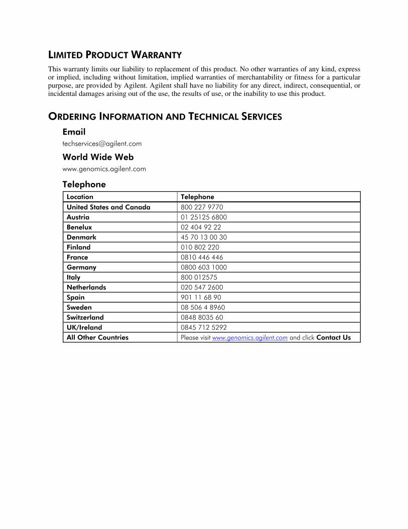
ORDERING INFORMATION AND TECHNICAL SERVICES Email [email protected]
World Wide Web
www.genomics.agilent.com
Telephone Location Telephone
United States and Canada 800 227 9770
Austria 01 25125 6800
Benelux 02 404 92 22
Denmark 45 70 13 00 30
Finland 010 802 220
France 0810 446 446
Germany 0800 603 1000
Italy 800 012575
Netherlands 020 547 2600
Spain 901 11 68 90
Sweden 08 506 4 8960
Switzerland 0848 8035 60
UK/Ireland 0845 712 5292
All Other Countries Please visit www.genomics.agilent.com and click Contact Us
Vitality hrGFP II Mammalian Expression Vectors
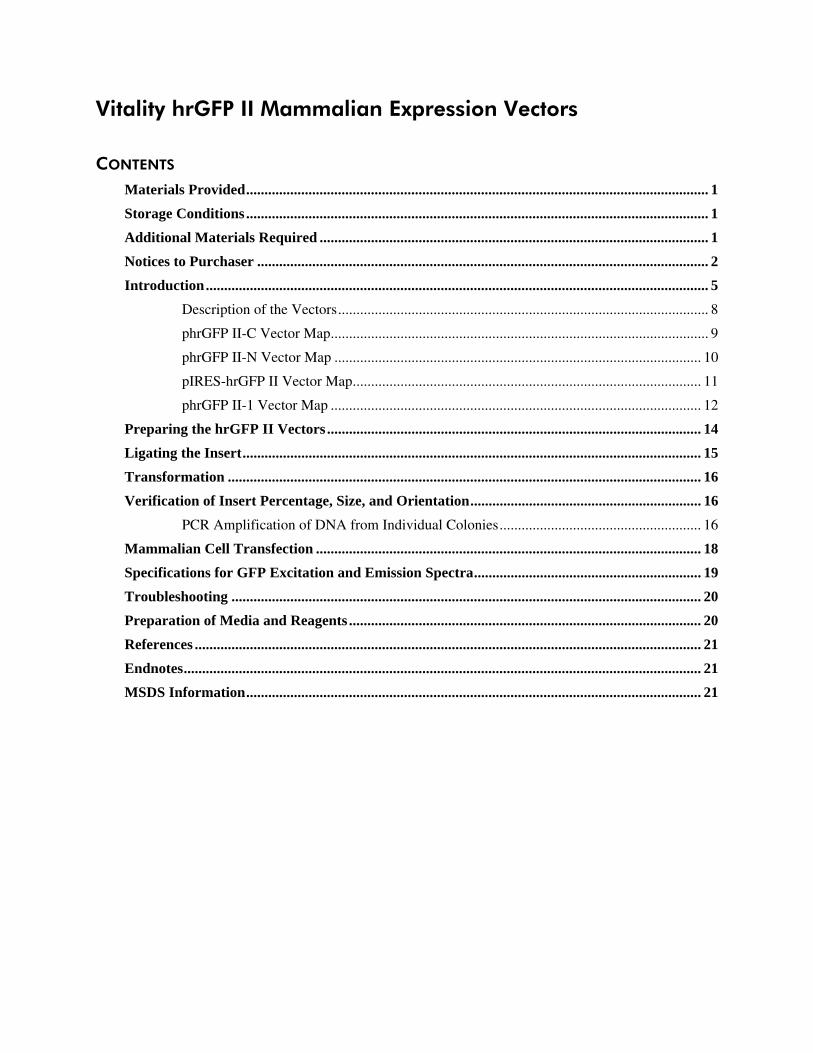
CONTENTS Materials Provided .............................................................................................................................. 1
Storage Conditions .............................................................................................................................. 1
Additional Materials Required .......................................................................................................... 1
Notices to Purchaser ........................................................................................................................... 2
Introduction ......................................................................................................................................... 5
Description of the Vectors ..................................................................................................... 8
phrGFP II-C Vector Map ....................................................................................................... 9
phrGFP II-N Vector Map .................................................................................................... 10
pIRES-hrGFP II Vector Map ............................................................................................... 11
phrGFP II-1 Vector Map ..................................................................................................... 12
Preparing the hrGFP II Vectors ...................................................................................................... 14
Ligating the Insert ............................................................................................................................. 15
Transformation ................................................................................................................................. 16
Verification of Insert Percentage, Size, and Orientation ............................................................... 16
PCR Amplification of DNA from Individual Colonies ....................................................... 16
Mammalian Cell Transfection ......................................................................................................... 18
Specifications for GFP Excitation and Emission Spectra .............................................................. 19
Troubleshooting ................................................................................................................................ 20
Preparation of Media and Reagents ................................................................................................ 20
References .......................................................................................................................................... 21
Endnotes ............................................................................................................................................. 21
MSDS Information ............................................................................................................................ 21
Vitality hrGFP II Mammalian Expression Vectors 1
Vitality hrGFP II Mammalian Expression Vectors
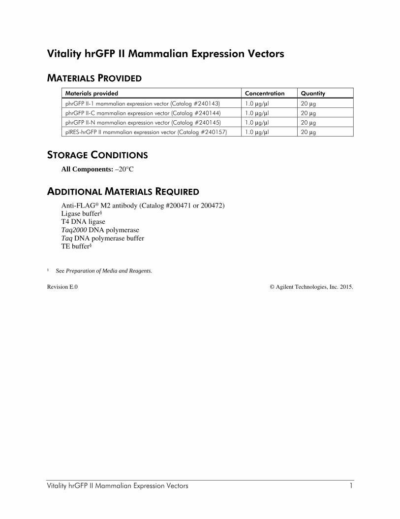
MATERIALS PROVIDED Materials provided Concentration Quantity
phrGFP II-1 mammalian expression vector (Catalog #240143) 1.0 μg/μl 20 μg
phrGFP II-C mammalian expression vector (Catalog #240144) 1.0 μg/μl 20 μg
phrGFP II-N mammalian expression vector (Catalog #240145) 1.0 μg/μl 20 μg
pIRES-hrGFP II mammalian expression vector (Catalog #240157) 1.0 μg/μl 20 μg
STORAGE CONDITIONS All Components: –20°C
ADDITIONAL MATERIALS REQUIRED Anti-FLAG® M2 antibody (Catalog #200471 or 200472) Ligase buffer§ T4 DNA ligase Taq2000 DNA polymerase Taq DNA polymerase buffer TE buffer§
§ See Preparation of Media and Reagents.
Revision E.0 © Agilent Technologies, Inc. 2015.
2 Vitality hrGFP II Mammalian Expression Vectors
NOTICES TO PURCHASER
CMV Promoter License Agreement The use of the CMV Promoter is covered under U.S. Patent Nos. 5,168,062 and 5,385,839 owned by the University of Iowa Research Foundation and licensed FOR RESEARCH USE ONLY. For further information, please contact UIRF at 319-335-4546.
IRES Sequence License Agreement Use of the translation enhancer of the pIRES-hrGFP II vector is covered by U.S. Patent No. 4,937,190 and is limited to use solely for research purposes. Any other use of the translation enhancer of the pIRES-hrGFP II vector requires a license from WARF. WARF can be reached at P.O. Box 7365 Madison, WI 53707-7365.
FLAG® License Agreement The enclosed DNA expression vector and/or antibody are specifically adapted for a method of producing selected protein molecules covered by one or more of the following patents owned by Sigma-Aldrich Co.: U.S. Patent Nos. (5,011912, 4,703,004, 4,782,137 and 4,851,341;EP Patent No. 150,126 (Austria, Belgium, Switzerland, France, United Kingdom, Italy, Netherlands and Sweden); EP Patent No. 335,899 (Belgium, Switzerland, Germany, France, United Kingdom, Italy, Luxembourg and Sweden); German Patent No. P3584260.1; Canadian Patent No. 1,307,752; and Japanese Patent Nos. 1,983,150 and 2,665,359. Your payment includes a limited license under these patents to make only the following uses of these products:
A. Vector License: You may use the enclosed vector to transform cells to produce proteins containing the amino acid sequence DYKDDDDK for research purposes provided, however, such research purposes do not include binding an unlicensed antibody to any portion of this amino acid sequence nor using such proteins for the preparation of antibodies having an affinity for any portion of this amino acid sequence.
B. Antibody License: You may only use the enclosed antibody for research purposes to perform a method of producing a protein in which the protein is expressed in a host cell and purified by use of the antibody in accordance with a claim in one of the above patents in force in a country where the use actually occurs so long as: (1) you perform such method with a DNA expression vector licensed from Sigma-Aldrich Co.; and (2) you do not bind (or allow others to bind) an unlicensed antibody to any DYKDDDDK epitope of any fusion protein that is produced by use of the method.
This license does not include any rights under any other patents. You are not licensed to use the vector and/or antibody in any manner or for any purposed not recited above. As used above, the term “unlicensed antibody” means any antibody which Sigma-Aldrich Co. has not expressly licensed pursuant to Paragraph B, above. Sigma-Aldrich Co. hereby expressly retains all rights in the above listed patents not expressly licensed hereunder.
If the terms and conditions of this License Agreement are acceptable to you, then you may open the vessel(s) containing the vector and/or antibody and, through such act of opening a vessel, will have shown your acceptance to these terms and conditions.
If the terms and conditions of this License Agreement are not acceptable to you, then please return the vessel(s) unopened to Agilent Technologies, Inc. for a complete refund of your payment.
For additional licensing information or to receive a copy of any of the above patents, please contact the Sigma-Aldrich Co. licensing department at telephone number 314-771-5765.
Vitality hrGFP II Mammalian Expression Vectors 3
Vitality Fluorescent Protein Products: Non-Commercial Research Use License Agilent agrees to sell, and Licensee agrees to purchase, Agilent Vitality fluorescent protein products provided herewith (referred to as the “Products”) on the following terms and conditions. (For purposes of this License, “Licensee” shall include any person or entity which ordered the Products or at any time uses the Products.) LICENSEE’S ACCEPTANCE OF DELIVERY AND/OR USE OF THE PRODUCTS SHALL CONSTITUTE LICENSEE’S BINDING AGREEMENT TO THE FOLLOWING TERMS AND CONDITIONS. IF LICENSEE IS UNWILLING TO ACCEPT SUCH TERMS AND CONDITIONS, AGILENT IS WILLING TO ACCEPT RETURN OF THE PRODUCTS PRIOR TO ANY USE OF THE PRODUCTS, FOR A FULL REFUND. 1. The Products, containing DNA sequences encoding for fluorescent protein or variants thereof, are proprietary or exclusively licensed to Agilent and licensed hereunder for non-commercial research purposes only. Licensee may modify only the non-coding region outside of the nucleic acid encoding the fluorescent protein of the Products to facilitate non-commercial research. Licensee shall not have any rights to (i) modify the coding region of the nucleic acid encoding the fluorescent protein of the Products, (ii) offer the Products, or any component, derivative or modification thereof, for resale, or (iii) distribute, transfer, or otherwise provide access to, the Products, or any component, derivative or modification thereof, to any third party for any purpose or use. 2. Except as set forth above, no other rights, express or implied, are conveyed to Licensee. No rights are granted to Licensee to use the Products for (i) the provision of services to any for-profit third party (e.g., screening and profiling), (ii) diagnostic applications, (iii) methods employed in screens to evaluate compounds (e.g., high throughput screening (“HTS”), (iv) profiling chemicals for selectivity, bioavailability, drug metabolism or toxicity, (v) use in vivo in multicellular organisms (and methods therein) for gene therapy, (vi) quality control or quality assurance processes, including food and environmental testing, or (vii) use in manufacturing. 3. The Products shall be used solely on premises under the control of Licensee, and in compliance with all laws, regulations, rules and guidelines applicable to the Products and their use, testing, handling, or other disposition thereof, or otherwise applicable to Licensee’s activities hereunder. 4. Title to the Products shall not transfer to Licensee. 5. Agilent warrants that, at the time of shipment, the Products will conform to the specifications which accompany the Products. This warranty limits Agilent’s liability to replacement of the Products. AGILENT MAKES NO OTHER WARRANTIES, EXPRESS OR IMPLIED, WITH RESPECT TO THE PRODUCTS, INCLUDING ANY WARRANTIES OF MERCHANTABILITY OR FITNESS FOR ANY PARTICULAR PURPOSE OR THAT THE PRODUCTS DO NOT INFRINGE ANY PROPRIETARY RIGHTS OF ANY THIRD PARTY. Licensee hereby waives, releases and renounces any and all warranties, guarantees, obligations, liabilities, rights and remedies, express or implied, arising by law or otherwise, with respect to the usefulness or freedom from defects of the Products, including, but not limited to, (a) any implied warranty or merchantability or fitness for a particular purpose, (b) any implied warranty arising from course of performance, course of dealing or usage in the trade, and (c) any obligation, right, liability, claim or remedy for (1) loss of use, revenue or profit, or any other damages, (2) infringement of third party intellectual property rights, and (3) incidental or consequential damages. 6. Licensee agrees to bear all risks associated with the Products and their use, testing, handling or other disposition thereof, and all risks associated with Licensee's use of the Products purchased hereunder. Licensee hereby assumes all risks of damage or injury to Licensee's facilities, employees
4 Vitality hrGFP II Mammalian Expression Vectors
or agents and to any third party arising from possession or use of the Products. Agilent shall have no liability to Licensee, its employees or agents or to any third party, regardless of the form or theory of action (whether contract, tort or otherwise, including, but not limited to, negligence and strict liability), for any direct, indirect, consequential, incidental or other damages arising out of or relating to the Products or this License. 7. Licensee shall indemnify, defend and hold Agilent, its affiliates, distributors, suppliers, directors, officers, employees and agents, harmless from and against any and all claims, actions, demands, liabilities, damages and expenses (including attorneys’ fees) relating to or arising out of any damage or injury, including, but not limited to, product liability and intellectual property infringement claims of any nature, alleged to have been caused by the Products or the use, testing, handling or other disposition thereof or Licensee’s activities hereunder. 8. Licensee may at any time properly dispose of the Products in a manner which ensures their prompt destruction and is consistent with all applicable laws, regulations, rules and guidelines. 9. No modification or waiver of any terms or conditions of this License shall be effective unless in writing signed by Licensee and an authorized representative of Agilent Technologies, Inc. For information on acquiring a license to use the Products for commercial purposes, including commercial research purposes, please contact Agilent Technologies, Inc., Business Development, 11011 North Torrey Pines Road, La Jolla, California 92037, telephone number (858) 373-6300, facsimile number 1-866-725-7207.
Vitality hrGFP II Mammalian Expression Vectors 5
INTRODUCTION The green fluorescent protein (GFP) has become an extremely versatile tool for tracking and quantifying biological entities in the fields of biochemistry, molecular and cell biology, as well as high throughput screening and gene discovery.1,2 GFPs have been identified in a wide range of coelenterates, and while recently the number of cloned GFPs has expanded, to date the best characterized proteins are those from the jellyfish Aequorea victoria and the anthazoan Renilla reniformis. We have isolated a cDNA clone for the R. reniformis GFP, and have fully humanized the gene using codons preferred in highly expressed human genes. The humanized recombinant GFP (hrGFP) cDNA was then subjected to random mutagenesis to generate the brighter variant, hrGFP II. The fluorescence spectra for hrGFP and hrGFP II are essentially identical to the published spectrum for the purified native protein, with the major excitation peak at 500 nm and the emission peak at 506 nm (see Figure 8).3 We have expressed the hrGFP II in a wide range of human, rodent, and simian cell lines (see Figures 1 and 2), and observed levels of fluorescence higher than that for the red-shifted, humanized variant of Aequorea GFP (EGFP) in all cell-types tested (see Figure 3). In addition, stable GFP-expressing cell lines are produced much more efficiently using the Agilent hrGFP compared with EGFP, since Aequorea GFP is often cytotoxic when expressed at low levels.4 Agilent offers four Vitality hrGFP II vectors* for mammalian expression. These vectors support a variety of expression configurations, thus providing ideal expression options for each specific application. The hrGFP II allows expressed genes to be easily visualized using fluorescence microscopy or fluorescence-activated cell sorting (FACS). We also offer a Vitality full-length hrGFP polyclonal antibody (Catalog #240141 and #240142) which recognizes the wild type green fluorescent protein, hrGFP, and the hrGFP II mutant as well as N- and C- terminal fusions to these proteins. Applications include Western blotting, immunoprecipitation, and flow cytometry. Because hrGFP is derived from a different organism, the hrGFP antibody does not cross react with the Aequorea victoria GFP variant, EGFP. * U.S. Patent No. 7,083,931.
6 Vitality hrGFP II Mammalian Expression Vectors
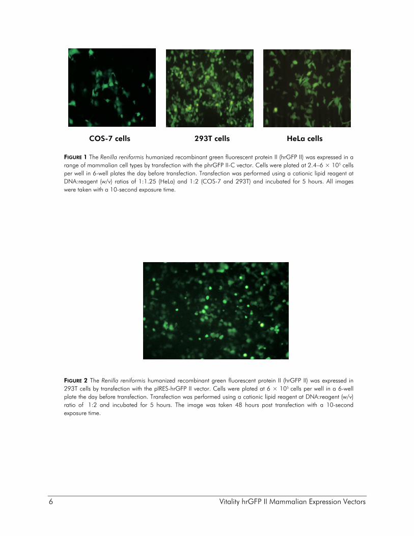
FIGURE 1 The Renilla reniformis humanized recombinant green fluorescent protein II (hrGFP II) was expressed in a range of mammalian cell types by transfection with the phrGFP II-C vector. Cells were plated at 2.4–6 × 10⁵ cells per well in 6-well plates the day before transfection. Transfection was performed using a cationic lipid reagent at DNA:reagent (w/v) ratios of 1:1.25 (HeLa) and 1:2 (COS-7 and 293T) and incubated for 5 hours. All images were taken with a 10-second exposure time.
FIGURE 2 The Renilla reniformis humanized recombinant green fluorescent protein II (hrGFP II) was expressed in 293T cells by transfection with the pIRES-hrGFP II vector. Cells were plated at 6 × 10⁵ cells per well in a 6-well plate the day before transfection. Transfection was performed using a cationic lipid reagent at DNA:reagent (w/v) ratio of 1:2 and incubated for 5 hours. The image was taken 48 hours post transfection with a 10-second exposure time.
293T cells COS-7 cells HeLa cells
Vitality hrGFP II Mammalian Expression Vectors 7
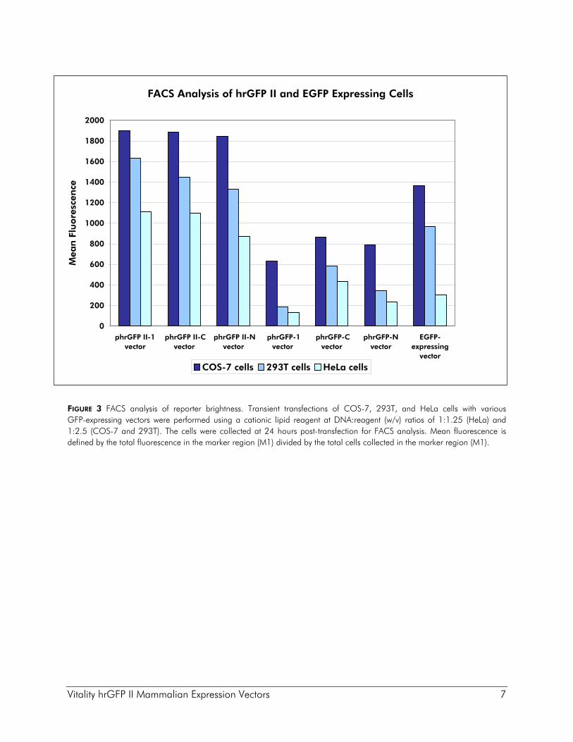
FIGURE 3 FACS analysis of reporter brightness. Transient transfections of COS-7, 293T, and HeLa cells with various GFP-expressing vectors were performed using a cationic lipid reagent at DNA:reagent (w/v) ratios of 1:1.25 (HeLa) and 1:2.5 (COS-7 and 293T). The cells were collected at 24 hours post-transfection for FACS analysis. Mean fluorescence is defined by the total fluorescence in the marker region (M1) divided by the total cells collected in the marker region (M1).
FACS Analysis of hrGFP II and EGFP Expressing Cells
0
200
400
600
800
1000
1200
1400
1600
1800
2000
phrGFP II-1vector
phrGFP II-Cvector
phrGFP II-Nvector
phrGFP-1vector
phrGFP-Cvector
phrGFP-Nvector
EGFP-expressing
vector
Mea
n F
luore
scen
ce
COS-7 cells 293T cells HeLa cells
8 Vitality hrGFP II Mammalian Expression Vectors
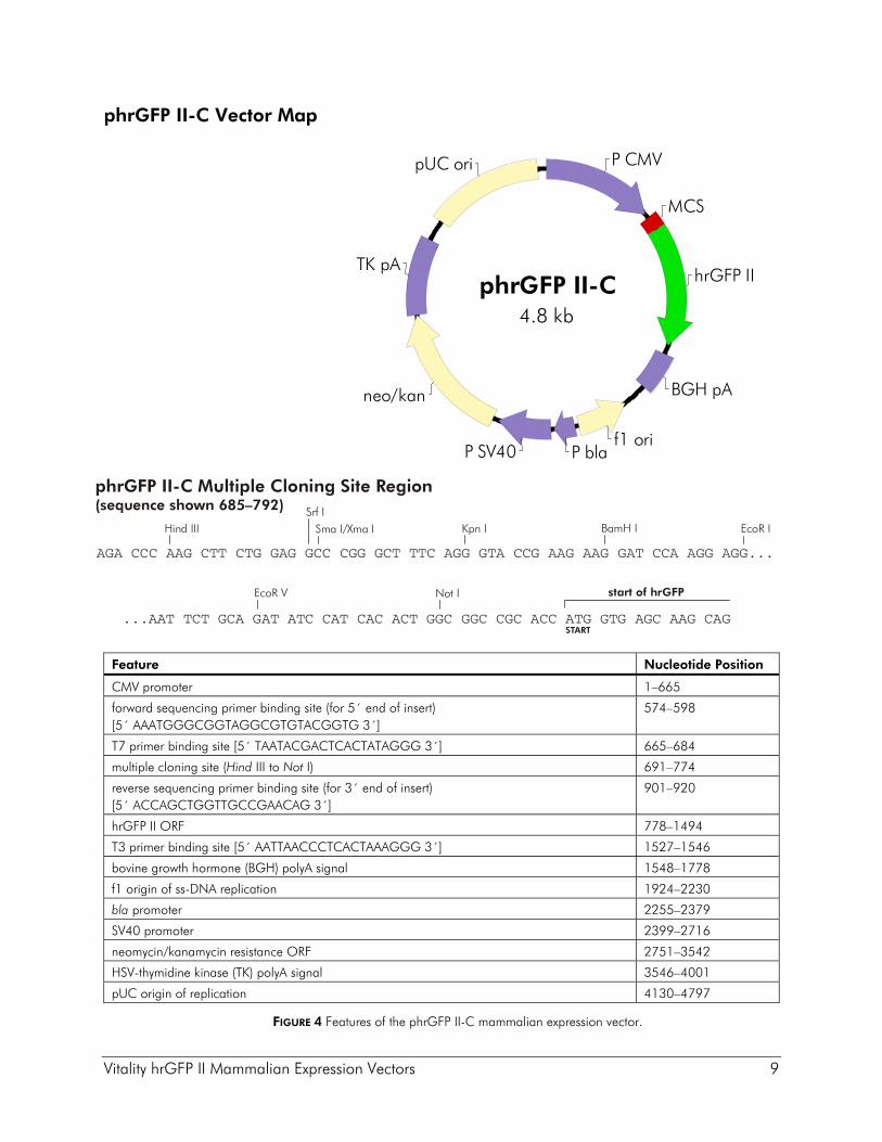
Description of the Vectors The phrGFP II-C, phrGFP II-N, pIRES-hrGFP II, and phrGFP II-1 mammalian expression vectors contain the CMV promoter, which directs constitutive, high-level expression of hrGFP II RNA transcripts of the hrGFP II reporter alone, the co-expressed hrGFP II reporter and gene of interest, or as fusion proteins in many cell lines. These cell lines include COS-7, 293T, HeLa, and CHO-KI. Each vector contains the neomycin/kanamycin-resistance gene under control of the β-lactamase promoter to provide kanamycin resistance in bacteria and the SV40 early promoter to provide G418 resistance in mammalian cells. In addition, all four hrGFP II vectors contain the bovine growth hormone polyadenylation sequence (BGHpA) for improved stability and translatability of mRNA.
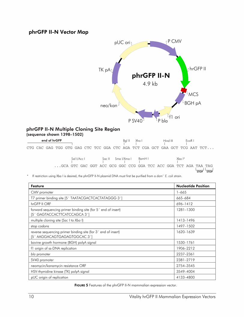
phrGFP II-C and -N Vectors The vectors phrGFP II-C and phrGFP II-N allow fusion of hrGFP II at either the C-terminus or the N-terminus of a protein of interest. phrGFP II-C contains a copy of the hrGFP II gene downstream of the MCS, allowing fusion of hrGFP II to the C-terminus of the protein of interest (see Figure 4). phrGFP II-N contains a copy of the hrGFP II gene that lacks a translational termination codon inserted upstream of the MCS to allow fusion of hrGFP II to the N-terminus of the protein of interest (see Figure 5).
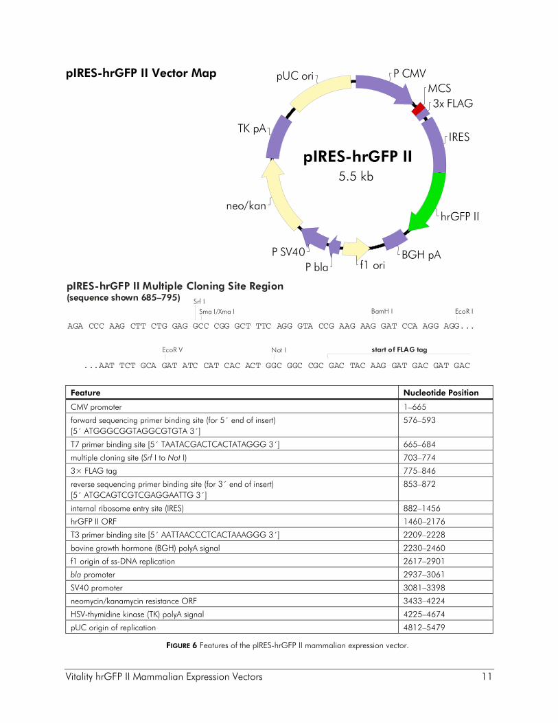
pIRES-hrGFP II Vector The pIRES-hrGFP II vector contains a bicistronic expression cassette in which the MCS is followed by the EMCV-IRES linked to the hrGFP II coding sequence. This design allows the expression of a gene of interest to be monitored at the single-cell level due to expression of hrGFP II on the same transcript. The gene of interest may be fused to three contiguous copies of the FLAG® epitope, provided by the vector (see Figure 6). For FLAG tag fusions, Western blot analysis using the anti-FLAG antibody at a 1:500 dilution will detect the protein of interest.
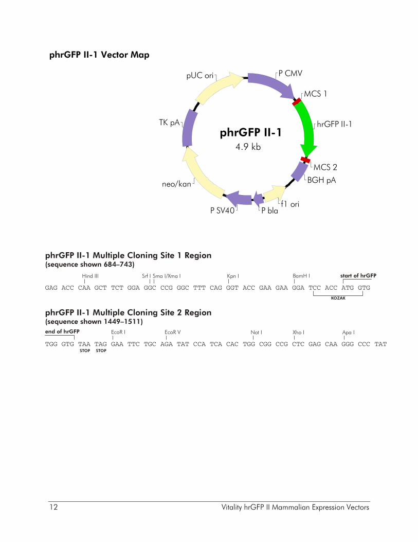
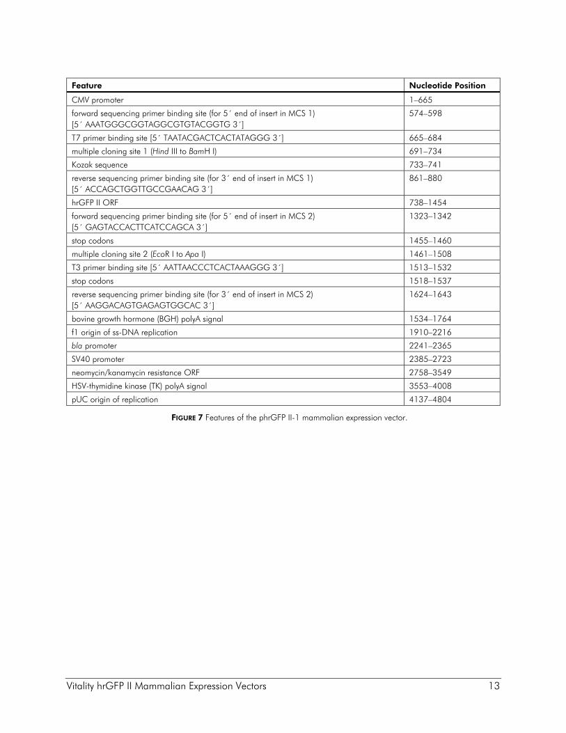
phrGFP II-1 Vector The phrGFP II-1 vector contains the hrGFP II gene, which includes a Kozak consensus sequence and termination codons directly between two multiple cloning sites for easy transfer of the hrGFP II module to new vectors (see Figure 7).
Vitality hrGFP II Mammalian Expression Vectors 9
phrGFP II-C Vector Map
Feature Nucleotide Position
CMV promoter 1–665
forward sequencing primer binding site (for 5´ end of insert) [5´ AAATGGGCGGTAGGCGTGTACGGTG 3´]
574–598
T7 primer binding site [5´ TAATACGACTCACTATAGGG 3´] 665–684
multiple cloning site (Hind III to Not I) 691–774
reverse sequencing primer binding site (for 3´ end of insert) [5´ ACCAGCTGGTTGCCGAACAG 3´]
901–920
hrGFP II ORF 778–1494
T3 primer binding site [5´ AATTAACCCTCACTAAAGGG 3´] 1527–1546
bovine growth hormone (BGH) polyA signal 1548–1778
f1 origin of ss-DNA replication 1924–2230
bla promoter 2255–2379
SV40 promoter 2399–2716
neomycin/kanamycin resistance ORF 2751–3542
HSV-thymidine kinase (TK) polyA signal 3546–4001
pUC origin of replication 4130–4797
FIGURE 4 Features of the phrGFP II-C mammalian expression vector.
P CMV
hrGFP II
MCS
BGH pAneo/kan
f1 ori
pUC ori
TK pA
P blaP SV40
phrGFP II-C4.8 kb
AGA CCC AAG CTT CTG GAG GCC CGG GCT TTC AGG GTA CCG AAG AAG GAT CCA AGG AGG...
...AAT TCT GCA GAT ATC CAT CAC ACT GGC GGC CGC ACC ATG GTG AGC AAG CAG
BamH I
START
start of hrGFP
EcoR IHind III
EcoR V
phrGFP II-C Multiple Cloning Site Region(sequence shown 685–792)
Sma I/Xma ISrf I
Kpn I
Not I
10 Vitality hrGFP II Mammalian Expression Vectors
phrGFP II-N Vector Map
* If restriction using Xba I is desired, the phrGFP II-N plasmid DNA must first be purified from a dam– E. coli strain.
Feature Nucleotide Position
CMV promoter 1–665
T7 primer binding site [5´ TAATACGACTCACTATAGGG 3´] 665–684
hrGFP II ORF 696–1412
forward sequencing primer binding site (for 5´ end of insert) [5´ GAGTACCACTTCATCCAGCA 3´]
1281–1300
multiple cloning site (Sac I to Xba I) 1413–1496
stop codons 1497–1502
reverse sequencing primer binding site (for 3´ end of insert) [5´ AAGGACAGTGAGAGTGGCAC 3´]
1620–1639
bovine growth hormone (BGH) polyA signal 1530–1761
f1 origin of ss-DNA replication 1906–2212
bla promoter 2237–2361
SV40 promoter 2381–2719
neomycin/kanamycin resistance ORF 2754–3545
HSV-thymidine kinase (TK) polyA signal 3549–4004
pUC origin of replication 4133–4800
FIGURE 5 Features of the phrGFP II-N mammalian expression vector.
P CMV
hrGFP II
MCS
BGH pA
pUC ori
f1 ori
TK pA
neo/kan
P SV40 P bla
phrGFP II-N4.9 kb
Sma I/Xma I
phrGFP II-N Multiple Cloning Site Region(sequence shown 1398–1502)
CTG CAC GAG TGG GTG GAG CTC TCC GGA CTC AGA TCT CGA GCT GAA GCT TCG AAT TCT...
...GCA GTC GAC GGT ACC GCG GGC CCG GGA TCC ACC GGA TCT AGA TAA TAG
BamH I
EcoR I
Sal I/Acc I Xba I*Sac II
Hind IIIXho IBgl II
STOP STOP
end of hrGFP
Vitality hrGFP II Mammalian Expression Vectors 11
pIRES-hrGFP II Vector Map Feature Nucleotide Position
CMV promoter 1–665
forward sequencing primer binding site (for 5´ end of insert) [5´ ATGGGCGGTAGGCGTGTA 3´]
576–593
T7 primer binding site [5´ TAATACGACTCACTATAGGG 3´] 665–684
multiple cloning site (Srf I to Not I) 703–774
3× FLAG tag 775–846
reverse sequencing primer binding site (for 3´ end of insert) [5´ ATGCAGTCGTCGAGGAATTG 3´]
853–872
internal ribosome entry site (IRES) 882–1456
hrGFP II ORF 1460–2176
T3 primer binding site [5´ AATTAACCCTCACTAAAGGG 3´] 2209–2228
bovine growth hormone (BGH) polyA signal 2230–2460
f1 origin of ss-DNA replication 2617–2901
bla promoter 2937–3061
SV40 promoter 3081–3398
neomycin/kanamycin resistance ORF 3433–4224
HSV-thymidine kinase (TK) polyA signal 4225–4674
pUC origin of replication 4812–5479
FIGURE 6 Features of the pIRES-hrGFP II mammalian expression vector.
P CMVMCS3x FLAG
IRES
hrGFP II
BGH pAf1 ori
pUC ori
P bla
neo/kan
P SV40
TK pA
pIRES-hrGFP II5.5 kb
AGA CCC AAG CTT CTG GAG GCC CGG GCT TTC AGG GTA CCG AAG AAG GAT CCA AGG AGG...
...AAT TCT GCA GAT ATC CAT CAC ACT GGC GGC CGC GAC TAC AAG GAT GAC GAT GAC
BamH I
start of FLAG tag
EcoR I
EcoR V
pIRES-hrGFP II Multiple Cloning Site Region(sequence shown 685–795)
Sma I/Xma ISrf I
Not I
12 Vitality hrGFP II Mammalian Expression Vectors
phrGFP II-1 Vector Map
P CMV
MCS 1
MCS 2
hrGFP II-1
BGH pA
f1 oriP blaP SV40
neo/kan
TK pA
pUC ori
phrGFP II-14.9 kb
Srf I Sma I/Xma I BamH I
GAG ACC CAA GCT TCT GGA GGC CCG GGC TTT CAG GGT ACC GAA GAA GGA TCC ACC ATG GTG
phrGFP II-1 Multiple Cloning Site 1 Region(sequence shown 684–743)
KOZAK
start of hrGFP
Not I
STOP
phrGFP II-1 Multiple Cloning Site 2 Region(sequence shown 1449–1511)
TGG GTG TAA TAG GAA TTC TGC AGA TAT CCA TCA CAC TGG CGG CCG CTC GAG CAA GGG CCC TAT
EcoR I
Hind III
EcoR V Xho I
Kpn I
STOP
end of hrGFP Apa I
Vitality hrGFP II Mammalian Expression Vectors 13
Feature Nucleotide Position
CMV promoter 1–665
forward sequencing primer binding site (for 5´ end of insert in MCS 1) [5´ AAATGGGCGGTAGGCGTGTACGGTG 3´]
574–598
T7 primer binding site [5´ TAATACGACTCACTATAGGG 3´] 665–684
multiple cloning site 1 (Hind III to BamH I) 691–734
Kozak sequence 733–741
reverse sequencing primer binding site (for 3´ end of insert in MCS 1) [5´ ACCAGCTGGTTGCCGAACAG 3´]
861–880
hrGFP II ORF 738–1454
forward sequencing primer binding site (for 5´ end of insert in MCS 2) [5´ GAGTACCACTTCATCCAGCA 3´]
1323–1342
stop codons 1455–1460
multiple cloning site 2 (EcoR I to Apa I) 1461–1508
T3 primer binding site [5´ AATTAACCCTCACTAAAGGG 3´] 1513–1532
stop codons 1518–1537
reverse sequencing primer binding site (for 3´ end of insert in MCS 2) [5´ AAGGACAGTGAGAGTGGCAC 3´]
1624–1643
bovine growth hormone (BGH) polyA signal 1534–1764
f1 origin of ss-DNA replication 1910–2216
bla promoter 2241–2365
SV40 promoter 2385–2723
neomycin/kanamycin resistance ORF 2758–3549
HSV-thymidine kinase (TK) polyA signal 3553–4008
pUC origin of replication 4137–4804
FIGURE 7 Features of the phrGFP II-1 mammalian expression vector.
14 Vitality hrGFP II Mammalian Expression Vectors
PREPARING THE HRGFP II VECTORS
♦ Ensure that the coding sequence of the insert is in the correct reading frame and that it contains an initiation codon or Kozak sequence.5 For gene fusions using the phrGFP II-C vector, ensure that the gene of interest lacks a termination codon, and reads in frame with the hrGFP II sequence (see the MCS sequence in Figure 4). For gene fusions using the phrGFP II-N vector, ensure that the gene of interest is inserted in frame with the hrGFP II coding sequence. If the insert lacks its own termination codon, termination codons at the 3´ end of the MCS may be used (see the MCS sequence in Figure 5).
♦ When cloning into the pIRES-hrGFP II vector, the gene of interest may be fused to three contiguous copies of the FLAG epitope, provided by the vector. For fusion with the FLAG tag, ensure that the gene of interest lacks a termination codon and that it reads in frame with the FLAG tag sequence (see the MCS sequence in Figure 6). A stop codon exists at the end of the FLAG tag sequence. Alternatively, to avoid fusion to the FLAG tag, include a termination codon at the end of the gene of interest.
♦ We recommend dephosphorylation of the digested vector with CIAP prior to ligation with the insert DNA. If more than one restriction enzyme is used, the background can be reduced further by electrophoresing the DNA on an agarose gel and removing the desired plasmid band through electroelution, leaving behind the small fragment that appears between the two restriction enzyme sites.
♦ After purification and ethanol precipitation of the DNA, resuspend in a volume of TE buffer (see Preparation of Media and Reagents) that will allow the concentration of the plasmid DNA to be the same as the concentration of the insert DNA (~0.1 μg/μl).
Vitality hrGFP II Mammalian Expression Vectors 15
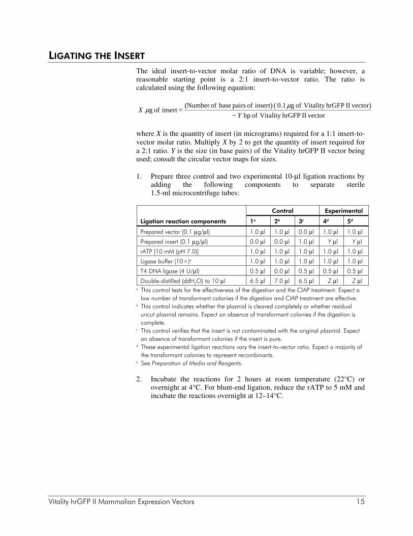
LIGATING THE INSERT The ideal insert-to-vector molar ratio of DNA is variable; however, a reasonable starting point is a 2:1 insert-to-vector ratio. The ratio is calculated using the following equation:
where X is the quantity of insert (in micrograms) required for a 1:1 insert-to-vector molar ratio. Multiply X by 2 to get the quantity of insert required for a 2:1 ratio. Y is the size (in base pairs) of the Vitality hrGFP II vector being used; consult the circular vector maps for sizes.
1. Prepare three control and two experimental 10-μl ligation reactions by adding the following components to separate sterile 1.5-ml microcentrifuge tubes:
Control Experimental
Ligation reaction components 1a 2b 3c 4d 5d
Prepared vector (0.1 μg/μl) 1.0 μl 1.0 μl 0.0 μl 1.0 μl 1.0 μl
Prepared insert (0.1 μg/μl) 0.0 μl 0.0 μl 1.0 μl Y μl Y μl
rATP [10 mM (pH 7.0)] 1.0 μl 1.0 μl 1.0 μl 1.0 μl 1.0 μl
Ligase buffer (10×)e 1.0 μl 1.0 μl 1.0 μl 1.0 μl 1.0 μl
T4 DNA ligase (4 U/μl) 0.5 μl 0.0 μl 0.5 μl 0.5 μl 0.5 μl
Double-distilled (ddH2O) to 10 μl 6.5 μl 7.0 μl 6.5 μl Z μl Z μl a This control tests for the effectiveness of the digestion and the CIAP treatment. Expect a
low number of transformant colonies if the digestion and CIAP treatment are effective. b This control indicates whether the plasmid is cleaved completely or whether residual
uncut plasmid remains. Expect an absence of transformant colonies if the digestion is complete.
c This control verifies that the insert is not contaminated with the original plasmid. Expect an absence of transformant colonies if the insert is pure.
d These experimental ligation reactions vary the insert-to-vector ratio. Expect a majority of the transformant colonies to represent recombinants.
e See Preparation of Media and Reagents.
2. Incubate the reactions for 2 hours at room temperature (22°C) or overnight at 4°C. For blunt-end ligation, reduce the rATP to 5 mM and incubate the reactions overnight at 12–14°C.
vectorII hrGFPVitality of bp ~ vector)II hrGFPVitality of g 0.1 ( insert) of pairs base of(Number
=insert of g Y
Xμμ
16 Vitality hrGFP II Mammalian Expression Vectors
TRANSFORMATION Transform competent bacteria with 1–2 μl of the ligation reaction, and plate the transformed bacteria on LB–kanamycin agar plates (see Preparation of Media and Reagents). Refer to references 6 and 7 for bacterial transformation protocols.
Note Competent cells with transformation efficiencies ≥5 × 10⁹ cfu/μg are available from Agilent.
VERIFICATION OF INSERT PERCENTAGE, SIZE, AND ORIENTATION Individual colonies can be examined to determine the percentage of vectors with inserts and the insert size and orientation by PCR directly from the colony or by restriction analysis.
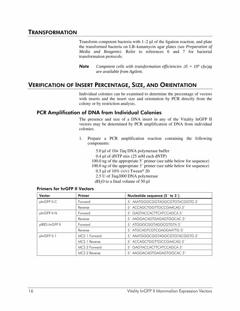
PCR Amplification of DNA from Individual Colonies The presence and size of a DNA insert in any of the Vitality hrGFP II vectors may be determined by PCR amplification of DNA from individual colonies.
1. Prepare a PCR amplification reaction containing the following components:
5.0 μl of 10× Taq DNA polymerase buffer 0.4 μl of dNTP mix (25 mM each dNTP) 100.0 ng of the appropriate 5´ primer (see table below for sequence) 100.0 ng of the appropriate 3´ primer (see table below for sequence) 0.5 μl of 10% (v/v) Tween® 20 2.5 U of Taq2000 DNA polymerase dH2O to a final volume of 50 μl
Primers for hrGFP II Vectors
Vector Primer Nucleotide sequence (5´ to 3´)
phrGFP II-C Forward 5´ AAATGGGCGGTAGGCGTGTACGGTG 3´
Reverse 5´ ACCAGCTGGTTGCCGAACAG 3´
phrGFP II-N Forward 5´ GAGTACCACTTCATCCAGCA 3´
Reverse 5´ AAGGACAGTGAGAGTGGCAC 3´
pIRES-hrGFP II Forward 5´ ATGGGCGGTAGGCGTGTA 3´
Reverse 5´ ATGCAGTCGTCGAGGAATTG 3´
phrGFP II-1 MCS 1 Forward 5´ AAATGGGCGGTAGGCGTGTACGGTG 3´
MCS 1 Reverse 5´ ACCAGCTGGTTGCCGAACAG 3´
MCS 2 Forward 5´ GAGTACCACTTCATCCAGCA 3´
MCS 2 Reverse 5´ AAGGACAGTGAGAGTGGCAC 3´
Vitality hrGFP II Mammalian Expression Vectors 17
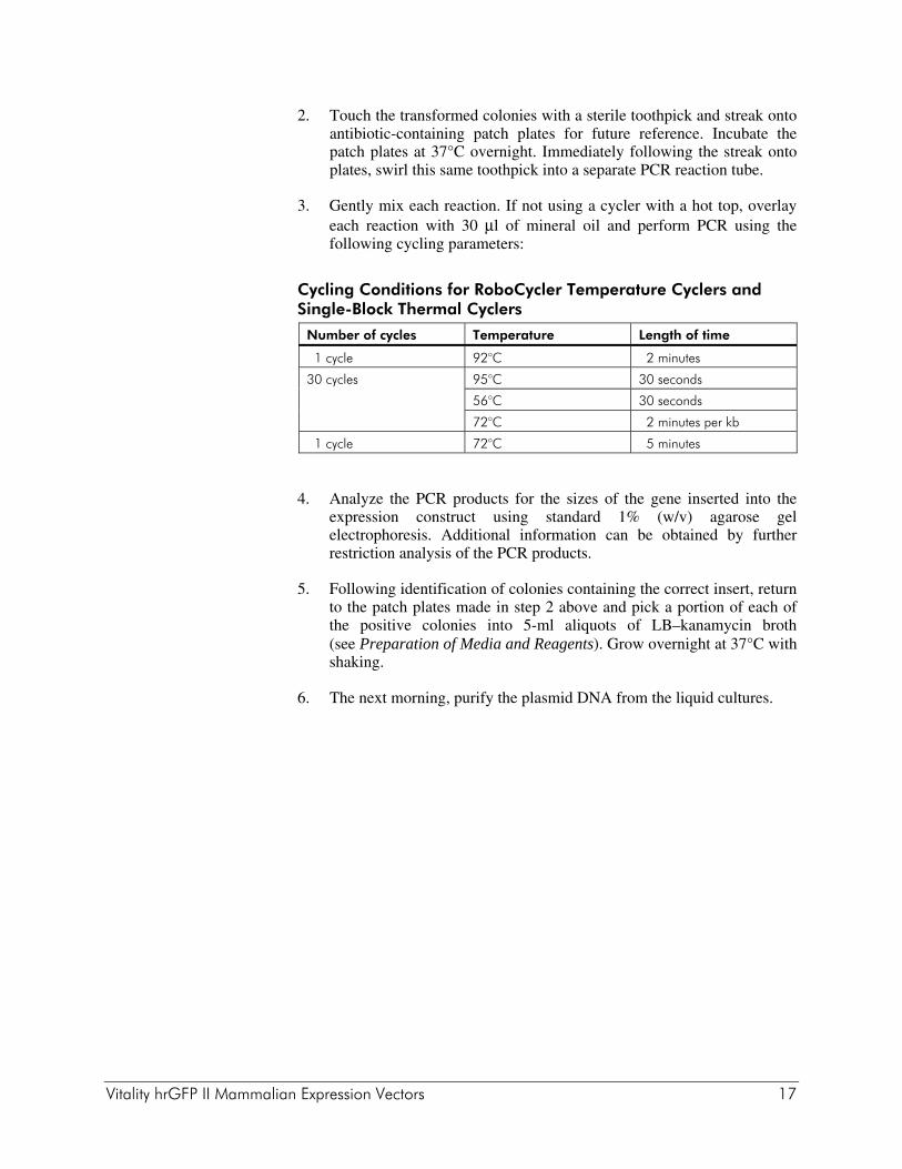
2. Touch the transformed colonies with a sterile toothpick and streak onto antibiotic-containing patch plates for future reference. Incubate the patch plates at 37°C overnight. Immediately following the streak onto plates, swirl this same toothpick into a separate PCR reaction tube.
3. Gently mix each reaction. If not using a cycler with a hot top, overlay each reaction with 30 μl of mineral oil and perform PCR using the following cycling parameters:
Cycling Conditions for RoboCycler Temperature Cyclers and Single-Block Thermal Cyclers
Number of cycles Temperature Length of time
1 cycle 92°C 2 minutes
30 cycles 95°C 30 seconds
56°C 30 seconds
72°C 2 minutes per kb
1 cycle 72°C 5 minutes
4. Analyze the PCR products for the sizes of the gene inserted into the expression construct using standard 1% (w/v) agarose gel electrophoresis. Additional information can be obtained by further restriction analysis of the PCR products.
5. Following identification of colonies containing the correct insert, return to the patch plates made in step 2 above and pick a portion of each of the positive colonies into 5-ml aliquots of LB–kanamycin broth (see Preparation of Media and Reagents). Grow overnight at 37°C with shaking.
6. The next morning, purify the plasmid DNA from the liquid cultures.
18 Vitality hrGFP II Mammalian Expression Vectors
MAMMALIAN CELL TRANSFECTION When the correct recombinant plasmids are confirmed, prepare enough DNA of appropriate purity for the mammalian cell transfection procedure to be carried out. Protocols for transfection of mammalian cell lines can be found in Sambrook, et al. (1989).6
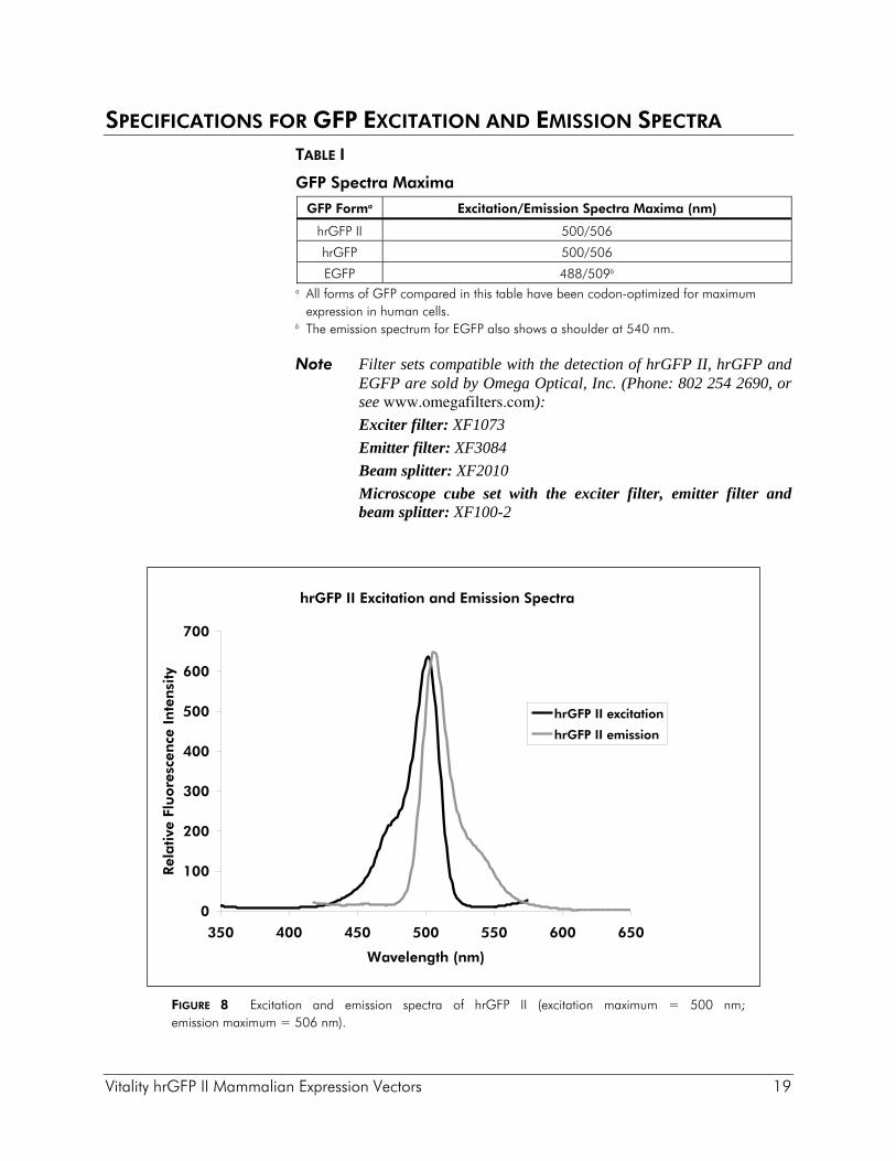
The efficiency of transfection will vary depending on the host cell line used. In most cases, mammalian host cell lines transfected with plasmids should show expression of hrGFP II 24–72 hours after transfection. Fluorescing cells growing in tissue culture dishes can be observed using an inverted fluorescence microscope. Fluorescence of populations of harvested cells can also be measured using FACS analysis or fluorometer assays. Table I lists excitation and emission spectra for Agilent hrGFP II and original hrGFP, as compared to EGFP. See Figure 8 for plotted hrGFP II spectra.
Vitality hrGFP II Mammalian Expression Vectors 19
SPECIFICATIONS FOR GFP EXCITATION AND EMISSION SPECTRA TABLE I
GFP Spectra Maxima
GFP Forma Excitation/Emission Spectra Maxima (nm)
hrGFP II 500/506
hrGFP 500/506
EGFP 488/509b a All forms of GFP compared in this table have been codon-optimized for maximum
expression in human cells. b The emission spectrum for EGFP also shows a shoulder at 540 nm.
Note Filter sets compatible with the detection of hrGFP II, hrGFP and EGFP are sold by Omega Optical, Inc. (Phone: 802 254 2690, or see www.omegafilters.com):
Exciter filter: XF1073
Emitter filter: XF3084
Beam splitter: XF2010
Microscope cube set with the exciter filter, emitter filter and beam splitter: XF100-2
FIGURE 8 Excitation and emission spectra of hrGFP II (excitation maximum = 500 nm; emission maximum = 506 nm).
hrGFP II Excitation and Emission Spectra
0
100
200
300
400
500
600
700
350 400 450 500 550 600 650
Wavelength (nm)
Rel
ativ
e Fl
uore
scen
ce I
nte
nsi
ty
hrGFP II excitation
hrGFP II emission
20 Vitality hrGFP II Mammalian Expression Vectors
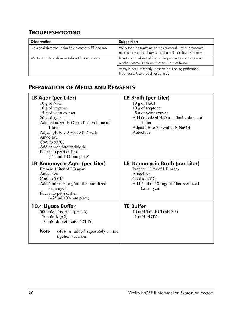
TROUBLESHOOTING Observation Suggestion
No signal detected in the flow cytometry F1 channel Verify that the transfection was successful by fluorescence microscopy before harvesting the cells for flow cytometry.
Western analysis does not detect fusion protein Insert is cloned out of frame. Sequence to ensure correct reading frame. Reclone if insert is out of frame.
Assay is not sufficiently sensitive or is being performed incorrectly. Use a positive control.
PREPARATION OF MEDIA AND REAGENTS
LB Agar (per Liter) 10 g of NaCl 10 g of tryptone 5 g of yeast extract 20 g of agar Add deionized H2O to a final volume of
1 liter Adjust pH to 7.0 with 5 N NaOH Autoclave Cool to 55°C. Add appropriate antibiotic. Pour into petri dishes
(~25 ml/100-mm plate)
LB Broth (per Liter) 10 g of NaCl 10 g of tryptone 5 g of yeast extract Add deionized H2O to a final volume of
1 liter Adjust pH to 7.0 with 5 N NaOH Autoclave
LB–Kanamycin Agar (per Liter) Prepare 1 liter of LB agar Autoclave Cool to 55°C Add 5 ml of 10-mg/ml filter-sterilized
kanamycin Pour into petri dishes
(~25 ml/100-mm plate)
LB–Kanamycin Broth (per Liter) Prepare 1 liter of LB broth Autoclave Cool to 55°C Add 5 ml of 10-mg/ml filter-sterilized
kanamycin
10× Ligase Buffer 500 mM Tris-HCl (pH 7.5) 70 mM MgCl2 10 mM dithiothreitol (DTT)
Note rATP is added separately in the ligation reaction
TE Buffer 10 mM Tris-HCl (pH 7.5) 1 mM EDTA
Vitality hrGFP II Mammalian Expression Vectors 21
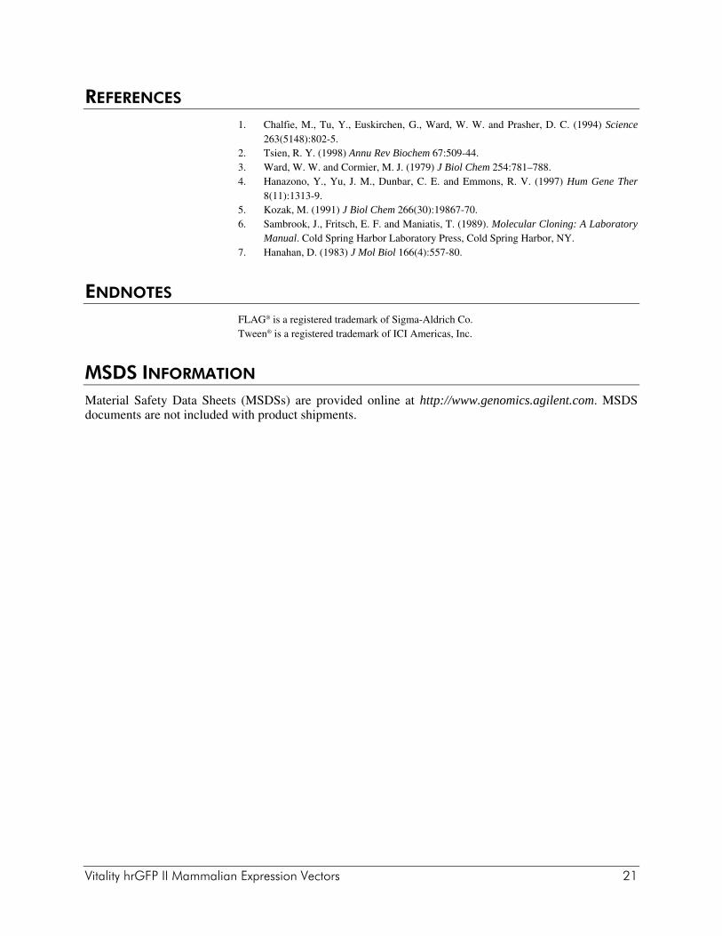
REFERENCES 1. Chalfie, M., Tu, Y., Euskirchen, G., Ward, W. W. and Prasher, D. C. (1994) Science
263(5148):802-5. 2. Tsien, R. Y. (1998) Annu Rev Biochem 67:509-44. 3. Ward, W. W. and Cormier, M. J. (1979) J Biol Chem 254:781–788. 4. Hanazono, Y., Yu, J. M., Dunbar, C. E. and Emmons, R. V. (1997) Hum Gene Ther
8(11):1313-9. 5. Kozak, M. (1991) J Biol Chem 266(30):19867-70. 6. Sambrook, J., Fritsch, E. F. and Maniatis, T. (1989). Molecular Cloning: A Laboratory
Manual. Cold Spring Harbor Laboratory Press, Cold Spring Harbor, NY. 7. Hanahan, D. (1983) J Mol Biol 166(4):557-80.
ENDNOTES FLAG® is a registered trademark of Sigma-Aldrich Co. Tween® is a registered trademark of ICI Americas, Inc.
MSDS INFORMATION Material Safety Data Sheets (MSDSs) are provided online at http://www.genomics.agilent.com. MSDS documents are not included with product shipments.


![2003 [New Comprehensive Biochemistry] Gene Transfer and Expression in Mammalian Cells Volume 38 __ Virus-based vectors f](https://img.pdfslide.us/doc/110x75/613ca5f79cc893456e1e7c1b/2003-new-comprehensive-biochemistry-gene-transfer-and-expression-in-mammalian.jpg)