Embed Size (px)
DESCRIPTION
The International Institute for Science, Technology and Education (IISTE). Science, Technology and Medicine Journals Call for Academic Manuscripts
Citation preview
Journal of Health, Medicine and Nursing www.iiste.org
An Open Access Journal
Vol. 4, 2014
1
Visual Perception and Electromagnetic Conception for the
Eyesight. Rhodopsin and Bacteriorhodopsin in Nano- and
Biotechnologies
Ignat Ignatov1*
Oleg Mosin2
1. Scientific Research Center of Medical Biophysics (SRCMB),
N. Kopernik Street, 32, Sofia 1111, Bulgaria
2. Biotechnology Department, Moscow State University of Applied Biotechnology,
Talalikhina Street, 33, Moscow 109316, Russian Federation
* E-mail of the corresponding author: [email protected]
Abstract
This article views predominately the structure and function of animal and bacterial photoreceptor pigments
(rhodopsin, iodopsin, bacteriorhodopsin) and their nano- and biotechnological usage. On an example of
bacterial pigment bacteriorhodopsin (BR) is described the method of its isolation from purple membranes of
halophilic bacterium Halobacterium halobium by cellular autolysis by distilled water, processing of bacterial
biomass by ultrasound at 22 KHz, alcohol extraction of low and high-weight molecular impurities, cellular
RNA, carotenoids and lipids, the solubilization with 0.5 % (w/v) SDS-Na and subsequent fractionation by
methanol and gel filtration chromatography on Sephadex G-200 Column balanced with 0.09 M Tris-borate
buffer (pH = 8.35) with 0.1% (w/v) SDS-Na and 2.5 mM EDTA. Within the framework of the research the
mechanism of color perception by the visual analyzer having the ability to analyze certain ranges of the
optical spectrum, as colors was studied along with an analysis of the additive mixing of two colors. It was
shown that in the mixing of electromagnetic waves with different wavelengths, the visual analyzer perceive
them as separate or average wave length corresponding to mix color.
Keywords: vision, rhodopsin, iodopsin, bacteriorhodopsin, additive color mixing
1. Introduction
Vision (visual perception) is a process of psycho-physiological processing of the images of surrounding
objects, carried out by the visual system, which allows getting an idea of the size, shape and color of
surrounding objects, their relative position and distance between them. By means of this animals can
receive 90% of all incoming information to the brain.
The function of the visual system is carried out through various interrelated complex structures designated
as visual analyzer, consisting of a peripheral part (retina, optic nerve, optic tract) and the central department
of combining stem and subcortical centers of the midbrain, as well as the visual cortex of the cerebral
Journal of Health, Medicine and Nursing www.iiste.org
An Open Access Journal
Vol. 4, 2014
2
hemispheres. The human eye can perceive only light waves of a certain length - from 380 to 770 nm.
Light rays from treated subjects pass through the optical system of the eye (cornea, lens and vitreous body)
and onto the retina, where the light-sensitive photoreceptor cells (rods and cones) are located. Light
incident on the photoreceptors, triggers a cascade of biochemical reactions of visual pigments (in particular,
the most studied of them is rhodopsin responsible for the perception of electromagnetic radiation in the
visible range), and in turn, the occurrence of nerve impulses, which are transmitted through the following
retinal neurons and further to the optic nerve. The optic nerve carries the nerve impulses into the lateral
geniculate body - subcortical center of vision, and thence to the cortical center, located in the occipital lobe
of the brain, where the visual image is formed.
Over the last decade have been obtained new data revealing the molecular basis of visual perception. It was
identified visual molecules of eucariotes (rhodopsin, iodopsin) and prokaryotes (bacteriorhodopsin)
involved in light perception and cleared up the mechanism of their action.
The structural research of rhodopsin and its affiliate chromophore proteins (iodopsin, bacteriorhodopsin)
and the analysis of their functions have been carried out in the Scientific Research Center of Medical
Biophysics (Bulgaria) throughout the last 20 years. The purpose of the research was the studying of basic
biochemical mechanisms associated with visual perception and some nano- and biotechnological
applications of visual phototransforming pigments as trans membrane chromo-protein bacteriorhodopsin
(BR), isolated from purple membranes of halobacterium Halobacterium halobium.
2. Materials and methods
2.1. Bacterial objects
As a BR producer was used a carotenoid strain of extreme photo-organo-heterotrophic halobacterium
Halobacterium halobium ET 1001, obtained from Moscow State University (Russia). The strain was
modified by selection of individual colonies on solid (2% (w/v) agarose) media with peptone and 4.3 M
NaCl.
2.2. Growth conditions
BR (yield 810 mg from 1 g biomass) was obtained in synthetic (SM) medium (g/l): D,L-alanine 0.43;
L-arginine 0.4; D,L-aspartic acid 0.45; L-cysteine 0.05; L-glutamic acid 1.3; L-lysine 0.06; D,
L-histidine 0.3; D,L-isoleucine 0.44; L-leucine 0.8; L-lysine 0.85; D,L-methionine 0.37;
D,L-phenylalanine 0.26; L-proline 0.05; D, L-serine 0.61; D,L-threonine 0.5; L-tyrosine 0.2;
D,L-tryptophan 0.5; D,L-valine 1.0, AMP 0.1; UMP 0.1; NaCl 250; MgSO4 7H2O 20; KCl 2;
NH4Cl 0.5; KNO3 0.1; KH2PO4 0.05; K2HPO4 0.05; Na+-citrate 0.5; MnSO4 2H2O – 3 10
-4; CaCl2
6H2O 0.065; ZnSO4 7H2O – 4.10
-5; FeSO4 7H2O – 5
.10
-4; CuSO4 5H2O – 5
.10
-5; glycerol 1.0, biotin –
1.10
-4; folic acid 1.5
. 10
-4, vitamin B12 – 2
.10
-5. The growth medium was autoclaved for 30 min at 0.5 atm,
the pH value was adjusted to 6.56.7 with 0.5 M KOH. Bacterial growth was performed in 500 ml
Erlenmeyer flasks (volume of the reaction mixture 100 ml) for 45 days at 35 0C on Biorad shaker (“Birad
Labs”, Hungary) under intense aeration and monochromatic illumination (3 lamps 1.5 lx). All further
manipulations for BR isolation were carried out with the use of a photomask lamp equipped with an orange
light filter.
Journal of Health, Medicine and Nursing www.iiste.org
An Open Access Journal
Vol. 4, 2014
3
2.3. Isolation of purple membranes (PM)
Biomass (1 g) was washed with distilled water and pelleted by centrifugation on T-24 centrifuge (“Carl
Zeiss”, Germany) (1500 g, 20 min). The precipitate was suspended in 100 ml of dist. H2O and kept for 3 h
at 4 0C. The reaction mixture was centrifuged (1500 g, 15 min), the pellet was resuspended in 20 ml dist.
H2O and disintegrated by infrasound sonication (22 kHz, 3 times 5 min) in an ice bath (0 0C). The cell
homogenate after washing with dist. H2O was resuspended in 10 ml of buffer containing 125 mM NaCl, 20
mM MgCl2, and 4 mM Tris-HCl (pH = 8.0), then 5 mg of RNA-ase (23 units of activity) was added . The
mixture was incubated for 2 h at 37 °C. Then 10 ml of the same buffer was added and kept for 1012 h at 4 0C. The aqueous fraction was separated by centrifugation (1500 g, 20 min), the PM precipitate was treated
with 50% (v/v) ethanol (5 times 7 ml) at 4 0C followed by separation of the solvent. This procedure was
repeated 6 times to give colorless washings. The protein content in the samples was determined
spectrophotometrically on DU-6 spectrophotometer (“Beckman Coulter”, USA) by the ratio D280/D568 (280 =
1.1.10
5; 568 = 6.3
.10
4 M
-1.сm
-1) (Neugebauer et al., 1978). PM regeneration is performed as described in
(Rudiger et al., 1997). Yield of PM fraction 120 mg (8085%).
2.4. Isolation of BR
Fraction PM (in H2O) (1 mg/ml) was dissolved in 1 ml of 0.5% (w/v) sodium dodecyl sulfate (SDS-Na),
and incubated for 57 h at 37 0C followed by centrifugation (1200 g, 15 min). The precipitate was
separated, than methanol was added to the supernatant in divided portions (3 times 100 ml) at 0 0C. The
reaction mixture was kept for 1415 h in ice bath at 4 0C and then centrifuged (1200 g, 15 min).
Fractionation procedure was performed three times, reducing the concentration of 0.5% SDS-Na to 0.2 and
0.1%. Crystal protein (output 810 mg) was washed with cold 2H2O (2 times 1 ml) and centrifuged (1200
g, 15 min).
2.5. Purification of BR
Protein sample (5 mg) was dissolved in 100 ml of buffer solution and placed on a column (150 10 mm),
stationary phase Sephadex G-200 ("Pharmacia", USA) (specific volume packed beads 3040 units per
1 g dry. Sephadex) equilibrated with buffer containing 0.1% (w/v) SDS-Na and 2.5 mM ETDA. Elution
proceeded by 0.09 M Tris-borate buffer containing 0.5 M NaCl, pH = 8.35 at a flow rate of 10 ml/cm2 .
h.
Combined protein fraction was subjected to freeze-drying, in sealed glass ampoules (10 50 mm) and
stored in frost camera at -10 0C.
2.6. Quantitative analysis of the protein
The procedure was performed in 12.5% (w/v) polyacrylamide gel (PAAG) containing 0.1% (w/v) SDS-Na.
The samples were prepared for electrophoresis by standard procedures (LKB protocol, Sweden).
Electrophoretic gel stained with Coomassie blue R-250 was scanned on a CDS-200 laser densitometer
(Beckman, USA) for quantitative analysis of the protein.
2.7.Absorption spectra
Absorption spectra of pigments were recorded on programmed DU-6 spectrophotometer (“Beckman Coulter”,
USA) at 280 nm and 750 nm.
2.8. IR-spectroscopy
IR-spectra were registered on Brucker Vertex IR spectrometer (“Brucker”, Germany) (a spectral range:
average IR – 370–7800 cm-1
; visible – 2500–8000 cm-1
; the permission – 0.5 cm-1
; accuracy of wave
number – 0.1 cm-1
on 2000 cm-1
).
Journal of Health, Medicine and Nursing www.iiste.org
An Open Access Journal
Vol. 4, 2014
4
2.9. Color analyzing
Colors were analyzed by using color analyzer “Tsvetan” (“Photopribor”, Cherkassk, Ukraine). Operating
relative absorbance, % from -80 to 70. Measurement error, ±5%. Response time from 0.4 to 63 sec. Overall
dimensions, 300 mm.
2.10. Scanning electron microscopy
The structural studies were carried out with using scanning electrom microscopy (SEM) on JSM 35 CF
(JEOL Ltd., Corea) device, equipped with X-ray microanalyzer “Tracor Northern TN”, SE detector,
thermomolecular pump, and tungsten electron gun (Harpin type W filament, DC heating); working
pressure: 10-4
Pa (10-6
Torr); magnification: 300.000, resolution: 3.0 nm, accelerating voltage: 1–30 kV;
sample size: 60–130 mm.
3. Results and discussion
3.1. Theoretical aspects of molecular basis of vision
The process of perception of light has a definite localization in photoreceptor light-sensitive cells of the
retina. The retina in its structure is a multilayer layer of nervous tissue that is sensitive to light, which lines
the inside of the back of the eyeball. Pigmented retina located at the membrane referred to as retinal
pigmented epithelium (RPE), which absorbs light passing through the retina. This prevents the reverse
reflection of the light through the retina and does not allow the vision to disperse.
Light enters through the eye and creates a complex biochemical reaction in the photoreceptor cells of the
retina. Photoreceptor cells are divided into two types that due to their characteristic form are designated as
rods and cones (Hubel, 1995). Rods are receptors of light of low intensity; they arranged in a colored layer
of the retina, in which is synthesized photochromic protein rhodopsin, responsible for color perception.
Cones on the contrary contain a group of visual pigments (iodopsin), and adapted to distinguish different
colors. Rods can perceive black and white images in the dim light, cones – to carry out color vision in
bright light. Human retina contains approximately 3 million of cones and 100 million of rods. Their
dimensions are very small – the length of about 50 mm, the diameter from 1 to 4 m.
Electrical signals generated by the rods and cones, are handled by other retinal cells – bipolar and ganglion
cells before they are transmitted to the brain via the optic nerve (Hogan et al., 1971). Additionally, there are
two intermediate layers of neurons. Horizontal cells transmit messages back and forth between the
photoreceptor cells, bipolar cells and each other. Amacrine cells of the retina are linked to bipolar cells,
ganglion cells, as well as with each other. Both types of these intermediate neurons play a major role in the
processing of visual information at the level of the retina before it is transmitted to the brain for final
processing.
Cones are approximately 100 times less sensitive to light than rods, but much better perceive the rapid
movement. The wand can be stimulated by a single photon. Cascade of molecular interactions enhances
this "quantum" of information into a chemical signal, which is then perceived by the nervous system. The
degree of enhancement signal varies depending on ambient light: rods are more sensitive under low than
under bright light. As a result, they operate effectively in a wide range of ambient light. Sensory system of
rods is packed up in clearly distinguishable cellular substructure that can be easily selected and investigated
in vitro in isolated state. This property makes them as indispensable object for further structural-functional
Journal of Health, Medicine and Nursing www.iiste.org
An Open Access Journal
Vol. 4, 2014
5
studies as well as studies of photoreceptor pigments (rhodopsin, iodopsin). These animal photoreceptor
pigments are used as models for studying of bacterial photoreceptor pigment bacteriorhodopsin (BR) from
purple membranes of halobacterium Halobacterium halobium.
3.2 Rhodopsin and its structural and functional properties
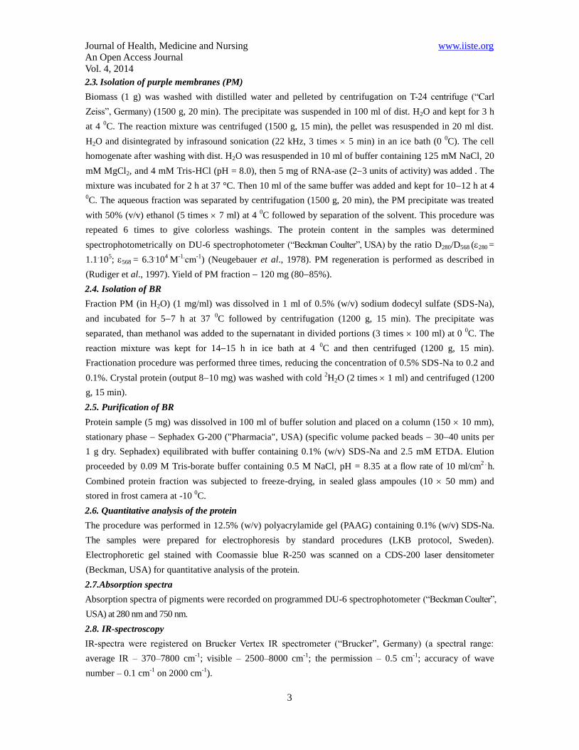
Rhodopsin (Nathans et al., 1986) is one of the most important integral photoreceptor proteins of rod cells,
which absorbs a photon and creates a biochemical response constituting a first step in a chain of events that
provide vision. Rhodopsin consists of two components – a colorless protein opsin and a chromophore
component 11-cis-retinal residue, acted as the light acceptor (Fig. 1). The absorption of a light photon by
11-cis-retinal “turns on” the enzymatic activity of opsin and further photosensitive biochemical cascade of
reactions that are responsible for vision (Liang et al., 2004).
Figure 1. Configuration of photosensitive chromophore of rhodopsin in the basic (unexcited) phase (at the
double bond is marked 11-cis-configuration)
Rhodopsin belongs to the group of the G-protein-coupled receptors (GPCR-receptors) of the retibylidene
protein family responsible for transmembrane signaling mechanism based on the interaction with
intracellular membrane G-proteins – universal intermediaries in the transmission of hormonal signals from
the cell membrane receptors to effectors proteins, causing the final cellular response. The establishment of
the spatial structure of rhodopsin is so important because rhodopsin as the “originator” of the family of
GPCR-receptors is a “model” for the structure and function of other receptors that it is extremely important
from fundamental scientific and practical points of view (Palczewski, 2006).
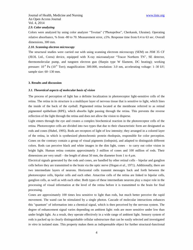
Spatial structure of rhodopsin was long defied by the study of "direct" methods – X-ray diffraction and
NMR spectroscopy, while the molecular structure of related to rhodopsin transmembrane chromoprotein
bacteriorhodopsin (Henderson et al., 1990) having a similar structure, performing the functions of
ATP-dependent transposes in the cell membranes of halophilic microorganisms pumped protons across the
cytoplasmic membrane of the cell and is involved in the anaerobic photosynthetic phosphorylation
(non-green synthesis), was determined as early as 1990. On the contrary the structure of rhodopsin
remained unknown until 2003 (Palczewski et al., 2000). The opsin fragment of the rhodopsin molecule has
348 amino acid residues in a polypeptide chain that is formed by seven transmembrane -helix segments
situated across the membrane and joined with short non-helix sections (Ovchinnikov et al., 1983). The
Journal of Health, Medicine and Nursing www.iiste.org
An Open Access Journal
Vol. 4, 2014
6
N-terminus of -helix is located in the extracellular region, while the C-terminus – in the cytoplasmic
region. The 11-cis-retinal residue is connected to one of the -helixes, located near the middle of the
membrane, so that its long axis is parallel to the membrane surface (Fig. 2). It was also determined the
dislocation of 11-cis-retinal aldimine bond with ε-amino group of Lys-296 residue located in the seventh
-helix. Thus, 11-cis-retinal is mounted in the center of a complex highly organized protein in the cellular
membrane comprising rods. This structure provides a photochemical "adjustment" of retinal residue,
affecting its absorption spectrum. The free 11-cis-retinal in a dissolved form has an absorption maximum in
the ultraviolet region - at a wavelength of 380 nm, while rhodopsin absorbs green light at 500 nm (Hargrave
et al., 1983). This shift in the wavelength of light is important from a functional point of view; it is aligned
with the spectrum of light that enters the retina.
Figure 2. The structure of rhodopsin according to computer modeling data
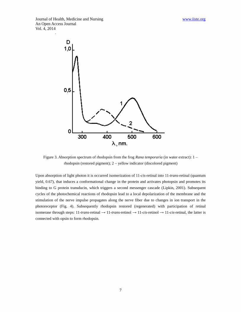
The absorption spectrum of rhodopsin is defined by properties of the chromophore - 11-cis-retinal residue
and opsin fragment. This range in vertebrates has two characteristic peaks – one in the ultraviolet (278 nm)
due to the opsin fragment, and the other – in the visible region (500 nm) corresponds to absorption of the
chromophore (Fig. 3). Further transformation of rhodopsin under the action of light to the final stable
product consists of a series of very fast intermediate stages. Investigating intermediates absorption spectra
of rhodopsin in extracts at low temperatures at which these products are stable, allows to describe in the
detail the photochemical changes of rhodopsin (Schertler, & Hargrave, 1995).
Journal of Health, Medicine and Nursing www.iiste.org
An Open Access Journal
Vol. 4, 2014
7
Figure 3. Absorption spectrum of rhodopsin from the frog Rana temporaria (in water extract): 1 –
rhodopsin (restored pigment); 2 – yellow indicator (discolored pigment)
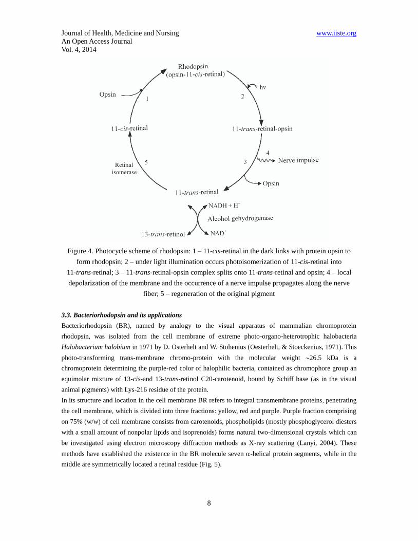
Upon absorption of light photon it is occurred isomerization of 11-cis-retinal into 11-trans-retinal (quantum
yield, 0.67), that induces a conformational change in the protein and activates photopsin and promotes its
binding to G protein transducin, which triggers a second messenger cascade (Lipkin, 2001). Subsequent
cycles of the photochemical reactions of rhodopsin lead to a local depolarization of the membrane and the
stimulation of the nerve impulse propagates along the nerve fiber due to changes in ion transport in the
photoreceptor (Fig. 4). Subsequently rhodopsin restored (regenerated) with participation of retinal
isomerase through steps: 11-trans-retinal → 11-trans-retinol → 11-cis-retinol → 11-cis-retinal, the latter is
connected with opsin to form rhodopsin.
Journal of Health, Medicine and Nursing www.iiste.org
An Open Access Journal
Vol. 4, 2014
8
Figure 4. Photocycle scheme of rhodopsin: 1 – 11-cis-retinal in the dark links with protein opsin to
form rhodopsin; 2 – under light illumination occurs photoisomerization of 11-cis-retinal into
11-trans-retinal; 3 – 11-trans-retinal-opsin complex splits onto 11-trans-retinal and opsin; 4 – local
depolarization of the membrane and the occurrence of a nerve impulse propagates along the nerve
fiber; 5 – regeneration of the original pigment
3.3. Bacteriorhodopsin and its applications
Bacteriorhodopsin (BR), named by analogy to the visual apparatus of mammalian chromoprotein
rhodopsin, was isolated from the cell membrane of extreme photo-organo-heterotrophic halobacteria
Halobacterium halobium in 1971 by D. Osterhelt and W. Stohenius (Oesterhelt, & Stoeckenius, 1971). This
photo-transforming trans-membrane chromo-protein with the molecular weight 26.5 kDa is a
chromoprotein determining the purple-red color of halophilic bacteria, contained as chromophore group an
equimolar mixture of 13-cis-and 13-trans-retinol C20-carotenoid, bound by Schiff base (as in the visual
animal pigments) with Lys-216 residue of the protein.
In its structure and location in the cell membrane BR refers to integral transmembrane proteins, penetrating
the cell membrane, which is divided into three fractions: yellow, red and purple. Purple fraction comprising
on 75% (w/w) of cell membrane consists from carotenoids, phospholipids (mostly phosphoglycerol diesters
with a small amount of nonpolar lipids and isoprenoids) forms natural two-dimensional crystals which can
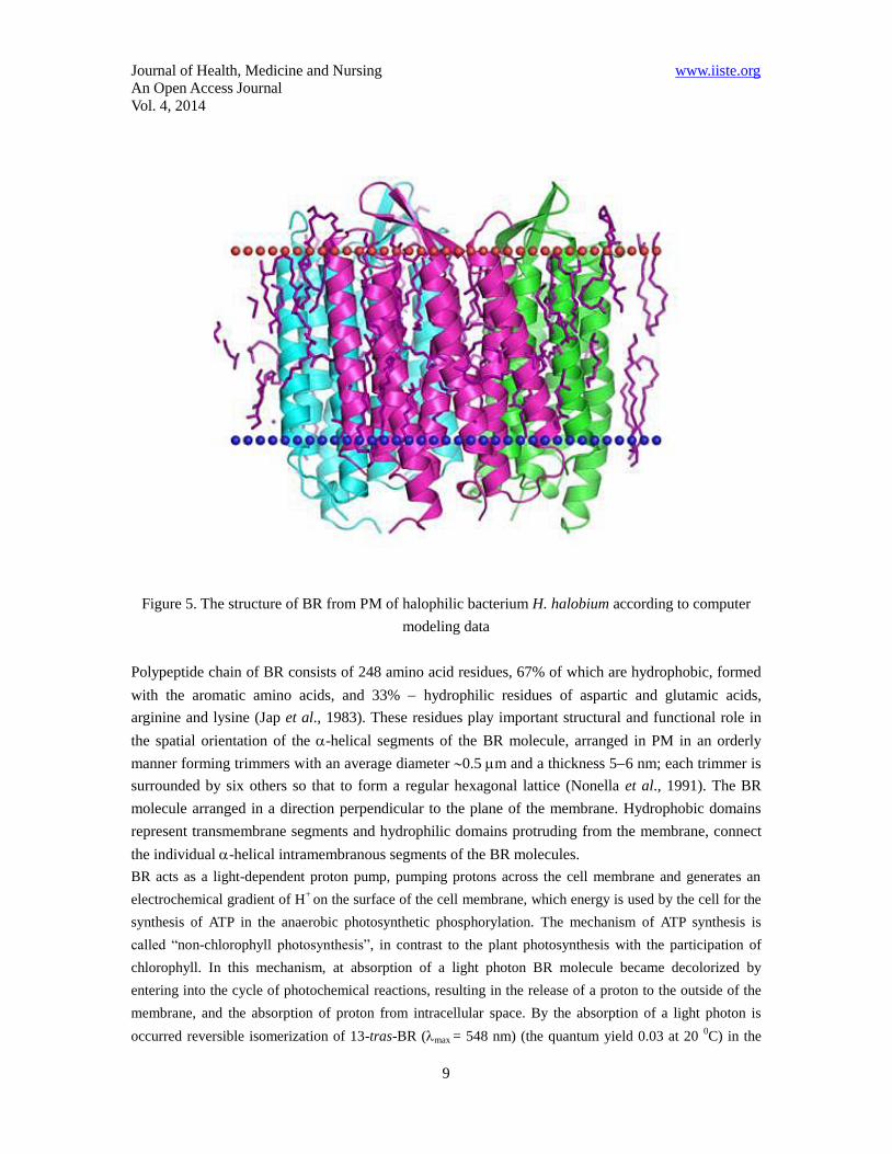
be investigated using electron microscopy diffraction methods as X-ray scattering (Lanyi, 2004). These
methods have established the existence in the BR molecule seven -helical protein segments, while in the
middle are symmetrically located a retinal residue (Fig. 5).
Journal of Health, Medicine and Nursing www.iiste.org
An Open Access Journal
Vol. 4, 2014
9
Figure 5. The structure of BR from PM of halophilic bacterium H. halobium according to computer
modeling data
Polypeptide chain of BR consists of 248 amino acid residues, 67% of which are hydrophobic, formed
with the aromatic amino acids, and 33% hydrophilic residues of aspartic and glutamic acids,
arginine and lysine (Jap et al., 1983). These residues play important structural and functional role in
the spatial orientation of the -helical segments of the BR molecule, arranged in PM in an orderly
manner forming trimmers with an average diameter 0.5 m and a thickness 56 nm; each trimmer is
surrounded by six others so that to form a regular hexagonal lattice (Nonella et al., 1991). The BR
molecule arranged in a direction perpendicular to the plane of the membrane. Hydrophobic domains
represent transmembrane segments and hydrophilic domains protruding from the membrane, connect
the individual -helical intramembranous segments of the BR molecules.
BR acts as a light-dependent proton pump, pumping protons across the cell membrane and generates an
electrochemical gradient of H+
on the surface of the cell membrane, which energy is used by the cell for the
synthesis of ATP in the anaerobic photosynthetic phosphorylation. The mechanism of ATP synthesis is
called “non-chlorophyll photosynthesis”, in contrast to the plant photosynthesis with the participation of
chlorophyll. In this mechanism, at absorption of a light photon BR molecule became decolorized by
entering into the cycle of photochemical reactions, resulting in the release of a proton to the outside of the
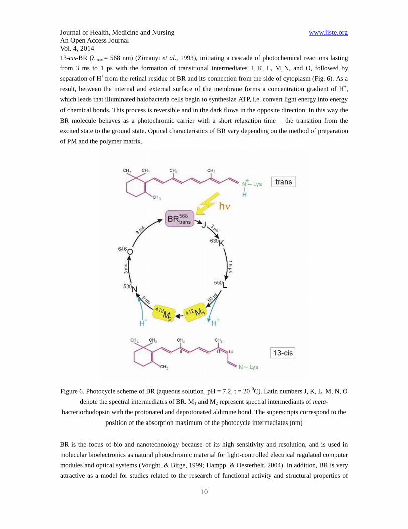
membrane, and the absorption of proton from intracellular space. By the absorption of a light photon is
occurred reversible isomerization of 13-tras-BR (max = 548 nm) (the quantum yield 0.03 at 20 0C) in the
Journal of Health, Medicine and Nursing www.iiste.org
An Open Access Journal
Vol. 4, 2014
10
13-cis-BR (max = 568 nm) (Zimanyi et al., 1993), initiating a cascade of photochemical reactions lasting
from 3 ms to 1 ps with the formation of transitional intermediates J, К, L, М, N, and O, followed by
separation of H+
from the retinal residue of BR and its connection from the side of cytoplasm (Fig. 6). As a
result, between the internal and external surface of the membrane forms a concentration gradient of H+,
which leads that illuminated halobacteria cells begin to synthesize ATP, i.e. convert light energy into energy
of chemical bonds. This process is reversible and in the dark flows in the opposite direction. In this way the
BR molecule behaves as a photochromic carrier with a short relaxation time the transition from the
excited state to the ground state. Optical characteristics of BR vary depending on the method of preparation
of PM and the polymer matrix.
Figure 6. Photocycle scheme of BR (aqueous solution, pH = 7.2, t = 20 0C). Latin numbers J, K, L, M, N, O
denote the spectral intermediates of BR. M1 and M2 represent spectral intermediants of meta-
bacteriorhodopsin with the protonated and deprotonated aldimine bond. The superscripts correspond to the
position of the absorption maximum of the photocycle intermediates (nm)
BR is the focus of bio-and nanotechnology because of its high sensitivity and resolution, and is used in
molecular bioelectronics as natural photochromic material for light-controlled electrical regulated computer
modules and optical systems (Vought, & Birge, 1999; Hampp, & Oesterhelt, 2004). In addition, BR is very
attractive as a model for studies related to the research of functional activity and structural properties of
Journal of Health, Medicine and Nursing www.iiste.org
An Open Access Journal
Vol. 4, 2014
11
photo-transforming membrane proteins in the native and photo-converting membranes (Wang et al., 2008).
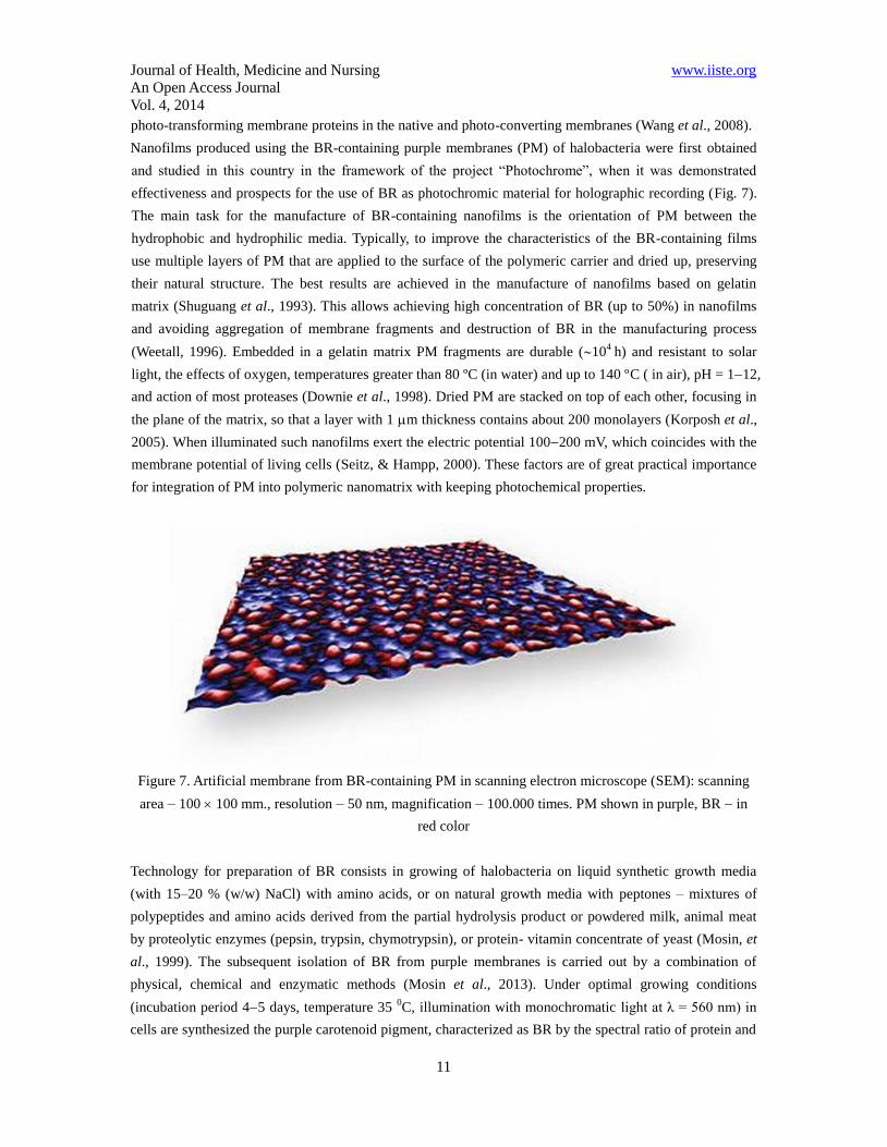
Nanofilms produced using the BR-containing purple membranes (PM) of halobacteria were first obtained
and studied in this country in the framework of the project “Photochrome”, when it was demonstrated
effectiveness and prospects for the use of BR as photochromic material for holographic recording (Fig. 7).
The main task for the manufacture of BR-containing nanofilms is the orientation of PM between the
hydrophobic and hydrophilic media. Typically, to improve the characteristics of the BR-containing films
use multiple layers of PM that are applied to the surface of the polymeric carrier and dried up, preserving
their natural structure. The best results are achieved in the manufacture of nanofilms based on gelatin
matrix (Shuguang et al., 1993). This allows achieving high concentration of BR (up to 50%) in nanofilms
and avoiding aggregation of membrane fragments and destruction of BR in the manufacturing process
(Weetall, 1996). Embedded in a gelatin matrix PM fragments are durable (104
h) and resistant to solar
light, the effects of oxygen, temperatures greater than 80 ºC (in water) and up to 140 ºC ( in air), pH = 112,
and action of most proteases (Downie et al., 1998). Dried PM are stacked on top of each other, focusing in
the plane of the matrix, so that a layer with 1 m thickness contains about 200 monolayers (Korposh et al.,
2005). When illuminated such nanofilms exert the electric potential 100200 mV, which coincides with the
membrane potential of living cells (Seitz, & Hampp, 2000). These factors are of great practical importance
for integration of PM into polymeric nanomatrix with keeping photochemical properties.
Figure 7. Artificial membrane from BR-containing PM in scanning electron microscope (SEM): scanning
area – 100 100 mm., resolution – 50 nm, magnification – 100.000 times. PM shown in purple, BR in
red color
Technology for preparation of BR consists in growing of halobacteria on liquid synthetic growth media
(with 15–20 % (w/w) NaCl) with amino acids, or on natural growth media with peptones – mixtures of
polypeptides and amino acids derived from the partial hydrolysis product or powdered milk, animal meat
by proteolytic enzymes (pepsin, trypsin, chymotrypsin), or protein- vitamin concentrate of yeast (Mosin, et
al., 1999). The subsequent isolation of BR from purple membranes is carried out by a combination of
physical, chemical and enzymatic methods (Mosin et al., 2013). Under optimal growing conditions
(incubation period 45 days, temperature 35 0C, illumination with monochromatic light at λ = 560 nm) in
cells are synthesized the purple carotenoid pigment, characterized as BR by the spectral ratio of protein and
Journal of Health, Medicine and Nursing www.iiste.org
An Open Access Journal
Vol. 4, 2014
12
chromophore fragments D280/D568 = 1.5:1.0 in the molecule.
Within the framework of the research we described an effective method for isolation of BR from PM of
photo-organo-heterotrophic halobacterium H. halobium consisted by cellular autolysis by distilled water,
processing of bacterial biomass by ultrasound at 22 KHz, location of PM fraction, purification of PM from
low and high-molecular weight impurities, cellular RNA, carotenoids and lipids, PM solubilization in 0.5%
(w/v) solution of the ionic detergent SDS-Na to form a microemulsion with the subsequent fractionation of
the protein by methanol (Mosin, & Ignatov, 2013a). The protein is localized in the PM; the release of low
molecular weight impurities and intracellular contents is reached by osmotic shock of cells with distilled
water in the cold after the removal of 4.3 M NaCl and the subsequent destruction of the cell membrane by
ultrasound at 22 kHz. For the destruction of cellular RNA the cellular homogenate was treated with Rnase I.
Fraction PM along with the desired protein in a complex with lipids and polysaccharides also contained
impurity of related carotenoids and proteins. Therefore, it was necessary to use special methods of
fractionation of the protein without damaging its native structure and dissociation.
BR being a transmembrane protein intricately penetrates bilipid layer in form of seven -helices; the use of
ammonium sulfate and other conventional agents to salting out did not give a positive result for isolation of
the protein. The resolving was in the translation of the protein to a soluble form by the colloidal dissolution
(solubilization) in an ionic detergent. Using as the ionic detergent SDS-Na was dictated by the need of
solubilization of the protein in a native, biologically active form in complex with 13-trans-retinal, because
BR solubilized in 0.5% (v/v) SDS-Na retains a native -helical configuration (Mosin, & Ignatov, 2013b).
Therefore, there is no need the use organic solvents as acetone, methanol and chloroform for purification of
lipids and protein, and precipitation and delipidization are combined in a single step, which significantly
simplifies the further fractionation. A significant advantage of this method is that the isolated protein in
complex with lipids and detergent molecules was distributed in the supernatant, and other high molecular
weight impurities – in unreacted precipitate, easily separated by centrifugation. Fractionation of solubilized
in 0.5% (w/v) SDS-Na protein and its subsequent isolation in crystalline form was achieved at 4 0C in three
steps precipitating procedure with methanol, reducing the concentration of detergent from 0.5, 0.25 and
0.1% (w/v) respectively. The final stage of BR purification involved the separation of the protein from
low-molecular-weight impurities by gel-permeation chromatography on dextran Sephadex G-200 Column
balanced with 0.09 M Tris-borate buffer (pH = 8.35) with 0.1% (w/v) SDS-Na and 2.5 mM EDTA (output of
the protein 810 mg).
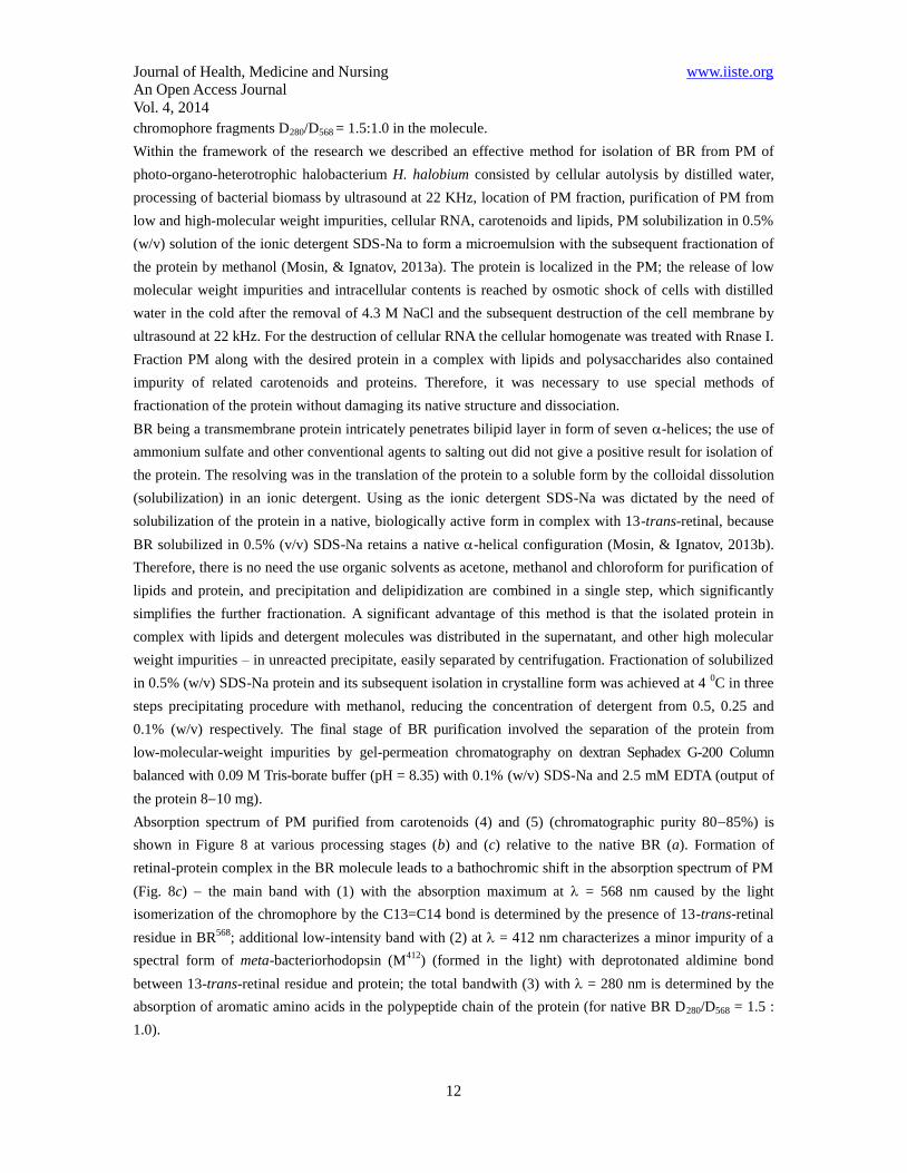
Absorption spectrum of PM purified from carotenoids (4) and (5) (chromatographic purity 8085%) is
shown in Figure 8 at various processing stages (b) and (c) relative to the native BR (a). Formation of
retinal-protein complex in the BR molecule leads to a bathochromic shift in the absorption spectrum of PM
(Fig. 8c) the main band with (1) with the absorption maximum at = 568 nm caused by the light
isomerization of the chromophore by the C13=C14 bond is determined by the presence of 13-trans-retinal
residue in BR568
; additional low-intensity band with (2) at = 412 nm characterizes a minor impurity of a
spectral form of meta-bacteriorhodopsin (M412
) (formed in the light) with deprotonated aldimine bond
between 13-trans-retinal residue and protein; the total bandwith (3) with = 280 nm is determined by the
absorption of aromatic amino acids in the polypeptide chain of the protein (for native BR D280/D568 = 1.5 :
1.0).
Journal of Health, Medicine and Nursing www.iiste.org
An Open Access Journal
Vol. 4, 2014
13
Figure 8. The absorption spectra of the PM (50% (v/v) ethanol) at various stages of processing: (a) –
natural BR; (b) – PM after intermediate treatment; (c) – PM purified from carotenoids. The bandwith (1) is
the spectral form of BR568
, (2) – impurity of spectral form of meta-bacteriorhodopsin (M412
), (3) – the total
absorption bandwith of aromatic amino acids, (4) and (5) – extraneous carotenoids. As a control used the
native BR
3.4. Iodopsin
Iodopsin is a violet, light-sensitive pigment of the retinal cone cells, responsible for color vision, and close
analogue of rhodopsin. This pigment consists of a protein photopsin linked with a chromophore, retinal
residue. According to the three-component theory of vision, it is believed that there have to be three types
of this pigment and accordingly three types of cones that are sensitive to blue, green and red light. Iodopsin
consists of three pigments – hlorolab, eritrolab and tsianolab. With the densitometry method W. Rushton
studied the coefficient of light absorption in the photo layers of the retina with different wavelengths
(Rushton, 1958). The hlorolab pigment absorbs the rays corresponding to yellow-green (450–630 nm
absorption band), the eritrolab – yellow and red (500-700 nm), and the tsianolab – blue-green ( 500–700
nm) parts of the visible spectrum (Wyszecki, & Stiles, 1982). Not yet been found and the different types of
cones.
3.5. The mechanism of color vision
Journal of Health, Medicine and Nursing www.iiste.org
An Open Access Journal
Vol. 4, 2014
14
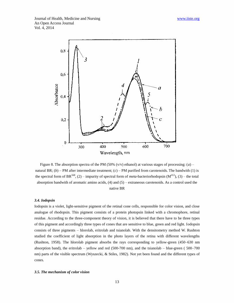
The retina has three types of cone cells – S, M and L cells, having a different sensitivity to different parts of
the visible range of the spectrum (Fig. 9). The cone cells of S type have a spectral range from 400 to 500
nm with a maximum peak at 420–440 nm, the cone cells of M type – from 450 to 630 nm with a maximum
peak at 534–555 nm, while the cone cells of L type – from 500 nm to 700 nm with a maximum peak at
564–580 nm. As the curves of the sensitivity of the cone cells overlap, it is impossible for monochromatic
light to stimulate only one type of cone cells. The other types of cone cells react though to a lesser degree.
The set of all possible values of the color combinations causing a visual reaction determines the human
color space. Human brain generally can discern approximately 10 million of different colors.
Figure 9. Spectral sensitivity of the different types of receptor cells (cones) in the retina
The electromagnetic waves spectrum stimulates the different types of cone cells from the three types S, L
and M to a different degree. The red light stimulates the L cone cells more than the M cone cells. The blue
light stimulates the S cone cells in the strongest way. The yellow-green light provides a strong stimulation
to the L and M cone cells, and a weaker stimulation to the S cone cells. The brain then combines the
information from all types of cone cells for different wavelengths and analyzes them as different colors.
3.6.Studying of additive mixing of colors
The analyses for the activity of the three types of cones – S, L and M in the perception of colors also show
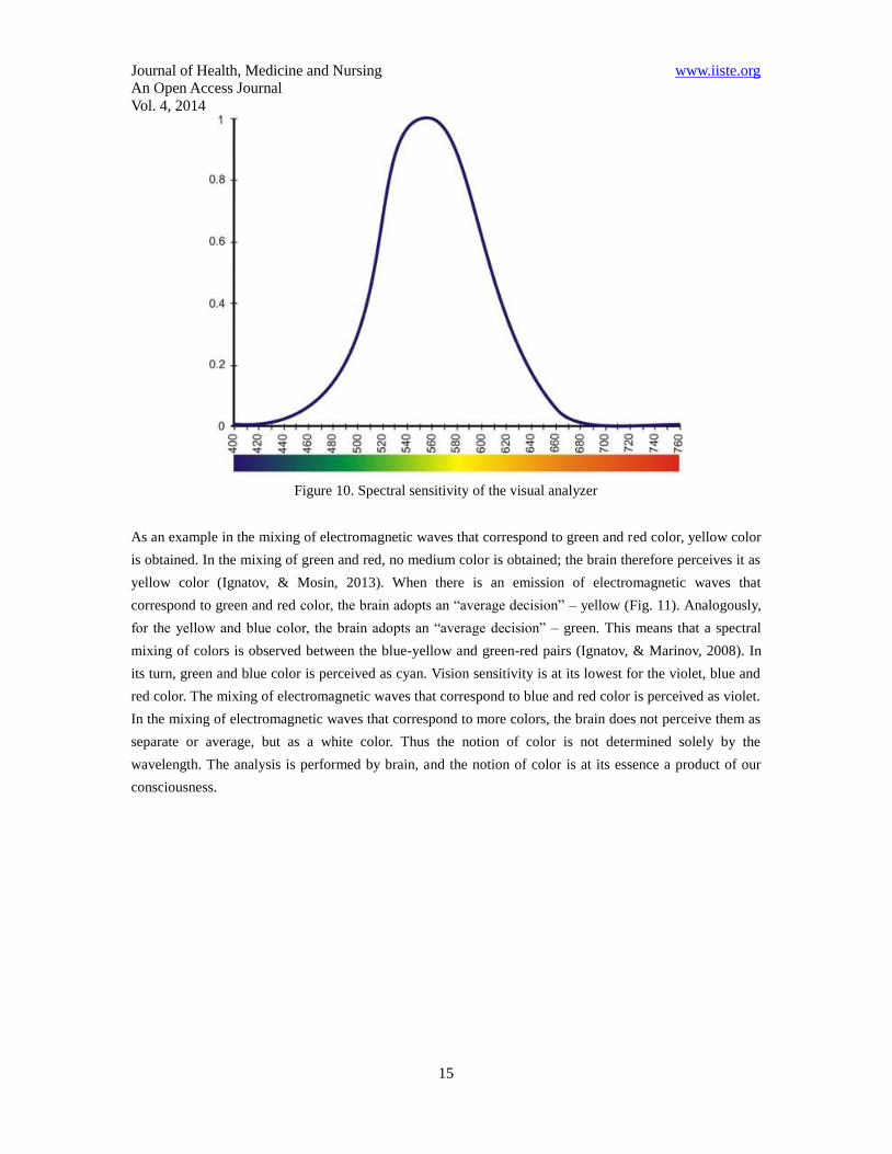
how the brain “deciphers” the colors. The foundation of this analysis, shown in Figure 10, was made by M.
Marinov and I. Ignatov in 2008. However, it is not clear whether the green color we perceive is a combined
effect of yellow and blue, or whether it corresponds to a wavelength of the green color from the visible
spectrum. Our brain can register the colors, i.e. the green color as a spectrometer, with certain lengths of the
electromagnetic waves. It can also register the green color as a mixture of yellow and blue. The full
perception of colors by the visual analyzer cannot be defined by a spectrometer (Hunt, 2004).
Journal of Health, Medicine and Nursing www.iiste.org
An Open Access Journal
Vol. 4, 2014
15
Figure 10. Spectral sensitivity of the visual analyzer
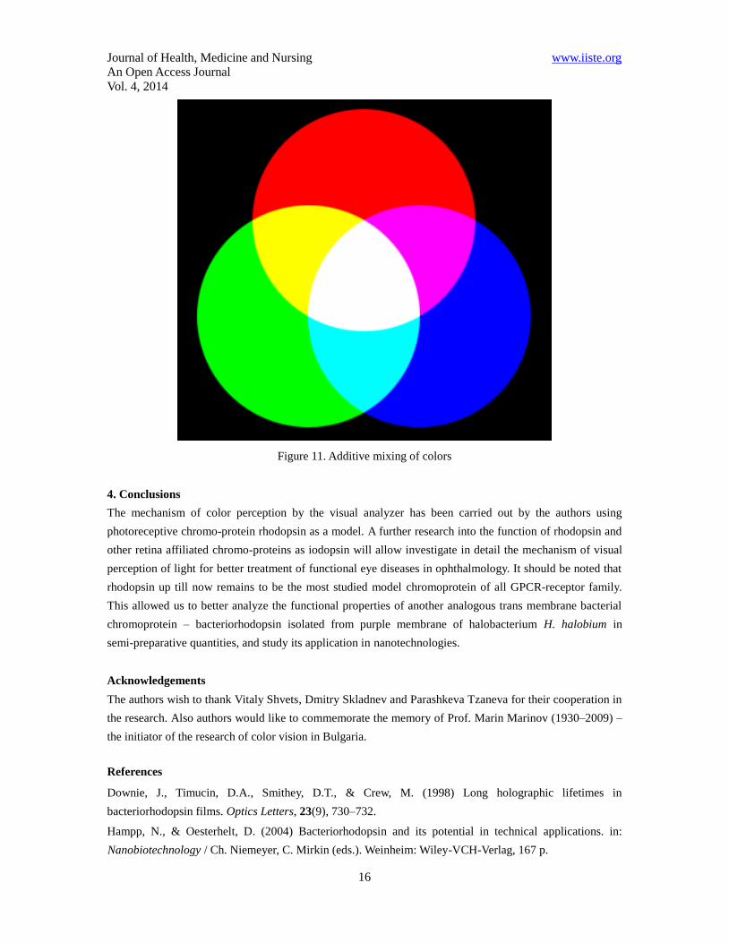
As an example in the mixing of electromagnetic waves that correspond to green and red color, yellow color
is obtained. In the mixing of green and red, no medium color is obtained; the brain therefore perceives it as
yellow color (Ignatov, & Mosin, 2013). When there is an emission of electromagnetic waves that
correspond to green and red color, the brain adopts an “average decision” – yellow (Fig. 11). Analogously,
for the yellow and blue color, the brain adopts an “average decision” – green. This means that a spectral
mixing of colors is observed between the blue-yellow and green-red pairs (Ignatov, & Marinov, 2008). In
its turn, green and blue color is perceived as cyan. Vision sensitivity is at its lowest for the violet, blue and
red color. The mixing of electromagnetic waves that correspond to blue and red color is perceived as violet.
In the mixing of electromagnetic waves that correspond to more colors, the brain does not perceive them as
separate or average, but as a white color. Thus the notion of color is not determined solely by the
wavelength. The analysis is performed by brain, and the notion of color is at its essence a product of our
consciousness.
Journal of Health, Medicine and Nursing www.iiste.org
An Open Access Journal
Vol. 4, 2014
16
Figure 11. Additive mixing of colors
4. Conclusions
The mechanism of color perception by the visual analyzer has been carried out by the authors using
photoreceptive chromo-protein rhodopsin as a model. A further research into the function of rhodopsin and
other retina affiliated chromo-proteins as iodopsin will allow investigate in detail the mechanism of visual
perception of light for better treatment of functional eye diseases in ophthalmology. It should be noted that
rhodopsin up till now remains to be the most studied model chromoprotein of all GPCR-receptor family.
This allowed us to better analyze the functional properties of another analogous trans membrane bacterial
chromoprotein – bacteriorhodopsin isolated from purple membrane of halobacterium H. halobium in
semi-preparative quantities, and study its application in nanotechnologies.
Acknowledgements
The authors wish to thank Vitaly Shvets, Dmitry Skladnev and Parashkeva Tzaneva for their cooperation in
the research. Also authors would like to commemorate the memory of Prof. Marin Marinov (1930–2009) –
the initiator of the research of color vision in Bulgaria.
References
Downie, J., Timucin, D.A., Smithey, D.T., & Crew, M. (1998) Long holographic lifetimes in
bacteriorhodopsin films. Optics Letters, 23(9), 730–732.
Hampp, N., & Oesterhelt, D. (2004) Bacteriorhodopsin and its potential in technical applications. in:
Nanobiotechnology / Ch. Niemeyer, C. Mirkin (eds.). Weinheim: Wiley-VCH-Verlag, 167 p.
Journal of Health, Medicine and Nursing www.iiste.org
An Open Access Journal
Vol. 4, 2014
17
Hargrave, P.A., McDowell, J.H., Curtis, D.R. et al. (1983) The structure of bovine rhodopsin. Biophys.
Struct. Mech., 9, 235–244.
Henderson, R., Baldwin, J., Ceska, T. et al. (1990) Model for the structure of bacteriorhodopsin based
on high-resolution electron cryo-microscopy. J. Mol. Biol., 213(4), 899–929.
Hogan, M. J., Alvarado, J. A., & Weddell, J. E. (1971) Histology of the Human Eye. Philadelphia: WB
Saunders Co., 115 p.
Hubel, D. (1995) Eye, Brain and Vision. Scientific American Library Series (Book 22), 2nd edition, New
York: W.H. Freeman Publ., 256.
Hunt, R.W.G. (2004). The Reproduction of Colour (6th ed.). Chichester: Wiley–IS&T Series in Imaging
Science and Technology, 724 .
Ignatov, I. (2005) Energy Biomedicine, Gea-Libris, Sofia, 1–88.
Ignatov, I. (2010) Which water is optimal for the origin (generation) of life? Euromedica, Hanover: 34-35.
Ignatov, I. (2011) Entropy and time in living matter, Euromedica: 74.
Ignatov, I. (2012) Origin of Life and Living Matter in Hot Mineral Water, Conference on the Physics,
Chemistry and Biology of Water, Vermont Photonics, USA.
Ignatov, I., & Mosin, O.V. (2012) Isotopic Composition of Water and its Temperature in Modeling of
Primordial Hydrosphere Experiments, VIII Intern. Conference Perspectives of the Development of Science
and Technique, Biochemistry and Biophysics, 15: 41–49.
Ignatov, I., Mosin, O. V. & Naneva, K. (2012) Water in the Human Body is Information Bearer about
Longevity, Euromedica, Hanover: 110-111.
Ignatov I., Mosin O.V. (2013) Possible Processes for Origin of Life and Living Matter with Modeling of
Physiological Processes of Bacterium Bacillus Subtilis in Heavy Water as Model System, Journal of
Natural Sciences Research, 3 (9): 65-76.
Ignatov, I., Mosin, O. V. (2013) Modeling of Possible Processes for Origin of Life and Living Matter in Hot
Mineral and Seawater with Deuterium, Journal of Environment and Earth Science, 3(14): 103-118.
Ignatov, I., Mosin, O. V. (2013) Structural Mathematical Models Describing Water Clusters, Journal of
Mathematical Theory and Modeling, 3 (11): 72-87.
Ignatov, I., Mosin, O. V. (2014) The Structure and Composition of Carbonaceous Fullerene Containing
Mineral Shungite and Microporous Crystalline Aluminosilicate Mineral Zeolite. Mathematical Model of
Interaction of Shungite and Zeolite with Water Molecules Advances in Physics Theories and Applications,
28: 10-21.
Ignatov, I., Mosin, O.V., Velikov, B., Bauer, E. & Tyminski, G. (2014) Longevity Factors and Mountain
Water as a Factor. Research in Mountain and Field Areas in Bulgaria, Civil and Environmental Research, 6
(4): 51-60.
Ignatov, I., Mosin, O. V., Niggli, H.&Drossinakis, Ch. (2014) Evaluating Possible Methods and Approaches
for Registering of Electromagnetic Waves Emitted from the Human Body, Advances in Physics Theories
and Applications, 30: 15-33.
Ignatov, I., Mosin, O.V.&Drossinakis, Ch. (2014) Infrared Thermal Field Emitted from Human Body.
Thermovision, Journal of Medicine, Physiology, Biophysics, 1:1-12.
Ignatov, I., Mosin, O.V.&Velikov, B. (2014) Longevity Factors and Mountain Water of Bulgaria in Factorial
Research of Longevity, Journal of Medicine, Physiology, Biophysics,1:13-33.
Journal of Health, Medicine and Nursing www.iiste.org
An Open Access Journal
Vol. 4, 2014
18
Ignatov, I.&Mosin,O.V. (2014) Visual Perception. Electromagnetic Conception for the Eyesight. Rhodopsin
and Bacteriodopsin, Journal of Medicine, Physiology and Biophysics, 2:1-19.
Ignatov, I.&Mosin,O.V. (2014) The Structure and Composition of Shungite and Zeolite. Mathematical
Model of Distribution of Hydrogen Bonds of Water Molecules in Solution of Shungite and Zeolite, Journal
of Medicine, Physiology and Biophysics, 2: 20-36.
Ignatov, I., Mosin,O.V., Velikov, B., Bauer, E.&Tyminski, G. (2014) Research of Longevity Factors and
Mountain Water as a Factor in Teteven Municipality, Bulgaria, Journal of Medicine, Physiology and
Biophysics, 2: 37-52.
Ignatov, I.&Mosin,O.V. (2014) Modeling of Possible Processes for Origin of Life and Living Matter in Hot
Mineral Water. Research of Physiological Processes of Bacterium Bacillus Subtilis in Hot Heavy Water,
Journal of Medicine, Physiology and Biophysics, 2: 53-70.
Ignatov, I.&Mosin,O.V. (2014) Mathematical Models of Distribution of Water Molecules Regarding
Energies of Hydrogen Bonds, Medicine, Physiology and Biophysics, 2: 71-94.
Ignatov, I.&Mosin,O.V. (2014) Studying of Phototransformans of Light Signal by Photoreceptor
Pigments – Rhodopsin, Iodopsin and Bacteriorhopsin and Additive Mixing of Colors, Journal of
Medicine, Physiology and Biophysics, 3:30-47.
Ignatov, I.&Mosin,O.V. (2014) Mathematical Models Describing Water Clusters as Interaction among
Water Molecules. Distributions of Energies of Hydrogen Bonds, Journal of Medicine, Physiology and
Biophysics, 3: 48-70.
Jap, B.K., Maestre M.F., Hayward, S.B., & Glaeser, R.M. (1983) Peptide-chain secondary structure of
bacteriorhodopsin. Biophys J., 43(1), 81–89.
Korposh, S.O., Sichka, M.Y., Trikur, I.I. et al. (2005) Films based on bacteriorhodopsin in sol-gel matrices.
Proc. of SPIE, 5956, Paper Number 595616, 312–320.
Lanyi, J.K. (2004) X-ray diffraction of bacteriorhodopsin photocycle intermediates. Molecular Membrane
Biology, 21(3), 143–150.
Liang, Y., Fotiadis, D., Maeda, T. et al. (2004) Rhodopsin signaling and organization in heterozygote
rhodopsin knockout mice. J. Biol. Chem., 279, 48189–48196.
Lipkin, V.M. (2001) Visual system. mechanisms of transmission and amplification of the visual signal in
eye retina. Soros Educational Journal, 7(9), 2–8 [in Russian].
Mosin, O.V., Skladnev, D.A., Egorova, T.A. & Shvets, V.I. (1996) Mass-spectrometric determination of
levels of enrichment of 2Н and
13С in molecules of amino acids of various bacterial objects. Bioorganic
Chemistry, 22(10–11):856–869.
Mosin, O.V., Skladnev, D.A & Shvets, V.I. (1999) The inclusion of deuterated aromatic amino acids in the
molecule of bacteriorhodopsin Halobacterium halobium. Applied Biochemistry and Microbiology, 35(1):
34-42.
Mosin, O.V., Shvets, V.I., Skladnev, D.A. & Ignatov, I. (2012) Synthesis of [2Н]bacteriorhodopsin labeled
by deuterium on residues of aromatic amino acids. Khimicheskaya Technologiya (Chemical Engineering),
Publishing House “Nauka & Technology” Moscow, 9: 553–564.
Mosin, O.V., Shvets, V.I., Skladnev, D.A. & Ignatov, I. (2013) Biosynthesis of trans-membrain
Journal of Health, Medicine and Nursing www.iiste.org
An Open Access Journal
Vol. 4, 2014
19
photo-transforming protein [2Н]bacteriorhodopsin, labeled with deuterium on residues of aromatic amino
acids [2,3,4,5,6-2H5]Phe, [3,5-
2H2]Tyr and [2,4,5,6,7-
2H5]Trp. Problems of Biological, Medical and
Pharmaceutical Chemistry, 8: 29–39.
Mosin,O.V.&Ignatov, I. (2014) Improved of Method for Isolation of Photochrome Transmembrane Protein
Bacteriorhodopsin from Purple Membranes of Halobacterium Halobacterium Halobium, Physiology and
Biophysics, 3:71-86.
Mosin,O.V.&Ignatov, I. (2014) The Natural Phototransforming Photochrome Membrane Protein
Bacteriorhodopsin from Halobacterium Halobacterium Halobium, European Journal of Molecular
Biotechnology, 1 (1): 25-40.
Nathans, J., Thomas, D., & Hogness, D.S. (1986) Molecular genetics of human color vision: the genes
encoding blue, green, and red pigments. Science, 232(47), 193–202.
Neugebauer, D.Ch., Zingsheim, H.P., & Oesterhelt, D. (1978) Recrystallization of the purple membrane in
vivo and in vitro. Journal Molecular Biology, 123, 247–257.
Nonella, M., Windemuth, A., & Schulten K. (1991) Structure of Bacteriorhodopsin and in situ isomerization
of retinal: A molecular dynamics study. Journal Photochem. Photobiol., 54(6), 937–948.
Oesterhelt, D., & Stoeckenius, W. (1971) Rhodopsin - like protein from the purple membrane of
Halobacterium halobium. Nature, 233(89), 149–160.
Ovchinnikov, Yu.A., Abdulaev, N.G., Feigina, M.Yu., Artamonov, I.D., & Bogachuk, A.S. (1983) Visual
rhodopsin: Whole amino acid sequence and topology in membrane. Bioorganic. chemistry, 10, 1331–1340.
Palczewski, K. (2006) G-protein-coupled receptor rhodopsin. Annu. Rev. Biochem., 75, 743–767.
Palczewski, K., Kumasaka, T., Hori, T. et al. (2000) Crystal structure of rhodopsin: a G-protein-coupled
receptor. Science, 289, 739–745.
Rudiger, M., Tittor, J., Gerwert, K., & Oesterhelt, D. (1997) Reconstitution of bacteriorhodopsin from the
apoprotein and retinal studied by Fourier-transformed infrared spectroscopy. Biochemistry, 36, 4867–4874.
Rushton, W.A.H. (1958). In: Visual problems of colour. N.P.L. Sump. (Ed.) London: Her Majesty’s
Stationary Office, 1, 71–101.
Schertler, G.F., & Hargrave, P.A. (1995) Projection structure of frog rhodopsin in two crystal forms. Proc.
Natl. Acad. Sci. U.S.A., 92, 11578–11582.
Seitz, A., & Hampp, N. (2000) Kinetic optimization of bacteriorhodopsin films for holographic
interferometry. J. Phys. Chem. B, 104(30), 7183–7192.
Shuguang, W.U., Ellerby, L.M., Cohan, J.S. et al. (1993) Bacteriorhodopsin encapsulated in transparent
sol-gel glass: a new biomaterial. Chem. Mater, 5, 115–120.
Vought, B.W., & Birge, R.R. (Eds.) (1999) Molecular electronics and hybrid computers. in: Wiley
Encyclopedia of Electrical and Electronics Engineering. NY: Wiley-Interscience, 490 p.
Wang, W.W., Knopf, G.K., & Bassi, A.S. (2008) Bioelectronic imaging array based on bacteriorhodopsin
film. IEEE Transactions on Nanobioscience, 7(4), 249–256.
Weetall, H. (1996) Retention of bacteriorhodopsin activity in dried sol-gel glass. Biosensors &
Bioelectronics, 11, 325–333.
Wyszecki, G., & Stiles, W.S. (1982). Color Science: Concepts and Methods, Quantitative Data and
Formulae (2nd ed.). New York: Wiley–IS&T Series in Pure and Applied Optics, 935 p.
Zimanyi, L., Cao, Y., Needleman, R., Ottolenghi, M., & Lanyi J.K. (1993) Pathway of poton uptake in the

Journal of Health, Medicine and Nursing www.iiste.org
An Open Access Journal
Vol. 4, 2014
20
bacteriorhodopsin photocycle. Biochemistry, 32, 7669–7678.
The IISTE is a pioneer in the Open-Access hosting service and academic event
management. The aim of the firm is Accelerating Global Knowledge Sharing.
More information about the firm can be found on the homepage:
http://www.iiste.org
CALL FOR JOURNAL PAPERS
There are more than 30 peer-reviewed academic journals hosted under the hosting
platform.
Prospective authors of journals can find the submission instruction on the
following page: http://www.iiste.org/journals/ All the journals articles are available
online to the readers all over the world without financial, legal, or technical barriers
other than those inseparable from gaining access to the internet itself. Paper version
of the journals is also available upon request of readers and authors.
MORE RESOURCES
Book publication information: http://www.iiste.org/book/
IISTE Knowledge Sharing Partners
EBSCO, Index Copernicus, Ulrich's Periodicals Directory, JournalTOCS, PKP Open
Archives Harvester, Bielefeld Academic Search Engine, Elektronische
Zeitschriftenbibliothek EZB, Open J-Gate, OCLC WorldCat, Universe Digtial
Library , NewJour, Google Scholar