Embed Size (px)
Citation preview

~ Pergamon Neuropsychologia, Vol. 33, No. 8. pp. 937-959, 1995
Copyright © 1995 Elsevier Science Ltd Printed m Great Britain. All rights reserved
002g--3932/95 $9.5ll + 0.00
0028-3932(95)00032-1
V I S U A L I N T E G R A T I O N I N T H E S P L I T B R A I N
MICHAEL C. CORBALLIS
Department of Psychology, University of Auckland, Auckland, New Zealand
(Received 21 October 1994; accepted 9 February 1995)
Abstract--A review of evidence suggests that subjects with forebrain commissurotomy can integrate information about location, orientation, and movement between visual hemifields, albeit with some loss of spatial and temporal resolution, even when it is flashed briefly in parafoveal vision. The classic disconnection syndrome for visual stimuli is largely explained by the lack of subcortical transfer of information about form. Voluntary location-based ('spotlight') attention is integrated interhemispherically in these subjects but object-based attention and controlled visual search can function concurrently and independently in the two hemispheres. The data support the distinction between two visual systems, each with its own perceptual and attentional subsystems: (1) a relatively precise cortical system dedicated primarily to the analysis of form and the fine-grained exploration of visual scenes; and (2) a 'second' subcortical system dedicated to a relatively low- resolution processing of movement and location, including 'spotlight' attention.
Key Words: commissurotomy; subcortical vision; visual perception; interhemispheric integration.
INTRODUCTION
Since the classic studies of Sperry and his colleagues in the 1960s, the disconnection syndrome resulting from surgical section of the forebrain commissures has become part of modern folklore. In an often quoted passage, Sperry wrote that the two hemispheres behaved as if they had "two separate conscious entities or minds running in parallel in the same cranium, each with its own sensations, perceptions, cognitive processes, learning experiences, and so on" [65, p. 318]. Moreover each hemisphere was revealed to have rather different modes of thought, elegantly elaborated by Bogen [3].
One of the simplest and most convincing ways to demonstrate the independence of the disconnected hemispheres has been through the use of visual input, since each visual hemifield projects to the contralateral hemisphere. Information can therefore be restricted to a single hemisphere by flashing information in a single hemifield while the subject maintains fixation. Experiments using this technique quickly confirmed that speech was located in the left hemisphere, since split-brained subjects could not name words or objects flashed in the left hemifield [27, 65, 66]. Some tests revealed superior performance with input restricted to the right hemifield, indicating superior processing in the right cerebral hemisphere. For example, one split-brained subject could carry out a simple mental- rotation test when the stimuli were flashed in the left hemifield, but failed to score above chance when they were flashed in the right hemifield [15], implying that input to the right hemifield was denied access to the dominant right hemisphere. In later testing, this subject's left hemisphere has achieved above-chance levels of performance, while never
937

938 M.C. CORBALLIS
reaching the level of competence of the right [16]. Another classic demonstration of disconnection is that split-brained subjects perform at little or no better than chance in judging whether stimuli in the two hemifields are the same or different [12, 17, 22, 27, 36, 59].
Despite these dramatic findings it has often been noted that, in their everyday lives, split-brained subjects do not behave as though the visual world were split; in Bogen's words, they display "social ordinariness" [5, p. 339]. For example, there is little sign of hemineglect [39, 49], as one might expect if only one cerebral hemisphere at a time views the world, although we shall see later that there may be exceptions to this. Of course there are compensatory mechanisms. The subject is free to make eye movements, thereby bringing visual stimuli on one side of the split across to the other side. At worst, then, this strategy might be an inconvenience, slowing down the rate at which each hemisphere can acquire visual information, and it almost certainly has a detrimental effect on complex visual skills such as reading [27, 71].
To some extent, moreover, the two hemispheres can act independently without conflict, giving the outward appearance of unitary behavior. For example, in simple confrontation testing, a split-brain subject asked to signal the onset of a stimulus, such as the wiggling of the examiner's fingers, can easily point to a stimulus in the left hemifield with the left hand and to a stimulus in the right hemifield with the right hand, and indeed may even do both simultaneously, indicating parallel processing in the two hemispheres [5]. In many everyday situations, the left hand and right hemisphere can presumably process events in the left hemifield, and the right hand and left brain can process events in the right hemifield, with little outward sign of disconnection.
As we shall see, however, there is evidence for at least some interhemispheric transfer of visual information even in tasks that require integration between hemifields and in which eye movements are controlled. This transfer is presumably accomplished via subcortical systems, although in some cases external cross-cuing may also play a role [4]. The following review will deal only with surgical cases, and not with cases of callosal agenesis, who present a rather different picture; these were dealt with elsewhere in this issue. Some subjects, described in this review as commissurotomized, have undergone complete forebrain commissurotomy, including section of the corpus callosum, anterior commissure, hippocampal commissure, and when present, the massa intermedia. Others, described as callosotomized, have undergone section of the corpus callosum only. There is at present no good evidence that these two classes of subjects differ with respect to the transfer or integration of visual information. I shall use the term split-brained to cover both classes. The review is organized in terms of a number of principles that have been proposed to govern the nature or extent of visual integration in split-brained subjects.
REVIEW OF EVIDENCE
Ambient vs focal perception
In what must be regarded as the pioneering study of interhemispheric visual integration in the human split brain, Trevarthen and Sperry [72] claimed that integration was possible within the so-called ambient system, but not within the focal system. This distinction was developed by Trevarthen [70] from earlier experiments carried out on split-brained monkeys. The monkeys had undergone section of the optic chiasm as well as of the

VISUAL INTEGRATION IN THE SPLIT BRAIN 939
forebrain commissures, so that each cerebral hemisphere received input only from the ipsilateral eye. By using orthogonally oriented polarizers, Trevarthen [68, 69] was able to present different patterns simultaneously to each eye, and so test the ability of the two hemispheres to learn different, even conflicting, discriminations.
When the stimuli were different forms, such as a circle and a cross, the animal could learn without apparent interference to reach with the same hand to what was seen by one hemisphere as a circle and the other hemisphere as a cross, but to avoid reaching when the stimuli were reversed. This suggested to Trevarthen that the discrimination was cortically mediated. But when the stimuli were a dark vs a light panel, or red vs blue, the animal responded on the basis of the stimulus seen by one eye only, the other view being suppressed. Later testing of each hemisphere separately showed that they both responded in the same way, treating one panel as positive and the other as negative, even though one hemisphere had been presented with the reverse discrimination. Trevarthen concluded that these discriminations must have been subcortically mediated, and the conflict resolved by internal suppression of the information received by one eye. In another case, in which the stimuli were a 5-point star vs a 6-point star, there was only partial suppression, suggesting both cortical and subcortical contributions. Trevarthen noted that the discriminations for which there was no conflict had to do with the processing of form, while those for which there was conflict were related to a different kind of visual processing.
He identified these two kinds of visual processing as focal and ambient, respectively. Extrapolating from his findings, he argued that focal vision is cortical, centered in the foveal region, and dedicated primarily to detailed vision and the identification of objects, while ambient vision is concerned with space at large around the body, and is correspondingly more sensitive to peripheral vision and to moving stimuli. It is largely subcortical, involving projections via the superior colliculus to the pulvinar nucleus of the thalamus, where it interacts with visual association areas of the cortex. This system is also sometimes referred to as the "second" visual system [4].
To demonstrate interhemispheric transfer within the ambient system in commissur- otomized humans, Trevarthen and Sperry [72] moved large shadowcast stimuli into peripheral vision and required the subjects to hold fixation while they viewed them. These procedures contrasted with the more usual procedures of flashing stationary stimuli close to central vision--procedures that favor focal over ambient vision. Three subjects (L.B., A.A. and N.G.) were able make quite accurate judgments about the relative motion of disks and lines located some 45 deg from fixation in the two hemifields, although all showed some degree of neglect of the stimulus on the left, and one subject (R.Y.) was unable to do the tasks because of persistent neglect.
The subjects were also able to make crude verbal statements about stimuli in the left hemifield. All four of them could verbally acknowledge the appearance of a simple stimulus, including a disk of white light or a dark line, as it moved in from the periphery, with one (L.B.) achieving sensitivity comparable to that of a normal subject. They also responded quite well to changes in size and brightness, but descriptions of shapes were poor. Nevertheless simple properties could be described, such as whether a shape was elongated or square, or whether it was single or composed of two parts. The subjects could also crudely identify colors in the left hemifield, at a level approximating that of a dichromat. Transfer might have been mediated by any of a number of possible subcortical routes, including those in the diencephalon (posterior commissure),

940 M.C. CORBALLIS
in the midbrain (superior colliculi, central gray, or reticular formation), or even possibly in the cerebellum [29, 71].
According to Trevarthen and Sperry, the classic disconnection syndrome in the split brain applies only to the focal system, which is cortically mediated. This explains why split-brained monkeys were able to sustain conflicting discriminations of visual forms in the two hemispheres. This was later demonstrated in eommissurotomized humans by Levy et al. [40] using so-called chimeric stimuli. These comprised half-pictures, such as a half- rose and a half-bee, joined at the midline, so that when centrally fixated each half projected to a different hemisphere. Each hemisphere, through the mechanism of perceptual completion, operated as though it perceived a whole picture. Under certain conditions these opposing percepts were held simultaneously, so that, to the subject's consternation, the left hemisphere might verbally report seeing a bee while the right hemisphere directed the left hand to point to a picture of a rose.
Low-level vs high-level information
Trevarthen and Sperry associated the focal system with central vision and the ambient system mainly with peripheral vision. Since most experiments demonstrating disconnec- tion have used fairly intricate patterns, such as digits, letters, words, or pictures, presented fairly close to the fovea, virtually no transfer would be expected. However it has long been known that commissurotomized subjects can report the presence of a stimulus flashed in the left hemifield, even if they cannot say what it was [5, 25]. More recent evidence suggests that each hemisphere may have at least some information concerning such low-level attributes as location or orientation of stimuli flashed to the other hemisphere. Trevarthen [71], for example, has acknowledged that there is some interhemispheric transfer of visual information presented near the fovea, and suggested that it is restricted to low-resolution, high-contrast stimuli.
Holtzman [32] showed that callosotomized subjects could quite accurately direct an eye movement to a specific location in one visual hemifield on the basis of a locational probe flashed briefly in the other. Location was specified relative to a 2 x 2 matrix of Xs, the probe being a circular shape briefly surrounding one of the Xs. The subjects could quickly and with reasonable accuracy direct their gaze to the equivalent position in matrix in the other hemifield, even though the two matrices differed in vertical location. However they could not direct eye movements across hemifields if the probe in one hemifield depicted a shape rather than a location. For example, if the probe was a cross, the subjects were unable to move their eyes to the cross in a 2 x 2 matrix that also comprised a circle, a square, and a triangle.
Holtzman suggested that this last result was due to a failure of resolution, since the geometric shapes each subtended only 0.25 deg of visual angle, whereas the matrices themselves subtend 2 deg in height and width. His conclusion was that surgical disconnection splits the visual world "for the perceptual resolution of detail" [32, p. 810; italics in original]. If so, then the divided visual world of the split-brained subject might be similar to that of having a blurring lens over one half of the visual field. It may be, however, that the disconnection is not simply a matter of resolution, but has to do also with the level of perceptual processing--that is, Holtzman's subjects may have been unable to transfer the information about the shapes, not because they were small, but because they were shapes. This interpretation seems to be preferred by Gazzaniga [26], who

VISUAL INTEGRATION IN THE SPLIT BRAIN 941
acknowledged the transfer of crude spatial information in the split brain, but was adamant that perceptual information remained disconnected (see also Ref. [63]).
Holtzman [32] also showed that a subject with hemianopia in the right hemifield, unlike the callosotomized subjects, was unable to direct eye movements to specific locations in that hemifield based on locational information in the other. From this he concluded that the transfer of spatial information requires the integrity of the cortex, even though it may be filtered through subcortical systems with low resolution.
The extent of perceptual transfer was documented further by Sergent [60], who reported that two commissurotomized subjects (L.B. and N.G.) could quickly and accurately perform a number of tasks requiring integration between the visual hemifields. For example, they could judge whether or not arrows flashed in opposite visual hemifields were aligned, whether an arrow in one hemifield pointed at a dot in the other, or whether lines in opposite hemifields formed an angle of greater or less than 90 deg. Sergent provided little evidence as to the precision of these judgments. However Corballis and Trudel [18] compared the performance of L.B. and D.K., a subject with section of the posterior corpus callosum only, with that of normal controls on a task requiring judgments of whether pairs of sloping 45-deg lines were aligned or not. L.B.'s performance was only very slightly worse with bilateral than with unilateral presentation, and did not differ from the normals' under any of these conditions. Curiously, D.K. failed to perform at better than chance when the lines were presented in opposite visual hemifields, but was as accurate as normals when they were within hemifields.
In a later study [11], the task was rendered more difficult by including lines of varying slope (see Fig. 1). L.B.'s performance was again only slightly worse under the bilateral than under the unilateral conditions. D.K. showed a sharp dip with bilateral as compared with unilateral presentation, but his performance this time was above chance. The results are summarized in Fig. 2. The fact that both L.B. and D.K. were able to perform at better than chance on this task with bilateral presentation implies subcortical transfer of information about both location and orientation. However the difference in performance level between these two subjects suggests that there may be considerable individual variation.
45
33.7
18.4 r~
VERTICAL MISALIGNMENT (DEG) 0 .33 .67
/ /
J /
J
/ / / /
/ /
/
J
Fig. 1. Examples of stimuli in alignment task.

942 M.C. CORBALLIS
ALIGNMENT TASK
d /
4.0
3 5
5 0
2 5
2 0
1.5
10
0 5
O0
L . B .
i i i
• 3 3 d e g L_ /
D.K.
! z~ • -!11
I ! I
7 !
• I
. . . . . . . I . . . . . i LVF BIL RVF LVF BIL RVF
Fig. 2. Values of d ' in alignment task for subjects L.B. and D.K. A score of 0 represents chance performance.
Although Trevarthen and Sperry [72] also demonstrated that commissurotomized subjects could judge the alignment of stimuli in the two hemifields, their stimuli were markedly different. Where they used large shadowcast stimuli in peripheral vision, the stimuli in our studies subtended only 1 deg of visual angle, and the inside edge of each line was only 0.5 deg from fixation in the bilateral condition. Moreover, the stimuli were flashed for only 100 msec. The results do not of course invalidate the distinction between ambient and focal vision, but they do suggest that the so-called second visual system may go beyond the realm of ambient vision.
Informational limits on transfer
There is also evidence that in some tasks transfer might act as a limited-capacity information channel, where the limiting factor is the amount of information rather than the degree of spatial resolution or the level of processing [45]. This limitation might apply to post-perceptual decisions rather than to perception itself, and in the simplest case transfer might be limited to the outcome of a binary decision. For example, Sergent [58] reported that a callosotomized (J.W.) could readily decide whether at least one of a pair of letters, one in each visual hemifield, was a vowel. In a later study [59] she showed that two commissurotomized subjects (L.B. and N.G.) could similarly decide accurately and rapidly whether at least one of two color patches, one in each hemifield, was green. These results seem to imply interhemispheric integration, since the subjects could not perform accurately by relying on the information in one visual hemifield only. However all that is necessary for accurate performance is the transfer of binary information ('green' or 'not green') from one hemisphere to the other.
Gazzaniga [26] has nevertheless proposed that these tasks might be accomplished without interhemispheric transfer at all. He suggested that each hemisphere might independently initiate a 'yes' response if it detects the critical stimulus (vowel or green patch), but that if no such response is forthcoming from either hemisphere after a certain

VISUAL INTEGRATION IN THE SPLIT BRAIN 943
time interval then a 'no' response is initiated. This implies that the time to respond 'no' should be systematically longer than the time to respond 'yes', and in a replication of Sergent's experiment [58] with two callosotomized subjects (J.W. and V.P.) Gazzaniga [26] showed this to be so. However the effect is scarcely evident in Sergent's later experiment [59], and she herself rejected this explanation.
In another study, Sergent [60] reported that two commissurotomized subjects, L.B. and N.G., were highly accurate in deciding whether the combined total of dots across the two hemifields was odd or even, and in deciding whether the sum of digits in the two hemifields was greater or less than 10. Although she claimed that these results implied integration at a sophisticated level of visual processing, they can be explained in terms of the transfer of binary information only. In the first of these tasks the subjects need only know whether the number of dots in each hemifield is odd or even, so that the transfer of binary information is again sufficient. Similarly, in deciding whether the sum of two digits exceeds 10, performance of above 50% can be obtained on the basis of one digit alone, and the addition of merely binary (high vs low) information about the other can raise performance to well over 90% [11, 18]. The transfer of binary information is also sufficient to explain why, in yet another study by Sergent [61], the commissurotomized subjects L.B., N.G. and A.A. were much better at judging whether digits in the two hemifields were the same or different with respect to the categories 'odd' and 'even' than with respect to the actual values of the digits. L.B. continued to score well above chance when the words 'O.DD' or 'EVEN' replaced one of the digits, but here too the transfer of binary information (odds vs even) is sufficient.
In all of these experiments, the subjects responded by pressing keys, and response times were typically short, of the order of a second or less, suggesting to Sergent [60] that the subjects would not have had time to make use of external cross-cuing strategies, such as those described by Bogen [4]. However it is not inconceivable that binary information might be transferred rapidly and accurately by cross-cuing that is not easily detectable, such as raising or lowering the tongue.
It is also possible of course that split-brain subjects may be able to transfer more than just binary information, but there is still reason to believe that the limiting factor is an informational one. It has been apparent for some time that some split-brained subjects, notably L.B., are able to report digits, letters, and even words flashed in the left hemifield [15, 37, 46, 77]. The amount of information conveyed by a given stimulus depends on the number of alternatives from which it is drawn [44]. Johnson [37] showed that the ability of L.B. in particular to vocalize letters and digits in the left hemifield improved as the size of the ensembles from which they were drawn decreased. We have also seen that L.B. can sometimes name words presented to the left hemifield, but can take an excessively long time to do so. If information transmission is restricted to binary messages, it may be achieved by what amounts to a game of 'Twenty Questions', in which the left hemisphere interrogates the right for binary answers to questions. This might well explain the effortful and often prolonged nature of the process.
Implicit vs explicit processing
One of the results reported by Sergent [60] is not so easily explained in terms of binary transfer. L.B. and N.G. were significantly above chance in making lexical decisions as to whether strings of four letters flashed bilaterally, with two letters in each hemifield, were words or nonwords. Again, they responded quickly by pressing keys, and their

944 M.C. CORBALLIS
performance contrasted markedly with their ability actually to name the words. N.G. scored 70.5% correct but was never actually able to name the word, while L.B. scored 75% and usually was able to name the word, but only after prolonged and apparently effortful thought, which contrasted markedly with his rapid lexical decisions.
Sergent interpreted these results as evidence for a distinction between "perceptual disunity and behavioral unity" [60, p. 1375] in the split brain. A characterization more in keeping with contemporary theory is that the rapid interhemispheric integration demonstrated by Sergent may have been implicit rather than explicit, and so falls into the same category as the implicit processing of information by those with amnesia, blindsight, hemineglect, agnosias, and other neurological disorders [55]. This might explain why the subjects could respond rapidly and accurately, yet remain unaware of the information upon which their responses were based.
Another possible explanation of the apparent dissociation shown by Sergent is that the manual decisions were governed by the right hemisphere, and the attempted verbal responses by the left. It is generally easier to guess a word from the first two letters than from the second two, although the decisions could not have been based entirely on the first two letters, since words and nonwords were matched in this respect. (They were also matched with respect to the second two letters.) Moreover evidence from hemineglect suggests that the right hemisphere may have greater access to information in the right hemifield than does the left to information in the left hemifield, perhaps as a particular consequence of its more general capacity for global processing [30].
A later study, however, has failed to replicate Sergent's findings. Corballis and Trudel [18] found that one of her subjects, L.B., and another subject (D.K.) with posterior callosal section failed to achieve better than chance performance on a similar lexical-decision task. This was confirmed in an extra testing session with L.B. [11]. One reason for this discrepancy may be that Sergent used a smaller pool of stimuli, and her subjects were familiar with the stimuli prior to the bilateral testing---conditions known to facilitate presumed subcortical transfer, especially in sophisticated subjects like L.B. and N.G. [37, 46]. Indeed it may well be that the transfer of binary information w a s sufficient to raise performance above chance, since for any pair of letters in one visual hemifield there were only two matching pairs in the other, one making up a word and the other a nonword. It is therefore possible that the subjects in Sergent's experiments had sufficient information to make a binary decision, but not enough to actually identify the word.
Sergent [61] reported further evidence which can be taken to imply the transfer of high- level information under conditions in which the subjects did not have explicit access to the information. She first showed that her three commissurotomized subjects, L.B., N.G. and A.A., scored poorly on a task requiring them to decide whether digits presented simultaneously in the two hemifields were the same or different. Only L.B. scored above chance. In contrast, all three subjects scored very highly when asked to judge which was the larger of the two digits, the one in the left hemifield or the one in the right hemifield, with L.B., N.G. and A.A. achieving scores of 98.6, 95.9, 86.1% correct, respectively. Despite these high scores and the rapidity of the decisions, A.A. and N.G. were unable to actually name the left digit, and L.B. was characteristically able to do so only after hesitation and delay. This again might be interpreted in terms of a dissociation between implicit and explicit processing.
On the strength of these results, Sergent then repeated the same-different task, but with altered instructions, so that the subjects were to press one key if one digit was higher than

VISUAL INTEGRATION IN THE SPLIT BRAIN 945
the other, irrespective of which one, and the other if the two were equal. A.A. proved unable to comprehend the instructions, but L.B. and N.G. now scored well above chance (90.3 and 90.1% correct, respectively). Their performance dropped substantially (N.G.'s to a chance level) when the instructions were again simply to judge the digits same or different! Sergent's explanation of these findings was that (implicit) transfer of quantitative information is possible in the split brain, but that information about name or shape is not.
There are again some difficulties with this work. First, it is possible to score well above 50% in judging which of two digits is the higher on the basis of just one of them. For example, if one inspects only the right-hemifield digit and responds 'right greater' if it exceeds 4 and 'left greater' otherwise, then accuracy can be as high as 78.6%. This still cannot explain the even higher performance of Sergent's subjects, but does at least warn against the assumption that lack of transfer results in 50% performance (as it does in judging same vs different). Moreover, the addition of merely binary information from the left-hemifield digits can raise performance to about 92%----closer to the level attained by L.B. and N.G., but still not quite close enough to explain it.
Again it has proven difficult to replicate this work. Seymour et al. [63] tested callosotomized subjects, while Corballis [12] tested the same subjects (L.B., N.G. and A.A.) tested by Sergent. In all cases performance on relative judgments was above 50%, but in no case did it exceed the 78.6% obtainable by basing the judgment on just one of the two digits (see also Ref. [11]). Seymour et al. also failed to replicate Sergent's finding that performance on same--different judgments was raised to above-chance levels when the subjects were instructed to focus on relative numerical values.
The digits presented by Sergent were fairly large, subtending 3 deg,* and were placed 7.5 deg from fixation, and one might ask whether these conditions might have favored the ambient system, producing a high level of transfer. If this were so, however, one might have expected a high level of performance on same--different judgments, but this was not the case. There is also evidence that judgments of sameness vs difference deteriorate, if anything, as the stimuli are moved away from fixation, at least over the range 1-4 deg [36]. Moreover Seymour et al. [63] presented stimuli under similar conditions (digits subtending 5 deg, and placed 7 deg from fixation), yet their subject J.W. scored only 75.7% on relative judgments. Finally, in an unpublished study, L.B. was retested on relative judgments under the same conditions as in Corballis's [12] Experiment 1, except that the digits were increased from 1.2 to 3 deg in height, and located 10 deg instead of 4.9 deg from fixation. His accuracy was 84.5% correct--above the maximum of 78.6 attainable by basing the judgment on just one of the two digits, but still well below the 98.6 reported by Sergent.
Sergent [62] extended her study of relative judgments to tasks in which the quantities were physically rather than symbolically represented. Three commissurotomized subjects (L.B., N.G. and A.A.) were well able to judge which visual hemifield contained the larger of two circles, or which contained a radial arm closer to the vertical. There were only three levels of each dimension, and by inspecting the stimulus on only one side it is possible to achieve a score of 83.3%---exactly the score achieved by A.A.L.B. and N.G. both scored higher than this, but this is scarcely surprising, given that the addition of merely binary information from the other side could in principle raise performance to 100%o.
*Sergent [56] indicates that the digits subtended 3 deg horizontally, but this seems unlikely as the digits would have differed in width. I therefore assume that the digits subtended 3 deg vertically.

946 M.C. CORBALLIS
In another study, Corballis and Sergent [17] tested the ability of the commissurotomized subject L.B. to compare the numbers of asterisks, ranging from 1 to 4, flashed in the two hemifields. Although at chance in deciding whether the numbers in the two hemifields were the same or different, L.B. was reasonably accurate at deciding which hemifield contained the larger number. His performance also exceeded that obtainable on the basis of the information in one hemifield, but was consistent with the transfer of only binary information from the other.
Overall, then, the evidence reviewed in this section provides little support for the transfer of high-level information about shape or quantity, or for sophisticated transfer at an implicit rather than an explicit level.
Spatial compression of transferred information? A rather more unexpected result was obtained in a study in which L.B. judged whether a
horizontal line extended further into the left or right hemifield. When the line was flashed for 100 msec, his performance did not rise above that attainable from inspection of the length on one side only. More interesting, however, was a strong bias to judge the right side longer, consistent with left hemineglect. As shown in Fig. 3, this bias disappeared when he was given unlimited time to inspect the line, without a fixation requirement. No bias was evident on a computerized test of line bisection with free viewing [13].
A possible interpretation of this result is that the left hemisphere receives a spatially compressed version of information in the left hemifield, interpreting the leftward portion of a centrally fixated line as shorter than it really is. This is consistent with one interpretation of why patients with left hemineglect tend to locate the center of a horizontal line as displaced to the right of its true center [45]. L.B. is evidently able to overcome the bias in free vision, either by moving his eyes or by shifting control to the right hemisphere, which may not suffer the same distorted view of stimuli in the right hemifield.
80
70 Cr~
Z Q .d 6O
C~ 50
c~
4O LH RH LH RH LH RH
L B - 1 0 0 L B - U L N - 1 0 0
Fig. 3. Proportion o f trials on which the right side of a horizontal line was judged to be the longer. LH = left-hand responses, R H = fight-hand responses; LB-100 = L.B.'s performance with 100-msec presentations; L B - U L = L . B . ' s performance with unlimited viewing; N - 1 0 0 f n o r m a l subjects'
performance with 100-msec presentations.
VISUAL INTEGRATION IN THE SPLIT BRAIN 947
Apparent motion across the midline
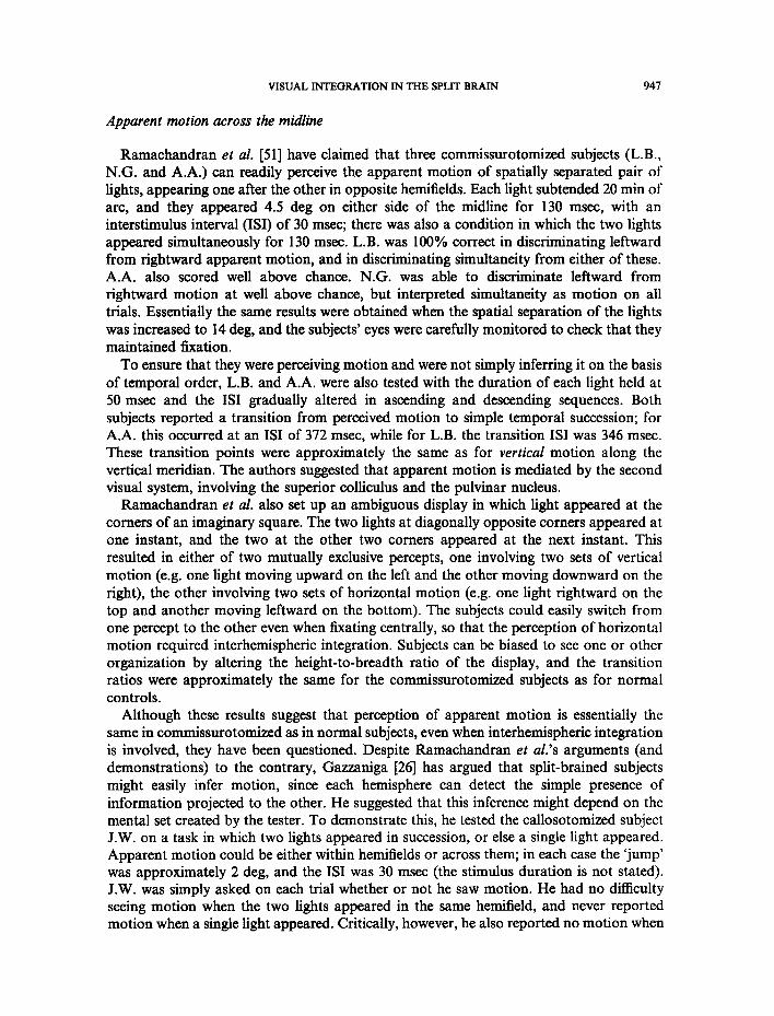
Ramachandran et al. [51] have claimed that three commissurotomized subjects (L.B., N.G. and A.A.) can readily perceive the apparent motion of spatially separated pair of lights, appearing one after the other in opposite hemitields. Each light subtended 20 min of arc, and they appeared 4.5 deg on either side of the midline for 130 msec, with an interstimulus interval (ISI) of 30 msec; there was also a condition in which the two lights appeared simultaneously for 130 msec. L.B. was 100% correct in discriminating leftward from rightward apparent motion, and in discriminating simultaneity from either of these. A.A. also scored well above chance. N.G. was able to discriminate leftward from rightward motion at well above chance, but interpreted simultaneity as motion on all trials. Essentially the same results were obtained when the spatial separation of the lights was increased to 14 deg, and the subjects' eyes were carefully monitored to check that they maintained fixation.
To ensure that they were perceiving motion and were not simply inferring it on the basis of temporal order, L.B. and A.A. were also tested with the duration of each light held at 50 msec and the ISI gradually altered in ascending and descending sequences. Both subjects reported a transition from perceived motion to simple temporal succession; for A.A. this occurred at an ISI of 372 msec, while for L.B. the transition ISI was 346 msec. These transition points were approximately the same as for vertical motion along the vertical meridian. The authors suggested that apparent motion is mediated by the second visual system, involving the superior colliculus and the pulvinar nucleus.
Ramachandran et al. also set up an ambiguous display in which light appeared at the comers of an imaginary square. The two lights at diagonally opposite comers appeared at one instant, and the two at the other two comers appeared at the next instant. This resulted in either of two mutually exclusive percepts, one involving two sets of vertical motion (e.g. one light moving upward on the left and the other moving downward on the right), the other involving two sets of horizontal motion (e.g. one light rightward on the top and another moving leftward on the bottom). The subjects could easily switch from one percept to the other even when fixating centrally, so that the perception of horizontal motion required interhemispheric integration. Subjects can be biased to see one or other organization by altering the height-to-breadth ratio of the display, and the transition ratios were approximately the same for the commissurotomized subjects as for normal controls.
Although these results suggest that perception of apparent motion is essentially the same in commissurotomized as in normal subjects, even when interhemispheric integration is involved, they have been questioned. Despite Ramachandran et al.'s arguments (and demonstrations) to the contrary, G ~ n i g a [26] has argued that split-brained subjects might easily infer motion, since each hemisphere can detect the simple presence of information projected to the other. He suggested that this inference might depend on the mental set created by the tester. To demonstrate this, he tested the callosotomized subject J.W. on a task in which two lights appeared in succession, or else a single light appeared. Apparent motion could be either within hemifields or across them; in each case the 'jump' was approximately 2 deg, and the ISI was 30 msec (the stimulus duration is not stated). J.W. was simply asked on each trial whether or not he saw motion. He had no difficulty seeing motion when the two lights appeared in the same hemifield, and never reported motion when a single light appeared. Critically, however, he also reported no motion when

~ 8 M.C. CORBALHS
the two lights appeared in opposite hemifields. That is, when he was 'set' to expect single lights on some trials, the report of motion between hemifields was replaced by report of a single light.
Trevarthen [71] has suggested that the discrepancy between Gazzaniga's and Ramachandran et al.'s findings might be explained in terms of a distinction between 'long-range' and 'short-range' motion systems [1, 6]. Gazzaniga's stimuli were separated by only 2 deg, which might have stimulated the higher-resolution short-range system, while Ramachandran et al. 's stimuli were separated by 9 deg, perhaps stimulating the low- resolution long-range system. Assuming the short-range system to be cortically mediated, it would be broken at the retinal midline by section of the forebrain commissures, whereas the long-range system would be integrated by subcortical commissures.
There are reasons to doubt this explanation. For one thing, recent evidence suggests that the distinction between short-range and long-range motion is not valid; Cavanagh [9] has shown that the distinction, such as it is, cannot be specified in terms of spatial dimensions, and argued instead for an alternative distinction between a low-level, automatic motion system and a higher-level, attention-based one. While these might be regarded as subcortical and cortical, respectively, the distinction does not seem to apply to that between Ramachandran et al.'s and Gazzaniga's experimental paradigms. More critically, unpublished experiments in our laboratory have failed to replicate Gazzaniga's result in the commissurotomized subject L.B., as well as in D.K., a subject with section of the posterior corpus callosum. In separate experiments involving binary decisions about lights appearing in opposite hemifields (as well as in the same hemifield), both subjects readily discriminated leftward from rightward motion, motion from simultaneity, a single light from simultaneous lights, and a single light from successive lights. In this last condition, essentially the same results were obtained whether the subjects were asked to discriminate between one light and two, or whether, as in Gazzaniga's experiment, they were asked simply to state whether they perceived motion or not. The lights were presented at spatial separations of either 2 or 7 deg, without affecting the results. The stimulus duration was held at approximately 133 msec, but the ISI was either 33 or 133 msec, again with little effect on the results. In fact performance was nearly perfect under all conditions, as shown in Fig. 4.
It is perhaps not surprising that split-brained subjects should be able to detect apparent motion within the normal temporal range, since the second visual system seems to play a special role in motion perception. The area of the visual cortex that appears to be critical for the perception of motion is the middle temporal lobe (MT). Lesions of this area in monkeys disrupts motion perception [64], and damage to the corresponding area in humans produces a specific deficit known as 'motion blindness' [31]. Area MT receives input from the superior eolliculus, both via the lateral geniculate and via the pulvinar nucleus [8]. Moreover, destruction of the striate cortex in the macaque does not eliminate the sensitivity of MT to motion, whereas subsequent destruction of the superior colliculus does [54], suggesting that the second visual system is indeed responsible for normal motion perception.
Yet there is evidence that temporal resolution across the midline in the split brain is poorer than that in normal subjects. In another study [14], the stimulus duration was reduced to 17 msec and ISI to 33 or 50 msec, and the commissurotomized subject L.B. was unable to discriminate successiveness from simultaneity when lights were flashed to opposite hemifields, virtually always reporting successive lights to be simultaneous. He

V I S U A L I N T E G R A T I O N IN T H E SPLIT B R A I N 949
c.j
0
<
Z
Eq
lO0
90
80
100
90
80
7O
L e f t v s r i g h t 60
_ i i _ _ i
~ °
70 1 VS 2 l i g h t s
60 ( s i m u l t a n e o u s }
LVF BIL RVF
S u c c e s s i v e v s
s i m u l t a n e o u s
L i
1 v s 2 l i g h t s
( s u c c e s s i v e )
_1 i i
LVF BIL RVF
SPATIAL LOCATION Fig. 4. Percent correct achieved by L.B. and D.K. in making decisions about lights. The four tasks were (1) judging whether successive pairs moved left or right; (2) judging whether pairs were successive or simultaneous; (3) judging whether one light or two simultaneous lights were presented; and (4) judging whether one or two successive lights were presented. The interstimulus intervals for successive presentations were either 33 or 133 reset, stimulus duration was 133 msec,
and spatial separation either 2 or 7 deg; data are averaged over these conditions.
3.5
3,0
2.5
2.0
d / 1.5
1.0
0.5
0.0
NORMAL CONTROLS L.B.
of' o . / \
"0
• 5 0 - m s ISI 0 3 3 - m s IS[
t I I I i [ ] I ~ i
LO LI B1L RI RO L0 LI BIL RI RO
SPATIAL LOCATION
Fig. 5. Values of d' achieved by normal controls and L.B. in judging whether pairs of lights were successive or simultaneous. There were six possible locations, spaced at 3.3-deg intervals, located symmetrieaUy about fixation. Lights appeared in adjacent locations: LO = left outer, LI = left inner, BIL = bilateral, RI = right inner, RO = right outer. Stimulus duration was approximately 17 msec, and interstimulus interval (ISI) either 33 or 50 msec; data are averaged over ISis. L.B. nearly always
reported that bilateral presentations were simultaneous.
scored above chance with unilateral presentations, a l though somewhat be low the level o f normal controls , who were also well above chance with bilateral presentation (see Fig. 5). It is debatable whether the stimuli were seen as in apparent motion in this study, since the ISis were be low the threshold for perceived m o t i o n as reported by N e u h a u s [47], and

950 M.C. CORBALLIS
subjects themselves were equivocal about this. Even so, the results do imply a distinction between cortical and subcortical mechanisms in the detection of temporal succession, with the cortical system tuned to a higher temporal resolution than the subeortical one.
Transfer of visual attention It has been suggested that visual attention may be unified in the split-brain, even if
visual perception is not [26]. I f so, this would go some distance toward explaining the apparent unity of consciousness in the split brain, especially since visual attention seems to constitute at least a part of what we mean by consciousness [19]. However visual attention is itself not a unitary phenomenon, and the experimental evidence has proven somewhat complicated.
One form of visual attention can be likened to a 'spotlight' that defines a region in space in which the processing of information is enhanced. If a person is cued to expect a signal in a particular location, which need not coincide with the point of fixation, then response to a target is speeded if it falls within that location and slowed if it does not, relative to a neutral condition in which there is no cue [50, 53]. These positive and negative effects of focused attention may be referred to as benefits and costs, respectively.
Two callosomotized subjects, J.W. and V.P., showed these effects even when the cue and target were presented to opposite visual hemifields [34]. In one experiment, 3 x 3 grids were displayed in both hemifields, and a cue ('X') appeared in one of the locations to indicate where a target digit was likely to appear. The task was to decide whether the digit was even or odd. Under one condition the cue appeared in the same hemifield as the target, and under it appeared in the opposite hemifield. In both cases the response time to the digit was increased if the digit appeared in the cued location, and slowed if it appeared in a non- cued location, relative to a condition in which there was no cue.
These results are perhaps not surprising, given the evidence of Holtzman [32], discussed earlier, that one of the subjects (J.W.) could direct an eye movement to a location in one hemifield on the basis of information presented to the other. Eye movements and spotlight attention are probably linked; indeed, it has even been proposed that spotlight attention is no more than the readiness to move the eyes to the cued location [53]. Moreover there is evidence in monkeys that the pulvinar nucleus, part of the second visual system, is involved in both eye movements and spotlight attention in the absence of eye movements [48].
In another experiment [35] with the callosotomized subject J.W. the digit could appear in only two locations, one in each hemifield, and the cue was an arrow pointing to the more likely location. Again, the cue was just as effective when it appeared in the hemifield opposite the target as when it appeared in the same hemifield. In a variation on this experiment [34] two arrows appeared, one in each hemifield. I f they pointed the same way, response times were again decreased if the digit appeared in the cued location and increased if it did not. But if the arrows pointed in opposite directions the response times were the same as those in which there were no cues. This result is consistent with other evidence that performance by one hemisphere depends on the processing load carried by the other, suggesting that the two hemispheres must share attentional resources [33].
It was on the basis of these findings, summarized in Fig. 6, that Gazzaniga drew the following general conclusion: "While it is clear perceptual processes are cleanly divided following commissurotomy, the attentional system remains unified in the split brain

VISUAL INTEGRATION IN THE SPLIT BRAIN 951
\ I~ .I~ /
ODD EVEN
2.1
u 1.9 ,la
>, 1.7 ~J {=
o 1.5 _1
8 ~.3
Bel~,~en
Wi th in
o n- I.I
X • ~ Wi th in -.~ I O 0 0
~ 95O O a D ~" v~" N 0 e~
or 900
\D I D s l ,,oo • --,. D iv ided " -~
o/ .. • --,. e - -~ Focused _J I 0 0 0
950 ODD EVEN 8,
o # 9oo
• Within o Between
I I I
Volid Neutral Invalid
Cue Type
• Within
y i i i
Valid Neutral Invalid Cue Type
/ ' ' ' v F D N I
Cue Type
Fig. 6. Experiments on visual attention in caUosotomized subjects. Top: Subjects (J.W. and P.S.) arc cued with an 'X' as to probable location of a digit to be judged odd or even. Left panels give examples of valid cues for within- and between-field conditions; cues may also be invalid, or absent (neutral). Right panel shows that within- and between-field cuing have approximately equal effects on response latency. Middle: Subject (J.W.) is cued with an arrow either on the same side (within field) or opposite side (between field) as the subsequent digit. Results again show equivalent effects. Bottom: Subject (J.W.) is given two arrows, one in each visual hemifield, which might cue the same side (focused attention) or opposite sides (divided attention). Results show that divided attention is no better than neutral condition, indicating that attention cannot be divided. (Reproduced with permission from GaT~mulga, M.S., Perceptual and attentional processes following caUosal section
in humans. Neuropsychologia 25, 119-133, 1987.)

952 M.C. CORBALLIS
patient and, consequently, can be viewed as a separate and fundamental property of human brain activity" [26, p. 131].
To complicate matters, however, there is more recent evidence that, under certain conditions, attention can be divided between the hemispheres. For example, spotlight attention can be divided when sensory rather than symbolic cues are used. In one study [52], single targets (Xs) were presented in four possible location boxes, arranged horizontally with two in each hemifield. A prior cue consisting of two lines flashed above and below one of the boxes marked the likely location of the target. Subjects' reaction times were longer to targets in an uncued box than to targets in a cued box, presumably reflecting the time it takes to re-orient attention. For normal subjects and a subject with section of the anterior two-thirds of the corpus callosum, the time to re-orient attention was about the same whether the locations of the cue and target were in the same or opposite hemifields. For two callosotomized subjects, however, it took considerably longer to shift attention between than within hemifields. That is, each hemisphere maintained a degree of attentional autonomy.
In another study [43], red or blue targets could appear in one of two boxes, each located in a different hemifield. The cue consisted of a brightening of a box. Normal subjects decided on the color of the target more quickly if it appeared in the cued box, and more slowly if it appeared in the uncued box, relative to neutral conditions in which both boxes were cued or in which there was a diffuse cue illuminating the whole screen, as shown in the top left panel of Fig. 7.
The top right panel of Fig. 7 shows the results for two callosotomized (J.W. and V.P.) and one commissurotomized (L.B.) subject. Cuing both boxes was essentially equivalent to a valid cue in either box, suggesting divided attention in the two hemispheres. However the effect of an invalid cue depended on whether the target was in the left or right hemifield, as shown in the bottom two panels of Fig. 7. The left panel, showing response times to left- hemifield targets, reveals the pattern that one would expect if the right hemisphere simply monitored the cuing of the left box; there was a benefit associated with cuing and a cost associated with no cue, compared with the neutral 'diffuse' condition. The right panel, showing response times to right-hemifield targets, indicates that the left hemisphere appeared to treat the uncued box as though it were cued. Mangun et al. suggest that the left hemisphere attended to the right-hemifield location regardless of cuing, but this does not really explain why response times were longer for the neutral 'diffuse' condition than for the invalid condition (when the lef t-hemifield location was cued).
Why should attention be divided when cued by sensory highlighting, but unified when cued by symbols, such as an arrow or the appearance of an 'X'? One possible answer is that attention based on direct sensory cues is automatic, while attention that depends on the prior decoding of symbolic cues is a more effortful, voluntary process [38]. This implies that automatic attention can proceed independently in the disconnected hemispheres of the split brain, while voluntary attention remains unified.*
To complicate matters even further, however, there appear to be situations in which even voluntary attention may be split. In a variant of the experiment using chimeric stimuli, described earlier, Levy et al. presented split faces such that a different half-face was
*There are other possible explanations for the discrepancy between the two sets of results. For example, in the experiments by Holtzman and his colleagues [34, 35], the interval between cue and target was considerably longer (1.5 see) than that in the experiments of Reuter-Lorenz and Fendrich [52] and Mangun et al. [44] (2004500 msec).

VISUAL INTEGRATION IN THE SPLIT BRAIN 953
>.
Z
.<
z o
5 8 0
5 6 0
6 4 0
520
5 0 0
780[ 7 6 0 ~
740
7 2 0 ~
7O0
6 8 0
6130
, , , /72t NORMAL 720
7 0 0
/ 6 8 0
/ /
/ z
i i _ _ I i
F D N IV
S P L I T - B R A I N L V F / •
/ /
/
/ /
/
6 4 0 i !
_~ _ ~ _ . t 6 2 0
F D N IV
6 6 0
640 I
_ . i
F 7BO --
760
740
7 2 0
700
6 8 0
660 r
6 4 0
620 J
F
CUE TYPE
S P L I T - B R A I N
/ /
/ ,/
/ /
_ 1 i i
D N IV
- - - i i - - T - ~
1 S P L I T - BRAIN RVF
,!
/ i . 1 / •
_ i _ i
D N IV
Fig. 7. Response latencies in experiment in which attention is cued by brightening of a box likely to contain the target stimulus (a red or a blue square). Data from commissurotomized subjects J.W., V.P. and L.B., and from 10 normal controls. Cues were either focused (F), with a cue on the side of the subsequent target, divided (D), with cues on both sides, neutral (N), with diffuse brightening signaling that the target could appear on either side, or invalid (IV), with the cue on the side opposite the subsequent target. Top panels show that the divided cue was equivalent to the neutral cue for the controls, but equivalent to the focused cue for the eommissurotomized subjects, indicating that attention was divided. Bottom panels show that expected cuing effects in the commissurotomized subjects were present for left-hemifield stimuli only. (Figures plotted from data
in Ref. [44].)
projected to each hemifield of the commissurotomized subjects N.G. and L.B. When naming the faces, the subjects tended to choose that displayed in the right hemifield, but when identifying them by pointing they tended to choose the one in the left hemifield. The subjects were also given trials in which the instruction was suddenly reversed. On some trials, just as the subject was about to point, the sample set was removed and the subject asked to name the face, while on other trials the subject was asked to point just as he or she was about to name the face. Again, naming favored the right-hemifield, pointing the left. This ability to switch rapidly from one mode to another suggests that the subjects were attending simultaneously to the two faces.
While spotlight attention may be described as location-based, the kind of attention in the experiment by Levy et al. is perhaps closer to what has been called object-based attention, in which the attentional focus is determined by object structure, independent of location [21, 73]. Object-based attention is presumably dependent on the analysis of form,

954 M.C. CORBALLIS
involving systems that are by-passed by the second visual system, or that are independent of it. There is evidence that neurons in area V4 and in the inferior temporal cortex of the monkey are modulated by attention to a visual stimulus [10, 20, 74]. We saw earlier that the callosotomized subject J.W. was unable to direct eye movement to a location in one hemifield when cued by a shape in the other [32]. Where attention depends on object structure, therefore, it is perhaps not surprising that it should be confined within the disconnected hemispheres of the split brain, and therefore divided between them.
Another example of divided voluntary attention in the split brain comes from an experiment on searching for a target in a visual array. There is evidence that voluntary attention is required when the target is composed of a conjunction of features that are also present in the distractors (e.g. searching for a red triangle among green triangles and red squares) [67]. Luck et al. [42] found that one callosotomized (J.W.) and one commissurotomized subject (L.B.) could search for a conjunction target in a visual array twice as quickly when the array was spread between the two hemifields as when it was confined to one, whereas normal subjects searched at the same (slower) rate in the two conditions. The implication is that the split-brained subjects were able to allocate attention equally between the hemispheres, and search for the targets in the two hemifields in parallel.
Again, the division of attention in this case may reflect cortical involvement. Trevarthen [70] suggested that the midbrain orienting (or attentional) mechanisms are augmented in primates by a cortical system with finer resolution and a more pronounced 'praxic' (or centrifugal') component, noting that frontal lesions may cause an inability to initiate eye movements for the solution of specific perceptual tasks [42]. More recent evidence for the role of the frontal lobes in directed visual search is summarized by Foster et al. [24]. Consequently, attention in visual search may be divided in the split brain because it is cortically mediated, while spotlight attention may be unified because it is subcortically mediated.
CONCLUSIONS
It is dear that there is considerable transfer of visual information in the split brain, even in cases where eye movements, cross-cuing, and transfer of nonperceptual information are either ruled out or taken into account. The most likely route is the so-called second visual system, which projects from the retina to the superior colliculus, and from there via the pulvinar nucleus to pre-striate areas of the visual cortex. Interhemispheric transfer might well be accomplished via the collicular commissure, but as noted earlier there are other possible transfer routes in the diencephalon, midbrain, or cerebellum [29, 71].
It has been common in the past to regard this second visual system as somehow complementary to the more extensively studied geniculostriate system, with its projections to pre-striate and temporal-lobe regions of the cortex. Thus Trevarthen [24] contrasts the 'ambient' system with the 'focal' system, Schneider [56] the 'where' system with the 'what' system, Gazzaniga [26] the attentional with the perceptual system, and Sergent [60] 'behavioral unity' with 'perceptual disunity'. Although each of these dichotomies may capture something of the difference between the two systems, none captures all. Moreover at least part of the difference seems to be quantitative rather than qualitative. For example, the geniculostriate system provides greater spatial and temporal resolution than the phylogenetically older second visual system. This is manifest in interhemispheric exchange

VISUAL INTEGRATION IN THE SPLIT BRAIN 955
through the two systems, with the corpus callosum providing more accurate spatial and temporal integration than the subcortical commissures.
But the geniculostriate system also offers some advantages that appear to be qualitative. In particular, it permits the analysis of visual form to the level necessary for the identification of objects and shapes, such as alphanumeric characters. This is no doubt achieved in part through enhanced spatial resolution, but presumably depends also on specialized processing of features in the striate and prestriate cortex that are inaccessible to the second visual system. We have seen that, in the monkey at least, the superior colliculus sends output to area MT, both directly and via the pulvinar nucleus [8]. The second visual system therefore by-passes the striate cortex and other prestriate areas presumed to be involved in the analysis of form.
As suggested by Trevarthen [70], the cortical system may also have introduced a 'praxic', centrifugal component not present in the subcortical system. By way of illustration Trevarthen writes: "The shift of gaze in solving a spatial riddle or searching out a hypothesized relationship may be called centrifugal in contrast to the visuomotor activity previously called centripetal" [70, p. 317; his italics]. One might add reading as another example. This praxic element may also apply to visual search in the absence of eye movements, as illustrated by the parallel search of the two hemifields in the split brain demonstrated by Luck et al. [41].
Gazzaniga's [26] suggestion that attention is integrated in the split brain while perception is divided is clearly no longer valid, as demonstrated in fact by more recent work in his own laboratory. Indeed the main principle governing whether attention is unified or divided may be essentially that governing whether perception is unified or divided; it depends on cortical involvement. Voluntary spotlight attention seems to be unified in the split brain [33, 34], probably because it depends on subcortical mechanisms involving the pulvinar nucleus. However object-based attention depends on cortical analysis, and is therefore divided. Similarly, the sequentially organized attentional shifts involved in visual search probably invoke frontal-lobe mechanisms rather than the pulvinar, again allowing for parallel processing in the divided hemispheres. It is of interest however that spotlight attention may be divided when it is induced by peripheral sensory cues, and is automatic rather than voluntary. At first glance (as it were) this may seem counterintuitive, since one might expect automatic processes to be associated with subcortical and voluntary mechanisms with cortical structures, rather than vice versa. On the other hand, one of characteristics of automatic processing is that it can proceed in parallel with other processes, while so-called controlled (or voluntary) processing demands more attentional resources and cannot be divided [57]. On these grounds, it is perhaps not surprising that automatic attention is more readily split.
It is tempting to suppose that the second visual system explains "blindsight" [75], the residual vision that occurs within a hemianopia or within a large scotoma following damage to the optic radiations or to the striate cortex. However the work of Holtzman [32], described earlier, suggested that hemianopia produces a more profound deficit of spatial localization in the 'blind' hemifield than does commissurotomy. Subjects with blindsight typically feel that they are merely guessing as to stimuli within the scotoma, and often require training if their performance is to rise above chance levels [78]; this contrasts with the ability of split-brained subjects to compare stimuli across the hemifields, at least in terms of location and orientation, and their occasional ability even to name stimuli in the left hemifield. Although there is still some question as to whether blindsight is a valid

956 M.C. CORBALLIS
phenomenon [7, 23, 28], it seems likely that it does operate at a largely unconscious level, and that the presence of the cortex is necessary for the effective registration in consciousness of input to the second visual system. This gives some support to the possibility of a subcortically-mediated behavioral unity despite perceptual disunity in the split brain, as suggested by Sergent [60], although some of her claims for this dissociation may have been exaggerated, as we have seen.
The poorer resolution of the second visual system need not imply that this system has been superseded in normal vision. As Trevarthen [70] proposed, it probably plays a normal role in locating objects or in detecting crude movement in ambient space, capturing visual events for more precise spatial and temporal analysis by the more focal cortical system. Even so, some of the results described above show that the two systems can be dissociated. For example, the disconnected hemispheres can simultaneously sustain conflicting discriminations or percepts based on shape, but cannot sustain simultaneous percepts based on luminance or color; again, the disconnected hemispheres can search visual arrays in parallel, but cannot voluntarily direct attentional spotlights simulta- neously to each hemifield.
Although the second visual system has a more global reach, this does not mean that it is not concerned with central vision. The evidence reviewed above suggests interhemispheric transfer of spatial information close to fixation in the split brain, albeit with some loss of resolution. Nor does the second visual system require sustained input; the evidence suggests interhemispheric integration of briefly flashed information in the split brain. Indeed a primary role of the second visual system is presumably to attract attention to events away from fixation, and one of the primary cues for attracting attention is abrupt visual onset [76]. It might also be noted that blindsight seems to be especially sensitive to flashed targets [2], and less sensitive to peripheral than to central vision [75].
Although this review suggests a greater degree of visual unity across the hemifields in the split brain than has been assumed in the past, this need not imply that splitting the brain does not split the mind. Through subcortical interchange, each hemisphere may indeed have considerable awareness of the hemifields on both sides of fixation, but this is not to say that each hemisphere has awareness of cortical processing in the other hemisphere. Because of the substantial role of the second visual system, it now appears that the disconnection syndrome is only partially revealed by visual experiments.
Acknowledgements---Some of the work described in this article was supported by grants from the Auckland University Research Committee and from the N.Z. Neurological Foundation. I thank Joseph Bogen, Colwyn Trevarthen, and David Milner for helpful comments.
REFERENCES 1. Anstis, S. M. Apparent movement. In Handbook of Ser~ory Physiology: Perception R. Held, H. Lcibowitz
and H.-L. Teuber (Editors), Vol. HI, Springer-Verlag, New York, 1978. 2. Barbur, J. L., Ruddoc, k, K. H. and Wateriield, V. A. Human visual responses in the absence of the geniculo-
calcarine projection. Brain 105, 905-928, 1980. 3. Bogen, J. E. The other side of the brain: II. An appositional mind. Bull. Los Angeles Neurol. Soc. 34, 135-162,
1969. 4. Bogen, J. E. Partial hemispheric independence with the noocommissures intact. In Brain Circuits and
Functions of the Mind: Essays in Honor of R. IV. Sperry, C. Trevarthen (Editor), pp. 211-230. Cambridge University Press, Cambridge, 1990.

VISUAL INTEGRATION IN THE SPLIT BRAIN 957
5. Bogen, J. E. The callosal syndromes. In Clinical Neuropsychology, 3rd edn, K. M. Heilman and E. Valenstein (F_xlitors), pp. 337-407, Oxford, New York, 1993.
6. Braddiek, O. J. Low-level and high-level processes in apparent motion. Phil. Trans. Roy. Soc. Lond. B 290, 137-151, 1980.
7. Campion, J., Latto, R. and Smith, Y. M. Is blindsight an effect of scattered light, spared cortex, or near- threshold vision? Behav. Brain Sci. 6, 423--486, 1983.
8. Carlson, N. R. Physiology of Behavior, 4th edn. Allyn & Bacon, Boston, 1991. 9. Cavanagh, J. P. Short-range vs long-range motion: Not a valid distinction. Spatial Vision 5, 303-309, 1991.
10. Chelazzi, L., Miller, E. K., Duncan, J. and Desimone, R. A neural basis for visual search in inferior temporal cortex. Nature 363, 345-347, 1993.
11. Corballis, M. C. Perceptual integration following eommissurotomy: A reappraisal. In New Horizons in Neuropsychology, M. Sugishita (Editor), pp. 139-158. Elsevier Science, Amsterdam, 1994.
12. Corballis, M. C. Can commissurotomized subjects compare digits between the two visual fields? Neuropsychologia 32, 1475-1486, 1994.
13. Corballis, M. C. Line bisection in a subject with complete forebrain eommissurotomy. Neuropsychology 9, 147-156, 1995.
14. Corballis, M. C. Hemispheric interaction in temporal judgments about spatially separated stimuli. Neuropsychology, in press.
15. Corballis, M. C. and Sergent, J. Tests of imagery in a commissurotomized subject. Neuropsychologia 25, 13- 26, 1988.
16. Corballis, M. C. and Sergent, J. Mental rotation in a commissurotomized subject. Neuropsychologia 27, 585- 597, 1989.
17. Corballis, M. C. and Sergent, J. Judgments of numerosity by commissurotomized subjects. Neuropsychologia 30, 865--876, 1992.
18. Corballis, M. C. and Trudel, C. I. The role of the forebrain commissures in interhemispherie integration. Neuropsychology 7, 1-19, 1993.
19. Crick, F. The Astonishing Hypothesis: The Scientific Search for the Soul. Scribners, New York, 1994. 20. Desimone, R. and Ungerleider, L. G. Neural mechanisms of visual processing in monkeys. In Handbook of
Neuropsychology, Vol. 2, F. Boiler and J. Grafman (Editors), pp. 267-299. Elsevier, New York, 1989. 21. Duncan, J. Selective attention and the organization of visual information. J. exp. Psychol.: Gen. 113, 501-
517, 1984. 22. Fendrieh, R. and Gazzaniga, M. S. Evidence of foveal splitting in a commissurotomy patient.
Neuropsychologia 27, 273-281, 1989. 23. Fendrieh, R., We,singer, C. M. and GaTJaniga, M. S. Residual vision in a scotoma: Implications for
blindsight. Science 258, 1489-1491, 1992. 24. Foster, J. K., Eskes, G. A. and Stuss, D. T. The cognitive neuropsyehology of attention: A frontal lobe
perspective. Cognit. NeuropsychoL 11, 133-147, 1994. 25. GazTaniga, M. S. The Bisected Brain. Appleton-Century-Crofts, New York, 1970. 26. Ga~7~niga, M. S. Perceptual and attentional processes following caUosal section in humans.
Neuropsychologia 25, 119-133, 1987. 27. G~Taniga, M. S., Bogen, J. E. and Sperry, R. W. Observations on visual perception after discotmexion of the
cerebral hemispheres in man. Brain 88, 221-236, 1965. 28. Gazzamga, M. S., Fendrieh, R. and Wessinger, C. M. Blindsight reconsidered. Curr. Direct Psychol. Sci. 3,
93-96, 1994. 29. Gliekstein, M. E. Brain pathways in the visual guidance of movement and the behavioral functions of the
cerebellum. In Brain Circuits and Functions of the Mind." Essays in Honor of R. W. Sperry, C. Trevarthen (Editor), pp. 157-167. Cambridge University Press, Cambridge, 1990.
30. Halligan, P. W. and Marshall, J. C. Toward a principled explanation of unilateral neglect. Cognit. Neurot~sychol. 11, 167-206, 1994.
31. Hess, R. H., Baker, C. L. and Zihl, J. The "motion blind" patient: Low-level spatial and temporal filters. J. Neurosci. 9, 1628-1640, 1989.
32. Holtzman, J. D. Interactions between cortical and subcortical visual areas: Evidence from human commissurotomy patients. Vis. Res. 24, 801-813, 1984.
33. Holtzman, J. D. and Gazzaniga, M. S. Dual task interactions due exclusively to limits in processing resources. Science 218, 1325-1327. 1982.
34. Holtzman, J. D., Sidtis, J. J., Volpe, B. T., Wilson, D. H. and Gazzaniga, M. S. Dissociation of spatial information for stimulus localization and the control of attention. Brain 104, 861-872, 1981.
35. Holtzman, J. D., Volpe, B. T. and Gn77aniga, M. S. Spatial orientation following commissural section. In Varieties of Attention, R. Parasuraman and D. R. Davies (Editors), pp. 375-394. Academic Press, New York, 1984.
36. Johnson, L. E. Bilateral visual cross-integration by human forebrain eommissurotomy subjects. Neuropsycholgia 22, 167-175, 1984.

958 M.C. CORBALLIS
37. Johnson, L. E. Vocal responses to left visual field stimuli following forebrain commissurotomy. Neuropsychologia 22, 153-166, 1984.
38. Jonides, J. Voluntary versus automatic control over the mind's eye movement. In Attention and Performance IX, J. Long and A. Baddeley (Editors), pp. 187-203. Lawrence Erlbaum, Hillsdale, New Jersey 1981.
39. Joynt, R. J. Inattention syndromes in split-brain man. Adv. Neurol. 81, 33-39, 1977. 40. Levy, J., Trevarthen, C. and Sperry, R. W. Perception of bilateral chimeric figures following hemispheric
deconnection. Brain 95, 61-78, 1972. 41. Luck, S. J., Hillyard, S. A., Mangun, G. R. and Gazzaniga, M. S. Independent bemispheric attentional
systems mediate visual search in split-brain patients. Nature 342, 543-545, 1989. 42. Luria, A. R., Karpov, B. A. and Yarbus, A. L. Disturbances of active visual perceptinn with lesions of the
frontal lobes. Cortex 2, 202-212, 1966. 43. Mangun, G. R., HiUyard, S. A., Luck, S. J., Handy, T., Plager, R., Clark, V. P., Loftus, W. and Gazzaniga,
M. S. Monitoring the visual world: H~nispheric asymmetries and subcortical processes in attention. J. cognit. Neurosci. 6, 267-275, 1994.
44. Miller, G. A. The magical number seven, plus or minus two: Some limits on our capacity for processing information. Psychol. Rev. 63, 81-97, 1956.
45. Milner, A. D., Harvey, M., Roberts, R. C. and Forster, S. V. Line bisection errors in visual neglect: Misguided action or size distortion? Neuropsychologia 31, 39-49, 1993.
46. Myers, J. J. and Sperry, R. W. Interhemispheric communication after section of the forebrain commissures. Cortex 21, 249-260, 1985.
47. Neuhaus, W. (1930). Experimentelle untersuchung der scheinbewegung. Arch. f~ir die gesamte Psychol. 75, 315-458, 1930.
48. Peterson, S. E., Robinson, D. L. and Morris, J. D. Contributions of the pulvinar to visual spatial attention. Neuropsycholgia 25, 97-105, 1987.
49. Plourde, G. and Sperry, R. W. Left hemisphere involvement in left spatial neglect from right sided lesions: A commissurotomy study. Brain 107, 95-106, 1984.
50. Posner, M. I. and Cohen, Y. Components of attention. In Attention and Performance, X. H. Bouma and D. Bowhuis (Editors), pp. 531-556. Lawrence Erlbaum, Hillsdale, New Jersey, 1984.
51. Ramachandran, V. S., Cronin-Golomb, A. and Myers, J. J. Perception of apparent motion by commissurotomy patients. Nature 320, 358-359, 1986.
52. Renter-Lorenz, P. A. and Fendrich, R. Orienting attention across the vertical meridian: Evidence from callosotomy patients. J. cognit. Neurosci. 2, 232-238, 1990.
53. Rizzolatti, G., Riggio, L., Daseola, I. and Umilta, C. Reorienting attention across the horizontal and vertical meridians: Evidence in favor of a premotor theory of attention. Neuropsychologia 25, 31-40, 1987.
54. Rodman, H. R., Gross, C. G. and Albright, T. D. Afferent basis of visual response properties in area MT of the macaque. I. Effects of straite cortex removal. J. Neurosci. 9, 2033-2050, 1989.
55. Schacter, D. L., McAndrews, M. P. and Moscovitch, M. Access to consciousness: Dissociations between implicit and explicit knowledge in neuropsychological disorders. In Thought without Language, L. Wciskrantz (Editor), pp. 242-278. Clarendon Press, Oxford, 1988.
56. Schneider, G. E. Contrasting visuomotor functions of teztum and cortex in the golden hamster. Psych. Forsch. 31, 52-62, 1967.
57. Schneider, W. and Shiffrin, R. M. Controlled and automatic information processing: I. Detection, search, and attention. Psychol. Rev. 92, 424-428, 1977.
58. Scrgent, J. Unified response to bilateral hemispheric stimulation by a split-brain patient. Nature 305, 800-802, 1983.
59. Scrgent, J. Subeortical coordination of hemisphere activity in commissurotomized patients. Brain 109, 357- 369, 1986.
60. Scrgent, J. A new look at the human split brain. Brain 110, 1375-1392, 1987. 61. Scrgent, J. Furtive incursions into bicameral minds: Integrating and coordinating role of subcortical
structures. Brain 113, 537-568, 1990. 62. Sergent, J. Processing of spatial relations within and between the disconnected cerebral hemispheres. Brain
114, 1025-1043, 1991. 63. Seymour, S. E., Renter-Lorenz, P. A. and Ga~n iga , M. S. The disconnection syndrome: Basic findings
reaffirmed. Brain 117, 105-115, 1994. 64. Siegei, R. M. and Andersen, R. A. Motion perceptual deficits following ibotenic acid lesions of the middle
temporal area (MT) in the behaving monkey. See. Neurosci. Abstr. 12, 1183, 1986. 65. Sperry, R. W. Mental unity following surgical disconnection of the cerebral hemispheres. Harvey Lecturer 62,
293-323, 1966-1967. 66. Sperry, R. W. Some effects of disconnecting the cerebral hemispheres. Science 217, 1223-1226, 1982. 67. Treisman, A. and Golad¢, G. A feature-integration theory of attention. Cognit. Psychol. 12, 97-136, 1980. 68. Trevarthen, C. B. Double visual learning in split-brain monkeys. Science 136, 258-259, 1962.

VISUAL INTEGRATION IN THE SPLIT BRAIN 959
69. Trevarthen, C. B. Functional interactions between the cerebral hemispheres of the spLit-brain monkey. In Functions of the Corpus Callosum, E. (3. Ettlinger (Editor), pp. 24-40. Ciba Foundation Study Group, No. 20, 1965.
70. Trevarthen, C. B. Two mechanisms of vision in primates. Psychol. Forsch. 31, 299-337, 1968. 71. Trevarthen, C. B. Integrative functions of the cerebral commissures. In Huna~ook ofNeuropsychology, VoL 4:
The Commissurotomized Brain, R. D. Nebes (Editor), pp. 49-83. Elsevier, Oxford, 1991. 72. Trevarthen, C. B. and Sperry, R. W. Perceptual unity of the ambient visual field in human commissurotomy
patients. Brain 96, 547-570, 1973. 73. Vecera, S. P. and Farah, M. J. Does visual attention select objects or locations? J. exp. PsychoL: Gen. 123,
146-160, 1994. 74. Walsh, V. and Perrett, D. I. Visual attention in the occipitotemporal processing stream of the macaque.
Cognit. Neurosci. 11, 243-263, 1994. 75. Weiskrantz, L. Blind.~ight. Oxford University Press, New York, 1986. 76. Yantis, S. and Jonides, J. Abrupt visual onsets and selective attention: Evidence from visual search. J. exp.
Psychol.: Hum. Percept. Perform. 10, 601-621, 1984. 77. Zaidel, D. W. Observations on right hemispheric language function. In Aphasia, F. C. Rose, R. Whurr and
M. Wyke (Editors), pp. 170-187. Whurr Publishers, London, 1988. 78. Zihl, J. and von Cramon, D. Restitution of visual function in patients with cerebral blindness. J. NeuroL
Neurasurg. Psychiat. 42, 312-322, 1979.