Embed Size (px)
Citation preview
Visual Acuity in Newborn and Preterm Infants MeasuredWith Grating Acuity Cards
Angela M. Brown, Ph.D., and Misao Yamamoto, M.D.
Binocular visual acuity of normal newborninfants, preterm newborn infants, and newborn, full-term infant patients with nonophthalmologic abnormalities was measured bymeans of grating acuity cards. Each test tookabout six minutes to complete, and 89% of thetests (154 of 174) were successful. Visual acuity of infants at 39 to 40 weeks of gestationalage was about 0.023 stripes per minute of are,or 0.69 cycles per degree (20/866). Between 34and 44 weeks of gestational age, visual acuityimproved at the rate of 0.46 octaves per month.This test is simple, fast, and reliable, andrequires no apparatus except the cardsthemselves.
ROUTINE MEASUREMENT of visual acuity innewborn infants requires a test that is reliable,successful on most infants, and easy to use.
The visual acuity of newborn infants hasbeen assessed behaviorally by several investigators!" and also by visual-evoked potential. 3,5,9·11 The visual acuities obtained behaviorally were generally near 20/800 (0.75 cycles perdegree). The visual acuity of newborns definedby visual-evoked potentials was between 20/800(0.75 cpd) and 20/420 (1.43 cpd).3,5,9,10 Norciaand Tyler" reported visual acuities measuredby visual-evoked potential near 20/125 (5 cpd)in infants less than 1 week old.
The visual acuities of individual infants wereoften not obtainable by behavioral techniques3,6,7 because the tests were generally longand newborn infants tend to be awake for onlyshort periods (however, Dubowitz and associ-
Accepted for publication April 21, 1986.From the Infant Vision Laboratory, University of
Washington, Seattle, Washington (Dr. Brown); and theDepartment of Ophthalmology, Kobe University Schoolof Medicine, Kobe, Japan (Dr. Yamamoto). This studywas supported in part by Kobe Children's Hospital.
Reprint requests to Angela M. Brown, Ph.D., InfantVision Laboratory, Guthrie Hall, NI-25, University ofWashington, Seattle, WA 98195.
ates5,S seemed to have better success than others). The techniques used in these behavioraland visual-evoked potential studies requiredboth an apparatus and several trained personnel. Therefore, those techniques are not reallysuitable for routine testing of infants in thehospital or clinic.
The purpose of our study was to develop atechnique for measuring visual acuity in infants under 44 weeks of gestational age. Thetechnique had to meet several criteria. First, ithad to produce valid, reproducible results. Second, it must be successful on most individualinfants. Third, it had to require a minimum ofresources: it must take little space in the crowded infant intensive-care unit and it must require no trained personnel other than thoselikely to be there anyway. Fourth, it must befast. To meet these requirements, we developed a variant of the grating acuity card technique" for use on newborn infants. This technique has been successful for infants more than1 month of age and has been shown to be muchfaster, more successful, and more convenientthan forced-choice preferential-looking" whichhas proven unsatisfactory in this application.The grating acuity card technique thereforeseemed a promising approach for use on newborns, who have very short alert periods. Sinceour data were collected, grating acuity cardshave also been used successfully for testinghealthy newborn infants by Dobson and associates.!' In this report, we describe the new stimuli and technique we developed. We also reportvisual acuity values for normal newborn infants, healthy preterm infants, and young infants hospitalized for nonneurologic illnesses.
SUbjects and Methods
The three groups of infants included in thisstudy were free of neurologic and ophthalmologic abnormalities. Two groups were in the
©AMERICAN JOURNAL OF OPHTHALMOLOGY 102:245-253, AUGUST, 1986 245
246 AMERICAN JOURNAL OF OPHTHALMOLOGY August, 1986
nursery of the infant intensive-care unit ofKobe Children's Hospital. The first group consisted of 24 preterm infants who were healthyexcept for their prematurity and the secondgroup consisted of 37 full-term infant patientswith nonneurologic diseases. The preterm infants were born before 36 weeks of gestation(average gestational age at birth was 32.4±2.4weeks) and they ranged from 33 to 42 weeks ofgestational age at the time they were tested.The full-term infant patients had been bornafter 36 weeks of gestation (average gestationalage at birth was 38.7±1.4 weeks); gestationalage at testing ranged up to 48 weeks. The thirdgroup consisted of 30 normal, full-term, newborn infants in the obstetrics ward of OhtaCommunity Hospital. These infants were bornat an average of 39.7±1.0 weeks of gestationalage and were less than 1 week old (average age,5.1 ±3.3·days).
The grating acuity card technique we developed required the participation of two adults,and we conducted the experiments ourselves.One of us (M. Y.) is an ophthalmologist. Heexamined the fundus of each subject at leastonce and read each patient's chart before eachtest. The other (A.M.B.) is a psychologist withtwo years of intensive experience in visualtesting of young infants. She was unaware ofthe age and diagnosis for all infants tested inKobe Children's Hospital. To this end, the databooks were kept in Japanese, which she couldnot read. We alternated across sessions as tester and observer. The observer presented thestimuli and observed the infant. The testertimed the tests, arranged the cards in order,selected the cards for the control phase of theprocedure, and wrote down the results. Someinfants were tested twice each week, once byeach observer.
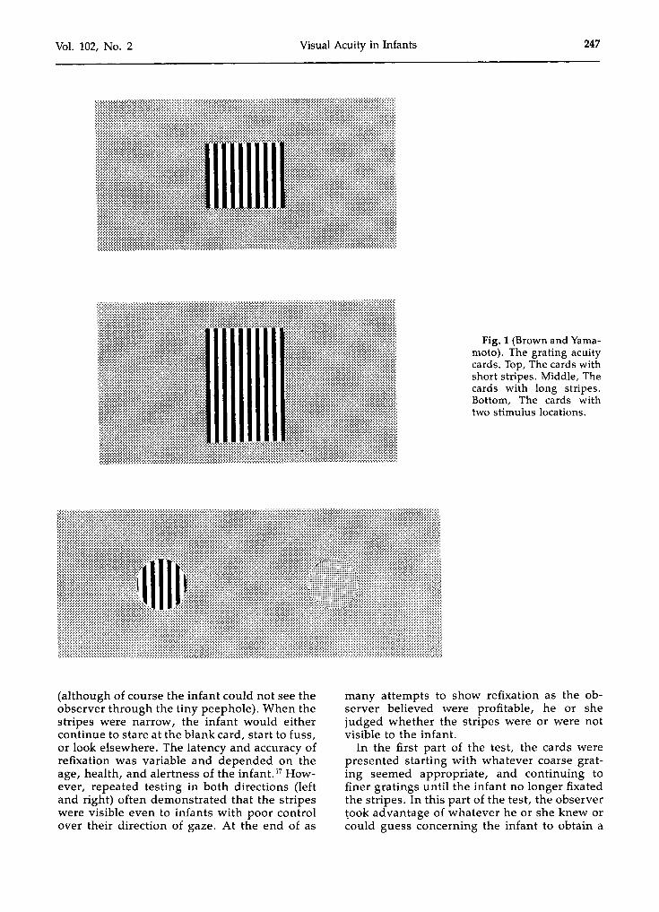
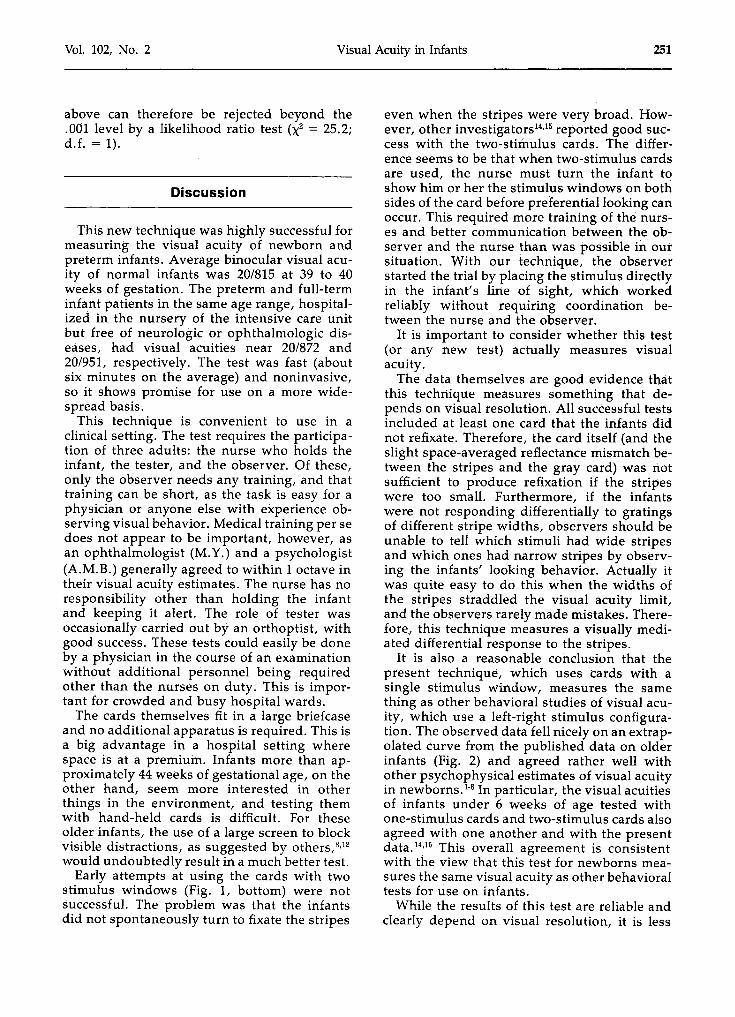
Three types of cards were used in this study(Fig. 1). Cards of the first type (Fig. 1, top) weregray and measured 35 x 56 cm. Each card had a12 x 14-cm central rectangular window. Apiece of photographic paper with black-andwhite stripes was attached to the back of eachcard so that it showed through the window.The contrast of the stripes was 0.87, calculatedas the difference in luminance between thewhite and black stripes, divided by the sum oftheir luminances. The space-averaged reflectance of the stripes was within 10% (0.05 10glOunits) of the reflectance of the gray card. In thecenter of a central black stripe was a 2-mmpeephole through which the observer couldwatch the infant.
The six cards were in a graded set: one cardwas blank, and the stripe widths were 0.11 to1.68 cm in I-octave steps. At a viewing distanceof 36 cm they were equivalent to between 0.1stripe per minute of arc (20/200) and 0.0063stripe per minute of arc (20/3200). The cardswere indistinguishable from the back. Cards ofthis type were used on all infants except for tenof the normal newborn infants.
The second type of card was similar to thefirst, except that the stripes were longer (23em), the card was taller (39 em), and the setincluded twice as many cards in 0.5-octavesteps (Fig. 1, middle). Cards of this type wereused on ten of the normal newborn infants.
Cards of the third type were identical to thegrating acuity cards of McDonald and associates. 12,15 These cards had two round stimuluswindows, to the right and left of the peephole;one window contained a grating and one wasblank.
All tests took place in the nursery where theinfants normally stayed. Testing took placeshortly before feeding, when the nursing staffreported them to be most alert. The infant washeld by a nurse and viewed the cards binocularly. Some pilot data were collected in the obstetrics ward of Kobe Kaisei Hospital where themothers held the infants. This was much lesssuccessful. The nurse stood with her back to awindow so that the cards were illuminated withdiffuse, indirect daylight. The luminance of thegray cards varied a bit, but was never below 300cd/rn-."
The observer decided whether the infantcould resolve the stripes by observing the infant's fixation behavior in response to displacement of the grating stimulus. First, the observer placed his or her face in the infant's view, asclose as possible to the infant's line of gaze.The distance between the infant's and observer's face was about 36 em at that point. Next,the observer held up the card at that same36-cm distance and observed the infant's facethrough the peephole. At that moment, theinfant and observer were looking at each other's eyes through the peephole in the card.Next, the observer moved the card about 20 cmto the right or left, and again held it stationaryfor several seconds while continuing to observethe infant. At that moment, the infant would belooking at a region of blank gray cardboard.When the stripes were wide (and presumablyvisible), the infant would turn his or her gazetowards them, thus reestablishing eye-to-eyecontact between the observer and the infant
Vol. 102, No. 2 Visual Acuity in Infants 247
Fig. 1 (Brown and Yamamoto). The grating acuitycards. Top, The cards withshort stripes. Middle, Thecards with long stripes.Bottom, The cards withtwo stimulus locations.
(although of course the infant could not see theobserver through the tiny peephole). When thestripes were narrow, the infant would eithercontinue to stare at the blank card, start to fuss,or look elsewhere. The latency and accuracy ofrefixation was variable and depended on theage, health, and alertness of the infant. 17 However, repeated testing in both directions (leftand right) often demonstrated that the stripeswere visible even to infants with poor controlover their direction of gaze. At the end of as
many attempts to show refixation as the observer believed were profitable, he or shejudged whether the stripes were or were notvisible to the infant.
In the first part of the test, the cards werepresented starting with whatever coarse grating seemed appropriate, and continuing tofiner gratings until the infant no longer fixatedthe stripes. In this part of the test, the observertook advantage of whatever he or she knew orcould guess concerning the infant to obtain a
248 AMERICAN JOURNAL OF OPHTHALMOLOGY August, 1986
fast, rough estimate of the infant's visual acuity. The provisional estimate was the stripewidth of the narrowest stripes the observerjudged the infant could see.
The second phase of the test was the controltest. The tester chose two cards near, but notnecessarily straddling, the provisional visualacuity limit. The tester had watched the firstpart of the test, and was urged to include anycards for which he or she had disagreed withthe observer. The observer was experimentallynaive in this control part of the test: the observer did not see the stripes or know either theirabsolute or relative stripe widths.
The observer decided whether the infantcould resolve the stripes on each control card.The observer could request additional cards,for example, the blank card or the card with thewidest stripes, as a reminder of the infant'sbehavior when the stripes were not visible or toverify that the infant was still alert.
If the results of the control phase of the testconfirmed those of the preliminary phase, thetest was considered final after two controlcards. If the observer was dissatisfied with thetest or if the tester found the results to beinconsistent with those of the preliminary test,then additional control cards were run untileither both observer and tester were satisfiedthat an assessment could be made from thedata or until the infant fell asleep. The finalvisual acuity estimate was the width of thefinest stripes that the observer judged the infant could resolve. All successful tests had toinclude at least one card the infant could resolve and one that he or she could not.
Immediately after the test was completed,the observer looked at the data and decidedwhat the visual acuity of the infant was. In 126of the 154 successful tests (90%), the preliminary and control phases of the test gave consistent results. In 102 of the tests, the controlcards were chosen so that visual acuity could beestimated from the control data alone. In 37tests, the visual acuity was taken from bothparts of the test together. This occurred whenever the control cards were not optimally chosen to estimate visual acuity, for example,when all of the control cards were seen or thejust-visible stripe width was not used as acontrol. It was important to have tests like thisif the observer was to be unable to guess thestripe widths of the control cards. In each ofthose tests, the results of the two halves of thetest were consistent. In the remaining 15 tests(10%), the subject was judged to see a particu-
lar stimulus on some trials but not on others. Inthose cases, the observer was allowed to takeinto account whether the infant was equallyalert throughout the experiment and how manyattempts were necessary before the infantlooked at the stimulus. In those cases, theresult of either the preliminary or the controltest was taken as the visual acuity.
The tester timed the test from the momentthe first card was presented until the observerjudged the visibility of the last card. Tests werescored as "attempts" if the clock was startedand as "successes" if the test was completedand a final visual acuity estimate was made.
Results
The two types of one-stimulus card used onthe normal newborn infants did not producesignificantly different visual acuity (t = 0.134;P>.5), population standard deviation (F = 1.86;d.f. = (9,13); P>.l), or test duration (t = 0.577;P>.5). Because none of these comparisons wassignificant, the data on normal newborns havebeen pooled across those two card types.
We were not successful with the twostimulus card and we stopped using it after afew attempts.
The success rate for normal newborns was83% (24 successes in 29 attempts). Of the 93tests done on the full-term infant patients,including retests, 87 (94%) were successful; 43of the 52 tests (83%) on preterm infants weresuccessful. The average success rate for allsubjects in Kobe Children's Hospital was 80%on the first attempt (49 of 59). Of the 52 infantstested two or more times, 51 (98%) were successfully tested on the first or second attempt.Therefore, if it is important for some reason totest the visual acuity of a particular healthyinfant, that test can almost surely be done ifseveral attempts are made, The problem seemsto be that finding the infant really awake ispartly a matter of luck, even when testing takesplace at the optimal time in the feeding schedule, and perseverance is sometimes necessaryfor success.
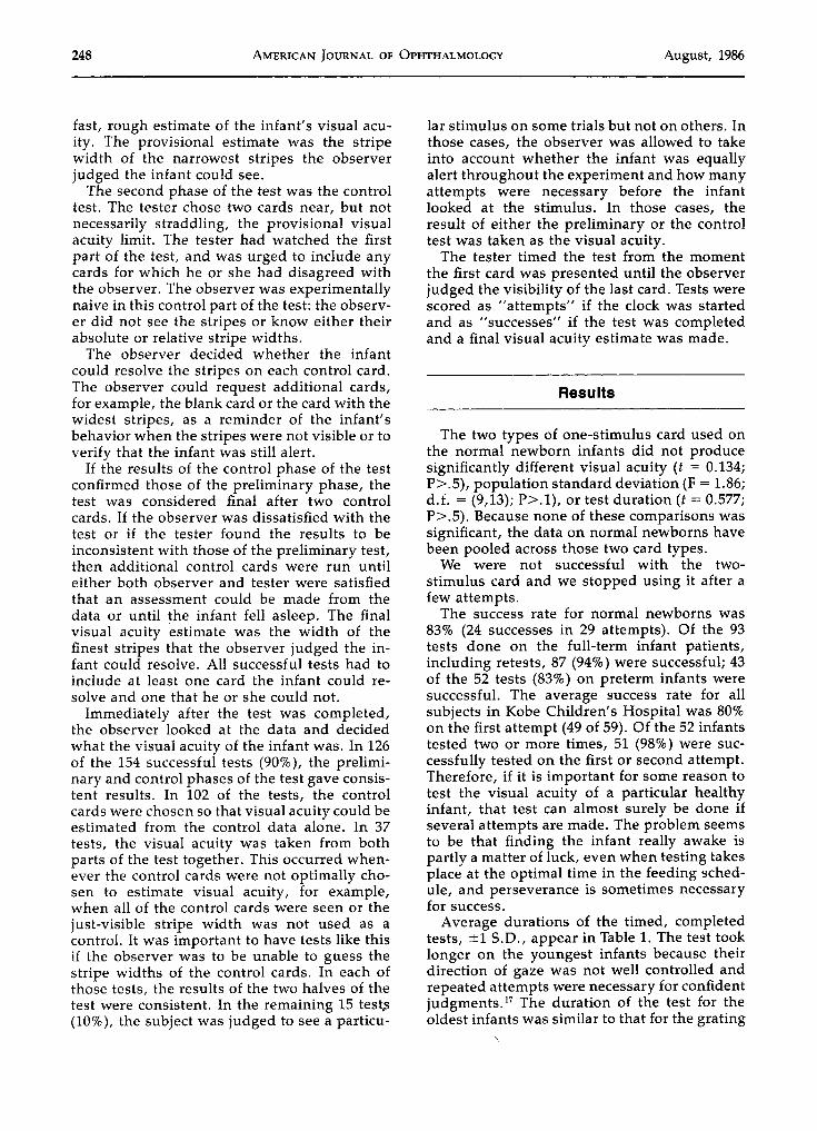
Average durations of the timed, completedtests, ±1 S.D., appear in Table 1. The test tooklonger on the youngest infants because theirdirection of gaze was not well controlled andrepeated attempts were necessary for confidentjudgments." The duration of the test for theoldest infants was similar to that for the grating
Vol. 102, No.2
GESTATIONAL AGE
AT TEST (WKS)
32 to 33
34·to 35
36 to 37
38 to 39
40 to 41
42 to 43
44 to 45
46 to 47
NO.'
1687
18
643
Visual Acuity in Infants
TABLE 1AVERAGE DURATION OF TESTS
KOBE CHILDREN'S HOSPITAL
MEAN (± S.D.) TEST
DURATION (MIN)!
4.25
8.86 ± 2.7
8.67 ± 3.3
7.52 ± 2.9
6.02 ± 1.9
5.51 ± 2.2
6.23 ± 2.6
6.03 ± 1.3
NO.'
911
1
249
OHTA COMMUNITY HOSPITAL
MEAN (± S.D.) TEST
DURATION (MIN)!
6.25 ± 2.5
6.19 ± 2.2
6.18
"No. for whom data are available. Data for preterm and full-term infants at the Kobe Children's Hospital are pooled. Each infant
contributed no more than a single data point.
!Standard deviation is undefined when No. = 1.
acuity cards used by McDonald and associates"on infants of comparable ages.
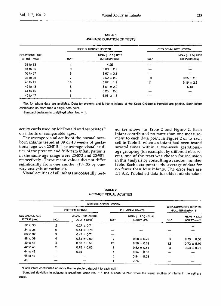
The average visual acuity of the normal newborn infants tested at 39 or 40 weeks of gestational age was 20/815. The average visual acuities of the preterm and full-term infant patientsin the same age range were 20/872 and 20/951,respectively. These mean values did not differsignificantly from one another (P>.05 by oneway analysis of variance).
Visual acuities of all infants successfully test-
ed are shown in Table 2 and Figure 2. Eachinfant contributed no more than one measurement to each data point in Figure 2 or to eachcell in Table 2: when an infant had been testedseveral times within a two-week gestationalage grouping (for example, by different observers), one of the tests was chosen for inclusionin this analysis by consulting a random numbertable. Each data point is the average of data forno fewer than four infants. The error bars are±1 S.E. Published data for older infants taken
TABLE 2AVERAGE VISUALACUITIES
PRETERM INFANTS
KOBE CHILDREN'S HOSPITAL
FULL-TERM INFANTS
OHTA COMMUNITY HOSPITAL
(FULL-TERM INFANTS)
GESTATIONAL AGE
AT TEST (WKS) NO.'
MEAN (± S.D.) VISUAL
ACUITY (cpo)! NO.'
MEAN (± S.D.) VISUAL
ACUITY (cPO)! NO.'
MEAN (± S.D.)
ACUITY (Cpo)!
32 to 33
34 to 35
36 to 3738 to 3940 to 41
42 to 4344 to 4546 to 4748
2994421
0.27 ± 0.71
0.41 ± 0.78
0.47 ± 0.710.63 ± 0.500.63 ± 0.50
0.75 ± 0.000.75
7 0.56 ± 0.79 9 0.75 ± 0.0020 0.59 ± 0.59 12 0.73 ± 0.40
8 0.82 ± 0.64 3 0.53 ± 0.71
4 0.94 ± 0.58
3 0.94 ± 0.581 0.75
"Each infant contributed no more than a single data point to each cell.
'Standard deviation in octaves is undefined when No. = 1 and is equal to zero when the visual acuities of infants in the cell areequal.
250 AMERICAN JOURNAL OF OPHTHALMOLOGY August, 1986
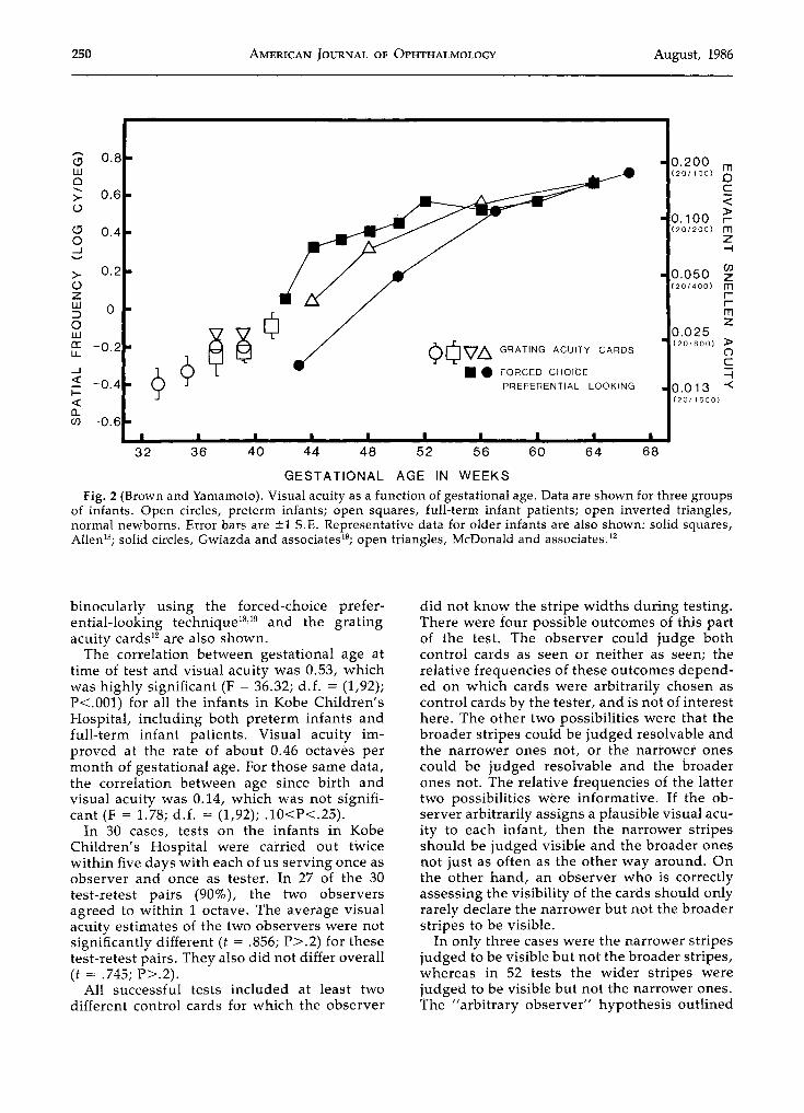
moc<~rmZ-i
(J)Zmrrmz~oC-i-<
6864
90\7.6. GRATING ACUITY CARDS
•• FORCED-CHOICE
PREFERENTIAL LOOKING
3632 40 44 48 52 56 60
GESTATIONAL AGE IN WEEKS
Fig. 2 (Brown and Yamamoto). Visual acuity as a function of gestational age. Data are shown for three groupsof infants. Open circles, preterm infants; open squares, full-term infant patients; open inverted triangles,normal newborns. Error bars are ±1 S.E. Representative data for older infants are also shown: solid squares,Allen": solid circles, Gwiazda and associates'", open triangles, McDonald and associates. 12
,.....,CJwCl<,
>oCJo...J......>ozW:::Jawa:u,
...J«I«0w
binocularly using the forced-choice preferential-looking technique'<" and the gratingacuity cards" are also shown.
The correlation between gestational age attime of test and visual acuity was 0.53, whichwas highly significant (F = 36.32; d.f. = (1,92);P<.OOl) for all the infants in Kobe Children'sHospital, including both preterm infants andfull-term infant patients. Visual acuity improved at the rate of about 0.46 octaves permonth of gestational age. For those same data,the correlation between age since birth andvisual acuity was 0.14, which was not significant (F = 1.78; d.f. = (1,92); .10<P<.25).
In 30 cases, tests on the infants in KobeChildren's Hospital were carried out twicewithin five days with each of us serving once asobserver and once as tester. In 27 of the 30test-retest pairs (90%L the two observersagreed to within 1 octave. The average visualacuity estimates of the two observers were notsignificantly different (t = .856; P>.2) for thesetest-retest pairs. They also did not differ overall(t = .745; P>.2).
All successful tests included at least twodifferent control cards for which the observer
did not know the stripe widths during testing.There were four possible outcomes of this partof the test. The observer could judge bothcontrol cards as seen or neither as seen; therelative frequencies of these outcomes depended on which cards were arbitrarily chosen ascontrol cards by the tester, and is not of interesthere. The other two possibilities were that thebroader stripes could be judged resolvable andthe narrower ones not, or the narrower onescould be judged resolvable and the broaderones not. The relative frequencies of the lattertwo possibilities were informative. If the observer arbitrarily assigns a plausible visual acuity to each infant, then the narrower stripesshould be judged visible and the broader onesnot just as often as the other way around. Onthe other hand, an observer who is correctlyassessing the visibility of the cards should onlyrarely declare the narrower but not the broaderstripes to be visible.
In only three cases were the narrower stripesjudged to be visible but not the broader stripes,whereas in 52 tests the wider stripes werejudged to be visible but not the narrower ones.The "arbitrary observer" hypothesis outlined
Vol. 102, No. 2 Visual Acuity in Infants 251
above can therefore be rejected beyond the.001 level by a likelihood ratio test (X2 = 25.2;d.f. = 1).
Discussion
This new technique was highly successful formeasuring the visual acuity of newborn andpreterm infants. Average binocular visual acuity of normal infants was 20/815 at 39 to 40weeks of gestation. The preterm and full-terminfant patients in the same age range, hospitalized in the nursery of the intensive care unitbut free of neurologic or ophthalmologic diseases, had visual acuities near 20/872 and20/951, respectively. The test was fast (aboutsix minutes on the average) and noninvasive,so it shows promise for use on a more widespread basis.
This technique is convenient to use in aclinical setting. The test requires the participation of three adults: the nurse who holds theinfant, the tester, and the observer. Of these,only the observer needs any training, and thattraining can be short, as the task is easy for aphysician or anyone else with experience observing visual behavior. Medical training per sedoes not appear to be important, however, asan ophthalmologist (M.Y.) and a psychologist(A.M.B.) generally agreed to within 1 octave intheir visual acuity estimates. The nurse has noresponsibility other than holding the infantand keeping it alert. The role of tester wasoccasionally carried out by an orthoptist, withgood success. These tests could easily be doneby a physician in the course of an examinationwithout additional personnel being requiredother than the nurses on duty. This is important for crowded and busy hospital wards.
The cards themselves fit in a large briefcaseand no additional apparatus is required. This isa big advantage in a hospital setting wherespace is at a premium. Infants more than approximately 44 weeks ofgestational age, on theother hand, seem more interested in otherthings in the environment, and testing themwith hand-held cards is difficult. For theseolder infants, the use of a large screen to blockvisible distractions, as suggested by others.v"would undoubtedly result in a much better test.
Early attempts at using the cards with twostimulus windows (Fig. 1, bottom) were notsuccessful. The problem was that the infantsdid not spontaneously turn to fixate the stripes
even when the stripes were very broad. However, other lnvestigators-v" reported good success with the two-stimulus cards. The difference seems to be that when two-stimulus cardsare used, the nurse must turn the infant toshow him or her the stimulus windows on bothsides of the card before preferential looking canoccur. This required more training of the nurses and better communication between the observer and the nurse than was possible in oursituation. With our technique, the observerstarted the trial by placing the stimulus directlyin the infant's lirte of sight, which workedreliably without requiring coordination between the nurse and the observer.
It is important to consider whether this test(or any new test) actually measures visualacuity.
The data themselves are good evidence thatthis techrtique measures something that depends on visual resolution. All successful testsincluded at least one card that the infants didnot refixate. Therefore, the card itself (and theslight space-averaged reflectance mismatch between the stripes and the gray card) was riotsufficient to produce refixation if the stripeswere too small. Furthermore, if the infantswere not responding differentially to gratingsof different stripe widths, observers should beunable to tell which stimuli had wide stripesand which ones had narrow stripes by observing the infants' looking behavior. Actually itwas quite easy to do this when the widths ofthe stripes straddled the visual acuity limit,and the observers rarely made mistakes. Therefore, this technique measures a visually mediated differential response to the stripes.
It is also a reasonable conclusion that thepresent technique, which uses cards with asingle stimulus window, measures the samething as other behavioral studies of visual acuity, which use a left-right stimulus configuration. The observed data fell nicely on an extrapolated curve from the published data on olderinfants (Fig. 2) and agreed rather well withother psychophysical estimates of visual acuityin newborns. 1
08 In particular, the visual acuitiesof infants under 6 weeks of age tested withone-stimulus cards and two-stimulus cards alsoagreed with one another and with the presentdata.":" This overall agreement is consistentwith the view that this test for newborns measures the same visual acuity as other behavioraltests for use on infants.
While the results of this test are reliable andclearly depend on visual resolution, it is less
252 AMERICAN JOURNAL OF OPHTHALMOLOGY August, 1986
clear that they are limited by immaturities located in the distal visual pathways: other partsof the nervous system, notably the oculomotorsystem, are necessary for successful refixationof the displaced grating, and immaturitiesthere could certainly limit performance. Allinfants in this study refixated at least one stimulus successfully, so it would be hard to arguethat the critical maturation is purely motor.However, these data are certainly consistentwith the hypothesis that the effectiveness ofthe gratings in producing differential responses in the optic nerve (for example) is relativelywell developed in newborn infants. Maturationof the visual sensitivity or acuity of the centralvisual nervous system could then account forthe improvement of behaviorally measured visual acuity with age. This view is consistentwith the finding of Norcia and Tyler" thatvisual acuity in young infants measured byvisual-evoked potential is somewhat higherthan that found in any behavioral experiment(however, other investigators using visualevoked potentials reported visual acuities closer to those reported here'': 5,9.10). Further research is necessary to trace the course ofmaturation of function in the different parts ofthe visual nervous system.
There are several reasons that it may beimportant to be able to assess the visual acuityof newborn infants. First, it is possible thatbehavioral assessment could disclose visualdisorders not apparent from ocular examination, just as behavioral assessment is necessaryfor the diagnosis of amblyopia in adults andyoung children. Evidence for this can onlybecome available if clinicians have a techniquesuitable for measuring visual acuity in newborns. The present report provides the firsttechnique that is really adequate for investigating this question. Second, it has been reportedthat behaviorally assessed visual performanceis correlated with neurologic status'i" and thatvisual performance is a sensitive predictor ofintelligence." This suggests that it may be useful to include a measure of visual function aspart of the neurologic examination of the neonate. Once these uses can be established, routine screening may benefit individual patientsand would also allow the development of goodepidemiologic data on a worldwide basis. Finally, even in cases in which the cause of poorvisual function is not well understood or effective treatment is not possible, knowledge of theextent of impairment may be valuable for genetic counseling and for early planning of the
education of the handicapped infant. All ofthese require a convenient technique for measuring the visual acuity of infants. This technique and ones like it will allow these and otherissues in the vision of newborns to be explored.
ACKNOWLEDGMENT
This research was performed at the KobeChildren's Hospital and Kobe Kaisei Hospital,Kobe, Japan, and the Ohta Community Hospital, Miyazu, Kyoto Prefecture, Japan.
References
1. Miranda, S. B.: Visual abilities and patternpreferences of premature infants and full-term neonates. J. Exp. Child Psychol. 10:189, 1970.
2. Dubowitz, L. M. S., Dubowitz, V., and Morante,A.: Visual function in the newborn. A study ofpreterm and full-term infants. Brain Dev. 2:15, 1980.
3. Baraldi, P., Ferrari, F., Fonda, S., and Penne, A.:Vision in the neonate (full-term and premature).Preliminary result of the application of some testingmethods. Doc. Ophthalmol. 51:101, 1981.
4. Morante, A., Dubowitz, L. M. S., Levene, M.,and Dubowitz, V.: The development of vjsual function in normal and neurologically abnormal pretermand fullterm infants. Dev. Med. Child Neurol.24:771, 1982.
5. Dubowitz, L. M. S., Mushin, J., Morante, A.,and Placzek, M.: The maturation of visual acuity inneurologically normal and abnormal newborn infants. Behav. Brain Res. 10:39, 1983.
6. Manning, K. A., Fulton, A. B., Hansen, R. M.,Mayer, D. L., Petersen, R. A., and Barg, D. c.: Preferentiallooking vision testing. Application to evaluation of high-risk, prematurely born infants andchildren. J. Pediatr. Ophthalmol. Strabismus 19:286,1984.
7. Shepard, P. A., Fagen, J. F., and Kleiner, K. A.:Visual pattern detection in preterm neonates. InfantBehav. Dev. 8:47, 1985.
8. Placzek, M., Mushin, J., and Dubowitz,L. M. S.: Maturation of the visual evoked responseand its correlation with visual acuity in pre-terminfants. Dev. Med. Child Neurol. 27:448, 1985.
9. Atkinson, J., Braddick, 0., and French, J.: Contrast sensitivity of the human neonate measured bythe visual evoked potential. Invest. Ophthalmol.Vis. Sci. 18:210, 1979.
10. Porciatti, V.: Temporal and spatial properties ofthe pattern-reversal VEPs in infants below 2 monthsof age. Hum. Neurobiol. 3:97, 1984.
11. Norcia, A. M., and Tyler, C. W.: Spatial frequency sweep VEP. Visual acuity during the firstyear of life. Vision Res. 25:1399, 1985.
12. McDonald, M. A., Dobson, V., Sebris, S. L.,
Vol. 102, No.2 Visual Acuity in Infants 253
Baitch, L., Varner, D., and Teller, D. Y.: The acuitycard procedure. A rapid test of infant acuity. Invest.Ophthalmol. Vis. Sci. 26:1158, 1985.
13. Teller, D. Y.: The forced-choice preferentiallooking procedure. A psychophysical technique foruse with human infants. Infant Behav. Dev. 2:135,1979.
14. Dobson, V., Schwartz, T. L., Sandstrom, D. J.,and Michel, L.: Binocular visual acuity in neonates.The acuity card procedure. Dev. Med. Child Neurol.,in press.
15. McDonald, M. A., Sebris, S. L., Mohn, G., andDobson, V.: Monocular acuity in normal infants. Theacuity card procedure. Am. J. Optom. Physiol. Opt.63:127, 1986.
16. Dobson, V., Salem, D., and Carson, J.: Visualacuity in infants--the effect of variations in stimulusluminance within the photopic range. Invest. Ophthalmol. Vis. Sci. 24:519, 1983.
17. Dubowitz, L. M. S., Dubowitz, V., Morante,A., and Verghote, M.: Visual function in the pretermand fullterm newborn infant. Dev. Med. Child Neur01. 22:465, 1980.
18. Allen, J. L.: The development of visual acuityin human infants during the early postnatal weeks,thesis. University of Washington, Seattle, 1979.
19. Gwiazda, J., Brill, S., Mohindra, 1., and Held,R.: Preferential looking acuity in infants from two tofifty-eight weeks of age. Am. J. Optom. Physiol. Opt.57:420, 1980.
20. Sigman, M., Kopp, C. B., Parmalee, A. H., andJeffrey, W. E.: Visual attention and neurologicalorganization in neonates. Child Dev. 44:461, 1973.
21. Miranda, S. B., Hack, M., Fantz, R. L., Fanaroff, A. A., and Klaus, M. H.: Neonatal pattern vision. A predictor of future mental performance? J.Pediatr. 91:642, 1978.