Embed Size (px)
Citation preview
Vision Rehabil itation AfterTraumatic Brain Injury
Sandra M. Fox, ODa,*, Paul Koons, MS, OMS, CLVT, CBISb, Sally H. Dang, ODc
KEYWORDS
� Traumatic brain injury � Visual dysfunction � Vision rehabilitation � Vision therapy� Concussion
KEY POINTS
� Visual dysfunctions and symptoms are commonly experienced after even mild traumaticbrain injury (TBI) despite excellent visual acuity.
� All individuals who have experienced a TBI/concussion should be screened for visionsymptoms and visual dysfunction.
� A TBI-specific eye examination is necessary to identify the visual sequelae of TBI as wellas address any vision/ocular issues that may be contributing to other post-TBI com-plaints, such as headache, photosensitivity, and vertigo.
� Recognizing and establishing your local vision rehabilitation network of professionals willoffer a comprehensive approach for the patient experiencing visual dysfunction and visualdeficits due to TBI.
� Combining office-based and home-based vision therapy training will maximize visual po-tential and functional results.
INTRODUCTION
Visual dysfunction and vision-related symptoms are common but often overlookedsequelae of traumatic brain injury (TBI). Approximately 70% of the brain is eitherdirectly involved with visual processing or is a component for other sensory process-ing.1,2 Six of the 12 cranial nerves pertain to vision and visual/ocular functions. In addi-tion, the areas of the brain that are most likely to be injured during a TBI (frontal,
Disclosure Statement: The views, opinions and/or findings expressed herein are those of the au-thors and do not necessarily reflect the views or the official policy of the Department of Vet-erans Affairs or the US government.a Surgical Service, Ophthalmology, Polytrauma Rehabilitation Center, South Texas VeteransHealthcare System, 7400 Merton Minter, San Antonio, TX 78229, USA; b Blind RehabilitationService, Major Charles Robert Soltes, Jr. O.D. Blind Rehabilitation Center (BRC), Tibor Rubin VAMedical Center, 5901 East 7th Street, Long Beach, CA 90822, USA; c Optometry Service, VALong Beach Healthcare System, 5901 East 7th Street, Long Beach, CA 90822, USA* Corresponding author.E-mail address: [email protected]
Phys Med Rehabil Clin N Am - (2018) -–-https://doi.org/10.1016/j.pmr.2018.09.001 pmr.theclinics.com1047-9651/18/Published by Elsevier Inc.

Fox et al2
occipital, temporal, and parietal lobes, as well as the long axonal fibers connecting themidbrain to the cortex) are vision related.1 Thus, it is not surprising that even a mildTBI/concussion can lead to significant visual sequelae that will adversely affect reha-bilitation and quality of life.Much of what we now know about how brain injury affects the visual system was
gleaned from research performed within the Department of Defense (DoD) and theDepartment of Veterans Affairs (VA) systems. Although earlier research postulated adifferent mechanism of action for blast-related TBI than non–blast-related TBI, morerecent research has found minimal difference in the visual sequelae between blast-related and non–blast-related TBI.1,3,4 This suggests that VA and DoD research con-cerning the assessment and management of the visual sequelae of mild TBI can beapplicable to those sustaining a mild TBI/concussion in the civilian setting.
VISUAL IMPAIRMENT VERSUS VISUAL DYSFUNCTION
Visual impairment or blindness occurs when visual acuity is decreased and or the vi-sual field is constricted. The incidence of diagnosed visual impairment and blindnessresulting from TBI ranges from approximately 9% to 38% depending on the definitionused, the mechanism of injury (blast vs non-blast) and the severity of the TBI, withmost cases occurring in blast-related moderate to severe TBI.3,5–7
Visual dysfunction refers to a disorder of any visual function, such as oculomotorand accommodation, visual spatial deficits, and photosensitivity. Visual dysfunctionsand symptoms are commonly experienced after TBI despite excellent visual acu-ity.1,2,4,5,7,8 They can contribute to headache and dizziness; cause diplopia, eye fa-tigue, and an inability to focus; adversely affect reading and all near tasks; andcontribute to photophobia.4,6,8–10 Undiagnosed, the visual sequela can affect school,work, and other activities of daily living.
Visual Symptoms and Dysfunction in Traumatic Brain Injury
Self-reported vision complaints, including blurry vision distance and near, eye strain,eye pain, double vision, bumping into objects, difficulty reading, and light sensitivity,range from 65% to 79% depending on the study design and patient popula-tion.1,3,4,6,8–10 Difficulty reading was a common complaint (32%–66%),1 as was lightsensitivity (33%–69%).1,4,8–10
The visual dysfunctions most often identified were convergence insufficiency,accommodative dysfunction, and photosensitivity.3,4,6,7,9–11
Other visual anomalies, such as visual field loss, cranial nerve disorder, strabismus,pursuit/saccade disorder, diplopia, and ocular injuries, are less frequently diagnosedand are more often found in moderate to severe TBI. The wide range in the frequencyof visual dysfunction in people with TBI is most likely due to differences in settings andpatient populations. Studies reporting data on unscreened individuals with TBI reportmuch lower rates of visual dysfunction than studies that use self-report measures toscreen for visual symptoms, highlighting the value of screening for visual dysfunctionin patients with TBI.
MILD TRAUMATIC BRAIN INJURYScreening for Visual Dysfunction in Mild Traumatic Brain Injury
Not all rehabilitation settings will have optometrists/ophthalmologists on staff to pro-vide an eye examination for persons who have experienced a mild TBI. In such cases,a method to screen patients with TBI for possible visual symptoms should beimplemented.

Vision Rehabilitation After Traumatic Brain Injury 3
Brain injury vision symptom survey questionnaireThe Brain Injury Vision Symptom Survey (BIVSS) Questionnaire is a 28-item self-administered survey for vision symptoms related to TBI.12 It probes multiple dimen-sions of vision-related behaviors, including eyesight clarity, visual comfort, diplopia,depth perception, dry eye, peripheral vision, light sensitivity, and reading and is thescreening tool of choice for visual symptoms related to mild TBI.
Screening protocol for therapistsAlthough symptom surveys are quite useful in identifying individuals who have visualsymptoms, a more in-depth screening protocol is necessary to determine if the visualsymptoms are such that an additional evaluation by an optometrist or ophthalmologistis required. Occupational therapists, vision rehabilitation therapists, and blind rehabil-itation outpatient specialists within the VA system can perform additional screeningtests to facilitate the appropriate referrals. A consensus panel of occupational thera-pists and optometrists suggested a screening protocol designed to identifyTBI-related vision disorders in adults.13 Table 1 uses their recommendations with afew updated modifications.In addition, although the consensus of this group did not include computerized
vision screening programs, such as the Home Therapy System (HTS) Binocular VisionAssessment (HTS Inc, Gold Canyon, AZ) and VERA vision screening software (VisualTechnology Applications, Philadelphia, PA), the computerized vision screening pro-grams do show promise, and in one small study showed excellent validity and repeat-ability for assessing near-related binocular vision problems and pursuit and saccadiceye movements.14,15 HTS Vision Therapy is a computer program that can be used asan in-office screening tool for accommodation, vergence, and eye movements (pur-suits and saccades). It also contains the Computerized Perceptual Therapy System,which evaluates visual perceptual areas including visual concentration, visual closure,visual processing, and visual sequential memory.
The Traumatic Brain Injury–Specific Vision Evaluation
A TBI-specific vision evaluation is indicated when a patient has experienced a TBI.Before the TBI vision evaluation, a comprehensive baseline refractive and ocularhealth examination is important to address non–TBI-related vision issues. It is
Table 1Screening tests for visual sequelae in mild traumatic brain injury
Test with corrective lenses if appropriate: older than 40, may need reading glasses
Symptom self-report Brain Injury Vision Symptom Survey
Distance visual acuity Distance Snellen chart
Near visual acuity chart Any near single letter/number chart
Accommodation Accommodative amplitude test
Convergence Near point of convergence
Eye alignment and binocular vision Stereo test
Saccades Developmental eye movement test
Pursuits Northeastern State University Collegeoptometry oculomotor test
Visual Fields Confrontation visual fields fingerCounting
Courtesy of Sandra Fox, OD, San Antonio TX, USA.

Fox et al4
common to have refractive error, such as latent hyperopia that is often symptomaticafter the TBI. A thorough damp refraction with dilation is helpful to detect latent hyper-opia, which will cause near or accommodative vision challenges. Ruling out any pre-existing ocular disease is important. Patients with TBI often have dry eye syndromethat adds to fluctuations in vision. It is recommended that patients with TBI have abaseline screening visual field test.In addition to visual sequelae such as oculomotor dysfunction and photosensitivity,
common complaints after mild TBI include headaches, vertigo, and difficulty readingand concentrating while reading. The role of the optometrist/ophthalmologist is toaddress any vision/ocular issues that may be contributing to these complaints.Box 1 from the Walter Reed National Military Medical Center Vision Center of Excel-
lence includes the eye/vision tests that are included as the basic components of aneye examination.
Patient History
Considering what we now know about mild TBI/concussion, a question pertaining tomilitary service and a history of high-impact sports should be a part of every medical/social history. Any positive responses should prompt additional TBI-related questionsdesigned to determine if there is a possible oculomotor dysfunction. See Box 2 foradditional TBI-related questions.
Additional Testing
Oculomotor dysfunctionIf a patient is symptomatic for oculomotor dysfunction, additional clinical testing mustbe performed to evaluate visual efficiency to determine the specific types and
Box 1
Basic eye care/vision examination by an eye care provider
Historya
Visual acuity
Refractive error measurement
External examination
Pupillary testing
Extraocular muscle testing/pursuits
Cover test (distance and near)
Confrontation visual field testing
Tonometry
Slit lamp biomicroscopy: anterior segment, cornea, macula, lens, and optic nerve
Binocular indirect ophthalmoscopy with scleral depressionb
Gonioscopyb
a It is recommended that assessment of medical history also include the question, “Have youbeen exposed to blast or sustained a head injury, concussion, or traumatic brain injury (TBI)?” Apositive response to this question would be a sufficient rationale to ask TBI-related ocular his-tory questions and conduct supplemental testing.
b If patient history indicates exposure to blast, head injury, concussion, and/or TBI.Previously published materials from Walter Reed National Military Medical Center/Vision
Center of Excellence. 2016; with permission.

Box 2
TBI-related ocular history questions
Did you have any neurologic problems or symptoms before your TBI (multiple sclerosis, stroke,brain tumor, severe headaches, other)?
When did your TBI occur (on what date)?
Did you lose consciousness during or after your TBI incident?
Were you disoriented or confused during or after your TBI incident?
Do you bump into objects and walls more now than before your injury?
Were your eyes, eyelids, or area around your eyes injured when your TBI event occurred?
Do you cover or close one eye at times since your injury?
Have you noticed a change in your vision since your injury?
Are you more sensitive to light, either indoors or outdoors, since your injury?
Have you had any double vision since your injury?
Have you noticed any changes in your peripheral vision since your injury?
Is your vision blurry at distance or near since your injury?
Have you noticed a change in your ability to read since your injury?
Do you lose your place while reading more now than before your injury?
How long can you read continuously before you need to stop?
Do you get headaches during/after reading more now than before your injury?
Do you have more difficulty remembering what you have read now than before your injury?
Data from Goodrich G, Martinsen G. Development of a mild traumatic brain injury-specificvision screening protocol: a Delphi study. J Rehabil Res Dev 2013;50(6):757–68; and Previouslypublished materials from Walter Reed National Military Medical Center/Vision Center of Excel-lence. 2016; with permission.
Vision Rehabilitation After Traumatic Brain Injury 5
severities of oculomotor dysfunction. Table 2 lists the oculomotor dysfunction param-eters that need to be tested and methods that can be used.
Visual information processingVisual information processing, which includes visual spatial information, visual anal-ysis, and visual motor integration, also needs to be evaluated. Table 3 pertains to vi-sual information processing function, description, and testing methods.Simple observation of the patient’s gait and balance can diagnose spatial localiza-
tion problems. This may or may not be present with motion sensitivity, which iscommon in crowded places. Patients may report feeling dizziness, nausea, andunsteadiness.
Treatment Strategies
Once the visual dysfunction has been identified, a treatment plan will need to be devel-oped to improve the visual efficiency. Box 3 describes the treatment strategies usedfor oculomotor dysfunction.
Rehabilitation Team
As with any rehabilitation plan, it is often necessary to have coordination of subse-quent referrals to other services. The goal is to understand how various vision

Table 2Recommended tests to evaluate for oculomotor dysfunctions
OculomotorParameter Testing
Eye alignment Distance and near cover test in multiple positions of gaze and head tiltPhorias (vertical and horizontal)Maddox rodModified Thorington
Vergence Vergence ranges (vertical and horizontal)Vergence facility
Convergenceamplitude
Near point of convergence Repeated measures
Accommodation Push-up method Repeated measuresMinus lens Repeated measuresAccommodative facility (monocular and binocular)Negative relative accommodation/Positive relative accommodation
(NRA/PRA)Near retinoscopyAccommodative convergence/accommodation (AC/A) ratio
Eye movements DuctionsVersionsPursuitSaccadesDevelopmental eye movement (DEM)King-Devick
Suppression check Worth 4 Dot (distance and near)Random dot stereopsis
Vestibulo-ocularreflex
(If positive, refer to audiology, otolaryngology, or vestibular physicaltherapist)
Dynamic visual acuityHead thrustLow-frequency head shake
Note: not all tests are required; italicized tests provide more comprehensive results as recognizedby our expert panel, but selection of tests is left to the clinical judgment of the eye care provider.
Previously published materials fromWalter Reed National Military Medical Center/Vision Centerof Excellence. 2016; with permission.
Fox et al6
problems affect function. See Table 4 for additional specialties that may need to beconsulted.
Plan of Care of Oculomotor Dysfunctions Associated with Traumatic Brain Injury
Fig. 1 is an algorithm outlining the process for the care of the patient with oculomotordysfunctions associated with TBI.
Vision Rehabilitation for Mild Traumatic Brain Injury
Vision rehabilitation after mild TBI can be further complicated by comorbidities andmust be considered when developing the rehabilitative plan.
Comorbidities
In the military/veteran TBI population, there is a high prevalence of comorbid drug/substance abuse and mental illness, further complicating the diagnosis and treatmentof visual symptoms. Among veterans with TBI, 89% had a comorbid psychiatric
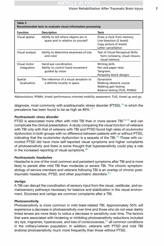
Table 3Recommended tests to evaluate visual information processing
Function Description Tests
Visual spatial Ability to tell where objects are inspace and in relation to yourself
Draw a clock from memoryLine bisection (2 levels)Copy picture (2 levels)Letter cancellation
Visual analysis Ability to determine awareness of sizeand color
Test of Visual Perceptual Skills:form constancy, visual closure,visual memory
Visual motorintegration
Hand-eye coordinationAbility to control hand movement
guided by vision
Writing skillsPen and paper testsTangramsParquetry block designs
Spatiallocalization
The reference of a visual sensation toa definite locality in space
DynavisionWalking obstacle courseWalking gait testingBalance testing (TUG, POMA)
Abbreviations: POMA, tinetti performance oriented mobility assessment; TUG, timed up and go.
Vision Rehabilitation After Traumatic Brain Injury 7
diagnosis, most commonly with posttraumatic stress disorder (PTSD),16 in which theprevalence has been found to be as high as 89%.17
Posttraumatic stress disorderPTSD is associated more often with mild TBI than in more severe TBI18,19 and cancomplicate the clinical presentation. A study comparing the visual function of veteranswith TBI only with that of veterans with TBI and PTSD found high rates of oculomotordysfunction in both groups with no difference between patients with or without PTSD,indicating that the oculomotor dysfunction is a sequela of the TBI.18 Those with co-morbid PTSD did have more self-reported visual symptoms and higher complaintsof photosensitivity and there is some thought that hypersensitivity could play a rolein the increased reporting of visual symptoms.20
Posttraumatic headachesHeadache is one of the most common and persistent symptoms after TBI and is morelikely to persist after mild TBI than moderate or severe TBI. The chronic symptom-atology of service members and veterans following TBI is an overlap of chronic post-traumatic headaches, PTSD, and other psychiatric disorders.21
VertigoA TBI can disrupt the coordination of sensory input from the visual, vestibular, and so-matosensory pathways necessary for balance and stabilization in the visual environ-ment. Dizziness and vertigo are common complaints after a TBI.
PhotosensitivityPhotosensitivity is more common in mild blast-related TBI. Approximately 50% willexperience a decrease in photosensitivity over time and those who do not wear darklytinted lenses are more likely to notice a decrease in sensitivity over time. The factorsthat were associated with hindering or inhibiting photosensitivity reductions includeddry eye, migraines, hyperacusis, and loss of consciousness,22 all common conditionsin the military/veteran population. In addition, veterans with PTSD and mild TBIendorse photosensitivity much more frequently than those without PTSD.

Box 3
Treatment strategies for oculomotor dysfunction
Correction of refractive error to improve vision, binocular alignment, and accommodativefunction
Added lenses to improve binocular alignment and accommodative function
When necessary, prism therapy to eliminate double vision and restore visual comfort
Office-based oculomotor rehabilitation (with home-reinforcement) using a variety ofprocedures to improve oculomotor function
When necessary, surgery for associated strabismus or other relevant oculomotor problems
Previously published materials from Walter Reed National Military Medical Center/Vision Cen-ter of Excellence. 2016; with permission.
Fox et al8
Before vision rehabilitation, it is important to rule out any visual or ocular conditionsthat may be contributing to headaches, vertigo, and photosensitivity, as well aswhether PTSD may be contributing to the symptomatology.
Vision Rehabilitation Team
The ideal setting for providing the patient with TBI with appropriate vision care andvision therapy is a team of providers working together in a vision clinic.23 Becausemost rehabilitation services for the population with mild TBI occur in an outpatientsetting, it is vital to establish a professional support network that evaluates and treatsvision-related issues.Vision rehabilitation specialists may include occupational therapists, certified low-
vision therapists, optometrists, and typically other trained blind rehabilitation special-ists with knowledge in vision therapy training. It is imperative that the vision therapistswork closely with the eye care practitioner’s plan of care and provide regular updatesas to the patient’s therapy progression, regression, and/or plateau of skills, as this mayrequire reevaluation of the vision therapy treatment plan.
Types of Vision Rehabilitation Programs
Vision rehabilitation settings in mild TBI may include an optometrist/ophthalmologistoffice or facilities with inpatient and/or outpatient rehabilitation clinics. Within VeteransAffairs Medical Centers, inpatient and outpatient clinics exist that offer specialty reha-bilitation programs aimed at evaluating and training those with visual dysfunctionsresulting from TBI.
Table 4Referral to appropriate facility-specific provider
Audiology/Otolaryngology/Vestibular PhysicalTherapy
Speech/Language Therapy
Blind/Low-Vision Rehabilitation Neurology/Neuro-Ophthalmic Care
Occupational Therapy Psychology/Psychiatry/Neuro-Psychiatry
Physical Therapy
Previously published materials fromWalter Reed National Military Medical Center/Vision Center ofExcellence. 2016; with permission.

Fig. 1. Algorithm for the care of oculomotor dysfunctions associated with TBI. (From WalterReed National Military Medical Center/Vision Center of Excellence. 2016; with permission.)
Vision Rehabilitation After Traumatic Brain Injury 9
Vision Therapy
The American Optometric Association defines vision therapy as a sequence of neuro-sensory and neuromuscular activities individually prescribed and monitored by thedoctor to develop, rehabilitate, and enhance visual skills and processing.24 The goalof vision therapy is to improve the speed, accuracy, and integration of oculomotorfunctions.Eye movements include motions that shift the direction of eye gaze, such as sac-
cades, pursuits, and vergences.25 Oculomotor dysfunctions associated with TBImay affect many areas of daily living, including reading, visual learning, and abilityto concentrate. Vision therapy is an accepted treatment of choice for the most

Fox et al10
common TBI-related visual dysfunctions. See Table 5 for management and treatmentoptions for common oculomotor dysfunctions associated with TBI.
Office-based and home-based vision therapy treatment strategiesOnce a vision therapy plan of care is developed, the patient (and family) should beeducated about the relationship between the visual deficit and dysfunction and theability to complete a visual task. A schedule should be created for regular inpatienttraining (office visit) with a home exercise program (HEP) for therapy reinforcement.The HTS program can be provided to the patient as an excellent home therapy toolfor those individuals with access to computers. Complying with the HEP is crucialfor success.
Duration of vision therapy and frequencyA patient’s motivation and commitment are key to success for any vision therapytraining program. The average duration for most vision therapy programs range
Table 5Management and treatment options for common oculomotor dysfunctions associated withtraumatic brain injury
Condition Primary Treatment Secondary Treatment
Accommodativeinsufficiency Ill-sustainedaccommodation
Plus-powered lenses Oculomotor rehabilitation
Accommodative excess Oculomotor rehabilitation
Convergence Insufficiency Oculomotor rehabilitation Prism lensesExtraocular muscle surgery
Convergence excess Plus-powered lenses Oculomotor rehabilitation
Fusional vergencedysfunction
Oculomotor rehabilitation
Divergence insufficiency Prism lenses Oculomotor rehabilitation
Divergence excess/basicexophoria
Oculomotor rehabilitation Extraocular muscle surgery
Basic esophoria Prism lenses Oculomotor rehabilitation
Vertical phoria Oculomotor rehabilitation andprism lenses
Extraocular muscle surgery
Saccadic dysfunction Oculomotor rehabilitation
Cranial nerve (CN) IIIpalsy
Fresnel prism, ptosis crutch, nearlenses
Extraocular muscle surgery
CN IV palsy Fresnel prism, distance and nearglasses, base down near yokedprism, reading stands, sectorocclusion, and/or full fieldocclusion
Prisms likely ineffective ifsignificant torsion
Oculomotor rehabilitationExtraocular muscle surgery
CN VI palsy Fresnel prism Oculomotor rehabilitationMedicationsExtraocular muscle surgery
Adapted from Scheiman M. Understanding and managing vision disorders after traumatic braininjury. A guide for military optometrists. Washington, DC; Office of the Surgeon General: 2011;and Previously published materials fromWalter Reed National Military Medical Center/Vision Cen-ter of Excellence. 2016; with permission.

Vision Rehabilitation After Traumatic Brain Injury 11
between 4 and 6 weeks, depending on the patient’s goals. It is recommended that pa-tients attend weekly in-office therapy in addition to home vision therapy to makecertain they are accurately performing the therapy.Office-based vision therapy training should resemble a “fitness center” for the eyes,
using a multitude of devices or techniques aimed at isolating and improving the spe-cific visual function.
Tint Evaluation
Based on the patient’s self-report or symptom checklist, the vision therapistshould evaluate each type of glare experienced by the patient, including outdoor,indoor, computer screen/iPhone, and night glare. Distant acuity charts may provebeneficial to evaluate visual clarity during indoor and outdoor tint evaluations. Forthose patients using darker tints, the goal is to decrease the tint level over time(increase light transmission levels) and improve their tolerance of brighter environ-ments. Exceptions would be patients who experience migraines and patients whohave abnormally large pupil size (often secondary to medication). PTSD as a co-morbidity can complicate the tint assessment and the goal of decreasing tintover time.
MODERATE TO SEVERE TRAUMATIC BRAIN INJURY
Individuals who experience a moderate or severe TBI will have visual sequelae likethose with mild TBI; however, in addition to oculomotor/accommodative dysfunctionand photosensitivity, they are more likely to experience ocular trauma whereby visualimpairment is more common. Also, the more severely the brain has been damaged,the more likely a visual field deficit will be present. The frequency of ocular injuriesin moderate to severe TBI ranges from 30% to 38% and includes orbital fractures,lid lacerations, traumatic cataracts, traumatic maculopathy, retinal hemorrhages, op-tic neuropathy, globe ruptures, angle recession, hyphema, and corneal injuries.4,6
Blindness or legal blindness in moderate to severe TBI was found in 13% to 14%3,6
and 18% to 32% were found to have visual field deficits.3,4
The Traumatic Brain Injury–Specific Eye Examination in Moderate to SevereTraumatic Brain Injury
The level of cognition may make a subjective evaluation challenging so theeye care provider may need to rely on objective findings only. The initial evaluationis likely to take place at bedside and the goal of this evaluation is to evaluateocular health because the patient with moderate/severe TBI is more likely tohave ocular trauma. More severe patients may be nonverbal or in a low level ofconsciousness. Assessing visual potential is crucial because visual tracking is apart of the Coma Recovery Scale and visual impairment/blindness is more likelyin the patient with more severe TBI. One study found that 65% of patients with dis-order of consciousness misdiagnosed as being in a vegetative state were blind orvision impaired.26
The visual potential is assessed by examining the ocular health, determining therefractive error, and using objective methods of assessing visual acuity, such as theoptokinetic drum and preferential looking (Teller Cards).
Vision Rehabilitation in Moderate to Severe Traumatic Brain Injury
Addressing the ocular health concerns and ensuring that any refractive error is cor-rected is primary in the acute setting. Frequent follow-up is indicated as the patient

Fox et al12
progresses and more subjective testing can be performed. Diplopia, visual impair-ment/blindness, and visual field deficits may become more apparent as the patient’scognition improves.Cotreatment among therapists often may prove beneficial due to incorporation of
multiple rehabilitation techniques that maximize training efforts and keep goals rele-vant to daily activities.Examples:
� Occupational therapist reviews patient’s daily task calendar, and vision therapistreinforces eye movement pursuit and fixation
� Vision therapist practices patient’s visual scanning techniques on white board tolocate letter, whereas speech and language pathologist practices pronunciationof letter/word
� Physical therapy provides training to patient for improved walking posture/gaitand walking endurance, whereas vision therapist incorporates bilateral scanningto locate wall targets
DiplopiaCranial nerve palsies are common after moderate to severe TBI and the resultantdiplopia will adversely affect rehabilitation. Patching is not ideal, especiallyfor mobility. Using a Fresnel press-on prism to eliminate the diplopia is preferablebecause the press-on prism eliminates the diplopia without significantly com-promising vision. The prism power can be changed as the palsy improves overtime.
Visual impairment/blindnessDetermining if the patient has suffered vision loss is critical to the rehabilitation pro-cess. Once it has been determined that a patient is visually impaired, low-vision eyecare providers and therapists with experience in low-vision rehabilitation will needto be consulted to partner with the patient’s TBI rehabilitation team. The rehabilitationteam should provide modifications and compensatory strategies that will be used forrehabilitation purposes.The VA provides blind and vision rehabilitation programs to eligible veterans and
active duty service members who are visually impaired. These training programs offerveterans the opportunity to acquire the skills necessary to regain independence andsuccessfully integrate into their family and community life. For more informationon blind rehabilitation services within the VA see http://www.prosthetics.va.gov/blindrehab/BRS_Coordinated_Care.asp.
Visual field lossVisual field deficits have been found in 35% of patients with TBI in a sample clinic pop-ulation having a range of visual symptoms.27 Deficits of all types may be present,ranging from hemianopia to small, scattered regions of reduced sensitivity (Table 6).27
Fig. 2 is an algorithm that outlines the steps and clinical decision points in the eyecare and rehabilitation process for patients with visual field loss associated with TBI/acquired brain injury.
Testing for visual field lossBox 4 outlines the screening tests recommended to evaluate the type of vision prob-lem the patient may be experiencing. These tests will indicate whether the visualdisturbance is related to possible visual field loss.
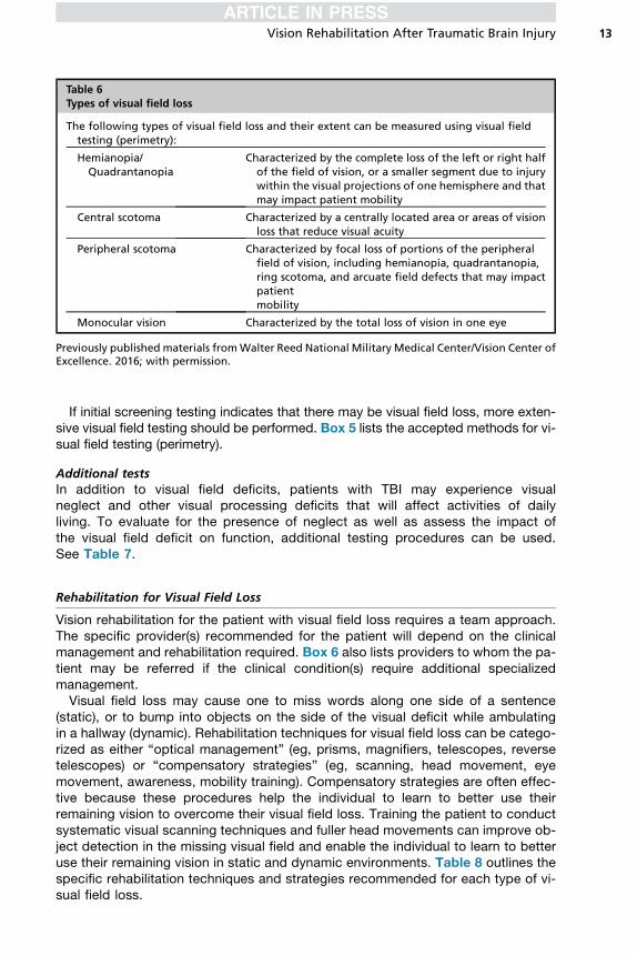
Table 6Types of visual field loss
The following types of visual field loss and their extent can be measured using visual fieldtesting (perimetry):
Hemianopia/Quadrantanopia
Characterized by the complete loss of the left or right halfof the field of vision, or a smaller segment due to injurywithin the visual projections of one hemisphere and thatmay impact patient mobility
Central scotoma Characterized by a centrally located area or areas of visionloss that reduce visual acuity
Peripheral scotoma Characterized by focal loss of portions of the peripheralfield of vision, including hemianopia, quadrantanopia,ring scotoma, and arcuate field defects that may impactpatientmobility
Monocular vision Characterized by the total loss of vision in one eye
Previously published materials fromWalter Reed National Military Medical Center/Vision Center ofExcellence. 2016; with permission.
Vision Rehabilitation After Traumatic Brain Injury 13
If initial screening testing indicates that there may be visual field loss, more exten-sive visual field testing should be performed. Box 5 lists the accepted methods for vi-sual field testing (perimetry).
Additional testsIn addition to visual field deficits, patients with TBI may experience visualneglect and other visual processing deficits that will affect activities of dailyliving. To evaluate for the presence of neglect as well as assess the impact ofthe visual field deficit on function, additional testing procedures can be used.See Table 7.
Rehabilitation for Visual Field Loss
Vision rehabilitation for the patient with visual field loss requires a team approach.The specific provider(s) recommended for the patient will depend on the clinicalmanagement and rehabilitation required. Box 6 also lists providers to whom the pa-tient may be referred if the clinical condition(s) require additional specializedmanagement.Visual field loss may cause one to miss words along one side of a sentence
(static), or to bump into objects on the side of the visual deficit while ambulatingin a hallway (dynamic). Rehabilitation techniques for visual field loss can be catego-rized as either “optical management” (eg, prisms, magnifiers, telescopes, reversetelescopes) or “compensatory strategies” (eg, scanning, head movement, eyemovement, awareness, mobility training). Compensatory strategies are often effec-tive because these procedures help the individual to learn to better use theirremaining vision to overcome their visual field loss. Training the patient to conductsystematic visual scanning techniques and fuller head movements can improve ob-ject detection in the missing visual field and enable the individual to learn to betteruse their remaining vision in static and dynamic environments. Table 8 outlines thespecific rehabilitation techniques and strategies recommended for each type of vi-sual field loss.
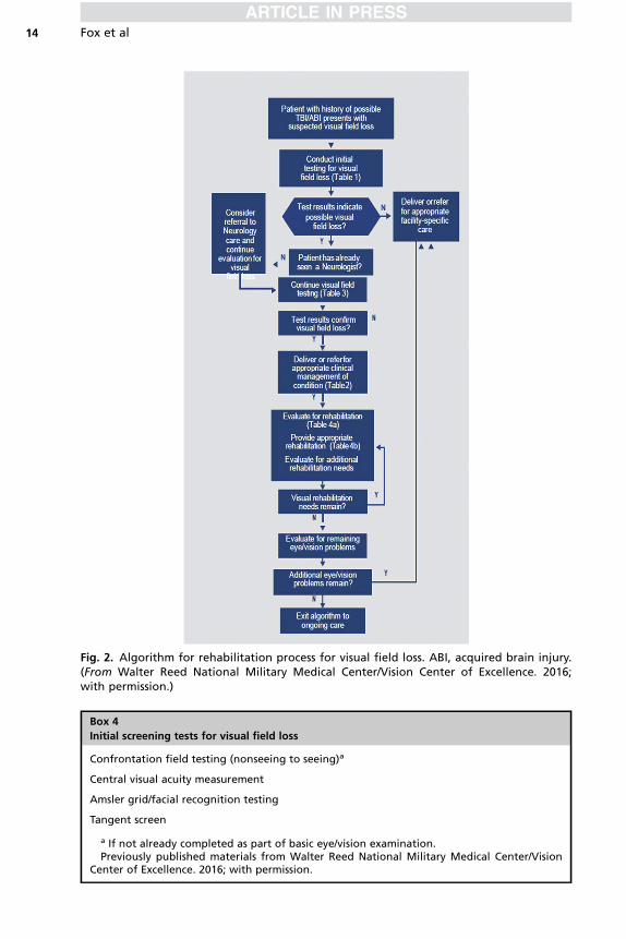
Fig. 2. Algorithm for rehabilitation process for visual field loss. ABI, acquired brain injury.(From Walter Reed National Military Medical Center/Vision Center of Excellence. 2016;with permission.)
Box 4
Initial screening tests for visual field loss
Confrontation field testing (nonseeing to seeing)a
Central visual acuity measurement
Amsler grid/facial recognition testing
Tangent screen
a If not already completed as part of basic eye/vision examination.Previously published materials from Walter Reed National Military Medical Center/Vision
Center of Excellence. 2016; with permission.
Fox et al14

Box 5
Visual field testing (perimetry)
Humphrey/Humphrey Esterman
Octopus
Goldmann
Previously published materials from Walter Reed National Military Medical Center/Vision Cen-ter of Excellence. 2016; with permission.
Vision Rehabilitation After Traumatic Brain Injury 15
REHABILITATION OVERVIEW
Visual dysfunction is commonly associated with TBI and often identified through visualsymptom questionnaires, such as the BIVSS. Recognizing the visual symptomologyrequiring a TBI-specific eye examination is a crucial starting point toward successfulpatient rehabilitation. Assessments must address a patient’s strengths, limitations,needs, preferences, and desired outcomes. Identifying and developing your rehabili-tative referral network is vital when dealing with a TBI patient population experiencingdeficits in visual function. Therapists or other rehabilitation clinicians may be the first to
Table 7Functional visual impact tests/procedures
Functional Task Visual Impact Test
Scanning � biVABA (portion)� DEM (adult)� King-Devick
Visual attention � biVABA� Rivermead (will rule out presence or absence of neglect)� Dynavision� Wayne fixation� Useful field of view
Reading/Near vision � biVABA� Smith-Kettlewell reading test (SK Read)� Pepper test� Minnesota low-vision reading test (MN Read)� Visagraph
Visual perception � Motor-free visual perception test (vertical is recommended butnot always available)
� Test of Visual Perceptual Skills (TVPS)� DVPT-Adult� Home Therapy System CPT Program
Functional independence � Functional Independence Measure (FIM)
Quality of life (QOL) � National Eye Institute Visual Functioning Questionnaire(NEI-VFQ-25) with 10-item euro-ophthalmic supplement
� College of Optometrists in Vision Development (COVD) Qualityof Life Assessment
Abbreviations: biVABA, brain injury visual assessment battery for adults; CPT, computer perceptualtherapy; DEM, developmental eye movement; DVPT, developmental visual perception test.
Previously published materials fromWalter Reed National Military Medical Center/Vision Centerof Excellence. 2016; with permission.

Box 6
Providers for clinical management and rehabilitation of visual field loss and related conditions
Optometrist/Ophthalmologist
Neurologist/Neuro-Ophthalmologist
Occupational/Physical therapist
Audiologista
Low-Vision or Blind Rehabilitation Specialist (Veterans Affairs facilities)
Certified Driver Evaluation Specialist
a Hearing loss may compound spatial awareness difficulties caused by visual field loss.Previously published materials from Walter Reed National Military Medical Center/Vision
Center of Excellence. 2016; with permission.
Fox et al16
notice a problem, therefore, 2-way communication (between medical and rehabilita-tion teams) is vital.Once an eye care provider identifies the visual deficits and/or dysfunction, a vision
rehabilitation plan of care should be offered to improve visual functioning and qualityof life. Successful vision rehabilitation training begins with education of the visualdysfunction to both the patient and family members. A combination of office andhome therapy will offer a comprehensive approach to improving function. Vision ther-apy training may appear repetitive to the patient; therefore, it is important to providemultiple therapeutic activities to keep the patient engaged in therapy and minimizemissed appointments.It is equally important for the rehabilitative team to continually update the
eye care practitioner regarding the patient’s progress, as the plan of care mayneed to be altered (ie, therapist informs that patient does not wear the prism
Table 8Rehabilitation of visual field loss
RehabilitationHemianopia/Quadrantanopia
CentralScotoma
PeripheralScotoma
MonocularVision
Awareness/Sensory integration x x x x
Environment training x x x x
Scanning x x x x
Reading strategies x x — —
Compensatory aids x x x —
Prisms x — — —
Near optical aids (magnifiers) — x — —
Telescopes — x — —
Reverse telescopes — — x —
Eccentric viewing x x — —
Mobility training x x x x
Fitness to drive x x x x
x, appropriate rehabilitation strategy; —, not appropriate.Previously published materials fromWalter Reed National Military Medical Center/Vision Center
of Excellence. 2016; with permission.

Vision Rehabilitation After Traumatic Brain Injury 17
lenses). Finally, goals should be realistic and aligned with the patient’s everydaytasks.
SUMMARY
The goal of all rehabilitation is to improve function and retain independence, therebyimproving quality of life. Vision is a major component in every aspect of rehabilitation.Speech language pathologists and neuropsychologists administer cognitive tests thatrequire reading. Physical therapists are working to improve mobility and balance,which are affected by vision. Occupational therapists are evaluating the ability toperform activities of daily living, and recreation therapists use games and crafts aspart of therapy, all highly dependent on vision.Knowledge of the patient’s visual acuity, visual fields, and oculomotor function is
crucial information for the rehabilitation team to accurately assess mobility and bal-ance or the higher-level visual skills, such as visual tracking and scanning, visual mem-ory, and visual cognition. Unfortunately, this information is often not available, and thepatient’s rehabilitation success can be significantly hampered.The optometrist’s role is to provide the rehabilitation team with this valuable infor-
mation. Knowing your eye care provider and rehabilitation network will provide ateam approach to identifying and improving potential visual dysfunctions caused byTBI.
REFERENCES
1. Capo-Aponte J, Jorgensen-Wagers K, Sosa J, et al. Visual dysfunctions atdifferent stages after blast and non-blast mild traumatic brain injury. Optom VisSci 2017;94(1):7–15.
2. Sutter P. Rehabilitation and management of visual dysfunction following traumaticbrain injury. In: Ashley MJ, Krych DK, editors. Traumatic brain injury rehabilitation.New York: CRC Press; 1995. p. 187–219.
3. Brahm KD, Wilgenburg HM, Kirby J, et al. Visual impairment and dysfunction incombat-injured service members with traumatic brain injury. Optom Vis Sci2009;86(7):817–25.
4. Goodrich GL, Flyg HM, Kirby JE, et al. Mechanisms of TBI and visual conse-quences in military and veteran populations. Optom Vis Sci 2013;90(2):106–12.
5. Bulson R, Jun W, Hayes J. Visual symptomology and referral patterns for Opera-tion Iraqi Freedom and Operation Enduring Freedom veterans with traumaticbrain injury. J Rehabil Res Dev 2012;49(7):1075–82.
6. Goodrich GL, Kirby J, Cockerham G, et al. Visual function in patients of a poly-trauma rehabilitation center: a descriptive study. J Rehabil Res Dev 2007;44(7):929–36.
7. Alvarez TL, Kim EH, Vicci VR, et al. Concurrent vision dysfunctions in conver-gence insufficiency with traumatic brain injury. Optom Vis Sci 2012;89(12):1740–51.
8. Stelmack JA, Frith T, Van Koevering D, et al. Visual function in patients followed ata Veterans Affairs polytrauma network site: an electronic medical record review.Optometry 2009;80:419–24.
9. Magone MT, Kwon E, Shin SY. Chronic visual dysfunction after blast-induced mildtraumatic brain injury. J Rehabil Res Dev 2014;51(1):71–80.
10. Lew HL, Poole JH, Vanderploeg RD, et al. Program development and definingcharacteristics of returning military in a VA Polytrauma Network Site. J RehabilRes Dev 2007;44(7):1027–34.

Fox et al18
11. Ciuffreda KJ, Kapoor N, Rutner D, et al. Occurrence of oculomotor dysfunctionsin acquired brain injury: a retrospective analysis. Optometry 2007;78(4):155–61.
12. Laukkanen H, Scheimann M, Hayes JT. Brain Injury Vision Symptom Survey(BIVSS) questionnaire. Optom Vis Sci 2017;94:43–50.
13. Radomski M, Finkelstein M, Llanos I, et al. Composition of a vision screen for ser-vice members with traumatic brain injury: consensus using a modified nominalgroup technique. Am J Occup Ther 2014;68(4):422–9.
14. Gallaway M, Mitchell GL. Validity of the VERA visual skills screening. Optometry2010;81:571–9.
15. Capo-Aponte JE, Tarbett AK, Urosevich TG, et al. Effectiveness of computerizedoculomotor vision screening in a military population: pilot study. J Rehabil ResDev 2012;49:1377–98.
16. Barker F, Cockerham G, Goodrich G, et al. Brain injury impact on the eye andvision. Optom Vis Sci 2017;94(1):4–6.
17. Bahraini N, Breshears R, Hernandez T, et al. Traumatic brain injury and post trau-matic stress disorder. Psychiatr Clin North Am 2014;37:55–75.
18. Goodrich GL, Martinsen GL, Flyg HM, et al. Visual function, traumatic brain injuryand posttraumatic stress disorder. J Rehabil Res Dev 2014;51(4):547–58.
19. Zatzick DF, Rivara FP, Jurkovich GJ, et al. Multisite investigation of traumatic braininjuries, posttraumatic stress disorder and self-reported health and cognitive im-pairments. Arch Gen Psychiatry 2010;67(12):1291–300.
20. Ragsdale KA, Neer SM, Beidel DC, et al. Posttraumatic stress disorder in OEF/OIF veterans with and without traumatic brain injury. J Anxiety Disord 2013;27:420–6.
21. Theeler B, Erickson J. Posttraumatic headache in military personnel and veteransof the Iraq and Afghanistan conflicts. Curr Treat Options Neurol 2012;(14):36–49.
22. Truong J, Cuiffreda K, Han E, et al. Photosensitivity in mild traumatic brain injury(mTBI): a retrospective analysis. Brain Inj 2014;28(10):1283–7.
23. Ripley D, Politzer T, Berryman A, et al. The vision clinic: an interdisciplinarymethod for assessment and treatment of visual problems after traumatic braininjury. NeuroRehabilitation 2010;27:231–5.
24. American Optometric Association Board of Trustees, 2009. Available at: www.aoa.org/Documents/optometrists/QI/definition-of-optometric-vision-therapy.pdf.Accessed October 12, 2018.
25. Sutter P. Rehabilitation and management of visual dysfunction following traumaticbrain injury. In: Sutter P, editor. Vision rehabilitation: multidisciplinary care of thepatient following brain injury. Boca Raton (FL): CRC Press; 2011. p. 309–46.
26. Andrews K, Murphy L, Munday R, et al. Misdiagnosis of the vegetative state:retrospective study in a rehabilitative unit. BMJ 1996;313:13–6.
27. Suchoff IB, Kapoor N, Ciuffreda KJ, et al. The frequency of occurrence, types andcharacteristics of visual field defects in acquired brain injury: a retrospectiveanalysis. Optometry 2008;79:259–65.