Embed Size (px)
Citation preview
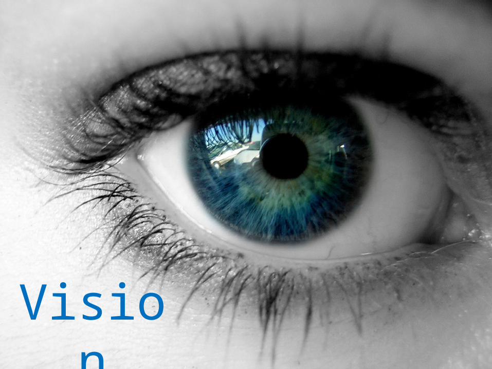
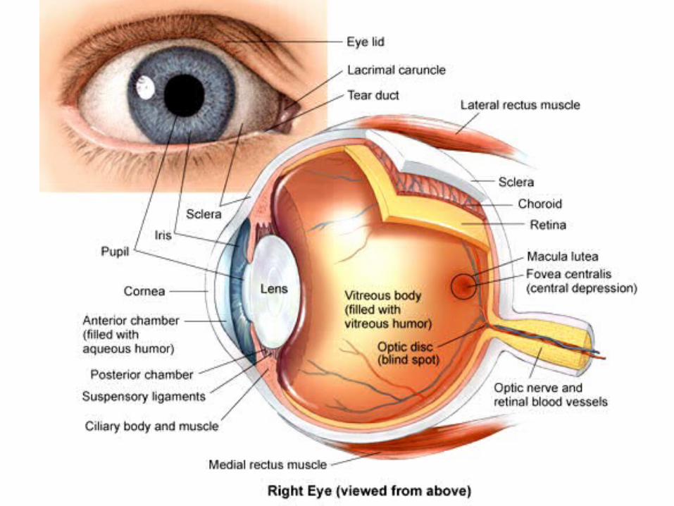
Vision
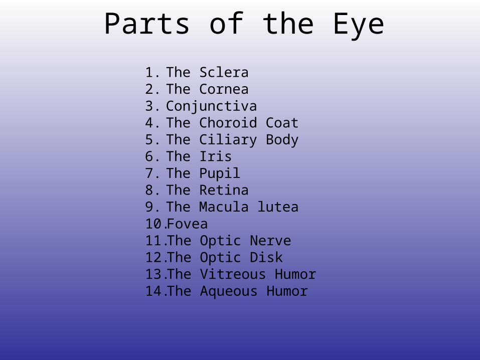
Parts of the Eye
1. The Sclera2. The Cornea3. Conjunctiva4. The Choroid Coat5. The Ciliary Body 6. The Iris7. The Pupil8. The Retina9. The Macula lutea10. Fovea11. The Optic Nerve12. The Optic Disk13. The Vitreous Humor 14. The Aqueous Humor

The Conjunctiva
• lines the inside of the eyelids and covers the sclera (white part of the eye). It is composed of rare stratified columnar epithelium.
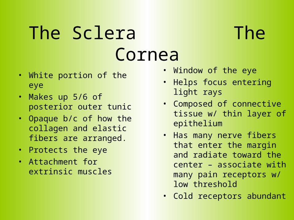
The Sclera The Cornea• Window of the eye
• Helps focus entering light rays
• Composed of connective tissue w/ thin layer of epithelium
• Has many nerve fibers that enter the margin and radiate toward the center – associate with many pain receptors w/ low threshold
• Cold receptors abundant
• White portion of the eye
• Makes up 5/6 of posterior outer tunic
• Opaque b/c of how the collagen and elastic fibers are arranged.
• Protects the eye
• Attachment for extrinsic muscles
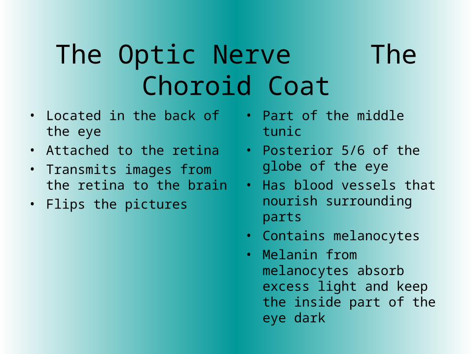
The Optic Nerve The Choroid Coat
• Located in the back of the eye
• Attached to the retina
• Transmits images from the retina to the brain
• Flips the pictures
• Part of the middle tunic
• Posterior 5/6 of the globe of the eye
• Has blood vessels that nourish surrounding parts
• Contains melanocytes
• Melanin from melanocytes absorb excess light and keep the inside part of the eye dark
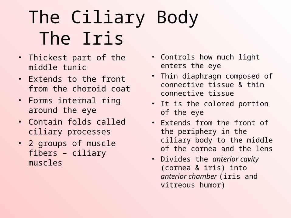
The Ciliary Body The Iris
• Thickest part of the middle tunic
• Extends to the front from the choroid coat
• Forms internal ring around the eye
• Contain folds called ciliary processes
• 2 groups of muscle fibers – ciliary muscles
• Controls how much light enters the eye
• Thin diaphragm composed of connective tissue & thin connective tissue
• It is the colored portion of the eye
• Extends from the front of the periphery in the ciliary body to the middle of the cornea and the lens
• Divides the anterior cavity (cornea & iris) into anterior chamber (iris and vitreous humor)
The Pupil The Lens • The circular opening in the center
of the iris
• Black spot in the center of the iris
• Controls how much light enters the retina
• Its diameters is usually 3-7 mm
• Transparent
• Held in position by fibers called suspensory ligaments (zonular fibers) from the ciliary processes
• Lacks blood vessels
• Located behind the iris and the pupil
• Composed of special epithelial cells
Vitreous Humor Aqueous Humor
• Front of iris and pupil• Watery fluid created by
epithelium on inner surface of ciliary body
• Separated into anterior and posterior chambers
• Maintains eye pressure• Nourishes front part of the eye
• Jelly- like fluid
• Fills the central core of the eye
• Helps maintain spherical shape to the eye
• this with collagenous fibers form the vitreous body
The Macula Lutea The Fovea Centralis
• Yellowish spot in the retina
• The central focusing spot
• Responsible for seeing details (reading) and color vision
• A depression in the macula lutea
• In the region of the retina that produces the sharpest vision
• Has no rods – only cones
The Optic Disk
• Lacks receptors, often called the blind spot
• The nerve fibers from the retina and became part of the optic nerve.
• A central artery and vein pass through here
• These vessels with vessels in the choroid coat supply blood to the cells of the inner tunic
• Jelly- like fluid
• Fills the central core of the eye
• Helps maintain spherical shape to the eye
• this with collagenous fibers form the vitreous body
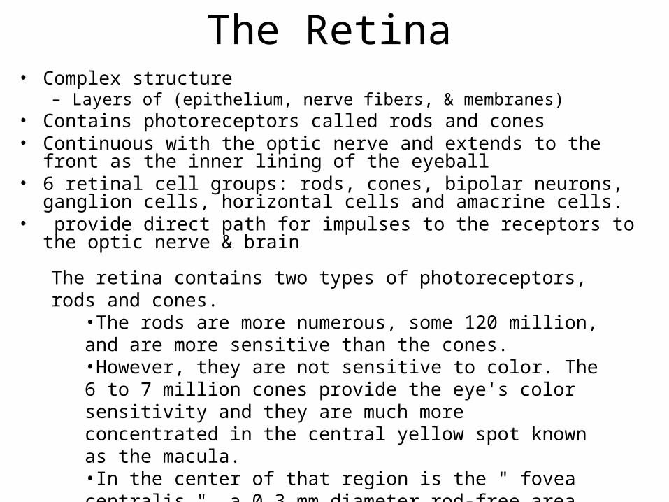
The Retina• Complex structure
– Layers of (epithelium, nerve fibers, & membranes)• Contains photoreceptors called rods and cones• Continuous with the optic nerve and extends to the front as the inner lining of
the eyeball • 6 retinal cell groups: rods, cones, bipolar neurons, ganglion cells, horizontal
cells and amacrine cells.• provide direct path for impulses to the receptors to the optic nerve & brain
The retina contains two types of photoreceptors, rods and cones. •The rods are more numerous, some 120 million, and are more sensitive than the cones. •However, they are not sensitive to color. The 6 to 7 million cones provide the eye's color sensitivity and they are much more concentrated in the central yellow spot known as the macula. •In the center of that region is the " fovea centralis ", a 0.3 mm diameter rod-free area with very thin, densely packed cones.
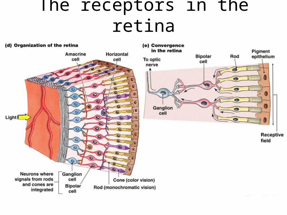
The receptors in the retina
Pigment epithelium
• the pigmented cell layer just outside the neurosensory retina
• nourishes retinal visual cells• firmly attached to the underlying choroid
and overlying retinal visual cells.
Rods Cones
• Responsible for black and white vision
• The rods are more numerous and more sensitive than the cones
• they are absent in the fovea centralis
• Responsible for color vision
• Densely packed in the fovea centralis
Horizontal cells Bipolar cells
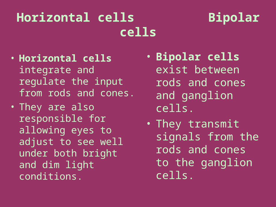
• Horizontal cells integrate and regulate the input from rods and cones.
• They are also responsible for allowing eyes to adjust to see well under both bright and dim light conditions.
• Bipolar cells exist between rods and cones and ganglion cells.
• They transmit signals from the rods and cones to the ganglion cells.
Amacrine cell Ganglion cell
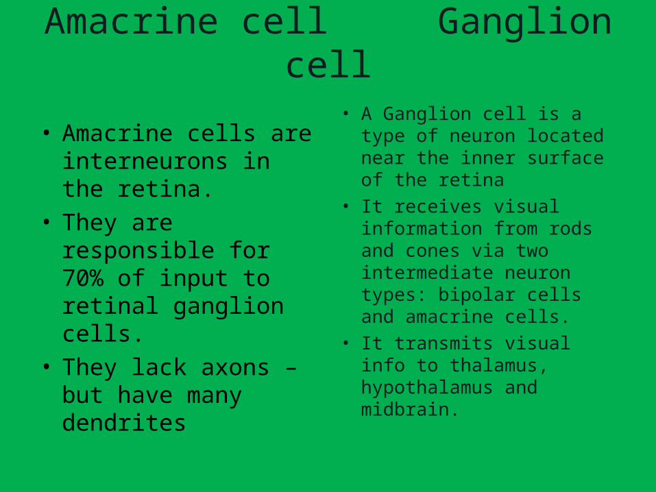
• A Ganglion cell is a type of neuron located near the inner surface of the retina
• It receives visual information from rods and cones via two intermediate neuron types: bipolar cells and amacrine cells.
• It transmits visual info to thalamus, hypothalamus and midbrain.
• Amacrine cells are interneurons in the retina.
• They are responsible for 70% of input to retinal ganglion cells.
• They lack axons – but have many dendrites
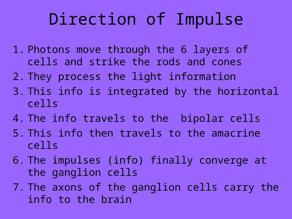
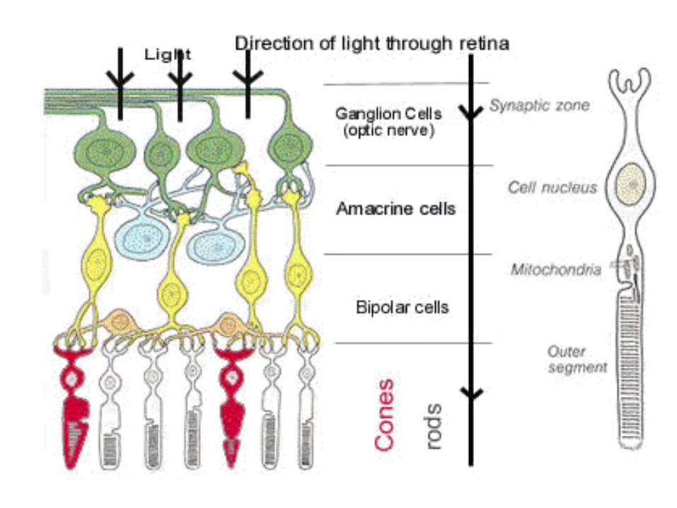
Direction of Impulse
1. Photons move through the 6 layers of cells and strike the rods and cones
2. They process the light information 3. This info is integrated by the horizontal cells4. The info travels to the bipolar cells5. This info then travels to the amacrine cells6. The impulses (info) finally converge at the
ganglion cells7. The axons of the ganglion cells carry the
info to the brain
Optic Nerve (Cranial nerve 2)
• The optic nerve, also known as cranial nerve 2, transmits visual information from the retina to the brain
• It is composed of the axons of the retinal ganglion cells
• The ganglion cell axons are myelinated by oligodendrocytes – since it is part of the CNS

THE END