Embed Size (px)
Citation preview
Department of Physics
Viruses
Seminar
Author: Eva Ule
Mentor: prof. dr. Rudolf Podgornik
December 2013
Abstract:
In this seminar we first take a look at the viruses in general, then we learn something about viral self-
assembly and about a model of a viral capsid. Finally, we describe the compression of virus-like
particles carrying surface dipole.
2
Contents 1 Introduction ......................................................................................................................... 3
2 Viruses ................................................................................................................................ 3
3 Viral self-assembly ............................................................................................................. 6
4 A simple model of a capsid and its energy ......................................................................... 8
5 Analysis of thin elastic cylindrical tube ............................................................................ 10
6 The compression of virus-like particles carrying surface dipoles..................................... 11
7 Conclusion ........................................................................................................................ 12
8 References ......................................................................................................................... 12
3
1 Introduction Viruses are often considered as something dangerous and unnecessary, because they cause a lot
of diseases. However, if we know well their properties, they can be used in many positive roles,
even in cancer treatment.
Viruses are really interesting objects and a lot about them is still unknown, so there is enough
work for all researches (physicists, biologists …).
Physical virology deals with thermodynamic of self-assembly, mechanical properties of viral
shells etc.
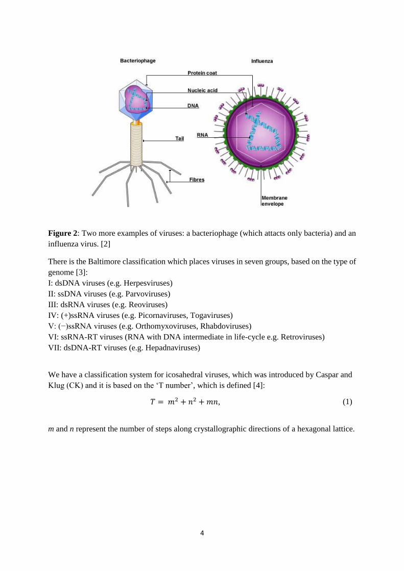
2 Viruses A virus is a very small infectious object. Viruses can infect animals, plants, bacteria, human
beings etc.
They consist of two parts: genetic material (DNA or RNA) and a capsid. The capsid is a protein
coat which protects genome. Viruses can be found in many shapes. The most common are
sphere-like and rod-like, but spherocylinders, cones and other shapes can also be seen. A lot of
viral families have icosahedral symmetry (half of all families), but genomes of these families
are not similar.
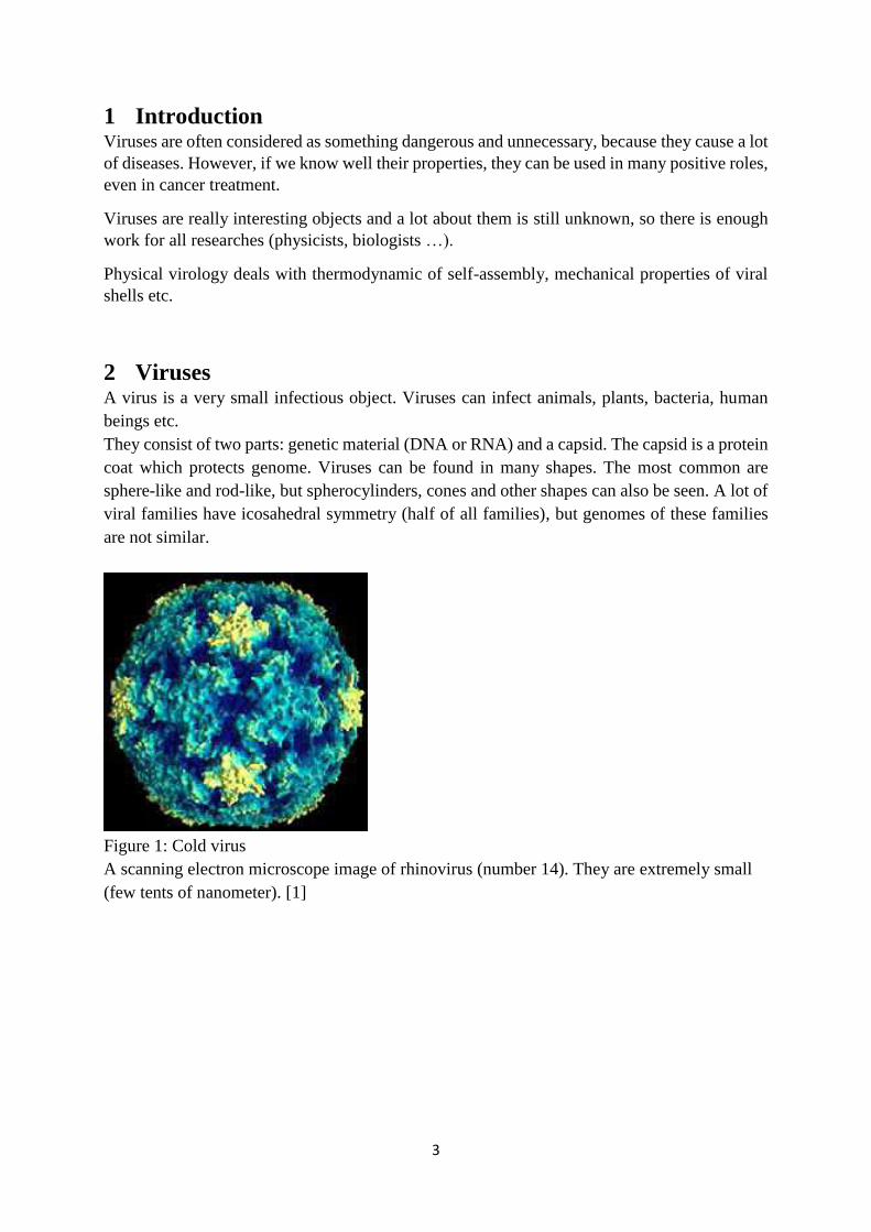
Figure 1: Cold virus
A scanning electron microscope image of rhinovirus (number 14). They are extremely small
(few tents of nanometer). [1]
4
Figure 2: Two more examples of viruses: a bacteriophage (which attacts only bacteria) and an
influenza virus. [2]
There is the Baltimore classification which places viruses in seven groups, based on the type of
genome [3]:
I: dsDNA viruses (e.g. Herpesviruses)
II: ssDNA viruses (e.g. Parvoviruses)
III: dsRNA viruses (e.g. Reoviruses)
IV: (+)ssRNA viruses (e.g. Picornaviruses, Togaviruses)
V: (−)ssRNA viruses (e.g. Orthomyxoviruses, Rhabdoviruses)
VI: ssRNA-RT viruses (RNA with DNA intermediate in life-cycle e.g. Retroviruses)
VII: dsDNA-RT viruses (e.g. Hepadnaviruses)
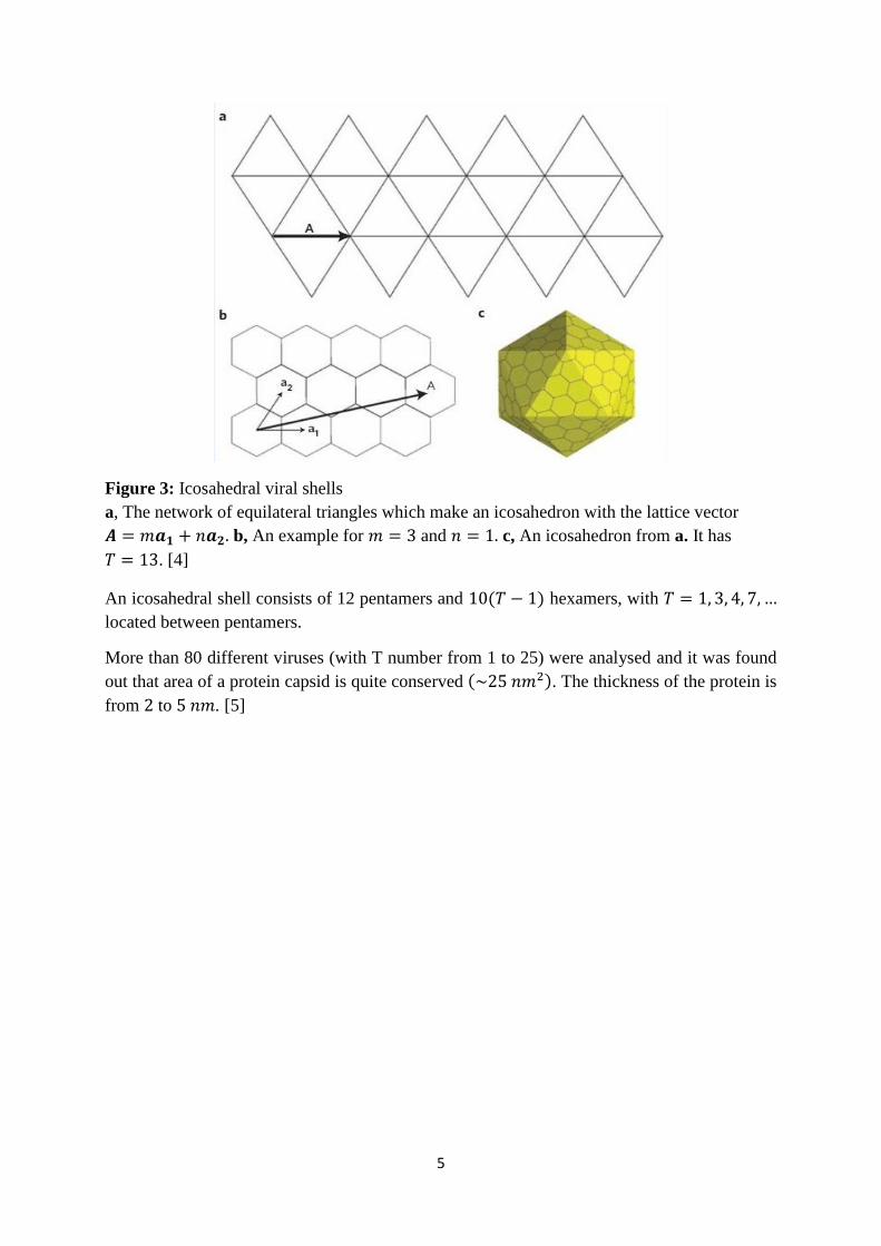
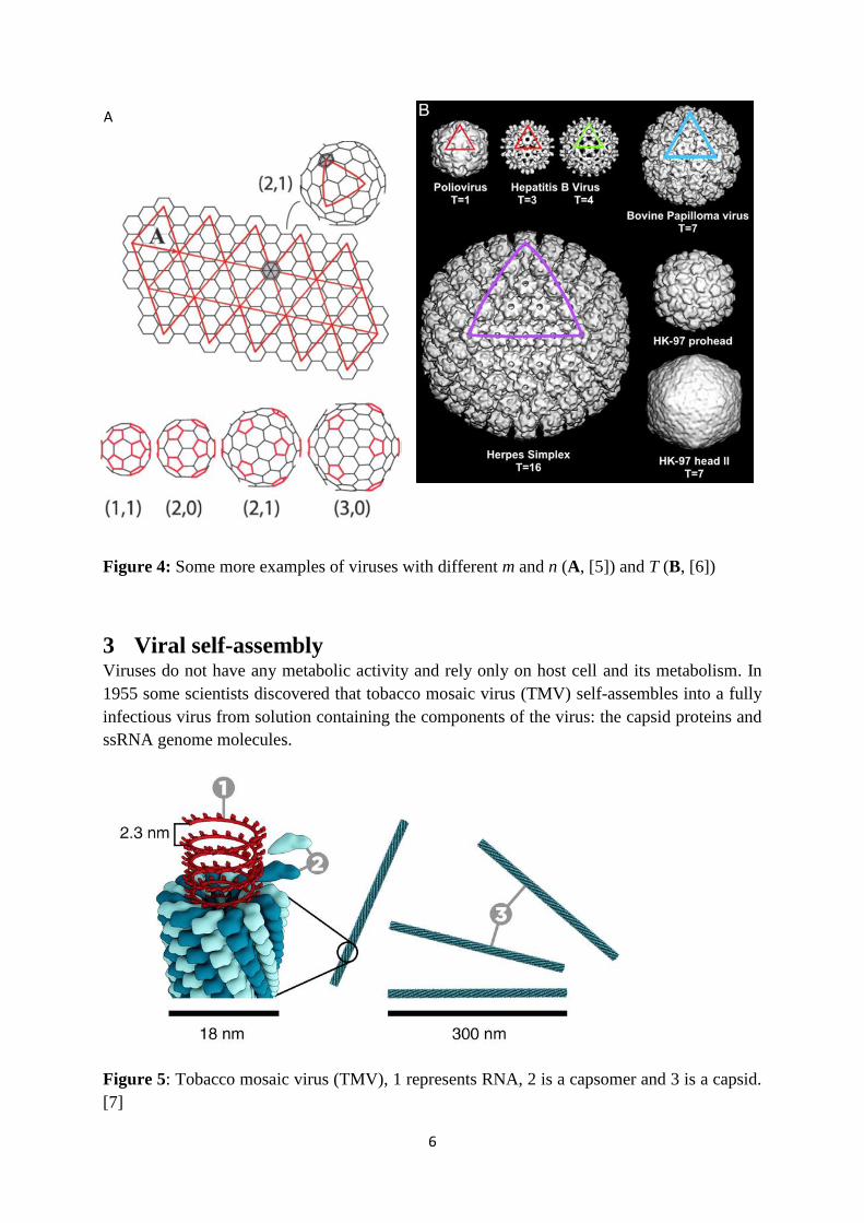
We have a classification system for icosahedral viruses, which was introduced by Caspar and
Klug (CK) and it is based on the ‘T number’, which is defined [4]:
𝑇 = 𝑚2 + 𝑛2 + 𝑚𝑛, (1)
m and n represent the number of steps along crystallographic directions of a hexagonal lattice.
5
Figure 3: Icosahedral viral shells
a, The network of equilateral triangles which make an icosahedron with the lattice vector
𝑨 = 𝑚𝒂𝟏 + 𝑛𝒂𝟐. b, An example for 𝑚 = 3 and 𝑛 = 1. c, An icosahedron from a. It has
𝑇 = 13. [4]
An icosahedral shell consists of 12 pentamers and 10(𝑇 − 1) hexamers, with 𝑇 = 1, 3, 4, 7, …
located between pentamers.
More than 80 different viruses (with T number from 1 to 25) were analysed and it was found
out that area of a protein capsid is quite conserved (~25 𝑛𝑚2). The thickness of the protein is
from 2 to 5 𝑛𝑚. [5]
6
Figure 4: Some more examples of viruses with different m and n (A, [5]) and T (B, [6])
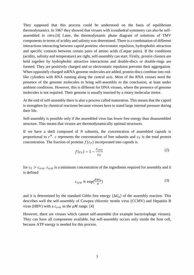
3 Viral self-assembly Viruses do not have any metabolic activity and rely only on host cell and its metabolism. In
1955 some scientists discovered that tobacco mosaic virus (TMV) self-assembles into a fully
infectious virus from solution containing the components of the virus: the capsid proteins and
ssRNA genome molecules.
Figure 5: Tobacco mosaic virus (TMV), 1 represents RNA, 2 is a capsomer and 3 is a capsid.
[7]
A
7
They supposed that this process could be understood on the basis of equilibrium
thermodynamics. In 1967 they showed that viruses with icosahedral symmetry can also be self-
assembled in vitro.[4] Later, the thermodynamic phase diagram of solutions of TMV
components in terms of acidity and salinity was determined. There is a combination of different
interactions interacting between capsid proteins: electrostatic repulsion, hydrophobic attraction
and specific contacts between certain pairs of amino acids (Caspar pairs). If the conditions
(acidity, salinity and temperature) are right, self-assembly can start. Firstly, protein clusters are
held together by hydrophobic attractive interactions and double-discs or double-rings are
formed. They are positively charged and so electrostatic repulsion prevents their aggregation.
When oppositely charged ssRNA genome molecules are added, protein discs combine into rod-
like cylinders with RNA running along the central axis. Most of the RNA viruses need the
presence of the genome molecules to bring self-assembly to the conclusion, at least under
ambient conditions. However, this is different for DNA viruses, where the presence of genome
molecules is not required. Their genome is usually inserted by a rotary molecular motor.
At the end of self-assembly there is also a process called maturation. This means that the capsid
is strengthen by chemical reactions because viruses have to stand large internal pressure during
their life.
Self-assembly is possible only if the assembled virus has lower free energy than disassembled
structure. This means that viruses are thermodynamically optimal structures.
If we have a shell composed of N subunits, the concentration of assembled capsids is
proportional to 𝑐𝑁. 𝑐 represents the concentration of free subunits and 𝑐𝑇 is the total protein
concentration. The fraction of proteins 𝑓(𝑐𝑇) incorporated into capsids is
𝑓(𝑐𝑇) ~ 1 −𝑐𝑐𝑟𝑖𝑡
𝑐𝑇 (2)
for 𝑐𝑇 > 𝑐𝑐𝑟𝑖𝑡. 𝑐𝑐𝑟𝑖𝑡 is a minimum concentration of the ingredients required for assembly and it
is defined
𝑐𝑐𝑟𝑖𝑡 ∝ exp(𝛽Δ𝐺0
𝑁) (3)
and it is determined by the standard Gibbs free energy (Δ𝐺0) of the assembly reaction. This
describes well the self-assembly of Cowpea chlorotic mottle virus (CCMV) and Hepatitis B
virus (HBV) with a 𝑐𝑐𝑟𝑖𝑡 in the 𝜇𝑀 range. [4]
However, there are viruses which cannot self-assemble (for example bacteriophage viruses).
They can have all components available, but self-assembly occurs only inside the host cell,
because ATP energy is needed for this process.
8
4 A simple model of a capsid and its energy As we find out in previous section the protein charge is very important in viral self-assembly,
thus we have to take a look at its energy.
The simplest distribution of the protein charge would be homogenously distributed on a protein.
This means that proteins would not want to be near each other because of the electrostatic
repulsion. However, we know that proteins assemble in a capsid so there has to be an attractive
interaction between them. It turns out that this is a combination of hydrophobic and van der
Waals interactions.
We take an uniformly positively charged (with surface charge density 𝜎) permeable infinitely
thin sphere of radius R and this is one really simple model of a capsid.[5] This can be solved
by treating the ions as ideal gas which adjust to the external potential and contribute to it via
their charge density. [5] This is Poisson-Boltzmann approach where we get a nonlinear
differential equation for the electrostatic potential 𝜙. It can be derived by minimizing the
appropriate free energy which has the form [5]
𝐹𝑃𝐵[𝜙(𝑟), ∇𝜙(𝑟), 𝑐𝑖(𝑟)] = ∫ 𝑓𝑃𝐵 (𝜙(𝑟), 𝛻𝜙(𝑟), 𝑐𝑖(𝑟)) 𝑑3𝑟
(4)
and the free energy density 𝑓𝑃𝐵 is
𝑓𝑃𝐵 (𝜙(𝑟), 𝛻𝜙(𝑟), 𝑐𝑖(𝑟))
= 1
2𝜀0𝜀∇𝜙(𝑟)2 + ∑ 𝑒𝑖
𝑖=±
𝑐𝑖(𝑟)𝜙(𝑟)
+ ∑ 𝑘𝐵𝑇[𝑐𝑖(𝑟) ln 𝑐𝑖(𝑟) − 𝑐𝑖(𝑟) − (𝑐0𝑖 ln 𝑐0
𝑖 − 𝑐0𝑖 )]
𝑖=±
+ 𝑒0𝜌𝑝(𝑟)𝜙(𝑟)
(5)
𝑒0𝜌𝑝(𝑟) is the charge density of the capsid, 𝑇 is the temperature, 𝑘𝐵 is the Boltzmann constant,
𝑐𝑖 are the concentrations of salt ions, 𝑐0𝑖 their bulk concentrations, 𝜀0𝜀 is the permittivity of
water and 𝑒0 is the electron charge. When we minimize the equation (5), we get a new form of
Poisson-Boltzmann equation
−𝜀𝜀0∇2𝜙(𝑟) = ∑ 𝑒𝑖𝑐0𝑖 𝑒−𝛽𝑒𝑖𝜙(𝑟) + 𝑒0𝜌𝑝(𝑟)
𝑖=±
(6)
with 𝛽 = 𝑘𝐵𝑇 and 𝑒𝑖 = ±𝑒0. If the electrostatic potentials in the solution are small
(𝑒0𝛽𝜙 ≪ 1) and we have a symmetric electrolyte (𝑐0+ = 𝑐0
− = 𝑐0), equation (6) becomes
−∇2𝜙(𝑟) =𝛽
𝜀𝜀0(∑ 𝑒𝑖
2𝑐0𝑖
𝑖±
) 𝜙(𝑟) +𝑒0𝜌𝑝(𝑟)
𝜀𝜀0+ ⋯
(7)
9
And this is called Debye-Hückel equation. We introduce Debye-Hückel length 𝜅−1, 𝜅 is
defined:
𝜅2 =𝛽
𝜀𝜀0(∑ 𝑒𝑖
2𝑐0𝑖
𝑖±
).
(8)
Our viruses are typically of the order 20 nm and 𝑐0 ≈ 150 𝑚𝑀. This gives us 𝜅−1 ≈ 1 𝑛𝑚, so
the limit of 𝜅𝑅 ≫ 1 is often implied. This helps us a lot to solve the equation (7) and we get the
expression for free energy [5]:
𝐹𝐷𝐻 =𝜋𝜎2𝑅2
𝜀𝜀0𝜅.
(9)
To estimate free energy we need some information. It is estimated that the surface charge
density is typically 𝜎 ≈ 1 𝑒0 /𝑛𝑚2, 𝑅 ≈ 20 𝑛𝑚 and 𝑐0 ≈ 100 𝑚𝑀. This gives us the result
𝐹𝐷𝐻 ≈ 104𝑘𝐵𝑇.
We can estimated the hydrophobic energy from the area of proteins engaged in protein-protein
contacts. The capsid with 𝑅 ≈ 20 𝑛𝑚 typically has triangulation number 𝑇 = 3, this means that
it consists of 180 protein subunits. The length of protein contacts in a capsid is approximately
3000 𝑛𝑚 and the thickness of capsid is about 2 𝑛𝑚. This gives us the total area of about
6000 𝑛𝑚2. The energy of attractive protein interactions for such cases is of the order of 10 𝑚𝐽
𝑚2
and if we multiply this with the total area of protein contacts, we get 𝐹𝐻𝑃 ≈ 104𝑘𝐵𝑇.
We can see that the electrostatic free energy and the hydrophobic are of the same order of
magnitude so the assembly and disassembly are in a balance. Viruses are able to self-assemble,
but they can disassemble too; when they want to put their genome into host cell.
It turns out that our simple model is quite good. When the model with the finite thickness of a
capsid is introduced, it turns out that the free energy is of the same order of magnitude as in the
previous case (with infinite thickness).
10
5 Analysis of thin elastic cylindrical tube We are often interested in elastic deformations of a system, but most of the studies are made in
the linear regime where we have small deformations. However, if we have bigger deformations,
nonlinear regime should be considered too.
It is interesting to take a look at experiment in which can be studied both small and large
deformations. This experiment is meant to model large deformations in materials made of thin
sheets such as carbon nanotubes, fullerenes, cellular membranes, virus capsids etc. However,
the experiment is made on macroscopic level, but it has been shown that is useful and accurate
for nanolevel as well.
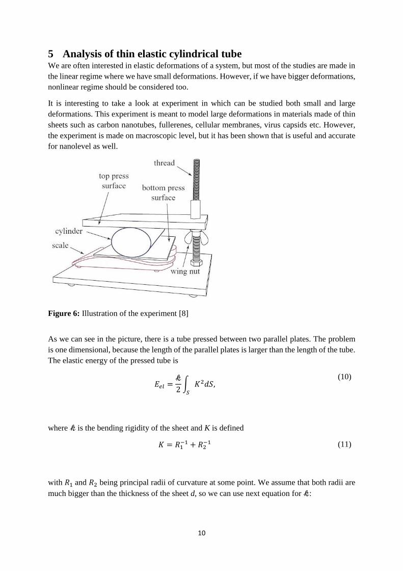
Figure 6: Illustration of the experiment [8]
As we can see in the picture, there is a tube pressed between two parallel plates. The problem
is one dimensional, because the length of the parallel plates is larger than the length of the tube.
The elastic energy of the pressed tube is
𝐸𝑒𝑙 =𝓀
2∫ 𝐾2𝑑𝑆,
𝑆
(10)
where 𝓀 is the bending rigidity of the sheet and K is defined
𝐾 = 𝑅1−1 + 𝑅2
−1
(11)
with 𝑅1 and 𝑅2 being principal radii of curvature at some point. We assume that both radii are
much bigger than the thickness of the sheet d, so we can use next equation for 𝓀:
11
𝓀 =𝐸𝑑3
12(1 − 𝜈2)
(12)
where E is Young modulus and 𝜈 is Poisson ratio.
6 The compression of virus-like particles carrying surface
dipoles The findings from section 5 can now be used for our further research about viruses.
We are interested in what happens when viruses are compressed under certain conditions.
We take N viruses and put them between two rigid plates. In this case a virus is treated as a
perfect sphere carrying a certain dipole moment distribution across the capsid thickness.
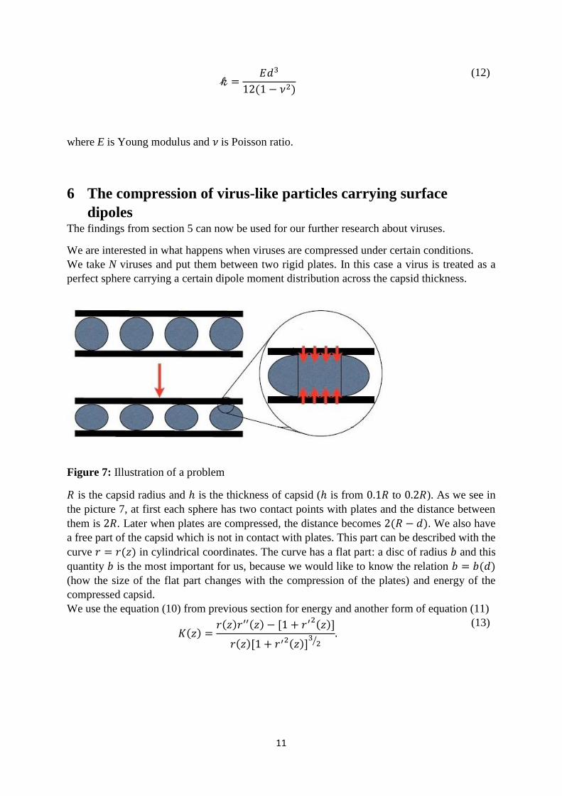
Figure 7: Illustration of a problem
𝑅 is the capsid radius and ℎ is the thickness of capsid (ℎ is from 0.1𝑅 to 0.2𝑅). As we see in
the picture 7, at first each sphere has two contact points with plates and the distance between
them is 2𝑅. Later when plates are compressed, the distance becomes 2(𝑅 − 𝑑). We also have
a free part of the capsid which is not in contact with plates. This part can be described with the
curve 𝑟 = 𝑟(𝑧) in cylindrical coordinates. The curve has a flat part: a disc of radius 𝑏 and this
quantity 𝑏 is the most important for us, because we would like to know the relation 𝑏 = 𝑏(𝑑)
(how the size of the flat part changes with the compression of the plates) and energy of the
compressed capsid.
We use the equation (10) from previous section for energy and another form of equation (11)
𝐾(𝑧) =𝑟(𝑧)𝑟′′(𝑧) − [1 + 𝑟′2(𝑧)]
𝑟(𝑧)[1 + 𝑟′2(𝑧)]3
2⁄.
(13)
12
This expression is then inserted in equation (Eel) for elastic energy. For obtaining total energy
functional we need to add the contribution of the deformation of the shell caused by pressing
the plates which, at the end, results in a complicated expression to be solved.
The radial dipole moment across the capsid thickness (in the figure 7 is marked red) introduces
jumps in the electric field at both plates and exactly this jumps and their size are our main
interest.
7 Conclusion Even though viruses are nano objects, there are a lot of things we can examine about them;
some of them are presented in this seminar. If we knew all their properties, our lives would be
much simpler, especially the treatment of diseases they cause. However, there is still a lot of
work to do because we do not understand many things, although we are daily surrounded by
various viruses. They are small so they can go almost everywhere and reach every cell. We
still do not know how to cope with many of them, therefore only further studies would help.
There is a quote which shows us how special creatures they are: “An inefficient virus kills its
host. A clever virus stays with it.” (James Lovelock) Not only that, they are able to self-
assemble, to survive in really unfriendly environment and to cause us many problems; we
should respect them because of their qualities.
8 References [1] http://www.waynesthisandthat.com/cold.htm (3 December 2013)
[2] http://www.bbc.co.uk/bitesize/higher/biology/cell_biology/viruses/revision/1/
(3 December 2013)
[3] http://en.wikipedia.org/wiki/Virus (20 November 2013)
[4] W. H. Roos, R. Bruinsma, G. J. L. Wuite, Nature Physics 6, 733–743 (2010)
[5] A. Šiber, A. L. Božič, R. Podgornik, Phys. Chem. Chem. Phys., 2012, 14, 3746–3765
[6] http://www.pnas.org/content/101/44/15549.long (25 November 2013)
[7] http://en.wikipedia.org/wiki/File:TMV_structure_full.png (9 December 2013)
[8] A. Šiber, H. Buljan, Phys. Rev. E 83, 067601 (2011)
[9] http://www.brainyquote.com/quotes/quotes/j/jameslovel314132.html
(22 December 2013)