Embed Size (px)
Citation preview
Virulence gene regulation by L-arabinose in Salmonella enterica
Javier López-Garrido1, Elena Puerta-Fernández2, Ignacio Cota,
and Josep Casadesús*
Departamento de Genética, Facultad de Biología, Universidad de Sevilla,
Apartado 1095, 41080 Sevilla, Spain
1 Present address: University of California San Diego, 9500 Gilman Drive, 4117 Natural
Sciences Building, La Jolla, CA 92093-0377, U. S. A.
2 Present address: Abengoa Research, Campus Palmas Altas, C/ Energía Solar 1, Edificio E,
41014 Sevilla, Spain
Genetics: Early Online, published on May 18, 2015 as 10.1534/genetics.115.178103
Copyright 2015.

2
Running title: Inhibition of Salmonella invasion by L-arabinose
Keywords: Salmonella pathogenicity island 1; Salmonella invasion; L-arabinose; HilD
* Corresponding author: Josep Casadesús, Departamento de Genética, Facultad de Biología,
Universidad de Sevilla, Apartado 1095, 41080 Sevilla, Spain. Telephone +34 95 455 7105.
Fax +34 95 455 7104. E-mail: [email protected]
3
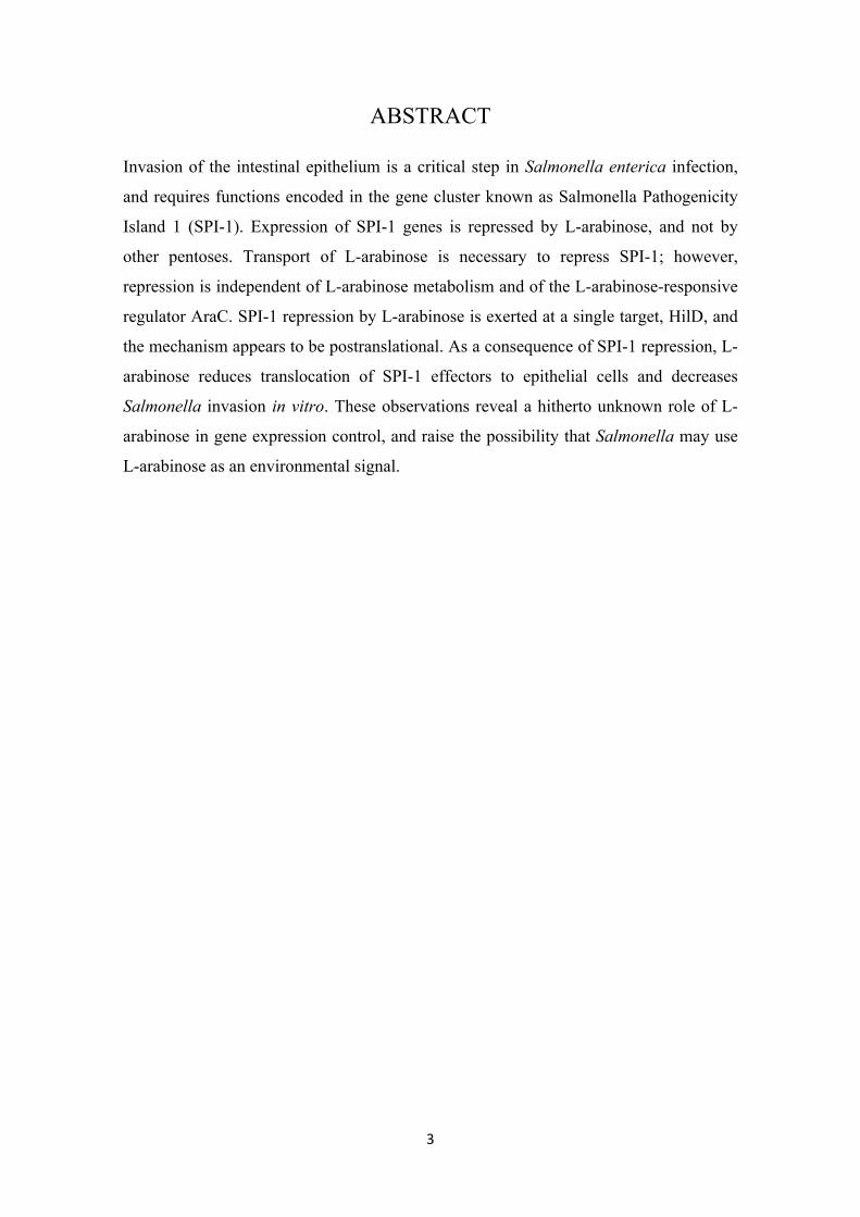
ABSTRACT
Invasion of the intestinal epithelium is a critical step in Salmonella enterica infection,
and requires functions encoded in the gene cluster known as Salmonella Pathogenicity
Island 1 (SPI-1). Expression of SPI-1 genes is repressed by L-arabinose, and not by
other pentoses. Transport of L-arabinose is necessary to repress SPI-1; however,
repression is independent of L-arabinose metabolism and of the L-arabinose-responsive
regulator AraC. SPI-1 repression by L-arabinose is exerted at a single target, HilD, and
the mechanism appears to be postranslational. As a consequence of SPI-1 repression, L-
arabinose reduces translocation of SPI-1 effectors to epithelial cells and decreases
Salmonella invasion in vitro. These observations reveal a hitherto unknown role of L-
arabinose in gene expression control, and raise the possibility that Salmonella may use
L-arabinose as an environmental signal.
4
INTRODUCTION
Invasion of epithelial cells by Salmonella enterica requires the expression of genes
located in a 40 kb region known as Salmonella pathogenicity island 1 (SPI-1) (Altier.
2005; Jones. 2005; Lostroh and Lee. 2001). SPI-1 is a cluster of approximately 40 genes
organized in several transcriptional units, and encodes a Type 3 Secretion System
(TTSS) and effector proteins that are translocated into epithelial cells (Lostroh and Lee.
2001). Inside the epithelial cell, the effector proteins trigger cytoskeleton
rearrangements necessary for Salmonella invasion (Darwin and Miller. 1999a). SPI-1
also encodes transcriptional activators responsible for its own expression: HilA, HilC,
HilD, and InvF (Ellermeier and Slauch. 2007; Lostroh and Lee. 2001). HilA is a
transcriptional activator of the OmpR/ToxR family (Bajaj et al. 1995; Lee et al. 1992),
and activates transcription of genes encoding components of the TTSS and the
transcriptional activator InvF (Bajaj et al. 1996). In association with the SicA
chaperone, InvF boosts transcription of the sicA/sip operon, which encodes effector
proteins (Darwin and Miller. 1999b; Eichelberg and Galan. 1999). Transcription of hilA
is directly activated by HilC and HilD, both members of the AraC/XylS family of
transcriptional regulators (Schechter and Lee. 2001). HilC and HilD relieve repression
of the hilA promoter by the nucleoid proteins H-NS and Hha (Olekhnovich and Kadner.
2006), and can activate expression of the invF and sicA/sip transcriptional units
independently of HilA (Akbar et al. 2003; Rakeman et al. 1999). Furthermore, HilD
activates transcription of hilC and of hilD itself (Ellermeier et al. 2005) by direct
binding to both promoters (Olekhnovich and Kadner. 2002). Together with a
transcriptional activator encoded outside SPI-1, RtsA (Ellermeier and Slauch. 2003),
SPI-1 transcription factors form a regulatory network that governs SPI-1 expression in
response to environmental and physiological factors (Figure S1) (Ellermeier and Slauch.
2007; Jones. 2005).
During passage through the digestive tract, Salmonella encounters environmental
conditions that affect SPI-1 expression. In the stomach, SPI-1 is repressed at acidic pH
(Bajaj et al. 1996; Behlau and Miller. 1993), thus preventing invasion. The proximal
part of the small intestine is under the influence of digestive fluids coming from the
stomach, and the pH remains slightly acidic. In the duodenum, SPI-1 is repressed by
5
bile, thereby inhibiting invasion of the proximal section of the small intestine (Prouty
and Gunn. 2000). In the large intestine, SPI-1 is repressed by the short chain fatty acids
propionate and butyrate, synthesized by the intestinal microbiota (Lawhon et al. 2002).
The gradients that repress SPI-1 along the digestive tract leave however a region, the
ileum, that permits SPI-1 expression. As a consequence, the ileum is the portion of the
small intestine that Salmonella preferentially invades (Jones et al. 1994).
In this study, we describe that the pentose L-arabinose represses SPI-1 expression. L-
arabinose is the second most abundant pentose in nature (Schadel et al. 2010; Seiboth
and Metz. 2011) and is found in hemicellulose and pectin in plant cell walls (Abedon et
al. 2006; Schadel et al. 2010). Salmonella can use L-arabinose as sole carbon source
(Gutnick et al. 1969). L-arabinose is transported into Salmonella cells by a permease
encoded by the araE gene (Lee et al. 1981; Lee et al. 1982). Inside the cell, L-arabinose
is sequentially transformed into L-ribulose, L-ribulose-5-phosphate, and D-xylulose-5-
phosphate, by the action of L-arabinose isomerase, ribulokinase, and L-ribulose-5-
phosphate-4-epimerase respectively (Englesberg. 1961; Englesberg et al. 1962). L-
ribulose-5-phosphate and D-xylulose-5-phosphate are substrates for the pentose
phosphate pathway, which produces glycolytic intermediates (Figure S2A). L-arabinose
isomerase, ribulokinase, and L-ribulose-5-phosphate-4-epimerase are encoded by the
araA, araB, and araD genes respectively (Englesberg. 1961; Englesberg et al. 1962),
arranged in a single transcriptional unit, the araBAD operon (Englesberg. 1961;
Englesberg et al. 1962; Gross and Englesberg. 1959; Lee et al. 1984). Expression of the
araBAD operon and the araE gene is induced by L-arabinose (Lee et al. 1980; Lee et al.
1982), and requires the transcriptional regulator AraC (Englesberg et al. 1965; Lee et al.
1981). The araC gene is located upstream of the araBAD operon but it is transcribed in
divergent orientation (Lee et al. 1984). AraC bound to L-arabinose activates
transcription from the araBAD and araE promoters. However, in the absence of L-
arabinose, AraC acts as a repressor (Schleif. 2010).
The phenomenon described in this study, SPI-1 repression by L-arabinose, is
independent of L-arabinose catabolism and of the regulatory protein AraC. We describe
epistasis analysis indicating that the target of L-arabinose within SPI-1 is the
transcription factor HilD, and present evidence that the inhibitory mechanism may be
6
postranslational. HilD inhibition causes SPI-1 repression, which results in impaired
secretion of SPI-1 effectors and reduced invasion of epithelial cells.
7
MATERIALS AND METHODS
Bacterial strains, plasmids, bacteriophages, and strain construction. All the
Salmonella enterica strains listed in Table 1 belong to serovar Typhimurium, and derive
from the mouse-virulent strain ATCC 14028. For simplicity, Salmonella enterica
serovar Typhimurium is often abbreviated as S. enterica. Targeted gene disruption was
achieved using pKD13 or pKD3 (Datsenko and Wanner. 2000). Antibiotic resistance
cassettes introduced during strain construction were excised by recombination with
plasmid pCP20 (Datsenko and Wanner. 2000). The oligonucleotides used for disruption
(labeled "UP" and "DO") are listed in Table S1, together with the oligonucleotides
(labeled "E") used for allele verification by the polymerase chain reaction. For the
construction of transcriptional and translational lac fusions in the Salmonella
chromosome, FRT sites generated by excision of Kmr cassettes (Datsenko and Wanner.
2000) were used to integrate either pCE37 or pCE40 (Ellermeier et al. 2002). An
exception was the hilD::lac477 translational fusion, constructed using the method
described by M. Hensel and co-workers (Gerlach et al. 2007). Unless specified
otherwise, all lac fusions used in this study are translational. Addition of a 3xFLAG
epitope tag to protein-coding DNA sequences was carried out using plasmid pSUB11
[Kmr, 3xFLAG] as template (Uzzau et al. 2001). Plasmid pIZ1902 (pXG10-hilD) was
constructed by cloning a DNA fragment encompassing the hilD transcription start point
and the hilD transcriptional terminator (Lopez-Garrido et al. 2014) on BrfBI-NheI
restriction sites in pXG10 (Urban and Vogel. 2007). Transductional crosses using phage
P22 HT105/1 int201 [(Schmieger. 1972) and G. Roberts, unpublished] were used for
strain construction operations involving chromosomal markers. The transduction
protocol was described elsewhere (Garzon et al. 1995). To obtain phage-free isolates,
transductants were purified by streaking on green plates. Phage sensitivity was tested by
cross-streaking with the clear-plaque mutant P22 H5.
Expression of araE from a heterologous promoter was achieved by replacing its native
promoter with the PLtetO promoter (Lutz and Bujard. 1997). A fragment containing the
cat gene and the PLtetO promoter was amplified by PCR using pXG1 as template (Urban
and Vogel. 2007), and primers PLtetOUP and PLtetODO (Table S1). The PCR product was
treated with DpnI to remove template traces. The construction was inserted in the
8
chromosome by λ Red recombinase-mediated recombination (Datsenko and Wanner.
2000), and Cmr colonies were selected. Insertion of the construction was verified by
PCR, using a pair of primers specific for the cat gene and the target gene (Table S1).
Growth conditions. Luria-Bertani (LB) broth was used as standard liquid medium.
Solid media were prepared by the addition of 1.5 % agar. L-arabinose, D-arabinose, D-
xylose, or sucrose was added from 20% stocks prepared in distilled water. Batch
cultures were performed in LB or LB supplemented with the appropriate sugar, and
incubated at 37ºC with shaking at 220 rpm. Samples were taken when the cultures had
reached stationary phase (O.D.600 2-2.5). For translocation and invasion assays, an
overnight bacterial culture of the appropriate strain was diluted 1:50 in LB and LB +
0.2% L-arabinose, and was incubated at 37ºC until stationary phase (O.D.600 ~2).
Carbon-free medium (NCE) (Maloy and Roth. 1983) supplemented with the appropriate
carbon source was used as minimal medium. Green plates were prepared according to
Chan and co-workers (Chan et al. 1972), except that methyl blue (Sigma Chemical Co,
St. Louis, MO) substituted for aniline blue.
pH curves. An overnight culture of Salmonella was diluted 1:50 in LB and LB
containing 0.01%, 0.02%, 0.05%, 0.1%, 0.2%, 0.5%, and 1% L-arabinose. The cultures
were incubated at 37ºC until O.D.600 ~2.5. The cultures were then centrifuged 20 min at
2,600 g, and the pH of the supernatant was determined using a pH meter Basic 20
(Crison Instruments, Barcelona, Spain).
Protein extracts and Western blot analysis. Total protein extracts were prepared from
bacterial cultures grown in LB or LB + L-arabinose at 37°C until stationary phase (final
O.D.600 ~2.5). Bacterial cells were collected by centrifugation (16,000 g, 2 min) and
suspended in 100 µl of Laemmli sample buffer [1.3% SDS, 10% (v/v) glycerol, 50 mM
Tris-HCl, 1.8% β-mercaptoethanol, 0.02% bromophenol blue, pH 6.8]. Proteins were
resolved by Tris-Tricine-PAGE, using 12% gels. Conditions for protein transfer have
been described elsewhere (Jakomin et al. 2008). Optimal dilutions of primary antibodies
were as follows: anti-Flag M2 monoclonal antibody (1:5,000, Sigma Chemical Co, St.
Louis, MO) and anti-GroEL polyclonal antibody (1:20,000, Sigma). Goat anti-mouse
horseradish peroxidase-conjugated antibody (1:5,000, BioRad, Hercules, CA) or goat
anti-rabbit horseradish peroxidase conjugated antibody (1:20,000, Santa Cruz
9
Biotechnology, Heildelberg, Germany) were used as secondary antibodies. Proteins
recognized by the antibodies were visualized by chemoluminescence using the luciferin-
luminol reagents in a LAS3000 mini imaging system (Fujifilm, Tokyo, Japan). For
quantification, the intensity of the bands was determined using the MultiGauge software
(Fujifilm, Tokyo, Japan). GroEL was used as loading control.
RNA extraction and Northern blot analysis. A 2 ml aliquot from a stationary culture
(O.D.600 ~2) was centrifuged at 16,000 g, 4ºC, for 5 min. The pellet was resuspended in
100 µl of a solution of lysozyme (3 mg/ml, Sigma Chemical Co, St. Louis, MO). Cell
lysis was facilitated by three consecutive freeze-thaw cycles. After lysis, RNA was
extracted using 1 ml of Trizol reagent (Invitrogen Co, Carlsbad, CA), according to
manufacturer instructions. Finally, total RNA was resuspended in 30 µl of RNase-free
water for subsequent uses. The quality and quantity of the RNA was determined using a
ND-1000 spectrophotometer (NanoDrop Technologies, Wilmington, DE). For Northern
blot analysis, 10 µg of total RNA was loaded per well and electrophoresed in denaturing
1% agarose formaldehyde gels. Transfer and fixation to Hybond-N+ membranes (GE
Healthcare, Little Chalfont, UK) were performed by vacuum using 0.05 M NaOH.
Filters were then hybridized using an internally labeled [(α-32P)UTP] riboprobe specific
for the upstream (5') 300 nucleotides of the hilD coding sequence, generated by in vitro
transcription using the MEGAscript T7 Transcription Kit (Invitrogen Co, Carlsbad,
CA). Hybridization was carried out at 65°C. As a control of RNA loading and transfer
efficiency, the filters were hybridized with a riboprobe for the RNase P mRNA gene
(rnpB). Images of radioactive filters were obtained with a FLA-5100 imaging system
(Fujifilm, Tokyo, Japan), and quantification was performed using MultiGauge software
(Fujifilm, Tokyo, Japan).
β-galactosidase assays. Levels of β-galactosidase activity were assayed using the
CHCl3-sodium dodecyl sulfate permeabilization procedure (Miller. 1972). Data are
averages and standard deviations of 3 or more independent experiments.
Preparation of HeLa cells for infection assays. HeLa cells (ATCC CCL2) were
cultured in tissue culture medium (Dulbecco’s modified essential medium supplemented
with 10% fetal calf serum and 2 mM L-glutamine). For routine cultivation, 60 µg/ml
penicillin and 100 µg/ml streptomycin were added to the culture medium. The day
10
before infection, approximately 1.5 x 10⁵ HeLa cells were seeded, using 24-well plates
(Costar, Corning, New York, NY). Each well contained 1 ml of tissue culture medium
without antibiotics. Cells were grown at 37ºC, 5% CO2 to obtain 80% confluency. One
hour before infection, the culture medium was removed and replaced by 0.5 ml fresh
tissue culture medium without antibiotics.
Translocation assays. Aliquots of bacterial suspensions were added to reach a
multiplicity of infection of 15:1 bacteria/HeLa cell. Plates were centrifuged at 200 g for
5 min, and incubated at 37ºC, 5% CO2 for 10 min. Infected cells were then washed 3
times with PBS and lysed with a 0.1 M HCl solution containing 0.1% Triton X-100.
Quantification of cAMP was performed using the Direct Cyclic AMP Enzyme
Immunoassay Kit (Array Designs, Ann Arbor, MI) following the manufacturer
instructions.
Invasion assays. Bacterial cultures were grown overnight at 37ºC in LB, diluted into
fresh medium (1:50), and incubated at 37ºC until stationary phase (O.D.600 ~2).
Bacterial suspensions were added to reach a multiplicity of infection of 50:1
bacteria/HeLa cell. HeLa cells were infected for 30 min, washed 3 times with PBS,
incubated in fresh tissue culture medium containing 100 µg/ml gentamicin for 1.5
hours, and washed 3 times with PBS. Numbers of viable intracellular bacteria were
obtained by lysing infected cells with 1% Triton X-100 (prepared in PBS) and
subsequent plating. Invasion rates were calculated as the ratio between viable
intracellular bacteria and viable bacteria added to infect the HeLa cells.
11
RESULTS
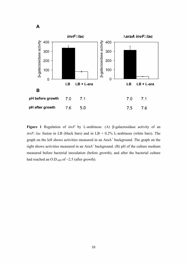
SPI-1 expression is downregulated in the presence of L-arabinose. pBAD vectors
are a set of plasmids that contain the PBAD promoter of the L-arabinose operon and the
regulatory gene araC (Guzman et al. 1995). In the presence of L-arabinose,
transcription from the PBAD promoter is turned on, while in its absence transcription is
extremely low. Use of pBAD vectors thus allows conditional, L-arabinose-dependent
expression of cloned genes (Guzman et al. 1995). We considered the possibility of
using pBAD vectors to study SPI-1 expression. As a preliminary control, we examined
whether SPI-1 expression was affected by the presence of L-arabinose (in a strain
without pBAD). For this purpose, we measured the β-galactosidase activity of an
invF::lac fusion in LB and in LB + 0.2% L-arabinose. As shown in Figure 1A,
invF::lac expression was reduced around 4-fold in LB + L-arabinose. This observation
suggested that the presence of L-arabinose in the culture medium might repress
expression of SPI-1 genes.
Figure 1
SPI-1 repression in the presence of L-arabinose is independent of L-arabinose
catabolism. The carbon sources for enteric bacteria in LB are catabolizable amino
acids, not sugars (Sezonov et al. 2007). In such conditions, enteric bacteria undergo
gluconeogenesis, and the culture medium is slightly alkalinized during growth.
However, if a catabolizable sugar is added to LB, Salmonella uses it as a carbon source
and undergoes glycolysis. As a consequence, pyruvic acid is produced and the culture
medium is acidified (Figure S2). Because Salmonella enterica is able to regulate SPI-1
expression in response to pH (Bajaj et al. 1996), we considered the possibility that
repression of SPI-1 by L-arabinose could be an indirect effect of acidification. We
measured the pH of LB and LB + 0.2% L-arabinose before inoculation (before growth)
and when the bacterial population had reached an O.D.600 of ~2.5 (after growth) (Figure
1B, left panel). Before growth, both LB and LB + L-arabinose showed a pH near
neutrality (7.0 and 7.1, respectively). However, after bacterial growth the pH of LB was
slightly alkaline (7.6) while the pH of LB + L-arabinose was 5.0. This difference
indicated that catabolism of L-arabinose by Salmonella acidifies indeed the culture
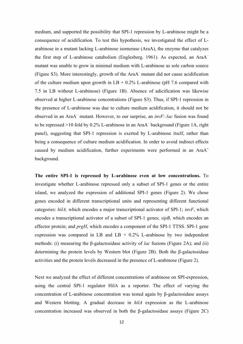
12
medium, and supported the possibility that SPI-1 repression by L-arabinose might be a
consequence of acidification. To test this hypothesis, we investigated the effect of L-
arabinose in a mutant lacking L-arabinose isomerase (AraA), the enzyme that catalyzes
the first step of L-arabinose catabolism (Englesberg. 1961). As expected, an AraA–
mutant was unable to grow in minimal medium with L-arabinose as sole carbon source
(Figure S3). More interestingly, growth of the AraA– mutant did not cause acidification
of the culture medium upon growth in LB + 0.2% L-arabinose (pH 7.6 compared with
7.5 in LB without L-arabinose) (Figure 1B). Absence of adicification was likewise
observed at higher L-arabinose concentrations (Figure S3). Thus, if SPI-1 repression in
the presence of L-arabinose was due to culture medium acidification, it should not be
observed in an AraA– mutant. However, to our surprise, an invF::lac fusion was found
to be repressed >10 fold by 0.2% L-arabinose in an AraA– background (Figure 1A, right
panel), suggesting that SPI-1 repression is exerted by L-arabinose itself, rather than
being a consequence of culture medium acidification. In order to avoid indirect effects
caused by medium acidification, further experiments were performed in an AraA¯
background.
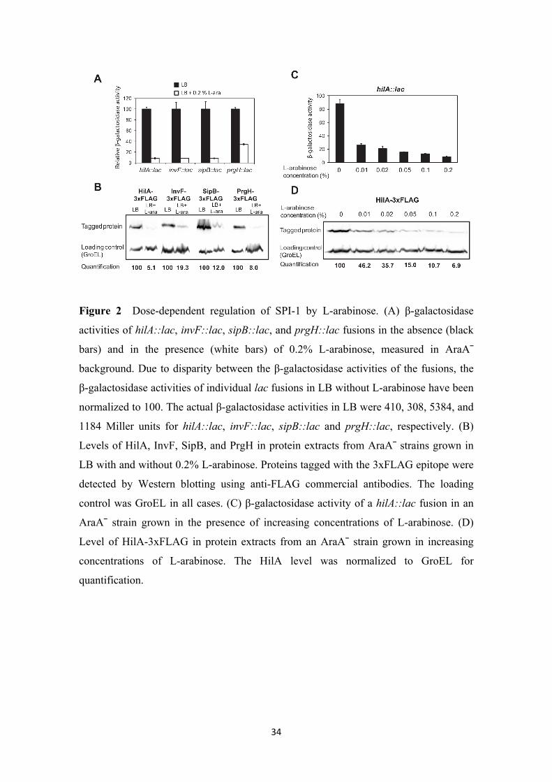
The entire SPI-1 is repressed by L-arabinose even at low concentrations. To
investigate whether L-arabinose repressed only a subset of SPI-1 genes or the entire
island, we analyzed the expression of additional SPI-1 genes (Figure 2). We chose
genes encoded in different transcriptional units and representing different functional
categories: hilA, which encodes a major transcriptional activator of SPI-1; invF, which
encodes a transcriptional activator of a subset of SPI-1 genes; sipB, which encodes an
effector protein; and prgH, which encodes a component of the SPI-1 TTSS. SPI-1 gene
expression was compared in LB and LB + 0.2% L-arabinose by two independent
methods: (i) measuring the β-galactosidase activity of lac fusions (Figure 2A); and (ii)
determining the protein levels by Western blot (Figure 2B). Both the β-galactosidase
activities and the protein levels decreased in the presence of L-arabinose (Figure 2).
Next we analyzed the effect of different concentrations of arabinose on SPI-expression,
using the central SPI-1 regulator HilA as a reporter. The effect of varying the
concentration of L-arabinose concentration was tested again by β-galactosidase assays
and Western blotting. A gradual decrease in hilA expression as the L-arabinose
concentration increased was observed in both the β-galactosidase assays (Figure 2C)
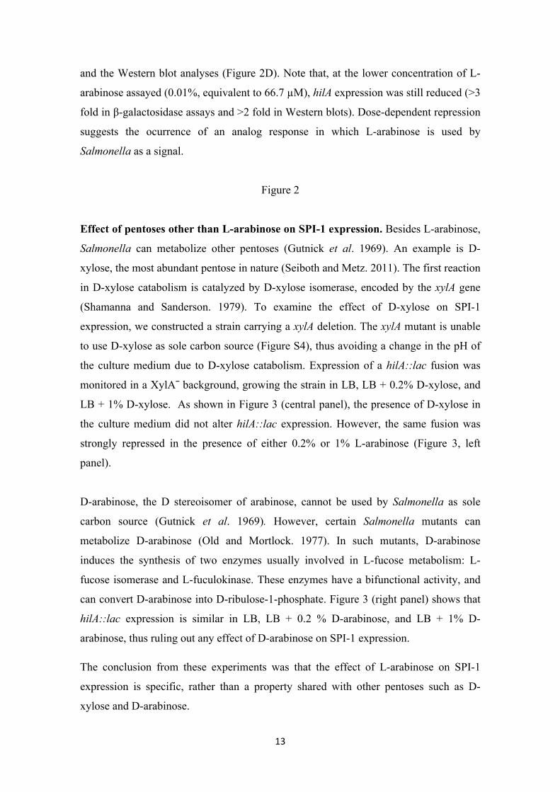
13
and the Western blot analyses (Figure 2D). Note that, at the lower concentration of L-
arabinose assayed (0.01%, equivalent to 66.7 µM), hilA expression was still reduced (>3
fold in β-galactosidase assays and >2 fold in Western blots). Dose-dependent repression
suggests the ocurrence of an analog response in which L-arabinose is used by
Salmonella as a signal.
Figure 2
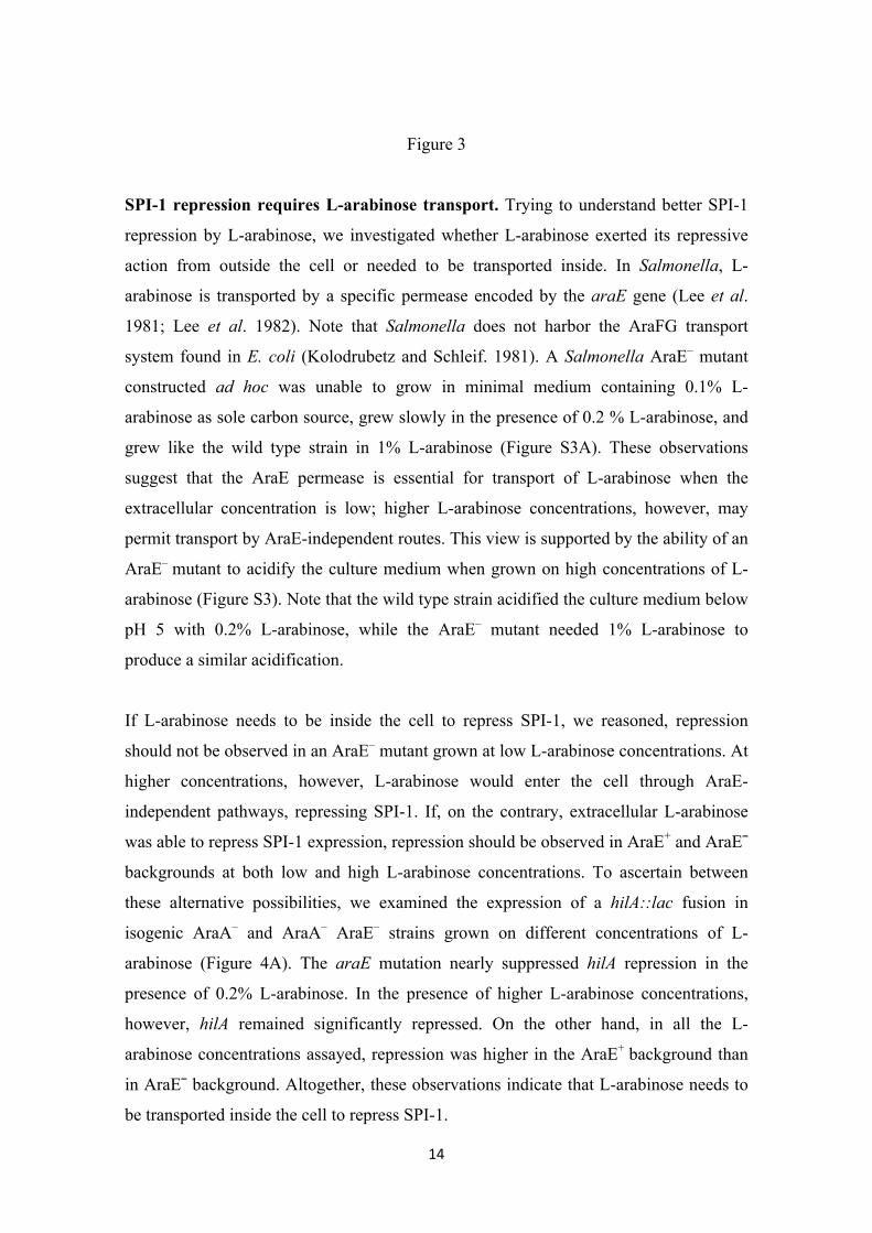
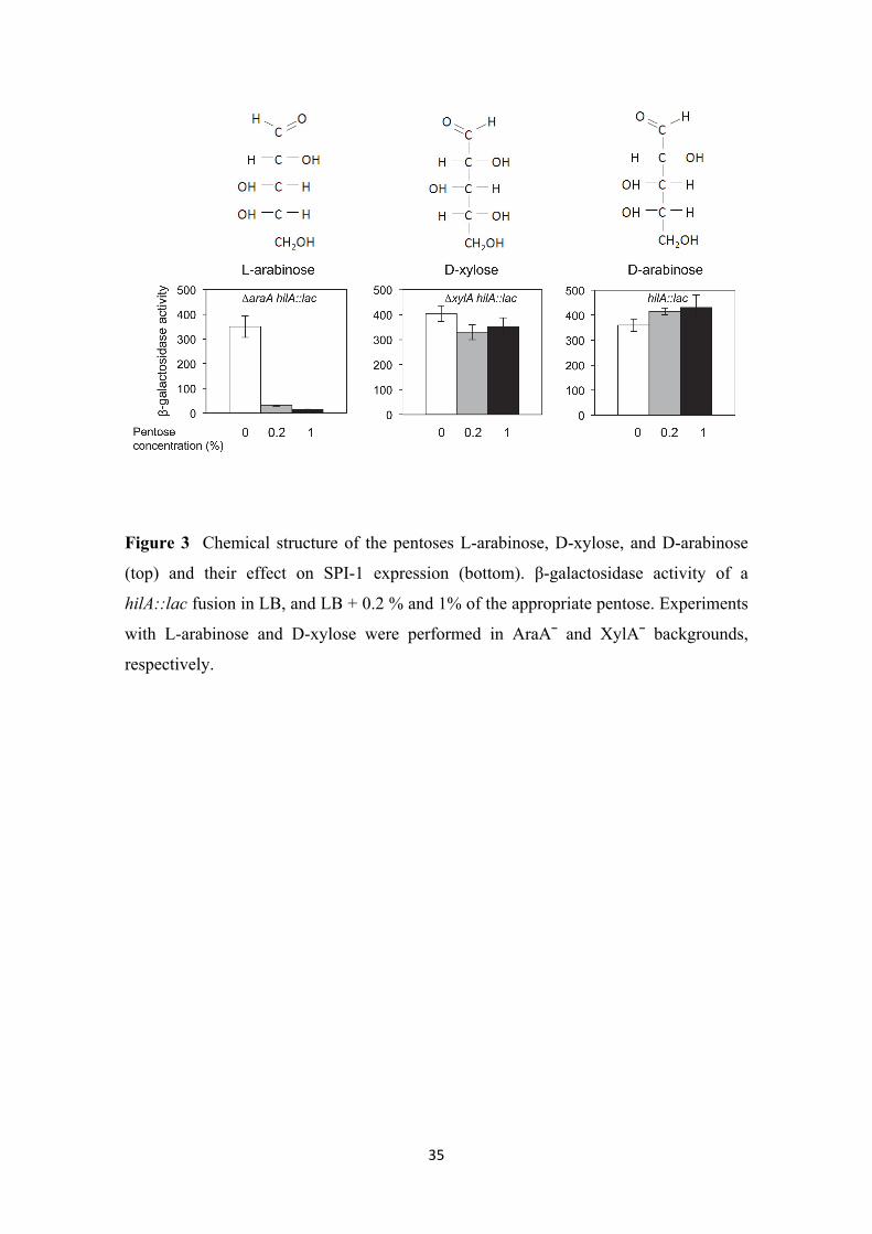
Effect of pentoses other than L-arabinose on SPI-1 expression. Besides L-arabinose,
Salmonella can metabolize other pentoses (Gutnick et al. 1969). An example is D-
xylose, the most abundant pentose in nature (Seiboth and Metz. 2011). The first reaction
in D-xylose catabolism is catalyzed by D-xylose isomerase, encoded by the xylA gene
(Shamanna and Sanderson. 1979). To examine the effect of D-xylose on SPI-1
expression, we constructed a strain carrying a xylA deletion. The xylA mutant is unable
to use D-xylose as sole carbon source (Figure S4), thus avoiding a change in the pH of
the culture medium due to D-xylose catabolism. Expression of a hilA::lac fusion was
monitored in a XylA¯ background, growing the strain in LB, LB + 0.2% D-xylose, and
LB + 1% D-xylose. As shown in Figure 3 (central panel), the presence of D-xylose in
the culture medium did not alter hilA::lac expression. However, the same fusion was
strongly repressed in the presence of either 0.2% or 1% L-arabinose (Figure 3, left
panel).
D-arabinose, the D stereoisomer of arabinose, cannot be used by Salmonella as sole
carbon source (Gutnick et al. 1969). However, certain Salmonella mutants can
metabolize D-arabinose (Old and Mortlock. 1977). In such mutants, D-arabinose
induces the synthesis of two enzymes usually involved in L-fucose metabolism: L-
fucose isomerase and L-fuculokinase. These enzymes have a bifunctional activity, and
can convert D-arabinose into D-ribulose-1-phosphate. Figure 3 (right panel) shows that
hilA::lac expression is similar in LB, LB + 0.2 % D-arabinose, and LB + 1% D-
arabinose, thus ruling out any effect of D-arabinose on SPI-1 expression.
The conclusion from these experiments was that the effect of L-arabinose on SPI-1
expression is specific, rather than a property shared with other pentoses such as D-
xylose and D-arabinose.
14
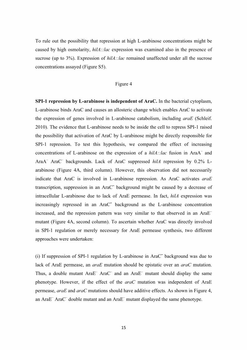
Figure 3
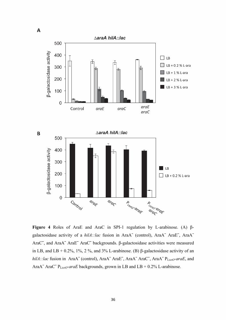
SPI-1 repression requires L-arabinose transport. Trying to understand better SPI-1
repression by L-arabinose, we investigated whether L-arabinose exerted its repressive
action from outside the cell or needed to be transported inside. In Salmonella, L-
arabinose is transported by a specific permease encoded by the araE gene (Lee et al.
1981; Lee et al. 1982). Note that Salmonella does not harbor the AraFG transport
system found in E. coli (Kolodrubetz and Schleif. 1981). A Salmonella AraE– mutant
constructed ad hoc was unable to grow in minimal medium containing 0.1% L-
arabinose as sole carbon source, grew slowly in the presence of 0.2 % L-arabinose, and
grew like the wild type strain in 1% L-arabinose (Figure S3A). These observations
suggest that the AraE permease is essential for transport of L-arabinose when the
extracellular concentration is low; higher L-arabinose concentrations, however, may
permit transport by AraE-independent routes. This view is supported by the ability of an
AraE– mutant to acidify the culture medium when grown on high concentrations of L-
arabinose (Figure S3). Note that the wild type strain acidified the culture medium below
pH 5 with 0.2% L-arabinose, while the AraE– mutant needed 1% L-arabinose to
produce a similar acidification.
If L-arabinose needs to be inside the cell to repress SPI-1, we reasoned, repression
should not be observed in an AraE– mutant grown at low L-arabinose concentrations. At
higher concentrations, however, L-arabinose would enter the cell through AraE-
independent pathways, repressing SPI-1. If, on the contrary, extracellular L-arabinose
was able to repress SPI-1 expression, repression should be observed in AraE+ and AraE¯
backgrounds at both low and high L-arabinose concentrations. To ascertain between
these alternative possibilities, we examined the expression of a hilA::lac fusion in
isogenic AraA– and AraA– AraE– strains grown on different concentrations of L-
arabinose (Figure 4A). The araE mutation nearly suppressed hilA repression in the
presence of 0.2% L-arabinose. In the presence of higher L-arabinose concentrations,
however, hilA remained significantly repressed. On the other hand, in all the L-
arabinose concentrations assayed, repression was higher in the AraE+ background than
in AraE¯ background. Altogether, these observations indicate that L-arabinose needs to
be transported inside the cell to repress SPI-1.
15
To rule out the possibility that repression at high L-arabinose concentrations might be
caused by high osmolarity, hilA::lac expression was examined also in the presence of
sucrose (up to 3%). Expression of hilA::lac remained unaffected under all the sucrose
concentrations assayed (Figure S5).
Figure 4
SPI-1 repression by L-arabinose is independent of AraC. In the bacterial cytoplasm,
L-arabinose binds AraC and causes an allosteric change which enables AraC to activate
the expression of genes involved in L-arabinose catabolism, including araE (Schleif.
2010). The evidence that L-arabinose needs to be inside the cell to repress SPI-1 raised
the possibility that activation of AraC by L-arabinose might be directly responsible for
SPI-1 repression. To test this hypothesis, we compared the effect of increasing
concentrations of L-arabinose on the expression of a hilA::lac fusion in AraA– and
AraA– AraC– backgrounds. Lack of AraC suppressed hilA repression by 0.2% L-
arabinose (Figure 4A, third column). However, this observation did not necessarily
indicate that AraC is involved in L-arabinose repression. As AraC activates araE
transcription, suppression in an AraC¯ background might be caused by a decrease of
intracellular L-arabinose due to lack of AraE permease. In fact, hilA expression was
increasingly repressed in an AraC¯ background as the L-arabinose concentration
increased, and the repression pattern was very similar to that observed in an AraE–
mutant (Figure 4A, second column). To ascertain whether AraC was directly involved
in SPI-1 regulation or merely necessary for AraE permease synthesis, two different
approaches were undertaken:
(i) If suppression of SPI-1 regulation by L-arabinose in AraC¯ background was due to
lack of AraE permease, an araE mutation should be epistatic over an araC mutation.
Thus, a double mutant AraE– AraC– and an AraE– mutant should display the same
phenotype. However, if the effect of the araC mutation was independent of AraE
permease, araE and araC mutations should have additive effects. As shown in Figure 4,
an AraE– AraC– double mutant and an AraE– mutant displayed the same phenotype.
16
(ii) If AraC is needed for AraE permease synthesis only, constitutive araE expression
should prevent AraC-mediated suppression of SPI-1 repression. AraC-independent
expression of araE was achieved by replacing the native araE promoter with the PLtetO
promoter (Lutz and Bujard. 1997). The resulting strain grew as well as the wild type in
minimal medium containing 0.1% L-arabinose (Figure S6A). In addition, it acidified the
culture medium like the wild type when grown in the presence of different
concentrations of L-arabinose, suggesting that AraE permease was produced indeed
(Figure S6). When araE was expressed constitutively, lack of AraC no longer
suppressed hilA repression by L-arabinose (Figure 4B).
Altogether, the above observations suggest that, once inside the cell, L-arabinose
represses SPI-1 expression by an AraC-independent mechanism.
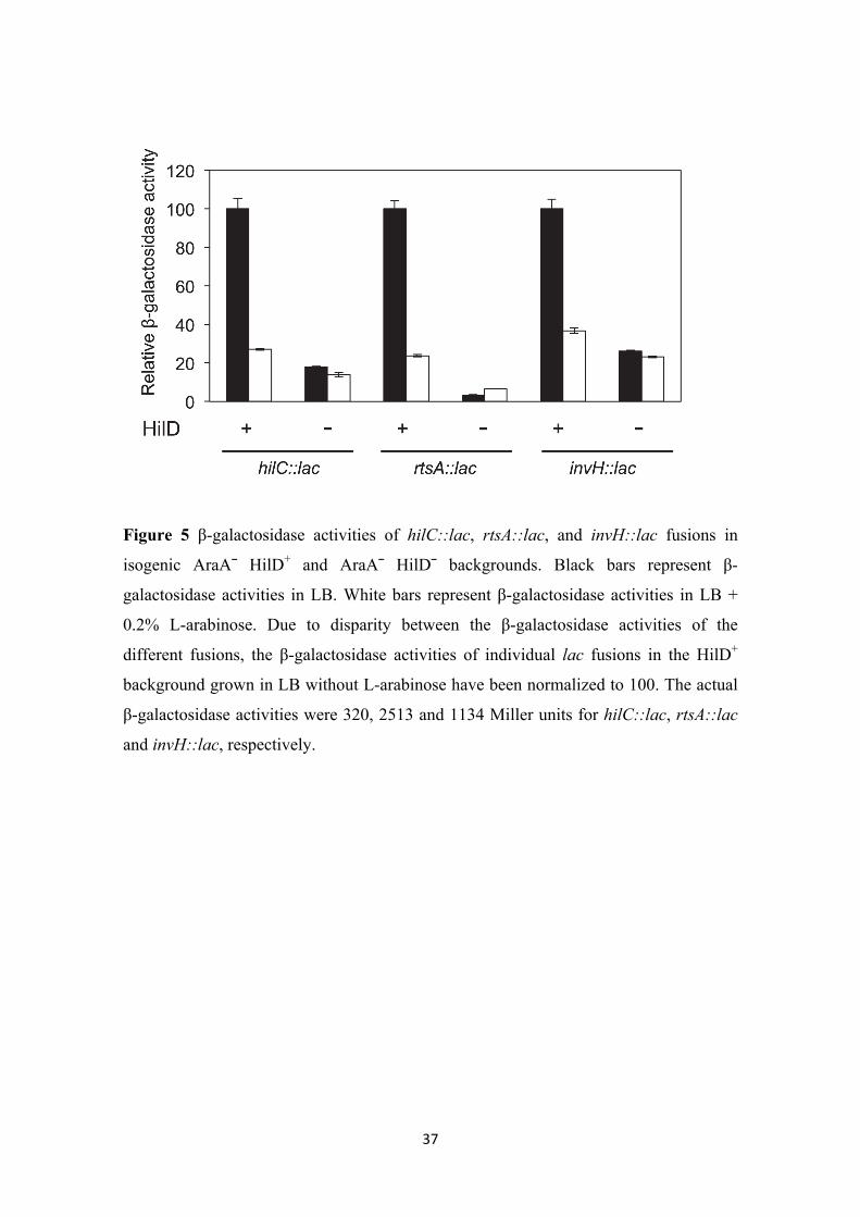
L-arabinose-dependent repression of SPI-1 is transmitted via HilD. SPI-1
expression is controlled by a regulatory network of SPI-1-encoded transcriptional
activators. On top of the network are the transcriptional activators HilA, HilC, and
HilD (Figure S1). The fact that hilA expression is regulated by L-arabinose might
indicate either that L-arabinose directly regulates hilA or that the regulatory input is
transmitted to hilA via HilC and/or HilD. Lack of HilC reduces hilA expression 2-3 fold
(Lopez-Garrido and Casadesus. 2010), while addition of 0.2% L-arabinose to the
culture medium represses hilA >10 fold. This discrepancy makes unlikely that HilC is
responsible for transmission of L-arabinose regulation. In contrast, lack of HilD reduces
hilA expression more than 100 fold (Lopez-Garrido and Casadesus. 2010). Thus, we
examined the possibility that L-arabinose might regulate SPI-1 expression via HilD. For
this purpose, we analyzed the effect of L-arabinose on the expression of three HilD-
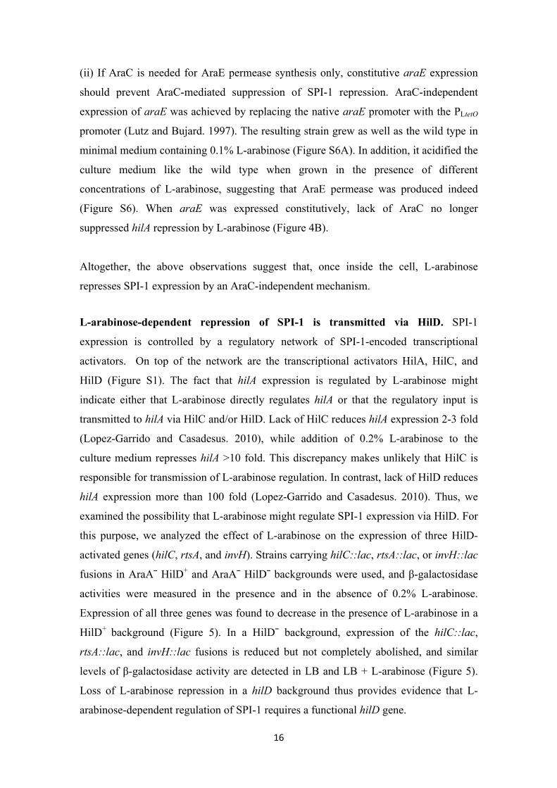
activated genes (hilC, rtsA, and invH). Strains carrying hilC::lac, rtsA::lac, or invH::lac
fusions in AraA¯ HilD+ and AraA¯ HilD¯ backgrounds were used, and β-galactosidase
activities were measured in the presence and in the absence of 0.2% L-arabinose.
Expression of all three genes was found to decrease in the presence of L-arabinose in a
HilD+ background (Figure 5). In a HilD¯ background, expression of the hilC::lac,
rtsA::lac, and invH::lac fusions is reduced but not completely abolished, and similar
levels of β-galactosidase activity are detected in LB and LB + L-arabinose (Figure 5).
Loss of L-arabinose repression in a hilD background thus provides evidence that L-
arabinose-dependent regulation of SPI-1 requires a functional hilD gene.
17
Figure 5
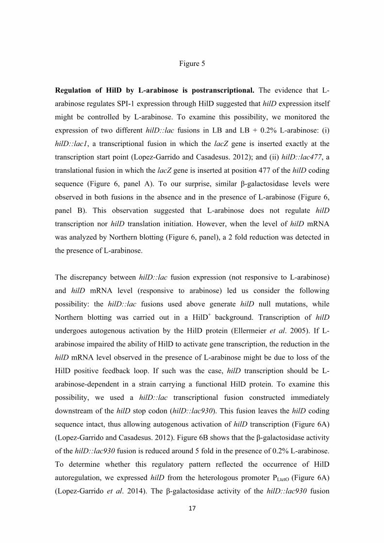
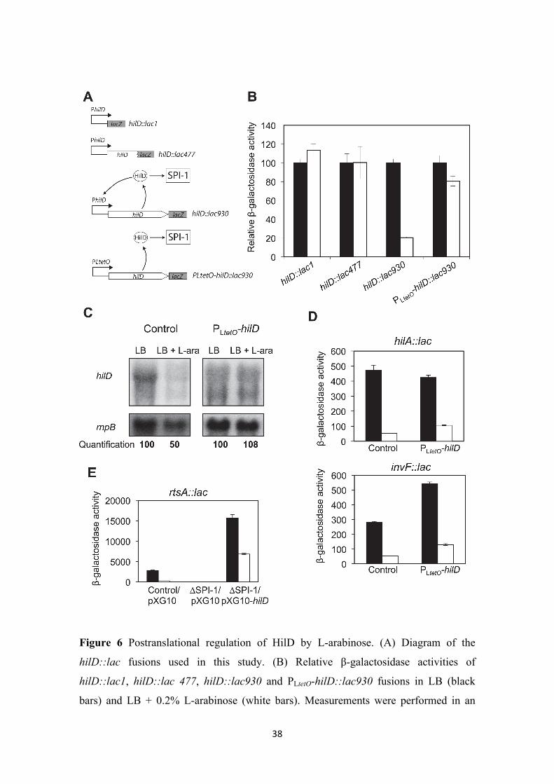
Regulation of HilD by L-arabinose is postranscriptional. The evidence that L-
arabinose regulates SPI-1 expression through HilD suggested that hilD expression itself
might be controlled by L-arabinose. To examine this possibility, we monitored the
expression of two different hilD::lac fusions in LB and LB + 0.2% L-arabinose: (i)
hilD::lac1, a transcriptional fusion in which the lacZ gene is inserted exactly at the
transcription start point (Lopez-Garrido and Casadesus. 2012); and (ii) hilD::lac477, a
translational fusion in which the lacZ gene is inserted at position 477 of the hilD coding
sequence (Figure 6, panel A). To our surprise, similar β-galactosidase levels were
observed in both fusions in the absence and in the presence of L-arabinose (Figure 6,
panel B). This observation suggested that L-arabinose does not regulate hilD
transcription nor hilD translation initiation. However, when the level of hilD mRNA
was analyzed by Northern blotting (Figure 6, panel), a 2 fold reduction was detected in
the presence of L-arabinose.
The discrepancy between hilD::lac fusion expression (not responsive to L-arabinose)
and hilD mRNA level (responsive to arabinose) led us consider the following
possibility: the hilD::lac fusions used above generate hilD null mutations, while
Northern blotting was carried out in a HilD+ background. Transcription of hilD
undergoes autogenous activation by the HilD protein (Ellermeier et al. 2005). If L-
arabinose impaired the ability of HilD to activate gene transcription, the reduction in the
hilD mRNA level observed in the presence of L-arabinose might be due to loss of the
HilD positive feedback loop. If such was the case, hilD transcription should be L-
arabinose-dependent in a strain carrying a functional HilD protein. To examine this
possibility, we used a hilD::lac transcriptional fusion constructed immediately
downstream of the hilD stop codon (hilD::lac930). This fusion leaves the hilD coding
sequence intact, thus allowing autogenous activation of hilD transcription (Figure 6A)
(Lopez-Garrido and Casadesus. 2012). Figure 6B shows that the β-galactosidase activity
of the hilD::lac930 fusion is reduced around 5 fold in the presence of 0.2% L-arabinose.
To determine whether this regulatory pattern reflected the occurrence of HilD
autoregulation, we expressed hilD from the heterologous promoter PLtetO (Figure 6A)
(Lopez-Garrido et al. 2014). The β-galactosidase activity of the hilD::lac930 fusion
18
transcribed from PLtetO was similar in LB and LB + 0.2% L-arabinose (Figure 6B).
Furthermore, the level of hilD mRNA did not decrease in the presence of L-arabinose
when hilD was transcribed from PLteto (Figure 6C). Altogether, these results suggest that
the target of L-arabinose is the HilD protein. A conceivable model is that L-arabinose
might interfere with HilD activity, thus affecting transcription of hilD itself and of the
remaining SPI-1 genes. This hypothesis is supported by the following observations:
(i) The effect of L-arabinose on the activity of hilA::lac and invF::lac fusions was
monitored upon hilD expression from PLtetO. Expression of both fusions was reduced
around 4 fold in the presence of 0.2% L-arabinose (Figure 6D).
(ii) The effect of L- arabinose on the activity of a rtsA::lac fusion was also monitored.
The rtsA gene is located outside SPI-1 but is directly activated by HilD. This trait
permits to study rtsA regulation in the absence of SPI-1, expressing hilD ectopically.
For this purpose, we cloned hilD on pXG-10 under the control of the PLtetO promoter.
Regulation of an rtsA::lac fusion by L-arabinose was examined in three different
backgrounds: AraA– with the pXG-10 vector (empty), AraA– ∆SPI-1 with the pXG-10
vector (empty), and AraA– ∆SPI-1 with pIZ1902 (pXG10-hilD). As shown in Figure
6E, the rtsA::lac fusion was regulated by L-arabinose is a strain with SPI-1. Deletion of
SPI-1 decreased β-galactosidase activity, and suppressed regulation by L-arabinose.
However, ectopic expression of hilD from a plasmid restored both the level of rtsA::lac
expression and regulation by L-arabinose in the absence of SPI-1.
These observations support the view that L-arabinose may interact with the HilD
protein, and confirms that HilD alone is sufficient for transmission of L-arabinose-
dependent regulation to other Salmonella genes.
Figure 6
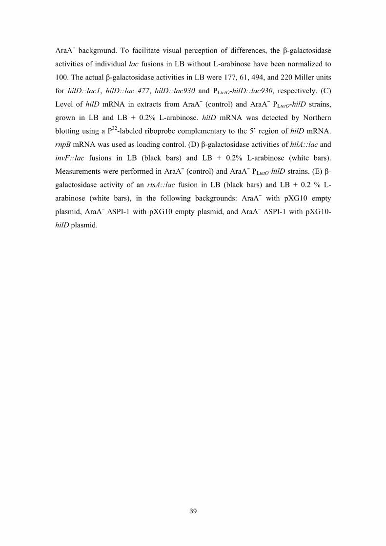
Translocation of SPI-1 effectors and epithelial cell invasion are reduced in the
presence of L-arabinose. To determine whether SPI-1 repression by L-arabinose
affected the interaction between Salmonella and host cells, we analyzed translocation of
SPI-1 effectors into eukaryotic HeLa cells (Figure 7A). A fusion of the SPI-1 gene sipA
with the cyaA gene of Bordetella pertussis was used for this purpose. Bordetella

19
adenylate cyclase requires calmodulin in order to synthesize cAMP (Glaser et al. 1989;
Greenlee et al. 1982; Wolff et al. 1980). Thus, cAMP will be produced only if the
SipA-CyaA fusion protein is translocated into eukaryotic cells. The sipA::cyaA fusion
was introduced in isogenic AraA–, AraA– AraE–, and AraA– PrgH– backgrounds, and
the strains were grown in LB and LB + 0.2% L-arabinose before mixing with HeLa
cells. Translocation was estimated by measuring the amount of cAMP, and was strongly
reduced in the presence of L-arabinose in the AraA– background (Figure 7, panel A).
However, in the AraA– AraE– background, addition of L-arabinose did not reduce
translocation, reflecting the need of L-arabinose transport to repress SPI-1 expression
(Figure 7, panel A). The AraA– PrgH– strain was used as negative control, since prgH
encodes an essential component of the SPI-1 T3SS. These results confirmed that SPI-1
repression by L-arabinose impairs the interaction between Salmonella and epithelial
host cells.
We then analyzed if the impaired translocation of SPI-1 effectors caused a reduction in
Salmonella invasion. The ability to invade epithelial HeLa cells was determined for
isogenic AraA–, AraA– AraE–, and AraA– ∆SPI-1 strains, grown on either LB or LB +
0.2% L-arabinose. Invasion by an AraA– strain decreased almost 100 fold in the
presence of arabinose. As expected, inactivation of AraE permease nearly suppressed
invasion inhibition in the presence of L-arabinose. In the absence of SPI-1, invasion was
reduced more that 5000 fold, and no further reduction was observed when L-arabinose
was added to the culture medium (Figure 7, panel B). These observations suggest that
SPI-1 repression in the presence of L-arabinose impairs Salmonella interaction with
eukaryotic epithelial cells, causing inhibition of invasion.
Figure 7
20
DISCUSSION
Regulation of virulence genes by sugars has been previously reported. In the Gram-
positive pathogen Listeria monocytogenes, expression of virulence genes regulated by
the master regulator PrfA is repressed in the presence of sugars transported by the
phosphoenolpyruvate-sugar phosphotransferase system (PTS) (de las Heras et al. 2011;
Milenbachs et al. 1997; Park and Kroll. 1993). However, repression is not observed in
the presence of non-PTS sugars (de las Heras et al. 2011; Joseph et al. 2008; Ripio et al.
1997; Stoll et al. 2008). In Streptococcus pyogenes, production of surface M protein, a
major virulence determinant, is affected by the sugar source (Pine and Reeves. 1978).
Transcription of the gene encoding the M protein is indirectly activated by carbon
catabolic repression (CCR) through the virulence gene regulator Mga. CCR also
controls virulence gene expression in Clostridium perfringens (Varga et al. 2004) and
Staphylococcus aureus (Morse et al. 1969). In Salmonella enterica, evidence exists that
PTS-dependent sugars may repress expression of invasion genes: Crp– Cya– mutants of
S. enterica serovar Choleraesuis are attenuated in pigs (Kennedy et al. 1999), which
correlates with the inability of Crp– mutants to secrete SPI-1 TTSS effectors (Chen et al.
2010). In addition, it has been reported that Mlc, a global regulator of carbon
metabolism that can be induced by the PTS-sugars glucose and mannose (Plumbridge.
2002), activates SPI-1 expression by repressing transcription of the hilE gene, which
encodes a negative regulator of SPI-1 (Lim et al. 2007).
The ability of L-arabinose, a non-PTS sugar, to regulate the expression of genes
involved in L-arabinose catabolism has been known for half a century [reviewed in
(Schleif. 2010)]. Like L-arabinose catabolism, SPI-1 repression requires transport of L-
arabinose by the AraE permease, a requirement that admits two alternative
interpretations: (i) L-arabinose has to be inside the cell to repress SPI-1; or (ii) transport
of L-arabinose by AraE is necessary for SPI-1 repression. The latter possibility seems
unlikely because a signal transduction system associated with AraE has not been
described. Furthermore, the view that SPI-1 repression is exerted by L-arabinose itself
is supported by two observations: (i) Salmonella AraE– mutants can grow with 1% L-
arabinose as sole carbon source, indicating the existence of alternative transport
21
mechanisms; (ii) L-arabinose represses SPI-1 expression in Salmonella AraE– mutants
if provided at concentrations of 1% or higher.
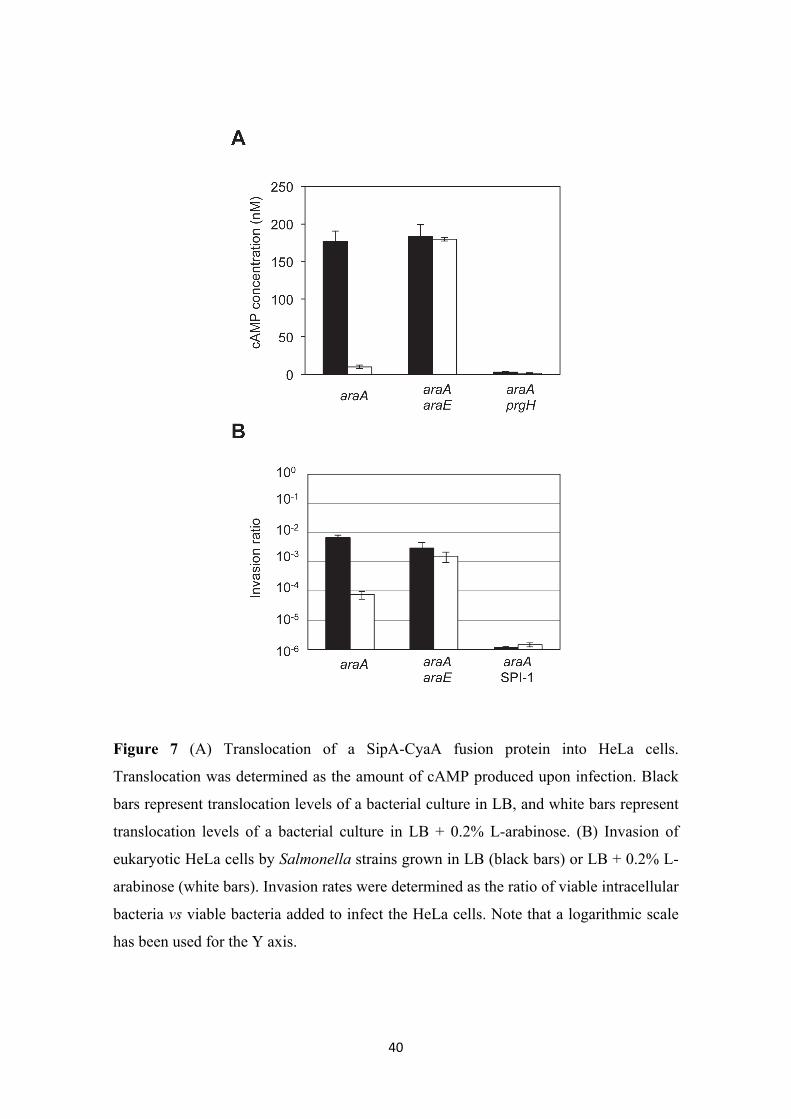
Epistasis analysis indicates that the target of L-arabinose is the transcription factor
HilD, and experiments with gene fusions suggest that regulation is postranscriptional.
Because HilD is an AraC-like transcription factor, it is tempting to propose that L-
arabinose might regulate HilD activity by direct binding to the protein. A tentative
model of SPI-1 regulation by L-arabinose is depicted in Figure 8.
Figure 8
L-arabinose is a plant-derived sugar, and the possession of a specific system for L-
arabinose catabolism may enable Salmonella to use L-arabinose as carbon source in L-
arabinose-rich environments. In addition, L-arabinose may serve as a signal for SPI-1
repression under certain circumstances. The observation that Salmonella grown in the
presence of L-arabinose fails to invade epithelial cells (Figure 7) suggests that L-
arabinose might inhibit invasion in the animal intestine. L-arabinose is poorly absorbed
during digestion by mammals (Cori. 1925) and chickens (Wagh and Waibel. 1967), and
its presence in the intestine after ingestion is supported by experimental evidence: PBAD
promoter expression is induced in the intestine of mice fed with plant components
(Loessner et al. 2009). Furthermore, L-arabinose catabolism is required for efficient
colonization of the large intestine by comensal and pathogenic strains of E. coli (Fabich
et al. 2008). L-arabinose supports efficient growth of Salmonella in vitro and might be a
good carbon source in the intestine. Thus, the presence of L-arabinose in the intestine
might be detected by Salmonella as a signal for repression of invasion. If such were the
case, L-arabinose-rich compounds in the diet could prevent infections by Salmonella.
Consistent with this possibility, dietary addition of arabinoxylooligosaccharides, made
of L-arabinose and D-xylose, has been shown to protect poultry against oral infection
by Salmonella enterica serovar Enteritidis (Eeckhaut et al. 2008).
SPI-1 repression by L-arabinose could also play a role outside the animal host. As a
plant-derived sugar, L-arabinose accumulates in the soil. It has been shown that
Salmonella is able to persist in the soil for long periods (Islam et al. 2004). Because
synthesis of the SPI-1 TTSS causes growth retardation (Ali et al. 2014; Sturm et al.
22
2011), SPI-1 repression by L-arabinose may improve the fitness of Salmonella in the
soil. On the other hand, Salmonella can colonize plant surfaces (Barak et al. 2002;
Brandl and Mandrell. 2002) as well as intercellular spaces inside the plants (Franz et al.
2007). Plant colonization by Salmonella might be used as a strategy for recolonizing
animal hosts (Tyler and Triplett. 2008). Salmonella mutants lacking components of the
SPI-1 TTSS colonize plants more efficiently than the wild type (Iniguez et al. 2005),
perhaps because the presence of a functional TTSS in the Salmonella surface triggers a
defense response by the plant (Iniguez et al. 2005). In such a scenario, detection of L-
arabinose during plant colonization might contribute to turn down SPI-1 expression.
An additional implication of this study concerns the use of L-arabinose and other sugars
for ectopic induction of gene expression, a tool routinely used in biological research.
Metabolizable sugars can produce changes in cell physiology. In addition, as shown
above, sugars can have metabolism-independent effects, potentially interfering with the
subject of study.
23
ACKNOWLEDGMENTS
This study was supported by grants BIO2010-15023 and BIO2013-44220-R from the
Ministerio de Economía y Competitividad (MINECO) of Spain and the European
Regional Fund, CSD2008-00013 from the Consolider-Ingenio 2010 Program of
MINECO, and CVI-5879 from the Consejería de Innovación, Ciencia y Empresa, Junta
de Andalucía, Spain. We are grateful to Dick D’Ari for encouraging the development of
this project, and to David Reyes and Karina Ramijan for performing experiments not
included in the manuscript. We appreciate the assistance provided by Modesto Carballo,
Laura Navarro, and Cristina Reyes of the Servicio de Biología, Centro de Investigación,
Tecnología e Innovación de la Universidad de Sevilla (CITIUS). J.L.G. and I.C. were
supported by fellowships of the MINECO program "Formación del Profesorado
Universitario".
24
LITERATURE CITED
Abedon, B. G., R. D. Hatfield and W. F. Tracy, 2006 Cell wall composition in juvenile
and adult leaves of maize (Zea mays L.). J. Agric. Food Chem. 54: 3896-3900.
Akbar, S., L. M. Schechter, C. P. Lostroh and C. A. Lee, 2003 AraC/XylS family
members, HilD and HilC, directly activate virulence gene expression independently of
HilA in Salmonella typhimurium. Mol. Microbiol. 47: 715-728.
Ali, S. S., J. Soo, C. Rao, A. S. Leung, D. H.-M. Ngai et al, 2014 Silencing by H-NS
potentiated the evolution of Salmonella. PLoS Pathog. 10: e1004500.
Altier, C., 2005 Genetic and environmental control of Salmonella invasion. J.
Microbiol. 43: 85-92.
Bajaj, V., C. Hwang and C. A. Lee, 1995 HilA is a novel OmpR/ToxR family member
that activates the expression of Salmonella typhimurium invasion genes. Mol.
Microbiol. 18: 715-727.
Bajaj, V., R. L. Lucas, C. Hwang and C. A. Lee, 1996 Co-ordinate regulation of
Salmonella typhimurium invasion genes by environmental and regulatory factors is
mediated by control of hilA expression. Mol. Microbiol. 22: 703-714.
Barak, J. D., L. C. Whitehand and A. O. Charkowski, 2002 Differences in attachment of
Salmonella enterica serovars and Escherichia coli O157:H7 to alfalfa sprouts. Appl.
Environ. Microbiol. 68: 4758-4763.
Behlau, I., and S. I. Miller, 1993 A PhoP-repressed gene promotes Salmonella
typhimurium invasion of epithelial cells. J. Bacteriol. 175: 4475-4484.
Brandl, M. T., and R. E. Mandrell, 2002 Fitness of Salmonella enterica serovar
Thompson in the cilantro phyllosphere. Appl. Environ. Microbiol. 68: 3614-3621.
Chan, R. K., D. Botstein, T. Watanabe and Y. Ogata, 1972 Specialized transduction of
tetracycline resistance by phage P22 in Salmonella typhimurium. II. Properties of a
high-frequency-transducing lysate. Virology 50: 883-898.

25
Chen, Z. W., S. L. Hsuan, J. W. Liao, T. H. Chen, C. M. Wu et al, 2010 Mutations in
the Salmonella enterica serovar Choleraesuis cAMP-receptor protein gene lead to
functional defects in the SPI-1 type III secretion system. Vet. Res. 41: 5.
Cori C. F., 1925 The fate of sugar in the animal body. I. The rate of absorption of
hexoses and pentoses from the intestinal tract. J Biol Chem 70: 691-715.
Darwin, K. H., and V. L. Miller, 1999a Molecular basis of the interaction of Salmonella
with the intestinal mucosa. Clin. Microbiol. Rev. 12: 405-428.
Darwin, K. H., and V. L. Miller, 1999b InvF is required for expression of genes
encoding proteins secreted by the SPI1 type III secretion apparatus in Salmonella
typhimurium. J. Bacteriol. 181: 4949-4954.
Datsenko, K. A., and B. L. Wanner, 2000 One-step inactivation of chromosomal genes
in Escherichia coli K-12 using PCR products. Proc. Natl. Acad. Sci. U. S. A. 97: 6640-
6645.
de las Heras, A., R. J. Cain, M. K. Bielecka and J. A. Vazquez-Boland, 2011 Regulation
of Listeria virulence: PrfA master and commander. Curr. Opin. Microbiol. 14: 118-127.
Eeckhaut, V., F. Van Immerseel, J. Dewulf, F. Pasmans, F. Haesebrouck et al, 2008
Arabinoxylooligosaccharides from wheat bran inhibit Salmonella colonization in broiler
chickens. Poult. Sci. 87: 2329-2334.
Eichelberg, K., and J. E. Galan, 1999 Differential regulation of Salmonella typhimurium
type III secreted proteins by pathogenicity island 1 (SPI-1)-encoded transcriptional
activators InvF and HilA. Infect. Immun. 67: 4099-4105.
Ellermeier, C. D., and J. M. Slauch, 2003 RtsA and RtsB coordinately regulate
expression of the invasion and flagellar genes in Salmonella enterica serovar
Typhimurium. J. Bacteriol. 185: 5096-5108.
Ellermeier, C. D., J. R. Ellermeier and J. M. Slauch, 2005 HilD, HilC and RtsA
constitute a feed forward loop that controls expression of the SPI1 type three secretion
system regulator hilA in Salmonella enterica serovar Typhimurium. Mol. Microbiol. 57:
691-705.
26
Ellermeier, C. D., A. Janakiraman and J. M. Slauch, 2002 Construction of targeted
single copy lac fusions using lambda red and FLP-mediated site-specific recombination
in bacteria. Gene 290: 153-161.
Ellermeier, J. R., and J. M. Slauch, 2007 Adaptation to the host environment:
Regulation of the SPI1 type III secretion system in Salmonella enterica serovar
Typhimurium. Curr. Opin. Microbiol. 10: 24-29.
Englesberg, E., 1961 Enzymatic characterization of 17 L-arabinose negative mutants of
Escherichia coli. J. Bacteriol. 81: 996-1006.
Englesberg, E., J. Irr, J. Power and N. Lee, 1965 Positive control of enzyme synthesis
by gene C in the L-arabinose system. J. Bacteriol. 90: 946-957.
Englesberg, E., R. L. Anderson, R. Weinberg, N. Lee, P. Hoffee et al, 1962 L-
arabinose-sensitive, L-ribulose 5-phosphate 4-epimerase-deficient mutants of
Escherichia coli. J. Bacteriol. 84: 137-146.
Fabich, A. J., S. A. Jones, F. Z. Chowdhury, A. Cernosek, A. Anderson et al, 2008
Comparison of carbon nutrition for pathogenic and commensal Escherichia coli strains
in the mouse intestine. Infect. Immun. 76: 1143-1152.
Franz, E., A. A. Visser, A. D. Van Diepeningen, M. M. Klerks, A. J. Termorshuizen et
al, 2007 Quantification of contamination of lettuce by GFP-expressing Escherichia coli
O157:H7 and Salmonella enterica serovar Typhimurium. Food Microbiol. 24: 106-112.
Garzon, A., D. A. Cano and J. Casadesus, 1995 Role of Erf recombinase in P22-
mediated plasmid transduction. Genetics 140: 427-434.
Gerlach, R. G., S. U. Holzer, D. Jackel and M. Hensel, 2007 Rapid engineering of
bacterial reporter gene fusions by using red recombination. Appl. Environ. Microbiol.
73: 4234-4242.
Glaser, P., A. Elmaoglou-Lazaridou, E. Krin, D. Ladant, O. Barzu et al, 1989
Identification of residues essential for catalysis and binding of calmodulin in Bordetella
pertussis adenylate cyclase by site-directed mutagenesis. EMBO J. 8: 967-972.
27
Greenlee, D. V., T. J. Andreasen and D. R. Storm, 1982 Calcium-independent
stimulation of Bordetella pertussis adenylate cyclase by calmodulin. Biochemistry 21:
2759-2764.
Gross, J., and E. Englesberg, 1959 Determination of the order of mutational sites
governing L-arabinose utilization in Escherichia coli B/r by transduction with phage
Plbt. Virology 9: 314-331.
Gutnick, D., J. M. Calvo, T. Klopotowski and B. N. Ames, 1969 Compounds which
serve as the sole source of carbon or nitrogen for Salmonella typhimurium LT-2. J.
Bacteriol. 100: 215-219.
Guzman, L. M., D. Belin, M. J. Carson and J. Beckwith, 1995 Tight regulation,
modulation, and high-level expression by vectors containing the arabinose PBAD
promoter. J. Bacteriol. 177: 4121-4130.
Iniguez, A. L., Y. Dong, H. D. Carter, B. M. Ahmer, J. M. Stone et al, 2005 Regulation
of enteric endophytic bacterial colonization by plant defenses. Mol. Plant Microbe
Interact. 18: 169-178.
Islam, M., J. Morgan, M. P. Doyle, S. C. Phatak, P. Millner et al, 2004 Persistence of
Salmonella enterica serovar Typhimurium on lettuce and parsley and in soils on which
they were grown in fields treated with contaminated manure composts or irrigation
water. Foodborne Pathog. Dis. 1: 27-35.
Jakomin, M., D. Chessa, A. J. Baumler and J. Casadesus, 2008 Regulation of the
Salmonella enterica std fimbrial operon by DNA adenine methylation, SeqA, and HdfR.
J. Bacteriol. 190: 7406-7413.
Jones, B. D., 2005 Salmonella invasion gene regulation: A story of environmental
awareness. J. Microbiol. 43 Spec No: 110-117.
Jones, B. D., N. Ghori and S. Falkow, 1994 Salmonella typhimurium initiates murine
infection by penetrating and destroying the specialized M cells of the Peyer’s patches. J.
Exp. Med. 180: 15-23.
28
Joseph, B., S. Mertins, R. Stoll, J. Schar, K. R. Umesha et al, 2008 Glycerol metabolism
and PrfA activity in Listeria monocytogenes. J. Bacteriol. 190: 5412-5430.
Kennedy, M. J., R. J. Yancey Jr, M. S. Sanchez, R. A. Rzepkowski, S. M. Kelly et al,
1999 Attenuation and immunogenicity of Delta-cya Delta-crp derivatives of Salmonella
choleraesuis in pigs. Infect. Immun. 67: 4628-4636.
Kolodrubetz, D., and R. Schleif, 1981 L-arabinose transport systems in Escherichia coli
K-12. J. Bacteriol. 148: 472-479.
Lawhon, S. D., R. Maurer, M. Suyemoto and C. Altier, 2002 Intestinal short-chain fatty
acids alter Salmonella typhimurium invasion gene expression and virulence through
BarA/SirA. Mol. Microbiol. 46: 1451-1464.
Lee, C. A., B. D. Jones and S. Falkow, 1992 Identification of a Salmonella typhimurium
invasion locus by selection for hyperinvasive mutants. Proc. Natl. Acad. Sci. U. S. A.
89: 1847-1851.
Lee, J. H., J. Nishitani and G. Wilcox, 1984 Genetic characterization of Salmonella
typhimurium LT2 ara mutations. J. Bacteriol. 158: 344-346.
Lee, J. H., S. Al-Zarban and G. Wilcox, 1981 Genetic characterization of the araE gene
in Salmonella typhimurium LT2. J. Bacteriol. 146: 298-304.
Lee, J. H., L. Heffernan and G. Wilcox, 1980 Isolation of ara-lac gene fusions in
Salmonella typhimurium LT2 by using transducing bacteriophage Mud(Ap lac). J.
Bacteriol. 143: 1325-1331.
Lee, J. H., R. J. Russo, L. Heffernan and G. Wilcox, 1982 Regulation of L-arabinose
transport in Salmonella typhimurium LT2. Mol. Gen. Genet. 185: 136-141.
Lim, S., J. Yun, H. Yoon, C. Park, B. Kim et al, 2007 Mlc regulation of Salmonella
pathogenicity island I gene expression via hilE repression. Nucleic Acids Res. 35:
1822-1832.
29
Loessner, H., S. Leschner, A. Endmann, K. Westphal, K. Wolf et al, 2009 Drug-
inducible remote control of gene expression by probiotic Escherichia coli Nissle 1917
in intestine, tumor and gall bladder of mice. Microbes Infect. 11: 1097-1105.
Lopez-Garrido, J., and J. Casadesus, 2012 Crosstalk between virulence loci: Regulation
of Salmonella enterica pathogenicity island 1 (SPI-1) by products of the std fimbrial
operon. PLoS One 7: e30499.
Lopez-Garrido, J., and J. Casadesus, 2010 Regulation of Salmonella enterica
pathogenicity island 1 by DNA adenine methylation. Genetics 184: 637-649.
Lopez-Garrido, J., E. Puerta-Fernandez and J. Casadesus, 2014 A eukaryotic-like 3'
untranslated region in Salmonella enterica hilD mRNA. Nucleic Acids Res. 42: 5894-
5906.
Lostroh, C. P., and C. A. Lee, 2001 The Salmonella pathogenicity island-1 type III
secretion system. Microbes Infect. 3: 1281-1291.
Lutz, R., and H. Bujard, 1997 Independent and tight regulation of transcriptional units
in Escherichia coli via the LacR/O, the TetR/O and AraC/I1-I2 regulatory elements.
Nucleic Acids Res. 25: 1203-1210.
Maloy, S. R., and J. R. Roth, 1983 Regulation of proline utilization in Salmonella
typhimurium: Characterization of put::Mud(Ap, lac) operon fusions. J. Bacteriol. 154:
561-568.
McClelland, M., K. E. Sanderson, J. Spieth, S. W. Clifton, P. Latreille et al, 2001
Complete genome sequence of Salmonella enterica serovar Typhimurium LT2. Nature
413: 852-856.
Milenbachs, A. A., D. P. Brown, M. Moors and P. Youngman, 1997 Carbon-source
regulation of virulence gene expression in Listeria monocytogenes. Mol. Microbiol. 23:
1075-1085.
Miller, J. H., 1972 Experiments in molecular genetics. Cold Spring Harbor Laboratory
Press, Cold Spring Harbor, NY.
30
Morse, S. A., R. A. Mah and W. J. Dobrogosz, 1969 Regulation of staphylococcal
enterotoxin B. J. Bacteriol. 98: 4-9.
Old, D., and R. D. Mortlock, 1977 The metabolism of D-arabinose by Salmonella
typhimurium. J. Gen. Microbiol. 101: 341-344.
Olekhnovich, I. N., and R. J. Kadner, 2006 Crucial roles of both flanking sequences in
silencing of the hilA promoter in Salmonella enterica. J. Mol. Biol. 357: 373-386.
Olekhnovich, I. N., and R. J. Kadner, 2002 DNA-binding activities of the HilC and
HilD virulence regulatory proteins of Salmonella enterica serovar Typhimurium. J.
Bacteriol. 184: 4148-4160.
Park, S. F., and R. G. Kroll, 1993 Expression of listeriolysin and phosphatidylinositol-
specific phospholipase C is repressed by the plant-derived molecule cellobiose in
Listeria monocytogenes. Mol. Microbiol. 8: 653-661.
Pine, L., and M. W. Reeves, 1978 Regulation of the synthesis of M protein by sugars,
Todd Hewitt broth, and horse serum, in growing cells of Streptococcus pyogenes.
Microbios 21: 185-212.
Plumbridge, J., 2002 Regulation of gene expression in the PTS in Escherichia coli: The
role and interactions of Mlc. Curr. Opin. Microbiol. 5: 187-193.
Prouty, A. M., and J. S. Gunn, 2000 Salmonella enterica serovar Typhimurium invasion
is repressed in the presence of bile. Infect. Immun. 68: 6763-6769.
Rakeman, J. L., H. R. Bonifield and S. I. Miller, 1999 A HilA-independent pathway to
Salmonella typhimurium invasion gene transcription. J. Bacteriol. 181: 3096-3104.
Ripio, M. T., K. Brehm, M. Lara, M. Suarez and J. A. Vazquez-Boland, 1997 Glucose-
1-phosphate utilization by Listeria monocytogenes is PrfA dependent and coordinately
expressed with virulence factors. J. Bacteriol. 179: 7174-7180.
Schadel, C., A. Blochl, A. Richter and G. Hoch, 2010 Quantification and
monosaccharide composition of hemicelluloses from different plant functional types.
Plant Physiol. Biochem. 48: 1-8.
31
Schechter, L. M., and C. A. Lee, 2001 AraC/XylS family members, HilC and HilD,
directly bind and derepress the Salmonella typhimurium hilA promoter. Mol. Microbiol.
40: 1289-1299.
Schleif, R., 2010 AraC protein, regulation of the L-arabinose operon in Escherichia
coli, and the light switch mechanism of AraC action. FEMS Microbiol. Rev. 34: 779-
796.
Schmieger, H., 1972 Phage P22-mutants with increased or decreased transduction
abilities. Mol. Gen. Genet. 119: 75-88.
Seiboth, B., and B. Metz, 2011 Fungal arabinan and L-arabinose metabolism. Appl.
Microbiol. Biotechnol. 89: 1665-1673.
Sezonov, G., D. Joseleau-Petit and R. D'Ari, 2007 Escherichia coli physiology in Luria-
Bertani broth. J. Bacteriol. 189: 8746-8749.
Shamanna, D. K., and K. E. Sanderson, 1979 Uptake and catabolism of D-xylose in
Salmonella typhimurium LT2. J. Bacteriol. 139: 64-70.
Stoll, R., S. Mertins, B. Joseph, S. Muller-Altrock and W. Goebel, 2008 Modulation of
PrfA activity in Listeria monocytogenes upon growth in different culture media.
Microbiology 154: 3856-3876.
Sturm, A., M. Heinemann, M. Arnoldini, A. Benecke, M. Ackermann et al, 2011 The
cost of virulence: Retarded growth of Salmonella typhimurium cells expressing type III
secretion system 1. PLoS Pathog. 7: e1002143.
Tyler, H. L., and E. W. Triplett, 2008 Plants as a habitat for beneficial and/or human
pathogenic bacteria. Annu. Rev. Phytopathol. 46: 53-73.
Urban, J. H., and J. Vogel, 2007 Translational control and target recognition by
Escherichia coli small RNAs in vivo. Nucleic Acids Res. 35: 1018-1037.
Uzzau, S., N. Figueroa-Bossi, S. Rubino and L. Bossi, 2001 Epitope tagging of
chromosomal genes in Salmonella. Proc. Natl. Acad. Sci. U. S. A. 98: 15264-15269.
32
Varga, J., V. L. Stirewalt and S. B. Melville, 2004 The CcpA protein is necessary for
efficient sporulation and enterotoxin gene (cpe) regulation in Clostridium perfringens. J.
Bacteriol. 186: 5221-5229.
Wagh, P. V., and P. E. Waibel, 1967 Alimentary absorption of L-arabinose and D-
xylose in chicks. Proc. Soc. Exp. Biol. Med. 124: 421-424.
Wolff, J., G. H. Cook, A. R. Goldhammer and S. A. Berkowitz, 1980 Calmodulin
activates prokaryotic adenylate cyclase. Proc. Natl. Acad. Sci. U. S. A. 77: 3841-3844.
33
Figure 1 Regulation of invF by L-arabinose. (A) β-galactosidase activity of an
invF::lac fusion in LB (black bars) and in LB + 0.2% L-arabinose (white bars). The
graph on the left shows activities measured in an AraA+ background. The graph on the
right shows activities measured in an AraA¯ background. (B) pH of the culture medium
measured before bacterial inoculation (before growth), and after the bacterial culture
had reached an O.D.600 of ~2.5 (after growth).
34
Figure 2 Dose-dependent regulation of SPI-1 by L-arabinose. (A) β-galactosidase
activities of hilA::lac, invF::lac, sipB::lac, and prgH::lac fusions in the absence (black
bars) and in the presence (white bars) of 0.2% L-arabinose, measured in AraA¯
background. Due to disparity between the β-galactosidase activities of the fusions, the
β-galactosidase activities of individual lac fusions in LB without L-arabinose have been
normalized to 100. The actual β-galactosidase activities in LB were 410, 308, 5384, and
1184 Miller units for hilA::lac, invF::lac, sipB::lac and prgH::lac, respectively. (B)
Levels of HilA, InvF, SipB, and PrgH in protein extracts from AraA¯ strains grown in
LB with and without 0.2% L-arabinose. Proteins tagged with the 3xFLAG epitope were
detected by Western blotting using anti-FLAG commercial antibodies. The loading
control was GroEL in all cases. (C) β-galactosidase activity of a hilA::lac fusion in an
AraA¯ strain grown in the presence of increasing concentrations of L-arabinose. (D)
Level of HilA-3xFLAG in protein extracts from an AraA¯ strain grown in increasing
concentrations of L-arabinose. The HilA level was normalized to GroEL for
quantification.
35
Figure 3 Chemical structure of the pentoses L-arabinose, D-xylose, and D-arabinose
(top) and their effect on SPI-1 expression (bottom). β-galactosidase activity of a
hilA::lac fusion in LB, and LB + 0.2 % and 1% of the appropriate pentose. Experiments
with L-arabinose and D-xylose were performed in AraA¯ and XylA¯ backgrounds,
respectively.
36
Figure 4 Roles of AraE and AraC in SPI-1 regulation by L-arabinose. (A) β-
galactosidase activity of a hilA::lac fusion in AraA¯ (control), AraA¯ AraE¯, AraA¯
AraC¯, and AraA¯ AraE¯ AraC¯ backgrounds. β-galactosidase activities were measured
in LB, and LB + 0.2%, 1%, 2 %, and 3% L-arabinose. (B) β-galactosidase activity of an
hilA::lac fusion in AraA¯ (control), AraA¯ AraE¯, AraA¯ AraC¯, AraA¯ PLtetO-araE, and
AraA¯ AraC¯ PLtetO-araE backgrounds, grown in LB and LB + 0.2% L-arabinose.
37
Figure 5 β-galactosidase activities of hilC::lac, rtsA::lac, and invH::lac fusions in
isogenic AraA¯ HilD+ and AraA¯ HilD¯ backgrounds. Black bars represent β-
galactosidase activities in LB. White bars represent β-galactosidase activities in LB +
0.2% L-arabinose. Due to disparity between the β-galactosidase activities of the
different fusions, the β-galactosidase activities of individual lac fusions in the HilD+
background grown in LB without L-arabinose have been normalized to 100. The actual
β-galactosidase activities were 320, 2513 and 1134 Miller units for hilC::lac, rtsA::lac
and invH::lac, respectively.
38
Figure 6 Postranslational regulation of HilD by L-arabinose. (A) Diagram of the
hilD::lac fusions used in this study. (B) Relative β-galactosidase activities of
hilD::lac1, hilD::lac 477, hilD::lac930 and PLtetO-hilD::lac930 fusions in LB (black
bars) and LB + 0.2% L-arabinose (white bars). Measurements were performed in an
39
AraA¯ background. To facilitate visual perception of differences, the β-galactosidase
activities of individual lac fusions in LB without L-arabinose have been normalized to
100. The actual β-galactosidase activities in LB were 177, 61, 494, and 220 Miller units
for hilD::lac1, hilD::lac 477, hilD::lac930 and PLtetO-hilD::lac930, respectively. (C)
Level of hilD mRNA in extracts from AraA¯ (control) and AraA¯ PLtetO-hilD strains,
grown in LB and LB + 0.2% L-arabinose. hilD mRNA was detected by Northern
blotting using a P32-labeled riboprobe complementary to the 5’ region of hilD mRNA.
rnpB mRNA was used as loading control. (D) β-galactosidase activities of hilA::lac and
invF::lac fusions in LB (black bars) and LB + 0.2% L-arabinose (white bars).
Measurements were performed in AraA¯ (control) and AraA¯ PLtetO-hilD strains. (E) β-
galactosidase activity of an rtsA::lac fusion in LB (black bars) and LB + 0.2 % L-
arabinose (white bars), in the following backgrounds: AraA¯ with pXG10 empty
plasmid, AraA¯ ∆SPI-1 with pXG10 empty plasmid, and AraA¯ ∆SPI-1 with pXG10-
hilD plasmid.
40
Figure 7 (A) Translocation of a SipA-CyaA fusion protein into HeLa cells.
Translocation was determined as the amount of cAMP produced upon infection. Black
bars represent translocation levels of a bacterial culture in LB, and white bars represent
translocation levels of a bacterial culture in LB + 0.2% L-arabinose. (B) Invasion of
eukaryotic HeLa cells by Salmonella strains grown in LB (black bars) or LB + 0.2% L-
arabinose (white bars). Invasion rates were determined as the ratio of viable intracellular
bacteria vs viable bacteria added to infect the HeLa cells. Note that a logarithmic scale
has been used for the Y axis.
41
Figure 8 Model of SPI-1 regulation by L-arabinose. L-arabinose is transported inside
the cell by AraE permease. AraE-independent transport may also occur when L-
arabinose is provided at high concentrations. Inside the cell, L-arabinose inhibits the
HilD protein. Repression of hilD by L-arabinose is further transmitted to the rest of SPI-
1. The regulatory effect is amplified by the feedback loop that activates hilD
transcription.
42
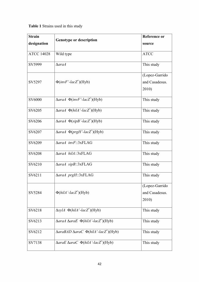
Table 1 Strains used in this study
Strain
designation Genotype or description
Reference or
source
ATCC 14028 Wild type ATCC
SV5999 ΔaraA This study
SV5297 Φ(invF’-lacZ+)(Hyb)
(Lopez-Garrido
and Casadesus.
2010)
SV6000 ΔaraA Φ(invF’-lacZ+)(Hyb) This study
SV6205 ΔaraA Φ(hilA’-lacZ+)(Hyb) This study
SV6206 ΔaraA Φ(sipB’-lacZ+)(Hyb) This study
SV6207 ΔaraA Φ(prgH’-lacZ+)(Hyb) This study
SV6209 ΔaraA invF::3xFLAG This study
SV6208 ΔaraA hilA::3xFLAG This study
SV6210 ΔaraA sipB::3xFLAG This study
SV6211 ΔaraA prgH::3xFLAG This study
SV5284 Φ(hilA’-lacZ+)(Hyb)
(Lopez-Garrido
and Casadesus.
2010)
SV6218 ΔxylA Φ(hilA’-lacZ+)(Hyb) This study
SV6213 ΔaraA ΔaraE Φ(hilA’-lacZ+)(Hyb) This study
SV6212 ΔaraBAD ΔaraC Φ(hilA’-lacZ+)(Hyb) This study
SV7138 ΔaraE ΔaraC Φ(hilA’-lacZ+)(Hyb) This study
43
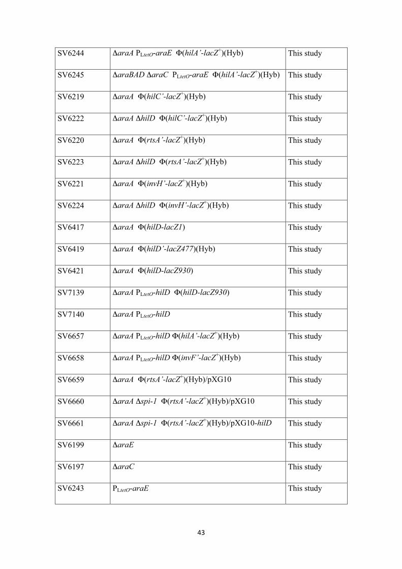
SV6244 ΔaraA PLtetO-araE Φ(hilA’-lacZ+)(Hyb) This study
SV6245 ΔaraBAD ΔaraC PLtetO-araE Φ(hilA’-lacZ+)(Hyb) This study
SV6219 ΔaraA Φ(hilC’-lacZ+)(Hyb) This study
SV6222 ΔaraA ΔhilD Φ(hilC’-lacZ+)(Hyb) This study
SV6220 ΔaraA Φ(rtsA’-lacZ+)(Hyb) This study
SV6223 ΔaraA ΔhilD Φ(rtsA’-lacZ+)(Hyb) This study
SV6221 ΔaraA Φ(invH’-lacZ+)(Hyb) This study
SV6224 ΔaraA ΔhilD Φ(invH’-lacZ+)(Hyb) This study
SV6417 ΔaraA Φ(hilD-lacZ1) This study
SV6419 ΔaraA Φ(hilD’-lacZ477)(Hyb) This study
SV6421 ΔaraA Φ(hilD-lacZ930) This study
SV7139 ΔaraA PLtetO-hilD Φ(hilD-lacZ930) This study
SV7140 ΔaraA PLtetO-hilD This study
SV6657 ΔaraA PLtetO-hilD Φ(hilA’-lacZ+)(Hyb) This study
SV6658 ΔaraA PLtetO-hilD Φ(invF’-lacZ+)(Hyb) This study
SV6659 ΔaraA Φ(rtsA’-lacZ+)(Hyb)/pXG10 This study
SV6660 ΔaraA Δspi-1 Φ(rtsA’-lacZ+)(Hyb)/pXG10 This study
SV6661 ΔaraA Δspi-1 Φ(rtsA’-lacZ+)(Hyb)/pXG10-hilD This study
SV6199 ΔaraE This study
SV6197 ΔaraC This study
SV6243 PLtetO-araE This study

44
SV6201 ΔxylA This study
SV6423 ΔaraA Φ(sipA’-cya+)(Hyb) This study
SV6424 ΔaraA ΔaraE Φ(sipA’-cya+)(Hyb) This study
SV6425 ΔaraA ΔprgH Φ(sipA’-cya+)(Hyb) This study