Embed Size (px)
Citation preview

Creative Education 2013. Vol.4, No.10A, 18-20 Published Online October 2013 in SciRes (http://www.scirp.org/journal/ce) http://dx.doi.org/10.4236/ce.2013.410A003
Copyright © 2013 SciRes. 18
Virtual Learning of the Digestive System: An Experience Developing an Undergraduate Course
Rodolfo E. Avila1, Hugo Juri1, María E. Samar2, Maria T. Mugnaini3, Carlos Soñez3, William Anderson4
1School of Medicine, National University of Córdoba, Córdoba, Argentina 2School of Dentistry, National University of Córdoba, Córdoba, Argentina
3College of Agriculture and Veterinary Medicine, National University of Río Cuarto, Córdoba, Argentina
4John Radcliffe Hospital, Oxford, UK Email: [email protected]
Received July 17th, 2013; revised August 17th, 2013; accepted August 25th, 2013
Copyright © 2013 Rodolfo E. Avila et al. This is an open access article distributed under the Creative Commons Attribution License, which permits unrestricted use, distribution, and reproduction in any medium, provided the original work is properly cited.
Using modern information and communication technology associated with distance education, we set out to produce an e-learning course to improve students’ understanding of the digestive system in an inter- disciplinary manner. To achieve this, we implemented a novel undergraduate teaching project in classes at the Faculties of Agricultural Sciences and Veterinary Medicine of the National University of Rio Cuarto, and Medical Sciences of the National University of Cordoba. In the context of other groups’ experiences we report our development of an undergraduate e-learning course, covering embryology, histology and cell biology in an integrated thematic unit. Keywords: Use of the Internet; Academic Integration Network; Virtual Teaching; Digestive System;
E-Learning
Introduction
Cell biology, histology and embryology are three core sub- jects which are taught early in medicine, dentistry and veter- inary science curricula. Collectively, they provide an essential foundation for the subsequent acquisition of clinical knowledge in each discipline; while conversely, an inability to assimilate such knowledge can lead to high failure and dropout rates in the early years of these degrees.
Teaching of these sciences has traditionally been delivered through classroom-based methods and can thus be limited by time and financial constraints. Increasingly, the availability of online resources and electronic learning or “e-learning”, is greatly facilitating this learning process while circumventing the need for expensive laboratory equipment (Goldberg, 2007; Silva Lópes, 2003).
Using information and communication technology associated with distance education, we set out to produce an e-learning course dedicated to the digestive system, aiming to promote an integrated comprehension. To achieve this, we implemented an undergraduate teaching project at the faculties of Agricultural Sciences and Veterinary Medicine of the National University of Rio Cuarto and also within Medical Sciences of the National University of Cordoba.
In this communication we describe our experience of using the Internet in a network of academic integration: virtual teaching of the digestive system as an integrated thematic unit of embryology, histology and cell biology.
Materials and Methodology
The practical tasks “Virtual Oral Cavity and Digestive Sys- tem” were prepared for the 2010 academic year and uploaded to the URL: http://www.histologiavirtual.com.ar/.
Those who took part as nodes were the Department of Cell Biology and General Embryology at the National University of Rio Cuarto, Argentina (Node 1) and at the National University of Cordoba, the departments of Cell Biology, Histology and Embryology (Node 2) and Medical Computing (Node 3). Images of organs within the digestive system of various species were obtained from departments participating in the network and used for the learning exercises (see Figures 1 and 2).
Image files have JPEG and GIF extensions. Digital images of histological cross-sections were obtained using an image analyzer with the “Image ProPlus” software connected to an Olympus Bx50 photomicroscope. Microsoft Office SharePoint Designer software was used as a designing and programming tool for the development of the website.
Results
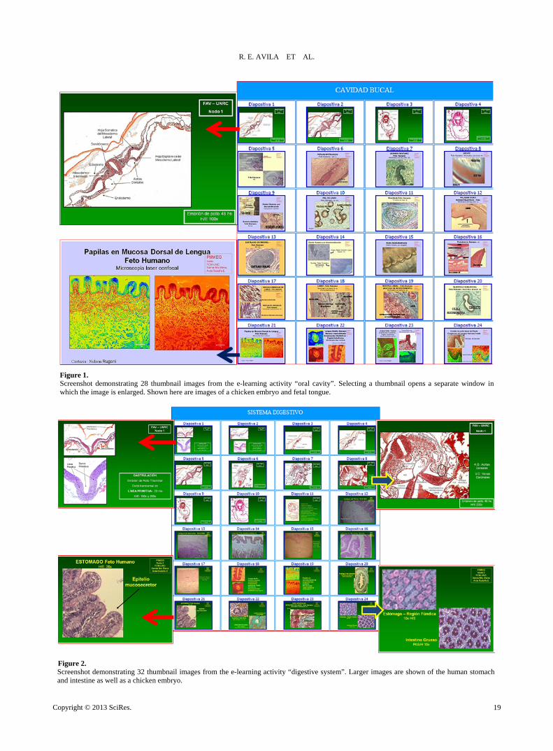
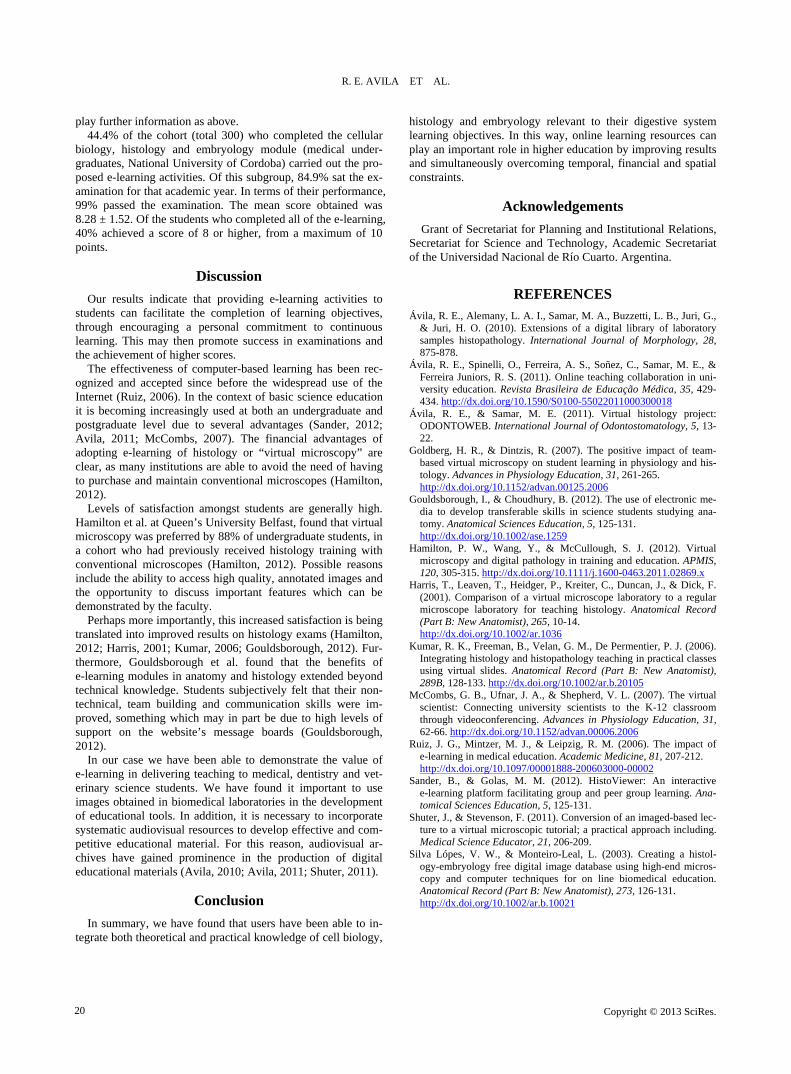
The Virtual Practical Task “Oral Cavity" consisted of a main page of 28 thumbnail images. Individual thumbnails could be selected, in order to present the user with an additional screen displaying an enlarged and fully-labelled image, with accom- panying text outlining the important structures. Similarly, the Virtual Practical Task “Digestive System” contained 32 thumb- nail images on the screen, which could also be selected to dis-
R. E. AVILA ET AL.
Figure 1. Screenshot demonstrating 28 thumbnail images from the e-learning activity “oral cavity”. Selecting a thumbnail opens a separate window in which the image is enlarged. Shown here are images of a chicken embryo and fetal tongue.
Figure 2. Screenshot demonstrating 32 thumbnail images from the e-learning activity “digestive system”. Larger images are shown of the human stomach and intestine as well as a chicken embryo.
Copyright © 2013 SciRes. 19
R. E. AVILA ET AL.
play further information as above.
44.4% of the cohort (total 300) who completed the cellular biology, histology and embryology module (medical under- graduates, National University of Cordoba) carried out the pro- posed e-learning activities. Of this subgroup, 84.9% sat the ex- amination for that academic year. In terms of their performance, 99% passed the examination. The mean score obtained was 8.28 ± 1.52. Of the students who completed all of the e-learning, 40% achieved a score of 8 or higher, from a maximum of 10 points.
Discussion
Our results indicate that providing e-learning activities to students can facilitate the completion of learning objectives, through encouraging a personal commitment to continuous learning. This may then promote success in examinations and the achievement of higher scores.
The effectiveness of computer-based learning has been rec-ognized and accepted since before the widespread use of the Internet (Ruiz, 2006). In the context of basic science education it is becoming increasingly used at both an undergraduate and postgraduate level due to several advantages (Sander, 2012; Avila, 2011; McCombs, 2007). The financial advantages of adopting e-learning of histology or “virtual microscopy” are clear, as many institutions are able to avoid the need of having to purchase and maintain conventional microscopes (Hamilton, 2012).
Levels of satisfaction amongst students are generally high. Hamilton et al. at Queen’s University Belfast, found that virtual microscopy was preferred by 88% of undergraduate students, in a cohort who had previously received histology training with conventional microscopes (Hamilton, 2012). Possible reasons include the ability to access high quality, annotated images and the opportunity to discuss important features which can be demonstrated by the faculty.
Perhaps more importantly, this increased satisfaction is being translated into improved results on histology exams (Hamilton, 2012; Harris, 2001; Kumar, 2006; Gouldsborough, 2012). Fur-thermore, Gouldsborough et al. found that the benefits of e-learning modules in anatomy and histology extended beyond technical knowledge. Students subjectively felt that their non- technical, team building and communication skills were im- proved, something which may in part be due to high levels of support on the website’s message boards (Gouldsborough, 2012).
In our case we have been able to demonstrate the value of e-learning in delivering teaching to medical, dentistry and vet- erinary science students. We have found it important to use images obtained in biomedical laboratories in the development of educational tools. In addition, it is necessary to incorporate systematic audiovisual resources to develop effective and com- petitive educational material. For this reason, audiovisual ar- chives have gained prominence in the production of digital educational materials (Avila, 2010; Avila, 2011; Shuter, 2011).
Conclusion
In summary, we have found that users have been able to in- tegrate both theoretical and practical knowledge of cell biology,
histology and embryology relevant to their digestive system learning objectives. In this way, online learning resources can play an important role in higher education by improving results and simultaneously overcoming temporal, financial and spatial constraints.
Acknowledgements
Grant of Secretariat for Planning and Institutional Relations, Secretariat for Science and Technology, Academic Secretariat of the Universidad Nacional de Río Cuarto. Argentina.
REFERENCES
Ávila, R. E., Alemany, L. A. I., Samar, M. A., Buzzetti, L. B., Juri, G., & Juri, H. O. (2010). Extensions of a digital library of laboratory samples histopathology. International Journal of Morphology, 28, 875-878.
Ávila, R. E., Spinelli, O., Ferreira, A. S., Soñez, C., Samar, M. E., & Ferreira Juniors, R. S. (2011). Online teaching collaboration in uni- versity education. Revista Brasileira de Educação Médica, 35, 429- 434. http://dx.doi.org/10.1590/S0100-55022011000300018
Ávila, R. E., & Samar, M. E. (2011). Virtual histology project: ODONTOWEB. International Journal of Odontostomatology, 5, 13- 22.
Goldberg, H. R., & Dintzis, R. (2007). The positive impact of team- based virtual microscopy on student learning in physiology and his- tology. Advances in Physiology Education, 31, 261-265. http://dx.doi.org/10.1152/advan.00125.2006
Gouldsborough, I., & Choudhury, B. (2012). The use of electronic me- dia to develop transferable skills in science students studying ana- tomy. Anatomical Sciences Education, 5, 125-131. http://dx.doi.org/10.1002/ase.1259
Hamilton, P. W., Wang, Y., & McCullough, S. J. (2012). Virtual microscopy and digital pathology in training and education. APMIS, 120, 305-315. http://dx.doi.org/10.1111/j.1600-0463.2011.02869.x
Harris, T., Leaven, T., Heidger, P., Kreiter, C., Duncan, J., & Dick, F. (2001). Comparison of a virtual microscope laboratory to a regular microscope laboratory for teaching histology. Anatomical Record (Part B: New Anatomist), 265, 10-14. http://dx.doi.org/10.1002/ar.1036
Kumar, R. K., Freeman, B., Velan, G. M., De Permentier, P. J. (2006). Integrating histology and histopathology teaching in practical classes using virtual slides. Anatomical Record (Part B: New Anatomist), 289B, 128-133. http://dx.doi.org/10.1002/ar.b.20105
McCombs, G. B., Ufnar, J. A., & Shepherd, V. L. (2007). The virtual scientist: Connecting university scientists to the K-12 classroom through videoconferencing. Advances in Physiology Education, 31, 62-66. http://dx.doi.org/10.1152/advan.00006.2006
Ruiz, J. G., Mintzer, M. J., & Leipzig, R. M. (2006). The impact of e-learning in medical education. Academic Medicine, 81, 207-212. http://dx.doi.org/10.1097/00001888-200603000-00002
Sander, B., & Golas, M. M. (2012). HistoViewer: An interactive e-learning platform facilitating group and peer group learning. Ana- tomical Sciences Education, 5, 125-131.
Shuter, J., & Stevenson, F. (2011). Conversion of an imaged-based lec- ture to a virtual microscopic tutorial; a practical approach including. Medical Science Educator, 21, 206-209.
Silva Lópes, V. W., & Monteiro-Leal, L. (2003). Creating a histol- ogy-embryology free digital image database using high-end micros- copy and computer techniques for on line biomedical education. Anatomical Record (Part B: New Anatomist), 273, 126-131. http://dx.doi.org/10.1002/ar.b.10021
Copyright © 2013 SciRes. 20