Embed Size (px)
Citation preview
lable at ScienceDirect
European Journal of Medicinal Chemistry 77 (2014) 38e46
Contents lists avai
European Journal of Medicinal Chemistry
journal homepage: http: / /www.elsevier .com/locate/ejmech
Original article
Virtual fragment screening on GPCRs: A case study on dopamine D3and histamine H4 receptors
Márton Vass a, Éva Schmidt a, Ferenc Horti a, György M. Keser}u b,*
aGedeon Richter Plc, H-1475, P.O.B. 27, Budapest, HungarybResearch Centre for Natural Sciences of the Hungarian Academy of Sciences, H-1525, P.O.B. 17, Budapest, Hungary
a r t i c l e i n f o
Article history:Received 9 October 2013Received in revised form11 February 2014Accepted 13 February 2014Available online 15 February 2014
Keywords:Fragment screeningFragment dockingVirtual screeningEnsemble dockingG protein-coupled receptorsDopamine D3 receptorHistamine H4 receptor
Abbreviations: GPCR, G protein-coupled receptordiscovery; LE, ligand efficiency; LELP, ligand-efficieMD, molecular dynamics; IFD, induced fit docoleoylphosphatidylcholine; RMSD, root-mean-squprecision.* Corresponding author.
E-mail addresses: [email protected],M. Keser}u).
http://dx.doi.org/10.1016/j.ejmech.2014.02.0340223-5234/� 2014 Elsevier Masson SAS. All rights re
a b s t r a c t
Prospective structure based virtual fragment screening methodologies on two GPCR targets namely thedopamine D3 and the histamine H4 receptors with a library of 12,905 fragments were evaluated.Fragments were docked to the X-ray structure and the homology model of the D3 and H4 receptors,respectively. Representative receptor conformations for ensemble docking were obtained frommoleculardynamics trajectories. In vitro confirmed hit rates ranged from 16% to 32%. Hits had high ligand efficiency(LE) values in the range of 0.31e0.74 and also acceptable lipophilic efficiency. The X-ray structure, thehomology model and structural ensembles were all found suitable for docking based virtual screening offragments against these GPCRs. However, there was little overlap among different hit sets and meth-odologies were thus complementary to each other.
� 2014 Elsevier Masson SAS. All rights reserved.
1. Introduction
Fragment-based lead discovery (FBLD) has become a feasiblealternative to traditional lead finding approaches in drug discoveryemployed both by industry and academic groups [1]. It has beendemonstrated that starting from polar, low molecular weight e
typically <250 Da or less than 20 heavy atoms e compounds, leadsand drugs with better physico-chemical properties can be achieved[2] even for difficult targets [3]. This view is supported by theincreasing number of drug candidates recently entering clinicaltrials and one already approved drug originating from a fragmenthit [4]. Since weakly binding fragments require sensitive, but typi-cally lower throughput biophysical detection methodologies (suchas SPR, NMR, XRD, MS), and also because fragments are usuallyoptimized using structural information, there is an ongoing interestin computational methodologies capable of predicting fragment
; FBLD, fragment-based leadncy-dependent lipophilicity;king; POPC, 1-palmitoyl-2-are deviation; SP, single
served.
binding and providing reliable binding modes for them. Moleculardocking is an in silico tool aiming to predict the binding mode andbinding free energy of druglike molecules. It has been shown thatvarious docking programs have similar performance in pose pre-diction for fragments (especially for fragments of high ligand effi-ciency) and druglike molecules [5e7], since fragments usuallyexploit the specific interactions available at protein hot spots [8]. Invirtual screening setups, where the objective is the ranking offragments by binding free energy, the modest enrichment of actives[9,10] shall be improved using more accurate binding free energyfunctions. One of these methods is the computationally intensiveMM-PBSA rescoring method that was used to improve enrichments[11,12]. It has also been suggested that incorporating receptorflexibility in docking (not only in the rescoring phase) might bebeneficial for virtual screening enrichments. Various protocols havebeen published taking into account different ranges of proteinflexibility [13]. The simplest approach is the use soft potentials toaccount for small side-chain movements. Larger movements mightbe considered using side chain rotamer libraries. Docking intoappropriately selected multiple protein conformations (ensembledocking) is a parallelizable and resource effective way of handlingthe flexibility of the entire protein. The most computationallyintensive methods attempt the simultaneous conformational sam-pling of the receptor and the ligand, such as Schrödinger’s Induced
M. Vass et al. / European Journal of Medicinal Chemistry 77 (2014) 38e46 39
Fit Docking (IFD) [14] application, or running molecular dynamics(MD) simulation on each individual protein-fragment complex.Different receptor conformations for ensemble docking can be ob-tained from multiple crystal structures or NMR structures if suchdata is available. However, structural studies on membrane pro-teins, such as G protein-coupled receptors (GPCRs) typicallyrepresent great challenges. Despite recent progress in GPCR crys-tallization still only a small percentage of structures have beenunveiled. In such a case homology modeling may be used to obtainan atomistic model of the receptor. Structurally diverse receptormodels can be obtained using different GPCR template structuresduring homology modeling. Diverse conformations may also besampled by MD simulation, Monte Carlo or low-mode conforma-tional search starting from a single homology model [15,16]. DeGraaf et al. used a homology model of the histamine H3 receptorand subsequent MD sampling to provide the conformations usedfor retrospective and prospective virtual fragment screening [17]. Inthe present study we performed prospective virtual fragmentscreening on the available dopamine D3 receptor crystal structureand a homology model of the histamine H4 receptor based on therecently solved histamine H1 receptor crystal structure. Snapshotsfrom all-atom membrane-embedded MD simulations were alsoused for ensemble virtual screening of the same fragment library.Screening performance of the different protocols was comparedanalyzing hit rates and hit compounds obtained by docking to thesingle structure and the conformational ensembles.
2. Computational methods
2.1. Homology modeling and crystal structure preparation
The construction of the histamine H4 receptor homology modelwas described previously [18]. Briefly, the H4 amino acid sequencefrom the UniProt server (http://www.uniprot.org/) was aligned tothe sequence of the template, the 3.1�A resolution X-ray structure ofthe human histamine H1 receptor (PDB code: 3RZE) using Prime 3.0[19]. The kink in helix TM4 was modeled based on the human b2-adrenergic receptor (PDB code: 2RH1). The JNJ7777120 ligand wasfirst manually docked into the receptor, and then the 5 �A envi-ronment of the ligand was subjected to minimization with two H-bond constraints using MacroModel 9.9 [20]. JNJ7777120 was re-docked into the minimized structure using IFD [14,21] in theSchrödinger Suite 2011. Finally the whole structure was subjectedto Impref restrained minimization in the Protein PreparationWizard [22]. Chain A of the dopamine D3 crystal structure (PDBcode: 3PBL) was subjected to the Protein Preparation Wizardworkflow with default settings, that included assigning bond or-ders, adding hydrogens, creating disulfide bonds, optimization ofthe H-bond network and finally a restrained minimization of thecomplex.
2.2. Molecular dynamics simulations and ensemble preparation
The details of molecular dynamics simulations were describedelsewhere [18]. Briefly, all-atom POPC membrane-embedded MDsimulations were run starting from the homology model of thehistamine H4 receptor JNJ7777120 complex and the crystal struc-ture of the dopamine D3 receptor eticlopride complex using ff99SBforce field for protein and GAFF force field for lipid and ligand atomsin the NAMD 2.7 [23] software. The systems were equilibrated withsubsequent steps of i) 3200 steps minimization with restrainedprotein and ligand atoms ii) 3200 steps unrestrained minimizationiii) heating in NVT ensemble to 310 K in 40 ps with restrainedprotein and ligand atoms iv) 1 nsMD simulation in NpzgTensemblewith restrained protein and ligand atoms v) 1 ns MD simulation in
NpzgT ensemble with gradual removal of the restraints. 20 nsproduction runs in NpzgT ensemble were conducted for both sys-tems. Receptor conformations of the two trajectories were clus-tered using the average linkage method in the ptraj program fromthe AmberTools package [24] based on the RMSD of the amino acidresidues that made up 90% cumulative occurrence in the 5 �Aenvironment of the ligand. This method provided 28 representativeconformations for the histamine H4 receptor and 27 representativeconformations for the dopamine D3 receptor. All representativeswere subjected to Impref restrained minimization in the ProteinPreparation Wizard.
2.3. Single structure and ensemble docking methodologies
We collected 12,905 fragment-like compounds from our in-house collection complying with an extended version of the Ruleof Three: having an MW � 300 Da, logP � 3, number of H-bonddonors and acceptors �3, number of rotatable bonds �6,PSA < 130 Å2, containing 1e3 rings and no reactive functionalities(see Property distributions and diversity assessment in theSupporting Information). The structures of these fragments wereprepared using LigPrep 2.5 [25]. The dominant protonation andtautomeric state at pH 7.4 was calculated using Epik 2.2 [26]. TheirlogP was calculated using the ChemAxon cxcalc utility [27]. In thesingle structure investigation the 12,905 fragments were dockedinto the binding site of the dopamine D3 X-ray structure and thehistamine H4 homology model. Then in the ensemble dockingapproach the fragment set was docked into the binding sites of allrepresentatives from the D3 and H4 MD trajectories. Glide 5.7 [28e31] software was used for docking. Grids for the initial homologymodel and crystal structure, as well as for the representatives fromthe MD trajectories were centered on the ligand centroids, and haddimensions of 14 � 14 � 14�A for the inner box (which contains theligand centroid during docking) and 44 � 44 � 44 �A for the outerbox (which contains all ligand atoms during docking) to ensure thatsampling of the binding mode was not biased by the grid size.Docking calculations were conducted using the single precision(SP) mode [5], with post-dock minimization performed for 15poses. Only the top pose for each fragment by the Emodel scoringfunction was saved, which were ranked by the GlideScore scoringfunction for each individual receptor conformation. For singlestructure docking to the D3 X-ray structure and the H4 homologymodel, the top 50 compounds from the GlideScore ranked list werechosen for biological testing. In the ensemble docking approachmean ranks and their standard deviations calculated over theensemble were used for evaluating each individual compound.Compounds having a mean rank lower than 500 were selected forbiological testing. This cutoff gave a similar number of compoundsto be tested as for the single structure case: 56 for the dopamine D3receptor and 50 for the histamine H4 receptor.
3. Results and discussion
3.1. Receptor binding sites
While the histamine H1 and H4 receptors share 40% amino acididentity in the transmembrane region and they recognize the sameendogenous ligand, there are substantial differences in theirbinding sites. For example Asn1474.57 in H4 is equivalent toTrp1584.56 in H1, Leu1755.39 to Lys1915.39, Glu1825.46 to Asn1985.46
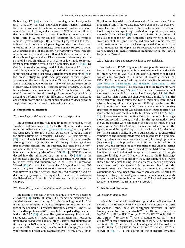
and Gln3477.42 to Gly4577.42. Also, mutation of Asn1474.57 andGlu1825.46 showed significant alteration to JNJ7777120 inhibitionconstants [32]. Thus the initial homology model featured twospecific H-bonds of JNJ7777120 to Asp943.32 and Glu1825.46 asshown in Fig. 1A. In the course of the molecular dynamics
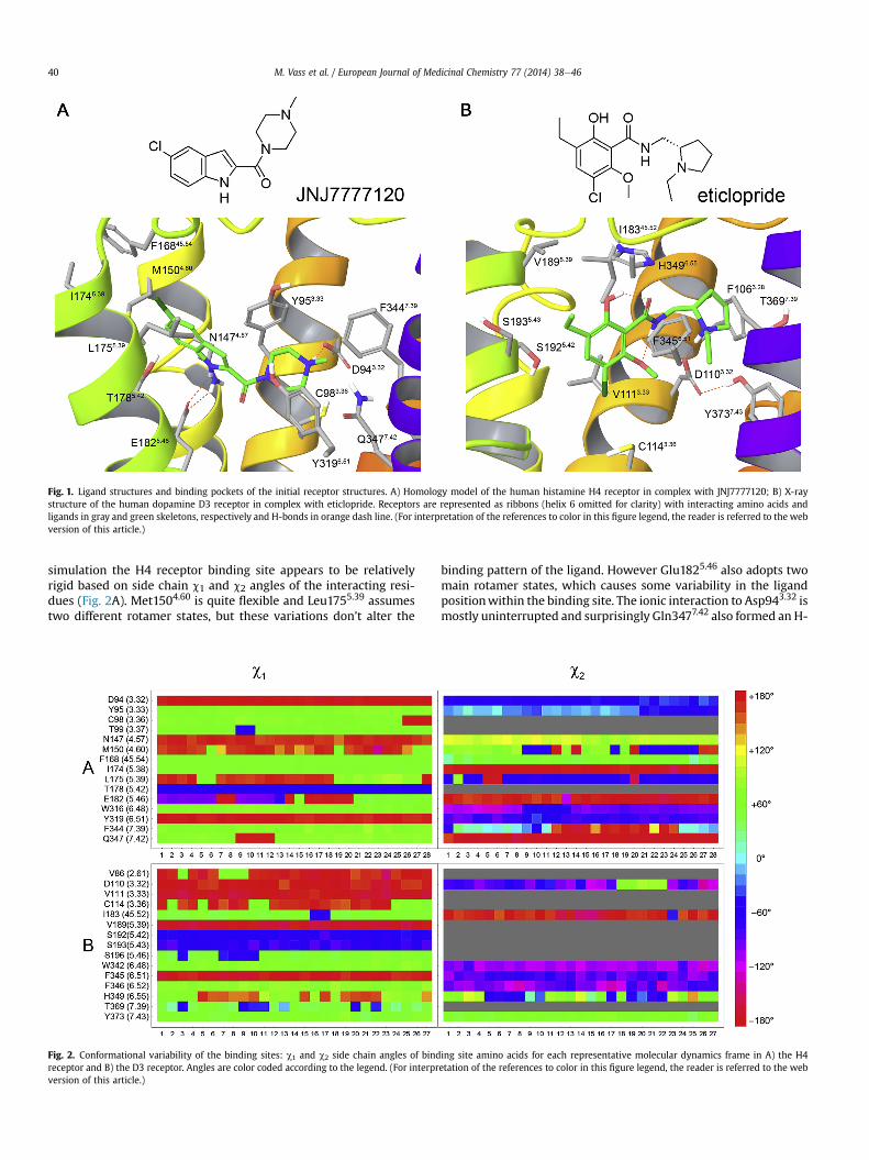
Fig. 1. Ligand structures and binding pockets of the initial receptor structures. A) Homology model of the human histamine H4 receptor in complex with JNJ7777120; B) X-raystructure of the human dopamine D3 receptor in complex with eticlopride. Receptors are represented as ribbons (helix 6 omitted for clarity) with interacting amino acids andligands in gray and green skeletons, respectively and H-bonds in orange dash line. (For interpretation of the references to color in this figure legend, the reader is referred to the webversion of this article.)
M. Vass et al. / European Journal of Medicinal Chemistry 77 (2014) 38e4640
simulation the H4 receptor binding site appears to be relativelyrigid based on side chain c1 and c2 angles of the interacting resi-dues (Fig. 2A). Met1504.60 is quite flexible and Leu1755.39 assumestwo different rotamer states, but these variations don’t alter the
Fig. 2. Conformational variability of the binding sites: c1 and c2 side chain angles of bindreceptor and B) the D3 receptor. Angles are color coded according to the legend. (For interprversion of this article.)
binding pattern of the ligand. However Glu1825.46 also adopts twomain rotamer states, which causes some variability in the ligandpositionwithin the binding site. The ionic interaction to Asp943.32 ismostly uninterrupted and surprisingly Gln3477.42 also formed an H-
ing site amino acids for each representative molecular dynamics frame in A) the H4etation of the references to color in this figure legend, the reader is referred to the web
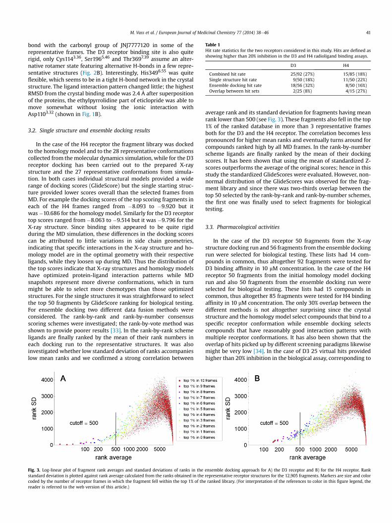
Table 1Hit rate statistics for the two receptors considered in this study. Hits are defined asshowing higher than 20% inhibition in the D3 and H4 radioligand binding assays.
D3 H4
Combined hit rate 25/92 (27%) 15/85 (18%)Single structure hit rate 9/50 (18%) 11/50 (22%)Ensemble docking hit rate 18/56 (32%) 8/50 (16%)Overlap between hit sets 2/25 (8%) 4/15 (27%)
M. Vass et al. / European Journal of Medicinal Chemistry 77 (2014) 38e46 41
bond with the carbonyl group of JNJ7777120 in some of therepresentative frames. The D3 receptor binding site is also quiterigid, only Cys1143.36, Ser1965.46 and Thr3697.39 assume an alter-native rotamer state featuring alternative H-bonds in a few repre-sentative structures (Fig. 2B). Interestingly, His3496.55 was quiteflexible, which seems to be in a tight H-bond network in the crystalstructure. The ligand interaction pattern changed little; the highestRMSD from the crystal binding mode was 2.4�A after superpositionof the proteins, the ethylpyrrolidine part of eticlopride was able tomove somewhat without losing the ionic interaction withAsp1103.32 (shown in Fig. 1B).
3.2. Single structure and ensemble docking results
In the case of the H4 receptor the fragment library was dockedto the homology model and to the 28 representative conformationscollected from themolecular dynamics simulation, while for the D3receptor docking has been carried out to the prepared X-raystructure and the 27 representative conformations from simula-tion. In both cases individual structural models provided a widerange of docking scores (GlideScore) but the single starting struc-ture provided lower scores overall than the selected frames fromMD. For example the docking scores of the top scoring fragments ineach of the H4 frames ranged from �8.093 to �9.920 but itwas�10.686 for the homology model. Similarly for the D3 receptortop scores ranged from �8.063 to �9.514 but it was �9.796 for theX-ray structure. Since binding sites appeared to be quite rigidduring the MD simulation, these differences in the docking scorescan be attributed to little variations in side chain geometries,indicating that specific interactions in the X-ray structure and ho-mology model are in the optimal geometry with their respectiveligands, while they loosen up during MD. Thus the distribution ofthe top scores indicate that X-ray structures and homology modelshave optimized protein-ligand interaction patterns while MDsnapshots represent more diverse conformations, which in turnmight be able to select more chemotypes than those optimizedstructures. For the single structures it was straightforward to selectthe top 50 fragments by GlideScore ranking for biological testing.For ensemble docking two different data fusion methods wereconsidered. The rank-by-rank and rank-by-number consensusscoring schemes were investigated; the rank-by-vote method wasshown to provide poorer results [33]. In the rank-by-rank schemeligands are finally ranked by the mean of their rank numbers ineach docking run to the representative structures. It was alsoinvestigated whether low standard deviation of ranks accompanieslow mean ranks and we confirmed a strong correlation between
Fig. 3. Log-linear plot of fragment rank averages and standard deviations of ranks in thestandard deviation is plotted against rank average calculated from the ranks obtained in thecoded by the number of receptor frames in which the fragment fell within the top 1% of threader is referred to the web version of this article.)
average rank and its standard deviation for fragments having meanrank lower than 500 (see Fig. 3). These fragments also fell in the top1% of the ranked database in more than 3 representative framesboth for the D3 and the H4 receptor. The correlation becomes lesspronounced for higher mean ranks and eventually turns around forcompounds ranked high by all MD frames. In the rank-by-numberscheme ligands are finally ranked by the mean of their dockingscores. It has been shown that using the mean of standardized Z-scores outperforms the average of the original scores; hence in thisstudy the standardized GlideScores were evaluated. However, non-normal distribution of the GlideScores was observed for the frag-ment library and since there was two-thirds overlap between thetop 50 selected by the rank-by-rank and rank-by-number schemes,the first one was finally used to select fragments for biologicaltesting.
3.3. Pharmacological activities
In the case of the D3 receptor 50 fragments from the X-raystructure docking run and 56 fragments from the ensemble dockingrun were selected for biological testing. These lists had 14 com-pounds in common, thus altogether 92 fragments were tested forD3 binding affinity in 10 mM concentration. In the case of the H4receptor 50 fragments from the initial homology model dockingrun and also 50 fragments from the ensemble docking run wereselected for biological testing. These lists had 15 compounds incommon, thus altogether 85 fragments were tested for H4 bindingaffinity in 10 mM concentration. The only 30% overlap between thedifferent methods is not altogether surprising since the crystalstructure and the homology model select compounds that bind to aspecific receptor conformation while ensemble docking selectscompounds that have reasonably good interaction patterns withmultiple receptor conformations. It has also been shown that theoverlap of hits picked up by different screening paradigms likewisemight be very low [34]. In the case of D3 25 virtual hits providedhigher than 20% inhibition in the biological assay, corresponding to
ensemble docking approach for A) the D3 receptor and B) for the H4 receptor. Rankrepresentative receptor structures for the 12,905 fragments. Markers are size and colore ranked library. (For interpretation of the references to color in this figure legend, the
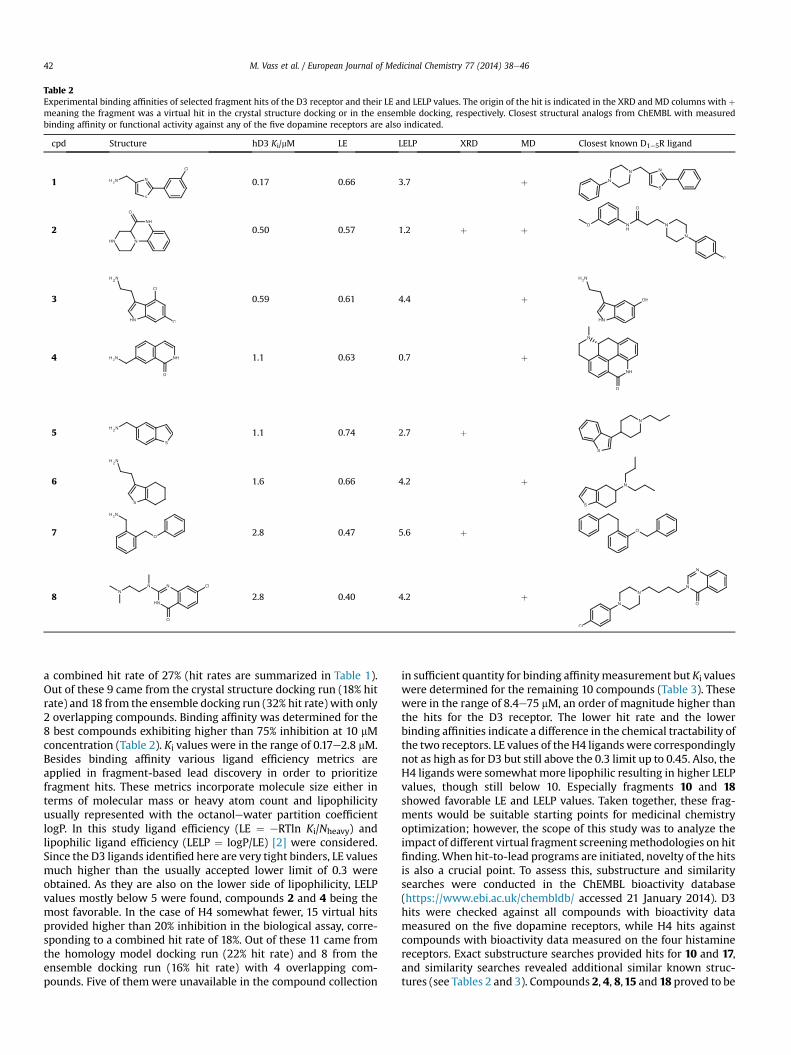
Table 2Experimental binding affinities of selected fragment hits of the D3 receptor and their LE and LELP values. The origin of the hit is indicated in the XRD and MD columns with þmeaning the fragment was a virtual hit in the crystal structure docking or in the ensemble docking, respectively. Closest structural analogs from ChEMBL with measuredbinding affinity or functional activity against any of the five dopamine receptors are also indicated.
cpd Structure hD3 Ki/mM LE LELP XRD MD Closest known D1e5R ligand
1 0.17 0.66 3.7 þ
2 0.50 0.57 1.2 þ þ
3 0.59 0.61 4.4 þ
4 1.1 0.63 0.7 þ
5 1.1 0.74 2.7 þ
6 1.6 0.66 4.2 þ
7 2.8 0.47 5.6 þ
8 2.8 0.40 4.2 þ
M. Vass et al. / European Journal of Medicinal Chemistry 77 (2014) 38e4642
a combined hit rate of 27% (hit rates are summarized in Table 1).Out of these 9 came from the crystal structure docking run (18% hitrate) and 18 from the ensemble docking run (32% hit rate) with only2 overlapping compounds. Binding affinity was determined for the8 best compounds exhibiting higher than 75% inhibition at 10 mMconcentration (Table 2). Ki values were in the range of 0.17e2.8 mM.Besides binding affinity various ligand efficiency metrics areapplied in fragment-based lead discovery in order to prioritizefragment hits. These metrics incorporate molecule size either interms of molecular mass or heavy atom count and lipophilicityusually represented with the octanolewater partition coefficientlogP. In this study ligand efficiency (LE ¼ eRTln Ki/Nheavy) andlipophilic ligand efficiency (LELP ¼ logP/LE) [2] were considered.Since the D3 ligands identified here are very tight binders, LE valuesmuch higher than the usually accepted lower limit of 0.3 wereobtained. As they are also on the lower side of lipophilicity, LELPvalues mostly below 5 were found, compounds 2 and 4 being themost favorable. In the case of H4 somewhat fewer, 15 virtual hitsprovided higher than 20% inhibition in the biological assay, corre-sponding to a combined hit rate of 18%. Out of these 11 came fromthe homology model docking run (22% hit rate) and 8 from theensemble docking run (16% hit rate) with 4 overlapping com-pounds. Five of them were unavailable in the compound collection
in sufficient quantity for binding affinitymeasurement but Ki valueswere determined for the remaining 10 compounds (Table 3). Thesewere in the range of 8.4e75 mM, an order of magnitude higher thanthe hits for the D3 receptor. The lower hit rate and the lowerbinding affinities indicate a difference in the chemical tractability ofthe two receptors. LE values of the H4 ligandswere correspondinglynot as high as for D3 but still above the 0.3 limit up to 0.45. Also, theH4 ligands were somewhat more lipophilic resulting in higher LELPvalues, though still below 10. Especially fragments 10 and 18showed favorable LE and LELP values. Taken together, these frag-ments would be suitable starting points for medicinal chemistryoptimization; however, the scope of this study was to analyze theimpact of different virtual fragment screeningmethodologies on hitfinding.When hit-to-lead programs are initiated, novelty of the hitsis also a crucial point. To assess this, substructure and similaritysearches were conducted in the ChEMBL bioactivity database(https://www.ebi.ac.uk/chembldb/ accessed 21 January 2014). D3hits were checked against all compounds with bioactivity datameasured on the five dopamine receptors, while H4 hits againstcompounds with bioactivity data measured on the four histaminereceptors. Exact substructure searches provided hits for 10 and 17,and similarity searches revealed additional similar known struc-tures (see Tables 2 and 3). Compounds 2, 4, 8,15 and 18 proved to be
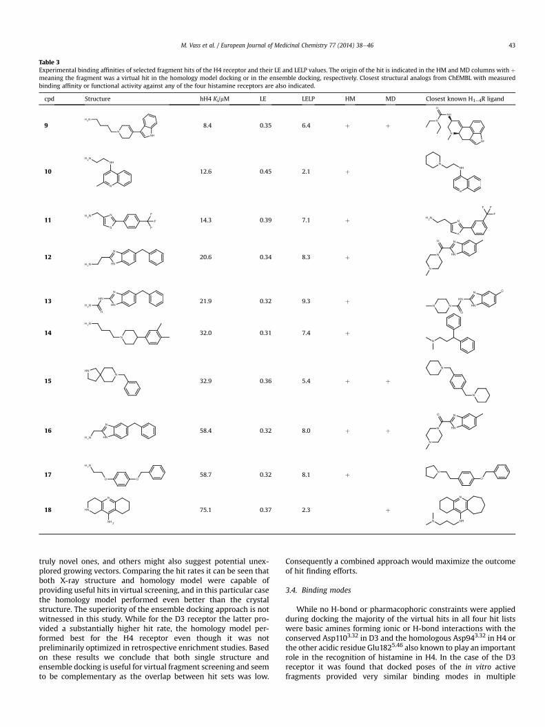
Table 3Experimental binding affinities of selected fragment hits of the H4 receptor and their LE and LELP values. The origin of the hit is indicated in the HM and MD columns with þmeaning the fragment was a virtual hit in the homology model docking or in the ensemble docking, respectively. Closest structural analogs from ChEMBL with measuredbinding affinity or functional activity against any of the four histamine receptors are also indicated.
cpd Structure hH4 Ki/mM LE LELP HM MD Closest known H1e4R ligand
9 8.4 0.35 6.4 þ þ
10 12.6 0.45 2.1 þ
11 14.3 0.39 7.1 þ
12 20.6 0.34 8.3 þ
13 21.9 0.32 9.3 þ
14 32.0 0.31 7.4 þ
15 32.9 0.36 5.4 þ þ
16 58.4 0.32 8.0 þ þ
17 58.7 0.32 8.1 þ
18 75.1 0.37 2.3 þ
M. Vass et al. / European Journal of Medicinal Chemistry 77 (2014) 38e46 43
truly novel ones, and others might also suggest potential unex-plored growing vectors. Comparing the hit rates it can be seen thatboth X-ray structure and homology model were capable ofproviding useful hits in virtual screening, and in this particular casethe homology model performed even better than the crystalstructure. The superiority of the ensemble docking approach is notwitnessed in this study. While for the D3 receptor the latter pro-vided a substantially higher hit rate, the homology model per-formed best for the H4 receptor even though it was notpreliminarily optimized in retrospective enrichment studies. Basedon these results we conclude that both single structure andensemble docking is useful for virtual fragment screening and seemto be complementary as the overlap between hit sets was low.
Consequently a combined approach would maximize the outcomeof hit finding efforts.
3.4. Binding modes
While no H-bond or pharmacophoric constraints were appliedduring docking the majority of the virtual hits in all four hit listswere basic amines forming ionic or H-bond interactions with theconserved Asp1103.32 in D3 and the homologous Asp943.32 in H4 orthe other acidic residue Glu1825.46 also known to play an importantrole in the recognition of histamine in H4. In the case of the D3receptor it was found that docked poses of the in vitro activefragments provided very similar binding modes in multiple
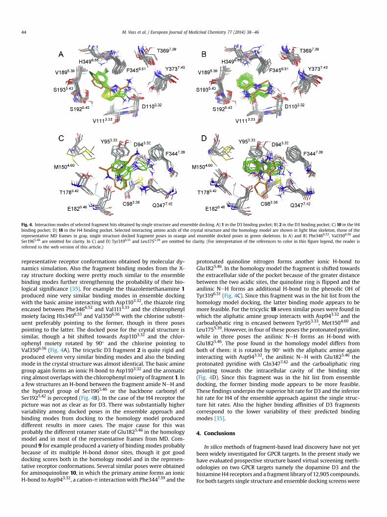
Fig. 4. Interaction modes of selected fragment hits obtained by single structure and ensemble docking. A) 1 in the D3 binding pocket; B) 2 in the D3 binding pocket; C) 10 in the H4binding pocket; D) 18 in the H4 binding pocket. Selected interacting amino acids of the crystal structure and the homology model are shown in light blue skeleton, those of therepresentative MD frames in gray, single structure docked fragment poses in orange and ensemble docked poses in green skeletons. In A) and B) Phe3466.52, Val3506.56 andSer1965.46 are omitted for clarity. In C) and D) Tyr3196.51 and Leu1755.39 are omitted for clarity. (For interpretation of the references to color in this figure legend, the reader isreferred to the web version of this article.)
M. Vass et al. / European Journal of Medicinal Chemistry 77 (2014) 38e4644
representative receptor conformations obtained by molecular dy-namics simulation. Also the fragment binding modes from the X-ray structure docking were pretty much similar to the ensemblebinding modes further strengthening the probability of their bio-logical significance [35]. For example the thiazolemethanamine 1produced nine very similar binding modes in ensemble dockingwith the basic amine interacting with Asp1103.32, the thiazole ringencased between Phe3466.52 and Val1113.33 and the chlorophenylmoiety facing His3496.55 and Val3506.56 with the chlorine substit-uent preferably pointing to the former, though in three posespointing to the latter. The docked pose for the crystal structure issimilar, though a bit shifted towards Asp1103.32 and the chlor-ophenyl moiety rotated by 90� and the chlorine pointing toVal3506.56 (Fig. 4A). The tricyclic D3 fragment 2 is quite rigid andproduced eleven very similar binding modes and also the bindingmode in the crystal structure was almost identical. The basic aminegroup again forms an ionic H-bond to Asp1103.32 and the aromaticring almost overlapswith the chlorophenyl moiety of fragment 1. Ina few structures an H-bond between the fragment amide NeH andthe hydroxyl group of Ser1965.46 or the backbone carbonyl ofSer1925.42 is percepted (Fig. 4B). In the case of the H4 receptor thepicture was not as clear as for D3. There was substantially highervariability among docked poses in the ensemble approach andbinding modes from docking to the homology model produceddifferent results in more cases. The major cause for this wasprobably the different rotamer state of Glu1825.46 in the homologymodel and in most of the representative frames from MD. Com-pound 9 for example produced a variety of binding modes probablybecause of its multiple H-bond donor sites, though it got gooddocking scores both in the homology model and in the represen-tative receptor conformations. Several similar poses were obtainedfor aminoquinoline 10, in which the primary amine forms an ionicH-bond to Asp943.32, a cation-p interactionwith Phe3447.39 and the
protonated quinoline nitrogen forms another ionic H-bond toGlu1825.46. In the homology model the fragment is shifted towardsthe extracellular side of the pocket because of the greater distancebetween the two acidic sites, the quinoline ring is flipped and theanilinic NeH forms an additional H-bond to the phenolic OH ofTyr3196.51 (Fig. 4C). Since this fragment was in the hit list from thehomology model docking, the latter binding mode appears to bemore feasible. For the tricyclic 18 seven similar poses were found inwhich the aliphatic amine group interacts with Asp943.32 and thecarboaliphatic ring is encased between Tyr953.33, Met1504.60 andLeu1755.39. However, in four of these poses the protonated pyridine,while in three poses the anilinic NeH forms an H-bond withGlu1825.46. The pose found in the homology model differs fromboth of them: it is rotated by 90� with the aliphatic amine againinteracting with Asp943.32, the anilinic NeH with Glu1825.46 theprotonated pyridine with Gln3477.42 and the carboaliphatic ringpointing towards the intracellular cavity of the binding site(Fig. 4D). Since this fragment was in the hit list from ensembledocking, the former binding mode appears to be more feasible.These findings underpin the superior hit rate for D3 and the inferiorhit rate for H4 of the ensemble approach against the single struc-ture hit rates. Also the higher binding affinities of D3 fragmentscorrespond to the lower variability of their predicted bindingmodes [35].
4. Conclusions
In silico methods of fragment-based lead discovery have not yetbeen widely investigated for GPCR targets. In the present study wehave evaluated prospective structure based virtual screening meth-odologies on two GPCR targets namely the dopamine D3 and thehistamineH4 receptors and a fragment library of 12,905 compounds.For both targets single structure and ensemble docking screenswere
M. Vass et al. / European Journal of Medicinal Chemistry 77 (2014) 38e46 45
performed. For the D3 receptor the X-ray structure with eticlopridewas available for the single structure screen, while a previouslyconstructed H1 receptor based homology model of H4 was utilized.Representative receptor conformations for ensemble docking weregeneratedbymoleculardynamics simulations. Around50virtual hitsfrom both methodologies for both receptors were measured in vitroand with a greater than 20% inhibition at 10 mM criterion confirmedhit rates ranged from 16% to 32%. The reported hits provided high LEand low LELP values and are suitable starting points for hit-to-leadoptimization. Analysis of the obtained binding modes providedinsight to the variation in hit rates of the different methodologies. Itwas found that the X-ray structure, the homology model and struc-tural ensembles are all suitable fordocking based virtual screening offragments against these GPCRs. However, there was little overlapamong their hit sets and were thus complementary to each other.Combined approaches should provide valuable starting points forfragment-based lead discovery for other GPCRs as well if an X-raystructure or a good quality homology model is available.
5. Experimental
5.1. Human recombinant D3 binding assay
Cell cultures (CHOeK1) expressing human D3 receptors (pur-chased from HD Euroscreen Fast, Belgium) were homogenized inbuffer solution (composition: 15 mM Tris, 2 mM MgCl2, 0.3 mMEDTA, 1 mM EGTA, pH ¼ 7.4 at 25 �C) in 4� v/w with a Douncetissue grinder and centrifuged at 40,000 g at 4 �C for 25 min. Thesupernatant was removed and the pellet was resuspended in 4� v/w buffer and recentrifuged. This process was repeated twice moreand the pellet was resuspended in buffer (composition: 75mM Tris,12.5 mM MgCl2, 0.3 mM EDTA, 1 mM EGTA, 250 mM Sucrose,pH ¼ 7.4 at 25 �C) at a volume of 12.5 mL/g original weight. Thepreparations were then aliquoted and stored at �70 �C.
The aliquotedmembranewas thawedandwashedonce inbindingbuffer containing 50 mM TriseHCl; 5 mM MgCl2, 5 mM KCl; 1 mMCaCl2, 120 mM NaCl, 1 mM EDTA. In the same buffer 3.3 mg protein/assay was incubated with 2 nM [3H]raclopride in the presence orabsence of test compound (to determine the binding inhibition of thetest compound or the total binding, respectively) for 120min at 25 �Cat avolume of 250 mL in 96DeepWell plate. Non-specific bindingwasdetermined in the presence of 10 mM haloperidol. After incubation,samples were filtered over UniFilter GF/B� using PerkinElmerHarvester and washed with 4 � 1 mL ice-cold binding buffer. Theplatewas dried at 40 �C for an hour and 40 mLMicroscint scintillationcocktail (PerkinElmer) was added to eachwell. The radioactivity wasdetermined in MicroBeta 2450 microplate counter (PerkinElmer).
SEMwas lower than 15% for single concentrationmeasurementsand lower than 7% for the hits. The ligand displacement experi-ments were repeated at least two times. The specific radioligandbinding is defined as the difference between total binding and thenon-specific binding determined in the presence of an excessamount of haloperidol. IC50 values (i.e. concentration of compoundgiving 50% inhibition of specific binding) were determined fromconcentrationedisplacement curves by sigmoidal fitting. The in-hibition constants (Ki) were calculated using the ChengePrusoffequation: Ki ¼ IC50/[1 þ (L/KD)], where [L] is the free radioligandconcentration and KD the affinity of the labeled ligand for receptor.KD was determined from the Scatchard plot. GraFit 6.0 (ErithracusSoftware, Horley, UK) software was used for curve fittings.
5.2. Human recombinant H4 binding assay
Membranes from CHO-K1 cells expressing human histamine H4receptors were purchased from PerkinElmer Life and Analytical
Sciences (Cat. No. ES-393-M400UA). Frozen membrane aliquotswere thawed at room temperature and diluted to 200-fold (15 mgprotein/500 mL diluted membrane/well) with binding buffer(50 mM TRISeHCl pH 7.4, 5 mM EDTA).
The assay was performed according to the PerkinElmer assayprotocol for human H4 receptor: 500 mL diluted membrane sus-pension (15 mg protein/assay) was incubated with [3H]histamine asradioligand. Final reaction volume was 550 mL and final radioligandconcentration was 4e7 nM. 10 mM histamine was used for deter-mination of non-specific binding. The samples were incubated at27 �C for 30 min and binding was terminated by vacuum filtrationthroughWhatmanGF/B glass fiber filters, pre-soaked in 0.5% PEI. Thefilters were washed 3-times with 4mL ice cold binding buffer. Filterswere transferred to vials, 4 mL Optiphase HiSafe scintillation cocktail(PerkinElmer) was added and radioactivity was determined byPackard TriCarb 2900 TR (PerkinElmer) liquid scintillation counter.
SEMwas lower than 15% for single concentrationmeasurementsand lower than 7% for the hits. The ligand displacement by thecompounds was determined using a minimum of six concentra-tions in duplicate or triplicate, and experiments were repeated atleast two times. The specific radioligand binding is defined as thedifference between total binding and the non-specific bindingdetermined in the presence of an excess of unlabeled ligand. IC50values (i.e. concentration of compound giving 50% inhibition ofspecific binding) were determined from concentrationedisplace-ment curves by sigmoidal fitting using Prism Software 4.0(GraphPad, San Diego, CA, U.S.A.). Ki values (i.e. inhibition con-stants) were calculated using the ChengePrusoff equation:Ki ¼ IC50/[1 þ (L/KD)], where [L] is the free radioligand concentra-tion and KD the affinity of the labeled ligand for receptor. KD wasdetermined from the Scatchard plot.
Acknowledgments
The authors are thankful to Ákos Tarcsay for valuable discus-sions, Balázs Jójárt, Gábor Paragi and Ferenc Bogár for the MDsimulation and subsequent clustering. This work was supported byCOST Action CM1207.
Appendix A. Supplementary data
Supplementary data related to this article can be found at http://dx.doi.org/10.1016/j.ejmech.2014.02.034.
References
[1] M. Baker, Fragment-based lead discovery grows up, Nature Reviews DrugDiscovery 12 (2013) 5e7.
[2] G.G. Ferenczy, G.M. Keser&udblac, How are fragments optimized? A retro-spective analysis of 145 fragment optimizations, Journal of Medicinal Chem-istry 56 (2013) 2478e2486.
[3] A. Stamford, C. Strickland, Inhibitors of BACE for treating Alzheimer’s disease:a fragment-based drug discovery story, Current Opinion in Chemical Biology17 (2013) 320e328.
[4] J. Tsai, J.T. Lee, W. Wang, J. Zhang, H. Cho, S. Mamo, R. Bremer, S. Gillette,J. Kong, N.K. Haass, K. Sproesser, L. Li, K.S. Smalley, D. Fong, Y.L. Zhu,A. Marimuthu, H. Nguyen, B. Lam, J. Liu, I. Cheung, J. Rice, Y. Suzuki, C. Luu,C. Settachatgul, R. Shellooe, J. Cantwell, S.H. Kim, J. Schlessinger, K.Y. Zhang,B.L. West, B. Powell, G. Habets, C. Zhang, P.N. Ibrahim, P. Hirth, D.R. Artis,M. Herlyn, G. Bollag, Discovery of a selective inhibitor of oncogenic B-Raf ki-nase with potent antimelanoma activity, Proceedings of the National Academyof Sciences of the United States of America 105 (2008) 3041e3046.
[5] M. Sándor, R. Kiss, G.M. Keser&udblac, Virtual fragment docking by Glide: avalidation study on 190 protein-fragment complexes, Journal of ChemicalInformation and Modeling 50 (2010) 1165e1172.
[6] M.L. Verdonk, I. Giangreco, R.J. Hall, O. Korb, P.N. Mortenson, C.W. Murray,Docking performance of fragments and druglike compounds, Journal of Me-dicinal Chemistry 54 (2011) 5422e5431.
[7] M. Vass, G.M. Keser}u, Fragments to link. A multiple docking strategy forsecond site binders, Medicinal Chemistry Communications 4 (2013) 510e514.
M. Vass et al. / European Journal of Medicinal Chemistry 77 (2014) 38e4646
[8] G.G. Ferenczy, G.M. Keser&udblac, Thermodynamics of fragment binding,Journal of Chemical Information and Modeling 52 (2012) 1039e1045.
[9] S. Kawatkar, H. Wang, R. Czerminski, D. Joseph-McCarthy, Virtual fragmentscreening: an exploration of various docking and scoring protocols for frag-ments using Glide, Journal of Computer-Aided Molecular Design 23 (2009)527e539.
[10] A. Kumar, K.Y. Zhang, Computational fragment-based screening using Roset-taLigand: the SAMPL3 challenge, Journal of Computer-Aided Molecular Design26 (2012) 603e616.
[11] S. Kawatkar, D. Moustakas, M. Miller, D. Joseph-McCarthy, Virtual fragmentscreening: exploration of MM-PBSA re-scoring, Journal of Computer-AidedMolecular Design 26 (2012) 921e934.
[12] T. Zhu, H. Lee, H. Lei, C. Jones, K. Patel, M.E. Johnson, K.E. Hevener, Fragment-based drug discovery using a multidomain, parallel MD-MM/PBSA screeningprotocol, Journal of Chemical Information and Modeling 53 (2013) 560e572.
[13] C.B. Rao, J. Subramanian, S.D. Sharma, Managing protein flexibility in dockingand its applications, Drug. Discovery Today 14 (2009) 394e400.
[14] W. Sherman, T. Day, M.P. Jacobson, R.A. Friesner, R. Farid, Novel procedure formodeling ligand/receptor induced fit effects, Journal of Medicinal Chemistry49 (2006) 534e553.
[15] D.J. Osguthorpe, W. Sherman, A.T. Hagler, Exploring protein flexibility:incorporating structural ensembles from crystal structures and simulationinto virtual screening protocols, Journal of Physical Chemistry B 116 (2012)6952e6959.
[16] C.N. Cavasotto, J.A. Kovacs, R.A. Abagyan, Representing receptor flexibility inligand docking through relevant normal modes, Journal of the AmericanChemical Society 127 (2005) 9632e9640.
[17] F. Sirci, E.P. Istyastono, H.F. Vischer, A.J. Kooistra, S. Nijmeijer, M. Kuijer,M. Wijtmans, R. Mannhold, R. Leurs, I.J. de Esch, C. de Graaf, Virtual fragmentscreening: discovery of histamine H3 receptor ligands using ligand-based andprotein-based molecular fingerprints, Journal of Chemical Information andModeling 52 (2012) 3308e3324.
[18] A. Tarcsay, G. Paragi, M. Vass, B. Jójárt, F. Bogár, G.M. Keser&udblac, Theimpact of molecular dynamics sampling on the performance of virtualscreening against GPCRs, Journal of Chemical Information and Modeling 53(2013) 2990e2999.
[19] Prime, Version 3.0, Schrödinger, LLC, New York, NY, 2012.[20] MacroModel, Version 9.9, Schrödinger, LLC, New York, NY, 2012.[21] Schrödinger Suite 2011 Induced Fit Docking Protocol; Glide Version 5.7,
Schrödinger, LLC, New York, NY, 2011. Prime version 3.0, Schrödinger, LLC,New York, NY, 2011.
[22] Schrödinger Suite 2011 Schrödinger Suite; Epik Version 2.2, Schrödinger, LLC,New York, NY, 2011. Impact version 5.7, Schrödinger, LLC, New York, NY,2011; Prime version 2.3, Schrödinger, LLC, New York, NY, 2011.
[23] J.C. Phillips, R. Braun, W. Wang, J. Gumbart, E. Tajkhorshid, E. Villa, C. Chipot,R.D. Skeel, L. Kalé, K. Schulten, Scalable molecular dynamics with NAMD,Journal of Computational Chemistry 26 (2005) 1781e1802.
[24] D.A. Case, T.E. Cheatham, T. Darden, H. Gohlke, R. Luo, K.M. Merz, A. Onufriev,C. Simmerling, B. Wang, R.J. Woods, The Amber biomolecular simulationprograms, JJournal of Computational Chemistry 26 (2005) 1668e1688.
[25] LigPrep, Version 2.5, Schrödinger, LLC, New York, NY, 2011.[26] Epik, Version 2.2, Schrödinger, LLC, New York, NY, 2011.[27] Calculator, Version 5.10.2, �, ChemAxon Ltd, 1998-2012.[28] Glide, Version 5.7, Schrödinger, LLC, New York, NY, 2011.[29] R.A. Friesner, J.L. Banks, R.B. Murphy, T.A. Halgren, J.J. Klicic, D.T. Mainz,
M.P. Repasky, E.H. Knoll, D.E. Shaw, M. Shelley, J.K. Perry, P. Francis,P.S. Shenkin, Glide: a new approach for rapid, accurate docking and scoring. 1.Method and assessment of docking accuracy, Journal of Medicinal Chemistry47 (2004) 1739e1749.
[30] T.A. Halgren, R.B. Murphy, R.A. Friesner, H.S. Beard, L.L. Frye, W.T. Pollard,J.L. Banks, Glide: a new approach for rapid, accurate docking and scoring. 2.enrichment factors in database screening, Journal of Medicinal Chemistry 47(2004) 1750e1759.
[31] R.A. Friesner, R.B. Murphy, M.P. Repasky, L.L. Frye, J.R. Greenwood,T.A. Halgren, P.C. Sanschagrin, D.T. Mainz, Extra precision Glide: docking andscoring incorporating a model of hydrophobic enclosure for protein-ligandcomplexes, Journal of Medicinal Chemistry 49 (2006) 6177e6196.
[32] H.D. Lim, C. de Graaf, W. Jiang, P. Sadek, P.M. McGovern, E.P. Istyastono,R.A. Bakker, I.J. de Esch, R.L. Thurmond, R. Leurs, Molecular determinants ofligand binding to H4R species variants, Molecular Pharmacology 77 (2010)734e743.
[33] R. Wang, S. Wang, How does consensus scoring work for virtual libraryscreening? An idealized computer experiment, Journal of Chemical Informa-tion and Computer Science 41 (2001) 1422e1426.
[34] J. Wielens, S.J. Headey, D.I. Rhodes, R.J. Mulder, O. Dolezal, J.J. Deadman,J. Newman, D.K. Chalmers, M.W. Parker, T.S. Peat, M.J. Scanlon, Parallelscreening of low molecular weight fragment libraries: do differences inmethodology affect hit identification? Journal of Biomolecular Screening 18(2013) 147e159.
[35] M. Orita, K. Ohno, M. Warizaya, Y. Amano, T. Niimi, Lead generation and ex-amples opinion regarding how to follow up hits, Methods in Enzymology 493(2011) 383e419.