Embed Size (px)
Citation preview
BioMed CentralVirology Journal
ss
Open AcceResearchThe role of NH4Cl and cysteine proteases in Human Papillomavirus type 16 infectionSarah A Dabydeen*†1 and Patricio I Meneses†1,2Address: 1Department of Microbiology and Immunology, H.M. Bligh Cancer Research Laboratory, School of Graduate and Postdoctoral Studies, Rosalind Franklin University of Medicine and Science, North Chicago, IL, USA and 2Department of Microbiology and Immunology, H.M. Bligh Cancer Research Laboratory, Chicago Medical School, Rosalind Franklin University of Medicine and Science, North Chicago, IL, USA
Email: Sarah A Dabydeen* - [email protected]; Patricio I Meneses - [email protected]
* Corresponding author †Equal contributors
AbstractBackground: The infectious pathway of the non-enveloped Human Papillomavirus Type 16(HPV16) includes binding to the cell surface, clathrin-mediated endocytosis, and penetration intoan endosome. HPV16 infection was shown to decrease in the presence of the lysosomotrophicneutralizing agent ammonium chloride (NH4Cl). NH4Cl neutralizes acidic endo-lysosomecompartments, thus suggesting that pH was responsible for PV capsid conformational changesleading endosome escape.
Results: However, our data suggested that NH4Cl blocked infection by preventing the movementof PV viral particles from the early endosome to the caveosome as was shown for JC virus [1,2].We have confirmed that HPV 16 infection requires the trafficking of reporter-virions to thecaveosome as is the case for BPV1 [3,4]. In this manuscript we propose that the observed decreasein infection of PV in the presence of NH4Cl was due to a loss of movement of reporter-virions tocaveosomes. We also demonstrate that cysteine proteases are involved in the infectious process,and that cathepsin B treatment of viral particles was shown to overcome the block of infectionobserved in the presence of furin inhibition. We confirmed the need for cathepsin B in HPV16infection using cathepsin B null mouse embryonic fibroblasts.
Conclusion: We present data that suggest HPV16 infection is in part mediated by cysteineproteases, and that NH4Cl blocks the intracellular trafficking of infectious viral particles. To ourknowledge this is the first demonstration that cysteine proteases influence the infection of a non-enveloped virus.
BackgroundHuman Papillomaviruses (HPVs) are non-envelopedDNA viruses that can infect the skin and mucous mem-branes. HPVs are known to cause cutaneous, cervical, andrespiratory warts and lesions [5-7]. The capsid of HPVs ismade of two virally encoded structural proteins L1 and L2[8-10]. The major capsid protein L1 is primarily involved
in attachment of the virus to the plasma membrane, whilethe minor capsid protein L2 functions in viral genometrafficking and encapsidation [11-15].
The infectious process begins via virion attachment to thecell surface through breaks in the skin. Although the vir-ion-cell binding process is still unclear it is thought to
Published: 20 July 2009
Virology Journal 2009, 6:109 doi:10.1186/1743-422X-6-109
Received: 20 April 2009Accepted: 20 July 2009
This article is available from: http://www.virologyj.com/content/6/1/109
© 2009 Dabydeen and Meneses; licensee BioMed Central Ltd. This is an Open Access article distributed under the terms of the Creative Commons Attribution License (http://creativecommons.org/licenses/by/2.0), which permits unrestricted use, distribution, and reproduction in any medium, provided the original work is properly cited.
Page 1 of 12(page number not for citation purposes)
Virology Journal 2009, 6:109 http://www.virologyj.com/content/6/1/109
occur by initial binding of the L1 protein on the virioncapsid to heparan sulfate (a cell surface proteoglycan), fol-lowed by binding to a secondary receptor, putatively anintegrin complex [16-18]. α6β4 has been shown to beable to mediate cell binding in studies showing that anti-bodies against α6 could block virion binding to the epi-thelial cells CV-1 and HaCaT keratinocytes [19]. However,α6β4 integrin may not be a necessary requirement forinfection since studies also indicate that some PVs caninfect cells such as BO-SV keratinocytes that lack this com-plex [20]. After attachment to the cell surface the HPV16virion is internalized via a mechanism that begins withclathrin mediated endocytosis [2,21,22]. N-terminuscleavage of L2 by furin, a calcium dependent serine endo-protease found at the plasma membrane, Golgi and endo-somes, has been suggested to be required for infection[23,24]. Our data suggests that after trafficking to theendosome, the reporter-virions may follow either aninfectious route or a noninfectious route ([3,4]). In theinfectious route, reporter-virions are moved to the caveo-lin-1 intracellular sorting pathway. This caveolin-1 path-way was shown to be necessary for infection, as infectionis blocked in cells where caveolin-1 protein levels werereduced using siRNA against caveolin-1 ([3,4]). Afterentering the caveosome, the virion was shown to traffic inan L2-mediated event to a region where it colocalized withthe endoplasmic reticulum (ER) t-SNARE syntaxin 18 andthe ER chaperone calnexin and ERp29 ([3,4,11,14]). Thenon-infectious pathway results in trafficking from theendosome to the lysosome where reporter-virions may beprocessed for degradation by the cell. This latter pathwaywas shown using a non-infectious L2 mutant virus andneutralizing antibodies [3]. It has been shown thatammonium chloride (NH4Cl) blocks infection of BovinePapillomavirus Type 1 (BPV1), a PV with similar kineticsto HPV16 [2]. NH4Cl neutralized the acidic endo-lyso-some compartments suggesting that pH was responsiblefor PV capsid conformational changes leading to viralgenome release. However, our data presented in this man-uscript suggested that ammonium chloride blocked infec-tion by preventing the movement of viral particles fromthe early endosome to the caveosome as was also shownfor JC virus [1]. In this manuscript we show that cysteineproteases and not pH may be responsible for changesleading to infection.
Cysteine proteases function as intracellular and extracellu-lar molecules [25]. The cysteine protease cathepsin B isassociated with caveolae. Caveolae are defined as smallinvaginations of the plasma membrane associated withlipid rafts that contain caveolin-1 [26,27]. Similar to cave-olae, endo-lysosomal compartments within cells containcathepsin B but in addition have cathepsin L. Both ofthese cathepsins are zymogens (pro-forms) that arecleaved into their active form [28,29]. The exact mecha-
nism of activation is not well understood however, activa-tion of pro-cathepsin B may occur by S100A10, a proteinfound in caveolae, while activation of pro-cathepsin Lmay occur by heparan sulfate, a possible receptor for PV[27,30]. In addition to caveolae and endosomes, cathep-sins have been found to be associated with lipid rafts, sug-gesting that cathepsins may be needed uponinternalization to break apart matrices on viral surfaces[25].
Cathepsins B and L have been implicated in the mecha-nism of binding, entry and disassembly of several envel-oped viruses. In the case of binding, treating Ebolavirusreporter-virions with cathepsin L enhanced infectivity bycleaving and removing a highly glycosylated mucindomain in the Ebolavirus glycoprotein and resulted inincreased binding suggesting that cathepsins are indeedpresent on the cell membrane [31]. Fusion of envelopedviruses such as Nipah, Hendra, SARS Coronavirus andMurine Coronavirus Mouse Hepatitis Virus has beenshown to be dependent on cathepsins B and L. In Nipahvirus, both cathepsin B and L were shown to cleave amembrane fusion protein required for virus-cell and cell-cell membrane fusion. Cleavage of the viral membranefusion protein into the correct size was necessary for mat-uration into a fusogenic form. Cathepsin B was shown tocleave the fusion protein in a cell-free system into twofragments but the smaller of these fragments migratedslower than fragments produced during the cleavage thatoccurs in infection, suggesting that the fusion protein wasnot cleaved at the correct size. Cathepsin L was able tocleave the fusion protein into fragments of the correct sizein the cell-free system suggesting that although cathepsinB and L are catalytically similar, they may have distinct tar-get/sequence specificity [32]. Similar to Nipah virus,cathepsin L was involved in cleaving the Hendra virusfusion protein into an active heterodimer [33]. In SARSCoronavirus infection, cathepsin L was needed to cleavethe spike protein, one of four major structural proteins,into two subunits: one having a high binding affinity tothe receptor and the other mediating fusion of viral andcellular membranes [34]. The requirement of cathepsinsfor fusion was also shown in Murine Coronavirus MouseHepatitis Virus (MHV), where proteolysis by cathepsin Band L was necessary for cleavage of the MHV-2 spike pro-tein [35]. In addition, proteolysis by cathepsins wasshown to be important for disassembly of Reovirus. Usingmouse embryonic fibroblasts derived from cathepsin B orL deficient and wild type mice, studies show that Reovirusdisassembly was prohibited in the absence of cathepsins Band L [36].
In this manuscript, we show a role for cathepsin B thatmay be important for HPV16 infection. It is, to our knowl-
Page 2 of 12(page number not for citation purposes)
Virology Journal 2009, 6:109 http://www.virologyj.com/content/6/1/109
edge, the first description of the role of cysteine proteaseson a non-enveloped virion.
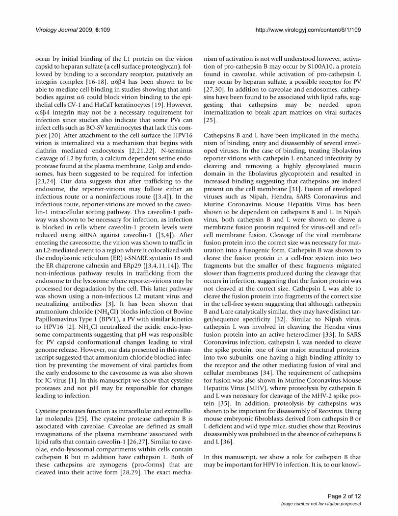
ResultsCysteine protease inhibitors inhibit HPV16 infectionTo determine the role of cysteine proteases on HPV16infection, we performed infections of 293 cells in the pres-ence of protease inhibitors at non-toxic concentrations[see Additional file 1] and compared them to the loss ofinfection in the presence of the lysosome pH neutralizingagent ammonium chloride (NH4Cl). The dose dependentconcentration effects of the inhibitors on HPV16 infectionare also shown [see Additional file 2]. Infection by HPV16DsRed reporter-virions was determined by FACS analysisof DsRed fluorescent cells after 48 hours. Compared tocells infected in the absence of inhibitor (Fig 1, infectedbar), we observed a 61.45% decrease in infection the pres-ence of NH4Cl (Fig 1, NH4Cl bar), a 41.38% and 37.30%decrease in the presence of permeable and non-permeablebroad targeting cysteine protease inhibitors (Fig. 1, E64and E64-d bar, respectively), and a statistically significantdecrease in infectivity of 53.75% and 51.11% in the pres-ence of specific cathepsin B or cathepsin L inhibitors (Fig1 cathepsin B and cathepsin L bars respectively) (p < 0.05
1-tail t test). The experiment was repeated in HaCaT cellsand shows that in the presence of either NH4Cl or thecysteine protease inhibitor E64, a decrease in infection isobserved compared to cells that were infected in theabsence of inhibitor [see Additional file 3]. These datasuggested a role for cysteine proteases in HPV16 infectiv-ity.
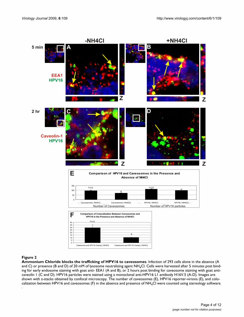
Ammonium chloride prevents HPV16 trafficking to caveosomesIn the presence of NH4Cl HPV16 infection levels wereshown to decrease. To determine if NH4Cl was able toblock the movement of HPV16 reporter-virions from earlyendosomes to caveosomes, we infected 293 cells in thepresence or absence of 20 mM NH4Cl. Infection by GFP-Reporter HPV16 reporter-virions was assessed by confocalmicroscopy and stereology counts. Data was collected at 5minutes post binding for early endosome staining (withgoat anti- EEA1, Fig. 2A and 2B, red) and 2 hours postbinding for caveolin-1 staining (with goat anti-caveolin 1,Fig 2C and 2D, red). HPV16 particles were stained using amonoclonal anti-HPV16 L1 antibody H16V.5 (Fig 2A–D,green). In the absence (Fig 2A) and presence of NH4Cl(Fig 2B) overlap is observed between HPV16 (Fig 2, green)
HPV16 infection is reduced in the presence of cysteine protease inhibitorsFigure 1HPV16 infection is reduced in the presence of cysteine protease inhibitors. Infection of 293 cells alone, with HPV 16 DsRed reporter-virions (mock control, Infected), or in the presence of: 20 mM of lysosome neutralizing agent NH4Cl; 10 μM of a non-permeable cysteine protease inhibitor E64; 10 μM of the permeable cysteine protease inhibitor E64-d; 6 μM of the permeable intracellular cathepsin B inhibitor CA-074ME; or 10 μM of the cathepsin L inhibitor. Infection was analyzed and compared 48 hours post binding by FACS of DSRED expression. Inhibitors were present for the duration of infection. Cells alone were analyzed for background fluorescence. Statistics were analyzed by 1 tailed t-test and found to be significant at P < 0.05.
Page 3 of 12(page number not for citation purposes)
Virology Journal 2009, 6:109 http://www.virologyj.com/content/6/1/109
Page 4 of 12(page number not for citation purposes)
Ammonium Chloride blocks the trafficking of HPV16 to caveosomesFigure 2Ammonium Chloride blocks the trafficking of HPV16 to caveosomes. Infection of 293 cells alone in the absence (A and C) or presence (B and D) of 20 mM of lysosome neutralizing agent NH4Cl. Cells were harvested after 5 minutes post bind-ing for early endosome staining with goat anti- EEA1 (A and B), or 2 hours post binding for caveosome staining with goat anti-caveolin 1 (C and D). HPV16 particles were stained using a monoclonal anti-HPV16 L1 antibody H16V.5 (A-D). Images are shown with z-stacks obtained by confocal microscopy. The number of caveosomes (E), HPV16 reporter-virions (E), and colo-calization between HPV16 and caveosomes (F) in the absence and presence of NH4Cl were counted using stereology software.
Virology Journal 2009, 6:109 http://www.virologyj.com/content/6/1/109
and the early endosomes (Fig 2A and 2B red) as indicatedby the yellow color (Fig 2A and 2B, yellow). We observeda decrease in colocalization between HPV16 (Fig 2 green)and caveolin-1 staining (Fig 2C and 2D, red) in the pres-ence of NH4Cl as compared to the absence of NH4Cl(compare Fig 2D to Fig 2C, yellow). A comparison of thepercent of HPV16 reporter-virions overlapping with cave-olin-1 in the absence or presence of NH4Cl by stereologyshowed a statistical significant change. The percent ofHPV16 reporter-virions colocalizing with caveosomesdecreased by 65.52% in the presence of NH4Cl (Fig 2FCaveosomes and HPV16 overlap -NH4Cl, and Caveosomeand HPV16 overlap + NH4Cl). This 65.52% decrease incolocalization was statistically significant at p < 0.05 1tailed T test. We did observe a drop in the overall numberof caveosomes, and no changes in the number of internal-ized viral particles (Fig 2E). The decrease in HPV16 infec-tivity was not due to the effects of NH4Cl on the HPV16reporter-virions [see Additional file 4]. These data suggestthat NH4Cl can influence that formation of caveosomesand of the movement of HPV16 reporter-virions intocaveosomes similar to JC virus.
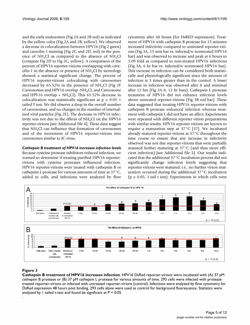
Cathepsin B treatment of HPV16 increases infection levelsBecause cysteine protease inhibitors reduced infection, wewanted to determine if treating purified HPV16 reporter-virions with cysteine proteases influenced infection.HPV16 reporter-virions were treated with cathepsin B orcathepsin L protease for various amounts of time at 37°C,added to cells, and infections were analyzed by flow
cytometry after 48 hours (for DsRED expression). Treat-ment of HPV16 with cathepsin B protease for 15 minutesincreased infectivity compared to untreated reporter-viri-ons (Fig 3A, 15 min bar vs. infected/w nontreated HPV16bar) and was observed to increase and peak at 6 hours to3.09 fold as compared to non-treated HPV16 infections(Fig 3A, 6 hr bar vs. infected/w nontreated HPV16 bar).This increase in infection can be considered both statisti-cally and physiologically significant since the amount ofinfection is 3 times greater than in the control. A lesserincrease in infection was observed after 8 and minimalafter 12 hrs (Fig 3A 8, 12 hr bars). Cathepsin L proteasetreatment of HPV16 did not enhance infection levelsabove untreated reporter-virions (Fig 3B red bar). Thesedata suggested that treating HPV16 reporter-virions withcathepsin B protease enhanced infection whereas treat-ment with cathepsin L did not have an affect. Experimentswere repeated with different reporter virion preparationswith similar results. HPV16 reporter-virions are known torequire a maturation step at 37°C [37]. We incubatedalready matured reporter-virions at 37°C throughout thetime course to ensure that any increase in infectivityobserved was not due reporter-virions that were partiallymatured further maturing at 37°C (and thus more effi-cient infection) [see Additional file 5]. Our results indi-cated that the additional 37°C incubation process did notsignificantly change infection levels suggesting thatreporter-virions were matured, i.e., no further virion mat-uration occurred during the additional 37°C incubation(p < 0.05, 1-tail t test). Experiments in which cells were
Cathepsin B treatment of HPV16 increases infectionFigure 3Cathepsin B treatment of HPV16 increases infection. HPV16 DsRed reporter-virions were incubated with (A) 37 pM cathepsin B protease or (B) 37 pM cathepsin L protease for various amounts of time. 293 cells were infected with protease treated reporter-virions or infected with untreated reporter-virions (control). Infections were analyzed by flow cytometry for DsRed expression 48 hours post binding. 293 cells alone were used as control for background fluorescence. Statistics were analyzed by 1 tailed t-test and found be significant at P < 0.05.
Page 5 of 12(page number not for citation purposes)
Virology Journal 2009, 6:109 http://www.virologyj.com/content/6/1/109
treated with cathepsin protease prior to infection showedno changes in infection (data not shown).
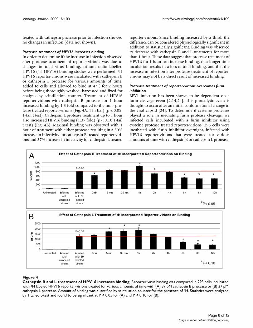
Protease treatment of HPV16 increases bindingIn order to determine if the increase in infection observedafter protease treatment of reporter-virions was due tochanges in total virus binding, tritium radio-labelledHPV16 (3H HPV16) binding studies were performed. 3HHPV16 reporter-virions were incubated with cathepsin Bor cathepsin L protease for various amounts of time,added to cells and allowed to bind at 4°C for 2 hoursbefore being thoroughly washed, harvested and fixed foranalysis by scintillation counter. Treatment of HPV16reporter-virions with cathepsin B protease for 1 hourincreased binding by 1.3 fold compared to the non- pro-tease treated reporter-virions (Fig. 4A, 1 hr bar) (p < 0.05,1-tail t test). Cathepsin L protease treatment up to 1 houralso increased HPV16 binding (1.37 fold) (p < 0.10 1-tailt test) (Fig. 4B). Maximal binding was observed with 1hour of treatment with either protease resulting in a 30%increase in infectivity for cathepsin B treated reporter viri-ons and 37% increase in infectivity for cathepsin L treated
reporter-virions. Since binding increased by a third, thedifference can be considered physiologically significant inaddition to statistically significant. Binding was observedto decrease with cathepsin B and L treatments for morethan 1 hour. These data suggest that protease treatment ofHPV16 for 1 hour can increase binding, that longer timeincubation results in a loss of total binding, and that theincrease in infection after protease treatment of reporter-virions may not be a direct result of increased binding.
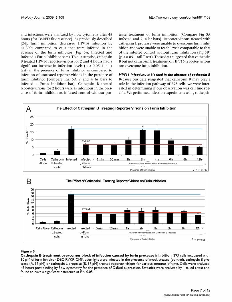
Protease treatment of reporter-virions overcomes furin inhibitionBPV1 infection has been shown to be dependent on afurin cleavage event [2,14,24]. This proteolytic event isthought to occur after an initial conformational change inthe viral capsid [24]. To determine if cysteine proteasesplayed a role in mediating furin protease cleavage, weinfected cells incubated with a furin inhibitor usingcysteine protease treated reporter-virions. 293 cells wereincubated with furin inhibitor overnight, infected withHPV16 reporter-virions that were treated for variousamounts of time with cathepsin B or cathepsin L protease,
Cathepsin B and L treatment of HPV16 increases bindingFigure 4Cathepsin B and L treatment of HPV16 increases binding. Reporter virus binding was compared in 293 cells incubated with 3H labeled HPV16 reporter-virions treated for various amounts of time with (A) 37 pM cathepsin B protease or (B) 37 pM cathepsin L protease. Amount of binding was quantified by scintillation counter for the presence of 3H. Statistics were analyzed by 1 tailed t-test and found to be significant at P < 0.05 for (A) and P < 0.10 for (B).
Page 6 of 12(page number not for citation purposes)
Virology Journal 2009, 6:109 http://www.virologyj.com/content/6/1/109
and infections were analyzed by flow cytometry after 48hours (for DsRED fluorescence). As previously described[24], furin inhibition decreased HPV16 infection by61.39% compared to cells that were infected in theabsence of the furin inhibitor (Fig. 5A, Infected andInfected + Furin Inhibitor bars). To our surprise, cathepsinB treated HPV16 reporter-virions for 2 and 4 hours had asignificant increase in infection levels (p < 0.05 1-tail ttest) in the presence of furin inhibitor as compared toinfection of untreated reporter-virions in the presence offurin inhibitor (compare Fig. 5A 2 and 4 hr bars toinfected + Furin inhibitor bar). Cathepsin B treatedreporter-virions for 2 hours were as infectious in the pres-ence of furin inhibitor as infected control without pro-
tease treatment or furin inhibition (Compare Fig 5AInfected and 2, 4 hr bars). Reporter-virions treated withcathepsin L protease were unable to overcome furin inhi-bition and were unable to reach levels comparable to thatof the infected control without furin inhibition (Fig 5B)(p < 0.05 1-tail T test). These data suggested that cathepsinB but not cathepsin L treatment of HPV16 reporter-virionscan overcome furin inhibition.
HPV16 Infectivity is blocked in the absence of cathepsin BBecause our data suggested that cathepsin B may play arole in the infection pathway of 293 cells, we were inter-ested in determining if our observation was cell line spe-cific. We performed infection experiments using cathepsin
Cathepsin B treatment overcomes block of infection caused by furin protease inhibitionFigure 5Cathepsin B treatment overcomes block of infection caused by furin protease inhibition. 293 cells incubated with 60 μM of furin inhibitor DEC-RVKR-CMK overnight were infected in the presence of mock treated (control), cathepsin B pro-tease (A, 37 pM) or cathepsin L protease (B, 37 pM) treated reporter-virions for various amounts of time. Cells were analyzed 48 hours post binding by flow cytometry for the presence of DsRed expression. Statistics were analyzed by 1 tailed t-test and found to have a significant difference at P < 0.05.
Page 7 of 12(page number not for citation purposes)
Virology Journal 2009, 6:109 http://www.virologyj.com/content/6/1/109
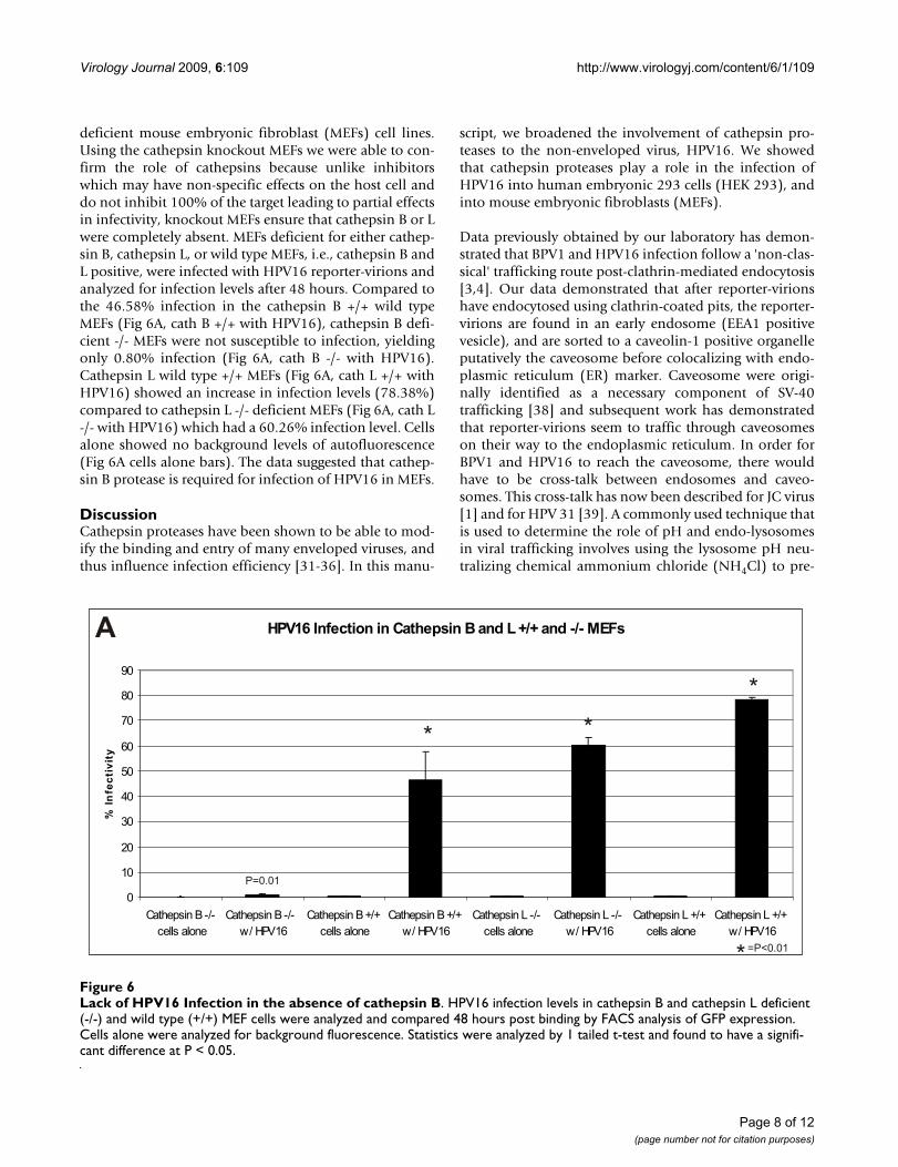
deficient mouse embryonic fibroblast (MEFs) cell lines.Using the cathepsin knockout MEFs we were able to con-firm the role of cathepsins because unlike inhibitorswhich may have non-specific effects on the host cell anddo not inhibit 100% of the target leading to partial effectsin infectivity, knockout MEFs ensure that cathepsin B or Lwere completely absent. MEFs deficient for either cathep-sin B, cathepsin L, or wild type MEFs, i.e., cathepsin B andL positive, were infected with HPV16 reporter-virions andanalyzed for infection levels after 48 hours. Compared tothe 46.58% infection in the cathepsin B +/+ wild typeMEFs (Fig 6A, cath B +/+ with HPV16), cathepsin B defi-cient -/- MEFs were not susceptible to infection, yieldingonly 0.80% infection (Fig 6A, cath B -/- with HPV16).Cathepsin L wild type +/+ MEFs (Fig 6A, cath L +/+ withHPV16) showed an increase in infection levels (78.38%)compared to cathepsin L -/- deficient MEFs (Fig 6A, cath L-/- with HPV16) which had a 60.26% infection level. Cellsalone showed no background levels of autofluorescence(Fig 6A cells alone bars). The data suggested that cathep-sin B protease is required for infection of HPV16 in MEFs.
DiscussionCathepsin proteases have been shown to be able to mod-ify the binding and entry of many enveloped viruses, andthus influence infection efficiency [31-36]. In this manu-
script, we broadened the involvement of cathepsin pro-teases to the non-enveloped virus, HPV16. We showedthat cathepsin proteases play a role in the infection ofHPV16 into human embryonic 293 cells (HEK 293), andinto mouse embryonic fibroblasts (MEFs).
Data previously obtained by our laboratory has demon-strated that BPV1 and HPV16 infection follow a 'non-clas-sical' trafficking route post-clathrin-mediated endocytosis[3,4]. Our data demonstrated that after reporter-virionshave endocytosed using clathrin-coated pits, the reporter-virions are found in an early endosome (EEA1 positivevesicle), and are sorted to a caveolin-1 positive organelleputatively the caveosome before colocalizing with endo-plasmic reticulum (ER) marker. Caveosome were origi-nally identified as a necessary component of SV-40trafficking [38] and subsequent work has demonstratedthat reporter-virions seem to traffic through caveosomeson their way to the endoplasmic reticulum. In order forBPV1 and HPV16 to reach the caveosome, there wouldhave to be cross-talk between endosomes and caveo-somes. This cross-talk has now been described for JC virus[1] and for HPV 31 [39]. A commonly used technique thatis used to determine the role of pH and endo-lysosomesin viral trafficking involves using the lysosome pH neu-tralizing chemical ammonium chloride (NH4Cl) to pre-
Lack of HPV16 Infection in the absence of cathepsin BFigure 6Lack of HPV16 Infection in the absence of cathepsin B. HPV16 infection levels in cathepsin B and cathepsin L deficient (-/-) and wild type (+/+) MEF cells were analyzed and compared 48 hours post binding by FACS analysis of GFP expression. Cells alone were analyzed for background fluorescence. Statistics were analyzed by 1 tailed t-test and found to have a signifi-cant difference at P < 0.05.
Page 8 of 12(page number not for citation purposes)
Virology Journal 2009, 6:109 http://www.virologyj.com/content/6/1/109
vent the acidification of vesicles, unfortunately thistreatment also results in the loss of fusion of intracellularvesicles. In fact, it was shown that NH4Cl prevented themovement of JC virus from endosome to caveosome.
The observed loss of viral infection using NH4Cl posedtwo hypotheses: 1) that the PV infection was preventingthe fusion of vesicles, and 2) that NH4Cl was preventingthe function of endo-lysosome proteases by preventingtheir conversion from "pro-inactive" form to "active"form. Our data shown in Figure 2 confirmed that indeedthere was loss of movement of HPV 16 reporter-virionsfrom endosomes to caveosome that could account for theloss of infection observed using NH4Cl (shown in Fig 1).Regarding the role of endo-lysosome proteases, wefocused on the cysteine proteases cathepsin B and L, twohighly abundant proteases in the endo-lysosome com-partments, and as mentioned above, cathepsin B and Lhave been previously shown to be involved in viral infec-tions. Our data showed that broad cysteine proteaseinhibitors and specific cathepsin B or L inhibitors wereable to decrease infection, thus suggesting that cysteineproteases were in part mediating HPV16 infection in 293cells.
Furin protease has been shown to be necessary for viralinfection by allowing the escape of the viral particle froman endosome (Richards RM, Lowy DR, Schiller JT, Day PM[24]). Richards and colleagues theorized that furinallowed the escape of reporter-virions from an endosomeas observed by the loss of endosome marker (EEA1) stain-ing overlap with reporter-virions. Our data supports a lossof EEA1 overlap with reporter-virions but show thatreporter-virions are moved to a caveolin-1 vesicle. It isunclear where Richards and colleagues propose viral par-ticles escape to. Because furin cleavage was shown to occurafter capsid conformation changes, we addressed if cathe-psins B or L were playing a role in capsid structuralchanges that aided the furin cleavage event. To our sur-prise the pre-treatment of purified viral particles withcathepsin B, but not cathepsin L, was able to overcome theblock of infection observed in the presence of furin inhib-itor. The significance of this finding needs further work. Itis possible that HPV16 utilizes cathepsin B as a "backup"mechanism for furin in order to establish infection.
In a recent study addressing the role of endosome pro-teases in the disassembly of HPV16 [24], the authorsnegated the role of cathepsin B and L in the HPV16 infec-tious process. The differences between our studies andtheirs may be due to the variation in inhibitors, cell lines,and quantity of viral particles used. In the paper by Rich-ards and colleagues the cathepsin B inhibitor used wasCA-074, a membrane impermeable inhibitor that wouldnot have shown an effect on an intracellular process (we
used both a permeable and non-permeable inhibitor); thecathepsin L inhibitor used was only described as "cathep-sin L inhibitor" and no further conclusions can be drawnas to the specificity of the inhibitor [24]. In addition, theobservations described by Richards and colleagues wereseen in the HPV 18 positive cervical carcinoma HeLa cellswhile our studies were performed in the adenovirus E1Atransformed human embryonic kidney 293 cell line andin MEFs derived from cathepsin B-deficient mice andcathepsin L-deficient mice [24]. HeLa cells were recentlyshown to be infected by a non-clathrin mediated endocy-tosis event, a finding that may also explain the differencesin both studies [40]. Finally the experiments performed inthe paper by Richards and colleagues, used a minimum of7.5 ng of PV while our experiments were carried out usingless than 0.33 ng of HPV16 (1 ng of VLPs has 30 millionparticles); a difference that may also contribute to the dif-ferences in results.
ConclusionIn summary, our data in this manuscript supports thatduring the process of infection, HPV16 is subjected to par-tial proteolysis by cathepsins at the plasma membrane, inthe endo-lysosome, or in the caveolin-1 positive vesicles.Work pursuing the role of cysteine proteases in HaCaTkeratinocytes and the specific biological significance andcellular site of cathepsin proteolysis is on-going.
MethodsCells, antibodies, proteases and inhibitors293 cells, a human embryonic kidney cell line (HEK),HaCaT cells, spontaneously immortalized human kerati-nocytes, and cathepsin B deficient (-/-), wild type (+/+),cathepsin L deficient (-/-) and wild type (+/+) mouseembryonic fibroblasts (MEFs) were grown in Dulbecco'sModified Eagle's medium (DMEM) supplemented with10% Fetal Bovine Serum (DMEM-10), and 100 IU/mLpenicillin/streptomycin. MEF cells were gifts from Dr. T. S.Dermody (Vanderbilt University School of Medicine,Nashville, TN). Goat anti-EEA1 (which recognizes earlyendosomes) antibody was purchased from Santa CruzBiotechnology, Santa Cruz, CA. Rabbit anti-caveolin 1antibody was purchased from Cell Signaling Technology(Danvers, MA). The monoclonal HPV16 L1 antibody,H16 V.5 was a generous gift from Dr. Neil Christensen(Penn State University, Hershey, PA). The followinginhibitors were obtained from Calbiochem (Gibbstown,NJ) and used at the following non-toxic concentration:CA-074Me (6 μM), Z-FF-FMK an irreversible cell permea-ble cathepsin L inhibitor (10 μM), E64 (10 μM), E64-d(10 μM). Furin inhibitor DEC-RVKR-CMK was obtainedfrom Biomol International (Plymouth Meeting, PA) andused at the non-toxic concentration of 60 μM. Ammo-nium chloride was used at a 20 mM concentration as perDay et al., (Sigma, St. Louis, MO) [2]. Cathepsin B pro-
Page 9 of 12(page number not for citation purposes)
Virology Journal 2009, 6:109 http://www.virologyj.com/content/6/1/109
tease (Calbiochem) and cathepsin L protease (R&D Sys-tems Minneapolis, MN) were used at a concentration of37 pM, a lower concentration compared to Ebert et al.,[36].
Flow cytometryInhibitors were added to 293 cells and allowed to incu-bate on cells at 37°C with 5% CO2 for 2 hours prior toinfection. After the 2 hr incubation period, the cells wereplaced on ice. HPV 16 reporter-virions containing aDsRed or GFP transgene were added to cells on ice for 2hours to allow for binding. At 2 hours, inhibitors andunbound virus were removed by washing with DMEM-10and replaced with 500 μl of warm DMEM-10 plus inhibi-tor. MEFs (not treated with inhibitors) were placed on icefor 2 hours in the presence of HPV16 reporter-virions toallow for binding. Unbound reporter-virions wereremoved by washing with DMEM-10 and replaced with500 μl of warm DMEM-10. The cells were incubated at37°C with 5% CO2 for 48 hours. Cells were harvestedusing trypsin. The cells were spun for 1 min at 16,100 × G,the pellet was washed 5× in 1× PBS and resuspended in300 ul of 1× PBS. 10,000 cells were counted on a fluores-cence activated cell sorter (FACS) and the number ofDsRed or GFP positive cells was used to determine the per-cent of infected cells (FACS performed at RFUMS Flowcytometry core). All experiments were repeated usingreporter-virions made from different preparations.
ImmunofluorescenceCells transfected on coverslips were washed 3× in 1× PBSand fixed in 4% paraformaldehyde for 20 min at 4°C.Paraformaldehyde was removed via 3 washes with 1×PBS. Cells were permeabilized with blocking buffer (0.2%fish skin gelatin (Sigma) and 0.2% Triton X-100 in PBS)for 5 min. The coverslips were washed 3× with 1× PBS andincubated with the appropriate primary antibody at 1:100dilution in blocking buffer (1:25 dilution for LAMP1 anti-body). Fluorescence labelled Alexa-flour donkey anti-mouse 488, goat anti-rabbit 594, chicken anti-goat 594,(Molecular Probes/Invitrogen, Eugene, OR) were used assecondary antibodies at 1:2,000 dilution in blockingbuffer in a 30 minute incubation. Coverslips were incu-bated for 5 minutes with TOPRO-3 (Invitrogen, CarlsbadCA) at 1:1000 dilution for nuclear staining. The coverslipswere mounted on glass slides using Prolong anti-fademounting medium (Invitrogen). Fluorescence confocalmicroscopy and stereology (the quantification of the per-centage of colocalization observed in the image, i.e.merged colors) were performed using an OlympusFluoview 300 microscope, and analyzed with Fluoviewand stereology software (Olympus, Melville, NY) at themicroscopy core of Rosalind Franklin University of Medi-cine and Science (RFUMS) (North Chicago, IL). Allimages are shown with z stacks.
Cytotoxicity AssayCytotoxicity studies for the various inhibitors were carriedout using the CellTiter-Glo Luminescent Cell ViabilityAssay Kit (Promega, Madison, WI). Cells were incubatedwith various concentrations of inhibitors for 48 hours andthen the supernatant and trypsinized cells were collected.100 μl of the harvested cell suspension was added to awell in a 96-well plate. The CellTiter-Glo substrate andbuffer were combined, and 100 μl was added to each wellcontaining sample. The reagent and cells are mixed for 5min on a shaker at room temperature. The 96-well platewas then allowed to rest at room temperature for 10 min-utes before being analyzed by a Bio-Tek Synergy HT PlateReader using the KC4 V3.4 software (Bio-Tek, Winooski,VT). All samples were analyzed in triplicate.
Reporter-virion production and purificationReporter-virions were made as described [37]. In brief,293TT cells were co-transfected with p16llwcha, a bicis-tronic HPV16 L1 and L2 plasmid and 8frb, the DsRed or8fwb, the GFP cDNA containing packaging plasmid. Con-structs and cells were gifts from Drs. Day and Schiller(National Cancer Institute, National Institute of Health,Bethesda, MD). Cells were harvested and lysed after 48hours. Reporter-virions were allowed to mature at 37°Cover night allowing for proper conformation of the capsidproteins. After a high salt extraction, reporter-virions werepurified on an optiprep gradient (27%–39%) via ultra-centrifugation. Titer of reporter-virions was determined byFACS for the percentage of DsRed or GFP positive cells 48hours after infection. Tritium labelled reporter-virionswere made with the addition of 3H 24 hours post transfec-tion.
Binding of radioactive reporter-virionsTritium labelled reporter-virions containing a DsRedtransgene were added to 293 cells on ice for 2 hours toallow for binding without internalization. The unboundreporter-virions were removed by washing with 1× PBS.Cells were harvested in 30 μl 1× PBS, spotted on What-mann paper and allowed to dry. The samples were fixed in5% Trichloroacetic acid (TCA) for 20 minutes and precip-itated in 95% ethanol for 20 minutes. The samples wereanalyzed with the LS 6500 Multipurpose ScintillationCounter (Beckman Coulter, Palatine, IL). All experimentswere repeated using reporter-virions made from differentpreparations.
Competing interestsThe authors declare that they have no competing interests.
Authors' contributionsSAD carried out the flow cytometric analysis, immunoflu-orescence analysis, radioactive studies, cytotoxicity analy-sis, and performed the statistical analysis. PIM and SAD
Page 10 of 12(page number not for citation purposes)
Virology Journal 2009, 6:109 http://www.virologyj.com/content/6/1/109
participated in the design of the study and drafted themanuscript. All authors read and approved the final man-uscript.
Additional material
AcknowledgementsThe funding for this work was provided by the H.M. Bligh Cancer Research Laboratory of the Rosalind Franklin University of Medicine and Science, North Chicago, IL; NIH/NCI grant K22:CA117971 to P.I.M; and by grant #07-034, and 09-15 from the American Cancer Society, IL-Division, Inc. to P.I.M.
References1. Querbes W, O'Hara BA, Williams G, Atwood WJ: Invasion of host
cells by JC virus identifies a novel role for caveolae in endo-somal sorting of noncaveolar ligands. J Virol 2006,80:9402-9413.
2. Day PM, Lowy DR, Schiller JT: Papillomaviruses infect cells via aclathrin-dependent pathway. Virology 2003, 307:1-11.
3. Laniosz V, Holthusen KA, Meneses PI: Bovine papillomavirus type1: from clathrin to caveolin. J Virol 2008, 82:6288-6298.
4. Laniosz V, Dabydeen SA, Havens MA, Meneses PI: Human Papillo-mavirus Type 16 Infection of Human Keratinocytes RequiresClathrin, Caveolin-1, and is Brefeldin A-Sensitive. J Virol 2009in press.
5. D'Souza G, Kreimer AR, Viscidi R, Pawlita M, Fakhry C, Koch WM,Westra WH, Gillison ML: Case-control study of human papillo-mavirus and oropharyngeal cancer. N Engl J Med 2007,356:1944-1956.
6. Greer CE, Wheeler CM, Ladner MB, Beutner K, Coyne MY, Liang H,Langenberg A, Yen TS, Ralston R: Human papillomavirus (HPV)type distribution and serological response to HPV type 6virus-like particles in patients with genital warts. J Clin Micro-biol 1995, 33:2058-2063.
7. Sinal SH, Woods CR: Human papillomavirus infections of thegenital and respiratory tracts in young children. Semin PediatrInfect Dis 2005, 16:306-316.
8. Baker TS, Newcomb WW, Olson NH, Cowsert LM, Olson C, BrownJC: Structures of bovine and human papillomaviruses. Analy-sis by cryoelectron microscopy and three-dimensional imagereconstruction. Biophys J 1991, 60:1445-1456.
9. Doorbar J, Gallimore PH: Identification of proteins encoded bythe L1 and L2 open reading frames of human papillomavirus1a. J Virol 1987, 61:2793-2799.
10. Trus BL, Roden RB, Greenstone HL, Vrhel M, Schiller JT, Booy FP:Novel structural features of bovine papillomavirus capsidrevealed by a three-dimensional reconstruction to 9 A reso-lution. Nat Struct Biol 1997, 4:413-420.
11. Bossis I, Roden RB, Gambhira R, Yang R, Tagaya M, Howley PM, Men-eses PI: Interaction of tSNARE syntaxin 18 with the papillo-mavirus minor capsid protein mediates infection. J Virol 2005,79:6723-6731.
12. Fay A, Yutzy WHt, Roden RB, Moroianu J: The positively chargedtermini of L2 minor capsid protein required for bovine pap-illomavirus infection function separately in nuclear importand DNA binding. J Virol 2004, 78:13447-13454.
13. Holmgren SC, Patterson NA, Ozbun MA, Lambert PF: The minorcapsid protein L2 contributes to two steps in the human pap-illomavirus type 31 life cycle. J Virol 2005, 79:3938-3948.
14. Laniosz V, Nguyen KC, Meneses PI: Bovine papillomavirus type 1infection is mediated by SNARE syntaxin 18. J Virol 2007,81:7435-7448.
Additional file 1Cytotoxicity of Inhibitors in 293 cells. 293 cells where incubated with various concentrations of a non-permeable cysteine protease inhibitor E64 (A), the permeable cysteine protease inhibitor E64-d (B), cathepsin L inhibitor (C), the intracellular cathepsin B inhibitor CA074-ME (D), furin inhibitor (E) and the lysosome neutralizing agent NH4Cl (F) for 48 hours. The cells and supernatant were analyzed for cytotoxicity using a plate reader. Cells alone were analyzed for background fluorescence.Click here for file[http://www.biomedcentral.com/content/supplementary/1743-422X-6-109-S1.pdf]
Additional file 2Dose Dependent Concentration of Inhibitors in 293 cells. 293 cells where incubated with various concentrations of a non-permeable cysteine protease inhibitor E64 (A), the permeable cysteine protease inhibitor E64-d (B), cathepsin L inhibitor (C), the intracellular cathepsin B inhibitor CA074-ME (D), furin inhibitor (E) and the lysosome neutralizing agent NH4Cl (F) overnight. The cells were then infected in the presence of inhibitor with HPV16 reporter-virions containing a GFP reporter gene. The cells were analyzed and compared 48 hours post binding by FACS analysis of GFP expression. Cells alone were analyzed for background flu-orescence. Statistics were analyzed by 1 tailed t-test and found to have a significant difference at P < 0.05.Click here for file[http://www.biomedcentral.com/content/supplementary/1743-422X-6-109-S2.pdf]
Additional file 3HPV16 infection is reduced in the presence of a cysteine protease inhibitor in HaCat cells. Infection of HaCat cells alone, with HPV 16 GFP reporter-virions (mock control, Infected), or in the presence of: 20 mM of lysosome neutralizing agent NH4Cl or 10 μM of a non-permeable cysteine protease inhibitor E64. Infection was analyzed and compared 48 hours post binding by FACS of GFP expression. Inhibitors were present for the duration of infection. Cells alone were analyzed for background fluo-rescence. Statistics were analyzed by 1 tailed t-test and found to be signif-icant at P < 0.05.Click here for file[http://www.biomedcentral.com/content/supplementary/1743-422X-6-109-S3.pdf]
Additional file 4NH4 Cl does not decrease HPV16 reporter-virions ability to infect. HPV16 GFP reporter-virions were incubated with (A) 20 mM NH4Cl for various amounts of time. 293 cells were infected with NH4Cl treated reporter-virions or infected with untreated reporter-virions (control). Infections were analyzed by flow cytometry for GFP expression 48 hours post binding. 293 cells alone were used as control for background fluores-cence. Statistics were analyzed by 1 tailed t-test and found be significant at P < 0.05.Click here for file[http://www.biomedcentral.com/content/supplementary/1743-422X-6-109-S4.pdf]
Additional file 5Additional Incubation of HPV16 reporter-virions at 37°C does not increase infectivity. HPV16 reporter-virions containing a GFP reporter gene were incubated at 37°C for various amounts of time. 293 cells were infected with the HPV16 reporter-virions and analyzed by flow cytometry for GFP expression 48 hours post binding. Statistics were analyzed by 1 tailed t-test and found to be significant at P < 0.05.Click here for file[http://www.biomedcentral.com/content/supplementary/1743-422X-6-109-S5.pdf]
Page 11 of 12(page number not for citation purposes)
Virology Journal 2009, 6:109 http://www.virologyj.com/content/6/1/109
Publish with BioMed Central and every scientist can read your work free of charge
"BioMed Central will be the most significant development for disseminating the results of biomedical research in our lifetime."
Sir Paul Nurse, Cancer Research UK
Your research papers will be:
available free of charge to the entire biomedical community
peer reviewed and published immediately upon acceptance
cited in PubMed and archived on PubMed Central
yours — you keep the copyright
Submit your manuscript here:http://www.biomedcentral.com/info/publishing_adv.asp
BioMedcentral
15. Zhou J, Stenzel DJ, Sun XY, Frazer IH: Synthesis and assembly ofinfectious bovine papillomavirus particles in vitro. J Gen Virol1993, 74(Pt 4):763-768.
16. Bousarghin L, Touze A, Combita-Rojas AL, Coursaget P: Positivelycharged sequences of human papillomavirus type 16 capsidproteins are sufficient to mediate gene transfer into targetcells via the heparan sulfate receptor. J Gen Virol 2003,84:157-164.
17. Joyce JG, Tung JS, Przysiecki CT, Cook JC, Lehman ED, Sands JA,Jansen KU, Keller PM: The L1 major capsid protein of humanpapillomavirus type 11 recombinant virus-like particlesinteracts with heparin and cell-surface glycosaminoglycanson human keratinocytes. J Biol Chem 1999, 274:5810-5822.
18. Shafti-Keramat S, Handisurya A, Kriehuber E, Meneguzzi G, SlupetzkyK, Kirnbauer R: Different heparan sulfate proteoglycans serveas cellular receptors for human papillomaviruses. J Virol 2003,77:13125-13135.
19. Evander M, Frazer IH, Payne E, Qi YM, Hengst K, McMillan NA: Iden-tification of the alpha6 integrin as a candidate receptor forpapillomaviruses. J Virol 1997, 71:2449-2456.
20. Sibbet G, Romero-Graillet C, Meneguzzi G, Saveria Campo M:alpha6 integrin is not the obligatory cell receptor for bovinepapillomavirus type 4. J Gen Virol 2000, 81(Pt 6):1629.
21. Bousarghin L, Touze A, Sizaret PY, Coursaget P: Human papillo-mavirus types 16, 31, and 58 use different endocytosis path-ways to enter cells. J Virol 2003, 77:3846-3850.
22. Muller M, Gissmann L, Cristiano RJ, Sun XY, Frazer IH, Jenson AB,Alonso A, Zentgraf H, Zhou J: Papillomavirus capsid binding anduptake by cells from different tissues and species. J Virol 1995,69:948-954.
23. Kamper N, Day PM, Nowak T, Selinka HC, Florin L, Bolscher J, HilbigL, Schiller JT, Sapp M: A membrane-destabilizing peptide incapsid protein L2 is required for egress of papillomavirusgenomes from endosomes. J Virol 2006, 80:759-768.
24. Richards RM, Lowy DR, Schiller JT, Day PM: Cleavage of the pap-illomavirus minor capsid protein, L2, at a furin consensussite is necessary for infection. Proc Natl Acad Sci USA 2006,103:1522-1527.
25. Mohamed MM, Sloane BF: Cysteine cathepsins: multifunctionalenzymes in cancer. Nat Rev Cancer 2006, 6:764-775.
26. Ohashi N, Yamamoto T, Uchida C, Togawa A, Fukasawa H, FujigakiY, Suzuki S, Kitagawa K, Hattori T, Oda T, et al.: Transcriptionalinduction of Smurf2 ubiquitin ligase by TGF-beta. FEBS Lett2005, 579:2557-2563.
27. Cavallo-Medved D, Dosescu J, Linebaugh BE, Sameni M, Rudy D,Sloane BF: Mutant K-ras regulates cathepsin B localization onthe surface of human colorectal carcinoma cells. Neoplasia2003, 5:507-519.
28. Caglic D, Pungercar JR, Pejler G, Turk V, Turk B: Glycosaminogly-cans facilitate procathepsin B activation through disruptionof propeptide-mature enzyme interactions. J Biol Chem 2007,282:33076-33085.
29. Salminen A, Gottesman MM: Inhibitor studies indicate thatactive cathepsin L is probably essential to its own processingin cultured fibroblasts. Biochem J 1990, 272:39-44.
30. Ishidoh K, Kominami E: Procathepsin L degrades extracellularmatrix proteins in the presence of glycosaminoglycans invitro. Biochem Biophys Res Commun 1995, 217:624-631.
31. Kaletsky RL, Simmons G, Bates P: Proteolysis of the Ebola virusglycoproteins enhances virus binding and infectivity. J Virol2007, 81:13378-13384.
32. Pager CT, Craft WW Jr, Patch J, Dutch RE: A mature andfusogenic form of the Nipah virus fusion protein requiresproteolytic processing by cathepsin L. Virology 2006,346:251-257.
33. Pager CT, Dutch RE: Cathepsin L is involved in proteolyticprocessing of the Hendra virus fusion protein. J Virol 2005,79:12714-12720.
34. Huang IC, Bosch BJ, Li F, Li W, Lee KH, Ghiran S, Vasilieva N, Der-mody TS, Harrison SC, Dormitzer PR, et al.: SARS coronavirus,but not human coronavirus NL63, utilizes cathepsin L toinfect ACE2-expressing cells. J Biol Chem 2006, 281:3198-3203.
35. Qiu Z, Hingley ST, Simmons G, Yu C, Das Sarma J, Bates P, Weiss SR:Endosomal proteolysis by cathepsins is necessary for murinecoronavirus mouse hepatitis virus type 2 spike-mediatedentry. J Virol 2006, 80:5768-5776.
36. Ebert DH, Deussing J, Peters C, Dermody TS: Cathepsin L andcathepsin B mediate reovirus disassembly in murine fibrob-last cells. J Biol Chem 2002, 277:24609-24617.
37. Buck CB, Thompson CD, Pang YY, Lowy DR, Schiller JT: Matura-tion of papillomavirus capsids. J Virol 2005, 79:2839-2846.
38. Pelkmans L, Kartenbeck J, Helenius A: Caveolar endocytosis ofsimian virus 40 reveals a new two-step vesicular-transportpathway to the ER. Nat Cell Biol 2001, 3:473-483.
39. Smith JL, Campos SK, Ozbun MA: Human papillomavirus type 31uses a caveolin 1- and dynamin 2-mediated entry pathwayfor infection of human keratinocytes. J Virol 2007,81:9922-9931.
40. Spoden G, Freitag K, Husmann M, Boller K, Sapp M, Lambert C, FlorinL: Clathrin- and caveolin-independent entry of human papil-lomavirus type 16 – involvement of tetraspanin-enrichedmicrodomains (TEMs). PLoS ONE 2008, 3:e3313.
Page 12 of 12(page number not for citation purposes)


![Virology Journal BioMed Central - Home - Springer · Virology Journal Research Open Access ... licensee BioMed Central Ltd. ... [20]. A schematic diagram of our experimental design](https://img.pdfslide.us/doc/110x75/5ada85c77f8b9a52528cf1bf/virology-journal-biomed-central-home-springer-journal-research-open-access-.jpg)