Embed Size (px)
Citation preview
Prof. Józef Dulak, PhD, DScDepartment of Medical Biotechnology
Faculty of Biochemistry, Biophysics and BiotechnologyRoom 3.025/3.07 Phone 664-63-75
Email: [email protected]
7 December 2015
Viral vectorsin clinical gene therapies
(3)
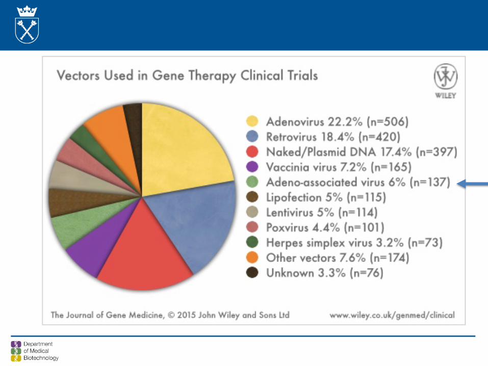
Drugs/gene therapy delivery to the eye
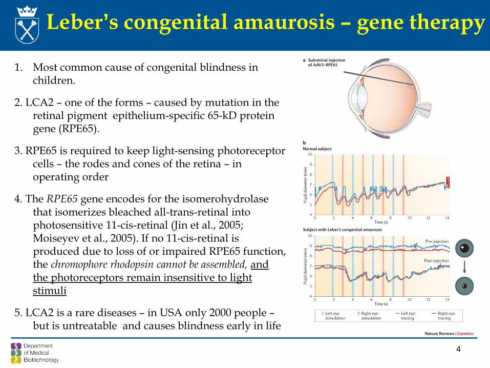
Leber’s congenital amaurosis – gene therapy
1. Most common cause of congenital blindness in children.
2. LCA2 – one of the forms – caused by mutation in the retinal pigment epithelium-specific 65-kD protein gene (RPE65).
3. RPE65 is required to keep light-sensing photoreceptorcells – the rodes and cones of the retina – in operating order
4. The RPE65 gene encodes for the isomerohydrolasethat isomerizes bleached all-trans-retinal into photosensitive 11-cis-retinal (Jin et al., 2005; Moiseyev et al., 2005). If no 11-cis-retinal is produced due to loss of or impaired RPE65 function, the chromophore rhodopsin cannot be assembled, and the photoreceptors remain insensitive to light stimuli
5. LCA2 is a rare diseases – in USA only 2000 people –but is untreatable and causes blindness early in life
4
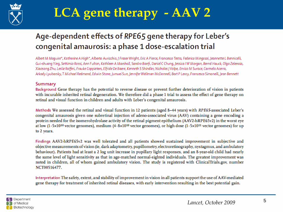
Lancet, October 2009 5
LCA gene therapy - AAV 2
• an incurable X-linked recessive degenerative disease of the retina and choroid• It has a prevalence of about 1:50 000, with northern Finland having the highest• Loss of night vision begins in the first decade of life and progresses with a gradual
loss of peripheral vision and legal blindness by the fi fth decade.• Choroideremia is caused by mutations in the CHM gene, which was one of the first
genes identified by use of positional cloning• Subsequently, prenylation deficiency due to absence of Rab escort protein-1 (REP1)
encoded by CHM was identified as the cause of retinal degeneration in choroideremia• Nearly all reported cases of choroideremia so far have been attributed to functionally
null mutations• That, combined with the slow rate of degeneration and small size of the CHM
protein coding sequence (1.9 kb), make gene therapy with AAV vectors an appealingtreatment strategy.
Choroideremia – another type of blindness
Choroideremia derives its name from the almost complete loss of the retina, choroid,and retinal pigment epithelium that leads to exposure of the underlying white sclera, which is a unique feature compared with other retinal degenerations.
RE MaclLaren et al., Lancet 2014; 383: 1129–37
• In The Lancet , Robert MacLaren and colleagues present data for sixpatients treated with subretinal delivery of an AAV vector encodingCHM , the gene disrupted in choroideremia, in a phase 1 trial.
• All patients recovered to baseline visual acuity despite detachment of the fovea as part of the surgical procedure.
• The authors did not notice any obvious detrimental effects resultingfrom detachment and treatment of the fovea, which is paramount sincethe ultimate goal of such an intervention is to prevent central visual loss.
• Two of the patients gained significant visual acuity.
HPN Scholl & JA Sahel, Lancet January 16, 2014
Gene therapy of choroideremia
8
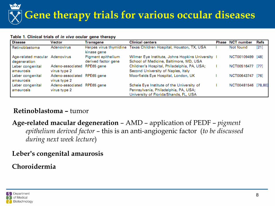
Gene therapy trials for various occular diseases
Retinoblastoma – tumor
Age-related macular degeneration – AMD – application of PEDF – pigment epithelium derived factor – this is an anti-angiogenic factor (to be discussedduring next week lecture)
Leber’s congenital amaurosis
Choroidermia
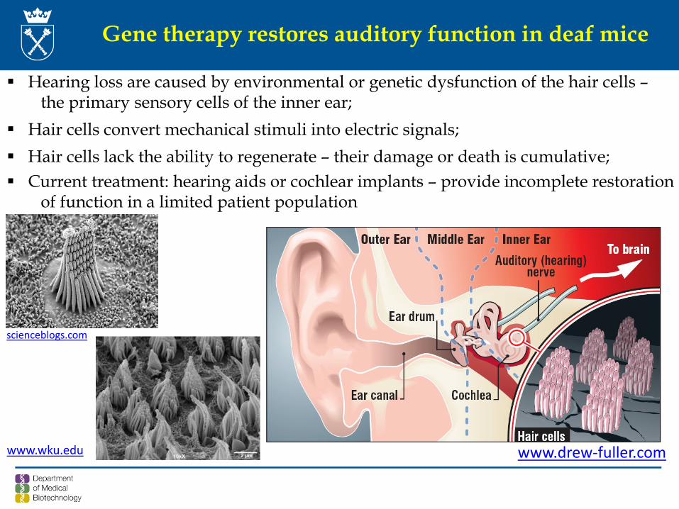
Gene therapy restores auditory function in deaf mice
Hearing loss are caused by environmental or genetic dysfunction of the hair cells –the primary sensory cells of the inner ear;
Hair cells convert mechanical stimuli into electric signals; Hair cells lack the ability to regenerate – their damage or death is cumulative; Current treatment: hearing aids or cochlear implants – provide incomplete restoration
of function in a limited patient population
scienceblogs.com
www.drew-fuller.comwww.wku.edu
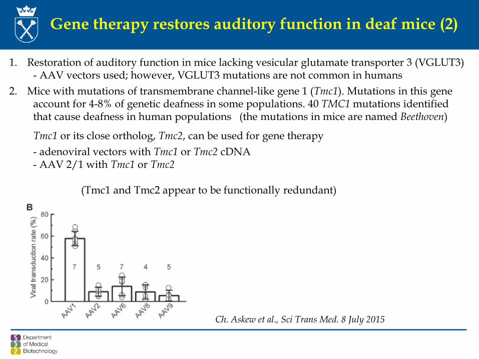
Gene therapy restores auditory function in deaf mice (2)
1. Restoration of auditory function in mice lacking vesicular glutamate transporter 3 (VGLUT3) - AAV vectors used; however, VGLUT3 mutations are not common in humans
2. Mice with mutations of transmembrane channel-like gene 1 (Tmc1). Mutations in this geneaccount for 4-8% of genetic deafness in some populations. 40 TMC1 mutations identifiedthat cause deafness in human populations (the mutations in mice are named Beethoven)
Tmc1 or its close ortholog, Tmc2, can be used for gene therapy- adenoviral vectors with Tmc1 or Tmc2 cDNA- AAV 2/1 with Tmc1 or Tmc2
(Tmc1 and Tmc2 appear to be functionally redundant)
Ch. Askew et al., Sci Trans Med. 8 July 2015
Nature, 27th November 2014
Outlook on Haemophilia
Haemophilia
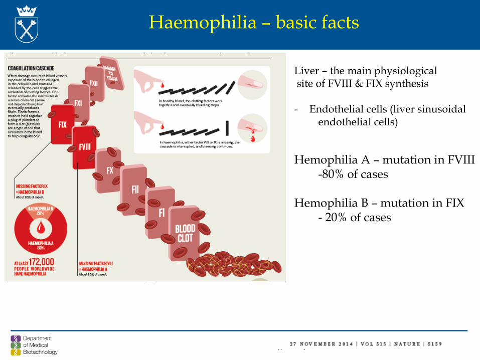
Haemophilia – basic facts
Liver – the main physiologicalsite of FVIII & FIX synthesis
- Endothelial cells (liver sinusoidalendothelial cells)
Hemophilia A – mutation in FVIII-80% of cases
Hemophilia B – mutation in FIX - 20% of cases
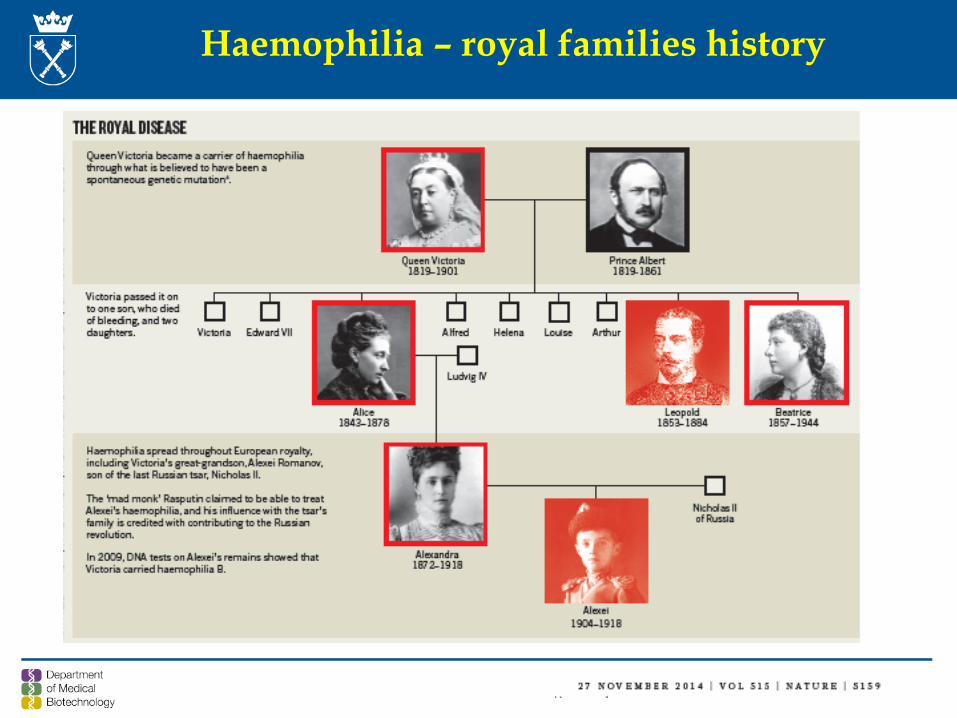
Haemophilia – royal families history
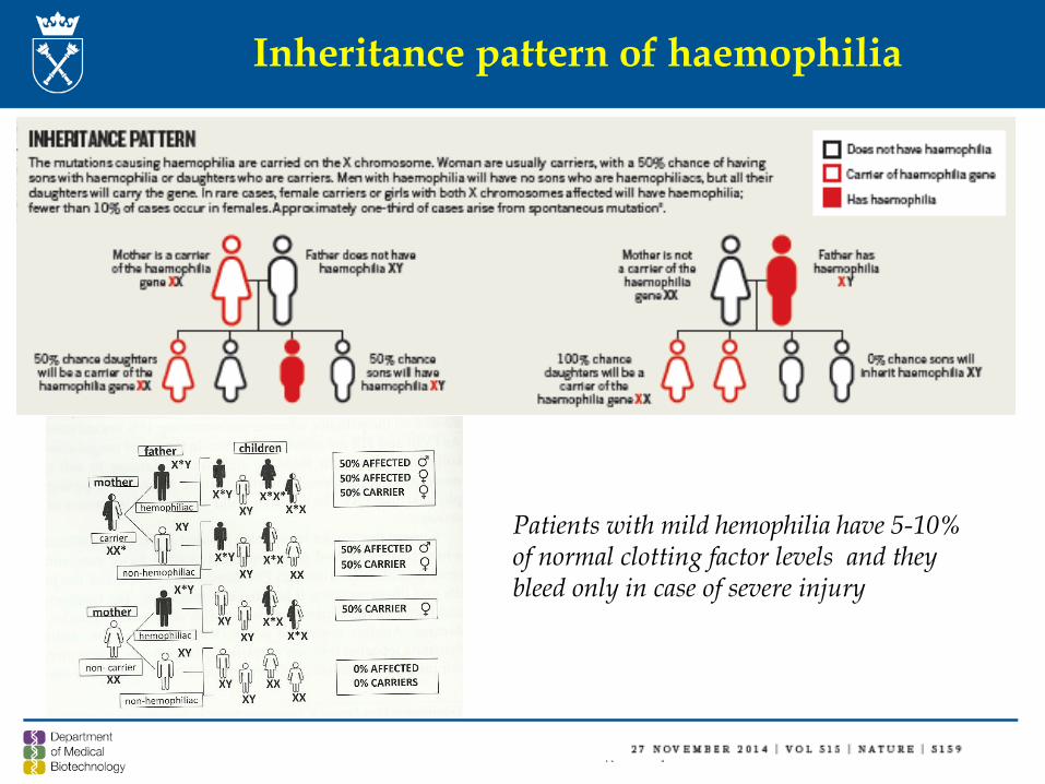
Inheritance pattern of haemophilia
Patients with mild hemophilia have 5-10%of normal clotting factor levels and theybleed only in case of severe injury
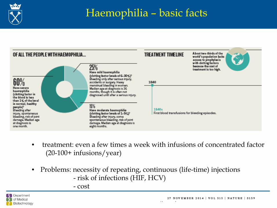
• treatment: even a few times a week with infusions of concentrated factor(20-100+ infusions/year)
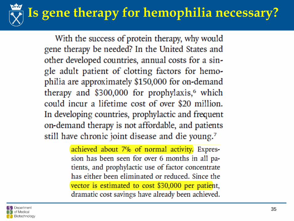
• Problems: necessity of repeating, continuous (life-time) injections- risk of infections (HIF, HCV)- cost
Haemophilia – basic facts
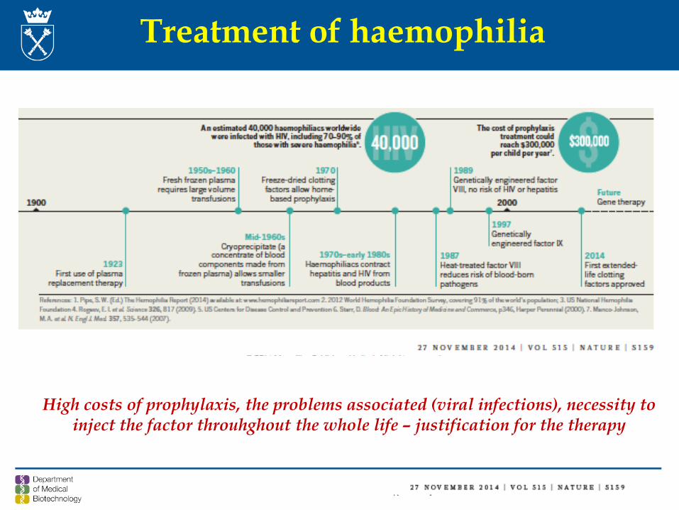
Treatment of haemophilia
High costs of prophylaxis, the problems associated (viral infections), necessity to inject the factor throuhghout the whole life – justification for the therapy
Hemofilia A and B and gene therapy
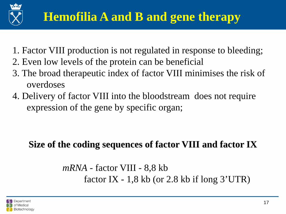
1. Factor VIII production is not regulated in response to bleeding; 2. Even low levels of the protein can be beneficial3. The broad therapeutic index of factor VIII minimises the risk of
overdoses4. Delivery of factor VIII into the bloodstream does not require
expression of the gene by specific organ;
mRNA - factor VIII - 8,8 kbfactor IX - 1,8 kb (or 2.8 kb if long 3’UTR)
Size of the coding sequences of factor VIII and factor IX
17
Roth DA et al., NEJM 2001; 344: 1735
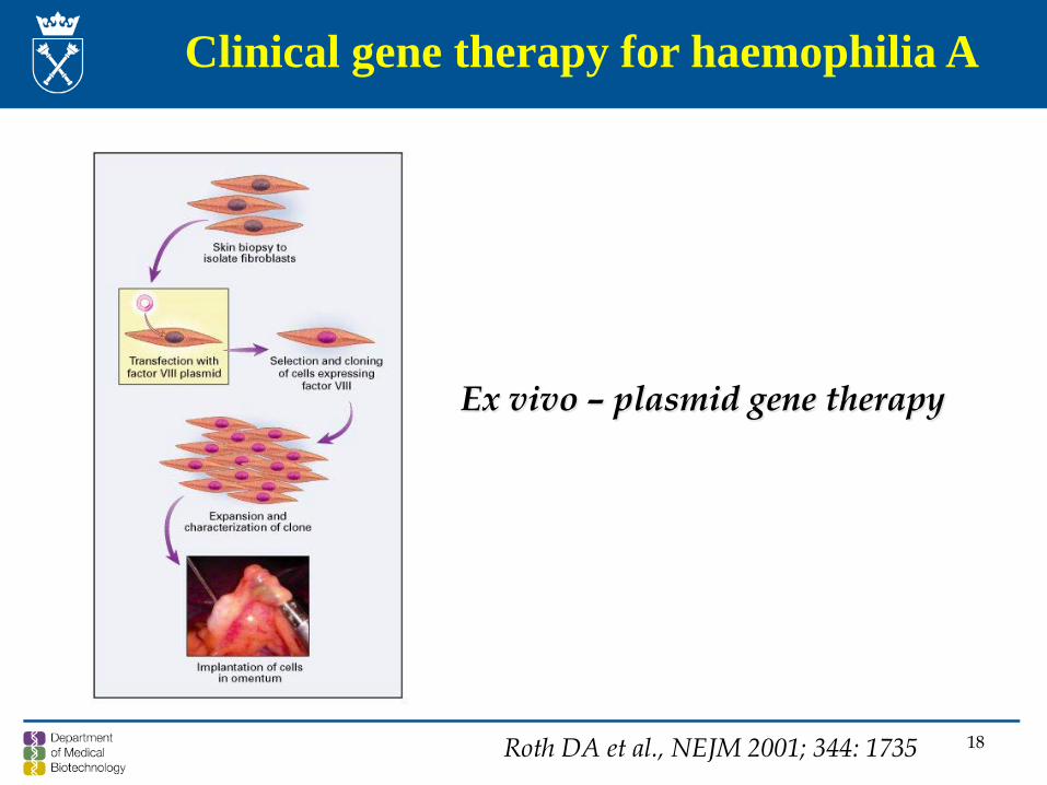
Clinical gene therapy for haemophilia A
Ex vivo – plasmid gene therapy
18
Roth DA et al., NEJM 2001; 344: 1735
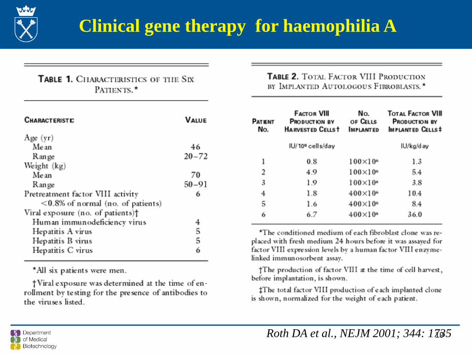
Clinical gene therapy for haemophilia A
19
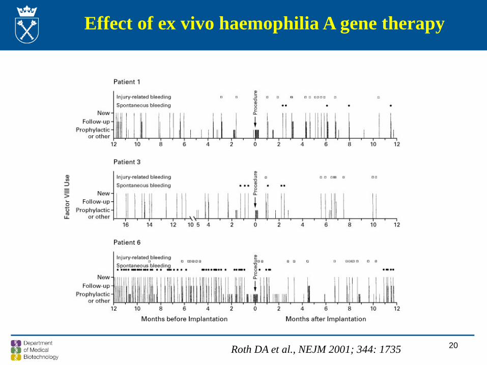
Effect of ex vivo haemophilia A gene therapy
Roth DA et al., NEJM 2001; 344: 1735 20
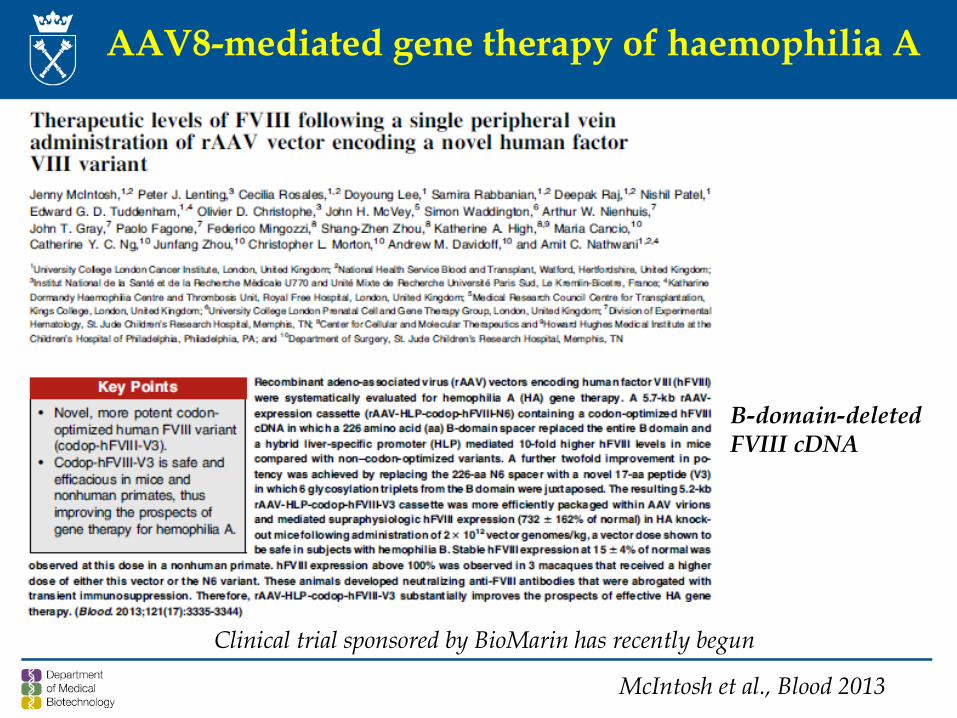
AAV8-mediated gene therapy of haemophilia A
McIntosh et al., Blood 2013
B-domain-deletedFVIII cDNA
Clinical trial sponsored by BioMarin has recently begun
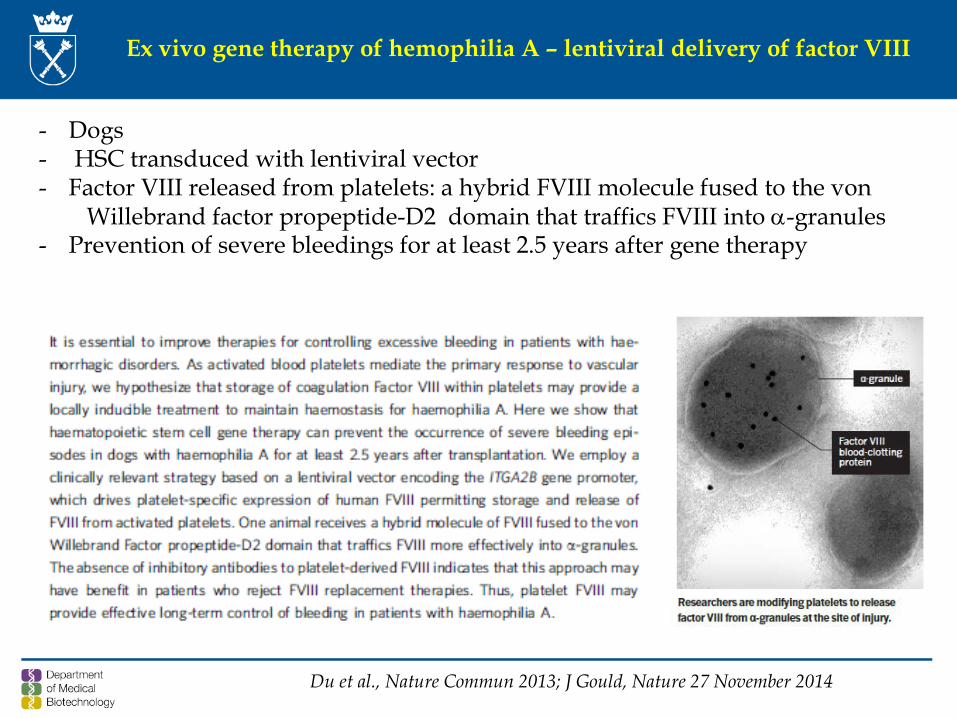
Ex vivo gene therapy of hemophilia A – lentiviral delivery of factor VIII
Du et al., Nature Commun 2013; J Gould, Nature 27 November 2014
- Dogs - HSC transduced with lentiviral vector- Factor VIII released from platelets: a hybrid FVIII molecule fused to the von
Willebrand factor propeptide-D2 domain that traffics FVIII into α-granules- Prevention of severe bleedings for at least 2.5 years after gene therapy
23NEJM, December 2011
„Christmas” disease
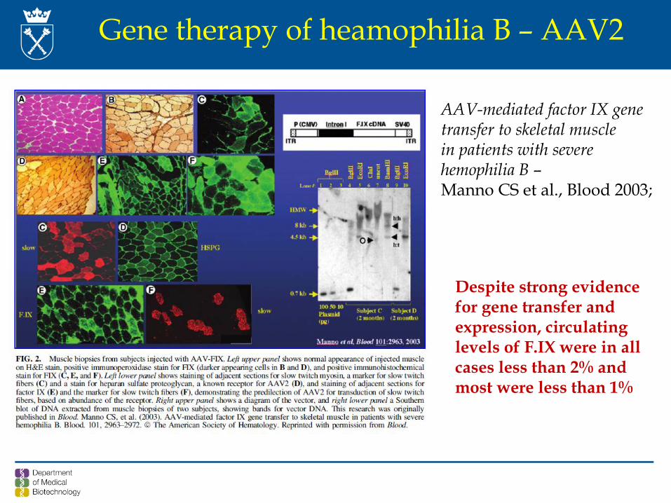
Gene therapy of heamophilia B – AAV2
AAV-mediated factor IX genetransfer to skeletal musclein patients with severehemophilia B –Manno CS et al., Blood 2003;
Despite strong evidence for gene transfer and expression, circulating levels of F.IX were in all cases less than 2% and most were less than 1%
25
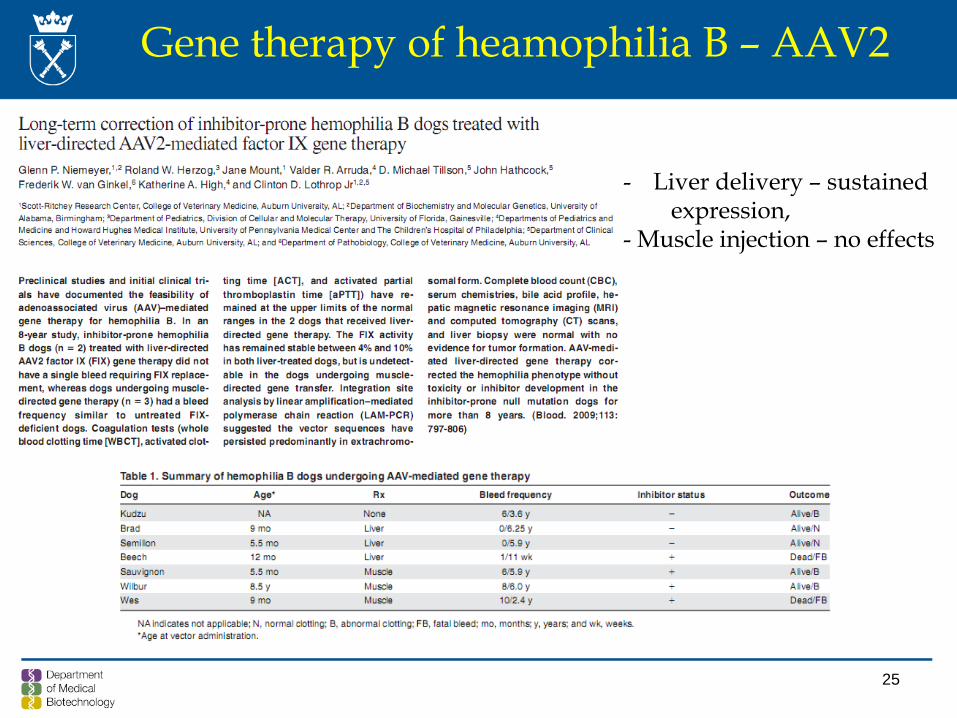
Gene therapy of heamophilia B – AAV2
- Liver delivery – sustainedexpression,
- Muscle injection – no effects
26
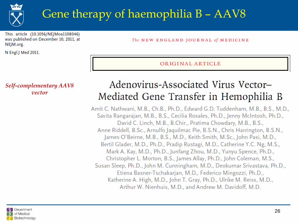
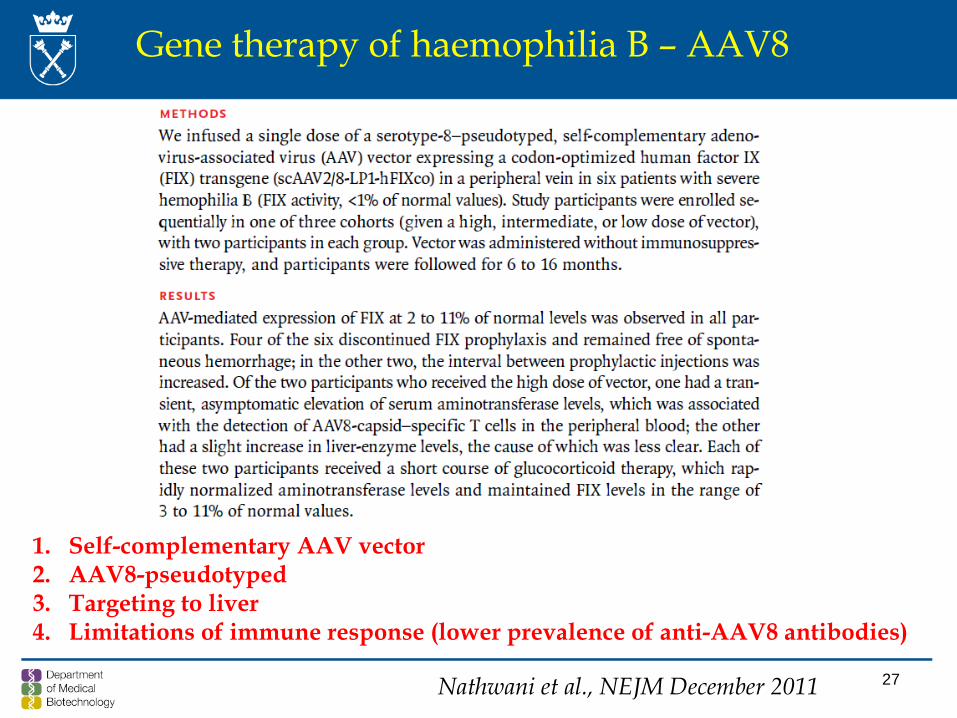
Gene therapy of haemophilia B – AAV8
Self-complementary AAV8vector
27
1. Self-complementary AAV vector2. AAV8-pseudotyped 3. Targeting to liver4. Limitations of immune response (lower prevalence of anti-AAV8 antibodies)
Gene therapy of haemophilia B – AAV8
Nathwani et al., NEJM December 2011
28
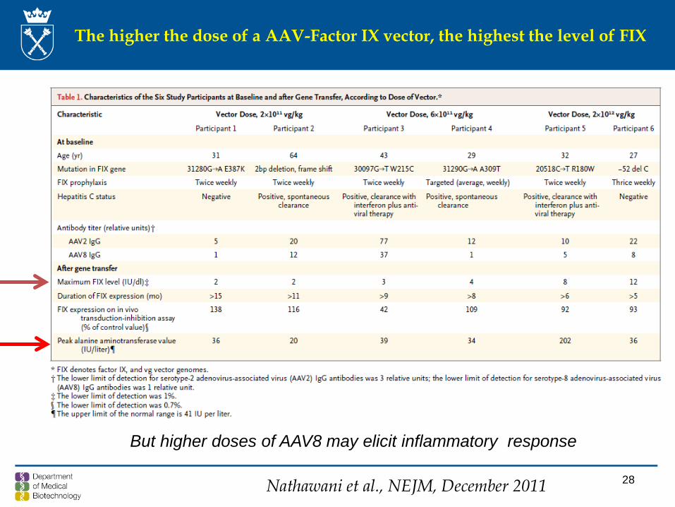
The higher the dose of a AAV-Factor IX vector, the highest the level of FIX
But higher doses of AAV8 may elicit inflammatory response
Nathawani et al., NEJM, December 2011
29
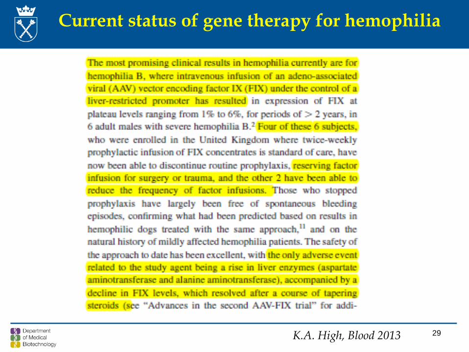
Current status of gene therapy for hemophilia
K.A. High, Blood 2013
MI Cancio et al., Appl Clin Genetics, 6: 91-101; 2013
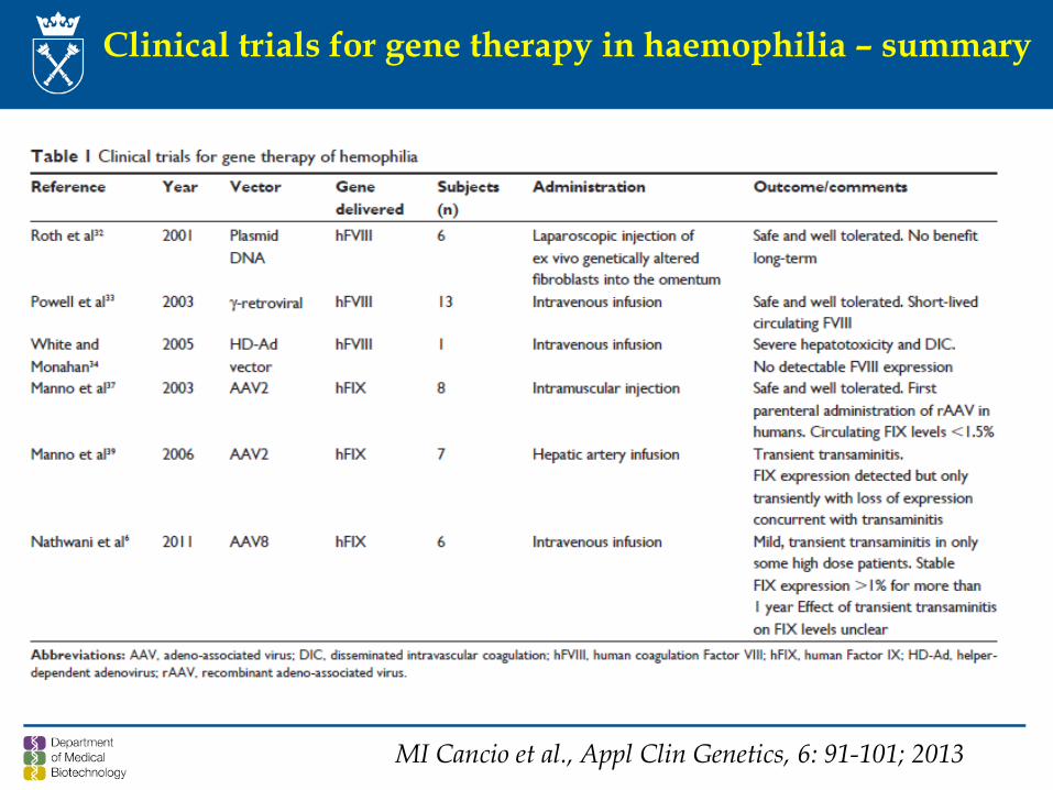
Clinical trials for gene therapy in haemophilia – summary
Clinical trials for gene therapy in haemophilia – summary
Nair et al., Advanced Textbook on Gene Therapy, Imperial College, London 2014
AAV2AAV8
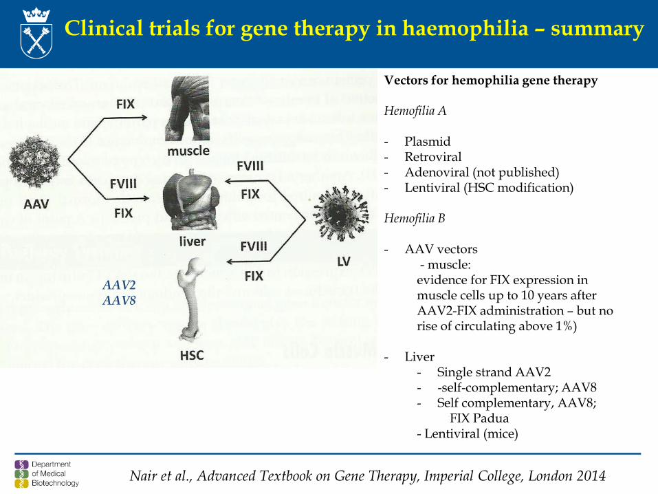
Vectors for hemophilia gene therapy
Hemofilia A
- Plasmid- Retroviral- Adenoviral (not published) - Lentiviral (HSC modification)
Hemofilia B
- AAV vectors- muscle: evidence for FIX expression in muscle cells up to 10 years afterAAV2-FIX administration – but norise of circulating above 1%)
- Liver- Single strand AAV2 - -self-complementary; AAV8- Self complementary, AAV8;
FIX Padua- Lentiviral (mice)
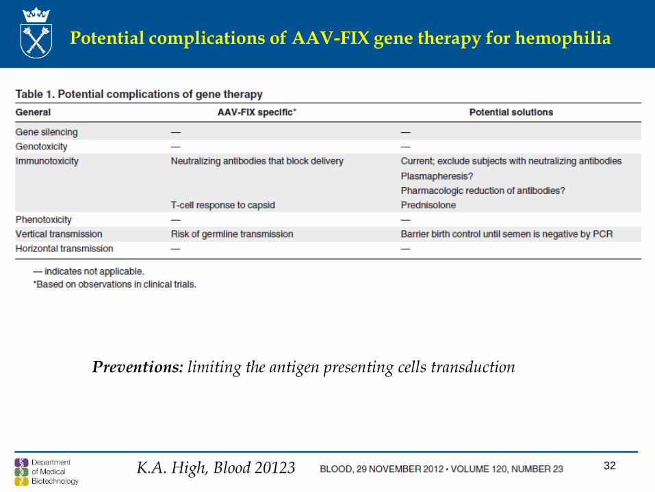
32K.A. High, Blood 20123
Potential complications of AAV-FIX gene therapy for hemophilia
Preventions: limiting the antigen presenting cells transduction
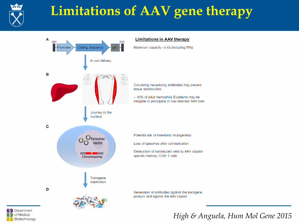
Limitations of AAV gene therapy
High & Anguela, Hum Mol Gene 2015
(Gene) therapy of haemophilia – problems
• About 40% of patients with haemophilia B produce antibodies against AAV – theywill be excluded from this type of gene therapy;
• Haemophilia A – is more difficult to target by gene therapy (longer gene of factorVIII);
• Shortened version of factor VIII is produced; • Still, the strong immune response against AAV8 limits the effectiveness of the
therapy - injections of AAV8 cannot be repeated; • The problem may be potentially overcome by ex vivo HSC therapy with lentiviral
vector harboring the factor VIII gene; • Potential of gene editing in therapy of haemophilia
• Recombinant-clotting factors – was associated with tens of thousands of haemophiliacs infected with HIV and HCV
• About 75% of patients with haemophilia still receive inadequate treatment, particularly in less-developed nations
• The fix will not lie in just one solution, but will be contextual and messy
S. Pemberton
35
Is gene therapy for hemophilia necessary?
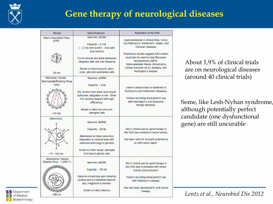
Gene therapy of neurological diseases
Lentz et al., Neurobiol Dis 2012
About 1,9% of clinical trialsare on neurological diseases(around 40 clinical trials)
Some, like Lesh-Nyhan syndrome,although potentially perfectcandidate (one dysfunctionalgene) are still uncurable
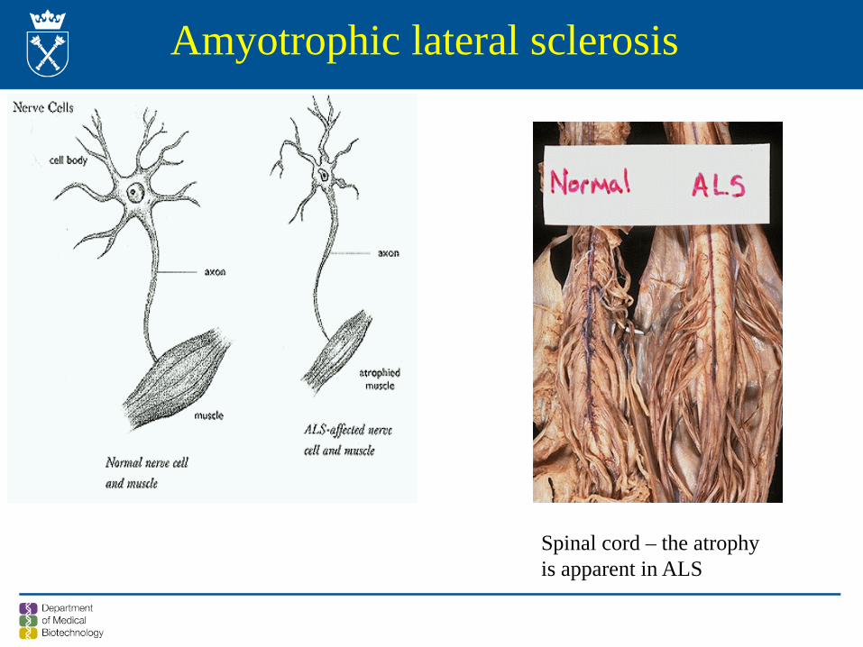
Amyotrophic lateral sclerosis
Spinal cord – the atrophy is apparent in ALS
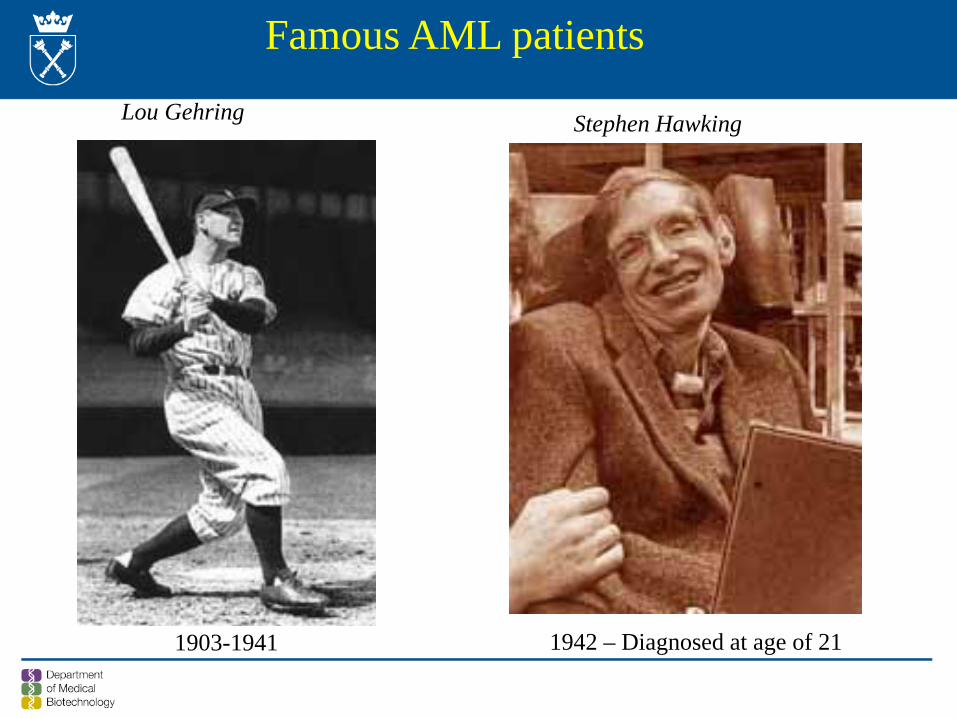
1903-1941 1942 – Diagnosed at age of 21
Famous AML patients
Lou Gehring Stephen Hawking
Amyotrophic lateral sclerosis (ALS) (1)
Incidence – 2-3:100 000
Onset at 50-60 years
Sporadic (SALS) – most instances (90-95%)Familial (FALS) – 5-10% - of these 20-25% are mapped to CuZnSOD
gene
Degeneration of motor neurons – progressive loss of the abilityto move, speak,
Usually fatal within 1-5 years of onset
No effective treatment available
40
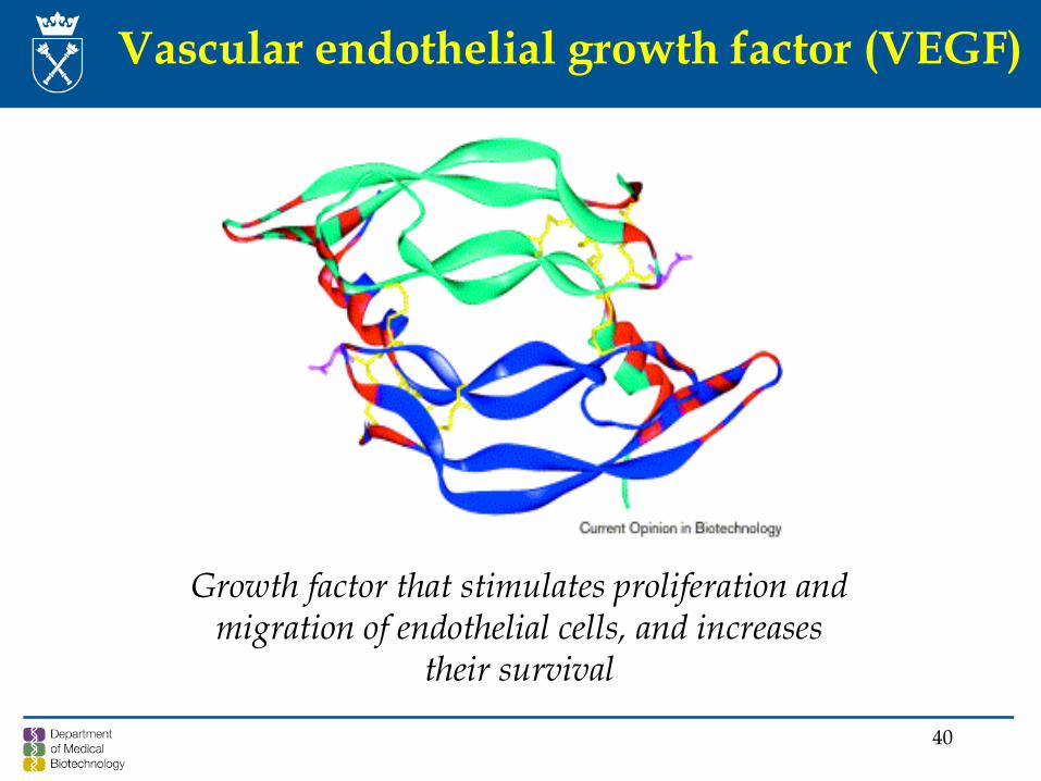
Vascular endothelial growth factor (VEGF)
Growth factor that stimulates proliferation and migration of endothelial cells, and increases
their survival
41
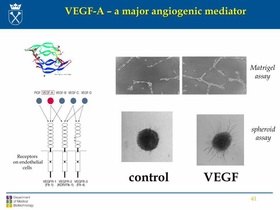
control VEGF
Matrigel assay
spheroid assay
VEGF-A – a major angiogenic mediator
Receptorson endothelial
cells
min
max
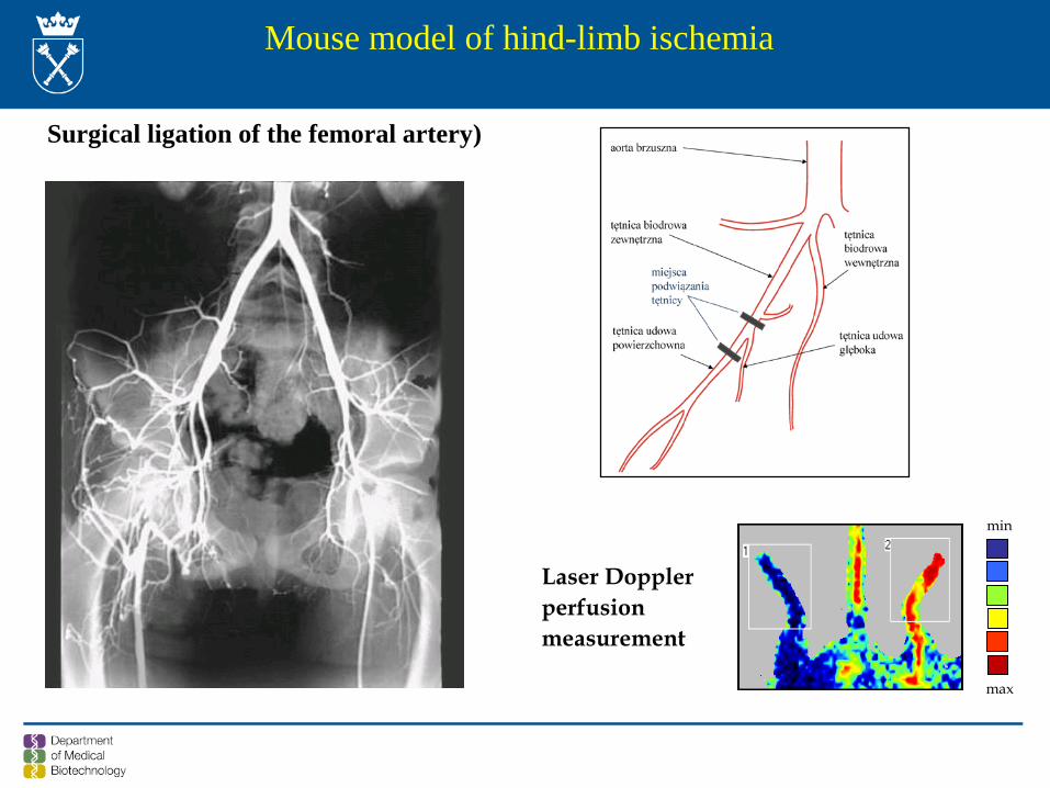
Laser Dopplerperfusionmeasurement
Mouse model of hind-limb ischemia
Surgical ligation of the femoral artery)
43
mea
n lo
cal b
lood
flow
in a
dduc
tor
mus
cle
(100
% -
bloo
d flo
w b
efor
e is
chem
ia)
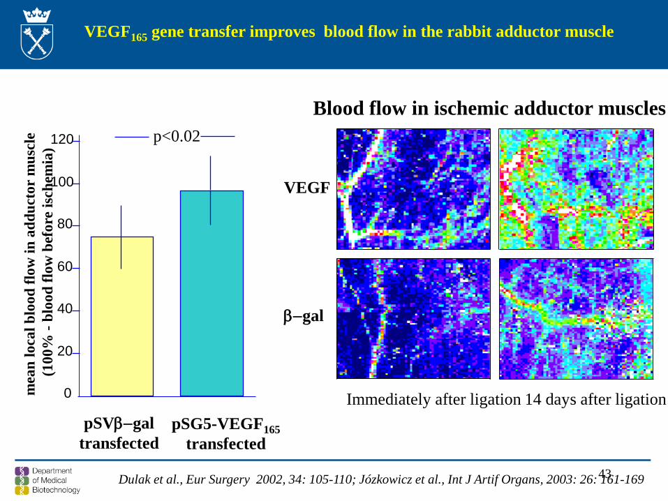
pSG5-VEGF165transfected
pSVβ−galtransfected
0
20
40
60
80
100
120 p<0.02
Immediately after ligation 14 days after ligation
β−gal
VEGF
Blood flow in ischemic adductor muscles
VEGF165 gene transfer improves blood flow in the rabbit adductor muscle
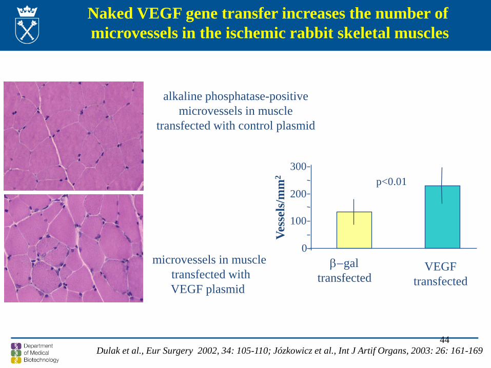
Dulak et al., Eur Surgery 2002, 34: 105-110; Józkowicz et al., Int J Artif Organs, 2003: 26: 161-169
44
Naked VEGF gene transfer increases the number of microvessels in the ischemic rabbit skeletal muscles
alkaline phosphatase-positive microvessels in muscle
transfected with control plasmid
microvessels in muscle transfected withVEGF plasmid
Vess
els/
mm
2β−gal
transfected VEGF
transfected
0
100
200
300p<0.01
Dulak et al., Eur Surgery 2002, 34: 105-110; Józkowicz et al., Int J Artif Organs, 2003: 26: 161-169
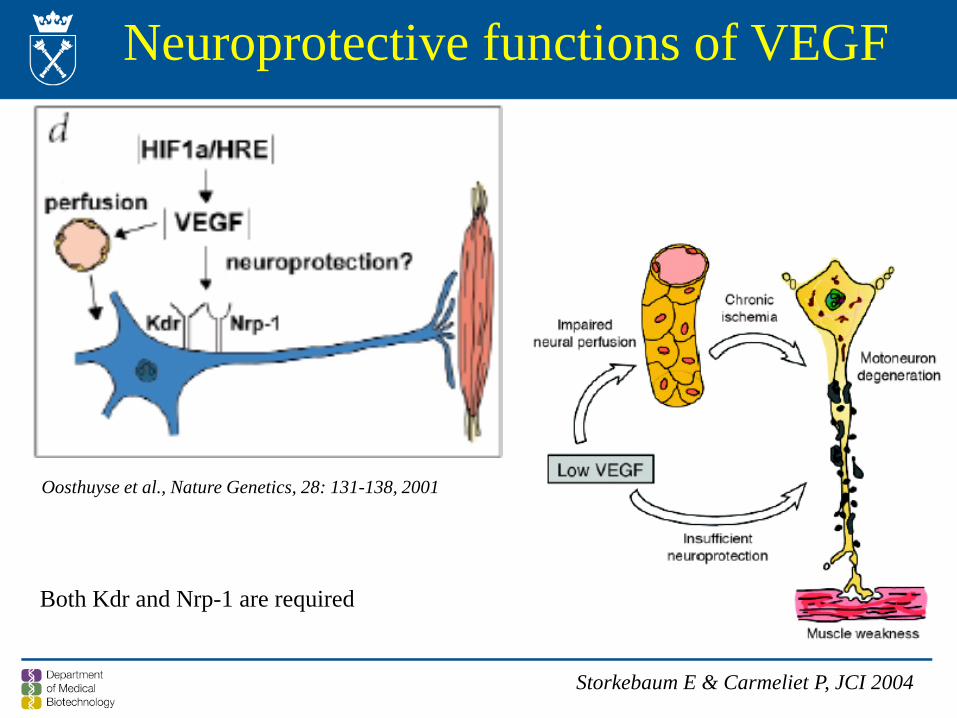
Neuroprotective functions of VEGF
Oosthuyse et al., Nature Genetics, 28: 131-138, 2001
Storkebaum E & Carmeliet P, JCI 2004
Both Kdr and Nrp-1 are required
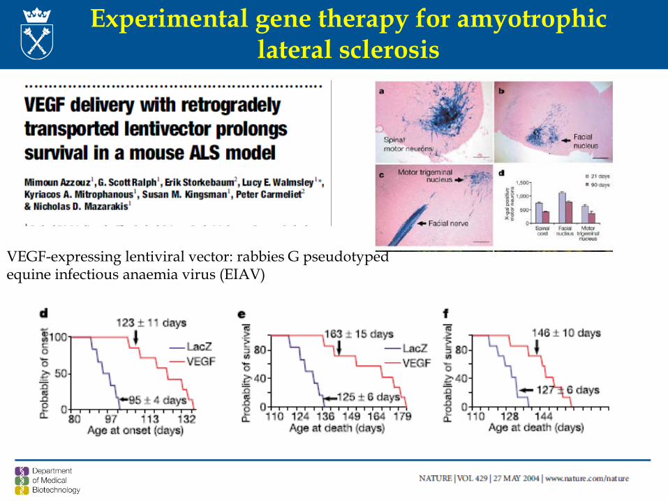
Experimental gene therapy for amyotrophiclateral sclerosis
VEGF-expressing lentiviral vector: rabbies G pseudotypedequine infectious anaemia virus (EIAV)

However, this was an experimental approach which so far has not beenundertaken in the clinical trials
48
First registered gene therapy drug in Europe.
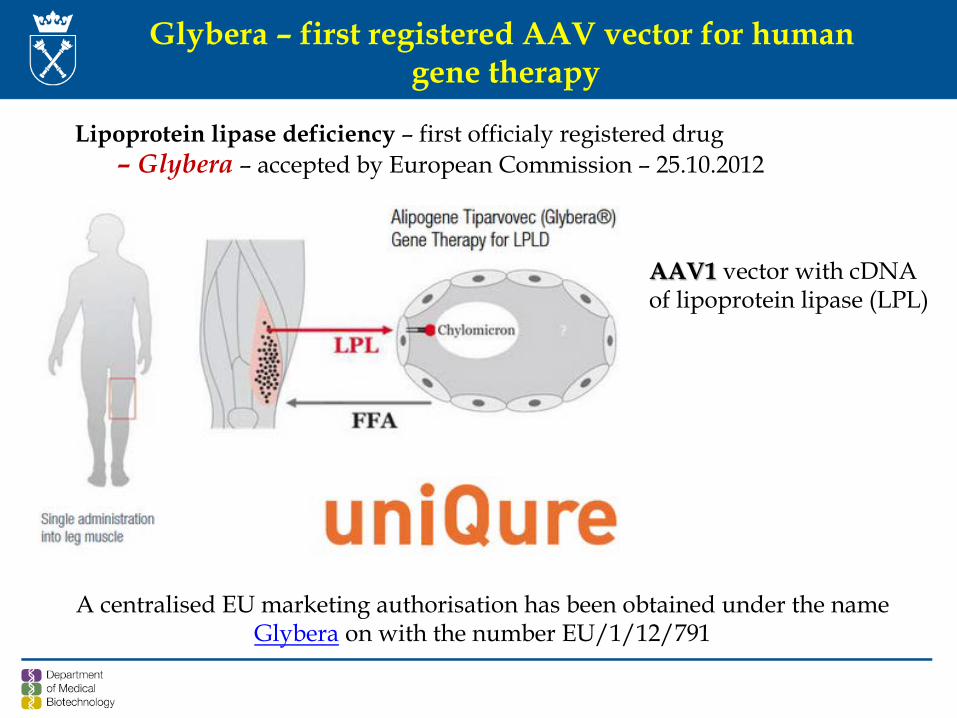
Glybera – first registered AAV vector for humangene therapy
Lipoprotein lipase deficiency – first officialy registered drug– Glybera – accepted by European Commission – 25.10.2012
AAV1 vector with cDNAof lipoprotein lipase (LPL)
A centralised EU marketing authorisation has been obtained under the name Glybera on with the number EU/1/12/791
Glybera• uniQure uses a naturally occurring variant of the LPL gene that has higher enzyme
activity than the normal version of the gene that encodes the protein;• The company produces Glybera using its insect cell-based manufacturing proces;• Clinicians administer Glybera in a one-time series of up to 60 intramuscular injections
in the legs. The patient is administered spinal anesthesia or deep sedation during the procedurę;
• In addition, an immunosuppressive regimen is recommended from three days prior to and for 12 weeks following Glybera administration;
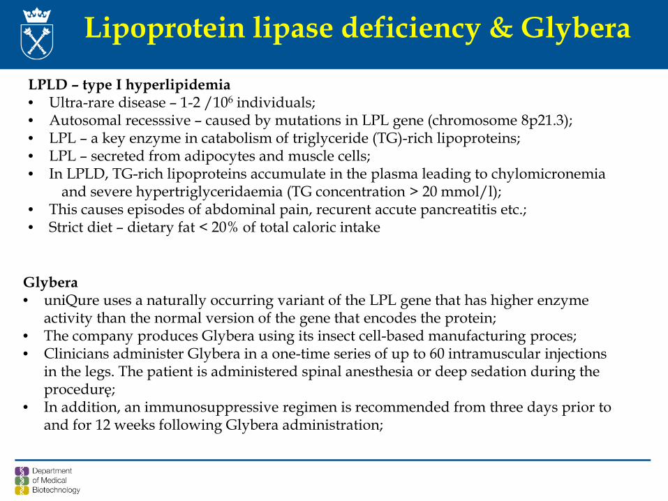
Lipoprotein lipase deficiency & Glybera
LPLD – type I hyperlipidemia• Ultra-rare disease – 1-2 /106 individuals; • Autosomal recesssive – caused by mutations in LPL gene (chromosome 8p21.3); • LPL – a key enzyme in catabolism of triglyceride (TG)-rich lipoproteins; • LPL – secreted from adipocytes and muscle cells; • In LPLD, TG-rich lipoproteins accumulate in the plasma leading to chylomicronemia
and severe hypertriglyceridaemia (TG concentration > 20 mmol/l);• This causes episodes of abdominal pain, recurent accute pancreatitis etc.; • Strict diet – dietary fat < 20% of total caloric intake
To read!
Hildegard Buning
Freely available in PubMed
Gene therapy is effective in a number of monogenic diseases1.Immunodeficiencies
- X-SCID immunodeficiency: retroviral vectors & hematopoietic stem cells- ADA- immunodeficiency - retroviral vectors & hematopoietic stem cells- chronic granulomatous diseases - retroviral vectors & hematopietic stem cells
2. Congential blindness:
- Leber’s congenital amaurosis – rAAV vectors
3. Hemophilia B – rAAV-8 vectors; liver-targeted delivery
4. Metabolic diseases - lipoprotein lipase deficiency - first registered drug
Some beneficial effects have been observed in treatment of:
1.Adrenoleukodystrophy – lentiviral vector & hematopoietic stem cells2.β-thalassemia – lentiviral vector & hematopoietic stem cells
SUMMARY
52

Examples of gene therapy strategies undertaken at the Department of Medical Biotechnology
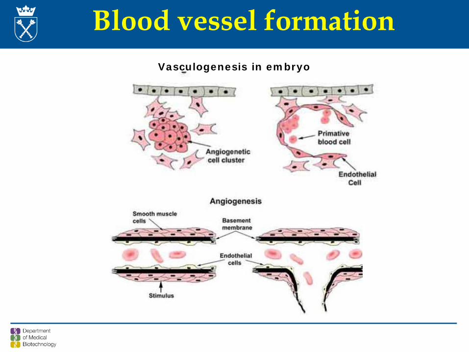
Blood vessel formationVasculogenesis in embryo
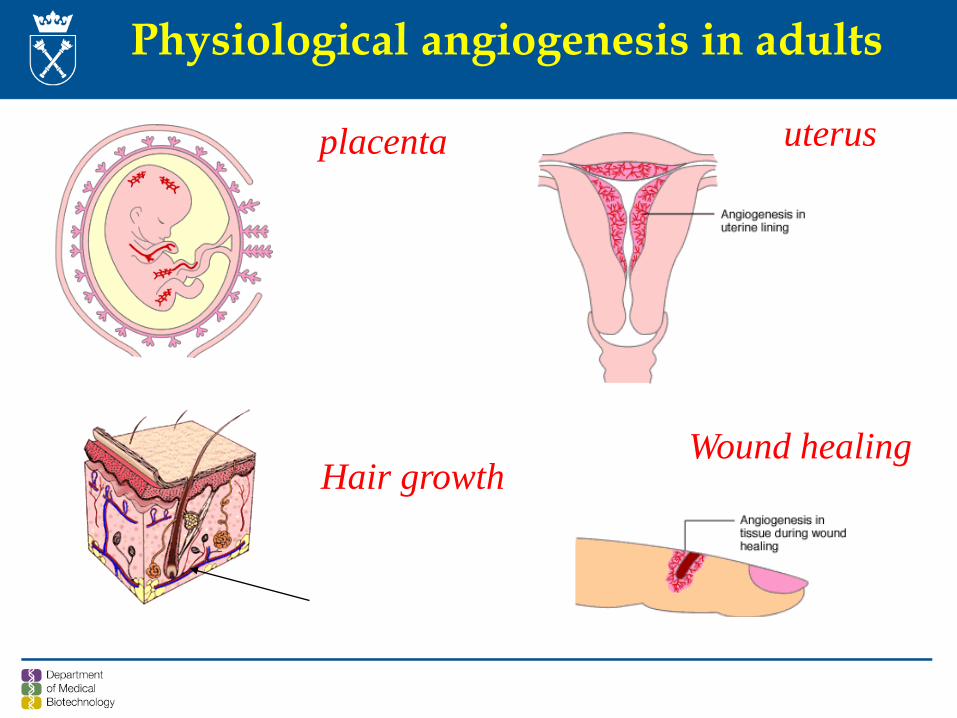
Physiological angiogenesis in adults
placenta uterus
Hair growthWound healing
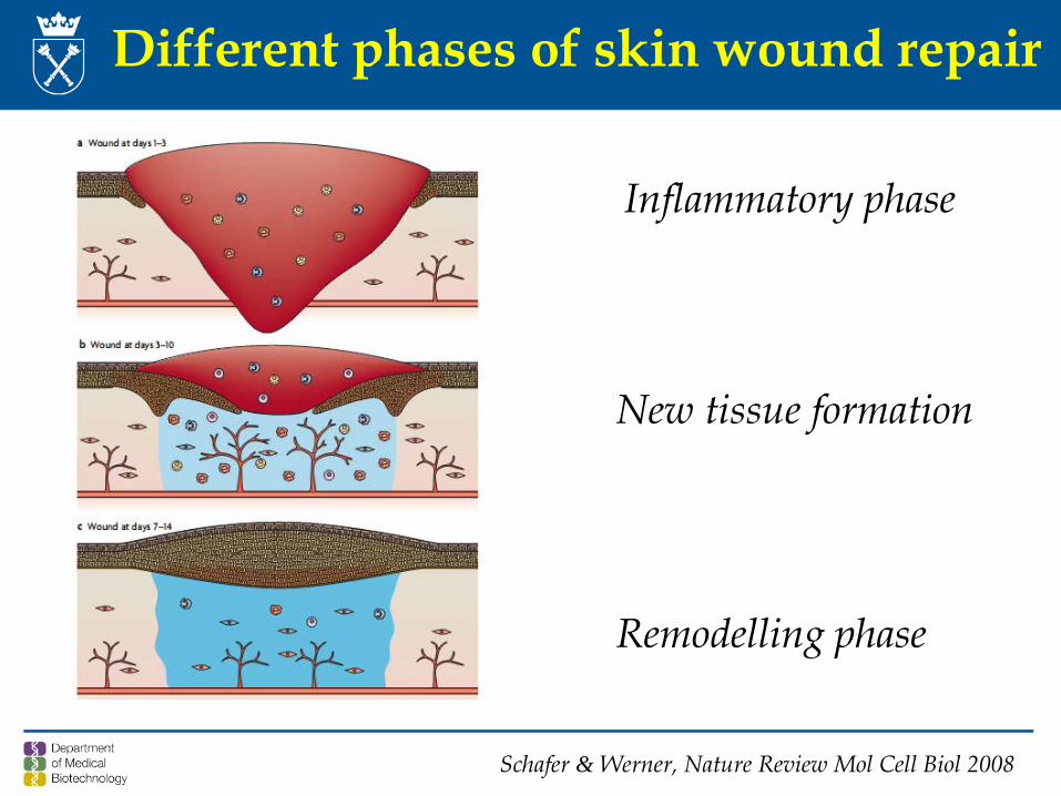
Different phases of skin wound repair
Inflammatory phase
New tissue formation
Remodelling phase
Schafer & Werner, Nature Review Mol Cell Biol 2008
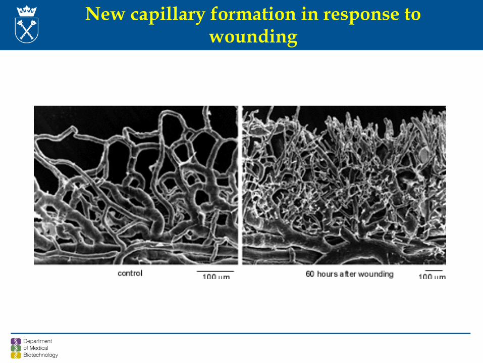
New capillary formation in response to wounding
0
20
40
60
80
100
0 1 2 3 4 5 6 7 8 10 11 13 15
days after wounding
% o
f the
initi
al w
ound
size
WT
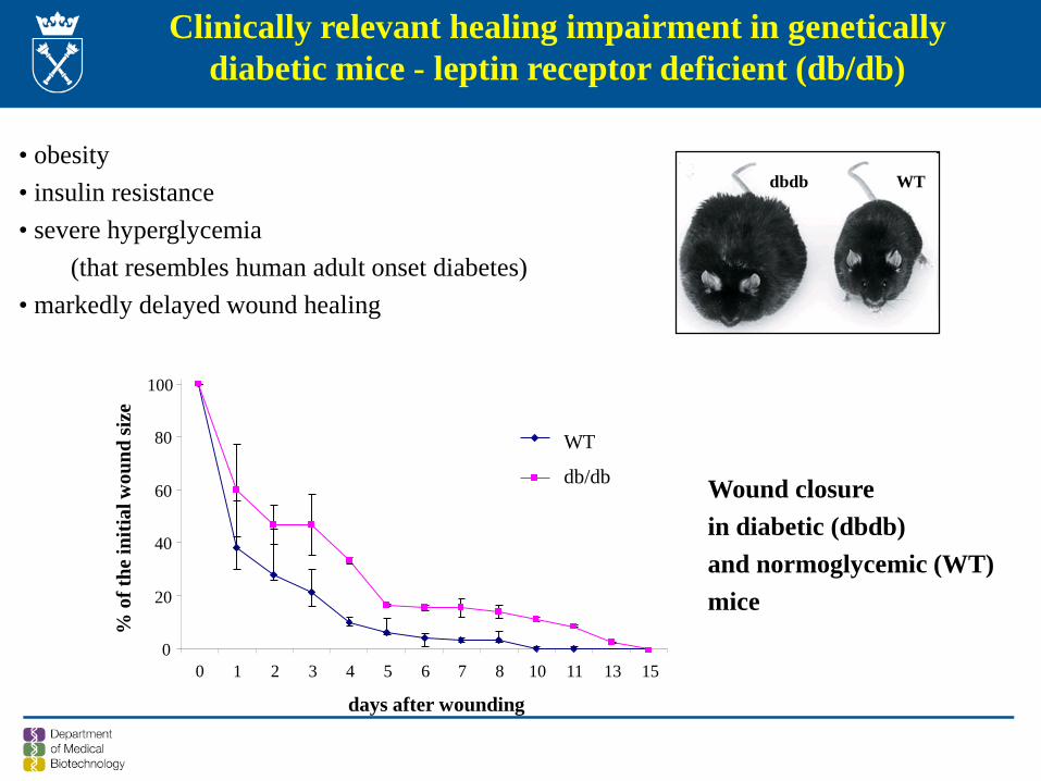
db/db Wound closurein diabetic (dbdb)and normoglycemic (WT)mice
dbdb WT
Clinically relevant healing impairment in genetically diabetic mice - leptin receptor deficient (db/db)
• obesity• insulin resistance• severe hyperglycemia
(that resembles human adult onset diabetes)• markedly delayed wound healing
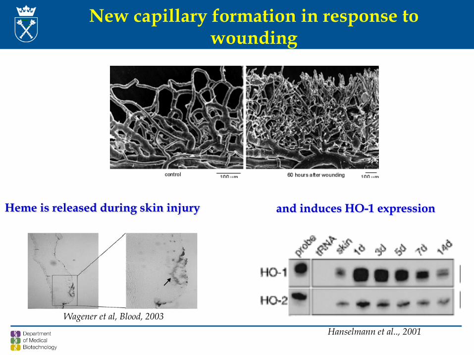
New capillary formation in response to wounding
Wagener et al, Blood, 2003
Heme is released during skin injury
Hanselmann et al.., 2001
and induces HO-1 expression
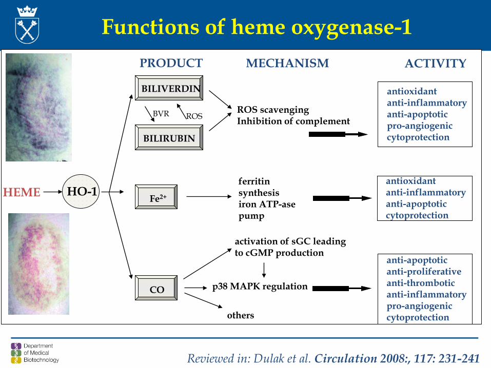
Functions of heme oxygenase-1
HO-1 Fe2+
antioxidant anti-inflammatoryanti-apoptoticpro-angiogeniccytoprotection
ACTIVITYPRODUCT MECHANISM
ferritin synthesisiron ATP-ase pump
ROSBVR
activation of sGC leadingto cGMP production
p38 MAPK regulation
BILIVERDIN
BILIRUBIN
CO
ROS scavengingInhibition of complement
anti-apoptoticanti-proliferative anti-thromboticanti-inflammatorypro-angiogeniccytoprotection
antioxidant anti-inflammatoryanti-apoptoticcytoprotection
HEME
others
Reviewed in: Dulak et al. Circulation 2008:, 117: 231-241
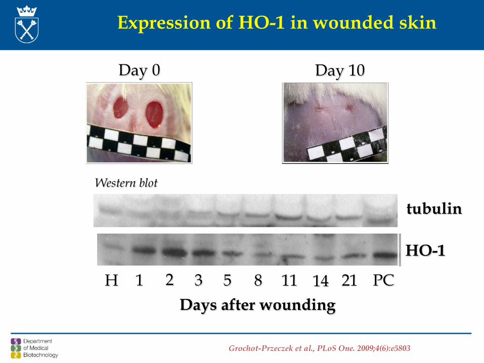
tubulin
HO-1
H 1 2 3 5 8 11 14 21 PCDays after wounding
Expression of HO-1 in wounded skin
Day 0 Day 10
Western blot
Grochot-Przeczek et al., PLoS One. 2009;4(6):e5803
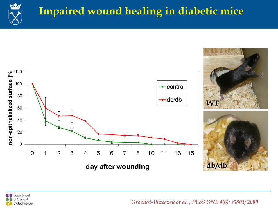
Impaired wound healing in diabetic mice
WT
db/db
Grochot-Przeczek et al. , PLoS ONE 4(6): e5803; 2009
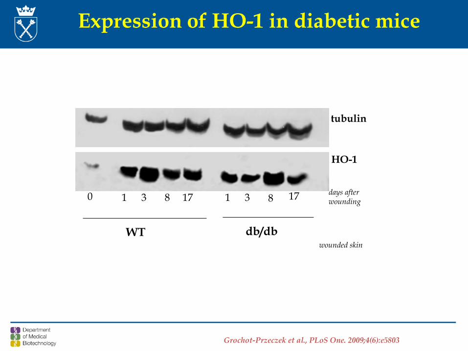
0 1 3 8 17 1 3 8 17
WT db/db
tubulin
HO-1
wounded skin
days after wounding
Expression of HO-1 in diabetic mice
Grochot-Przeczek et al., PLoS One. 2009;4(6):e5803
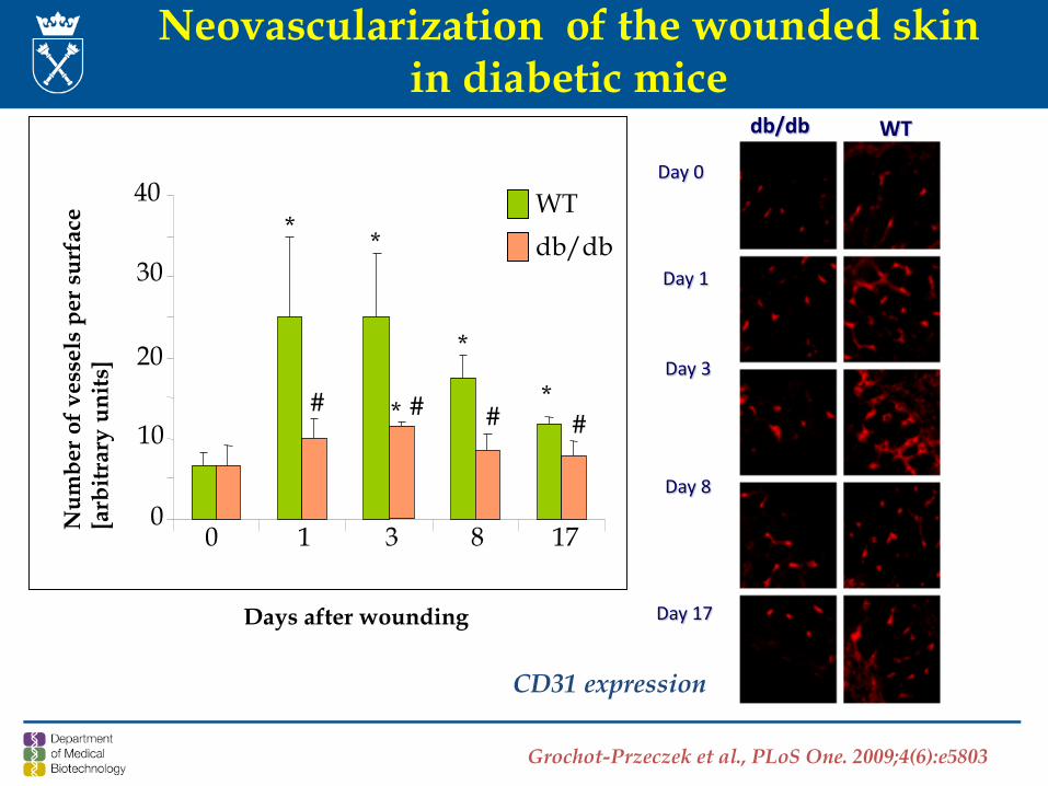
Days after wounding
Num
ber o
f ves
sels
per s
urfa
ce[a
rbitr
ary
units
]
0
10
20
30
40
0 1 3 8 17
* *
*
** ## # #
WTdb/db
db/db WT
Day 1
Day 3
Day 8
Day 17
Day 0
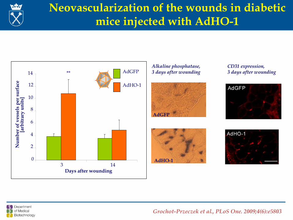
Neovascularization of the wounded skin in diabetic mice
Grochot-Przeczek et al., PLoS One. 2009;4(6):e5803
CD31 expression
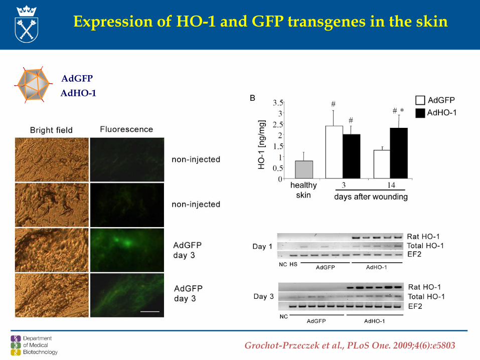
Expression of HO-1 and GFP transgenes in the skin
AdGFPAdHO-1
Grochot-Przeczek et al., PLoS One. 2009;4(6):e5803
Alkaline phosphatase, 3 days after wounding
Neovascularization of the wounds in diabeticmice injected with AdHO-1
Days after wounding
Num
ber o
f ves
sels
per
sur
face
[a
rbitr
ary
units
]
14
3 140
2
4
6
8
10
12
** AdGFP
AdHO-1
AdGFP
AdHO-1
CD31 expression, 3 days after wounding
Grochot-Przeczek et al., PLoS One. 2009;4(6):e5803
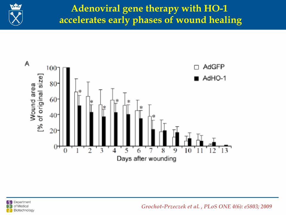
Adenoviral gene therapy with HO-1 accelerates early phases of wound healing
Grochot-Przeczek et al. , PLoS ONE 4(6): e5803; 2009
- HO-1 improves cutaneous wound healing, which is possiblyassociated with increased angiogenesis.
- Impaired induction of HO-1 in wounded skin appears to contribute tothe delayed wound healing in diabetic mice.
- Overexpression of HO-1 by gene transfer may facilitate woundhealing in diabetic mice.
Conclusions
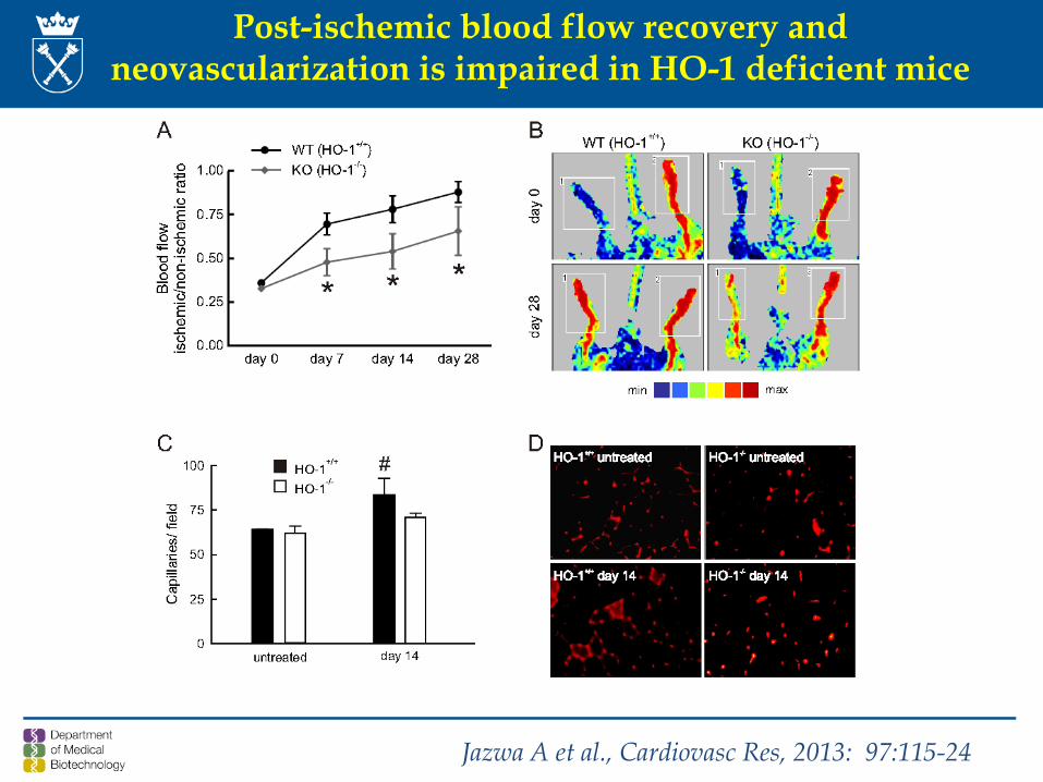
Post-ischemic blood flow recovery and neovascularization is impaired in HO-1 deficient mice
Jazwa A et al., Cardiovasc Res, 2013: 97:115-24
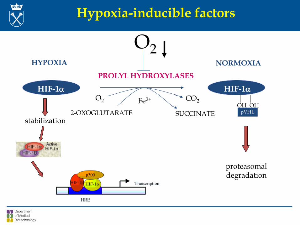
HIF-1α HIF-1α
2-OXOGLUTARATE
O2 CO2
PROLYL HYDROXYLASES
OH OHpVHL
Fe2+
SUCCINATE
NORMOXIAHYPOXIA
proteasomaldegradation
stabilization
O2
Hypoxia-inducible factors
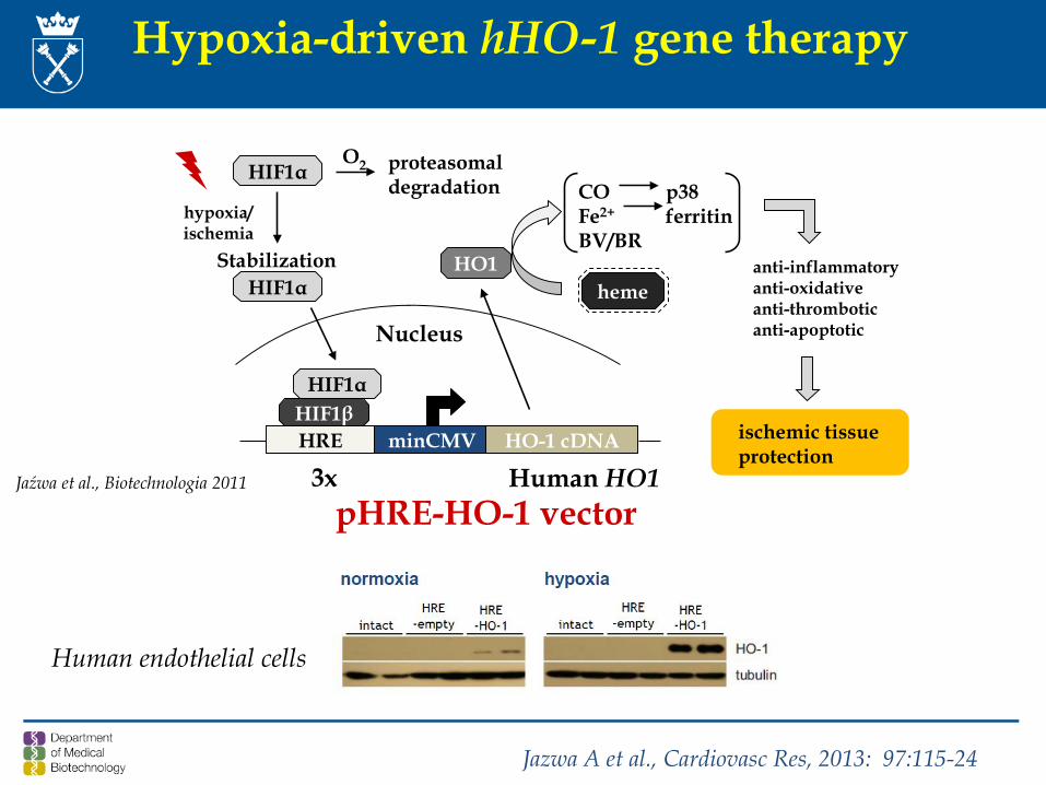
Hypoxia-driven hHO-1 gene therapy
pHRE-HO-1 vector
anti-inflammatoryanti-oxidativeanti-thromboticanti-apoptotic
HIF1αO2 proteasomal
degradationhypoxia/ischemia
Stabilization
HIF1βHRE minCMV HO-1 cDNA
Nucleus
HO1HIF1α
HIF1α
ischemic tissueprotection
heme
CO p38Fe2+ ferritinBV/BR
3x Human HO1Jaźwa et al., Biotechnologia 2011
Jazwa A et al., Cardiovasc Res, 2013: 97:115-24
Human endothelial cells
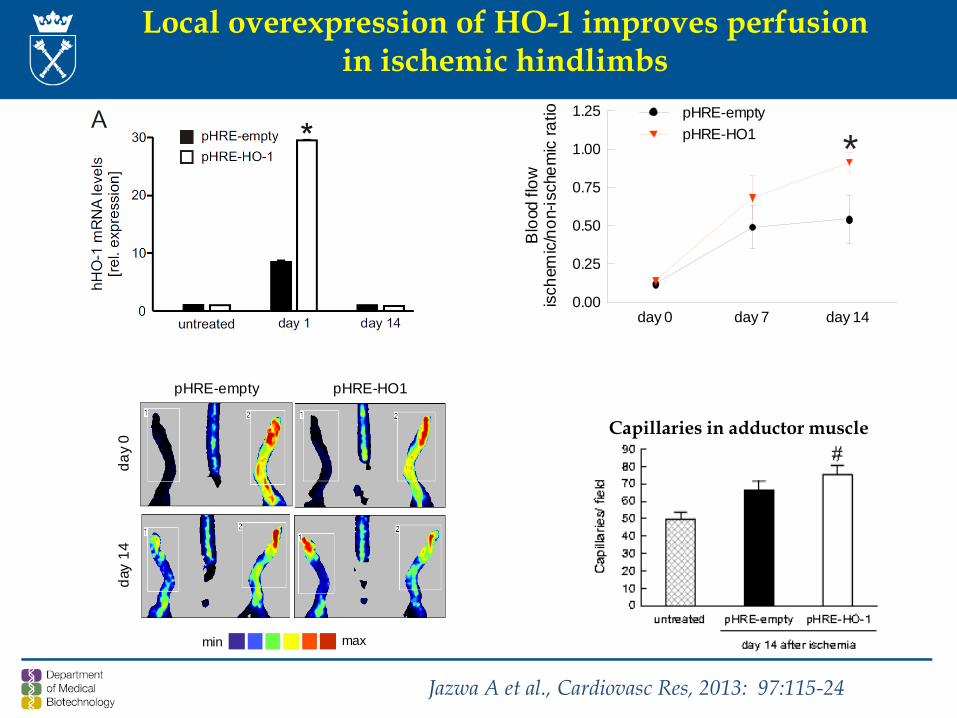
Local overexpression of HO-1 improves perfusionin ischemic hindlimbs
pHRE-empty
day
0
pHRE-HO1
day
14
min max
day 0 day 7 day 140.00
0.25
0.50
0.75
1.00
1.25 pHRE-emptypHRE-HO1
Blo
od fl
owis
chem
ic/n
on-is
chem
ic ra
tio
*
Capillaries in adductor muscle
Jazwa A et al., Cardiovasc Res, 2013: 97:115-24
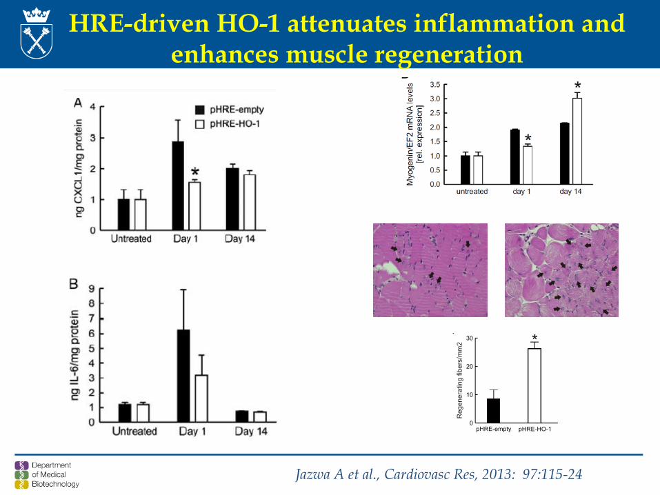
HRE-driven HO-1 attenuates inflammation and enhances muscle regeneration
Jazwa A et al., Cardiovasc Res, 2013: 97:115-24
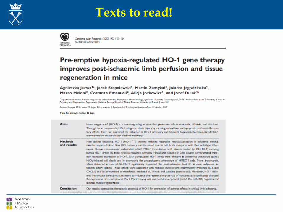
Texts to read!
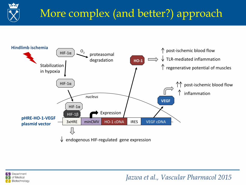
pHRE-HO-1-VEGF plasmid vector
post-ischemic blood flow
TLR-mediated inflammation
regenerative potential of muscles
HIF-1α O2 proteasomaldegradation
Stabilizationin hypoxia
HIF-1β
3xHRE minCMV HO-1 cDNA
Expression
nucleus
HO-1
HIF-1α
Hindlimb ischemia
HIF-1α
IRES VEGF cDNA
VEGF
post-ischemic blood flow
inflammation
endogenous HIF-regulated gene expression
More complex (and better?) approach
Jazwa et al., Vascular Pharmacol 2015
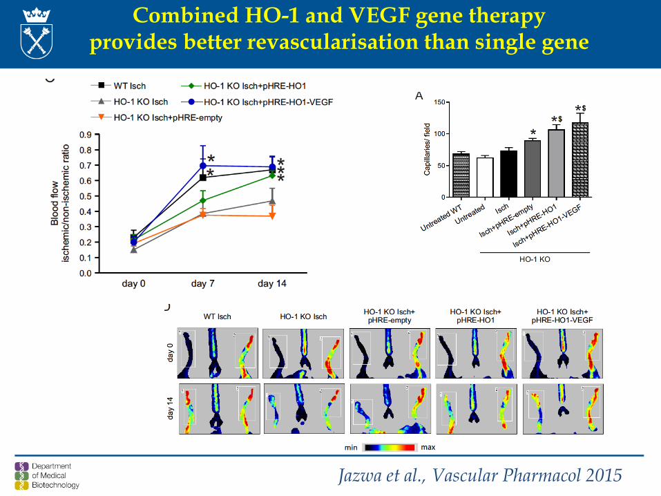
Combined HO-1 and VEGF gene therapyprovides better revascularisation than single gene
Jazwa et al., Vascular Pharmacol 2015
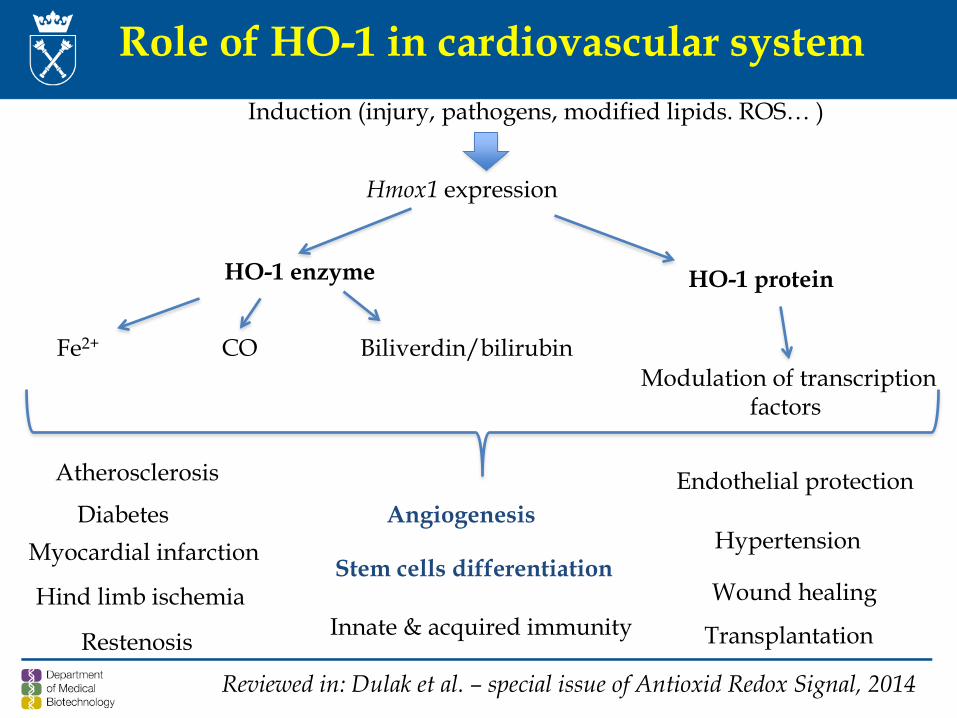
Role of HO-1 in cardiovascular system Induction (injury, pathogens, modified lipids. ROS… )
Hmox1 expression
HO-1 enzyme HO-1 protein
CO Biliverdin/bilirubin
Atherosclerosis
Myocardial infarction
Hind limb ischemia
Diabetes
Stem cells differentiation Wound healing
Restenosis Innate & acquired immunity Transplantation
Modulation of transcriptionfactors
Endothelial protection
Fe2+
Hypertension Angiogenesis
Reviewed in: Dulak et al. – special issue of Antioxid Redox Signal, 2014
Text to read!

Exam – planned on 1st February (Monday), room D107, 1 pm