Embed Size (px)
Citation preview
Prof. Józef Dulak, PhD, DScDepartment of Medical Biotechnology
Faculty of Biochemistry, Biophysics and BiotechnologyRoom 3.025/3.07 Phone 664-63-75
Email: [email protected]
26th October 2015
Viral vectorsPart I
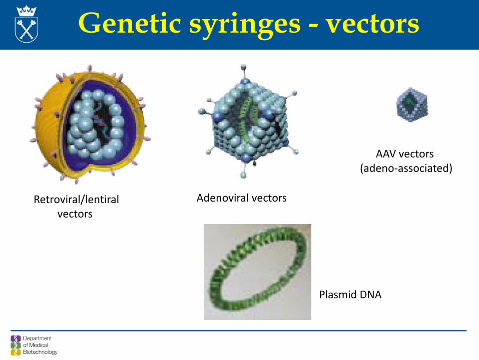
Genetic syringes - vectors
Retroviral/lentiralvectors
Adenoviral vectors
AAV vectors (adeno-associated)
Plasmid DNA
3
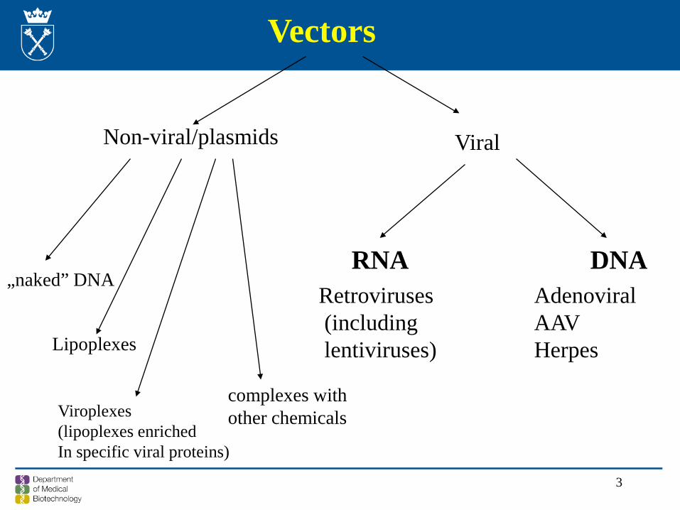
Vectors
Non-viral/plasmids Viral
RNA DNA Retroviruses(including lentiviruses)
AdenoviralAAV Herpes
„naked” DNA
Lipoplexes
Viroplexes(lipoplexes enriched In specific viral proteins)
complexes withother chemicals
4
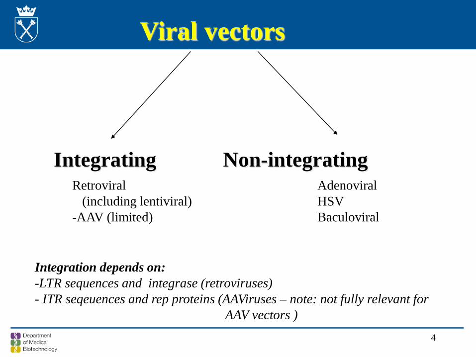
Viral vectors
Integrating Non-integratingRetroviral
(including lentiviral)-AAV (limited)
Adenoviral HSV Baculoviral
Integration depends on:-LTR sequences and integrase (retroviruses) - ITR seqeuences and rep proteins (AAViruses – note: not fully relevant for
AAV vectors )
5
Episomal transgene
Advantage - no risk of insertional mutagenesis
Disadvantage - transient expression - repeated treatments may be necessary
6
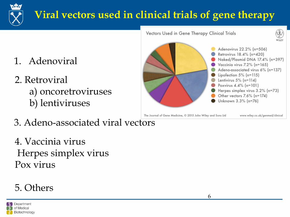
Viral vectors used in clinical trials of gene therapy
1. Adenoviral
2. Retrovirala) oncoretrovirusesb) lentiviruses
3. Adeno-associated viral vectors
4. Vaccinia virusHerpes simplex virus
Pox virus
5. Others
7
Integrated transgene
Advantage: - perpetual - may provide a stable expression and a cure
Disadvantage: - random insertion may lead to silencing of a transgene
or inactivation or dysregulated activation of host genes
- unknown, long-term effects of the transgene
8
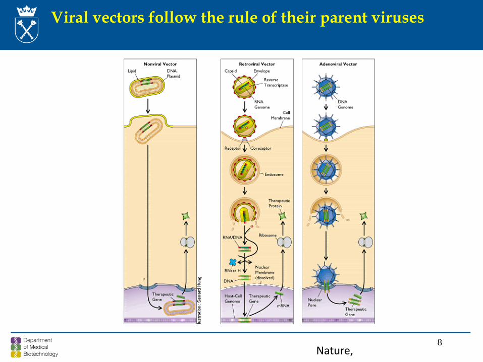
Viral vectors follow the rule of their parent viruses
Nature,
9
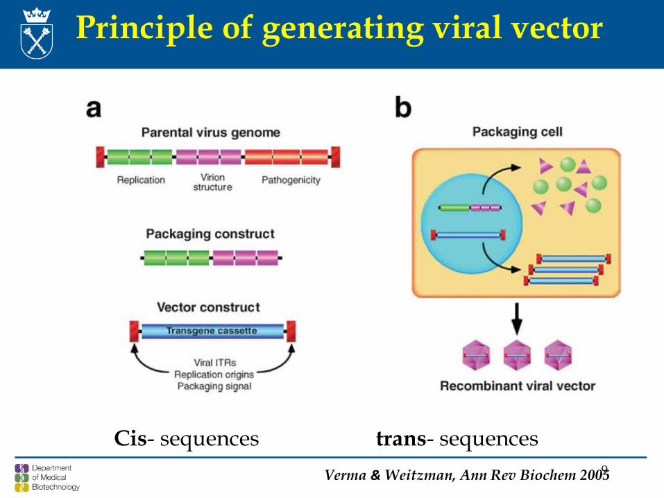
Principle of generating viral vector
Verma & Weitzman, Ann Rev Biochem 2005
Cis- sequences trans- sequences
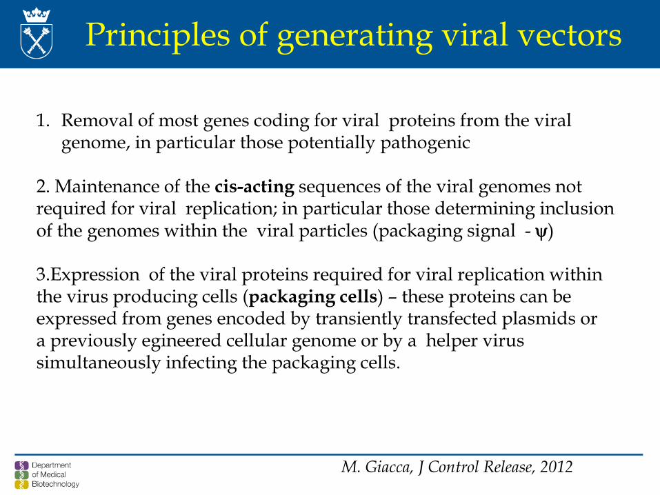
Principles of generating viral vectors
1. Removal of most genes coding for viral proteins from the viralgenome, in particular those potentially pathogenic
2. Maintenance of the cis-acting sequences of the viral genomes not required for viral replication; in particular those determining inclusionof the genomes within the viral particles (packaging signal - ψ)
3.Expression of the viral proteins required for viral replication withinthe virus producing cells (packaging cells) – these proteins can be expressed from genes encoded by transiently transfected plasmids ora previously egineered cellular genome or by a helper virussimultaneously infecting the packaging cells.
M. Giacca, J Control Release, 2012
11
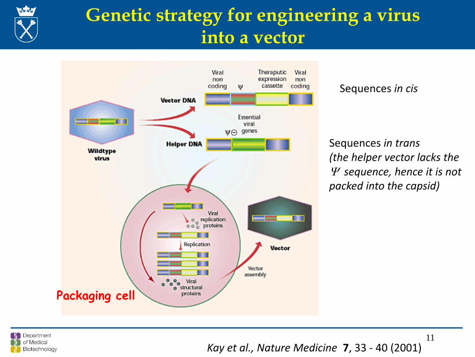
Genetic strategy for engineering a virusinto a vector
Kay et al., Nature Medicine 7, 33 - 40 (2001)
Packaging cell
Sequences in cis
Sequences in trans(the helper vector lacks the Ψ sequence, hence it is notpacked into the capsid)
12
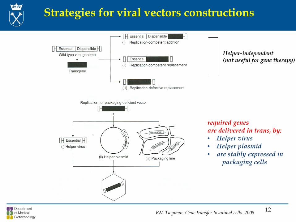
Strategies for viral vectors constructions
RM Twyman, Gene transfer to animal cells. 2005
Helper-independent(not useful for gene therapy)
required genesare delivered in trans, by:• Helper virus• Helper plasmid• are stably expressed in
packaging cells
13
Retroviral vectors
1. Oncoretroviruses: a) Alpharetrovirus – Avian leucosis virus (RSV) b) Betaretrovirus – Mouse mammary tumor virus (MMTV)c) Gammaretrovirus – Murine leukemia virus (Mo-MLV)d) Deltaretrovirus – Bovine leukemia viruse) Epsilonretrovirus – Walleye dermal sarcoma virus
2. Lentivirus – HIV, HTLV, BLV
3. Spumavirus – human spumavirus(human foamy virus – HMV)
Family Retroviridae
14
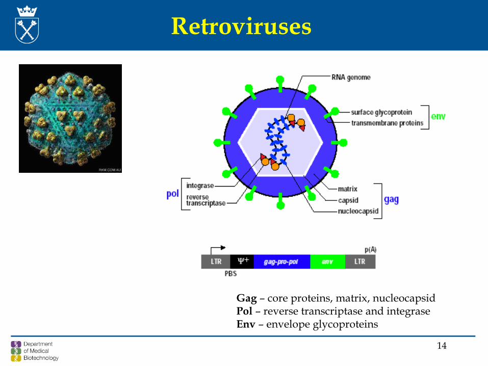
Retroviruses
Gag – core proteins, matrix, nucleocapsidPol – reverse transcriptase and integraseEnv – envelope glycoproteins
15
Genomic organization of retrovirus
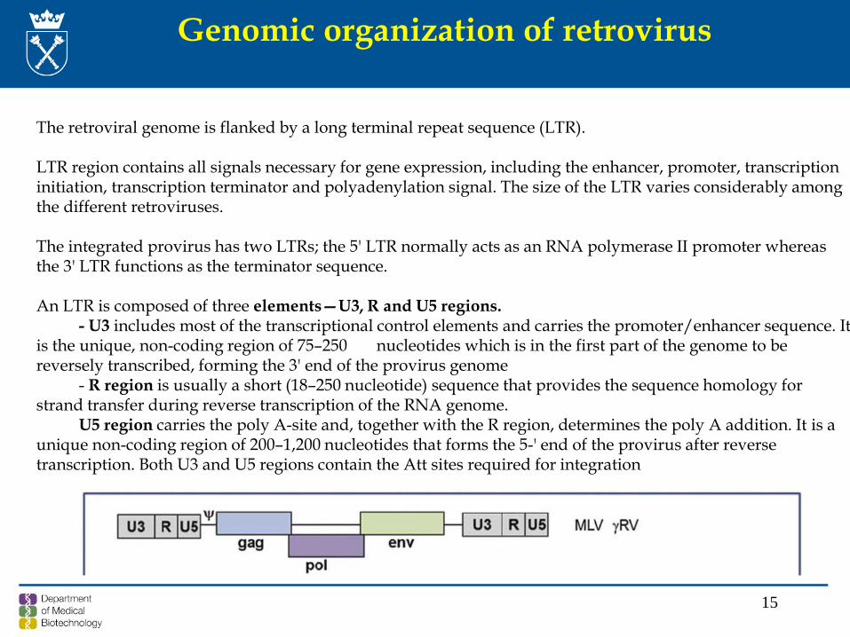
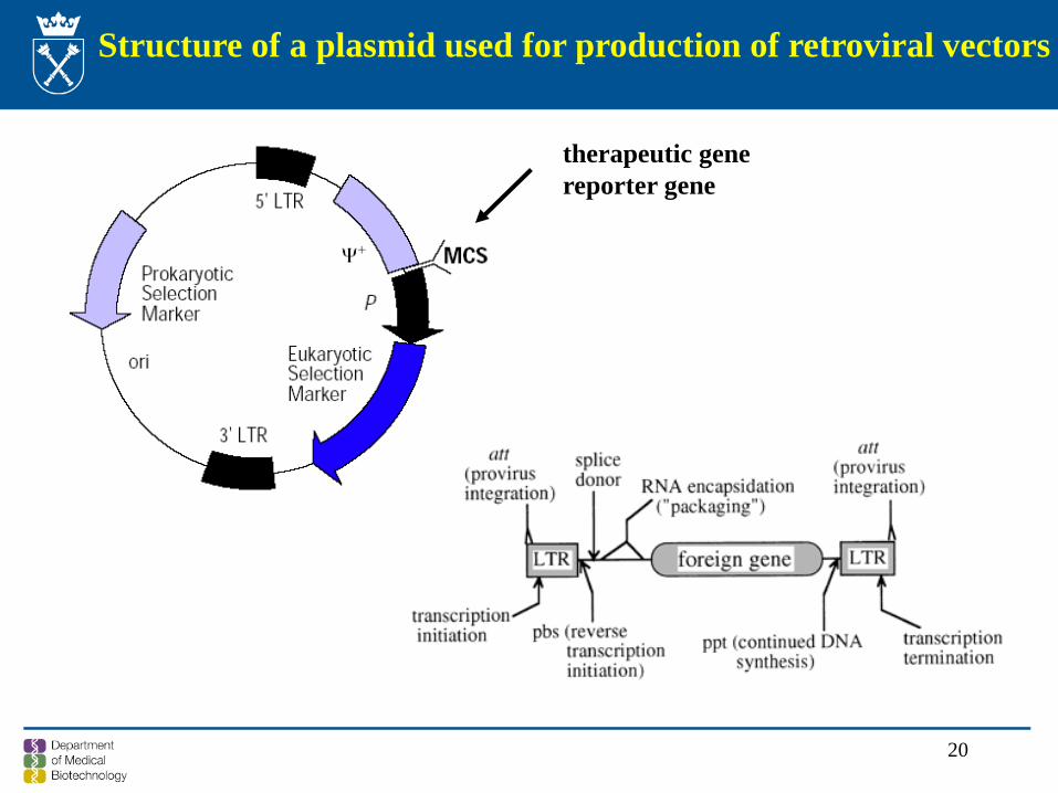
The retroviral genome is flanked by a long terminal repeat sequence (LTR).
LTR region contains all signals necessary for gene expression, including the enhancer, promoter, transcriptioninitiation, transcription terminator and polyadenylation signal. The size of the LTR varies considerably amongthe different retroviruses.
The integrated provirus has two LTRs; the 5' LTR normally acts as an RNA polymerase II promoter whereasthe 3' LTR functions as the terminator sequence.
An LTR is composed of three elements—U3, R and U5 regions. - U3 includes most of the transcriptional control elements and carries the promoter/enhancer sequence. It
is the unique, non-coding region of 75–250 nucleotides which is in the first part of the genome to be reversely transcribed, forming the 3' end of the provirus genome
- R region is usually a short (18–250 nucleotide) sequence that provides the sequence homology for strand transfer during reverse transcription of the RNA genome.
U5 region carries the poly A-site and, together with the R region, determines the poly A addition. It is a unique non-coding region of 200–1,200 nucleotides that forms the 5-' end of the provirus after reversetranscription. Both U3 and U5 regions contain the Att sites required for integration
16
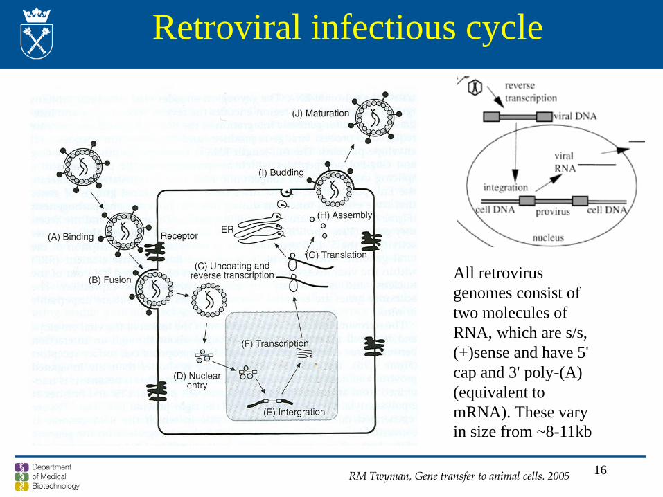
Retroviral infectious cycle
All retrovirus genomes consist of two molecules of RNA, which are s/s, (+)sense and have 5' cap and 3' poly-(A) (equivalent to mRNA). These vary in size from ~8-11kb
RM Twyman, Gene transfer to animal cells. 2005
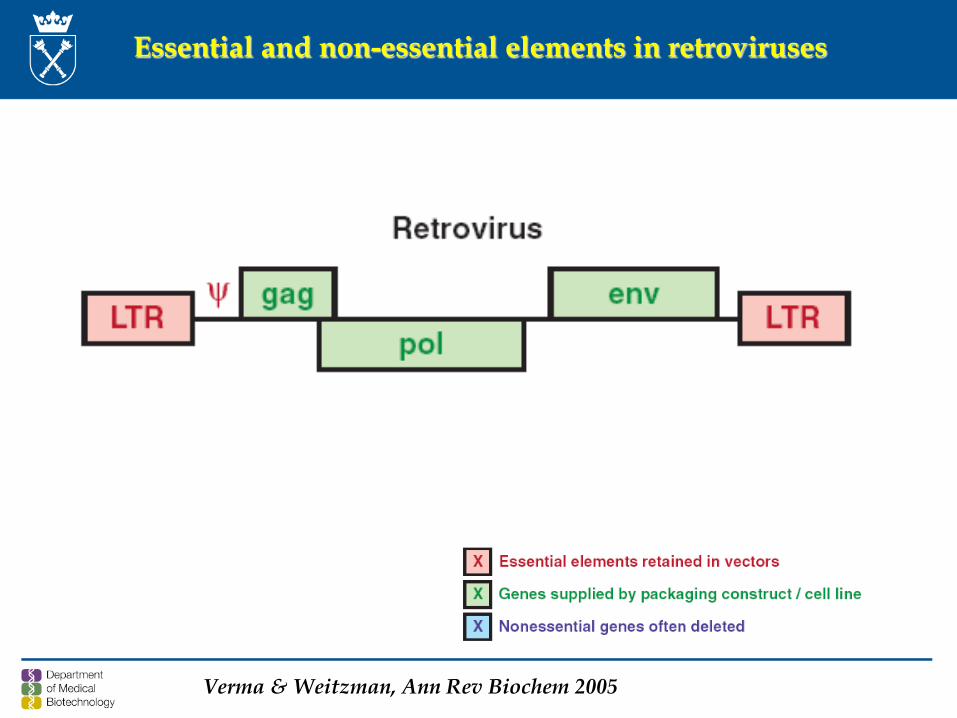
Essential and non-essential elements in retroviruses
Verma & Weitzman, Ann Rev Biochem 2005
18
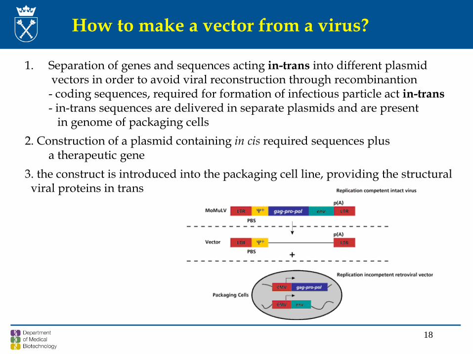
How to make a vector from a virus?
1. Separation of genes and sequences acting in-trans into different plasmidvectors in order to avoid viral reconstruction through recombinantion- coding sequences, required for formation of infectious particle act in-trans - in-trans sequences are delivered in separate plasmids and are present
in genome of packaging cells2. Construction of a plasmid containing in cis required sequences plus
a therapeutic gene3. the construct is introduced into the packaging cell line, providing the structural
viral proteins in trans
19
Construction of retrovirus vector
1. Construction of retrovirus vector as a recombinant plasmid in E.coli
2. Introduction of a plasmid into a packaging cell line
3. Incorporation of vector transcripts into transmissible virus particles
20
Structure of a plasmid used for production of retroviral vectors
therapeutic genereporter gene
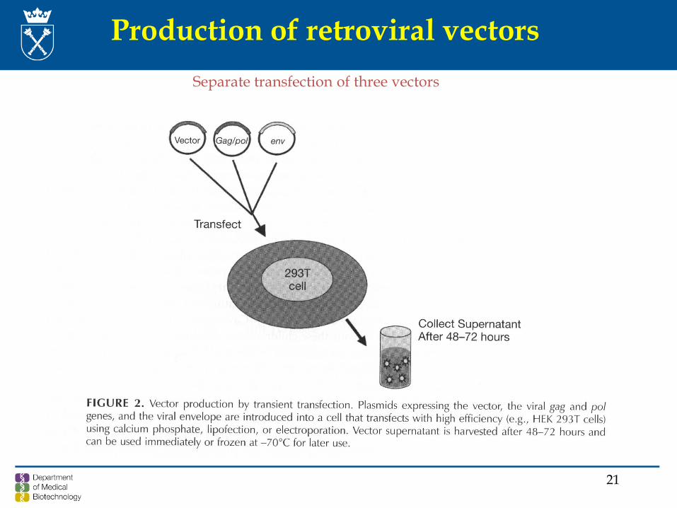
21
Production of retroviral vectorsSeparate transfection of three vectors
22
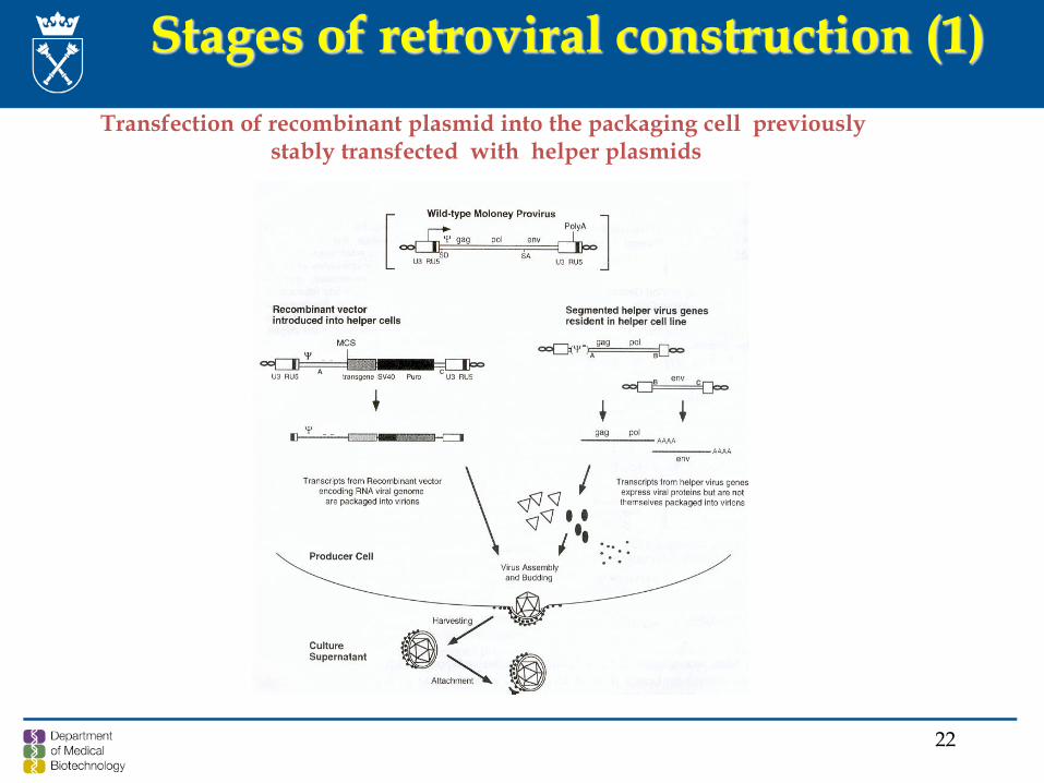
Stages of retroviral construction (1) Transfection of recombinant plasmid into the packaging cell previously
stably transfected with helper plasmids
23
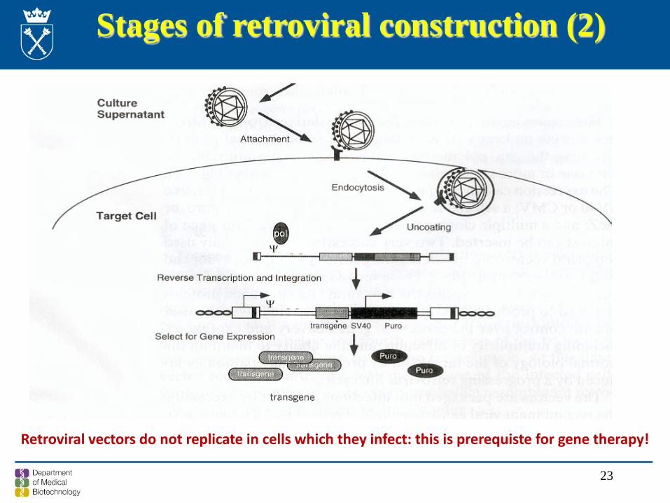
Stages of retroviral construction (2)
Retroviral vectors do not replicate in cells which they infect: this is prerequiste for gene therapy!
24
Features of retroviral vectors
Due to destruction of a natural, well-functioning genecomposition, required for the formation of infectiousvirus, the resulting system of retroviral vectorsynthesis suffers from:
- low efficiency of packaging of vectors in comparison to wild type viruses
- formation of a large number of defective particles, which inhibit the transduction efficiency
25
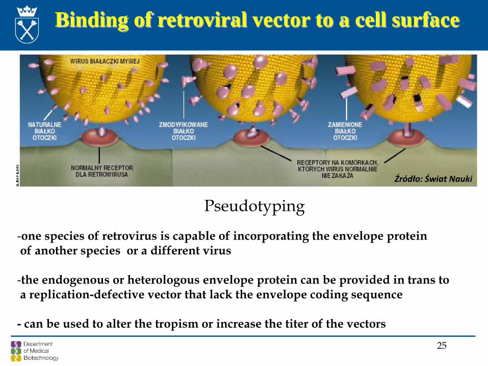
Binding of retroviral vector to a cell surface
Pseudotyping
-one species of retrovirus is capable of incorporating the envelope protein of another species or a different virus
-the endogenous or heterologous envelope protein can be provided in trans to a replication-defective vector that lack the envelope coding sequence
- can be used to alter the tropism or increase the titer of the vectors
Źródło: Świat Nauki
G protein of vesicular stomatitis virus (VSVG)
An envelope virus belonging to the family Rhabdoviridae
- Binds the phospholipids present on virtually allmammalian cell membranes & triggers endocytosis of the viral particles
- Lowering the pH in endosomes activates the fusiogenicproperties of VSVG, which determine fusion of the viralenvelope with the endosomal membrane and release of the virion content into the cytosol
27
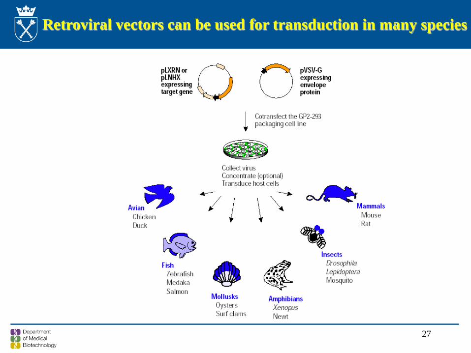
Retroviral vectors can be used for transduction in many species
28
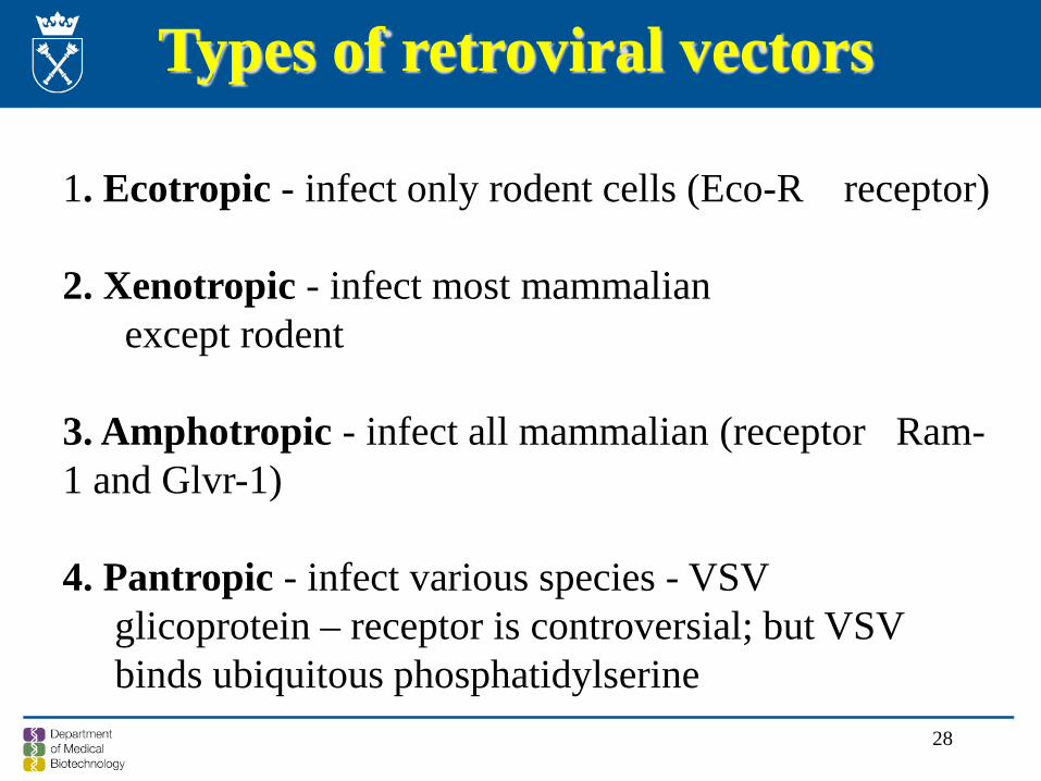
1. Ecotropic - infect only rodent cells (Eco-R receptor)
2. Xenotropic - infect most mammalianexcept rodent
3. Amphotropic - infect all mammalian (receptor Ram-1 and Glvr-1)
4. Pantropic - infect various species - VSV glicoprotein – receptor is controversial; but VSV binds ubiquitous phosphatidylserine
Types of retroviral vectors
Targeting retroviral vectors to specific cell types
1. Neuronal – RVG/VSVG chimeric proteins – a fusion productbetween the external domain of Rabies virus glycoprotein and the cytoplasmic domain of VSVG
2. LCMV-GP – lymphotropic choriomeningitis virus – specificinfection of glioma cells while sparing neurons – for brain cancertherapy
3. Targeting to dendritic cells – by binding to C-type lectins, using the proteins from
- glycoprotein from Sindbis virus- gp64 – a major envelope protein of baculovirus- Aura virus glycoproteins
30
Retroviral gene delivery is the method of choice for gene expression in higher organisms because it is generally faster, more reliable, and has broader utility than
alternative gene transfer methods.
Infection efficiencies of >90%
Efficient transduction of "difficult" cell types including primary, explant, embryonic stem (ES) and embryonic carcinoma (EC) cells
The host range of retroviruses has been expanded by pseudotyping the vectors with heterologous viral glycoproteins and receptor-specific ligands. This is possible because one species of retrovirus is capable of incorporating the
envelope from another species or type of retrovirus. Therefore, the envelope protein can be provided in trans so that the virus produced can infect cells
based on the tropism of that envelope protein
Transduction without variability or loss of expression
Significance of retroviral vectors
31
Retroviral Vectors as Gene Delivery Tools
The ability to transduce a variety of cell types
The ability to integrate efficiently into the genomic DNA of the dividing or mitotically active recipient cells
The ability to express the transduced gene at high levels
Capacity for long-term persistence and stable transmission of the gene to all future progeny of the transduced cell
Up to 6.5 kb of foreign gene sequence can be packaged in a retroviral vector. This is adequate for most applications
Ability to be manufactured in large quantities to meet very stringent safety specifications
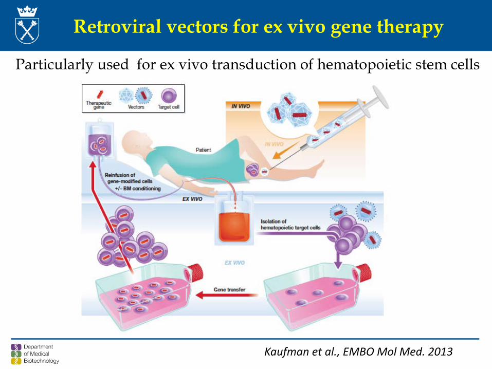
Retroviral vectors for ex vivo gene therapy
Particularly used for ex vivo transduction of hematopoietic stem cells
Kaufman et al., EMBO Mol Med. 2013
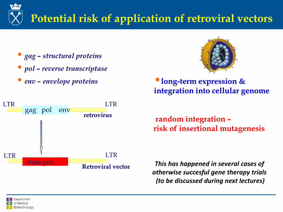
Potential risk of application of retroviral vectors
•long-term expression & integration into cellular genome
gag pol envLTR LTR
retrovirus
transgenLTRLTR
Retroviral vector
• gag – structural proteins
• pol – reverse transcriptase
• env – envelope proteins
random integration –risk of insertional mutagenesis
This has happened in several cases of otherwise succesful gene therapy trials
(to be discussed during next lectures)
34
Side effects of MMLV-based retroviral vectors prompted investigations
of the mechanisms of integration and search for the new, safer vectors
35
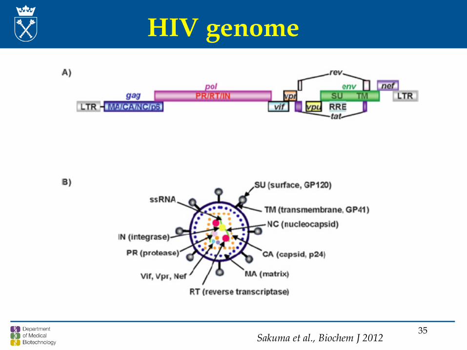
HIV genome
Sakuma et al., Biochem J 2012
36
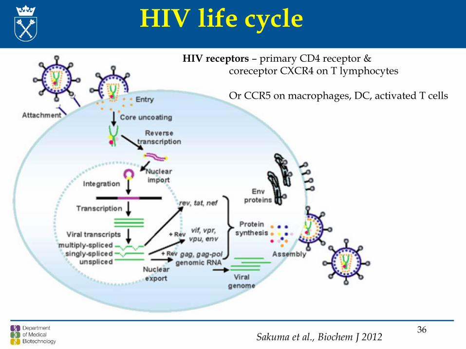
HIV life cycle
Sakuma et al., Biochem J 2012
HIV receptors – primary CD4 receptor & coreceptor CXCR4 on T lymphocytes
Or CCR5 on macrophages, DC, activated T cells
37
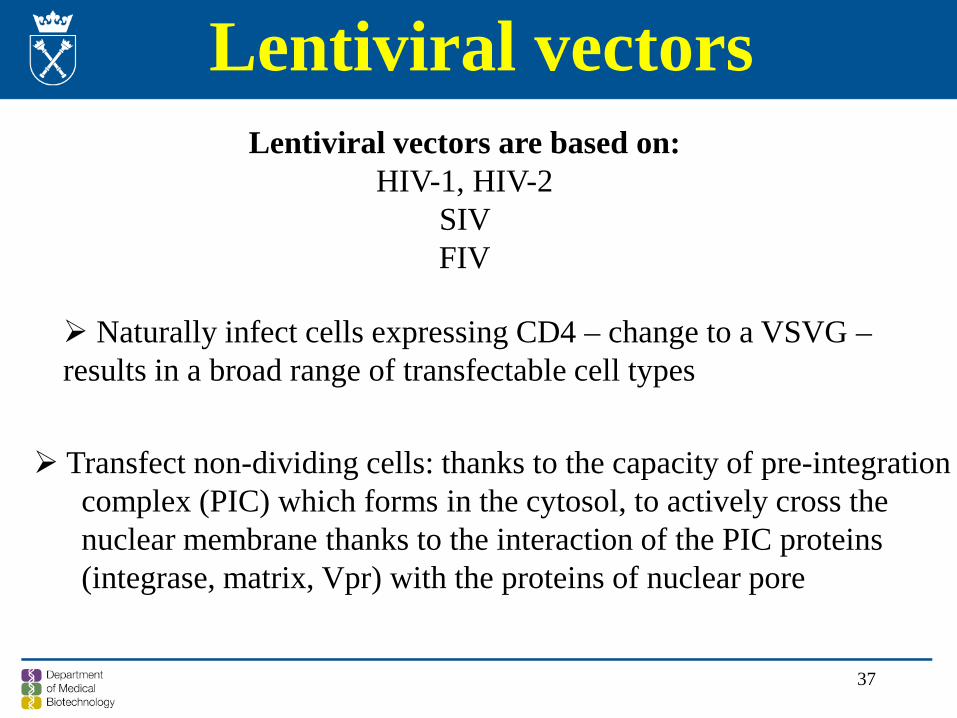
Lentiviral vectorsLentiviral vectors are based on:
HIV-1, HIV-2SIV FIV
Naturally infect cells expressing CD4 – change to a VSVG –results in a broad range of transfectable cell types
Transfect non-dividing cells: thanks to the capacity of pre-integrationcomplex (PIC) which forms in the cytosol, to actively cross the nuclear membrane thanks to the interaction of the PIC proteins(integrase, matrix, Vpr) with the proteins of nuclear pore
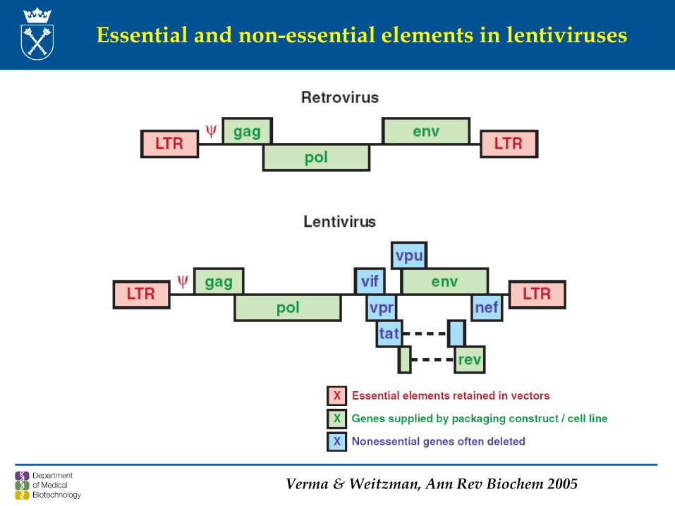
Essential and non-essential elements in lentiviruses
Verma & Weitzman, Ann Rev Biochem 2005
39
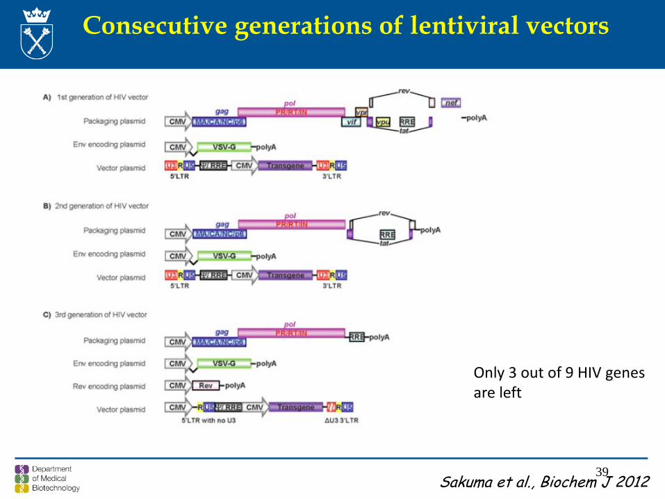
Consecutive generations of lentiviral vectors
Sakuma et al., Biochem J 2012
Only 3 out of 9 HIV genesare left
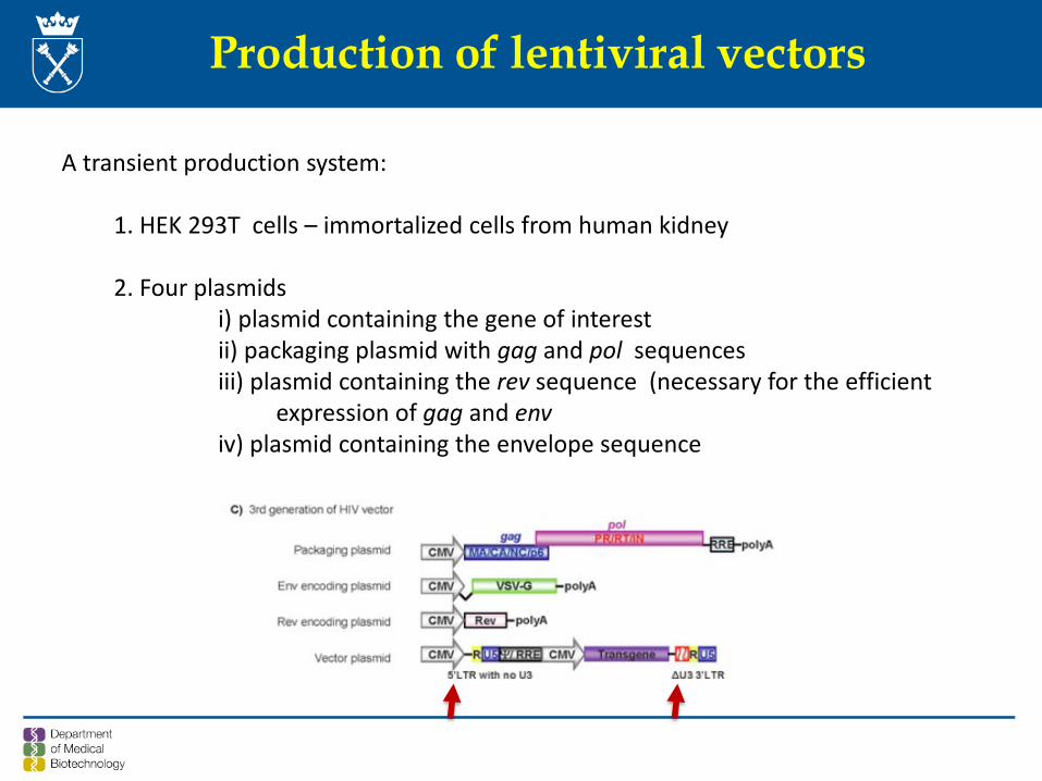
Production of lentiviral vectors
A transient production system:
1. HEK 293T cells – immortalized cells from human kidney
2. Four plasmidsi) plasmid containing the gene of interestii) packaging plasmid with gag and pol sequencesiii) plasmid containing the rev sequence (necessary for the efficient
expression of gag and enviv) plasmid containing the envelope sequence
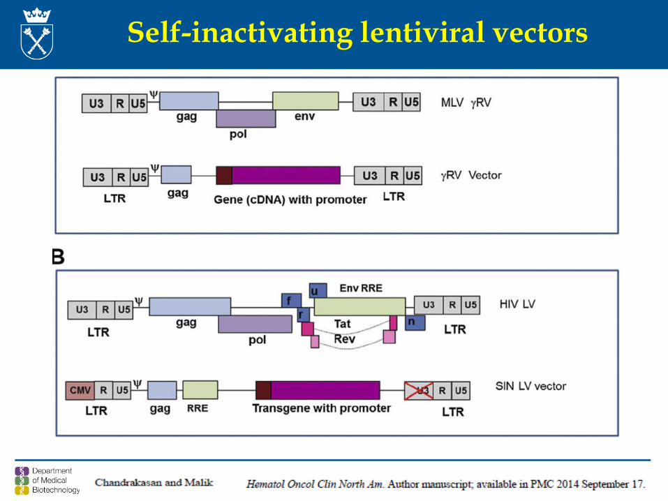
Self-inactivating lentiviral vectors
Self-inactivation of lentiviral vectors
Deletion of 3’-LTR elements, including its TAT-boc, Sp-1, NF-kB and NFAT binding site
This reduces the likehood of:
1. Propagation of spontaneously replication-competent recombinant HIV-like viruses;
1. insertional activation of cellular oncogenes by residual promtoer activitiesof integrated LTRs;
3. Mobilisation of integrated vectors by wild-type virus;
4. Transcriptional interference and suppresion by LTRs
Safety of lentiviral vectors
1. Replication deficient
1. Six out of nine genes involved in the lentiviral pathogenecity are removed;envvif – viral infectivity factorVpr – viral protein r Vpu – viral protein u Nef – negative regulatory factorTat – transactivator
3. Self –inactivated
4. Integration properties different than oncoretroviruses
Self-inactivating lentiviruses: deletion of 299 bp in 3’ LTR causes after transductioninactivation of 5’LTR, decreasing the risk of recombination and vector mobilisation
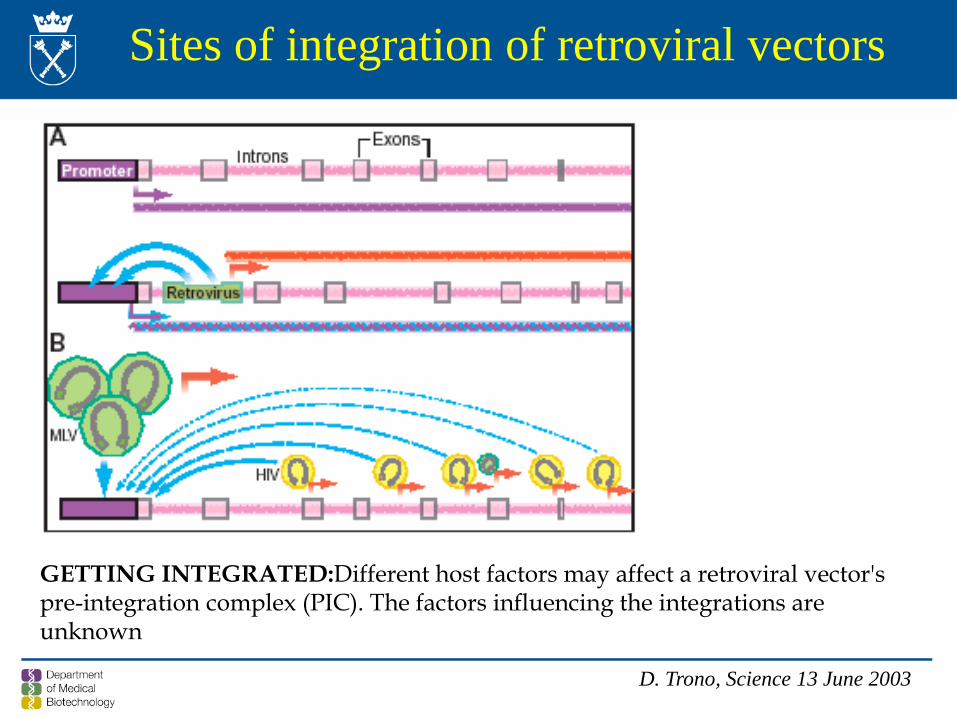
Sites of integration of retroviral vectors
D. Trono, Science 13 June 2003
GETTING INTEGRATED:Different host factors may affect a retroviral vector's pre-integration complex (PIC). The factors influencing the integrations are unknown
45
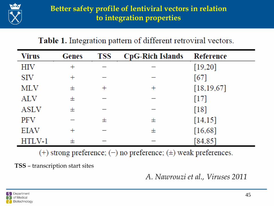
A. Nawrouzi et al., Viruses 2011TSS – transcription start sites
Better safety profile of lentiviral vectors in relationto integration properties
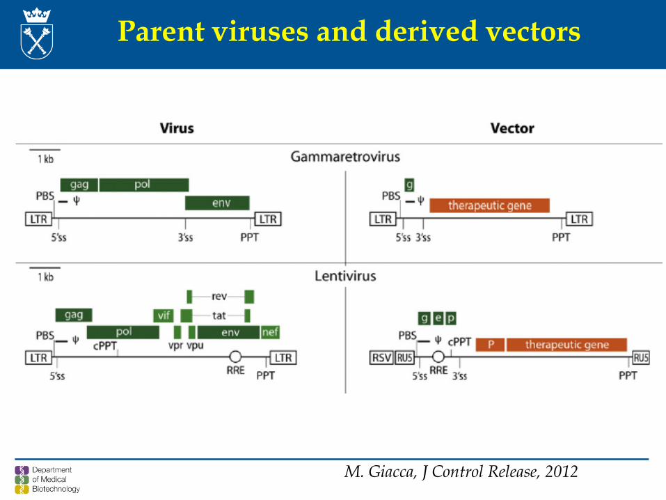
M. Giacca, J Control Release, 2012
Parent viruses and derived vectors
47
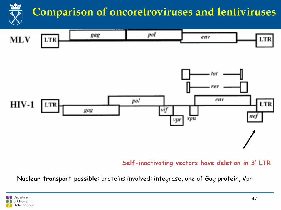
Comparison of oncoretroviruses and lentiviruses
Self-inactivating vectors have deletion in 3’ LTR
Nuclear transport possible: proteins involved: integrase, one of Gag protein, Vpr
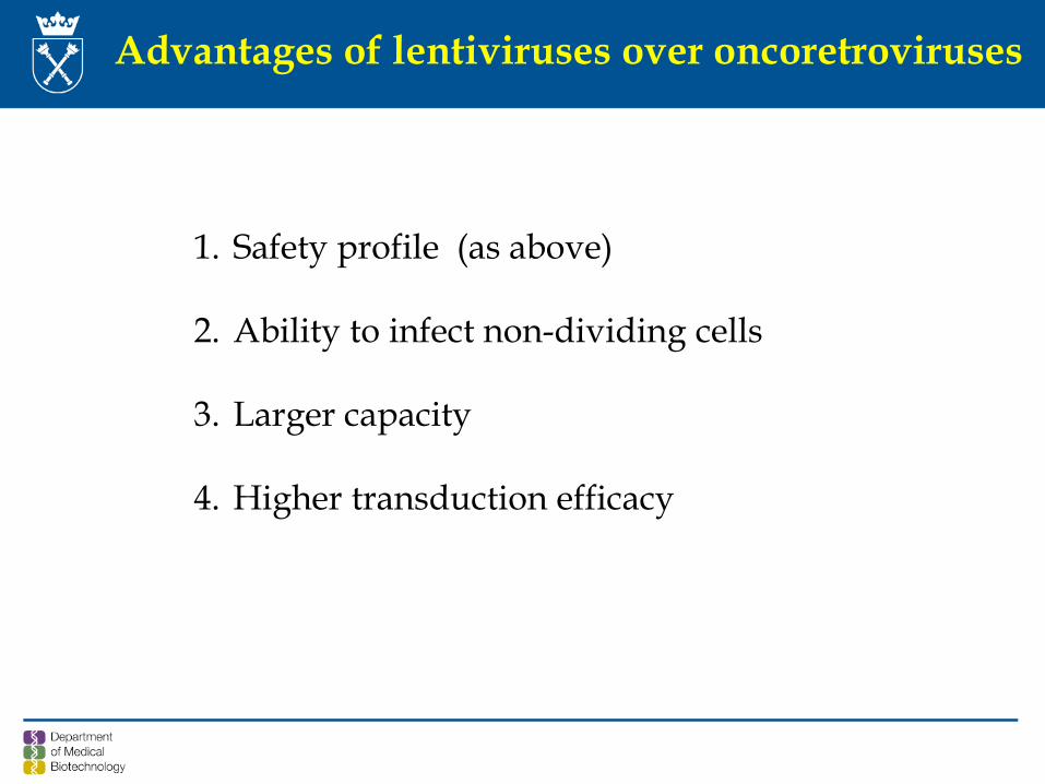
Advantages of lentiviruses over oncoretroviruses
1. Safety profile (as above)
2. Ability to infect non-dividing cells
3. Larger capacity
4. Higher transduction efficacy
49
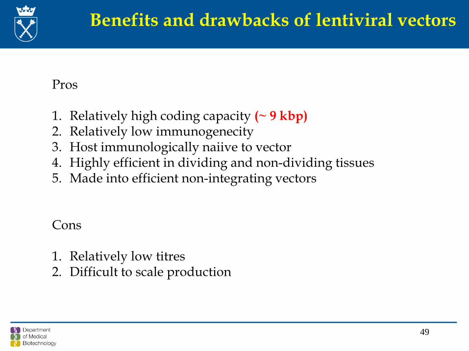
Benefits and drawbacks of lentiviral vectors
Pros
1. Relatively high coding capacity (~ 9 kbp) 2. Relatively low immunogenecity3. Host immunologically naiive to vector4. Highly efficient in dividing and non-dividing tissues5. Made into efficient non-integrating vectors
Cons
1. Relatively low titres2. Difficult to scale production
50
Titer (miano) of viral vectors
Concentration of viral particles and/or virionswhich are able to transduce the cells
The titer represents only a small fractionof a total number of viral particles
51
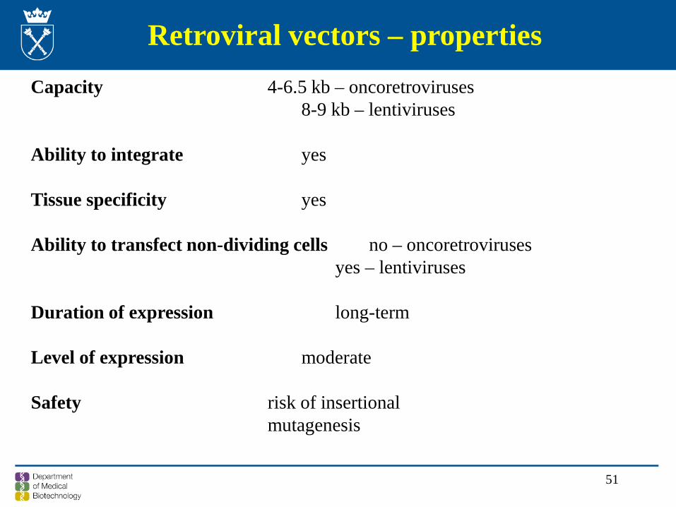
Retroviral vectors – propertiesCapacity 4-6.5 kb – oncoretroviruses
8-9 kb – lentiviruses
Ability to integrate yes
Tissue specificity yes
Ability to transfect non-dividing cells no – oncoretrovirusesyes – lentiviruses
Duration of expression long-term
Level of expression moderate
Safety risk of insertionalmutagenesis
52
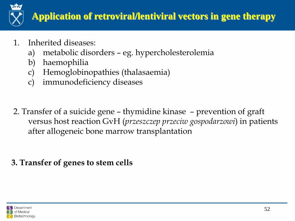
Application of retroviral/lentiviral vectors in gene therapy
1. Inherited diseases: a) metabolic disorders – eg. hypercholesterolemiab) haemophiliac) Hemoglobinopathies (thalasaemia)c) immunodeficiency diseases
2. Transfer of a suicide gene – thymidine kinase – prevention of graftversus host reaction GvH (przeszczep przeciw gospodarzowi) in patientsafter allogeneic bone marrow transplantation
3. Transfer of genes to stem cells
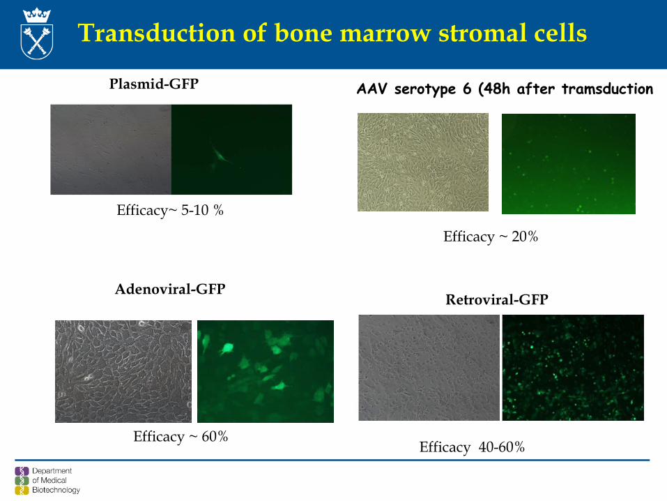
Transduction of bone marrow stromal cells
Retroviral-GFP Adenoviral-GFP
Plasmid-GFP
Efficacy 40-60%Efficacy ~ 60%
AAV serotype 6 (48h after tramsduction
Efficacy ~ 20%
Efficacy~ 5-10 %
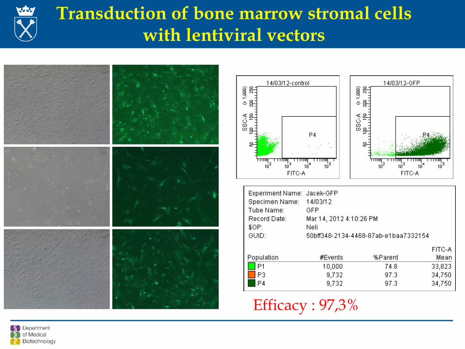
Efficacy : 97,3%
Transduction of bone marrow stromal cellswith lentiviral vectors

55
Texts to read !