Embed Size (px)
Citation preview
REVIEW
Viral Regulation of RNA Granules in Infected Cells
Qiang Zhang1 • Nishi R. Sharma2 • Zhi-Ming Zheng2 • Mingzhou Chen1
Received: 15 January 2019 / Accepted: 2 April 2019 / Published online: 29 April 2019� The Author(s) 2019
AbstractRNA granules are cytoplasmic, microscopically visible, non-membrane ribo-nucleoprotein structures and are important
posttranscriptional regulators in gene expression by controlling RNA translation and stability. TIA/G3BP/PABP-specific
stress granules (SG) and GW182/DCP-specific RNA processing bodies (PB) are two major distinguishable RNA granules
in somatic cells and contain various ribosomal subunits, translation factors, scaffold proteins, RNA-binding proteins, RNA
decay enzymes and helicases to exclude mRNAs from the cellular active translational pool. Although SG formation is
inducible due to cellular stress, PB exist physiologically in every cell. Both RNA granules are important components of the
host antiviral defense. Virus infection imposes stress on host cells and thus induces SG formation. However, both RNA and
DNA viruses must confront the hostile environment of host innate immunity and apply various strategies to block the
formation of SG and PB for their effective infection and multiplication. This review summarizes the current research
development in the field and the mechanisms of how individual viruses suppress the formation of host SG and PB for virus
production.
Keywords Stress granules (SG) � P-bodies (PB) � RNA virus � DNA virus
Introduction
While the intracellular environment and embedded cellular
machinery provide the needed vital force and necessary
materials for viruses to replicate after infection, these host
machineries are not available to these foreign invaders at
ease. In fact, viruses have to counter the multiple layers of
intracellular defense to replicate and establish their domi-
nance for their propagation. RNA granules (Thomas et al.
2011) are dynamic non-membrane subcellular structures
(Ivanov et al. 2018) containing translationally silenced
messenger ribonucleoproteins (mRNPs), which play an
important role in regulation of cellular homeostasis, RNA
metabolism and gene expression at the posttranscriptional
level (Anderson and Kedersha 2009). Stress granules (SG)
and processing bodies (PB) (Eulalio et al. 2007) are two of
RNA granules well characterized in yeast and mammalian
cells (Poblete-Duran et al. 2016) and are important com-
ponents of the host cell antiviral defense.
SG are non-membranous, transiently assembled cyto-
plasmic aggregates of 48S mRNPs and associated proteins
(Stohr et al. 2006; Buchan and Parker 2009), where stalled
translation preinitiation complexes (PICs) repress the
translation of nonessential mRNAs (Anderson et al. 2015)
and modulate cell signaling by sequestering key signal
translation proteins (Kedersha et al. 2013). Thus, SG are
thought to be the aggregates of stable, translationally silent
mRNAs (Kedersha and Anderson 2002). A variety of
environmental stresses, including viral infection, can trig-
ger SG formation in eukaryotic cells (Anderson and Ked-
ersha 2008). In contrast, PB can exist in the absence of
stress (Stoecklin and Kedersha 2013), which are sites of
active mRNA decay (Decker and Parker 2012). SG initiate
global translational arrest by storing mRNA (Anderson and
Kedersha 2009) for exchange with either polysomes for
translation or PB for degradation (Kedersha et al. 2005).
RNA-binding proteins TIA-1 (Kedersha et al. 1999; Gilks
Qiang Zhang and Nishi R. Sharma contributed equally to this work.
& Zhi-Ming Zheng
& Mingzhou Chen
1 State Key Laboratory of Virology and Modern Virology
Research Center, College of Life Sciences, Wuhan
University, Wuhan 430072, China
2 Tumor Virus RNA Biology Section, RNA Biology
Laboratory, National Cancer Institute, National Institutes of
Health, Frederick, MD 21702, USA
123
Virologica Sinica (2019) 34:175–191 www.virosin.orghttps://doi.org/10.1007/s12250-019-00122-3 www.springer.com/12250(0123456789().,-volV)(0123456789().,-volV)
et al. 2004), G3BP (Tourriere et al. 2003; Matsuki et al.
2013) and PABP (Ma et al. 2009; Smith and Gray 2010;
Burgess et al. 2011) are three fundamental components of
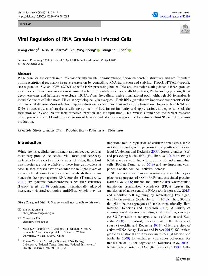
SG during stress (Fig. 1). GW182 and de-capping/de-
adenylating enzymes are specific components of PB
(Kedersha et al. 2005), where siRNA- or miRNA-guided
mRNAs are processed and degraded (Liu et al. 2005)
(Fig. 1). Virus infection imposes stress on host cells
(McInerney et al. 2005) and thereby induces SG formation.
SG can shut off the translation of bulk mRNAs (Poblete-
Duran et al. 2016) to regulate gene expression and com-
partmentalization of heterologous viral RNAs and proteins.
At the same time, viruses must take strategies to confront
these responses and maximize their own replication effi-
ciency (White and Lloyd 2012) by inhibition of SG for-
mation and disruption of PB assembly via virally encoded
factors.
Viral Regulation of RNA Stress GranuleFormation
SG Formation and Induction of SG by RNA VirusInfections
The process of SG formation can be artificially divided into
the following steps (Fig. 2): (1) accumulation of stalled
translation initiation complexes (Panas et al. 2016) in
response to various types of stress; (2) the RNA-binding
proteins such as RAS-GTPase-activating protein SH3
domain-binding protein 1 (G3BP1) and T cell-restricted
intracellular antigen 1 (TIA1) bind mRNAs and aggregate
to nucleate SG formation. Self-aggregation of G3BP1
(Tourriere et al. 2003) and the binding of TIA1 and TIAR
(TIA-1-related protein) to polysome-free mRNAs, which
exposes prion-like domains (Gilks et al. 2004), trigger
mRNP aggregation. The aggregation of proteins is
dynamic, and can rapidly exchange between SG and
cytosol (Kedersha et al. 2000, 2005). (3) large SG aggre-
gate from smaller foci via posttranslational modification
and microtubule transport (McCormick and Khaperskyy
2017). Many SG proteins undergo multiple post-transla-
tional modifications (Jayabalan et al. 2016; Protter and
Parker 2016). For example, G3BP1 must be demethylated
(Tsai et al. 2016), dephosphorylated (Kedersha et al. 2016)
and poly(ADP)-ribosylated (Leung et al. 2011) to promote
SG nucleation. Accordingly, SG formation also requires
ongoing transport of mRNPs along with an intact micro-
tubule cytoskeleton (Ivanov et al. 2003). Theoretically,
viral interference with any of these important steps may
modulate SG formation in cells. In fact, many viral factors
can interfere with SG formation and/or function. Mean-
while, SG can entrap viral RNA in some cases (McCor-
mick and Khaperskyy 2017). Therefore, SG are thought to
be antiviral (Rozelle et al. 2014). Thus, to illustrate the
relationship between SG and RNA viruses would be
important for us to better understand the interactions of
host and viruses.
Up to the present, SG can be divided into two types
according to their formation mode. Type I SG formation
depends on phosphorylation of eukaryotic translation initi-
ation factor-2a (eIF2a) by one of the eIF2 kinases—double-
stranded RNA (dsRNA)-activated protein kinase or protein
kinase R (PKR) (Srivastava et al. 1998; Garcia et al. 2007;
Onomoto et al. 2012), PKR-like ER kinase (PERK) (Harding
et al. 2000a, b), general control non-derepressible protein 2
(GCN2) (Wek et al. 1995; Deng et al. 2002) or haeme-reg-
ulated inhibitor (HRI) (McEwen et al. 2005), which are
activated by distinct types of stress. Phosphorylated eIF2astably binds to eIF2b, which prevents the recycle of eIF2 andregeneration of the eIF2-GTP-Met-tRNAi
Met ternary com-
plex. Thus, eIF2a phosphorylation blocks recognition of the
No arsenite, no SG + arsenite, SG No arsenite, PB
IPAD + 281WG1-AIT1-AIT
Fig. 1 Mammalian RNA granules. HeLa cells immunostaining with
anti-TIA-1 (left and middle, red) show stress granules (SG) during
stress of NaAS2O3 (?arsenite, middle) and with anti-GW182 show
processing bodies (PB) under physiological condition. Arrows
indicate granules (SG or PB).
176 Virologica Sinica
123

initiation codon and joining of the large ribosomal subunit,
resulting in accumulation of stalled 48S mRNPs (Jackson
et al. 2010). Type II SG formation is independent of eIF2aphosphorylation, but requires eIF4F complex disruption
such as inhibition of eIF4A RNA helicase (Bordeleau et al.
2006; Dang et al. 2006) or disruption of eIF4E activity (von
der Haar et al. 2004; Fournier et al. 2013) for recognition and
binding ofRNAcap structure. The stress induced by nutrient,
energy, oxygen or growth factor insufficiency inhibits
mTOR complex 1 (mTORC1), whose activity is required for
the dissociation of 4EBPs from eIF4E (Fujimura et al. 2012)
and enables eIF4E to form the eIF4F complex, and thus
blocks assembly of pre-initiation complexes (Zoncu et al.
2011).
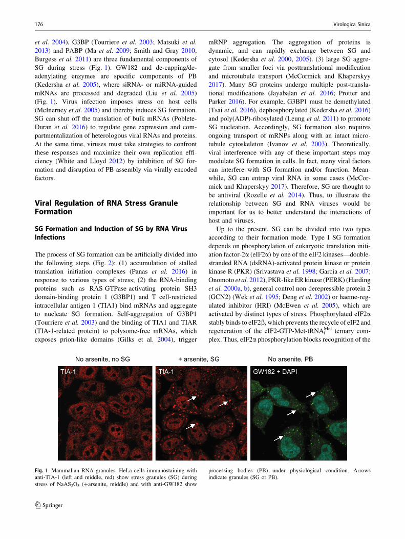
Type I SG formation induced by viruses is the most and
best-studied example (Table 1, Fig. 3A). Various RNA
products derived by viruses including long dsRNA (Rojas
et al. 2010), 50-triphosphate RNA (50-ppp-RNA) (Nalla-
gatla et al. 2007), dsRNA that is formed by the antiparallel
mRNA transcripts of some DNA viruses (Willis et al.
2011), and human immunodeficiency virus (HIV) transac-
tivation-response region (TAR) RNA hairpins (Heinicke
et al. 2009), can be recognized by PKR. The activated PKR
initiates SG assembly through eIF2a phosphorylation. For
instance, the persistent phosphorylation of eIF2a (Montero
et al. 2008) during rotavirus infection is PKR-dependent as
a consequence of the accumulation of viral dsRNA in the
cytoplasm outside the viroplasms (virus-induced cytoplas-
mic inclusion bodies called viroplasms [VMs]) (Rojas et al.
2010). Even though eIF2a is phosphorylated in rotavirus-
infected cells, the formation of SG is prevented and viral
proteins are efficiently translated, suggesting that the virus
prevents the assembly of these structures presumably
downstream of eIF2a phosphorylation to allow the trans-
lation of its mRNAs (Mazroui et al. 2006). Very recently,
Dhillon and Rao found that rotavirus induces formation
and sequestration of remodeled SG and PB in the VMs
which contain the majorities of their components but
selective exclusion of a few proteins (G3BP1 and ZBP1 for
SG, DDX6, EDC4 and Pan3 for PB), to promote virus
replication (Dhillon and Rao 2018). Oceguera et al.
demonstrated that viral RNA of rotavirus could interact
with several RNA binding proteins (RBPs) (Xrn1, Dcp1,
Ago2, Hur) and interfere with their subcellular localization
(Oceguera et al. 2018). Lindquist et al. (Lindquist et al.
2011) first determined that SG induction by respiratory
syncytial virus (RSV) was mediated by PKR-dependent
eIF2a phosphorylation. The RSV-mediated SG formation
was significantly reduced in PKR-knockdown cells (Lind-
quist et al. 2010). In addition, it has been shown that
Hepatitis C virus (HCV) strongly activates PKR via the 50-untranslated region (UTR) of its genome (Toroney et al.
2010), thereby inducing SG. NS1-mutant Influenza virus A
(IAV) (Khaperskyy et al. 2012; Mok et al. 2012; Ng et al.
2013) and C protein-deficient Sendai virus (SeV) (Takeu-
chi et al. 2008) lead to significant activation of PKR and
eIF2a phosphorylation. Besides, PERK could be activated
by high levels of glycoproteins produced from enveloped
Fig. 2 Viruses induce SG formation. Type I SG formation: RNAs
derived from rotavirus, RSV and HCV activate PKR; High levels of
glycoproteins produced from enveloped virus activate PERK; HCMV
infection activates PERK; Sindbis virus genomic RNA activates
GCN2. Type II SG formation: RVFV attenuates mTOR signaling to
inhibite 4EBP phosphorylation. All above lead to the formation of
stalled translation complexes to initiate the assembly of SG.
Q. Zhang et al.: Virus and RNA Granules 177
123
Table 1 Regulation of SG by viruses.
Genome Virus family Virus Type Mechanism References
dsDNA Herpesviridae HCMV Induction Modifies the UPR and activates PERK Isler et al. (2005)
Inhibition pTRS1 and pIRS1 antagonize PKR to facilitate virus
replication
Ziehr et al. (2016)
KSHV Inhibition ORF57 interacts with PKR and PACT to inhibit PKR
activation
Sharma et al. (2017)
HSV-1 Inhibition vhs and Us11 protein play a key role in blocking the
activation of PKR
Sciortino et al. (2013)
HSV-2 Inhibition vhs localizes to SG and its endoribonuclease activity
is required to disrupt SG formation
Finnen et al.
(2012, 2014, 2016)
Poxviridae VV Inhibition Sequesters crucial SG components within DNA
factories
Katsafanas and Moss
(2007), Zaborowska
et al. (2012)
Induction Untranslated mRNA accumulation in viral DNA
factories induces RNA granules formation
Meng and Xiang (2019)
dsRNA Reoviridae Rotavirus Induction Phosphorylation of eIF2a is PKR-dependent as a
consequence of the accumulation of viral dsRNA
Montero et al. (2008),
Rojas et al. (2010)
Modulation Induces formation and sequestration in the VMs of
remodeled SG and PB
Dhillon and Rao (2018)
(?)ssRNA Picornaviridae PV Inhibition Viral 3C protease cleaves G3BP White et al. (2007)
FMDV Inhibition Viral 3C protease cleaves G3BP Ye et al. (2018)
Leader Protease Cleaves G3BP1 and G3BP2 Visser et al. (2019)
TMEV Inhibition Express the leader (L) protein to inhibit G3BP1
aggregation
Borghese and Michiels
(2011)
Mengovirus Inhibition Express the leader (L) protein to inhibit G3BP1
aggregation
Borghese and Michiels
(2011)
EV71 Modulation 2A protease inhibits typical SG formation but
induces atypical SG formation by cleaving eIF4GI
Yang et al. (2018)
Caliciviridae FCV Inhibition NS6Pro cleaves G3BP1 Humoud et al. (2016)
Togaviridae SINV Induction Genomic RNA activates GCN2 Berlanga et al. (2006)
Flaviviridae WNV Inhibition 30-end viral genome captures TIA-1/TIAR Li et al. (2002, Emara and
Brinton (2007)
DENV Inhibition 30-end viral genome captures TIA-1/TIAR Li et al. (2002), Emara and
Brinton (2007), Ward
et al. (2011)
30-UTR interacts with G3BP1, G3BP2, Caprin1 and
USP10
JEV Inhibition Recruits G3BP and USP10 to the perinuclear region Tu et al. (2012)
NS2A interact with PKR and prevent PKR
dimerization
Ward et al. (2011)
HCV Induction Activates PKR via the 50- UTR of its genome Toroney et al. (2010)
Inhibition NS5A protein binds to the PKR dimerization domain
to inhibit PKR activation
Toroney et al. (2010)
Modulate GADD34 and PP1 to de-phosphorylate
eIF2aRuggieri et al. (2012)
HCV-JFH1 Modulation Redistributes several SG components to the HCV
replication complex (RC)
Ariumi et al. (2011),
Garaigorta et al. (2012),
Pene et al. (2015)
ZIKV Inhibition Induces the redistribution of TIAR to the viral RNA
replication sites
Hou et al. (2017)
(-)ssRNA Arenaviridae JUNV Inhibition N and GPC impair the phosphorylation of eIF2a Linero et al. (2011)
Bunyaviridae RVFV Inhibition RVFV attenuates mTOR signaling to inhibite 4EBP
phosphorylation
Habjan et al. (2009),
Ikegami et al. (2009),
Hopkins et al. (2015)
178 Virologica Sinica
123
viruses (Chan and Egan 2005), and general control non-
derepressible-2 (GCN2) could be activated by Sindbis virus
(SINV) genomic RNA (Berlanga et al. 2006), both leading
to phosphorylation of eIF2a. GCN2 prevents replication of
SINV in the early stages of the viral replicative cycle by
blocking the synthesis of NSPs from SINV RNA (Berlanga
et al. 2006; Frolova et al. 2006; Gorchakov et al. 2008).
Viruses also induce SG formation independent of eIF2aphosphorylation (Table 1). The most typical example is
from Rift Valley fever virus (RVFV) (Habjan et al. 2009;
Ikegami et al. 2009) (Fig. 2). RVFV (Hopkins et al. 2015)
infection attenuates Akt/mTOR signaling and inhibits
4EBP phosphorylation and translation of 50-TOP mRNAs,
subsequently leading to an inhibition of global protein
translation. 50-TOP–containing mRNAs are indeed tar-
geted to PB, where RVFV uses these cellular mRNAs for
cap-snatching (Hopkins et al. 2015). This can reflect that
SG may interact with PB in a process that is thought to
result in the exchange of mRNA cargos (Kedersha et al.
2008). Whether any virus induces SG formation to cause
translation inhibition due to the destruction of eIF4G or
eIF4A is worth exploring in the future.
RNA Viruses Modulate SG Formation or Assembly
SG formation shuts off bulk host protein synthesis. How-
ever, all viruses depend on the host translation apparatus
for their gene expression. Therefore, viruses, as intracel-
lular parasites, have to modulate the stress response path-
way and SG assembly to translate their proteins for virus
replication. RNA viruses modulate stress response pathway
at different levels of SG formation (Table 1): One is to
regulate eIF2a phosphorylation, and the other is to regulate
the process of SG nucleation.
RNA Viruses Modulate eIF2a Phosphorylation to Interferewith SG Formation
In some cases, viral gene products can act as antagonists by
targeting the virus-activated eIF2a kinases such as PKR or
even by directly modulating the phosphorylation of eIF2a(Fig. 3A). IAV NS1 (Khaperskyy et al. 2012; Ng et al.
2013), Middle East respiratory syndrome coronavirus
(MERS-CoV) accessory protein 4a (Rabouw et al. 2016;
Nakagawa et al. 2018), and Ebola virus (EBOV) multi-
functional protein VP35 (Nelson et al. 2016; Le Sage et al.
2017) bind viral dsRNA and prevent the viral dsRNA from
PKR binding to inhibit SG formation. Inhibition of SG
formation facilitates the translation of viral mRNAs,
leading to efficient virus replication. HCV NS5A protein
(Toroney et al. 2010) binds to the PKR dimerization
domain to inhibit PKR activation. Japanese encephalitis
virus (JEV) NS2A protein (Tu et al. 2012) might similarly
interact with PKR and then prevent PKR dimer formation.
SeV (Takeuchi et al. 2008) and measles virus (MV)
(Okonski and Samuel 2013) encode a C protein to limit the
accumulation of dsRNA to inhibit SG formation. It seems
that a portion of RNA viruses encode RNA binding pro-
teins to antagonize the activity of PKR. There are also
other groups of RNA viruses which directly modulate the
phosphorylation of eIF2a without PKR. Junı́n virus
(JUNV) prevents SG assembly by impairing the
Table 1 (continued)
Genome Virus family Virus Type Mechanism References
Coronaviridae MERS-
CoV
Inhibition Accessory protein 4a bind viral dsRNA and prevent
the viral dsRNA from PKR binding
Rabouw et al. (2016),
Nakagawa et al. (2018)
Filoviridae EBOV Inhibition VP35 bind viral dsRNA and prevent the viral dsRNA
from PKR binding
Nelson et al. (2016, Le
Sage et al. (2017)
Modulation SG proteins are selectively sequestered within virus
inclusions and co-localize with viral RNA to form
inclusion-bound granules
Nelson et al. (2016)
Rhabdoviridae VSV Modulation Induces formation of the SG-like structures that co-
localize with viral replication proteins and RNA
Dinh et al. (2013)
Paramyxoviridae MV Inhibition Encode a C protein to limit the accumulation of
dsRNA
Okonski and Samuel
(2013)
SeV Inhibition Encode a C protein to limit the accumulation of
dsRNA
Takeuchi et al. (2008)
Trailer RNA captures TIAR from SG Iseni et al. (2002)
RSV Induction Mediated by PKR-dependent eIF2a phosphorylation. Lindquist et al. (2011)
Inhibition Sequestration of OGT in IBs Fricke et al. (2013)
HPIV3 Inhibition IBs shield viral RNAs from recognition by PKR Hu et al. 2018)
Q. Zhang et al.: Virus and RNA Granules 179
123
phosphorylation of eIF2a through its nucleoprotein (N) and
glycoprotein precursor (GPC) (Linero et al. 2011). How-
ever, its mechanism remains to be elucidated, although it
may be similar to HCV. Ruggieri and colleagues reported
that HCV rapidly de-phosphorylated eIF2a through protein
phosphatase 1 (PP1) and its regulatory subunit GADD34
(growth arrest and DNA-damage-inducible 34) (Kojima
et al. 2003; Clavarino et al. 2012; Ruggieri et al. 2012).
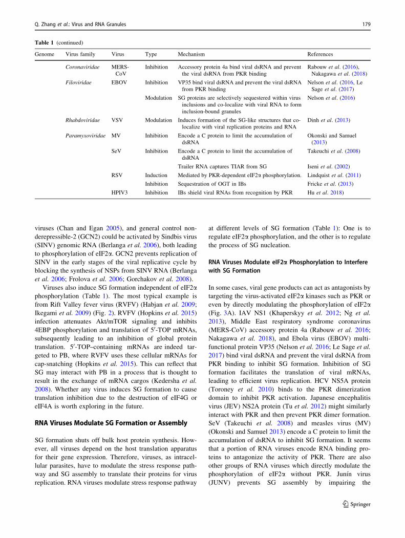
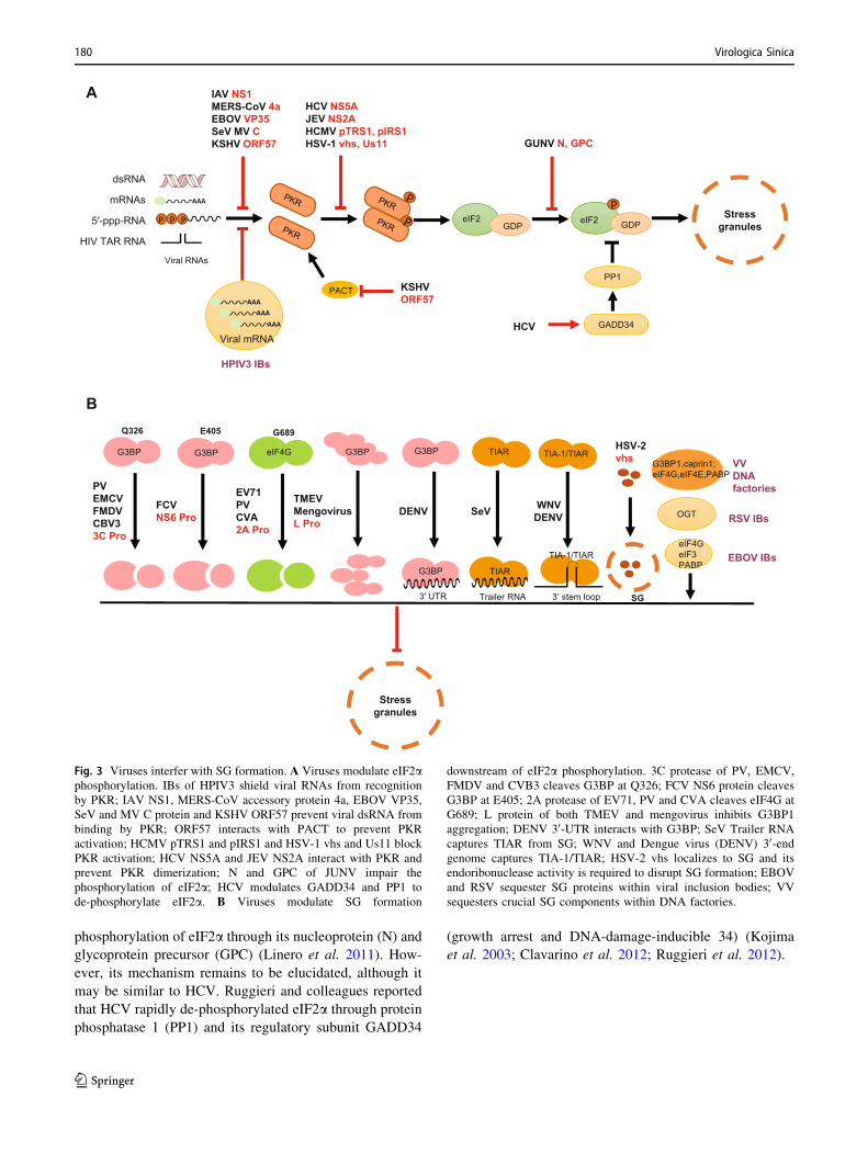
A
B
Fig. 3 Viruses interfer with SG formation. A Viruses modulate eIF2aphosphorylation. IBs of HPIV3 shield viral RNAs from recognition
by PKR; IAV NS1, MERS-CoV accessory protein 4a, EBOV VP35,
SeV and MV C protein and KSHV ORF57 prevent viral dsRNA from
binding by PKR; ORF57 interacts with PACT to prevent PKR
activation; HCMV pTRS1 and pIRS1 and HSV-1 vhs and Us11 block
PKR activation; HCV NS5A and JEV NS2A interact with PKR and
prevent PKR dimerization; N and GPC of JUNV impair the
phosphorylation of eIF2a; HCV modulates GADD34 and PP1 to
de-phosphorylate eIF2a. B Viruses modulate SG formation
downstream of eIF2a phosphorylation. 3C protease of PV, EMCV,
FMDV and CVB3 cleaves G3BP at Q326; FCV NS6 protein cleaves
G3BP at E405; 2A protease of EV71, PV and CVA cleaves eIF4G at
G689; L protein of both TMEV and mengovirus inhibits G3BP1
aggregation; DENV 30-UTR interacts with G3BP; SeV Trailer RNA
captures TIAR from SG; WNV and Dengue virus (DENV) 30-endgenome captures TIA-1/TIAR; HSV-2 vhs localizes to SG and its
endoribonuclease activity is required to disrupt SG formation; EBOV
and RSV sequester SG proteins within viral inclusion bodies; VV
sequesters crucial SG components within DNA factories.
180 Virologica Sinica
123
RNA Viruses Cleave/Sequester/Redistribute Stress Granule-Nucleating Proteins to Interfere with SG Assembly
Several RNA viruses have been shown to express viral
effectors that can actively disrupt the accumulation of SG
through cleavage of SG components (Fig. 3B). Poliovirus
(PV) induces SG formation in early phase but induces SG
disassembly at later stages via cleavage of G3BP by viral
3C, thus preventing SG formation (White et al. 2007).
Similar findings were also reported for encephalomy-
ocarditis virus (EMCV) (Ng et al. 2013), foot-and-mouth
disease virus (FMDV) (Ye et al. 2018; Visser et al. 2019),
coxsackievirus B3 (CBV3) (Fung et al. 2013) and feline
calicivirus (FCV) (Humoud et al. 2016). FCV infection
does not cause accumulation of SG, despite an increased
phosphorylation of eIF2a (Humoud et al. 2016). This is
because FCV NS6Pro, a 3C-like proteinase, cleaves
G3BP1 at a site different from the poliovirus 3C pro-
teinase. Unlike FCV, murine norovirus (MNV) does not
cleave G3BP1 and thus does not inhibit SG formation
during virus infection (Humoud et al. 2016). In general,
picornaviruses inhibit SG formation by viral 2A/L or 3C
cleaving the major components of SG. In recent study,
Yang et al. found that the 2A protease of picornavirus
(EV71, PV, CVA) inhibits typical SG formation, which is
PKR and eIF2a phosphorylation-dependent, but induces
atypical SG formation by cleaving eIF4GI to sequester
cellular mRNA and release viral mRNA, thereby facili-
tating viral infection (Yang et al. 2018). In other words, the
2A protease can transform the overall translation machin-
ery favorable for productive viral infection by induction of
atypical SG while blocking the typical SG in the presence
of G3BP cleavage by viral 3C protease during viral
infection (Yang et al. 2018).
Redistribution or sequestering SG components to the
viral replication sites is another strategy used by many
viruses to impair SG assembly in infected cells (Fig. 3B).
ZIKV infection induces the redistribution of TIAR to the
viral RNA replication sites (Hou et al. 2017); SeV Trailer
RNA captures TIAR from SG (Iseni et al. 2002); West Nile
Virus (WNV) and Dengue virus (DENV) 30-end viral
genome captures TIA-1/TIAR (Li et al. 2002; Emara and
Brinton 2007; Xia et al. 2015); DENV 30-UTR interacts
with G3BP1, G3BP2, Caprin1 and USP10 (Ward et al.
2011; Reineke et al. 2015); JEV recruits G3BP and USP10
to the perinuclear region through the interaction of JEV
core protein with Caprin-1, a SG-associated cellular factor
(Ward et al. 2011). Theiler murine encephalomyelitis virus
(TMEV) and mengovirus, a strain of EMCV, express the
leader (L) protein to inhibit G3BP1 aggregation (Borghese
and Michiels 2011). Sequestration or redistribution of SG
components by viruses through protein–protein and pro-
tein-RNA interactions not only prevents SG assembly, but
also facilitates viral genome replication. HCV-JFH1
infection redistributes several SG components, including
G3BP1, ataxin-2 (ATX2), and poly(A)-binding protein 1
(PABP1), to the HCV replication complex (RC) (Ariumi
et al. 2011; Pene et al. 2015), and co-opts G3BP1 to
mediate efficient viral replication by interaction with NS5B
and the 50 end of the HCV minus-strand RNA (Ariumi
et al. 2011; Garaigorta et al. 2012).
RNA Virus Inclusion Bodies (IBs) Emergingas a New Strategy Used by Viruses to Resist SG
Studies on Human parainfluenza virus type 3 (HPIV3) (Hu
et al. 2018), RSV (Rincheval et al. 2017), EBOV (Hoenen
et al. 2012), Rabies virus (RABV) (Lahaye et al. 2009) and
Vesicular stomatitis virus (VSV) (Heinrich et al. 2010)
showed that inclusion bodies (IBs) of negative stranded
RNA viruses are the sites of viral RNA synthesis. A recent
study suggested an emerging role of IBs in HPIV3 repli-
cation by shielding newly synthesized viral RNA from the
antiviral effect of SG (Hu et al. 2018) (Fig. 3B). Seques-
tration of O-linked N-acetylglucosamine (OGN) trans-
ferase (OGT), an enzyme that catalyzes the
posttranslational addition of OGN to protein targets, in
RSV IBs was also proposed to regulate SG nucleation and
suppression of SG formation (Fricke et al. 2013) (Fig. 3B).
Viral transcription and replication of RABV take place
within Negri bodies (NBs), which are IB-like structures
(Lahaye et al. 2009). RABV-induced SG are normally
located closely to NBs. Viral mRNAs rather than viral
genomic RNA accumulate in the SG-like structures toge-
ther with cellular mRNAs were found to be specially
transported from NBs to SG-like structures (Nikolic et al.
2016). VSV infection also induces formation of the SG-like
structures that co-localize with viral replication proteins
and RNA, which are different from canonical SG (Dinh
et al. 2013). SG proteins (eIF4G, eIF3, PABP) are selec-
tively sequestered within Ebola virus inclusion bodies and
co-localize with viral RNA to form inclusion body-bound
granules, which are functionally and structurally different
from canonical SG, probably leading to inhibit the antiviral
role of SG (Nelson et al. 2016) (Fig. 3B). Collectively,
these findings provoke more investigations on the roles of
viral IBs in viral replication and resisting cellular
responses.
DNA Viruses Regulate SG Formation
Unlike RNA viruses, the regulation of SG formation during
infection with DNA viruses is poorly understood. It was
reported that human cytomegalovirus (HCMV) infection
modifies the unfolded protein response (UPR) and activates
PERK (Fig. 2), but limiting the amount of phosphorylated
Q. Zhang et al.: Virus and RNA Granules 181
123
eIF2a to maintain translation (Isler et al. 2005). Kaposi’s
sarcoma-associated herpesvirus (KSHV) ORF57 (Sharma
et al. 2017) interacts with PKR and PKR-activating protein
(PACT) (Patel et al. 2000) to inhibit PKR binding dsRNA
and prevent PACT-PKR interaction in the PKR pathway
(Li et al. 2006), respectively. HCMV pTRS1 and pIRS1
antagonize PKR to facilitate virus replication (Ziehr et al.
2016). The HSV-1 vhs (Sciortino et al. 2013) and Us11
protein (Cassady and Gross 2002) play a key role in
blocking the activation of PKR. Smiley and colleagues also
demonstrated that infection with virion host shutoff protein
(vhs)-defective herpes simplex virus 1 (HSV-1) triggers SG
formation, and PKR is essential for SG formation in the
absence of vhs (Dauber et al. 2016) (Fig. 3A). Finnen et al.
previously established that herpes simplex virus 2 (HSV-2)
infection impacts stress granule accumulation in response
to oxidative stress (Finnen et al. 2012). They also
demonstrated that disruption of SG is mediated by vhs
(Finnen et al. 2014), whose endoribonuclease activity is
required to disrupt SG formation (Finnen et al. 2016).
HSV-2 vhs indeed have the ability to localize to SG
(Finnen et al. 2016) (Fig. 3B). This implies that removal of
RNA from SG promotes its disassembly and that intact
RNA is crucial for maintaining SG structure. It will be
interesting to test the function of endoribonucleases in SG
disassembly. Vaccinia virus (VV) sequesters crucial
translation initiation factors, such as G3BP1, Caprin1,
eIF4E, PABP and eIF4G (Katsafanas and Moss 2007;
Simpson-Holley et al. 2011; Zaborowska et al. 2012),
within cytoplasmic viral DNA factories to utilize SG
components for different purposes (Fig. 3B). A recent
study (Meng and Xiang 2019) suggested that the RNA
granules are resulted from untranslated mRNA accumula-
tion in viral DNA factories (Liu and Moss 2016) and TIA-1
is probably not required for granule formation and anti-
poxviruses. Instead, the granules formation is most likely
driven by an array of RNA–protein interactions and
requires no specific SG components (Sivan et al. 2018;
Meng and Xiang 2019).
Viral Regulation of RNA Processing BodyAssembly
Assembly of P-Bodies (PB)
PB were first reported in the scientific literature by Bash-
kirov et al. in 1997, and described as ‘‘small granules or
discrete, prominent foci’’ or as the cytoplasmic location of
the mouse exoribonuclease mXrn1p (Bashkirov et al.
1997). Like SG, PB lack outer lipid membrane and now are
recognized to be the sites where non-translating mRNAs
accumulate for different fates including decay, storage, or
returning to translation. A variety of enzymes involved in
mRNA deadenylation (Ccr1, Caf1, Not1) (Sheth and Par-
ker 2006), decapping (Dcp1/2, Lsm1-7, Edc3proteins)
(Ingelfinger et al. 2002; Yu et al. 2005), nonsense-medi-
ated decay (NMD) proteins (SMG5-6-7, UPF1) (Ingelfin-
ger et al. 2002; Durand et al. 2007), in addition to
scaffolding proteins (Ge-1/Hedls) (Yu et al. 2005) and
translation control factors (CPEB, eIF4E-T) (Andrei et al.
2005; Wilczynska et al. 2005), are the components of PB
and used as routine markers to distinguish these granules.
Nonetheless, some components (APOBEC3G, BRF1,
DDX3, FAST, TTP, Rap55) (McEwen et al. 2005; Sen and
Blau 2005; Gallois-Montbrun et al. 2007; Chen et al. 2008)
have also been shown to be shared by both SG and PB,
suggesting a substantial linkage of these two structures and
movement of mRNAs between both RNA granules. Inter-
estingly, among these components, PB also include RNA-
induced silencing complex (RISC) or miRNA associated
argonaute (Ago) proteins (also shared with SG) and the
GW182 protein which provides scaffolding activities for
RISC to function, suggesting PB being the sites of miRNA
mediated translation repression. The scaffolding activity of
GW182 is critical for PB and knockdown of GW182
expression disrupts PB formation (Liu et al. 2005). Nota-
bly, GW182 has been shown to bind to Ago2 which is
critical for miRNA function and PB formation (Liu et al.
2005). Recent evidence indicates that GW182 can recruit
up to three molecules of Ago2 via its three GW motifs
(glycine-tryptophan repeats) while each Ago protein has a
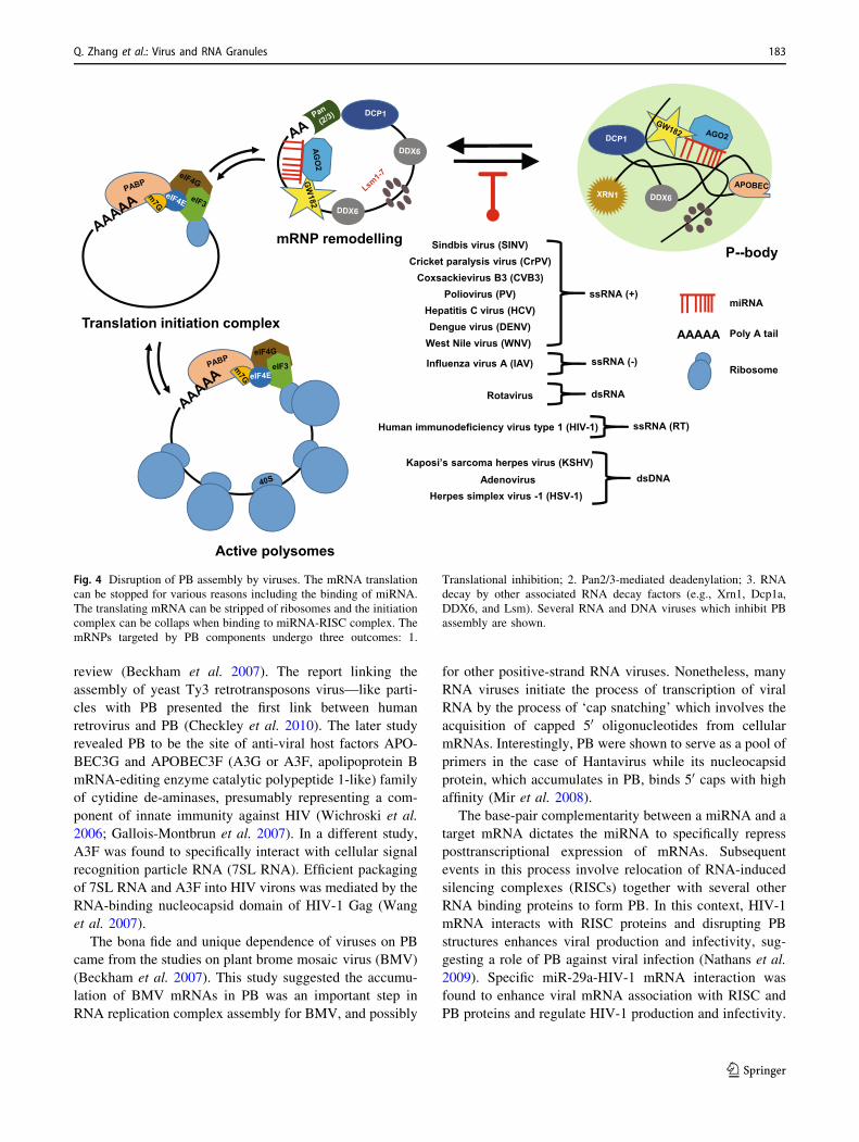
single GW182-binding site (Elkayam et al. 2017) (Fig. 4).
By applying fluorescence-activated particle sorting to
purify PB in combination with mass spectrometry, Hub-
stenberger et al. identified 125 proteins that are signifi-
cantly associated with PB (Hubstenberger et al. 2017). By
labeling several PB-localized proteins with a BirA
(E. coli biotin ligase) enzyme in combination with mass
spectrometry after streptavidin pulldown, Youn
et al. identified 38 proteins in the PB (Youn et al. 2018).
ISGs (interferon stimulated genes) can also be found in PB
during virus infection (Hebner et al. 2006).
RNA Viruses and PB
In comparison to viral regulation of SG, interaction of virus
and PB was not much explored. It is an assumption
that RNA viruses must regulate RNA decay pro-
cesses/machinery to prevent degradation of virus genomes
and mRNAs. Recently, some progress has been made to
understand the relationship between PB components and
some viruses in the context of viral gene expression. The
data in published literatures are summarized in (Table 2).
Mutation induced in the PB core components to affect the
viral life cycles are well studied and tabulated in an earlier
182 Virologica Sinica
123
review (Beckham et al. 2007). The report linking the
assembly of yeast Ty3 retrotransposons virus—like parti-
cles with PB presented the first link between human
retrovirus and PB (Checkley et al. 2010). The later study
revealed PB to be the site of anti-viral host factors APO-
BEC3G and APOBEC3F (A3G or A3F, apolipoprotein B
mRNA-editing enzyme catalytic polypeptide 1-like) family
of cytidine de-aminases, presumably representing a com-
ponent of innate immunity against HIV (Wichroski et al.
2006; Gallois-Montbrun et al. 2007). In a different study,
A3F was found to specifically interact with cellular signal
recognition particle RNA (7SL RNA). Efficient packaging
of 7SL RNA and A3F into HIV virons was mediated by the
RNA-binding nucleocapsid domain of HIV-1 Gag (Wang
et al. 2007).
The bona fide and unique dependence of viruses on PB
came from the studies on plant brome mosaic virus (BMV)
(Beckham et al. 2007). This study suggested the accumu-
lation of BMV mRNAs in PB was an important step in
RNA replication complex assembly for BMV, and possibly
for other positive-strand RNA viruses. Nonetheless, many
RNA viruses initiate the process of transcription of viral
RNA by the process of ‘cap snatching’ which involves the
acquisition of capped 50 oligonucleotides from cellular
mRNAs. Interestingly, PB were shown to serve as a pool of
primers in the case of Hantavirus while its nucleocapsid
protein, which accumulates in PB, binds 50 caps with high
affinity (Mir et al. 2008).
The base-pair complementarity between a miRNA and a
target mRNA dictates the miRNA to specifically repress
posttranscriptional expression of mRNAs. Subsequent
events in this process involve relocation of RNA-induced
silencing complexes (RISCs) together with several other
RNA binding proteins to form PB. In this context, HIV-1
mRNA interacts with RISC proteins and disrupting PB
structures enhances viral production and infectivity, sug-
gesting a role of PB against viral infection (Nathans et al.
2009). Specific miR-29a-HIV-1 mRNA interaction was
found to enhance viral mRNA association with RISC and
PB proteins and regulate HIV-1 production and infectivity.
Fig. 4 Disruption of PB assembly by viruses. The mRNA translation
can be stopped for various reasons including the binding of miRNA.
The translating mRNA can be stripped of ribosomes and the initiation
complex can be collaps when binding to miRNA-RISC complex. The
mRNPs targeted by PB components undergo three outcomes: 1.
Translational inhibition; 2. Pan2/3-mediated deadenylation; 3. RNA
decay by other associated RNA decay factors (e.g., Xrn1, Dcp1a,
DDX6, and Lsm). Several RNA and DNA viruses which inhibit PB
assembly are shown.
Q. Zhang et al.: Virus and RNA Granules 183
123
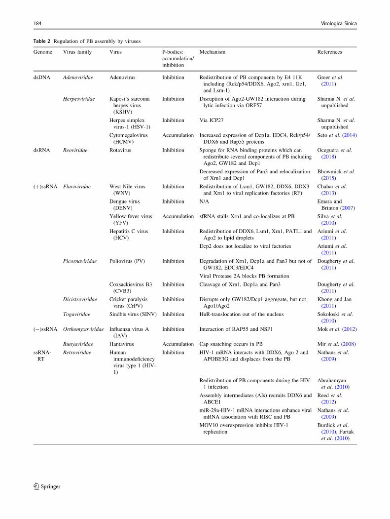
Table 2 Regulation of PB assembly by viruses
Genome Virus family Virus P-bodies:
accumulation/
inhibition
Mechanism References
dsDNA Adenoviridae Adenovirus Inhibition Redistribution of PB components by E4 11K
including (Rck/p54/DDX6, Ago2, xrn1, Ge1,
and Lsm-1)
Greer et al.
(2011)
Herpesviridae Kaposi’s sarcoma
herpes virus
(KSHV)
Inhibition Disruption of Ago2-GW182 interaction during
lytic infection via ORF57
Sharma N. et al.
unpublished
Herpes simplex
virus-1 (HSV-1)
Inhibition Via ICP27 Sharma N. et al.
unpublished
Cytomegalovirus
(HCMV)
Accumulation Increased expression of Dcp1a, EDC4, Rck/p54/
DDX6 and Rap55 proteins
Seto et al. (2014)
dsRNA Reoviridae Rotavirus Inhibition Sponge for RNA binding proteins which can
redistribute several components of PB including
Ago2, GW182 and Dcp1
Oceguera et al.
(2018)
Decreased expression of Pan3 and relocalization
of Xrn1 and Dcp1
Bhowmick et al.
(2015)
(?)ssRNA Flaviviridae West Nile virus
(WNV)
Inhibition Redistribution of Lsm1, GW182, DDX6, DDX3
and Xrn1 to viral replication factories (RF)
Chahar et al.
(2013)
Dengue virus
(DENV)
Inhibition N/A Emara and
Brinton (2007)
Yellow fever virus
(YFV)
Accumulation sfRNA stalls Xrn1 and co-localizes at PB Silva et al.
(2010)
Hepatitis C virus
(HCV)
Inhibition Redistribution of DDX6, Lsm1, Xrn1, PATL1 and
Ago2 to lipid droplets
Ariumi et al.
(2011)
Dcp2 does not localize to viral factories Ariumi et al.
(2011)
Picornaviridae Poliovirus (PV) Inhibition Degradation of Xrn1, Dcp1a and Pan3 but not of
GW182, EDC3/EDC4
Dougherty et al.
(2011)
Viral Protease 2A blocks PB formation
Coxsackievirus B3
(CVB3)
Inhibition Cleavage of Xrn1, Dcp1a and Pan3 Dougherty et al.
(2011)
Dicistroviridae Cricket paralysis
virus (CrPV)
Inhibition Disrupts only GW182/Dcp1 aggregate, but not
Ago1/Ago2
Khong and Jan
(2011)
Togaviridae Sindbis virus (SINV) Inhibition HuR-translocation out of the nucleus Sokoloski et al.
(2010)
(-)ssRNA Orthomyxoviridae Influenza virus A
(IAV)
Inhibition Interaction of RAP55 and NSP1 Mok et al. (2012)
Bunyaviridae Hantavirus Accumulation Cap snatching occurs in PB Mir et al. (2008)
ssRNA-
RT
Retroviridae Human
immunodeficiency
virus type 1 (HIV-
1)
Inhibition HIV-1 mRNA interacts with DDX6, Ago 2 and
APOBE3G and displaces from the PB
Nathans et al.
(2009)
Redistribution of PB components during the HIV-
1 infection
Abrahamyan
et al. (2010)
Assembly intermediates (AIs) recruits DDX6 and
ABCE1
Reed et al.
(2012)
miR-29a-HIV-1 mRNA interactions enhance viral
mRNA association with RISC and PB
Nathans et al.
(2009)
MOV10 overexpression inhibits HIV-1
replication
Burdick et al.
(2010), Furtak
et al. (2010)
184 Virologica Sinica
123
HIV Nef interacts with Ago2 via its glycine-tryptophan
region and functions as a viral suppressor of RNAi (Aqil
et al. 2013). While overexpression of Mov10, a component
of PB and an ATP-dependent 50-30 RNA helicase, inhibits
HIV production (Burdick et al. 2010; Furtak et al. 2010),
Mov10 and APOBEC3G localization to PB is not required
for HIV virion incorporation and antiviral activity (Izumi
et al. 2013). It becomes clear that Mov10 inhibits virus
infection by enhancing RIG-I-MAVS-Independent IFN
Induction (Cuevas et al. 2016) and stabilizing A3G from
degradation (Chen et al. 2017).
The anticipated evidence of viral disruption of PB also
came from the study with poliovirus (PV), a plus-strand
RNA virus showing that PB are disrupted during PV
infection in cells by 4 h post infection (Dougherty et al.
2011). This function is attributed to viral proteinase 3C
which degrades several components of PB including Xrn1
and Dcp1a, but not affecting others such as GW182, Edc3
and Edc4. Rotaviruses disassemble PB by using viral RNA
as a sponge for RNA binding proteins to redistribute sev-
eral PB components, including Ago2, GW182 and
Dcp1 PB (Oceguera et al. 2018). In fact, rotavirus disrupts
PB through multiple mechanisms. The viral NSP1 protein
seems to degrade PB component Pan3, while relocalizing
other two components (Xrn1 and Dcp1a) (Bhowmick et al.
2015). Intriguingly, exclusion of SG and PB components
from the viroplasm is important for rotavirus replication
and progeny virus production (Dhillon and Rao 2018).
DNA Viruses and PB
While RNA viruses have evolved to co-opt or modulate the
assembly of PB, this effect is rather unclear during infec-
tion by DNA viruses. Since most of the DNA viruses
replicate and assemble in the nucleus, therefore as pro-
posed for RNA viruses, accumulation of viral RNAs in PB
for assembly cannot be a strategy required by DNA viruses.
However, the close relationship of PB with translational
repression reasonably provides a foundation for PB being
antiviral cellular components against DNA viruses. Thus it
is assumed that those factories suppressing mRNA trans-
lation would inhibit protein production of DNA viruses. To
fight back, the DNA viruses have to develope strategies to
bypass this antagonism mediated by PB for their survival
and productive infection (Table 2).
Adenovirus E4 11 k, the product of E4 ORF3, accu-
mulates viral late mRNA transcripts and at least five pro-
teins of PB (Rck/p54/DDX6, Ago2, xrn1, Ge1, and Lsm-1)
in the E4 11 k-induced cytoplasmic aggresomes. Redistri-
bution of the PB components to the aggresomes, not to the
PB, leads to inactivate or destroy these proteins. E4 11 k
protein interacts with RNA helicase DDX6, one of the PB
proteins, for its redistribution. Because PB are the sites for
mRNA degradation, their alteration by E4 11 k suggests a
role of E4 11 k in viral late mRNA accumulation (Greer
et al. 2011).
The role of PB in regulation of cytomegalovirus infec-
tion remains elusive. First, HCMV infection does not
affect, but rather accumulates the formation of PB; second,
PB formed during HCMV infection do not contain Ago2;
third, HCMV prevents viral IE1 mRNA, a major IE gene
product to encode a critical protein for viral gene expres-
sion and replication, from colocalization with PB (Seto
et al. 2014).
By generating a transgenic mice deficient of PB com-
ponent LSm14A (or Rap55), recent studies showed that
LSm14A plays a critical and specific role in the induction
of antiviral cytokines (IFN-b, IFN-a, and IL-6) in dendritic
cells (DCs). DNA viruses (HSV-1 and murine herpesvirus
68) and RNA virus VSV trigger this induction, but Sendai
virus lacks such an effect (Anderson and Kedersha 2009;
Liu et al. 2016). LSm14A deficiency specifically down-
regulates MITA/STING (stimulator of interferon genes)
level in DCs by impairing its nuclear mRNA precursor
processing. In contrast to its role in mRNA decay, this
study revealed a role of LSm14 in nuclear mRNA pre-
cursor processing and cell-specific regulatory mechanism
of antiviral immune responses (Liu et al. 2016).
KSHV kaposin B, a latent protein linked with cancer
progression, induces PB dispersion (Corcoran et al. 2015).
Kaposin B activates the stress-responsive kinase MK2 in
endothelial cells (ECs) to selectively block the decay of
AU-rich mRNAs (ARE-mRNAs) which encode pro-in-
flammatory cytokines and angiogenic factors and to
reprogram ECs through post-transcriptional control of EC
gene expression and secretion. KSHV ORF57 protein
inhibits the formation of PB during lytic infection by dis-
rupting the essential interaction of Ago2 with GW182
(unpublished data). These data provide the first evidence
that a tumor virus RNA-binding protein ORF57 antago-
nizes the RNA regulatory pathway of host antiviral
defenses during lytic infection.
Remarks and Perspectives
SG are highly dynamic structures (Jain et al. 2016), which
constantly exchange their components to regulate gene
expression and are thought to be antiviral. SG composition
appears to vary according to the inducing stimulus
(Table 3). It’s clear that SG assembly/disassembly is a
tightly regulated process which accompanies rearrange-
ments of RNA and proteins (Wheeler et al. 2016).
Although significant advances have been made to under-
stand how viruses regulate SG formation, our current
knowledge is not suffucient to fully elucidate the
Q. Zhang et al.: Virus and RNA Granules 185
123
machanism how SG are regulated in living cells. Further
works are needed to address the following questions: First,
is there any pathway to be a target for antiviral drug
development? Second, do SG function as platforms that
potentiate virus recognition? Third, is any unexplored
pathway leading to SG formation which could be visual-
ized by fluorescence in situ hybridization techniques—in-
cluding single molecule RNA tracking methods in
combination with super-resolution microscopy? Using
viruses as a research tool will definitely teach us how the
host fights virus infections and how the viruses get away
from its host resistance.
PB affect viral infections in multiple ways. Thus, it is
difficult to generalize a common viral strategy in a par-
ticular virus group to interact with the components of PB.
The noticed evidence is that viruses in the same family
may show extremely distant behavior when they come to
interact with PB (Table 2). More studies on virus inter-
actions with PB will be required to characterize the PB to
be proviral or antiviral in a context-dependent manner.
Other key questions in the field for future studies are: (1)
to understand the mechanisms that regulate PB formation
in cells. Viral manipulation of PB may provide a better
platform to understand this regulation; (2) to determine
which viral RNA species preferentially travel through
these RNA granules and which ones do not? (3) to iden-
tify the RNA elements dictating viral RNA to escape from
SG and PB. Thus, discovery of virus regulations of PB
assembly represents a new paradigm of virus-host
interactions.
Acknowledgements This work was supported by grants from the
China Natural Science Foundation (81825015 and 31630086), the
Natural Science Foundation of Hubei Province Innovation Group
(2017CFA022), and Intramural Research Program of NCI/NIH
(1ZIASC010357 to ZMZ).
Compliance with Ethical Standards
Conflict of interest The authors declare that they have no conflict of
interest.
Animal and Human Rights Statement This article does not contain
any studies with human or animal subjects performed by any of the
authors.
Open Access This article is distributed under the terms of the Creative
Commons Attribution 4.0 International License (http://creative
commons.org/licenses/by/4.0/), which permits unrestricted use, dis-
tribution, and reproduction in any medium, provided you give
appropriate credit to the original author(s) and the source, provide a
link to the Creative Commons license, and indicate if changes were
made.
References
Abrahamyan LG, Chatel-Chaix L, Ajamian L, Milev MP, Monette A,
Clement JF, Song R, Lehmann M, DesGroseillers L, Laughrea
Table 3 Viruses and SG components.
Genome Virus family Virus SG components Effects on viral
replication*
References
Proteins RNAs
dsDNA Poxviridae Vaccinia virus G3BP, Caprin-1,
eIF4G, eIF4E
Viral but not host
mRNA
SG stimulate viral
translation
Katsafanas and
Moss (2007)
(?)ssRNA Picornaviridae EV71 Sam68, TIA-1,
TIAR
Cellular but not
viral mRNA
Induced aSG beneficial to
viral translation
Yang et al. (2018)
EV71-2AC110S eIF4G, G3BP, TIA-
1
Viral and cellular
mRNA
EV71-2AC110S induced
tSG inhibit viral
translation
Yang et al. (2018)
TMEV LM60V eIF3, TIA-1, PTB,
G3BP
No viral RNA
sequestered in
SG
N/A Borghese and
Michiels (2011)
(-)ssRNA Rhabdoviridae Rabies Virus G3BP1, TIA-1,
PAPB
Viral and cellular
mRNA
Efficient for virus
infection
Nikolic et al.
(2016)
VSV Viral replication
proteins and TIA-
1, TIAR, PCBP2
Viral RNA No effect on viral protein
synthesis despite eIF2
phosphorylation
Dinh et al. (2013)
Paramyxoviridae HPIV3 TIA-1, G3BP,
eIF4A, eIF4E,
eIF4G
?vRNA (the
mRNA and the
anti-genome
RNA)
Inhibition of SG
formation facilitates
HPIV3 replication
Hu et al. (2018)
RSV G3BP, HuR, eIF3g,TIA-1
Genomic RNA SG promote RSV
replication
Lindquist et al.
(2010)
*N/A not available; aSG, atypical SG; tSG, typical SG.
186 Virologica Sinica
123
M, Boccaccio G, Mouland AJ (2010) Novel Staufen1 ribonu-
cleoproteins prevent formation of stress granules but favour
encapsidation of HIV-1 genomic RNA. J Cell Sci 123:369–383
Anderson P, Kedersha N (2008) Stress granules: the Tao of RNA
triage. Trends Biochem Sci 33:141–150
Anderson P, Kedersha N (2009) RNA granules: post-transcriptional
and epigenetic modulators of gene expression. Nat Rev Mol Cell
Biol 10:430–436
Anderson P, Kedersha N, Ivanov P (2015) Stress granules, P-bodies
and cancer. Biochim Biophys Acta 1849:861–870
Andrei MA, Ingelfinger D, Heintzmann R, Achsel T, Rivera-Pomar R,
Luhrmann R (2005) A role for eIF4E and eIF4E-transporter in
targeting mRNPs to mammalian processing bodies. RNA
11:717–727
Aqil M, Naqvi AR, Bano AS, Jameel S (2013) The HIV-1 Nef protein
binds argonaute-2 and functions as a viral suppressor of RNA
interference. PLoS ONE 8:e74472
Ariumi Y, Kuroki M, Kushima Y, Osugi K, Hijikata M, Maki M,
Ikeda M, Kato N (2011) Hepatitis C virus hijacks P-body and
stress granule components around lipid droplets. J Virol
85:6882–6892
Bashkirov VI, Scherthan H, Solinger JA, Buerstedde JM, Heyer WD
(1997) A mouse cytoplasmic exoribonuclease (mXRN1p) with
preference for G4 tetraplex substrates. J Cell Biol 136:761–773
Beckham CJ, Light HR, Nissan TA, Ahlquist P, Parker R, Noueiry A
(2007) Interactions between brome mosaic virus RNAs and
cytoplasmic processing bodies. J Virol 81:9759–9768
Berlanga JJ, Ventoso I, Harding HP, Deng J, Ron D, Sonenberg N,
Carrasco L, de Haro C (2006) Antiviral effect of the mammalian
translation initiation factor 2alpha kinase GCN2 against RNA
viruses. EMBO J 25:1730–1740
Bhowmick R, Mukherjee A, Patra U, Chawla-Sarkar M (2015)
Rotavirus disrupts cytoplasmic P bodies during infection. Virus
Res 210:344–354
Bordeleau ME, Cencic R, Lindqvist L, Oberer M, Northcote P,
Wagner G, Pelletier J (2006) RNA-mediated sequestration of the
RNA helicase eIF4A by Pateamine A inhibits translation
initiation. Chem Biol 13:1287–1295
Borghese F, Michiels T (2011) The leader protein of cardioviruses
inhibits stress granule assembly. J Virol 85:9614–9622
Buchan JR, Parker R (2009) Eukaryotic stress granules: the ins and
outs of translation. Mol Cell 36:932–941
Burdick R, Smith JL, Chaipan C, Friew Y, Chen J, Venkatachari NJ,
Delviks-Frankenberry KA, Hu WS, Pathak VK (2010) P body-
associated protein Mov10 inhibits HIV-1 replication at multiple
stages. J Virol 84:10241–10253
Burgess HM, Richardson WA, Anderson RC, Salaun C, Graham SV,
Gray NK (2011) Nuclear relocalisation of cytoplasmic poly(A)-
binding proteins PABP1 and PABP4 in response to UV
irradiation reveals mRNA-dependent export of metazoan
PABPs. J Cell Sci 124:3344–3355
Cassady KA, Gross M (2002) The herpes simplex virus type 1 U(S)11
protein interacts with protein kinase R in infected cells and
requires a 30-amino-acid sequence adjacent to a kinase substrate
domain. J Virol 76:2029–2035
Chahar HS, Chen S, Manjunath N (2013) P-body components LSM1,
GW182, DDX3, DDX6 and XRN1 are recruited to WNV
replication sites and positively regulate viral replication. Virol-
ogy 436:1–7
Chan SW, Egan PA (2005) Hepatitis C virus envelope proteins
regulate CHOP via induction of the unfolded protein response.
FASEB J 19:1510–1512
Checkley MA, Nagashima K, Lockett SJ, Nyswaner KM, Garfinkel
DJ (2010) P-body components are required for Ty1 retrotrans-
position during assembly of retrotransposition-competent virus-
like particles. Mol Cell Biol 30:382–398
Chen D, Wilkinson CR, Watt S, Penkett CJ, Toone WM, Jones N,
Bahler J (2008) Multiple pathways differentially regulate global
oxidative stress responses in fission yeast. Mol Biol Cell
19:308–317
Chen C, Ma X, Hu Q, Li X, Huang F, Zhang J, Pan T, Xia J, Liu C
(2017) Moloney leukemia virus 10 (MOV10) inhibits the
degradation of APOBEC3G through interference with the Vif-
mediated ubiquitin-proteasome pathway. Retrovirology 14:56
Clavarino G, Claudio N, Dalet A, Terawaki S, Couderc T, Chasson L,
Ceppi M, Schmidt EK, Wenger T, Lecuit M, Gatti E, Pierre P
(2012) Protein phosphatase 1 subunit Ppp1r15a/GADD34 reg-
ulates cytokine production in polyinosinic:polycytidylic acid-
stimulated dendritic cells. Proc Natl Acad Sci U S A
109:3006–3011
Corcoran JA, Johnston BP, McCormick C (2015) Viral activation of
MK2-hsp27-p115RhoGEF-RhoA signaling axis causes
cytoskeletal rearrangements, p-body disruption and ARE-mRNA
stabilization. PLoS Pathog 11:e1004597
Cuevas RA, Ghosh A, Wallerath C (2016) MOV10 provides antiviral
activity against RNA viruses by enhancing RIG-I-MAVS-
independent IFN induction. J Immunol 196:3877–3886
Dang Y, Kedersha N, Low WK, Romo D, Gorospe M, Kaufman R,
Anderson P, Liu JO (2006) Eukaryotic initiation factor 2alpha-
independent pathway of stress granule induction by the natural
product pateamine A. J Biol Chem 281:32870–32878
Dauber B, Poon D, Dos Santos T, Duguay BA, Mehta N, Saffran HA,
Smiley JR (2016) The herpes simplex virus virion host shutoff
protein enhances translation of viral true late mrnas indepen-
dently of suppressing protein kinase R and stress granule
formation. J Virol 90:6049–6057
Decker CJ, Parker R (2012) P-bodies and stress granules: possible
roles in the control of translation and mRNA degradation. Cold
Spring Harb Perspect Biol 4:a012286
Deng J, Harding HP, Raught B, Gingras AC, Berlanga JJ, Scheuner
D, Kaufman RJ, Ron D, Sonenberg N (2002) Activation of
GCN2 in UV-irradiated cells inhibits translation. Curr Biol
12:1279–1286
Dhillon P, Rao CD (2018) Rotavirus induces formation of remodeled
stress granules and P bodies and their sequestration in viroplasms
to promote progeny virus production. J Virol 92:e01363–18
Dinh PX, Beura LK, Das PB, Panda D, Das A, Pattnaik AK (2013)
Induction of stress granule-like structures in vesicular stomatitis
virus-infected cells. J Virol 87:372–383
Dougherty JD, White JP, Lloyd RE (2011) Poliovirus-mediated
disruption of cytoplasmic processing bodies. J Virol 85:64–75
Durand S, Cougot N, Mahuteau-Betzer F, Nguyen CH, Grierson DS,
Bertrand E, Tazi J, Lejeune F (2007) Inhibition of nonsense-
mediated mRNA decay (NMD) by a new chemical molecule
reveals the dynamic of NMD factors in P-bodies. J Cell Biol
178:1145–1160
Elkayam E, Faehnle CR, Morales M, Sun J, Li H, Joshua-Tor L
(2017) Multivalent recruitment of human argonaute by GW182.
Mol Cell 67:646–658.e643
Emara MM, Brinton MA (2007) Interaction of TIA-1/TIAR with
West Nile and dengue virus products in infected cells interferes
with stress granule formation and processing body assembly.
Proc Natl Acad Sci U S A 104:9041–9046
Eulalio A, Behm-Ansmant I, Schweizer D, Izaurralde E (2007)
P-body formation is a consequence, not the cause, of RNA-
mediated gene silencing. Mol Cell Biol 27:3970–3981
Finnen RL, Pangka KR, Banfield BW (2012) Herpes simplex virus 2
infection impacts stress granule accumulation. J Virol
86:8119–8130
Finnen RL, Hay TJ, Dauber B, Smiley JR, Banfield BW (2014) The
herpes simplex virus 2 virion-associated ribonuclease vhs
interferes with stress granule formation. J Virol 88:12727–12739
Q. Zhang et al.: Virus and RNA Granules 187
123
Finnen RL, Zhu M, Li J, Romo D, Banfield BW (2016) Herpes
simplex virus 2 virion host shutoff endoribonuclease activity is
required to disrupt stress granule formation. J Virol
90:7943–7955
Fournier MJ, Coudert L, Mellaoui S, Adjibade P, Gareau C, Cote MF,
Sonenberg N, Gaudreault RC, Mazroui R (2013) Inactivation of
the mTORC1-eukaryotic translation initiation factor 4E pathway
alters stress granule formation. Mol Cell Biol 33:2285–2301
Fricke J, Koo LY, Brown CR, Collins PL (2013) p38 and OGT
sequestration into viral inclusion bodies in cells infected with
human respiratory syncytial virus suppresses MK2 activities and
stress granule assembly. J Virol 87:1333–1347
Frolova E, Gorchakov R, Garmashova N, Atasheva S, Vergara LA,
Frolov I (2006) Formation of nsP3-specific protein complexes
during Sindbis virus replication. J Virol 80:4122–4134
Fujimura K, Sasaki AT, Anderson P (2012) Selenite targets eIF4E-
binding protein-1 to inhibit translation initiation and induce the
assembly of non-canonical stress granules. Nucleic Acids Res
40:8099–8110
Fung G, Ng CS, Zhang J, Shi J, Wong J, Piesik P, Han L, Chu F,
Jagdeo J, Jan E, Fujita T, Luo H (2013) Production of a
dominant-negative fragment due to G3BP1 cleavage contributes
to the disruption of mitochondria-associated protective stress
granules during CVB3 infection. PLoS ONE 8:e79546
Furtak V, Mulky A, Rawlings SA, Kozhaya L, Lee K, Kewalramani
VN, Unutmaz D (2010) Perturbation of the P-body component
Mov10 inhibits HIV-1 infectivity. PLoS ONE 5:e9081
Gallois-Montbrun S, Kramer B, Swanson CM, Byers H, Lynham S,
Ward M, Malim MH (2007) Antiviral protein APOBEC3G
localizes to ribonucleoprotein complexes found in P bodies and
stress granules. J Virol 81:2165–2178
Garaigorta U, Heim MH, Boyd B, Wieland S, Chisari FV (2012)
Hepatitis C virus (HCV) induces formation of stress granules
whose proteins regulate HCV RNA replication and virus
assembly and egress. J Virol 86:11043–11056
Garcia MA, Meurs EF, Esteban M (2007) The dsRNA protein kinase
PKR: virus and cell control. Biochimie 89:799–811
Gilks N, Kedersha N, Ayodele M, Shen L, Stoecklin G, Dember LM,
Anderson P (2004) Stress granule assembly is mediated by
prion-like aggregation of TIA-1. Mol Biol Cell 15:5383–5398
Gorchakov R, Garmashova N, Frolova E, Frolov I (2008) Different
types of nsP3-containing protein complexes in Sindbis virus-
infected cells. J Virol 82:10088–10101
Greer AE, Hearing P, Ketner G (2011) The adenovirus E4 11 k
protein binds and relocalizes the cytoplasmic P-body component
Ddx6 to aggresomes. Virology 417:161–168
Habjan M, Pichlmair A, Elliott RM, Overby AK, Glatter T, Gstaiger
M, Superti-Furga G, Unger H, Weber F (2009) NSs protein of
rift valley fever virus induces the specific degradation of the
double-stranded RNA-dependent protein kinase. J Virol
83:4365–4375
Harding HP, Novoa I, Zhang Y, Zeng H, Wek R, Schapira M, Ron D
(2000a) Regulated translation initiation controls stress-induced
gene expression in mammalian cells. Mol Cell 6:1099–1108
Harding HP, Zhang Y, Bertolotti A, Zeng H, Ron D (2000b) Perk is
essential for translational regulation and cell survival during the
unfolded protein response. Mol Cell 5:897–904
Hebner CM, Wilson R, Rader J, Bidder M, Laimins LA (2006)
Human papillomaviruses target the double-stranded RNA pro-
tein kinase pathway. J Gen Virol 87:3183–3193
Heinicke LA, Wong CJ, Lary J, Nallagatla SR, Diegelman-Parente A,
Zheng X, Cole JL, Bevilacqua PC (2009) RNA dimerization
promotes PKR dimerization and activation. J Mol Biol
390:319–338
Heinrich BS, Cureton DK, Rahmeh AA, Whelan SP (2010) Protein
expression redirects vesicular stomatitis virus RNA synthesis to
cytoplasmic inclusions. PLoS Pathog 6:e1000958
Hoenen T, Shabman RS, Groseth A, Herwig A, Weber M, Schudt G,
Dolnik O, Basler CF, Becker S, Feldmann H (2012) Inclusion
bodies are a site of ebolavirus replication. J Virol 86:11779–11788
Hopkins KC, Tartell MA, Herrmann C, Hackett BA, Taschuk F,
Panda D, Menghani SV, Sabin LR, Cherry S (2015) Virus-
induced translational arrest through 4EBP1/2-dependent decay
of 50-TOP mRNAs restricts viral infection. Proc Natl Acad Sci U
S A 112:E2920–2929
Hou S, Kumar A, Xu Z, Airo AM, Stryapunina I, Wong CP, Branton
W, Tchesnokov E, Gotte M, Power C, Hobman TC (2017) Zika
virus hijacks stress granule proteins and modulates the host stress
response. J Virol. https://doi.org/10.1128/jvi.00474-17
Hu Z, Wang Y, Tang Q, Yang X, Qin Y, Chen M (2018) Inclusion
bodies of human parainfluenza virus type 3 inhibit antiviral
stress granule formation by shielding viral RNAs. PLoS Pathog
14:e1006948
Hubstenberger A, Courel M, Benard M, Souquere S, Ernoult-Lange
M, Chouaib R, Yi Z, Morlot JB, Munier A, Fradet M, Daunesse
M, Bertrand E, Pierron G, Mozziconacci J, Kress M, Weil D
(2017) P-Body purification reveals the condensation of repressed
mRNA regulons. Mol Cell 68:144–157.e145
Humoud MN, Doyle N, Royall E, Willcocks MM, Sorgeloos F, van
Kuppeveld F, Roberts LO, Goodfellow IG, Langereis MA,
Locker N (2016) Feline calicivirus infection disrupts assembly of
cytoplasmic stress granules and induces G3BP1 cleavage. J Virol
90:6489–6501
Ikegami T, Narayanan K, Won S, Kamitani W, Peters CJ, Makino S
(2009) Rift Valley fever virus NSs protein promotes post-
transcriptional downregulation of protein kinase PKR and
inhibits eIF2alpha phosphorylation. PLoS Pathog 5:e1000287
Ingelfinger D, Arndt-Jovin DJ, Luhrmann R, Achsel T (2002) The
human LSm1-7 proteins colocalize with the mRNA-degrading
enzymes Dcp1/2 and Xrnl in distinct cytoplasmic foci. RNA
8:1489–1501
Iseni F, Garcin D, Nishio M, Kedersha N, Anderson P, Kolakofsky D
(2002) Sendai virus trailer RNA binds TIAR, a cellular protein
involved in virus-induced apoptosis. EMBO J 21:5141–5150
Isler JA, Skalet AH, Alwine JC (2005) Human cytomegalovirus
infection activates and regulates the unfolded protein response.
J Virol 79:6890–6899
Ivanov PA, Chudinova EM, Nadezhdina ES (2003) Disruption of
microtubules inhibits cytoplasmic ribonucleoprotein stress gran-
ule formation. Exp Cell Res 290:227–233
Ivanov P, Kedersha N, Anderson P (2018) Stress granules and
processing bodies in translational control. Cold Spring Harb
Perspect Biol. https://doi.org/10.1101/cshperspect.a032813
Izumi T, Burdick R, Shigemi M, Plisov S, Hu WS, Pathak VK (2013)
Mov10 and APOBEC3G localization to processing bodies is not
required for virion incorporation and antiviral activity. J Virol
87:11047–11062
Jackson RJ, Hellen CU, Pestova TV (2010) The mechanism of
eukaryotic translation initiation and principles of its regulation.
Nat Rev Mol Cell Biol 11:113–127
Jain S, Wheeler JR, Walters RW, Agrawal A, Barsic A, Parker R
(2016) ATPase-modulated stress granules contain a diverse
proteome and substructure. Cell 164:487–498
Jayabalan AK, Sanchez A, Park RY, Yoon SP, Kang GY, Baek JH,
Anderson P, Kee Y, Ohn T (2016) NEDDylation promotes stress
granule assembly. Nat Commun 7:12125
Katsafanas GC, Moss B (2007) Colocalization of transcription and
translation within cytoplasmic poxvirus factories coordinates
viral expression and subjugates host functions. Cell Host
Microbe 2:221–228
188 Virologica Sinica
123
Kedersha N, Anderson P (2002) Stress granules: sites of mRNA triage
that regulate mRNA stability and translatability. Biochem Soc
Trans 30:963–969
Kedersha NL, Gupta M, Li W, Miller I, Anderson P (1999) RNA-
binding proteins TIA-1 and TIAR link the phosphorylation of
eIF-2 alpha to the assembly of mammalian stress granules. J Cell
Biol 147:1431–1442
Kedersha N, Cho MR, Li W, Yacono PW, Chen S, Gilks N, Golan
DE, Anderson P (2000) Dynamic shuttling of TIA-1 accompa-
nies the recruitment of mRNA to mammalian stress granules.
J Cell Biol 151:1257–1268
Kedersha N, Stoecklin G, Ayodele M, Yacono P, Lykke-Andersen J,
Fritzler MJ, Scheuner D, Kaufman RJ, Golan DE, Anderson P
(2005) Stress granules and processing bodies are dynamically
linked sites of mRNP remodeling. J Cell Biol 169:871–884
Kedersha N, Tisdale S, Hickman T, Anderson P (2008) Real-time and
quantitative imaging of mammalian stress granules and process-
ing bodies. Methods Enzymol 448:521–552
Kedersha N, Ivanov P, Anderson P (2013) Stress granules and cell
signaling: more than just a passing phase? Trends Biochem Sci
38:494–506
Kedersha N, Panas MD, Achorn CA, Lyons S, Tisdale S, Hickman T,
Thomas M, Lieberman J, McInerney GM, Ivanov P, Anderson P
(2016) G3BP-Caprin1-USP10 complexes mediate stress granule
condensation and associate with 40S subunits. J Cell Biol
212:845–860
Khaperskyy DA, Hatchette TF, McCormick C (2012) Influenza A
virus inhibits cytoplasmic stress granule formation. FASEB J
26:1629–1639
Khong A, Jan E (2011) Modulation of stress granules and P bodies
during dicistrovirus infection. J Virol 85:1439–1451
Kojima E, Takeuchi A, Haneda M, Yagi A, Hasegawa T, Yamaki K,
Takeda K, Akira S, Shimokata K, Isobe K (2003) The function
of GADD34 is a recovery from a shutoff of protein synthesis
induced by ER stress: elucidation by GADD34-deficient mice.
FASEB J 17:1573–1575
Lahaye X, Vidy A, Pomier C, Obiang L, Harper F, Gaudin Y, Blondel
D (2009) Functional characterization of Negri bodies (NBs) in
rabies virus-infected cells: evidence that NBs are sites of viral
transcription and replication. J Virol 83:7948–7958
Le Sage V, Cinti A, McCarthy S, Amorim R, Rao S, Daino GL,
Tramontano E, Branch DR, Mouland AJ (2017) Ebola virus
VP35 blocks stress granule assembly. Virology 502:73–83
Leung AK, Vyas S, Rood JE, Bhutkar A, Sharp PA, Chang P (2011)
Poly(ADP-ribose) regulates stress responses and microRNA
activity in the cytoplasm. Mol Cell 42:489–499
Li W, Li Y, Kedersha N, Anderson P, Emara M, Swiderek KM,
Moreno GT, Brinton MA (2002) Cell proteins TIA-1 and TIAR
interact with the 30 stem-loop of the West Nile virus comple-
mentary minus-strand RNA and facilitate virus replication.
J Virol 76:11989–12000
Li S, Peters GA, Ding K, Zhang X, Qin J, Sen GC (2006) Molecular
basis for PKR activation by PACT or dsRNA. Proc Natl Acad
Sci U S A 103:10005–10010
Lindquist ME, Lifland AW, Utley TJ, Santangelo PJ, Crowe JE Jr
(2010) Respiratory syncytial virus induces host RNA stress
granules to facilitate viral replication. J Virol 84:12274–12284
Lindquist ME, Mainou BA, Dermody TS, Crowe JE Jr (2011)
Activation of protein kinase R is required for induction of stress
granules by respiratory syncytial virus but dispensable for viral
replication. Virology 413:103–110
Linero FN, Thomas MG, Boccaccio GL, Scolaro LA (2011) Junin
virus infection impairs stress-granule formation in Vero cells
treated with arsenite via inhibition of eIF2alpha phosphorylation.
J Gen Virol 92:2889–2899
Liu R, Moss B (2016) Opposing roles of double-stranded RNA
effector pathways and viral defense proteins revealed with
CRISPR-Cas9 Knockout cell lines and vaccinia virus mutants.
J Virol 90:7864–7879
Liu J, Valencia-Sanchez MA, Hannon GJ, Parker R (2005)
MicroRNA-dependent localization of targeted mRNAs to mam-
malian P-bodies. Nat Cell Biol 7:719–723
Liu TT, Yang Q, Li M, Zhong B, Ran Y, Liu LL, Yang Y, Wang YY,
Shu HB (2016) LSm14A plays a critical role in antiviral immune
responses by regulating MITA level in a cell-specific manner.
J Immunol 196:5101–5111
Ma S, Bhattacharjee RB, Bag J (2009) Expression of poly(A)-binding
protein is upregulated during recovery from heat shock in HeLa
cells. FEBS J 276:552–570
Matsuki H, Takahashi M, Higuchi M, Makokha GN, Oie M, Fujii M
(2013) Both G3BP1 and G3BP2 contribute to stress granule
formation. Genes Cells 18:135–146
Mazroui R, Sukarieh R, Bordeleau ME, Kaufman RJ, Northcote P,
Tanaka J, Gallouzi I, Pelletier J (2006) Inhibition of ribosome
recruitment induces stress granule formation independently of
eukaryotic initiation factor 2alpha phosphorylation. Mol Biol
Cell 17:4212–4219
McCormick C, Khaperskyy DA (2017) Translation inhibition and
stress granules in the antiviral immune response. Nat Rev
Immunol 17:647–660
McEwen E, Kedersha N, Song B, Scheuner D, Gilks N, Han A, Chen
JJ, Anderson P, Kaufman RJ (2005) Heme-regulated inhibitor
kinase-mediated phosphorylation of eukaryotic translation initi-
ation factor 2 inhibits translation, induces stress granule
formation, and mediates survival upon arsenite exposure.
J Biol Chem 280:16925–16933
McInerney GM, Kedersha NL, Kaufman RJ, Anderson P, Liljestrom
P (2005) Importance of eIF2alpha phosphorylation and stress
granule assembly in alphavirus translation regulation. Mol Biol
Cell 16:3753–3763
Meng X, Xiang Y (2019) RNA granules associated with SAMD9-
mediated poxvirus restriction are similar to antiviral granules in
composition but do not require TIA1 for poxvirus restriction.
Virology 529:16–22
Mir MA, Duran WA, Hjelle BL, Ye C, Panganiban AT (2008)
Storage of cellular 50 mRNA caps in P bodies for viral cap-
snatching. Proc Natl Acad Sci U S A 105:19294–19299
Mok BW, Song W, Wang P, Tai H, Chen Y, Zheng M, Wen X, Lau
SY, Wu WL, Matsumoto K, Yuen KY, Chen H (2012) The NS1
protein of influenza A virus interacts with cellular processing
bodies and stress granules through RNA-associated protein 55
(RAP55) during virus infection. J Virol 86:12695–12707
Montero H, Rojas M, Arias CF, Lopez S (2008) Rotavirus infection
induces the phosphorylation of eIF2alpha but prevents the
formation of stress granules. J Virol 82:1496–1504
Nakagawa K, Narayanan K, Wada M, Makino S (2018) Inhibition of
stress granule formation by middle east respiratory syndrome
coronavirus 4a accessory protein facilitates viral translation,
leading to efficient virus replication. J Virol 92:e00902–18
Nallagatla SR, Hwang J, Toroney R, Zheng X, Cameron CE,
Bevilacqua PC (2007) 50-triphosphate-dependent activation of
PKR by RNAs with short stem-loops. Science 318:1455–1458
Nathans R, Chu CY, Serquina AK, Lu CC, Cao H, Rana TM (2009)
Cellular microRNA and P bodies modulate host-HIV-1 interac-
tions. Mol Cell 34:696–709
Nelson EV, Schmidt KM, Deflube LR, Doganay S, Banadyga L,
Olejnik J, Hume AJ, Ryabchikova E, Ebihara H, Kedersha N, Ha
T, Muhlberger E (2016) Ebola virus does not induce stress
granule formation during infection and sequesters stress granule
proteins within viral inclusions. J Virol 90:7268–7284
Q. Zhang et al.: Virus and RNA Granules 189
123
Ng CS, Jogi M, Yoo JS, Onomoto K, Koike S, Iwasaki T, Yoneyama
M, Kato H, Fujita T (2013) Encephalomyocarditis virus disrupts
stress granules, the critical platform for triggering antiviral
innate immune responses. J Virol 87:9511–9522
Nikolic J, Civas A, Lama Z (2016) Rabies virus infection induces the
formation of stress granules closely connected to the viral
factories. PLoS Pathog 12:e1005942
Oceguera A, Peralta AV, Martinez-Delgado G, Arias CF, Lopez S
(2018) Rotavirus RNAs sponge host cell RNA binding proteins
and interfere with their subcellular localization. Virology
525:96–105
Okonski KM, Samuel CE (2013) Stress granule formation induced by
measles virus is protein kinase PKR dependent and impaired by
RNA adenosine deaminase ADAR1. J Virol 87:756–766
Onomoto K, Jogi M, Yoo JS, Narita R, Morimoto S, Takemura A,
Sambhara S, Kawaguchi A, Osari S, Nagata K, Matsumiya T,
Namiki H, Yoneyama M, Fujita T (2012) Critical role of an
antiviral stress granule containing RIG-I and PKR in viral
detection and innate immunity. PLoS ONE 7:e43031
Panas MD, Ivanov P, Anderson P (2016) Mechanistic insights into
mammalian stress granule dynamics. J Cell Biol 215:313–323
Patel CV, Handy I, Goldsmith T, Patel RC (2000) PACT, a stress-
modulated cellular activator of interferon-induced double-
stranded RNA-activated protein kinase, PKR. J Biol Chem
275:37993–37998
Pene V, Li Q, Sodroski C, Hsu CS, Liang TJ (2015) Dynamic
interaction of stress granules, DDX3X, and IKK-alpha mediates
multiple functions in hepatitis C virus infection. J Virol
89:5462–5477
Poblete-Duran N, Prades-Perez Y, Vera-Otarola J, Soto-Rifo R,
Valiente-Echeverria F (2016) Who regulates whom? an over-
view of RNA granules and viral infections. Viruses 8:E180
Protter DSW, Parker R (2016) Principles and properties of stress
granules. Trends Cell Biol 26:668–679
Rabouw HH, Langereis MA, Knaap RC, Dalebout TJ (2016) Middle
east respiratory coronavirus accessory protein 4a inhibits PKR-
mediated antiviral stress responses. PLoS Pathog 12:e1005982
Reed JC, Molter B, Geary CD, McNevin J, McElrath J, Giri S, Klein
KC, Lingappa JR (2012) HIV-1 Gag co-opts a cellular complex
containing DDX6, a helicase that facilitates capsid assembly.
J Cell Biol 198:439–456
Reineke LC, Kedersha N, Langereis MA, van Kuppeveld FJ, Lloyd
RE (2015) Stress granules regulate double-stranded RNA-
dependent protein kinase activation through a complex contain-
ing G3BP1 and Caprin1. MBio 6:e02486
Rincheval V, Lelek M, Gault E, Bouillier C, Sitterlin D, Blouquit-
Laye S, Galloux M, Zimmer C, Eleouet JF, Rameix-Welti MA
(2017) Functional organization of cytoplasmic inclusion bodies
in cells infected by respiratory syncytial virus. Nat Commun
8:563
Rojas M, Arias CF, Lopez S (2010) Protein kinase R is responsible for
the phosphorylation of eIF2alpha in rotavirus infection. J Virol
84:10457–10466
Rozelle DK, Filone CM, Kedersha N, Connor JH (2014) Activation of
stress response pathways promotes formation of antiviral
granules and restricts virus replication. Mol Cell Biol
34:2003–2016
Ruggieri A, Dazert E, Metz P, Hofmann S, Bergeest JP, Mazur J,
Bankhead P, Hiet MS, Kallis S, Alvisi G, Samuel CE, Lohmann
V, Kaderali L, Rohr K, Frese M, Stoecklin G, Bartenschlager R
(2012) Dynamic oscillation of translation and stress granule
formation mark the cellular response to virus infection. Cell Host
Microbe 12:71–85
Sciortino MT, Parisi T, Siracusano G, Mastino A, Taddeo B, Roizman
B (2013) The virion host shutoff RNase plays a key role in
blocking the activation of protein kinase R in cells infected with
herpes simplex virus 1. J Virol 87:3271–3276
Sen GL, Blau HM (2005) Argonaute 2/RISC resides in sites of
mammalian mRNA decay known as cytoplasmic bodies. Nat
Cell Biol 7:633–636
Seto E, Inoue T, Nakatani Y, Yamada M, Isomura H (2014)
Processing bodies accumulate in human cytomegalovirus-in-
fected cells and do not affect viral replication at high multiplicity
of infection. Virology 458–459:151–161
Sharma NR, Majerciak V, Kruhlak MJ, Zheng ZM (2017) KSHV
inhibits stress granule formation by viral ORF57 blocking PKR
activation. PLoS Pathog 13:e1006677
Sheth U, Parker R (2006) Targeting of aberrant mRNAs to
cytoplasmic processing bodies. Cell 125:1095–1109
Silva PA, Pereira CF, Dalebout TJ, Spaan WJ, Bredenbeek PJ (2010)
An RNA pseudoknot is required for production of yellow fever
virus subgenomic RNA by the host nuclease XRN1. J Virol
84:11395–11406
Simpson-Holley M, Kedersha N, Dower K, Rubins KH, Anderson P,
Hensley LE, Connor JH (2011) Formation of antiviral cytoplas-
mic granules during orthopoxvirus infection. J Virol
85:1581–1593
Sivan G, Glushakow-Smith SG, Katsafanas GC, Americo JL, Moss B
(2018) Human host range restriction of the vaccinia virus C7/K1
double deletion mutant is mediated by an atypical mode of
translation inhibition. J Virol 92:e01329–18
Smith RW, Gray NK (2010) Poly(A)-binding protein (PABP): a
common viral target. Biochem J 426:1–12
Sokoloski KJ, Dickson AM, Chaskey EL, Garneau NL, Wilusz CJ,
Wilusz J (2010) Sindbis virus usurps the cellular HuR protein to
stabilize its transcripts and promote productive infections in
mammalian and mosquito cells. Cell Host Microbe 8:196–207
Srivastava SP, Kumar KU, Kaufman RJ (1998) Phosphorylation of
eukaryotic translation initiation factor 2 mediates apoptosis in
response to activation of the double-stranded RNA-dependent
protein kinase. J Biol Chem 273:2416–2423
Stoecklin G, Kedersha N (2013) Relationship of GW/P-bodies with
stress granules. Adv Exp Med Biol 768:197–211
Stohr N, Lederer M, Reinke C, Meyer S, Hatzfeld M, Singer RH,
Huttelmaier S (2006) ZBP1 regulates mRNA stability during
cellular stress. J Cell Biol 175:527–534
Takeuchi K, Komatsu T, Kitagawa Y, Sada K, Gotoh B (2008) Sendai
virus C protein plays a role in restricting PKR activation by
limiting the generation of intracellular double-stranded RNA.
J Virol 82:10102–10110
Thomas MG, Loschi M, Desbats MA, Boccaccio GL (2011) RNA
granules: the good, the bad and the ugly. Cell Signal 23:324–334
Toroney R, Nallagatla SR, Boyer JA, Cameron CE, Bevilacqua PC
(2010) Regulation of PKR by HCV IRES RNA: importance of
domain II and NS5A. J Mol Biol 400:393–412
Tourriere H, Chebli K, Zekri L, Courselaud B, Blanchard JM,
Bertrand E, Tazi J (2003) The RasGAP-associated endoribonu-
clease G3BP assembles stress granules. J Cell Biol 160:823–831
Tsai WC, Gayatri S, Reineke LC, Sbardella G, Bedford MT, Lloyd
RE (2016) Arginine demethylation of G3BP1 promotes stress
granule assembly. J Biol Chem 291:22671–22685
Tu YC, Yu CY, Liang JJ, Lin E, Liao CL, Lin YL (2012) Blocking
double-stranded RNA-activated protein kinase PKR by Japanese
encephalitis virus nonstructural protein 2A. J Virol 86:10347–10358
Visser LJ, Medina GN, Rabouw HH, de Groot RJ, Langereis MA, de
Los T, van Kuppeveld FJM (2019) Foot-and-mouth disease virus
leader protease cleaves G3BP1 and G3BP2 and inhibits stress
granule formation. J Virol 93:e00922–18
von der Haar T, Gross JD, Wagner G, McCarthy JE (2004) The
mRNA cap-binding protein eIF4E in post-transcriptional gene
expression. Nat Struct Mol Biol 11:503–511
190 Virologica Sinica
123
Wang T, Tian C, Zhang W, Luo K, Sarkis PT, Yu L, Liu B, Yu Y, Yu
XF (2007) 7SL RNA mediates virion packaging of the antiviral
cytidine deaminase APOBEC3G. J Virol 81:13112–13124
Ward AM, Bidet K, Yinglin A, Ler SG, Hogue K, Blackstock W,
Gunaratne J, Garcia-Blanco MA (2011) Quantitative mass
spectrometry of DENV-2 RNA-interacting proteins reveals that
the DEAD-box RNA helicase DDX6 binds the DB1 and DB2 30
UTR structures. RNA Biol 8:1173–1186
Wek SA, Zhu S, Wek RC (1995) The histidyl-tRNA synthetase-
related sequence in the eIF-2 alpha protein kinase GCN2
interacts with tRNA and is required for activation in response to
starvation for different amino acids. Mol Cell Biol
15:4497–4506
Wheeler JR, Matheny T, Jain S, Abrisch R, Parker R (2016) Distinct
stages in stress granule assembly and disassembly. Elife
5:e18413
White JP, Lloyd RE (2012) Regulation of stress granules in virus
systems. Trends Microbiol 20:175–183
White JP, Cardenas AM, Marissen WE, Lloyd RE (2007) Inhibition
of cytoplasmic mRNA stress granule formation by a viral
proteinase. Cell Host Microbe 2:295–305
Wichroski MJ, Robb GB, Rana TM (2006) Human retroviral host
restriction factors APOBEC3G and APOBEC3F localize to
mRNA processing bodies. PLoS Pathog 2:e41
Wilczynska A, Aigueperse C, Kress M, Dautry F, Weil D (2005) The
translational regulator CPEB1 provides a link between dcp1
bodies and stress granules. J Cell Sci 118:981–992
Willis KL, Langland JO, Shisler JL (2011) Viral double-stranded
RNAs from vaccinia virus early or intermediate gene transcripts
possess PKR activating function, resulting in NF-kappaB
activation, when the K1 protein is absent or mutated. J Biol
Chem 286:7765–7778
Xia J, Chen X, Xu F, Wang Y, Shi Y, Li Y, He J, Zhang P (2015)
Dengue virus infection induces formation of G3BP1 granules in
human lung epithelial cells. Arch Virol 160:2991–2999
Yang X, Hu Z, Fan S, Zhang Q, Zhong Y, Guo D, Qin Y, Chen M
(2018) Picornavirus 2A protease regulates stress granule forma-
tion to facilitate viral translation. PLoS Pathog 14:e1006901
Ye X, Pan T, Wang D, Fang L, Ma J, Zhu X, Shi Y, Zhang K, Zheng
H, Chen H, Li K, Xiao S (2018) Foot-and-mouth disease virus
counteracts on internal ribosome entry site suppression by
G3BP1 and Inhibits G3BP1-mediated stress granule assembly
via post-translational mechanisms. Front Immunol 9:1142
Youn JY, Dunham WH, Hong SJ, Knight JDR, Bashkurov M, Chen
GI, Bagci H, Rathod B, MacLeod G, Eng SWM, Angers S,
Morris Q, Fabian M, Cote JF, Gingras AC (2018) High-density
proximity mapping reveals the subcellular organization of
mRNA-associated granules and bodies. Mol Cell
69:517–532.e511
Yu JH, Yang WH, Gulick T, Bloch KD, Bloch DB (2005) Ge-1 is a
central component of the mammalian cytoplasmic mRNA
processing body. RNA 11:1795–1802
Zaborowska I, Kellner K, Henry M, Meleady P, Walsh D (2012)
Recruitment of host translation initiation factor eIF4G by the
vaccinia virus ssDNA-binding protein I3. Virology 425:11–22
Ziehr B, Vincent HA, Moorman NJ (2016) Human cytomegalovirus
pTRS1 and pIRS1 antagonize protein kinase R To facilitate virus
replication. J Virol 90:3839–3848
Zoncu R, Efeyan A, Sabatini DM (2011) mTOR: from growth signal
integration to cancer, diabetes and ageing. Nat Rev Mol Cell
Biol 12:21–35
Q. Zhang et al.: Virus and RNA Granules 191
123


![CALIFORNIA [ADVANCE RELEASE] · Sh Sh MgCp SG SG SG SG SG SG SG SG SG Fe Fe Gr-s Gr-s Per CS Pum Pum Salt Salt Salt S-o S-o Zeo Dia Bent Bent Bent B B Clay Clay Dia DS DS DS DS DS](https://img.pdfslide.us/doc/110x75/5d435e0888c993ea558bc1de/california-advance-release-sh-sh-mgcp-sg-sg-sg-sg-sg-sg-sg-sg-sg-fe-fe-gr-s.jpg)

![006 the Formation of Protective Feco3 Corrosion Product Layers in Co2 Corrosion (51300-96006-Sg)[1]](https://img.pdfslide.us/doc/110x75/55cf93be550346f57b9e3edb/006-the-formation-of-protective-feco3-corrosion-product-layers-in-co2-corrosion.jpg)