Embed Size (px)
Citation preview

Viral Infections In Which Cardiovascular
Manifestations Predominate
OST 524
Cardiovascular System
M. J. Patterson, MD, PhD

Myocarditis-Pericarditis
• Etiology: cardiotropism
• Pathology
• Clinical features
• Diagnosis
• Immunity
• Epidemiology
• Prophylaxis and treatment

Viral Infections with Involvement of the Hematopoeitic and Lymphatic Systems
• Epstein Barr Virus (EBV): Infectious mononucleosis• EBV: Burkitt's lymphoma• Human herpes HHV6, HHV7, HHV8 • Human Parvovirus B19: transient aplastic anemia • Bone marrow failure• Malignant association - other

CMV and cardiovascular disease

Cardiac Malformations as Part of Rubella Embryopathy
• Etiology: vascular endothelial tropism

Myocarditis - Pericarditis
• Etiology– Virus should always be part of the differential
diagnosis of primary acute myocarditis– Clinical evidence suggesting involvement of the heart
has been reported for essentially all known viruses– Cardiotropism: viral receptor substances

Myocarditis - Pericarditis
• Etiology– Most commonly incriminated viruses: enterovirus 30 nm,
RNA: Coxsackie B, Coxsackie A, ECHO, polio• Cox B esp 2,3,4,5
• Cox A
• ECHO
– Occasionally myopericardial involvement in course of any viral infection
• often manifested only by EKG modification
• does not necessarily imply an anatomic alteration of the myocardium

Viruses That Have Been Shown to Cause Myocarditis
• Common– Coxsackievirus A
– Coxsackievirus B
– Echovirus
– Human immunodeficiency virus
– Influenza
• Less Common– Adenovirus family– Arbovirus– Epstein-Barr virus– Herpes simplex virus type 1– Human cytomegalovirus– Measles virus– Respiratory syncytial virus– Rubella virus– Varicella-zoster virus

Myocarditis - Pericarditis
• Pathology– Relatively nonspecific– Cardiac lesions: dilation and hypertrophy, esp. of left ventricle,
edema, interstitial infiltrate of mononuclear cells, isolated necrosis of myocardial fibers, inflammation and necrosis resulting in foci for sclerosis
– Diffuse cellular necrosis in other organs in coxsackie infections– Pericarditis rarely occurs without clinical or histologic evidence
of myocarditis– Immune-mediated pathology

Circulation 89:2422, 1994

Inflammatory CytokinesCytokine Principal Cell of Origin Principal Action
IL-2 Activated T cell Autocrine T-cell growth factor. Stimulates production of IL-2, TNF-, Activates natural killer cells
IL-1 Activated macrophages, endothelial cells
Stimulates T-cell activation. Induction of inflammatory metabolites. Activates endothelial cells and stimulates cytokine production.
IL-6 Monocytes, macrophages, T cells, endothelial cells
Stimulates differentiation of B cells. Stimulates production of plasma proteins by hepatocytes.
INF Activated T cells Activates monocytes. Increases production of oxygen radicals by macrophages. Increases expression of MHC class I and II antigens.
TNF- Activated macrophages Activates endothelial cells. Stimulates production of cytokines. Can induce direct lysis of some cell types.
IL-8 Activated macrophages, lymphocytes, endothelial cells
Chemo-attractant for neutrophils and causes neutrophil stimulation.
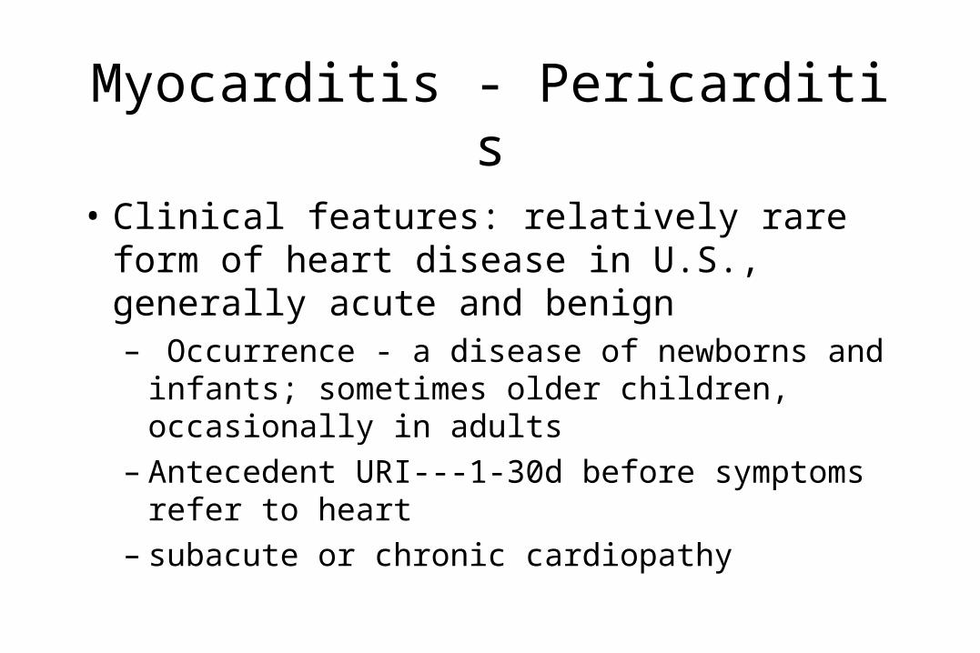
Myocarditis - Pericarditis
• Clinical features: relatively rare form of heart disease in U.S., generally acute and benign– Occurrence - a disease of newborns and infants;
sometimes older children, occasionally in adults– Antecedent URI---1-30d before symptoms refer to
heart– subacute or chronic cardiopathy

Signs and Symptoms of Viral Myocarditis
• Symptoms– Fatigue
– Dyspnea
– Palpitation
– Chest pain
– Syncope
• Signs– Pericardial rub– Sinus tachycardia– Atrial or ventricular
arrhythmias– Conduction disturbances– Cardiomegaly– Right or left S3 or S4 gallop
sounds– Congestive heart failure

New England Journal of Medicine 343:1391 2000

Infectious Causes of Pericarditis
• Bacterial– Actinomyces– Bacteroides fragilis– Borrelia burgdorferi– Brucella– Campylobacter– Chlamydia– Enterococcus sp.– Escherichia coli– Fusobacterium nucleatum– Haemophilus influenzae– Klebsiella pneumoniae– Legionella– Listeria monocytogenes
– Mycobacterium avius-intracellulare– Mycobacterium tuberculosis– Mycoplasma pneumoniae– Neisseria gonorrhea– Neisseria meningitis– Nocardia asteroides– Peptostreptococcus– Pseudomonas aeruginosa– Prevotella sp.– Salmonella– Staphylococcus aureus– Streptococcus pneumoniae– Streptococcus (group C)

InfectiousCauses of Pericarditis
• Viral– Adenovirus– Coxsackie A– Coxsackie B– Cytomegalovirus– Echovirus– Epstein Barr virus– Hepatitis B– Herpes simplex– HIV– Influenza– Mumps– Varicella Zoster
• Fungal– Aspergillus
– Blastomyces dermatitidis
– Candida
– Coccidioides Immitis
– Cryptococcus neoformans
– Histoplasma capsulatum
• Parasitic – Entamoeba histolytica
– Schistosoma
– Toxocara canis
– Toxoplasma gondii

Noninfectious Causes of Pericarditis
• Collagen vascular diseases– Rheumatic fever– Rheumatoid arthritis– Scleroderma– CREST syndrome– Systemic lupus erythematosus– Sarcoidosis– Sjögren's syndrome– Mixed connective tissue disease– Vasculitis, including temporal
arteritis– Polyarteritis
• Drug-induced– Minoxidil
– Bleomycin
– Procainamide
– Hydralazine
– Azathioprine
• Inflammatory bowel disease– Ulcerative colitis
– Crohn’s disease

Noninfectious Causes of Pericarditis
• Neoplastic– Primary (benign or malignant)– Metastatic to pericardium
• Other– Fabry’s disease– Uremia– Löffler's syndrome– Thalassemia– Acute myocardial infarction – Kawasaki’s Disease– Dissection aortic aneurysm – Post-radiation– Pregnancy
• Other– Myxedema – Dego's disease– Cardiac Injury
• Traumatic• Dressler’s syndrome
– Stevens-Johnson syndrome– Polymyositis– Dermatomyositis– Behçet's syndrome– Addisonian crisis– Gout – Whipple’s disease

Criteria for Diagnosis of Myopericarditis
• ECG manifestation– ST-T or T wave changes or
– Low QRS voltage or
– A-V conduction defects or
– Intraventricular conduction defects
• Plus 2 or more symptoms– Precordial left-sided chest
pain
– Signs and symptoms of congestive heart failure
– Cardiomegaly
– Fever
– Pericardial friction rub

Myocarditis - Pericarditis
• Diagnosis– Appropriate specimens for viral diagnosis
• Isolation of agent: pericardial fluid, T.S., R. S. first few days of illness, heart tissue at autopsy or biopsy
• Serology: 4-fold rise in titre by neutralization, complement fixation, hemagglutination inhibition; allows identification of a specific recent infection which is circumstantial evidence with a high index of suspicion when correlated with clinical findings.
– Etiological diagnosis of viral carditis is difficult

Disease Category: Myocarditis-pericarditis
Source
Viral Agents Usually Sought
Throat Swab
Rectal Swab
CSF Urine Pericardial Fluid
Other
Enterovirus ++ +++ - - ++ *
Myxovirus +++ - - - ++ *
Paramyxovirus +++ - - - ++ *
• *Because it is frequently very difficult to isolate and/or associate these agents with the disease in question, it is emphasized that serological tests are particularly important to insure a diagnosis.
• N.B. In general, it is important to remember that viral shedding often diminishes rapidly after the onset of illness; therefore, it is important to attempt to collect specimens as early as possible - including an acute serum sample.

Criteria for Viral Myocarditis
• High-order association– Isolation of virus from myocardium, endocardium or
pericardial fluid
or– Demonstration of viral antigen in the myocardium
endocardium or pericardium by immunofluorescent or immunoperoxidase assay, etc.

Criteria for Viral Myocarditis
• Moderate-order association– Isolation of virus from pharynx or feces, and a fourfold rise in
type-specific neutralizing, hemagglutination-inhibiting or complement-fixing antibodies
or
– Isolation of virus from pharynx or feces, and a concomitant titer in serum of 1/32 or more of type-specific IgM-neutralizing or hemagglutination-inhibiting antibodies.

Criteria for Viral Myocarditis
• Low-order association– Isolation of virus from pharynx or feces.– A fourfold rise in type-specific neutralizing,
hemagglutination inhibiting, or complement-fixing antibodies
– A single serum with a titer of 1/32 or greater of type-specific IgM neutralizing or hemagglutination inhibiting antibodies

Histologic Criteria for the Classification of Viral Myocarditis (“Dallas Criteria”)
• Initial Biopsy– Active myocarditis with or without fibrosis
• Presence of inflammatory infiltrate and damage of adjacent myocytes
• Frank necrosis that may consist of vacuolization, irregular cellular outlines, and cellular disruption with lymphocytes closely applied to the cell surface
• Uninvolved myocardium often appears normal
– Borderline myocarditis (may require biopsy)• Inflammatory infiltrate or myocyte damage not seen on light microscopy
• Diagnostic changes evident on additional cuts of original biopsy, which suggest active myocarditis and do not require a repeat biopsy
– No evidence of myocarditis

Histologic Criteria for the Classification of Viral Myocarditis (“Dallas Criteria”)
• Subsequent Biopsies– Ongoing myocarditis
• Degree of abnormality is equal to or worse than that of the original biopsy
– Resolving myocarditis• Inflammatory infiltrate is less and repair is evident
– Resolved myocarditis• No remaining inflammatory infiltrate and no evidence of persistent
cellular necrosis

Myocarditis - Pericarditis
• Immunity:– Need to see 4-fold rise due to ubiquity of the agents
and persistence of titers– Chronicity postulated due to lesions representing an
immune response

Myocarditis - Pericarditis
• Epidemiology:– Season: random through year– Spread: fecal-oral and respiratory– Age– Other factors:
• Physical exercise• Nutrition• Volume load on circulatory system• Pregnancy• Sex• Corticosteroids• Diabetes
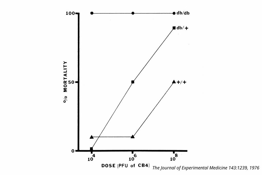
The Journal of Experimental Medicine 143:1239, 1976
Myocarditis - Pericarditis
• Prophylaxis and treatment:– Chronic sequelae constitute an argument for search for
specific treatment and prevention– Controlled studies of effects of therapeutic measures
are needed– Bed rest and supportive therapy

Proposed Therapies of Postviral and Idiopathic Myocarditis
Category Therapy Comment
Conventional therapy of congestive heart failure
Digitalis and diuretics Digitalis may decrease interleukin-1 and tumor necrosis factor-
Angiotensins-converting enzyme inhibitors and angiotensin-II receptor antagonists
May have a direct immunomodulatory effect
Bed rest, -blockers Both beneficial and deleterious effects in murine models
Immunosuppressive therapy Corticosteroids Documented use in humans
Cyclosporine Documented use in humans
Azathioprine Documented use in humans
FK506
OKT3 Documented use in humans
Many others

Proposed Therapies of Postviral and Idiopathic Myocarditis
Category Therapy Comment
Immunomodulatory therapy
Gamma globulin Documented use in humans
Coxsackie B3 vaccine FK565 Immunostimulant action inhibits replication
Immunoadsorption
Antiviral therapy Ribavirin
Interferon
Anticytokine therapy Anti-tumor necrosis factor antibody
Vesnarinone One of several phosphodiesterase inhibitors that inhibit cytokine release
Amiodarone
Miscellaneous Margatoxin One of several T-cell potassium-channel blockers
Calcium antagonists May prevent microvascular spasm
N-monomethyl-l-arginine
Inhibition of nitric oxide synthesis may prevent myocyte injury and reversible depression

Viral Infections with Involvement of the Hematopoietic and Lymphatic Systems

Epstein-Barr Virus, Infectious mononucleosis
• EBV herpes group virus, lymphotropic – 1889 Pfeiffer - "drusenfieber" - glandular fever– 1968 - Henle's: after long history attributed an essential virus
role in the disease to a virus of the herpes group– EB virus = Epstein Barr virus, a herpes type virus named for
cell line in which it was first detected– Transforms (i.e., releases from normal regulatory control)
human B lymphocytes which then interact with the T lymphocytes (atypical lymphs of mono)

New England Journal of Medicine343:482 2000

New England Journal of Medicine 343:483 2000

JAMA 278:511, 1997

Various Forms of Infection by EB Virus in Man
• Productive replicative infection– Virus replication leading to cell death (as in the oropharynx of some infected
individuals)
• Nonproductive infection– Can be activated to productive cycle
• Latent infection– Virus genome express to give LYDMA and EBNA (as in peripheral B cells of all
infected individuals)
• Malignant transformation– Virus genome expressed to give early antigen and cell changes of malignancy (as in
BL showing LYDMA, EBNA, EMA, and NPC showing EBNA)
• In marmosets EB virus certainly induces malignant transformation with EBNA expression to give malignant lymphomas

Pediatrics in Review7:36, 1985

Clinical Findings in Heterophile Antibody-Positive Infectious Mononucleosis
No. of Patients 270 56,200 100 100
Symptoms (% of patients)
•Sore throat 88 70 NS NS
•Malaise 50 43 NS 76
•Headache 62 37.5 NS 55
•Nausea, vomiting, anorexia 27 7.1 NS 43
•Myalgia 21 12.5 NS NS

Clinical Findings in Heterophile Antibody-Positive Infectious Mononucleosis
No. of Patients 270 56,200 100 100
Signs (% of patients)
•Fever 65 97.5 94 79
•Lymphadenopathy >90 100 94 95
•Pharyngitis 85 83 NS 91
•Exudative 63 22 69 49
•Splenomegaly 50 NS 63 51
•Palpebral edema 18 36 11 5
•Palatal petechiae 47 25 NS 13
•Rash 25 3 15 12
•Jaundice 10 8 8 0
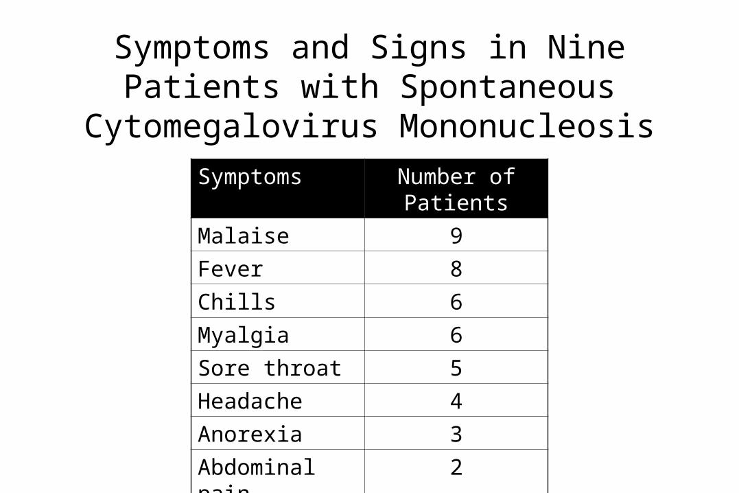
Symptoms and Signs in Nine Patients with Spontaneous Cytomegalovirus Mononucleosis
Symptoms Number of Patients
Malaise 9
Fever 8
Chills 6
Myalgia 6
Sore throat 5
Headache 4
Anorexia 3
Abdominal pain 2
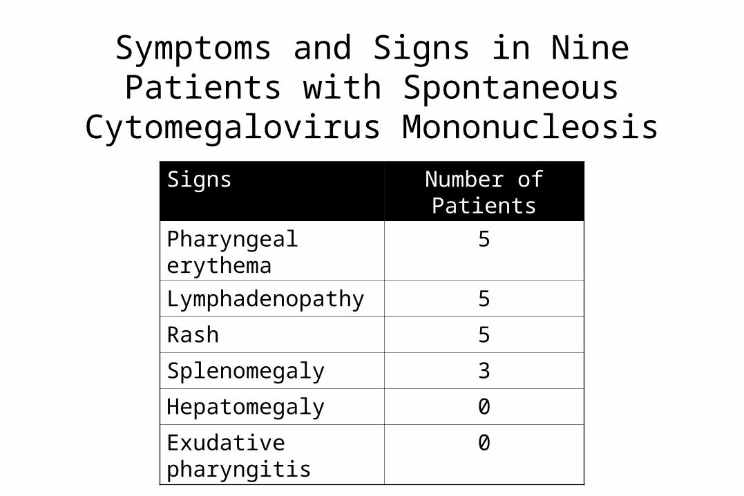
Symptoms and Signs in Nine Patients with Spontaneous Cytomegalovirus Mononucleosis
Signs Number of Patients
Pharyngeal erythema 5
Lymphadenopathy 5
Rash 5
Splenomegaly 3
Hepatomegaly 0
Exudative pharyngitis 0

Clinical Disorders Associated Etiologically with Epstein-Barr Virus
Primary infection Evidence for etiology
(+ to ++++)
•Infectious mononucleosis ++++
•Congenital infection with fetal abnormalities ++++
•Acute neurologic disease (Guillain Barré, Bell’s Palsy, meningoencephalitis)
+++
•Acquire agammaglobulinemia, aplastic anemia, lymphoma
+++
•Lymphoproliferative lesions including lymphomas in renal and other organ transplant recipients
++
•Tonsillopharyngitis ++
•Thrombocytopenia ++
•Pneumonia ++
•Reye’s syndrome ++
•Hemophagocytic syndrome +
•Acute arthritis +
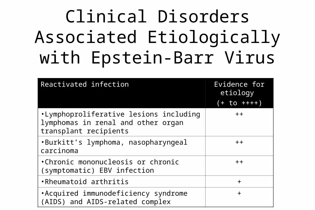
Clinical Disorders Associated Etiologically with Epstein-Barr Virus
Reactivated infection Evidence for etiology
(+ to ++++)
•Lymphoproliferative lesions including lymphomas in renal and other organ transplant recipients
++
•Burkitt’s lymphoma, nasopharyngeal carcinoma ++
•Chronic mononucleosis or chronic (symptomatic) EBV infection
++
•Rheumatoid arthritis +
•Acquired immunodeficiency syndrome (AIDS) and AIDS-related complex
+

Complications of Infectious Mononucleosis
• Neurologic– Meningoencephalitis– Aseptic meningitis– Guillain-Barré syndrome– Facial or other peripheral nerve
paralysis– Transverse myelitis– Optic neuritis– Seizures– Coma– Acute psychosis– Acute cerebellar ataxia
• Hematologic – Autoimmune hemolytic anemia
– Thrombocytopenic purpura
– Granulocytopenia
– Pancytopenia
– DIC

Complications of Infectious Mononucleosis
• Cardiac– Myocarditis
– Pericarditis
• Respiratory– Pharyngeal edema with
airway obstruction
– Interstitial pneumonia
– Pleuritis
• Hepatic– Cholestatic jaundice
– Massive hepatic necrosis causing liver failure
• Splenic Rupture

Signs and Symptoms of Hemophagocytic Lymphohistiocytosis
Organ System
Clinical Findings Laboratory Findings
General Fever, edema
Bone Marrow Anemia Hemophagocytosis, cytopenia 2 lines
Immune system
Splenomegaly, lymphadenopathy ↓ Natural killer cell activity, ↑ serum cytokines, ↑ soluble IL-2 receptor
Liver Jaundice, hepatomegaly ↑ Triglycerides, ↓ fibrinogen, ↑ ferritin,
↑ LDH, coagulopathy, ↑ transaminases,
↑ bilirubin, DIC
Lungs Cough Infiltrates on chest x-ray
Skin Generalized maculopapular rash
CNS Irritability, stiff neck, seizure, CN palsy, ataxia
↑ Protein in CSF, hemophagocytosis in CSF

“Chronic Mononucleosis”Clinical Findings and Reported Complaints Among 39 Patients with
Suspected Chronic Infectious Mononucleosis
Complaint Patients No. (%)
Fatigue 29 (74)Nervous system 28 (73)Depression 27 (70)Pharyngitis 25 (64)Fever 24 (63)Lymphadenopathy 23 (59)Myalgia 21 (56)
Complaint Patients
No. (%)
Dyslogia 20 (53)
Arthritis/arthralgia 19 (51)
Splenomegaly 9 (22)
Weight loss 9 (22)
Rash 5 (12)
Hepatomegaly 4 (10)
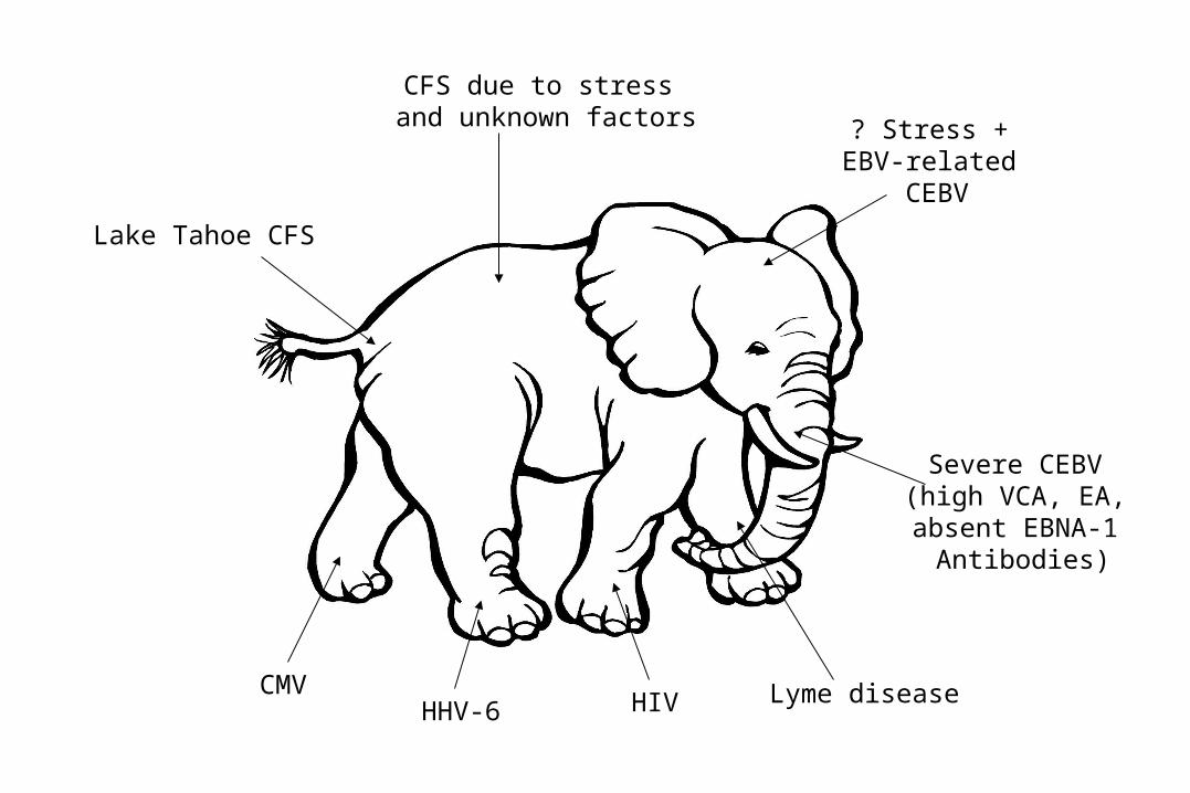
CFS due to stress and unknown factors
Lake Tahoe CFS
? Stress + EBV-related
CEBV
Severe CEBV (high VCA, EA, absent EBNA-1
Antibodies)
CMVHIVHHV-6
Lyme disease

Timeline graph from 1800 to the present of other diseases with symptoms very similar to CFS
1800 1850 1900 1950 2000
Chronic Fatigue Syndrome
Postviral Fatigue Syndrome
Chronic Candidiasis
Chronic Mononucleosis, Chronic EBV
Total Allergy Syndrome
Myalgic Encephalomyelitis, Epidemic Neuromyasthenia
Hypoglycemia
Chronic Brucellosis
Da Costa's Syndrome
Neurasthenia
Febricula, Vapors

Summary of the Working Definition of CFS
• Major criteria– Persistent or relapsing fatigue or easy fatigability that
does not resolve with bed rest and is severe enough to reduce average daily activity by ≥ 50
– Satisfactory exclusion of other chronic conditions, including preexisting psychiatric disease

Summary of the Working Definition of CFS
• Minor criteria– Mild fever (37.5-38.0ºC oral if document by patient) or chills– Sore throat– Lymph node pain in anterior or posterior cervical or axillary chains– Unexplained, generalized muscle weakness– Muscle discomfort, myalgia– Prolonged (≥ 24 h) generalized fatigue after previously tolerable levels of exercise– New generalized headaches– Migratory, noninflammatory arthralgia– Neuropsychologic symptoms: photophobia, transient visual scotomata, forgetfulness,
excessive irritability, confusion, difficulty thinking, inability to concentrate or depression
– Sleep disturbance– Patient description of initial onset of symptoms as acute or subacute
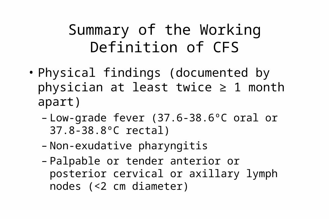
Summary of the Working Definition of CFS
• Physical findings (documented by physician at least twice ≥ 1 month apart)– Low-grade fever (37.6-38.6ºC oral or 37.8-38.8ºC
rectal)– Non-exudative pharyngitis– Palpable or tender anterior or posterior cervical or
axillary lymph nodes (<2 cm diameter)

Epstein-Barr Virus, Infectious mononucleosis
• Laboratory diagnosis– Blood smear with "atypical" lymphocytes– Heterophile agglutination (nonspecific reaction with
abs which agglutinate HRBC or SRBC)– Anti EB virus abs

Clinical and laboratory manifestations of infectious mononucleosis. The predominant symptoms, signs, laboratory changes and EB virus-specific serologic findings during classic infectious mononucleosis are depicted in four panels. Arrow A indicates asymptomatic prodrome; arrow B, peak of clinical illness; and arrow C, early convalescence, during which the EB virus-associated neuropathies usually occur.
Pediatrics in Review 7:37, 1985

Disorders Associated with >20% Atypical Lymphocytes
• EBV mononucleosis
• Viral hepatitis
• CMV mononucleosis

Disorders Associated with <20% Atypical Lymphocytes
• Infections– Mumps– Varicella– Rubeola– Rubella– Roseola infantum (HHV6)– Herpes simplex– Herpes zoster– Influenza– Tuberculosis
– Brucellosis
– Toxoplasmosis
– Syphilis
– Smallpox
– Malaria
– Babesiosis
– RMSF
– Ehrlichiosis

Disorders Associated with <20% Atypical Lymphocytes
• Non-Infectious– Drug hypersensitivity
reactions
– Drug fever
– Dermatitis herpetiformis
– Radiation therapy
– Stress
– Lead intoxication

Interpretation of EBV Serology
IgG-VCA IgM-VCA EBV Nuclear Antigen
EBV Early Antigen
No evidence of infection
<10 <10 <2 <10
Acute infection
>10 ≥10 <2 ≥20
Convalescent infection
>10 Variable >2 Variable
Remote past infection
≥10 <10 >2 ≤20

EBV Toxoplasmosis
Rubella HIV CMV HHV-6 HAV/HBV
Physical Findings
•Pharyngitis ++
(exudative/
non-exudative)
+
(non-exudative)
+
(non-exudative)
±
(non-exudative)
+
(non-exudative)
+
(non-exudative)
±
(non-exudative)
•Lymphadenopathy Bilateral posterior
cervical/generalized lymph-adenopathy
Unilateral single node involvement
Occipital postauricular generalized
lymph-adenopathy
Localized node
enlargement generalized
lymph-adenopathy
Bilateral posterior cervical/
generalized lymph-
adenopathy
Bilateral posterior cervical
adenopathy
None/mild general
adenopathy
•Splenomegaly +++ ± - - ± ± -
Lab Abnormalities
•Leukopenia + - ± + + ±
•Atypical lymphocytosis
20% <5% <5% - ≥ 20% <10% <5%
• SGOT/SGPT + ± - - + + +++
•Thrombocytopenia + - ± + + ± -
•Mono spot + -* -* -* -* -* -*
*rarely false positive Mono spot test

Epstein-Barr Virus, Infectious mononucleosis
• Epidemiology:– Children and young adults– Droplet spread probably– Communicability period and incubation period

Epstein-Barr Virus, Infectious mononucleosis
• Immunity:– EB virus (or one closely related antigenically) might
operate in an opportunistic way whenever it finds actively proliferating lymphocytes

Epstein-Barr Virus, Infectious mononucleosis
• Prophylaxis and treatment: – Symptomatic and supportive– Acyclovir– Corticosteroids

Burkitt's disease
• African lymphoma starting as jaw or orbital tumor, then involvement of maxillary bones, kidneys, ovaries, thyroid, parotid
• Epidemiology– Central Africa– Case concentration: children 7-8 years old
• Associated etiology– Herpes-group virus: EB virus (from cell line of a Burkitt
lymphoma established by Epstein and Barr)– DNA, 180 nm enveloped

Annual Review of Microbiology31:424, 1977

Other
• HHV 6, HHV7, HHV8
• Human Parvovirus B19: transient aplastic crisis
• Bone marrow failure
• Malignant association
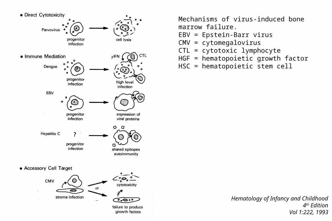
Hematology of Infancy and Childhood4th Edition
Vol 1:222, 1993
Mechanisms of virus-induced bone marrow failure. EBV = Epstein-Barr virusCMV = cytomegalovirusCTL = cytotoxic lymphocyteHGF = hematopoietic growth factorHSC = hematopoietic stem cell

Infectious Causes of Cancer
Clinical Infectious Diseases32:679, 2001

Established Association Between an Infectious Agent and a Malignancy
Pathogen Malignancy
Helicobacter pylori Gastric carcinoma
Helicobacter pylori Mucosal-associated lymphoid tissue
Schistosoma haematobium
Bladder cancer
HTLV-1 Adult T-cell leukemia/lymphoma
HTLV-11 Hairy cell leukemia
HBV Liver cancer
HHV-8 Kaposi sarcoma
EBV Lymphoproliferative disorders
EBV Nasopharyngeal carcinoma
EBV Burkitt’s lymphoma
HPV Anogenital carcinoma, cervical cancer

CMV and cardiovascular disease

Cardiac Malformations as Part of Rubella Embryopathy
• Rubella virus predilection for vascular endothelium: patent ductus arteriosus, atrial septal defect, ventricular septal defect, lesions of myocardial fibers, alterations in renal arteries, pulmonary artery stenosis, and also thrombocytopenic purpura