Embed Size (px)
Citation preview

DISEASES OF AQUATIC ORGANISMSDis Aquat Org
Vol. 101: 69–86, 2012doi: 10.3354/dao02501
Published October 10
INTRODUCTION
Freshwater eels of the genus Anguilla have extra -ordinary catadromous lifecycles, with the spawninggrounds of some species located in the ocean sev-eral thousands of kilometres away from their fresh-water growth habitats in lakes and rivers of themainland (van Ginneken & Maes 2005). The wildfreshwater eel stocks have shown strong declinesworldwide since the 1980s (Dekker et al. 2003,
Stone 2003). The cause of the decline is unknown,but is probably multifactorial, with suggested factorsincluding pollution, habitat loss, fisheries, migrationbarriers, and diseases (Dekker 2004). Indeed, theswimbladder nematode Anguillicoloides crassus(Székely et al. 2009), several pathogenic bacteria(Esteve & Alcaide 2009) and certain viruses (Haenenet al. 2009) have been suggested to play a contribu-tory role in the decline of the wild European eelAnguilla anguilla stock.
© Inter-Research 2012 · www.int-res.com*Corresponding author. Email: [email protected]
REVIEW
Viral diseases of wild and farmed European eelAnguilla anguilla with particular reference
to the Netherlands
Steven J. van Beurden1,2, Marc Y. Engelsma1, Ineke Roozenburg1, Michal A. Voorbergen-Laarman1, Peter W. van Tulden1, Sonja Kerkhoff1,
Anton P. van Nieuwstadt1, Aart Davidse1, Olga L. M. Haenen1,*
1Central Veterinary Institute, Wageningen UR, PO Box 65, 8200 AB Lelystad, The Netherlands2Department of Infectious Diseases and Immunology, Faculty of Veterinary Medicine, Utrecht University, PO Box 80.165,
3508 TD Utrecht, The Netherlands
ABSTRACT: Diseases are an important cause of losses and decreased production rates in fresh -water eel farming, and have been suggested to play a contributory role in the worldwide declinein wild freshwater eel stocks. Three commonly detected pathogenic viruses of European eelAnguilla anguilla are the aquabirnavirus eel virus European (EVE), the rhabdovirus eel virusEuropean X (EVEX), and the alloherpesvirus anguillid herpesvirus 1 (AngHV1). In general, all 3viruses cause a nonspecific haemorrhagic disease with increased mortality rates. This review pro-vides an overview of the current knowledge on the aetiology, prevalence, clinical signs and grosspathology of these 3 viruses. Reported experimental infections showed the temperature depend-ency and potential pathogenicity of these viruses for eels and other fish species. In addition to thepublished literature, an overview of the isolation of pathogenic viruses from wild and farmed A.anguilla in the Netherlands during the past 2 decades is given. A total of 249 wild A. anguilla, 39batches of glass eels intended for farming purposes, and 239 batches of farmed European eelswere necropsied and examined virologically. AngHV1 was isolated from wild A. anguilla yellowand silver eels from the Netherlands from 1998 until the present, while EVEX was only found spo-radically, and EVE was never isolated. In farmed A. anguilla AngHV1 was also the most com-monly isolated virus, followed by EVE and EVEX.
KEY WORDS: Anguillid herpesvirus 1 · Eel virus European · Eel virus European X
Resale or republication not permitted without written consent of the publisher
Dis Aquat Org 101: 69–86, 2012
Anguilla anguilla and the Japanese eel A. japon-ica are traditionally consumed in several Europeancountries and Japan, respectively (Heinsbroek 1991).Historically, eels for consumption were wild-caught,but European eel fisheries have recently been sub-ject to limitations because of the population decline(Council of the European Union 2007). In Japan, eelfarming for consumption purposes started at the endof the 19th century. Eel farming has developed fromnon-intensive polyculture in outdoor ponds to inten-sive indoor culture in greenhouses since the 1970s.Eel farming in Europe has its origin in Italy, butgradually moved to northwestern Europe, where itchanged into an intensive form of aquaculture afterthe Japanese example.
As artificial reproduction of freshwater eel is notyet possible on a commercial scale, production forconsumption is still based entirely on catches of wildglass eels or elvers. This leads to the potential intro-duction of eel disease agents in aquaculture pro -duction systems. Anguilla anguilla is nowadays pro-duced generally in intensive recirculation systems ata regulated water temperature. With an annual pro-duction of about 4000 t in the previous decade, theNetherlands is the most important eel-producingcountry in Europe (FAO 2012). High stocking densi-ties make detection and control of diseases vital forsustainable farming. Prevention and treatment of eelviral diseases is particularly difficult, as commercialvaccines are not available.
When high-density Japanese eel pond cultureexponentially grew in the late 1960s and early 1970sin Japan, Anguilla anguilla and American glass eelsand elvers A. rostrata were imported and stocked,and catastrophic viral disease outbreaks occurredfrequently (Heinsbroek 1991). Permanently growingcell lines were developed from A. japonica kidneyand ovary cells, and used for virus isolation (Chen &Kou 1981, Chen et al. 1982). During the many out-breaks, new viruses were isolated and shown to bethe causative agent (T. Sano 1976, T. Sano et al. 1977,M. Sano et al. 1990). Although initial descriptionswere usually thorough and detailed, nomenclaturewas ambiguous. Hence, several virus isolates wereinitially presented as a new virus, and later demon-strated to be highly similar to an already describedvirus.
Identification of pathogenic eel viruses is furthercomplicated by the non-pathognomonic clinical signsand gross pathology of these eel viral diseases. Inaddition, virus isolation from clinically healthy eels(Castric & Chastel 1980, Bucke 1981, Castric et al.1984, Chen et al. 1985, Shchelkunov et al. 1989, Hae-
nen et al. 2002), as well as double infections with dif-ferent viruses (Ahne & Thomas 1985, Haenen et al.2002, van Ginneken et al. 2004, Varvarigos et al.2011), have been observed. Several diagnostic assayshave been developed for the detection of eel viruses,more recently with a focus on molecular assays. Ineel farming, identification of the causative agent canbe used to take adequate quarantine and water tem-perature regulation measures in order to reduce clin-ical signs and losses.
In this literature review, we give an overview ofthe current knowledge on the aetiology, geographi-cal distribution, clinical signs, mortality and grosspathology of pathogenic European eel viruses. Inaddition, we present a retrospective analysis of diag-nostic data from the Dutch National Reference Labo-ratory (NRL) for Fish Diseases over the period 1990to 2011, which provides a historical overview on theviruses isolated from wild and farmed A. anguilla inthe Netherlands in the past 2 decades. The 3 viralagents that are observed most commonly in A. ang -uilla are the aquabirnavirus eel virus European(EVE), the rhabdovirus eel virus European X (EVEX)and the alloherpesvirus anguillid herpesvirus 1(AngHV1).
EEL VIRUS EUROPEAN
Aetiology
Since 1969, serious outbreaks of a new disease,called branchionephritis or viral kidney disease ofAnguilla japonica, occurred every winter whenwater temperatures were below 20°C in eel cultureponds in Japan (T. Sano et al. 1981). The aetiologicalagent was isolated for the first time from imported A.anguilla using the rainbow trout gonad cell line RTG-2 (Wolf & Quimby 1962) in 1973, and tentativelynamed eel virus European (EVE) (T. Sano 1976). EVEwas subsequently also isolated from A. japonica, andRiver’s postulates were fulfilled. The type of cyto-pathic effect (CPE) caused by EVE resembled thetype of CPE caused by the aquabirnavirus infectiouspancreatic necrosis virus (IPNV) (T. Sano 1976, T.Sano et al. 1981). Electron microscopy (EM) revealedthat EVE virions were non-enveloped polyhedronswith a diameter of 68 to 77 nm, only present in thecytoplasm of infected cells. EVE also resembledIPNV in terms of biological properties, such aspolypeptide composition and the bisegmented dou-ble-stranded RNA genome (Nishimura et al. 1981a,T. Sano et al. 1981, Hedrick et al. 1983a). Hence, EVE
70

van Beurden et al.: Viral diseases of European eel
is a tentative member of the genus Aquabirnavirus inthe family Birnaviridae. Other names for EVE orIPNV of eel include eel virus (EV) [Berlin] (Schwanz-Pfitzner et al. 1984) and pillar cell necrosis virus(PCNV; Lee et al. 1999a, 2001).
Aquabirnaviruses form an antigenically diversegroup of viruses, with the type species IPNV beingthe aetiological agent of an acute contagious systemicdisease of several species of freshwater and marinefish, molluscs and crustaceans (Rodriguez Saint-Jeanet al. 2003). Mortality caused by IPNV in salmonids ishigh in fry and fingerlings, but rare in older fish. Sur-vivors of the epizootic disease may become lifelongcarriers. Host specificity and cell tropism are deter-mined by viral proteins encoded by the larger RNAsegment A (M. Sano et al. 1992), and the occurrenceof natural reassortment has recently been shown(Romero-Brey et al. 2009). Interspecies transmissionhas not yet been demonstrated, but it would explainthe wide range of host species (Bandin & Dopazo2011). Historically, aquabirnavirus isolates weregrouped as 1 of the 3 major serotypes, designated Ab,Sp and VR-299 (Macdonald & Gower 1981).
Neutralisation tests confirmed the close relation-ship of EVE with IPNV (T. Sano 1976, T. Sano et al.1981), later specified to IPNV type Ab (Hudson et al.1981, Okamoto et al. 1983). EVE and IPNV type Abwere also found to be similar in polypeptide and RNAcomposition, and clearly distinguishable from IPNVstrains VR-299 and Sp (T. Sano et al. 1981, Hedricket al. 1983a). Using cross-neutralisation assays withalmost 200 IPNV isolates, Hill & Way (1995) later pro-posed a new serological classification, consisting ofserogroup A containing serotypes A1 to A9, andserogroup B containing the single serotype B1. Theprevalence of the different serotypes is geographical,with the aquatic birnaviruses in the USA generallybelonging to serotype A1, those in South Americaand Asia to serotypes A1 to A3, those in Europe to A2to A5, and those in Canada to A6 to A9 (Blake et al.2001). More recent phylogenetic analyses based ondeduced amino acid sequences of the VP2 and VP5genes of larger RNA segment A showed that Japan-ese and Taiwanese EVE strains group with IPNVstrain Ab in genogroup 3 (Blake et al. 2001, Zhang &Suzuki 2004).
Geographical distribution
Anguilla anguilla elvers were first imported intoJapan in 1968, after which the epizootics of bran-chionephritis started occurring in A. japonica (T.
Sano et al. 1981). It was therefore suggested — andallegedly proven— that EVE entered Japan with theimport of A. anguilla elvers (T. Sano et al. 1981,Hedrick et al. 1983a). EVE/IPNV type Ab and IPNVtype VR-299 were later also isolated from various A.japonica farms in Taiwan (Chen et al. 1985, Hsu et al.1989, 1993). EVE/PCNV from diseased A. japonica inJapan was serologically most similar to IPNV sero -type Sp (Lee et al. 1999a), but genetically closerrelated to strain Ab (Lee et al. 2001).
In 1977, Castric & Chastel (1980) isolated an IPNV-like agent called B6 from Anguilla anguilla elversalong the French Atlantic coast, and showed its relat-edness to IPNV serotype Sp by serum neutralisationtests. EVE related to IPNV serotype Ab was repeat-edly isolated from an eel farm in the UK (Bucke 1981,Hudson et al. 1981). IPNV type Ab or EVE was iso-lated from different populations of wild and farmedA. japonica in Taiwan (Hedrick et al. 1983b, Wu et al.1987). Several viruses isolated from the blood (4 iso-lates) and gonads (1 isolate) of A. anguilla with stom-atopapillomas in Germany were identified as IPNVsubtype Ab by serum neutralisation tests (Ahne et al.1987). The first of these viral isolates — isolated in1968 — was tentatively named EV [Berlin], but latercharacterised as a birnavirus (Schwanz-Pfitzner et al.1984). IPNV types Ab and Sp were isolated frompools of A. anguilla elvers and eels from Denmark,the UK and France (Jørgensen et al. 1994). J. Plumb(Auburn University) isolated EVE serotype Ab fromA. rostrata (cited in McAllister & Owens 1995). Dou-ble infections of farmed A. anguilla with EVE andAngHV1 were re ported in the Netherlands (Haenenet al. 2002) and Greece (Varvarigos et al. 2011), anddouble infections with EVE and EVEX were reportedin Germany (Ahne & Thomsen 1985) and Italy (vanGinneken et al. 2004). Overall, EVE has beendetected in A. japonica in Japan and Taiwan, in A.anguilla in Japan, France, the UK, Germany, Den-mark, the Netherlands and Greece, and in A. rostratain the USA.
Clinical signs and mortality
EVE has been isolated from apparently healthyand diseased eels. Moribund Anguilla japonicashowed rigidity or spasm of the body, retractedabdomen, congestion of the anal fin, and occasion-ally diffuse congestion on the abdomen and gills (T.Sano 1976, T. Sano et al. 1981). Wu et al. (1987) iso-lated an EVE-like virus from farmed A. japonicashowing some pathological symptoms such as ulcer-
71

Dis Aquat Org 101: 69–86, 2012
ative lesions over the body, congestion of the fins,atrophy of the muscles and a deformed trunk. Bucke(1981) and Hudson et al. (1981), however, isolatedEVE from A. anguilla with no external lesions orabnormalities. Chen et al. (1985) isolated EVE fromhealthy and diseased A. japonica, and during an out-break of branchionephritis with nearly 100% mortal-ity in certain ponds. EVE/PCNV was isolated frommass mortalities among farmed A. japonica since thelate 1980s, in which the eels showed no other exter-nal pathological signs, except for loss of appetite andgeneral weakness (Lee et al. 1999a). In short, themost commonly reported clinical signs of EVE-infec-tion in eel are an abnormal shape of the trunk, andcongestion of the skin, fins and gills.
Gross pathology
Gross internal findings in Anguilla japonica and A.anguilla were some enlargement of the kidney, anempty gut, and in some cases ascites (T. Sano 1976,T. Sano et al. 1981). Histopathological findings in -cluded tubular and renal interstitial necrosis in thekidney, and occasionally focal necrosis in the liverand spleen. Wu et al. (1987) found hypertrophy andnecrosis of the liver in A. japonica, while Hudson etal. (1981) occasionally found petechial haemorrhagesin the liver of A. anguilla. The A. japonica from whichUeno et al. (1984) isolated a birna virus similar to EVEhad nephroblastoma, clinically manifested as whitish,swollen and solid kidneys. EVE/PCNV caused gilldisease, characterised by aneurysmal haematomaformations in the gill lamellae and necrosis of the pil-lar cells (Lee et al. 1999a). Although EVE was iso-lated repeatedly from A. anguilla with stomatopapil-lomas, attempts to initiate tumour production inhealthy eels by inoculation with this virus failed, sug-gesting no causative relationship (McAllister et al.1977). Taken together, the most common grosspathological findings of EVE infection in eel areenlargement of the kidneys, necrosis or petechialhaemorrhages in the liver, and gill disease.
Experimental infections
Experimental infections of eel and rainbow troutwith EVE have yielded variable results. T. Sano(1976) tested the infectivity of EVE for Anguillajaponica glass eels and young eels experimentally bybath immersion and by intraperitoneal injection,respectively. Cumulative mortality over a 20 d period
was 60% for the glass eels, which were held at 15 to20°C (T. Sano 1976), and 55 to 75% for the youngeels, which were held at 8 to 14°C (T. Sano 1976, T.Sano et al. 1981). Moribund young eels showed mus-cular spasm or rigidity, slight petechiae of theabdominal skin and congestion of the anal fin. EVEwas reisolated from gill, spleen, gut and kidney tis-sue of the moribund young eels, and from wholeglass eels. In a sub sequent infection trial of A. japon-ica with EVE and with IPNV strain d’Honnicthun, nomortality occurred (T. Sano et al. 1981). It was possi-ble to reisolate EVE, but not IPNV, from the injectedeels. No significant mortalities were observed in A.anguilla elvers (0.25 and 0.50 g) infected withEVE/IPNV strain B6 by bath immersion or sprinklingat 8−11 and 21.5°C, or in young A. anguilla (3 g)infected with EVE/IPNV strain B6 by intraperitonealinjection at 17°C (Castric & Chastel 1980). The Abserotype of EVE isolated from farmed A. anguillafrom the UK did not cause disease in eels either(Hudson et al. 1981). The EVE-like virus isolated byUeno et al. (1984) caused 40% mortality in juvenileA. japonica after intraperitoneal injection at 20 to25°C. EVE/ PCNV caused about 70% mortality in A.japonica (~40 g) during a 21 d experiment at 25°C,after intramuscular injection (Lee et al. 1999a). Allinoculated eels showed aneurysmal haematoma for-mation in gill lamellae and stasis of gill filamentalarteries, similar to natural infections, and it was pos-sible to reisolate the virus from diseased gill fila-ments. Larger eels (~120 g) showed only limited gilldisease and no significant mortality.
Castric & Chastel (1980) showed the pathogenicityof EVE/IPNV strain B6 for rainbow trout Oncorhyn-chus mykiss fry, by bath immersion at 8 to 11°C,reaching cumulative mortalities of 82% over a 2 moperiod. Affected fish showed typical signs of infec-tious pancreatic necrosis (IPN), and strain B6 was eas-ily reisolated from dead fry. However, T. Sano et al.(1981) did not find any significant signs or mortalityin rainbow trout fry exposed to EVE by bath immer-sion at 10°C over a 40 d period. The Ab serotypes ofEVE isolated from farmed Anguilla anguilla and A.japonica from the UK and Japan, respectively, didnot cause disease in rainbow trout fry either (Hudsonet al. 1981, Okamoto et al. 1983, M. Sano et al. 1992).The EVE isolate serotype Ab from A. rostrata ap -peared to be non-virulent to brook trout Salvelinusfontinalis fry 42 d old at 12°C; a Japanese EVE isolatewas weakly virulent (3% virus associated mortality),and another Japanese EVE isolate was highly viru-lent (87% virus-associated mortality) (McAllister &Owens 1995). The EVE-like virus isolated by Ueno et
72

van Beurden et al.: Viral diseases of European eel
al. (1984) resulted in 25 to 35% mortality in intraperi-toneally injected juvenile common carp Cyprinuscarpio and hybrid tilapia at 20 to 25°C. An infectiontrial with tilapia at 10 to 16°C resulted in a cumula-tive mortality of 80% (Ueno et al. 1984).
In conclusion, outcomes of infection trials with EVEin elvers, young eels and rainbow trout fry vary. EVEseems to be pathogenic for rainbow trout fry, but notalways for juvenile eels. The observed differencesare most likely due to varying experimental condi-tions, such as age of the experimental fish, EVE strainused, infection method, and water temperature.
EEL VIRUS AMERICAN AND EEL VIRUSEUROPEAN X
Aetiology
In the 1970s, 2 rhabdoviruses were isolated fromimported eel in Japan. The first was isolated fromyoung Anguilla rostrata imported from Cuba in 1974and designated eel virus American (EVA) (T. Sano1976). A second related rhabdovirus was isolated 2 yrlater from A. anguilla elvers imported from Franceand designated eel virus European X (EVEX) (T.Sano et al. 1977). EVA and EVEX are morphologi-cally (enveloped bullet-shaped particles of 136−160 ×53−84 nm), serologically, physicochemically andgenetically (single-stranded RNA genome) highlysimilar, and regarded as 2 strains of a single virusspecies (Hill et al. 1980, Nishimura et al. 1981b, vanBeurden et al. 2011). Synonymous names used forEVEX/EVA were rhabdovirus anguilla (Hill et al.1980, Shchelkunov et al. 1989) and rhabdoviral dermatitis of Japanese eel (Kobayashi & Miyazaki1996). EVEX/EVA belongs to the group of fish vesi -culovirus-like isolates, family Rhabdoviridae, orderMononegavirales (Galinier et al. 2012).
Geographical distribution
The first detection of EVEX in Europe originatesfrom the early 1980s, when the EVEX-like viruses C30,B44 and D13 were isolated during a virological surveyon the wild Anguilla anguilla elver population of theLoire estuary along the French Atlantic coast (Castric& Chastel 1980, Castric et al. 1984). EVEX was subse-quently isolated from A. anguilla from Germany(Ahne & Thomsen 1985, Ahne et al. 1987), and fromA. anguilla elvers imported from Germany to Russia(Shchelkunov et al. 1989). In a comprehensive study
on the occurrence of virus infections in pools of elversand eels in Europe from 1977 to 1992, Jørgensen et al.(1994) isolated EVEX in A. anguilla from France, theUK, Denmark and Sweden. Van Ginneken et al.(2004) detected EVEX in wild eels originating fromvarious geographic regions. In retrospective sequenceanalyses of these samples, however, only a singlevirus strain was observed, most likely having anEVEX-infected eel farm in Italy as its source (M. Y.Engelsma et al. unpubl. data). In the Netherlands,EVEX was later isolated from wild A. anguilla after aswim tunnel experiment (van Ginneken et al. 2005),and from various A. anguilla farms (Haenen et al.2002, van Beurden et al. 2011). In Japan, a rhab-dovirus causing dermatitis in A. japonica was isolatedand shown to be serologically similar to EVEX andEVA (Kobayashi & Miyazaki 1996). Overall, EVA hasbeen isolated from A. rostrata imported from Cuba,EVEX has been detected in A. anguilla from France,Germany, the UK, Denmark, Sweden, Italy and theNetherlands, and rhabdoviral dermatitis has been di-agnosed in A. japonica in Japan.
Clinical signs and mortality
In the initial description of EVA by T. Sano (1976),the infected Anguilla rostrata showed clear externalsymptoms: most eels had a tendency to bend thehead down, and showed intense congestion in thepectoral and anal fin and diffuse congestion over theabdominal skin. The American eels showed anunusually high mortality of 59% over a 170 d rearingperiod. A. japonica infected with rhabdoviral der-matitis during post-harvest stocking showed cuta-neous erosion and ulceration (Kobayashi & Miyazaki1996). This disease occasionally occurred and causedmass mortalities during the pre-shipping stocking.
EVEX-infected farmed Anguilla anguilla from Italyshowed clinical signs such as haemorrhages and redskin areas (van Ginneken et al. 2004). However, onseveral occasions, EVEX has also been isolated fromapparently healthy A. anguilla elvers (Castric &Chastel 1980, Castric et al. 1984, Shchelkunov et al.1989). Although Ahne et al. (1987) repeatedly iso-lated EVEX from A. anguilla with stomatopapilloma,they did not think there was a causative relationship.
Gross pathology
Internal gross pathology findings specific for natu-rally EVA- or EVEX-infected eels have never been
73

Dis Aquat Org 101: 69–86, 2012
recorded. Histopathological examination of EVA-infected Anguilla rostrata revealed intense haemor-rhages and degeneration in the skeletal muscles,hyperaemia of the branchial vessels and haemor-rhages in the Bowman’s space and tubuli (T. Sano1976).
Experimental infections
Several infection trials with EVA and EVEX in eeland other fish species have been performed.Nishimura et al. (1981b) tested the pathogenicity ofEVA in carp and rainbow trout, and the pathogenic-ity of EVEX in Anguilla japonica, ayu Plecoglossusaltivelis, carp and rainbow trout. Mortality and posi-tive virus reisolation was only observed in EVA andEVEX bath-infected rainbow trout fry (0.2 to 0.3 g).Diseased trout became dark in colour, lost theirappetite, became apathetic, gathered at the bottomof the aquarium and died quickly. Internally, grosshaemorrhages in the kidney were most noticeable.Gross pathological and histopathological findingswere very similar to that caused by infectioushematopoietic necrosis virus (IHNV). Mortality in -creased with the water temperature, being lowest at10°C and highest at 20°C. In general, EVEX seemedto be more virulent (cumulative mortality was nearly100% at 20°C) than EVA (cumulative mortality was26% at 20°C). EVA and EVEX were reisolated frommost of the dead trout, and from some of the surviv-ing trout. Hill et al. (1980) and Hill & Williams (1984)confirmed that EVA and EVEX caused mortality inrainbow trout fry, and that the clinical symptomswere indistinguishable from those caused by viralhemorrhagic septicemia virus (VHSV). However,Castric & Chastel (1980) were not able to reproducethese results in several infection trials with EVEXand eel rhabdoviruses B12 and C30 in rainbow troutfry, and A. anguilla elvers and young eels.
Shchelkunov et al. (1989) injected EVEX intraperi-toneally in 4 yr old Anguilla anguilla kept at 10.5 to13.5°C, which resulted repeatedly in signs of haem-orrhages in the interradial tissue of the fins, on themucous membrane of the mouth and in the eyeball,exudate in the peritoneal cavity, oedema and ana -emia of internal organs, and mortality rates up to37.5%. Their EVEX isolate — originating from im -ported eel from Germany — did not appear to bepathogenic for yearlings of common carp and rain-bow trout (I. S. Shchelkunov pers. comm.).
Intracutaneous injection of the EVA/EVEX-likevirus causing rhabdoviral dermatitis in Anguilla
japonica resulted in extensive cutaneous erosivelesions with haemorrhage, and histopathologicalfindings similar to naturally infected eels (Kobayashi& Miyazaki 1996). The rhabdovirus resulted in 25 to50% mortality at 15°C, 25% mortality at 20°C at thehighest infective dose, and no mortality at 25°C(Kobayashi et al. 1999). The virus was reisolated frommoribund and surviving fish.
In a swim tunnel experiment simulating the migra-tion of Anguilla anguilla to the Sargasso Sea, EVEX-infected silver eels developed petechial haemorrhagesall over the body, bloody abdominal fluid and ana -emia, and died after swimming only 1000 to 1500 km(van Ginneken et al. 2005). Since the virus-negativeanimals were able to swim 5500 km successfully —the estimated distance from Europe to the SargassoSea — it was hypothesised that EVEX infection mightimpair the eels’ natural spawning migration.
In conclusion, EVEX has experimentally beenshown to be pathogenic for Anguilla anguilla and A.japonica, causing external haemorrhages, anaemiaand mortality up to 50%.
EVEX and EVA have also been found to be patho-genic to rainbow trout fry, causing internal haemor-rhages and mortality up to 100%. These results couldnot be confirmed by Castric & Chastel (1980), how-ever.
ANGUILLID HERPESVIRUS 1
Aetiology
In 1985, herpesvirus-like particles were observedby EM in skin lesions of wild-caught Anguillaanguilla reared in a raceway system in Hungary atwater temperatures of 26 to 28°C (Békési et al. 1986).The eels showed several skin lesions associated withmortality. The observed herpesvirus could not be iso-lated, however. In the same year, undefined mortali-ties were observed in A. japonica and A. anguillafarmed in recirculation systems at 30°C in Japan (M.Sano et al. 1990). The causative virus was isolatedsuccessfully in the eel kidney cell line EK-1 (Chen etal. 1982). Using EM, virions were shown to be com-posed of an icosahedral nucleocapsid (triangulationnumber or T = 16) with a mean diameter of 110 nmmade up of hollow capsomers, a surrounding tegu-ment, and an envelope with a diameter ranging from185 to 210 nm, including spikes, i.e. the typical mor-phology of a herpesvirus. The isolated eel herpesviruswas tentatively named herpesvirus anguillae (M.Sano et al. 1990) and later designated anguillid her-
74

van Beurden et al.: Viral diseases of European eel
pesvirus 1 (AngHV1). Synonymous names includeeel herpesvirus in Formosa (EHVF; Ueno et al. 1992,1996), gill herpesvirus of eel (Lee et al. 1999b) andEuropean eel herpesvirus (Chang et al. 2002).AngHV1 isolates from A. anguilla in Europe and Asiaare serologically and molecularly highly similar, andcan be considered as a single virus species (Chang etal. 2002, Rijsewijk et al. 2005, Waltzek et al. 2009).AngHV1 belongs to the genus Cyprinivirus, in thefamily Alloherpesviridae of the order Herpesvirales(van Beurden et al. 2010, ICTV 2012).
Geographical distribution
After the first isolation in Japan, several her-pesviruses were isolated from Anguilla japonica inEast Asia. From 1988 to 1990, a herpesvirus was iso-lated from diseased A. japonica in Taiwan, whichwas designated EHVF (Ueno et al. 1992). In a subse-quent study, EHVF was shown to be highly similar toAngHV1 in cross-neutralisation tests, structural pro-tein analysis and Western blot (Ueno et al. 1996). In1992, a herpesvirus was isolated from A. japonica,reared in warm water ponds, showing erosive andulcerative cutaneous lesions (Kobayashi & Miyazaki1997). From 1993 to 1995, a herpesviral gill diseaseaccompanied by mass mortality occurred in A. japon-ica reared in warm water ponds in Japan (Lee et al.1999b). The virus was designated gill herpesvirus ofeel, but was identified as AngHV1 by virus neutrali-sation. The presence of AngHV1 DNA was demon-strated in asymptomatic farmed A. japonica and A.rostrata in Taiwan by PCR (Shih 2004).
In Europe, several uncharacterised herpesviruseswere isolated from apparently healthy wild andfarmed Anguilla anguilla from France (Jørgensen etal. 1994). In 1998, a herpesvirus was isolated fromdiseased farmed A. anguilla in the Netherlands, andantigenically identified as AngHV1 (Davidse et al.1999). Another herpesvirus, isolated from A. anguillain Taiwan, was named European eel herpesvirus andshown to be genetically highly similar to AngHV1(Chang et al. 2002). AngHV1 was subsequently iso-lated from wild A. anguilla from the Netherlands(van Ginneken et al. 2004, Haenen et al. 2010) andGermany (only PCR-positive) (Jakob et al. 2009), andfrom farmed A. anguilla from the Netherlands (Hae-nen et al. 2002, van Ginneken et al. 2004) and Greece(Varvarigos et al. 2011). The Dutch AngHV1 isolateswere shown to be antigenically and geneticallyrelated to the Japanese AngHV1 isolate (Davidse etal. 1999, van Nieuwstadt et al. 2001, Rijsewijk et al.
2005). Overall, AngHV1 has been reported in A.japonica and A. anguilla in Japan and Taiwan, A.anguilla in the Netherlands, Germany and Greece,and A. rostrata in Taiwan.
Clinical signs and mortality
Clinical and pathological findings of AngHV1 infec-tions varied among and within outbreaks, and weregenerally stress-induced (Chang et al. 2002, Haenenet al. 2002). Morbidity was high in some outbreaks(Davidse et al. 1999), and observed mortalities rangedfrom almost 0 up to 30% (M. Sano et al. 1990, Davidseet al. 1999, Chang et al. 2002, Haenen et al. 2002).
With regard to behavioural changes, apathy (Hae-nen et al. 2002) and a loss of appetite (Lee et al. 1999b)were recorded. The skin of affected eels showed vary-ing degrees of erythema (M. Sano et al. 1990, vanGinneken et al. 2004), petechial and non-petechialhaemorrhages (Davidse et al. 1999, Lee et al. 1999b,Haenen et al. 2002, van Ginneken et al. 2004), erosiveand ulcerative lesions (Kobayashi & Miyazaki 1997,Davidse et al. 1999, Haenen et al. 2002), and varicella(Ueno et al. 1992), sometimes with a patchy appear-ance (Davidse et al. 1999, Haenen et al. 2002) or in-creased mucus secretion (Chang et al. 2002). Themost affected regions included the head, mouth(Davidse et al. 1999), operculum (Davidse et al. 1999,Lee et al. 1999b), abdominal body surface (Lee et al.1999b, Chang et al. 2002), and anal and urogenital re-gion (Chang et al. 2002). The fins also showed haem-orrhages (Davidse et al. 1999, Lee et al. 1999b, Hae-nen et al. 2002) and sometimes ulcerative lesions(Haenen et al. 2002) or bloody congestion of the analfin (Chang et al. 2002). Although Haenen et al. (2010)observed fin haemorrhages in 72% of AngHV1- infected Anguilla anguilla silver eels, this clinical signwas found to be nonspecific.
Chang et al. (2002) found that affected eels onlyshowed pale and swollen gills. However, most otherstudies reported more severe pathological changes,such as increased mucus secretion (Ueno et al. 1992,Chang et al. 2002), varying degrees of erythema (M.Sano et al. 1990), haemorrhages (Ueno et al. 1992,Davidse et al. 1999, Lee et al. 1999b, Haenen et al.2002), partial fusion of the branchial lamellae result-ing in mild necrosis (M. Sano et al. 1990), destructionof the filament tips (Lee et al. 1999b), and congestion(Lee et al. 1999b, Haenen et al. 2002). Overall, themost common clinical findings include apathy, vary-ing degrees of skin and fin haemorrhages, and con-gestion of the gills.
75
Dis Aquat Org 101: 69–86, 2012
Gross pathology
In natural AngHV1 infections, the internal find-ings ranged from clinically normal (Chang et al.2002) to severely affected organs. The most appar-ent internal findings included paleness of the liver(Ueno et al. 1992, Davidse et al. 1999, Haenen et al.2002), multi focal haemorrhages in the liver (Ueno etal. 1992, Davidse et al. 1999, Haenen et al. 2002),swelling of the kidney (Ueno et al. 1992, Lee et al.1999b), and distension of the gall bladder (Uenoet al. 1992, Haenen et al. 2002). Less frequent find-ings include hep atic congestion (Lee et al. 1999b),marked enteritis (Ueno et al. 1992), an enlargedspleen (Davidse et al. 1999), pink fat caused bysmall diffuse haemorrhages (Davidse et al. 1999)and ascites (Haenen et al. 2002).
Experimental infections
Several experimental infections with AngHV1have been performed and described, but severe dis-ease was not generally induced. Ueno et al. (1992)injected Anguilla japonica (~112 g) and commoncarp (~15.2 g) intraperitoneally with AngHV1/EHVF,and observed the fish for 60 d at a water temperatureof 10 to 19°C. Infected fish showed only increasedmucus secretion on the gills, but no skin haemor-rhages. Internally, the liver seemed to be slightlypaler than normal. The eels showed no mortality, butAngHV1 was reisolated from the liver and kidneyfrom all infected fish. The carp showed 37% mor -tality, and the virus was reisolated from all of thedead and the majority of the surviving fish. Similarly,Shih et al. (2003) did not observe any pathologicalchanges until 7 wk after intraperitoneal infectionwith AngHV1 of A. japonica (~14.9 g) kept at 25°C.
Kobayashi & Miyazaki (1997) injected Anguillajaponica intracutaneously. The experimental infec-tion did not result in any mortality after 14 d at 25°C,and cutaneous lesions — histologically similar to nat-urally infected A. japonica —were only observed atthe site of injection.
Lee et al. (1999b) infected smaller Anguilla japon-ica (25 to 40 g) with AngHV1/gill herpesvirus of eelby intramuscular injection, and larger A. japonica(130 g) by gill arch injection. After 5 to 10 d at 25°C,some smaller eels showed skin haemorrhages at thesite of injection and some haemorrhages in the gills.The virus was reisolated from the gills and kidneysfrom some moribund smaller eels. In the larger eelsin fected by gill arch injection, after 21 d, slight
necrotic lesions were observed in gill filaments, butAngHV1 was not reisolated.
Hangalapura et al. (2007) infected post-larval An-guilla anguilla (~5.1 g) by bath immersion withAngHV1. During the 21 d rearing period at 24°C, 15%of the inoculated eels showed clinical signs, such ashaemorrhages extending from the lower jaw, throat,operculum and pectoral fins, ventrally down to thetail. Virus infection was monitored by clinical signs,PCR, virus isolation, histopathology and immunohis-tochemistry, which all showed good correlation.
Van Nieuwstadt et al. (2001) experimentally demon -strated persistence of AngHV1 infection in farmed A.anguilla. Outwardly healthy and virus isolation-neg-ative farmed A. anguilla (150 to 200 g) were shown tohave antibodies specific for AngHV1. After beingkept for several days at 23°C, some eels demon-strated either spontaneous or dexamethasone-pro-voked recrudescence of AngHV1, suggestive for theability of AngHV1 to establish a latent infection.
In conclusion, experimental infection of Anguillajaponica and A. anguilla with AngHV1 resulted in alimited number of animals showing various degreesof external haemorrhages and pathology of the gills.AngHV1 did not cause any mortality in experimen-tally infected eels.
OTHER VIRUSES ISOLATED FROM ANGUILLA ANGUILLA
In addition to the 3 well-characterised eel virusesdescribed above (EVE, EVEX, and AngHV1), severalother viruses have been isolated from diseasedAnguilla anguilla in the past. Discussed below areEV-1, EV-2, an orthomyxovirus-like isolate, andother rhabdoviruses isolated from A. anguilla.Descriptions of most of these isolates are limited,however, hampering proper taxonomic classificationand assessment of their pathogenicity.
EV-1
In the early 1970s, Wolf & Quimby (1973) isolated avirus designated EV-1 from tumour and internalorgan homogenates from Anguilla anguilla withstomatopapilloma originating from Germany. As EV-1 did not cause a lytic CPE in RTG-2 and fatheadminnow (FHM; Gravell & Malsberger 1965) cells, butexhibited a CPE characterised by pyknotic, necroticfoci and massive syncytia, it was concluded that EV-1 was a virus other than EV [Berlin]/EVE (McAllister
76
van Beurden et al.: Viral diseases of European eel
et al. 1977). Using EM, small polyhedral particleswere observed in the cytoplasm of infected cells. Therelation of EV-1 to EV [Berlin]/EVE or the tumour isunknown, and no infection trials were performed.Another yet uncharacterised virus was isolated laterfrom another tumour-bearing A. anguilla from Ger-many, and suggested to be similar to EV-1 based onits type of CPE (Ahne & Thomsen 1985).
EV-2 and another orthomyxovirus-like isolate from Anguilla anguilla
From the homogenates from which EV-1 was iso-lated, T. Nagabayashi and K. Wolf isolated anothervirus designated EV-2, causing a CPE characterisedby diffuse foci of pyknotic cell masses and syncytia inFHM cells (McAllister et al. 1977, Nagabayashi &Wolf 1979). EM analysis revealed moderately pleo-morphic 80−140 nm spheroid particles, possessingradially arranged 10 nm surface projections. By EMand indirect immunofluorescent microscopy, virusparticles were observed only in the cytoplasm ofinfected cells, not in the nucleus. With the viralnucleic acid identified as RNA, the virus characteris-tics pointed in the direction of an orthomyxovirus-like agent. Intraperitoneal injection of EV-2 in NorthAmerican elvers resulted in a cumulative mortality of50% over a 3 mo period. However, virus could onlybe recovered from 25% of the moribund eels, and nosignificant histopathological changes were observed.
Another orthomyxovirus-like agent was isolatedfrom wild-caught Anguilla anguilla elvers showing dis-ease and high mortality (94.4%) directly after arrival ata Dutch eel farm (Munro et al. 2011). The elversshowed vertical swimming behaviour, loss of appetite,and yellow skin patches at the ventral body surface.Necropsy findings included pale gills with a congestedepithelium, a pale liver and kidney, a congested gallbladder, haemorrhages in the spleen, and gas bubblesin the haemorrhagic intestine. The orthomyxovirus-like agent was isolated in the FHM cell line, and char-acterised by EM. Biochemical characterisation con-firmed that the virus was an en veloped RNA virus.
Other rhabdoviruses isolated from Anguilla anguilla
When Castric et al. (1984) isolated 5 rhabdovirusesfrom Anguilla anguilla elvers from the Loire estuary,the 3 isolates C30, B44 and D13 were serologically sim-ilar or closely related to EVEX, while 2 other isolates,
B6 and B12, were classified as lyssaviruses—now ten-tative members of the genus Novirhabdovirus. Thelyssavirus-like isolates failed to produce any mortal-ity in A. anguilla elvers, young eel, and rainbow troutfry under various experimental conditions (Castric &Chastel 1980). Later, 3 more lyssavirus-like agentswere isolated from pools of A. anguilla adults andelvers from France (Jørgensen et al. 1994).
A rhabdovirus isolate, L59X, antigenically related toVHSV, was isolated from a pool of Anguilla anguillaelvers originating from the River Loire and severalcoastal rivers of Brittany, France (Castric et al. 1992).The virus appeared to be highly pathogenic forintraperitoneally infected rainbow trout fry (3 mo old)at 13°C, with a cumulative mortality of 89%, butmuch less pathogenic for 5 mo old rainbow trout fin-gerlings, with a mortality of only 15%. The patho-genicity of the isolated virus for eel, as well as the ori-gin of the elver contamination, remains unknown.
In 1998, an IHNV isolate, DF13/98, was isolatedfrom diseased farmed Anguilla anguilla kept at 23°Cin Germany (Bergmann et al. 2003). Rainbow trout ofvarious age and weight classes were infected withDF13/98 by immersion and reared for 28 d at 9°C.Fingerlings of 2.5 to 3 g did not show any clinicalsigns, but 28% of the fish died during the experi-ment. In larger rainbow trout of 15 to 20 g and 40 to50 g, no symptoms or mortality were observed. IHNVwas not reisolated from any of the infected fish at theend of the experiment. The pathogenicity of the newIHNV type DF13/98 for eel has not been studied yet.
VIRUSES ISOLATED FROM ANGUILLA ANGUILLA IN THE NETHERLANDS
The Dutch NRL for Fish Diseases is the only fullyequipped diagnostic fish disease laboratory in theNetherlands. Since its establishment in 1985, it hasregularly received batches of Anguilla anguilla glasseels and yellow eels from Dutch eel farms for clinicaldiagnostics or disease screening purposes. From 1998onwards, wild A. anguilla yellow and silver eels fromDutch open water caught by fyke nets have been pre-sented to the NRL as well. Data on all batches and in-dividual eels tested for the presence of viruses overthe period 1990 to 2011 are presented below.
Diagnostic procedures
Live Anguilla anguilla are transported to the DutchNRL for Fish Diseases for diagnostic research. Upon
77
Dis Aquat Org 101: 69–86, 2012
arrival, the eels are checked for clinical symptoms,anaesthetised, and euthanised by decapitation. Thebody cavity of each eel is opened and the internalorgans are examined. Per eel, the spleen, kidney andliver are pooled for virus isolation, and from 1999onwards, gills are collected separately. Appliedmaterials and methods for cell culture and virus iso-lation have been published previously (Haenen et al.2002). Briefly, 10% organ suspensions were pre-pared and filtered and unfiltered inoculated ontopermissive cell lines at 15, 20 and 26°C. From 1990 to1999, RTG-2 (Wolf & Quimby 1962) was used forvirus isolation from eels, in which EVE, EVEX andseveral other uncharacterised eel viruses could besuccessfully isolated. From 1996 onwards, the EK-1cell line (Chen et al. 1982) was used, in whichAngHV1 could be additionally isolated. Two blindpassages of 7 to 10 d were performed. After theappearance of CPE, the causative virus was de -termined by subsequent virus-specific assays. ForEVE testing, an immunoperoxidase monolayer assay(IPMA) was developed (O. L. M. Haenen et al.unpubl. data). For EVEX testing, an indirect fluores-cent antibody test (IFAT) was developed, and from2008 on, real-time RT-PCR was used (van Beurden etal. 2011). For AngHV1 testing, from 1996 to 2005, anIPMA was used (Davidse et al. 1999), and from 2005on, a PCR assay was used (Rijsewijk et al. 2005). If all3 virus typing tests appeared to be negative, theuncharacterised virus was concentrated and charac-terised by EM as described previously (Haenen et al.2002). If no CPE developed after 2 blind passages incell culture, the 10% organ suspensions were consid-ered virus-negative.
Viruses isolated from wild eels (1998−2011)
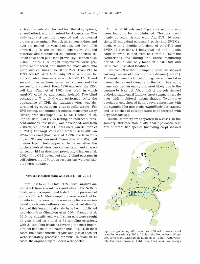
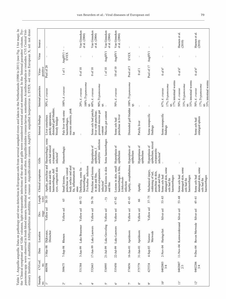
From 1998 to 2011, a total of 249 wild Anguilla an-guilla eels from several rivers and lakes in the Nether-lands were necropsied and tested for the presence ofviruses (Table 1). Most samplings were carried out formonitoring purposes, while some samplings were ini-tiated by disease outbreaks or unusual eel die-offs.Parts of this longitudinal study have been publishedelsewhere (van Ginneken et al. 2004, Haenen et al.2010). A. anguilla yellow and silver eels were caughtall year round on a total of 31 sampling occasions,with 21 sampling locations covering the most impor-tant eel habitats in the Netherlands (Fig. 1). In mostcases, the pooled internal organs and gills of each eelwere separately processed for virus isolation. In 14cases, the organs of up to 10 eels were pooled.
A total of 36 eels and 5 pools of multiple eelswere found to be virus-infected. The most com-monly detected viruses were AngHV1 (10 occa-sions, 35 individual eels and 3 pools) and EVEX (1pool), with 2 double infections of AngHV1 andEVEX (2 occasions, 1 individual eel and 1 pool).AngHV1 was isolated from eels from all over theNetherlands and during the entire monitoringperiod. EVEX was only found in 1998, 2001 and2010 from 3 isolated locations.
Eels from 29 of the 31 sampling occasions showedvarying degrees of clinical signs of disease (Table 1).The most common clinical findings were fin and skinhaemorrhages and damage to the skin. Internally,many eels had an empty gut, most likely due to thecapture by fyke net. About half of the eels showedpathological internal findings, most commonly a paleliver with multifocal haemorrhages. Twenty-fivebatches of eels showed light to severe infections withthe swimbladder nematode Anguillicoloides crassus,and 15 batches of eels appeared to be infected withTrypanosoma spp.
Unusual mortality was reported in 5 cases. In theJanuary 2001 case from a lake near Apeldoorn, sev-eral different fish species (including carp) showed
78
Fig. 1. Anguilla anguilla. Locations of 31 wild European eelsampling occasions (1998 to 2011) in the Netherlands. Num-bers correspond with sample numbers in Table 1, with virus-infected sites shown in bold. Blue lines: main waterways
van Beurden et al.: Viral diseases of European eel 79
Sam
ple
no.
CV
I re
f.no
.D
ate
Loc
atio
nD
ev.
stag
eL
engt
h (c
m)
Clin
ical
sym
ptom
sG
ills
Inte
rnal
fin
ding
sIn
tern
al p
aras
ites
Vir
us-
posi
tive
Vir
usSo
urce
1a 3
-Mar
-98
Dijk
wat
er,
4885
98D
reis
chor
Yel
low
eel
30–3
5A
path
y, p
etec
hiae
and
som
e sk
in a
nd f
in
haem
orrh
ages
, dul
l ey
es, c
onge
sted
ski
n
Hae
mor
rhag
ic, s
ome
eels
had
mix
ed
ecto
para
sitic
infe
ctio
nb
Liv
er s
omet
imes
pa
tchy
app
eara
nce,
di
sten
ded
gall
blad
der,
bloo
dy h
indg
ut
30%
A. c
rass
usPo
ol o
f 20
––
2a 7
-Sep
-98
Rhe
nen
Yel
low
eel
65Sm
all l
esio
ns,
5096
75pe
tech
iae
on v
entr
al
body
sur
face
and
ana
lfi
n, in
flam
mat
ion
of
anus
Hae
mor
rhag
icPa
le li
ver
with
som
e ha
emor
rhag
es,
muc
ous
ente
ritis
, pin
kfa
t
100%
A. c
rass
us1
of 1
Ang
HV
1 +
EV
EX
–
20%
A. c
rass
us
100%
Tryp
anos
oma
sp.
60%
A. c
rass
us
90%
Tryp
anos
oma
sp.
6c22
-Jul
-99
Lak
e L
auw
ers
Yel
low
eel
67–8
2L
esio
ns in
ski
n, s
mal
l53
5450
haem
orrh
ages
in f
ins,
Icht
hyob
odo
infe
ctio
n
Hyp
erpl
asia
of
epith
eliu
mSo
me
eels
had
pe
tech
iae
in li
ver
50%
A. c
rass
us10
of
10A
ngH
V1
Van
Gin
neke
net
al.
(200
4)
9a 4
-Sep
-03
Nie
uwe
6255
34M
erw
ede
Yel
low
eel
37–7
5M
echa
nica
l inj
ury,
re
dden
ing
of ta
il,
pete
chia
e in
ski
n,
mou
th a
nd f
ins
Hyp
erpl
asia
of
epith
eliu
m, m
ixed
ec
topa
rasi
ticin
fect
ionb
Som
e no
nspe
cifi
c fi
ndin
gs–
Pool
of
17A
ngH
V1
–
67%
A. c
rass
us
17%
Tryp
anos
oma
sp.
17%
inte
stin
al w
orm
s
50%
A. c
rass
us
33%
Tryp
anos
oma
sp.
33%
inte
stin
al w
orm
s
50%
A. c
rass
us
17%
Tryp
anos
oma
sp.
33%
inte
stin
al w
orm
s
3c 3
-Jun
-99
Lak
e B
rase
mer
Yel
low
eel
60–7
2D
arke
ning
, som
e fi
n 53
1384
haem
orrh
ages
,Tr
icho
dina
infe
ctio
n
ne ke nniG na
V–
0 1 f o 0–
–et
al.
(200
4)
4c17
-Jun
-99
Lak
e L
auw
ers
Yel
low
eel
58–7
0Pe
tech
iae
and
lesi
ons
5326
63in
ski
n an
d fi
ns,
mix
ed e
ctop
aras
itic
infe
ctio
nb
Hyp
erpl
asia
of
epith
eliu
m, I
.m
ultif
iliis
infe
ctio
n
Som
e ee
ls h
ad p
atch
yliv
er w
ith p
etec
hiae
; gr
eeni
sh m
ucou
s gu
t co
nten
t
0 of
10
–V
an G
inne
ken
et a
l. (2
004)
5c21
-Jul
-99
Lak
e G
reve
ling
Yel
low
eel
5349
9573
Smal
l les
ions
in s
kin
and
fins
Som
e ha
emor
rhag
esM
ucou
s gu
t con
tent
–1
of 1
0A
ngH
V1
Van
Gin
neke
net
al.
(200
4)
7a16
-Jan
-01
Ape
ldoo
rnY
ello
w e
el45
–63
Apa
thy,
eno
phth
alm
usH
yper
plas
ia o
f 57
4856
epith
eliu
mD
iste
nded
gal
l bla
dder
100%
Try
pano
som
asp
.Po
ol o
f 5
EV
EX
–
8a31
-Jan
-01
Ape
ldoo
rnY
ello
w e
el66
Apa
thy
Hyp
erpl
asia
of
5757
79ep
ithel
ium
Patc
hy li
ver
–0
of 1
––
10c
0402
003
3/4
2-N
ov-0
4H
arin
gvlie
tSi
lver
eel
53–8
3So
me
eels
had
da
mag
ed s
kin,
som
e ee
ls h
ad f
in
haem
orrh
ages
–So
me
nons
peci
fic
find
ings
0 of
6d
––
11c
0402
007
2/3
11-N
ov-0
4K
ornw
erde
rzan
dSi
lver
eel
53–6
8So
me
eels
had
da
mag
ed s
kin,
som
e ee
ls h
ad f
in
haem
orrh
ages
––
0 of
6d
–H
aene
n et
al.
(201
0)
12c
0402
206
3/4
9-D
ec-0
4B
oven
Mer
wed
eSi
lver
eel
45–6
1So
me
eels
had
da
mag
ed s
kin,
som
e ee
ls h
ad f
in
haem
orrh
ages
–So
me
eels
had
en
larg
ed s
plee
n0
of 6
d–
Hae
nen
et a
l. (2
010)
Tab
le 1
. An
gu
illa
an
gu
illa
. Dat
a on
pat
hol
ogy
and
vir
us
det
ecti
on in
Eu
rop
ean
eel
s fr
om s
ever
al s
amp
led
riv
ers
and
lak
es in
the
Net
her
lan
ds
(199
8 to
201
1) (s
ee F
ig. 1
for
map
). In
the
‘Cli
nic
al s
ymp
tom
s’ a
nd
‘Gil
ls’ c
olu
mn
s, li
gh
t ec
top
aras
itic
infe
ctio
ns
of t
he
skin
an
d g
ills
wer
e co
nsi
der
ed n
orm
al a
nd
not
men
tion
ed. I
n t
he
‘In
tern
al p
aras
ites
’ col
um
n, T
ry-
pan
osom
asp
p. w
ere
obse
rved
in fr
esh
blo
od s
mea
rs; i
nte
stin
al w
orm
s w
ere
mos
t oft
en A
ng
uil
lico
loid
es c
rass
us
larv
ae, s
omet
imes
Aca
nth
ocep
hal
aor
ces
tod
es. C
VI:
Cen
tral
Vet
-er
inar
y In
stit
ute
; I.
mu
ltif
ilii
s: I
chth
yop
hth
iriu
s m
ult
ifil
iis;
A.
cras
sus:
An
gu
illi
colo
ides
cra
ssu
s; A
ng
HV
1: a
ng
uil
lid
her
pes
viru
s 1;
EV
EX
: ee
l vi
rus
Eu
rop
ean
X;
nd
: n
ot d
one

Dis Aquat Org 101: 69–86, 201280
Sam
ple
no.
CV
I re
f.no
.D
ate
Loc
atio
nC
linic
al s
ympt
oms
Gill
sIn
tern
al f
indi
ngs
Inte
rnal
par
asite
sV
irus
-po
sitiv
eV
irus
Sour
ce
13c
14-D
ec-0
4R
iver
Rhi
neY
ello
w e
el80
–81
Som
e sk
in d
amag
es
4022
377
and
pete
chia
e, h
eavy
haem
orrh
ages
in th
e fi
ns
–B
row
nish
blo
od10
0% A
. cra
ssus
Pool
of
2–
–
14c
14-D
ec-0
4R
iver
Rhi
neY
ello
w e
el62
–75
Pete
chia
e in
ski
n an
d40
2241
8sm
all h
aem
orrh
ages
infi
ns
––
25%
A. c
rass
usPo
ol o
f 4
––
100%
A. c
rass
us
100%
Tryp
anos
oma
sp.
100%
inte
stin
al
wor
ms
80%
A. c
rass
us
10%
Tryp
anos
oma
s p.
50%
inte
stin
al w
orm
s
A. c
rass
us
Inte
stin
al w
orm
s
90%
.A. c
rass
us
40%
Tryp
anos
oma
sp.
50%
inte
stin
al w
orm
s
70%
A. c
rass
us
40%
inte
stin
al w
orm
s
80%
A. c
rass
us
40%
Tryp
anos
oma
s p.
60%
inte
stin
al w
orm
s
90%
A. c
rass
us
20%
Tryp
anos
oma
s p.
40%
inte
stin
al w
orm
s
80%
A. c
rass
us
40%
Tryp
anos
oma
sp.
50%
inte
stin
al w
orm
s
60%
A. c
rass
us
30%
Tryp
anos
oma
sp.
20%
inte
stin
al w
orm
s
15c
14-D
ec-0
4R
iver
Rhi
neY
ello
w e
el78
–90
Smal
l hae
mor
rhag
es
4022
419
in f
ins
–O
ne e
el h
ad p
etec
hiae
in m
uscl
es a
nd
mes
ente
ria
100%
A. c
rass
usPo
ol o
f 2
––
16a
12-M
ay-0
5L
ake
IJss
elm
eer
Yel
low
eel
28–3
5G
reen
ish
skin
, 50
0824
2ha
emor
rhag
ic h
ead,
bl
iste
rs o
n he
ad a
nd
alon
g la
tera
l lin
e, d
ull
eyes
Bro
wni
sh, m
yxos
pore
infe
ctio
n–
–3 fo looP
revil eg naro yhct aP
17c
26-A
ug-0
5B
ened
en/B
oven
5015
220
Mer
wed
eSi
lver
eel
43–8
5Fi
n ha
emor
rhag
esTr
icho
dina
infe
ctio
n,so
me
eels
had
D
acty
logy
rus
infe
ctio
n
–10
of
10d
Ang
HV
1H
aene
n et
al.
(201
0)
18a
8-S
ep-0
5B
oven
Mer
wed
eY
ello
w e
el62
–67
Bro
ken
back
, ski
nned
,50
1613
9m
echa
nica
l les
ions
, sm
all f
in
haem
orrh
a ges
Con
gest
ed (
pale
, br
own)
gill
s w
ith g
asbu
bble
dis
ease
Som
e no
nspe
cifi
c fi
ndin
gsPo
ol o
f 10
dA
ngH
V1
–
19c
28-S
ep-0
5D
en O
ever
Silv
er e
el52
–84
Som
e sm
all f
in
5017
527
haem
orrh
ages
–So
me
eels
had
co
nges
ted
swim
blad
der
3 of
10d
Ang
HV
1H
aene
n et
al.
(201
0)
20c
5-O
ct-0
5B
ened
en
5017
879
Mer
wed
eSi
lver
eel
55–9
2So
me
eels
had
da
mag
ed s
kin,
fin
ha
emor
rha g
es
––
8 of
10d
Ang
HV
1H
aene
n et
al.
(201
0)
21c
12-O
ct-0
5K
ornw
erde
rzan
dSi
lver
eel
56–8
2Fi
n ha
emor
rhag
es–
–50
1831
91
of 1
0dA
ngH
V1
Hae
nen
et a
l. (2
010)
22c
14-O
ct-0
5L
ekSi
lver
eel
58–7
3Fi
n ha
emor
rhag
es–
Som
e ee
ls h
ad
5018
401
cong
este
dsw
imbl
adde
r
0 of
10d
–H
aene
n et
al.
(201
0)
23c
10-N
ov-0
5D
en O
ever
Silv
er e
el57
–84
Fin
haem
orrh
ages
, 50
2030
5so
me
eels
had
da
mag
ed s
kin
––
0 of
10d
–H
aene
n et
al.
(201
0)
24c
16-N
ov-0
5N
ieuw
e 50
2075
2M
erw
ede
Silv
er e
el50
–76
Fin
haem
orrh
ages
, so
me
eels
had
m
echa
nica
l inj
ury
––
0 of
10d
–H
aene
n et
al.
(201
0)
Dev
.st
age
Len
gth
(cm
)
Tab
le 1
. (co
nti
nu
ed)

van Beurden et al.: Viral diseases of European eel 81
Sam
ple
no.
CV
I re
f.no
.D
ate
Loc
atio
nC
linic
al s
ympt
oms
Gill
sIn
tern
al f
indi
ngs
Inte
rnal
par
asite
sV
irus
-po
sitiv
eV
irus
Sour
ce
100%
A. c
rass
us
33%
Tryp
anos
oma
s p.
100%
inte
stin
al
wor
ms
27a
29-A
ug-0
7O
oste
rsch
elde
Yel
low
eel
7022
947
46Sk
in le
sion
s,
haem
orrh
ages
in a
nal
fin
–E
nlar
ged
sple
en, s
ome
nons
peci
fic
find
ings
–Po
ol o
f 5
Ang
HV
1–
50%
A. c
rass
us
50%
inte
stin
al w
orm
s
Som
e ee
ls h
ad A
.cr
assu
s
Som
e in
test
inal
w
orm
s
26a
18-O
ct-0
6So
uthw
est
6029
414
Dre
nthe
––
3 fo loo P–
–snif larotcep de
R05–04
l ee wolle
Y
28a
18-D
ec-0
7L
ake
Vee
rse
Yel
low
eel
40–7
3A
path
y, la
rge
infe
cted
7032
848
skin
wou
nds,
da
mag
ed ta
il,
redd
ened
ana
l fin
Pale
and
ha
emor
rhag
icgi
lls
Dar
k sp
leen
, enl
arge
dpa
le li
ver
with
pe
tech
iae,
pin
k fa
t and
mus
cles
, bro
wn
kidn
e ys
Pool
of
4–
–
29a
12-A
ug-1
0W
orku
mY
ello
w e
el28
–60
Apa
thy,
hae
mor
rhag
es10
0134
02in
mou
th, s
kin
and
fins
, dam
aged
ski
n,
red
infl
amed
dor
sal
fin
–D
iste
nded
gal
l bla
dder
Pool
of
11A
ngH
V1
+E
VE
X–
30c
30-S
ep-1
0O
ldam
btm
eer
Yel
low
eel
40–5
6–
ndnd
ndPo
ol o
f 10
––
1001
6086
31c
8-J
un-1
1N
oord
zeek
anaa
lY
ello
w e
el32
–42
–11
0103
68Tr
icho
dina
infe
ctio
n –
Som
e ee
ls h
ad A
.cr
assu
sPo
ol o
f 13
––
a Sam
plin
g fo
r di
agno
stic
pur
pose
s be
caus
e of
dis
ease
out
brea
k or
unu
sual
eel
die
-off
b Dou
ble
or tr
iple
infe
ctio
n w
ith T
rich
odin
a, I
chth
yobo
do, D
acty
logy
rus,
Gyr
odac
tylu
s, o
r Ic
hthy
opht
hiri
us m
ultif
iliis
c Sa
mpl
ing
for
mon
itori
ng p
urpo
ses
d Vir
us is
olat
ion
only
per
form
ed a
t 20°
C
70%
A. c
rass
us
20%
Tryp
anos
oma
sp.
25c
2-D
ec-0
5L
ekSi
lver
eel
62–8
1So
me
eels
had
fin
50
2178
7ha
emor
rhag
es–
Som
e ee
ls h
ad
haem
orrh
agic
swim
blad
der
2 of
10d
Ang
HV
1H
aene
n et
al.
(201
0)
60%
inte
stin
al w
orm
s
Dev
.st
age
Len
gth
(cm
)
Tab
le 1
. (co
nti
nu
ed)
Dis Aquat Org 101: 69–86, 2012
mortality, which made a causative role of isolatedEVEX unlikely. In the September 2003 and Septem-ber 2005 cases from the rivers Nieuwe Merwede andBoven Merwede, respectively, AngHV1 was isolatedfrom pools of silver Anguilla anguilla, but the causeof death was likely due to mechanical injury causedby hydroelectric power plant turbines in combinationwith a low water level during a hot summer. For theincreased mortality rates of several fish species in-cluding A. anguilla in October 2006 from SouthwestDrenthe, no infectious cause could be identified. Inthe August 2007 case, wild A. anguilla catches fromthe Oosterschelde estuary had declined by as muchas 90%. The diseased eels showed bacterially in-fected skin wounds and AngHV1 was isolated.
Overall, pathogenic viruses were isolated from wildAnguilla an gui lla from 12 different locations in theNetherlands during the past 14 yr. EVEX was onlydetected on 3 occasions, while AngHV1 was isolated12 times. Clinical signs and pathological findings didnot correlate with virus infection, and the effect ofthese pathogenic viruses on the local A. anguilla pop-ulations in the different areas is unclear.
Viruses isolated from glass eels(1990−2011)
From 1990 up to the present,Anguilla anguilla farmers from theNetherlands occasionally submit-ted glass eels intended for farm ingpurposes to the Dutch NRL for FishDiseases for clinical diagnostics.These wild-caught glass eels orig -inated from different estuariesalong the western European coast.Glass eels were either checked forthe presence of pathogenic agentsbefore stocking, or showed clinicalsigns of disease and/or increasedmortality during the first weeksafter arrival at the eel farms. Perbatch, up to 10 glass eels werepooled, eutha nised and ground ina mortar. Ten percent suspensionswere then tested for the presenceof pathogenic viruses by inocula-tion on permissive cell lines.
Over the 22 yr monitoring pe -riod, only 39 batches of glass eelswere tested, making it difficult todetect trends in virus prevalence.
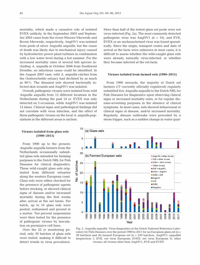
More than half of the tested glass eel pools were notvirus-infected (Fig. 2a). The most commonly detectedpathogenic virus was AngHV1 (n = 10), and EVE,EVEX or an uncharacterised virus was found sporad-ically. Since the origin, transport routes and date ofarrival at the farm were unknown in most cases, it isdifficult to assess whether the wild-caught glass eelswere already naturally virus-infected, or whetherthey became in fected at the eel farm.
Viruses isolated from farmed eels (1990−2011)
From 1990 onwards, the majority of Dutch eelfarmers (17 currently officially registered) regularlysubmitted live Anguilla anguilla to the Dutch NRL forFish Diseases for diagnostics upon observing clinicalsigns or increased mortality rates, or for regular dis-ease-screening purposes in the absence of clinicalsymptoms. In most cases, eels showed behavioural orclinical signs of disease, and/or increased mortality.Regularly, disease outbreaks were preceded by astress trigger, such as a sudden change in water qual-
82
Fig. 2. Anguilla anguilla. Virus diagnostics at the Dutch National Reference Labo-ratory for Fish Diseases over the period 1990 to 2011 for (a) European glass eel (n =39 batches) and (b) farmed European eel (n = 239 batches). AngHV1: anguillidherpesvirus 1; EVE: eel virus European; EVEX: eel virus European X; other
viruses: all viruses other than AngHV1, EVE and EVEX
van Beurden et al.: Viral diseases of European eel
ity or size-sorting of the eels. In the case of farmed A.anguilla, internal organs and gills of up to 10 clini-cally diseased eels from the same system or farm —and not individual eels — were pooled and tested forthe presence of pathogenic viruses as describedabove for wild eels (see ‘Viruses isolated from wildeels (1998−2011)’).
A total of 239 batches of farmed Anguilla anguillawere tested over a 22 yr period. More than half ofthe tested pools were negative for virus isolation(Fig. 2b). The most commonly isolated viruses wereAngHV1 (n = 37), EVE (n = 28) and EVEX (n = 7).EVE and EVEX outbreaks mostly occurred at eelfarms with water temperatures of 15 to 20°C,whereas AngHV1 generally caused disease at higherwater temperatures (around 26°C). Double infectionswith 2 pathogenic viruses were regularly found, mostcommonly AngHV1 with EVE (n = 10) and AngHV1with EVEX (n = 5). This phenomenon has seriousimplications for eel virus diagnostics, in which thepresence of >1 virus species should always be con-sidered. In addition, a double infection limits the pos-sibility of controlling the disease outbreak by adjust-ing the water temperature to a non-permissivetemperature for both viruses.
Occasionally, a yet uncharacterised virus was iso-lated and typed by EM; in most of these cases, areovirus-like agent was found. In general, EVE waspredominantly found from 1990 to 1997, whileAngHV1 was predominantly found from 1997 to2010. As AngHV1 does not propagate in the RTG-2cell line, which was used until 1999, but can be iso-lated in the EK-1 cell line, which has been used since1996, AngHV1 might have been present — but notdetected — in samples collected and tested before1996. In addition, some Dutch eel farmers immunisenewly arrived elvers with AngHV1-infected waterfrom the ongrow system, inducing a mild viral infec-tion and supposedly protective immunisation (O. L.M. Haenen unpubl.). This may have biased the num-ber of reported AngHV1 infections in farmed eels.
In most cases, the investigated Anguilla anguillashowed nonspecific clinical signs of disease, suchas fin and skin haemorrhages, and sometimes localbacterial skin infections. EVE-infected farmed A.anguilla showed congestion of the skin, fins and gills,with severe fin haemorrhages, and anae mia.AngHV1 and EVEX infections were more often char-acterised by reddening of the fins and petechial skinhaemorrhages, generally concentrated in the headregion and ventral part of the body. SeverelyAngHV1-infected A. anguilla sometimes showed atypical tiger-like haemorrhagic pattern in the skin.
Internally, various pathological findings were re -ported, with the liver being most commonly affected,characterised by paleness and multifocal smaller andlarger haemorrhages.
In conclusion, the most commonly detected virusesfrom farmed Anguilla anguilla in the Netherlandswere AngHV1, EVE and EVEX. Viral disease out-breaks were usually stress-triggered, temperature-dependent, and accompanied by secondary infec-tions. Virus-infected eels usually showed clinicalsigns of disease, but clinical signs alone were notfound to be a marker for virus infection.
FUTURE RESEARCH DIRECTIONS
Our current knowledge on pathogenic Europeaneel viruses is hampered by a number of factors. First,there is a lack of peer-reviewed publications on theprevalence, clinical signs, mortality and gross pathol-ogy of these viruses, especially with regard to thewild Anguilla anguilla stock. Only a handful of stud-ies have investigated the virological status of wild A.anguilla in Europe (Castric & Chastel 1980, Castric etal. 1984, Jørgensen et al. 1994, van Ginneken et al.2004, Jakob et al. 2009, Haenen et al. 2010). As clini-cal signs and gross pathology were not recorded inmost studies, the results only give an indication of thepresence of particular viruses in the wild A. anguillastock. The recent concern about the decline of thewild A. anguilla stock opens up new possibilities forstudying the general health status of the wild A.anguilla population and the potential role of diseasesin its decline (Haenen et al. 2012), which will supportscientifically based restocking strategies.
Second, several of the eel viruses described earlyon are incompletely characterised. Many of theseviruses were initially described as separate species,but are likely separate isolations of the same virusspecies. A solution might be found nowadays bysequencing analyses of the original isolates, such asEV [Berlin], EVE and EVA.
Third, the susceptibility of different freshwater eelspecies to the various virological agents is unknown.Many of the viruses presented in the present reviewwere initially isolated in Japan, after shipment ofAnguilla anguilla and A. rostrata for farming pur-poses. The identified viruses were subsequently iso-lated from Japanese eel too, and successful infectiontrials demonstrated the sus ceptibility of A. japonicaand A. anguilla to similar viruses. With regard to A.rostrata, only 3 publications briefly report on the iso-lation or detection of pathogenic viruses (T. Sano
83
Dis Aquat Org 101: 69–86, 2012
1976, McAllister & Owens 1995, Shih 2004). At leastone virus has now been found in A. japonica and notin A. anguilla or A. rostrata (Mizutani et al. 2011).Concerning the introduction of new viruses via theimport of foreign eel species, it is worth mentioningthat A. anguilla is currently considered a criticallyendangered species by the IUCN (Freyhof & Kottelat2010), and the export of live A. anguilla is hencerestricted (CITES 2007).
CONCLUSIONS
The most commonly observed pathogenic virusesin Anguilla anguilla are AngHV1, EVE and EVEX.All 3 viruses may cause a haemorrhagic disease withincreased mortality rates, but have been isolatedfrom seemingly healthy eels too. In addition, latencyhas been suggested for AngHV1, and a carrier statefor EVE. AngHV1 seems to be host-restricted to A.japonica and A. anguilla, while EVEX and EVE havealso been shown to be able to cause disease in rain-bow trout fry. In the Netherlands, AngHV1 has beenregularly isolated from wild and farmed A. anguilla,EVEX sporadically, and EVE only from farmed eels.Viral disease in farmed eel is usually stress-triggeredand temperature-dependent. Future research shouldfocus on the genetic characterisation of historical iso-lates, the health status of the wild eel populations allover Europe, the potential role of diseases in thedecline of the A. anguilla stock, and virus screeningof farmed eel batches for restocking purposes intothe wild.
Acknowledgements. We thank former employees of theDutch NRL for Fish and Shellfish Diseases for their assis-tance in necropsies and virus diagnostics of European eels:Sonja Dijkstra, Imke Wijmenga, Rob Zwart, Betty vanGelderen, Françoise Keuzenkamp and Grytsje Wybenga.We also thank former interns of the Dutch NRL for Fish andShellfish Diseases for their contribution to the developmentof diagnostic assays for the most relevant European eelviruses: Annemiek Botter, Madelon Willemsen, Bart Jansen,Annette Boerlage, Marco de Mik and Jurjen van Tellingen.This study received financial support from the Dutch Min-istry of Economic Affairs, Agriculture and Innovation.
LITERATURE CITED
Ahne W, Thomsen I (1985) The existence of three differentviral agents in a tumour bearing European eel (Anguillaanguilla). Zentralbl Veterinarmed B 32: 228−235
Ahne W, Schwanz-Pfitzner I, Thomsen I (1987) Serologicalidentification of 9 viral isolates from European eels(Anguilla anguilla) with stomatopapilloma by means ofneutralization tests. J Appl Ichthyol 3: 30−32
Bandin I, Dopazo CP (2011) Host range, host specificity andhypothesized host shift events among viruses of lowervertebrates. Vet Res 42: 67
Békési L, Horváth I, Kovács-Gayer E, Csaba G (1986) De mon -stration of herpesvirus like particles in skin lesions of Eu-ropean eel (Anguilla anguilla). J Appl Ichthyol 2: 190−192
Bergmann SM, Fichtner D, Skall HF, Schotfeldt HJ, OlesenNJ (2003) Age- and weight-dependent susceptibility ofrainbow trout Onchorhynchus mykiss to isolates of infec-tious haematopoietic necrosis virus (IHNV) of varyingvirulence. Dis Aquat Org 55: 205−210
Blake S, Ma JY, Caporale DA, Jairath S, Nicholson BL (2001)Phylogenetic relationships of aquatic birnaviruses basedon deduced amino acid sequences of genome segment AcDNA. Dis Aquat Org 45: 89−102
Bucke D (1981) A note on IPNV from eels in the U.K. BullEur Assoc Fish Pathol 1: 12
Castric J, Chastel C (1980) Isolation and characterizationattempts of three viruses from European eel, Anguillaanguilla: preliminary results. Ann Inst Pasteur Virol 131:435−448
Castric J, Rasschaert D, Bernard J (1984) Evidence of lyssa -viruses among rhabdovirus isolates from the Europeaneel Anguilla anguilla. Ann Inst Pasteur Virol 135: 35−55
Castric J, Jeffroy J, Bearzotti M, De Kinkelin P (1992) Isola-tion of viral haemorrhagic septicaemia virus (VHSV)from wild elvers Anguilla anguilla. Bull Eur Assoc FishPathol 12: 21−23
Chang PH, Pan YH, Wu CM, Kuo ST, Chung HY (2002) Iso-lation and molecular characterization of herpesvirusfrom cultured European eels Anguilla anguilla in Tai-wan. Dis Aquat Org 50: 111−118
Chen SN, Kou GH (1981) A cell line derived from Japaneseeel (Anguilla japonica) ovary. Fish Pathol 16: 129−137
Chen SN, Ueno Y, Kou GH (1982) A cell line derived fromJapanese eel (Anguilla japonica) kidney. Proc Natl SciCounc Repub China B 6: 93−100
Chen SN, Kou GH, Hedrick RP, Fryer JL (1985) The occur-rence of viral infections of fish in Taiwan. In: Ellis AE (ed)Fish and shellfish pathology. Academic Press, London,p 313–319
CITES (Convention on International Trade in EndangeredSpecies of Wild Fauna and Flora) (2007) Inclusion ofAnguilla anguilla (L.) in Appendix II. CITES 14th MeetConf Parties, The Hague, 3−15 Jun 2007. Proposal 18.CITES, Geneva
Council of the European Union (2007) Council Regulation(EC) No. 1100/2007 of 18 September 2007 establishingmeasures for the recovery of the stock of European eel.Off J Eur Union L248: 17−23
Davidse A, Haenen OLM, Dijkstra SG, van Nieuwstadt AP,van der Vorst TJK, Wagenaar F, Wellenberg GJ (1999)First isolation of herpesvirus of eel (Herpesvirus Anguil-lae) in diseased European eel (Anguilla anguilla L.) inEurope. Bull Eur Assoc Fish Pathol 19: 137−141
Dekker W (2004) Slipping through our hands: populationdynamics of the European eel. PhD dissertation, Univer-sity of Amsterdam
Dekker W, Casselman JM, Cairns DK, Tsukamoto K, Jelly-man D, Lickers H (2003) Worldwide decline of eelresources necessitates immediate action, Québec Decla-ration of Concern. Fisheries 28: 28−30
Esteve C, Alcaide E (2009) Influence of diseases on the wildeel stock: the case of Albufera Lake. Aquaculture 289:143−149
84
van Beurden et al.: Viral diseases of European eel
FAO (Food and Agriculture Organization of the UnitedNations) (2012) Cultured aquatic species informationprogramme: Anguilla anguilla (Linnaeus, 1758). Fish-eries and Aquaculture Department, FAO, Rome. www.fao. org/ fishery/ culturedspecies/Anguilla_anguilla(accessed Feb 2012)
Freyhof J, Kottelat M (2010) Anguilla anguilla. In: IUCNRed List of Threatened Species. www.iucnredlist.org(accessed Feb 2012)
Galinier R, van Beurden S, Amilhat E, Castric J and others(2012) Complete genomic sequence and taxonomic posi-tion of eel virus European X (EVEX), a rhabdovirus ofEuropean eel. Virus Res 166: 1−12
Gravell M, Malsberger RG (1965) A permanent cell linefrom the fathead minnow (Pimephales promelas). AnnNY Acad Sci 126: 555−565
Haenen OLM, Dijkstra SG, Tulden PW, Davidse A, vanNieuwstadt AP, Wagenaar F, Wellenberg GJ (2002) Her-pesvirus anguillae (HVA) isolations from disease out-breaks in cultured European eel, Anguilla anguilla in theNetherlands since 1996. Bull Eur Assoc Fish Pathol 22:247−257
Haenen O, van Ginneken V, Engelsma M, van den ThillartG (2009) Impact of eel viruses on recruitment of Euro-pean eel. In: van den Thillart G, Dufour S, Rankin J (eds)Spawning migration of the European eel. Reproductionindex, a useful tool for conservation management. Fish& Fisheries Series, Vol 30. Springer Verlag, Berlin,p 387−400
Haenen OLM, Lehmann J, Engelsma MY, Stürenberg FJ,Roozenburg I, Kerkhoff S, Klein Breteler J (2010) Thehealth status of European silver eels, Anguilla anguilla,in the Dutch River Rhine Watershed and Lake IJs-selmeer. Aquaculture 309: 15−24
Haenen OLM, Mladineo I, Konecny R, Yoshimizu M andothers (2012) Diseases of eels in an international per-spective: workshop on eel diseases at the 15th Interna-tional Conference on Diseases of Fish and Shellfish,Split, Croatia, 2011. Bull Eur Assoc Fish Pathol 32:109−115
Hangalapura BN, Zwart R, Engelsma MY, Haenen O (2007)Pathogenesis of Herpesvirus anguillae (HVA) in juvenileEuropean eel Anguilla anguilla after infection by bathimmersion. Dis Aquat Org 78: 13−22
Hedrick RP, Okamoto N, Sano T, Fryer JL (1983a) Biochem-ical characterization of eel virus European. J Gen Virol64: 1421−1426
Hedrick RP, Fryer JL, Chen SN, Kou GH (1983b) Character-istics of four birnaviruses isolated from fish in Taiwan.Fish Pathol 18: 91−97
Heinsbroek LTN (1991) A review of eel culture in Japan andEurope. Aquacult Fish Manage 22: 57−72
Hill BJ, Way K (1995) Serological classification of Infectiouspancreatic necrosis (IPN) virus and other aquatic bir-naviruses. Annu Rev Fish Dis 5: 55−77
Hill BJ, Williams RF (1984) Comparative studies on the path-ogenicity of the eel rhabdovirus (EVEX) and VHS virusfor rainbow trout and eels at different temperatures. In:Fish Diseases, Fourth Session of the Cooperative Pro-gramme of Research on Aquaculture, Cadiz (Spain),26−30 Oct 1981. Acuigrup, Madrid, p 17–27
Hill BJ, Williams RF, Smale CJ, Underwood BO, Brown F(1980) Physicochemical and serological characterizationof two rhabdoviruses isolated from eels. Intervirology 14:208−212
Hsu YL, Chen BS, Wu JL (1989) Comparison of RNAs andpolypeptides of infectious pancreatic necrosis virus iso-lates from eel and rainbow trout. J Gen Virol 70:2233−2239
Hsu YL, Chen BS, Wu JL (1993) Demonstration of infectiouspancreatic necrosis virus strain VR-299 in Japanese eel,Anguilla japonica Temminck & Schlegel. J Fish Dis 16:123−129
Hudson E, Bucke D, Forrest A (1981) Isolation of infectiouspancreatic necrosis virus from eels, Anguilla anguilla L.,in the United Kingdom. J Fish Dis 4: 429−431
ICTV (International Committee on Taxonomy of Viruses)(2012) Virus Taxonomy: 2011 Release (current). www.ictvonline. org/virusTaxonomy.asp?version=2011 (accessedSep 2012)
Jakob E, Neuhaus H, Steinhagen D, Luckhardt B, Hanel R(2009) Monitoring of Herpesvirus anguillae (HVA) infec-tions in European eel, Anguilla anguilla (L.), in northernGermany. J Fish Dis 32: 557−561
Jørgensen P, Castric J, Hill B, Ljungberg O, De Kinkelin P(1994) The occurrence of virus infections in elvers andeels (Anguilla anguilla) in Europe with particular refer-ence to VHSV and IHNV. Aquaculture 123: 11−19
Kobayashi T, Miyazaki T (1996) Rhabdoviral dermatitis inJapanese eel, Anguilla japonica. Fish Pathol 31: 183−190
Kobayashi T, Miyazaki T (1997) Characterization and path-ogenicity of a herpesvirus isolated from cutaneouslesions in Japanese eel, Anguilla japonica. Fish Pathol32: 89−95
Kobayashi T, Shiino T, Miyazaki T (1999) The effect of watertemperature on rhabdoviral dermatitis in the Japaneseeel, Anguilla japonica Temminck and Schleger. Aqua-culture 170: 7−15
Lee NS, Nomura Y, Miyazaki T (1999a) Gill lamellar pillarcell necrosis, a new birnavirus disease in Japanese eels.Dis Aquat Org 37: 13−21
Lee NS, Kobayashi J, Miyazaki T (1999b) Gill filamentnecrosis in farmed Japanese eels, Anguilla japonica(Temminck & Schlegel), infected with Herpesvirusanguillae. J Fish Dis 22: 457−463
Lee NS, Miyata M, Miyazaki T (2001) Genome sequence ofa VP2/NS junction region of pillar cell necrosis virus(PCNV) in cultured Japanese eel Anguilla japonica. DisAquat Org 44: 179−182
Macdonald RD, Gower DA (1981) Genomic and phenotypicdivergence among three serotypes of aquatic bir-naviruses (infectious pancreatic necrosis virus). Virology114: 187−195
McAllister PE, Owens WJ (1995) Assessment of the virulenceof fish and molluscan isolates of infectious pancreaticnecrosis virus for salmonid fish by challenge of brooktrout, Salvelinus fontinalis (Mitchill). J Fish Dis 18: 97−103
McAllister PE, Nagabayashi T, Wolf K (1977) Viruses of eelswith and without stomatopapillomas. Ann NY Acad Sci298: 233−244
Mizutani T, Sayama Y, Nakanishi A, Ochiai H and others(2011) Novel DNA virus isolated from samples showingendothelial cell necrosis in the Japanese eel, Anguillajaponica. Virology 412: 179−187
Munro ES, Stewart KJ, Murray WJ, Haenen OLM (2011)Characterization of a virus isolated from farmed Euro-pean eel elvers (Anguilla anguilla) undergoing a mortality event in the Netherlands. In: 15th Int Conf DisFish Shellfish. Dalmacijapapir Publishing House, Split,abstract no. 0–140
85

Dis Aquat Org 101: 69–86, 2012
Nagabayashi T, Wolf K (1979) Characterization of EV-2, avirus isolated from European eels (Anguilla anguilla)with stomatopapilloma. J Virol 30: 358−364
Nishimura T, Fukuda H, Yamazaki H, Sano T (1981a) Con-centration and purification of eel virus European, (EVE).Fish Pathol 16: 75−83
Nishimura T, Toba M, Ban F, Okamoto N, Sano T (1981b) Eelrhabdovirus, EVA, EVEX and their infectivity to fishes.Fish Pathol 15: 173−184
Okamoto N, Sano T, Hedrick RP, Fryer JL (1983) Antigenicrelationships of selected strains of infectious pancreaticnecrosis virus and European eel virus. J Fish Dis 6: 19−25
Rijsewijk F, Pritz-Verschuren S, Kerkhoff S, Botter A,Willemsen M, van Nieuwstadt T, Haenen O (2005)Development of a polymerase chain reaction for thedetection of Anguillid herpesvirus DNA in eels based onthe herpesvirus DNA polymerase gene. J Virol Methods124: 87−94
Rodriguez Saint-Jean S, Borrego JJ, Perez-Prieto SI (2003)Infectious pancreatic necrosis virus: biology, pathogene-sis, and diagnostic methods. Adv Virus Res 62: 113−165
Romero-Brey I, Bandin I, Cutrin JM, Vakharia VN, DopazoCP (2009) Genetic analysis of aquabirnaviruses isolatedfrom wild fish reveals occurrence of natural reassortmentof infectious pancreatic necrosis virus. J Fish Dis 32:585−595
Sano T (1976) Viral diseases of cultured fishes in Japan. FishPathol 10: 221−226
Sano T, Nishimura T, Okamoto N, Fukuda H (1977) Studieson viral diseases of Japanese fishes 7. A rhabdovirus iso-lated from European eel, Anguilla anguilla. Bull Jpn SocSci Fish 43: 491−495
Sano T, Okamoto N, Nishimura T (1981) A new viral epi-zootic of Anguilla japonica Temminck and Schlegel.J Fish Dis 4: 127−139
Sano M, Fukuda H, Sano T (1990) Isolation and characteri-zation of a new herpesvirus from eel. In: Perkins FO,Cheng TC (eds) Pathology in marine sciences. AcademicPress, San Diego, CA, p 15−31
Sano M, Okamoto N, Fukuda H, Saneyoshi M, Sano T (1992)Virulence of infectious pancreatic necrosis virus is asso-ciated with the larger RNA segment (RNA segment A).J Fish Dis 15: 283−293
Schwanz-Pfitzner I, Ozel M, Darai G, Gelderblom H (1984)Morphogenesis and fine structure of eel virus (Berlin), amember of the proposed Birnavirus group. Arch Virol 81:151−162
Shchelkunov IS, Skurat EK, Sivolotskaia VA, Sapot’ko KV,Shimko VV, Linnik Y (1989) Rhabdovirus anguilla in eelin the USSR and its pathogenicity for fish. Vopr Virusol34: 81−84
Shih HH (2004) A polymerase chain reaction for detectingHerpesvirus anguillae in asymptomatic eels. Taiwania49: 1−6
Shih HH, Hu CW, Wang CS (2003) Detection of Herpesvirusanguillae infection in eel using in situ hybridization.J Appl Ichthyol 19: 99−103
Stone R (2003) Freshwater eels are slip-sliding away. Sci-ence 302: 221−222
Székely C, Palstra A, Molnár K, van den Thillart G (2009)Impact of the swim-bladder parasite on the health and
performance of European eels. In: van den Thillart G,Dufour S, Rankin J (eds) Spawning migration of theEuropean eel. Reproduction index, a useful tool for con-servation management. Fish & Fisheries Series, Vol 30.Springer Verlag, Berlin, p 201−226
Ueno Y, Chen SN, Kou GH, Hedrick RP, Fryer JL (1984)Characterization of a virus isolated from Japanese eel(Anguilla japonica) with nephroblastoma. Bull Inst ZoolAcad Sin 23: 47−55
Ueno Y, Kitao T, Chen SN, Aoki T, Kou GH (1992) Charac-terization of a herpes-like virus isolated from culturedJapanese eels in Taiwan. Fish Pathol 27: 7−17
Ueno Y, Shi JW, Yoshida T, Kitao T, Sakai M, Chen SN, KouGH (1996) Biological and serological comparisons of eelherpesvirus in Formosa (EHVF) and Herpesvirus anguil-lae (HVA). J Appl Ichthyol 12: 49−51
van Beurden SJ, Bossers A, Voorbergen-Laarman MH, Hae-nen OL and others (2010) Complete genome sequenceand taxonomic position of anguillid herpesvirus 1. J GenVirol 91: 880−887
van Beurden SJ, Voorbergen-Laarman MA, Roozenburg I,Boerlage AS, Haenen OL, Engelsma MY (2011) Develop-ment and validation of a two-step real-time RT-PCR forthe detection of eel virus European X in European eel,Anguilla anguilla. J Virol Methods 171: 352−359
van Ginneken VJT, Maes GE (2005) The European eel(Anguilla anguilla, Linnaeus), its lifecycle, evolution andreproduction: a literature review. Rev Fish Biol Fish 15:367−398
van Ginneken V, Haenen O, Coldenhoff K, Willemze R andothers (2004) Presence of eel viruses in eel species fromvarious geographic regions. Bull Eur Assoc Fish Pathol24: 268−272
van Ginneken V, Ballieux B, Willemze R, Coldenhoff K andothers (2005) Hematology patterns of migrating Euro-pean eels and the role of EVEX virus. Comp BiochemPhysiol C 140: 97−102
van Nieuwstadt AP, Dijkstra SG, Haenen OLM (2001) Per-sistence of herpesvirus of eel Herpesvirus anguillae infarmed European eel Anguilla anguilla. Dis Aquat Org45: 103−107
Varvarigos P, Vendramin N, Cappellozza E, Bovo G (2011)First confirmation of herpes virus anguillae (HVA) andinfectious pancreatic necrosis (IPN) virus infecting Euro-pean eel Anguilla anguilla farmed in Greece. Bull EurAssoc Fish Pathol 31: 101−111
Waltzek TB, Kelley GO, Alfaro ME, Kurobe T, Davison AJ,Hedrick RP (2009) Phylogenetic relationships in the fam-ily Alloherpesviridae. Dis Aquat Org 84: 179−194
Wolf K, Quimby MC (1962) Established eurythermic line offish cells in vitro. Science 135: 1065−1066
Wolf K, Quimby MC (1973) Fish viruses: buffers and meth-ods for plaquing eight agents under normal atmosphere.Appl Microbiol 25: 659−664
Wu JL, Chang CY, Hsu YL (1987) Characteristics of an infec-tious pancreatic necrosis like virus isolated from Japan-ese eel (Anguilla japonica). Bull Inst Zool Acad Sin 26:201−214
Zhang CX, Suzuki S (2004) Aquabirnaviruses isolated frommarine organisms form a distinct genogroup from otheraquabirnaviruses. J Fish Dis 27: 633−643
86
Editorial responsibility: Dieter Steinhagen,Hannover, Germany
Submitted: February 22, 2012; Accepted: July 3, 2012Proofs received from author(s): September 11, 2012