Embed Size (px)
Citation preview
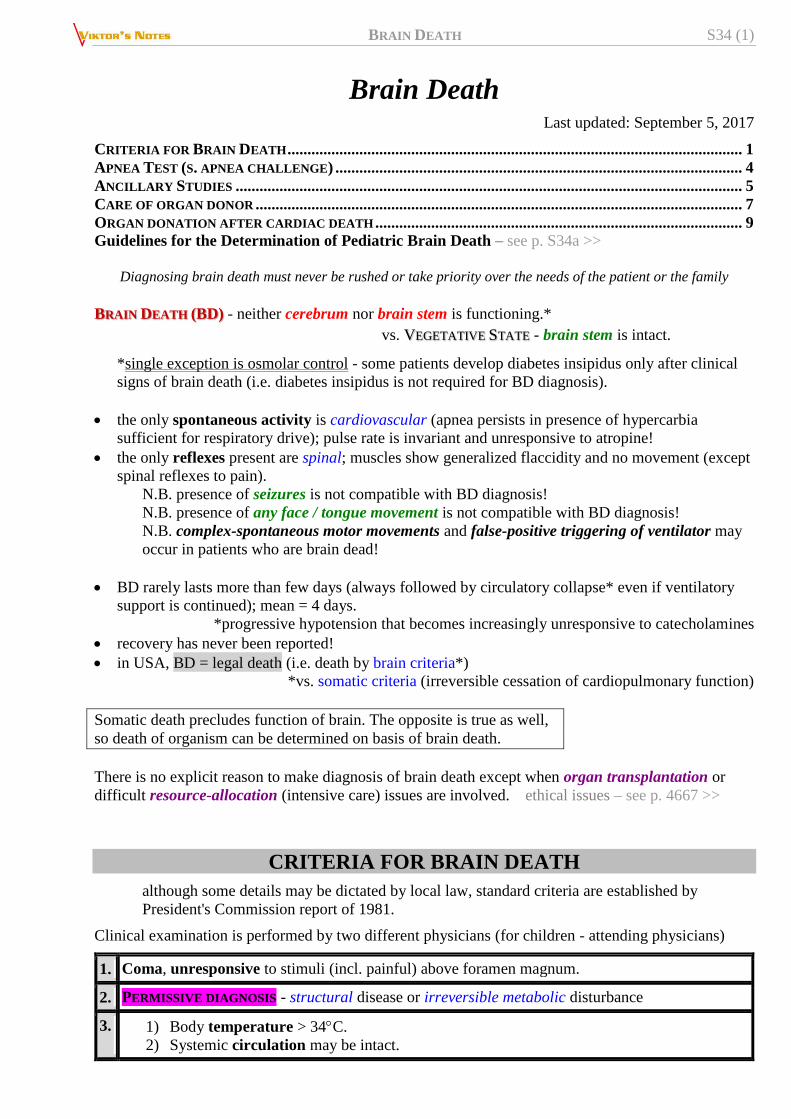
BRAIN DEATH S34 (1)
Brain Death Last updated: September 5, 2017
CRITERIA FOR BRAIN DEATH .................................................................................................................. 1 APNEA TEST (S. APNEA CHALLENGE) ...................................................................................................... 4 ANCILLARY STUDIES ............................................................................................................................... 5
CARE OF ORGAN DONOR .......................................................................................................................... 7 ORGAN DONATION AFTER CARDIAC DEATH ............................................................................................ 9 Guidelines for the Determination of Pediatric Brain Death – see p. S34a >>
Diagnosing brain death must never be rushed or take priority over the needs of the patient or the family
BRAIN DEATH (BD) - neither cerebrum nor brain stem is functioning.*
vs. VEGETATIVE STATE - brain stem is intact.
*single exception is osmolar control - some patients develop diabetes insipidus only after clinical
signs of brain death (i.e. diabetes insipidus is not required for BD diagnosis).
the only spontaneous activity is cardiovascular (apnea persists in presence of hypercarbia
sufficient for respiratory drive); pulse rate is invariant and unresponsive to atropine!
the only reflexes present are spinal; muscles show generalized flaccidity and no movement (except
spinal reflexes to pain).
N.B. presence of seizures is not compatible with BD diagnosis!
N.B. presence of any face / tongue movement is not compatible with BD diagnosis!
N.B. complex-spontaneous motor movements and false-positive triggering of ventilator may
occur in patients who are brain dead!
BD rarely lasts more than few days (always followed by circulatory collapse* even if ventilatory
support is continued); mean = 4 days.
*progressive hypotension that becomes increasingly unresponsive to catecholamines
recovery has never been reported!
in USA, BD = legal death (i.e. death by brain criteria*)
*vs. somatic criteria (irreversible cessation of cardiopulmonary function)
Somatic death precludes function of brain. The opposite is true as well,
so death of organism can be determined on basis of brain death.
There is no explicit reason to make diagnosis of brain death except when organ transplantation or
difficult resource-allocation (intensive care) issues are involved. ethical issues – see p. 4667 >>
CRITERIA FOR BRAIN DEATH
although some details may be dictated by local law, standard criteria are established by
President's Commission report of 1981.
Clinical examination is performed by two different physicians (for children - attending physicians)
1. Coma, unresponsive to stimuli (incl. painful) above foramen magnum.
2. PERMISSIVE DIAGNOSIS - structural disease or irreversible metabolic disturbance
3. 1) Body temperature > 34C.
2) Systemic circulation may be intact.

BRAIN DEATH S34 (2)
3) Serum electrolytes must be WNL + no known endocrine disturbances + absence of drug
intoxication (incl. ethanol, sedatives, potentially anesthetizing or paralyzing drugs).
HYPOTENSION, HYPOTHERMIA, and METABOLIC DISTURBANCES should be corrected!
Pentobarbital level should be < 10
4. ADULTS
known structural cause – at least 6 hours observation (absent brain function)
others (incl. anoxic-ischemic brain damage) – at least 24 hours observation
CHILDREN
< 7 days of age or prematures – BD diagnosis inappropriate (i.e. wait until age 7 days)
7 ÷ 30 days – observation at least 24 hours
older children – observation at least 12 hours (24 hours if anoxic-ischemic brain damage)
Child's brain is more resilient - more difficult determination of BD!
5. Absence of cephalic reflexes, incl. pupillary, corneal, oculocephalic, oculovestibular (caloric),
gag, sucking, swallowing, cough, stereotyped posturing.
Decorticate or decerebrate posturing is not compatible with BD diagnosis!
Purely spinal reflexes may be present (incl. tendon reflexes, plantar responses, limb flexion to
noxious stimuli, tonic neck reflexes).
6. Apnea off ventilator (with ongoing oxygenation) for duration sufficient to produce hypercarbic
respiratory drive (usually 10-20 min to achieve PaCO2 ≥ 60 mmHg).
7. Optional confirmatory studies:
1. EEG - isoelectric for 30 minutes at maximal gain.
2. Absent brain stem evoked responses.
3. Absent cerebral circulation demonstrated by radiographic / radioisotope / MR angiography.
assessment of BD after cardiopulmonary resuscitation or other severe acute brain injuries must
be deferred for 24 hours if there are concerns or inconsistencies in the examination.
when death results from criminal assault, or there is possibility of litigation regarding death,
extra care must be taken and legal counsel may be advisable before making determination of
brain death.
First stage - demonstrate DEEP UNRESPONSIVE COMA with apnea* and no response to painful CENTRAL
stimuli (PERIPHERAL stimuli may elicit spinal reflex movements and may confuse family).
*always first check if patient is triggering the ventilator = “breathing over the
vent” (i.e. real f > ventilator set f)
spinal cord mediated reflex movements (flexor plantar reflexes, flexor withdrawal, muscle stretch
reflexes, and even abdominal and cremasteric reflexes) can be compatible with brain death, and
may occasionally consist of complex movements, including bringing one or both arms up to face,
or sitting up ("Lazarus" sign) especially with hypoxemia (thought to be due to spinal cord
ischemia stimulating surviving motor neurons in upper cervical cord).
N.B. if complex integrated motor movements occur, it is recommended that
confirmatory testing be performed prior to pronouncement of brain death
Second stage - demonstrate PERMISSIVE DIAGNOSIS; i.e. there must be diagnosis adequate to explain
death of brain (including brain stem!).
– this need not be ETIOLOGICAL diagnosis (e.g. massive intracerebral hemorrhage
qualifies as permissive diagnosis, even if etiology of hemorrhage is unknown).
– this does not require demonstration of ANATOMICAL lesion (e.g. history of prolonged
anoxia would suffice).
– this requires documentation of IRREVERSIBILITY.

BRAIN DEATH S34 (3)
Exclusion criteria (irreversibility and BD cannot be determined): 1sedative drugs, 2hypothermia (< 32.2°C), 3shock (MAP < 55 or SBP < 90), 4neuromuscular
blockade.
– below 32.2°C (90° F), pupils may be fixed and dilated, respirations may be difficult to
detect, and recovery is possible!
– shock (SBP < 90 mmHg) and anoxia can produce lethargy.
– immediately post-resuscitation: shock or anoxia may cause fixed and dilated pupils;
atropine may cause slight dilatation but not unreactivity
N.B. neuromuscular blockage (e.g. for intubation) does not affect pupils
because iris lacks nicotinic receptors
– should be no evidence of remediable exogenous or endogenous intoxication, including
drug or metabolic (barbiturates, benzodiazepines, meprobamate, methaqualone,
trichloroethylene, paralytics, hepatic encephalopathy, hyperosmolar coma ... ).
N.B. for patients coming out of pentobarbital coma, wait until level <
10 mcg/mL
– pseudocholinesterase deficiency (prevalence 1/3000) can cause succinylcholine to last
up to 8 hours (instead of 5 mins); H: twitch monitor can rule-out NMB (place electrodes
immediately behind eye or across zygomatic arch)
Third stage - demonstrate no detectable function above level foramen magnum:
midbrain – absent pupillary light reflex (most easily assessed by bright light of
ophthalmoscope); unreactive pupils may be either at midposition (as they will be in
death) or dilated (in setting of dopamine infusion); pupils should not be constricted!
pons:
1) absent corneal reflex - eye closing to corneal (not scleral) stimulation
2) no inducible eye movements:
a) absent doll’s eyes (contraindicated if C-spine not cleared)
OR
b) absent oculovestibular reflex (cold water calorics): instill 60-100 ml ice water
into one ear (do not do if TM perforated) with HOB at 30° - wait at least 1
minute for response, and 5 min before testing the opposite side.
Brain death is excluded if any eye movement is noted!
medulla:
1) absent oropharyngeal (gag) reflex to stimulation of posterior pharynx
2) absent cough reflex, i.e. no cough response to bronchial suctioning
3) apnea test (last test to perform! - elevating PaCO2, increases ICP which could
precipitate herniation and vasomotor instability)
Fourth stage - period of observation with sequential testing; recommended observation periods during
which time the patient fulfills criteria of clinical brain death before the patient may be pronounced
dead:
N.B. there is insufficient evidence to determine minimally acceptable observation
period to ensure that neurologic functions have ceased irreversibly!
1) well established overwhelming brain damage from an irreversible condition (e.g. massive
intracerebral hemorrhage), some experts will pronounce death following a single valid brain
death exam in conjunction with a clinical confirmatory test.
2) well established irreversible condition and clinical confirmatory tests are used: 6 hours.
3) well established irreversible condition and no clinical confirmatory tests are used: 12 hours
4) if diagnosis is uncertain and no clinical confirmatory tests: 12-24 hours
5) if anoxic injury is cause of brain death: 24 hours (may be shortened if cessation of CBF is
demonstrated)

BRAIN DEATH S34 (4)
when BD criteria are met, it is legal time of death – artificial ventilation and blood pressure
support are no longer an option (unless organ harvesting is intended).
– if BD patient is maintained on mechanical ventilation, brain gradually undergoes
autolytic process.
– removal of ventilator results in terminal rhythms (most often complete heart block
without ventricular response, junctional rhythms, or ventricular tachycardia).
– purely spinal motor movements may occur in moments of terminal apnea: back
arching, neck turning, leg stiffening, upper extremity flexion.
N.B. BD diagnosis is made primarily by clinical methods!
APNEA TEST (s. APNEA CHALLENGE)
- observing brain stem response to hypercapnia without producing hypoxemia.
although acidosis, rather than hypercapnia, is real afferent trigger for ventilation, PaCO2 ≥ 60
mmHg (50 mmHg in United Kingdom) is usually endpoint for this test.
Additional requirement for children, PaCO2 ≥ 20 mmHg above the baseline
in order to prevent hypoxemia (→ arrhythmias, MI):
preapneic oxygenation (15 minutes of 100% O2 ventilation) is required before starting test;
also adjust ventilator to bring PaCO2 to 40 mmHg (to shorten test time and thus reduce risk of
hypoxemia).
during the test - supplemental oxygen by diffusion:
a) 100% O2 flow administered at 6* L/min through either pediatric oxygen cannula
or 14 F tracheal suction catheter (with side port covered with adhesive tape)
passed to estimated level of carina
*N.B. too high flow may wash out CO2 and it might be difficult
to achieve Paco2 ≥ 60 mmHg
b) continuous positive airway pressure (CPAP) with 10 cmH2O pressure - does not
provide ventilation, so it does not interfere with observation for spontaneous
respirations.
N.B. some patients with cardiorespiratory dysfunction still may not tolerate ≈ 10 minutes of
apnea (necessary to raise PaCO2 to 60 mmHg) without becoming hypoxemic & hypotensive →
take blood gases sample and stop apnea test → perform CONFIRMATORY TEST (see below)
instead.
in absence of ventilation, PaCO2 passively rises 3 mmHg/min;
although it may be possible to predict this point by following trend in end-tidal CO2
(PetCO2) measurements, there is enough discrepancy between arterial blood PaCO2 and
PetCO2 to indicate use of arterial measurement.
starting from normocapnia, average time to reach Paco2 60 mmHg is 6 minutes
(sometimes as long as 12 minutes may be necessary)
visual observation is standard method for detecting respiratory movement; this may be
supplemented by airway pressure monitoring.
test is aborted prematurely if:
a) patient breathes - incompatible with brain death
b) significant hypotension occurs
c) if O2 saturation drops below 80% (on pulse oximeter)
d) significant cardiac arrhythmias occur
if apnea test cannot be safely completed, ancillary study should be performed. see below
PaCO2 60 mmHg will adequately stimulate ventilatory drive within 120 seconds in functioning
brain;

BRAIN DEATH S34 (5)
– if patient remains apneic ≥ 2 minutes despite PaCO2 60 mmHg, diagnosis of apnea is
confirmed.
– any respiratory movement negates diagnosis of apnea.
not valid with severe COPD or CHF
ANCILLARY STUDIES
Indicated if:
a) patient is unable to tolerate apnea test.
b) some portion of examination cannot be performed (e.g. face too swollen to examine eyes,
slowly cleared barbiturates present in blood).
usually indicated only for potential organ donors, because there is no requirement that death be
diagnosed in order to withdraw supportive measures, but at times may be helpful for patient's
family (it is commonly accepted that respirator can be disconnected from brain-dead patient, but
problems may arise because of inadequate explanation and preparation of family by physician).
EEG
– electrocerebral silence (ECS)
example – EEG in brain-dead patient following attempted resuscitation after cardiopulmonary
arrest:

BRAIN DEATH S34 (6)
N.B. EEG is prone to false-positive (e.g. artifacts that cannot be distinguished from cerebral
activity with certainty) and false-negative (due to hypothermia, shock or hypnosedative drug
intoxication) results.
residual EEG activity (alpha coma-like activity, low-voltage fast waves, sleep-like slowing with
spindle activity) may persist for some days following brain death.
some guidelines require EEG confirmation of brain death in children < 1 yr of age.
EEG does not detect brainstem activity.
ECS does exclude reversible coma - at least 6 hours observation is recommended in conjunction
with ECS.
definition of ECS on EEG: no electrical activity > 2 µV with the following requirements:
recording from scalp or referential electrode pairs ≥ 10 cm apart
8 scalp electrodes and ear lobe reference electrodes
inter-electrode resistance < 10,000 ohms (or impedance < 6,000 ohms) but over 100 ohms
sensitivity of 2 µV/mm
time constants 0.3-0.4 sec for part of recording
no response to stimuli (pain, noise, light)
record > 30 mins
repeat EEG in doubtful cases
qualified technologist and electroencephalographer with ICU EEG experience
telephone transmission not permissible
EVOKED RESPONSES
1. BAER - absent (apart from wave I and early part of wave II, which are generated peripherally);
– in many patients with suspected BD, however, all BAER components (incl. wave I) are absent
- not possible to exclude other causes (such as technical factors of deafness) for absent
response.
2. Bilateral absence of all somatosensory evoked responses later than N13-N14 is supportive of
brain death.
TESTS OF CEREBRAL PERFUSION
- most definitive confirmatory tests!!! (in some countries, used as indication for terminating life-
support).
blood does not flow intracranially above foramen magnum* (static column of contrast medium)
* absence of intracranial flow at level of carotid bifurcation or circle of Willis; filling of superior
sagittal sinus may occur in delayed fashion
some conditions may mimic this pattern: arterial dissection, embolic / arteritic occlusion just
beyond ophthalmic artery, severe catheter-induced spasm, subintimal injection.
1. Conventional contrast four-vessel angiography
2. Cerebral Radionuclide Angiogram (CRAG)
can be performed at the bedside with a general purpose scintillation camera with low energy
collimator.
may not detect minimal blood flow to the brain, especially brainstem, therefore 6 hours
observation in conjunction with CRAG is recommended unless there is a clear etiology of
overwhelming brain injury (e.g. massive hemorrhage or GSW).
indications:
1) where complicating conditions are present, e.g. hypothermia, hypotension (shock),
drug intoxication, severe facial trauma where ocular findings may be difficult or
confusing

BRAIN DEATH S34 (7)
2) severe COPD or CHF where apnea testing may not be valid
3) to shorten observation period, especially when organ donation is a possibility
technique:
o scintillation camera is positioned for an AP head and neck view
o inject 20-30 mCi of 99mTc-labeled serum albumin or pertechnetate in a volume of 0.5-1.5
ml into a proximal IV port, or a central line, followed by a 30 ml NS flush
o perform serial dynamic images at 2 second intervals for 60 seconds, then obtain static
images with 400,000 counts in AP and then lateral views at 5, 15 & 30 minutes after
injection
o if a study needs to be repeated because of a previous non-diagnostic study or a previous
exam incompatible with brain death, a period of 12 hours should lapse.
findings:
o no uptake in brain parenchyma = "hollow skull phenomenon":
o termination of carotid circulation at skull base, and lack of uptake in ACA and MCA
distributions (absent "candelabra effect").
o there may be delayed or faint visualization of dural venous sinuses even with brain death
due to connections between extracranial circulation and venous system.
3. In some areas, transcranial Doppler blood flow velocity measurements are considered adequate.
small peaks in early systole without diastolic flow or reverberating flow (indicative of
significantly increased ICP).
initial absence of doppler signals cannot be used as criteria for brain death since 10% of
patients do not have temporal insonation windows.
ATROPINE
in brain death, 1 amp of atropine (1 mg) IV should not affect heart rate due to absence of vagal
tone (normal response to atropine of increased heart rate rules out brain death, but absence of
response is not helpful since some conditions such as Guillain-Barre may blunt response).
systemic atropine in usual doses causes slight pupillary dilatation, but does not eliminate reaction
to light (therefore, to eliminate uncertainty, examine pupils before giving atropine).
CARE OF ORGAN DONOR
once brain death occurs, cardiovascular instability eventually ensues, generally within 3-5 days -
management with pressors is required.
fluid and electrolyte imbalances from loss of hypothalamic regulation must be normalized.
in some instances a beating-heart cadaver can be maintained for months

BRAIN DEATH S34 (8)
Hypotension and urinary output control:
1. Control hypotension through volume expansion whenever possible (after brain death, ADH
production ceases, producing diabetes insipidus with high urine output, thus copious fluid
administration is anticipated (> 250-500 ml/hr is common). Most centers prefer AVOIDING
exogenous ADH (risk of renal shutdown)
o start with D5 - 1/4 NS + 20 mEq KCI/L (replaces free water) “replace urine cc for cc plus 100
cc/hr maintenance”
o use colloid (FFP, albumin) if unable to maintain BP by replacement.
2. Vasopressors if still hypotensive:
o start with low dose dopamine, increase up to 10 µg/kg/min, add dobutamine if still hypotensive
at this dosage
3. If UO is still > 300 ml/hr after above measures, use ADH analog
N.B. aqueous vasopressin (Pitressin®) is preferred over DDAVP to avoid renal shutdown!
4. Thyroglobulin IV converts some cells from anaerobic to aerobic metabolism - may help stave off
cardiovascular collapse.

BRAIN DEATH S34 (10)
BIBLIOGRAPHY Mark S. Greenberg “Handbook of Neurosurgery” 7th ed. (2010); Publisher: Thieme Medical Publishers; ISBN-13: 978-
1604063264 (chapter 13) >>
Goetz “Textbook of Clinical Neurology”, 1st ed., 1999 (13-15 p.)
Rowland “Merritt's Textbook of Neurology”, 9th ed., 1995 (26 p.)
Goldman “Cecil Textbook of Medicine”, 21st ed., 2000 (2027-2028 p.)
“Harrison's Principles of Internal Medicine”, 1998, ch. 24
Behrman “Nelson Textbook of Pediatrics”, 15th ed., 1996 (1718-1719 p.)
“The Merck Manual”, 17th ed., 1999 (ch. 170)
NMS Medicine 2000, Pediatrics 2000, Physiology 2001
Viktor’s Notes℠ for the Neurosurgery Resident
Please visit website at www.NeurosurgeryResident.net