Embed Size (px)
Citation preview

STRUCTURAL BASIS OF GLYCOGEN BIOSYNTHESIS REGULATION IN BACTERIA
Javier Cifuente1,2,3,§, Natalia Comino1,2,3,§, Julene Madariaga-Marcos2,3,§, Sonia López-Fernández2,3,
Mikel García-Alija1,2,3, Jon Agirre4, David Albesa-Jové1,2,3,5,*, Marcelo E. Guerin1,2,3,5,*
From the 1Structural Biology Unit, CIC bioGUNE, Bizkaia Technology Park, 48160 Derio, Spain,
2Unidad de Biofísica, Centro Mixto Consejo Superior de Investigaciones Científicas - Universidad del
País Vasco/Euskal Herriko Unibertsitatea (CSIC,UPV/EHU), Barrio Sarriena s/n, Leioa, Bizkaia, 48940,
Spain, 3Departamento de Bioquímica, Universidad del País Vasco, Spain, 4York Structural Biology
Laboratory, Department of Chemistry, The University of York, YO10 5DD, UK, 5IKERBASQUE,
Basque Foundation for Science, 48013, Bilbao, Spain.
Running title: Regulation of Glycogen Biosynthesis in Bacteria
*To whom correspondence may be addressed: Dr. David Albesa-Jové and Prof. Marcelo E. Guerin,
Structural Biology Unit, CIC bioGUNE, Bizkaia Technology Park, 48160 Derio, Spain, Tel +34 944 061
309, E-mail: David Albesa-Jové, [email protected]; Marcelo E. Guerin (lead contact),
§ These authors equally contributed to this work

SUMMARY
ADP-glucose pyrophosphorylase (AGPase) catalyzes the rate-limiting step of bacterial glycogen and
plant starch biosynthesis, the most common carbon storage polysaccharides in nature. A major challenge
is to understand how AGPase activity is regulated by metabolites in the energetic flux within the cell.
Here we report crystal structures of the homotetrameric AGPase from Escherichia coli in complex with
its physiological positive and negative allosteric regulators, fructose-1,6-bisphosphate (FBP) and
adenosine 5'-monophosphate (AMP), and sucrose in the active site. FBP and AMP bind to partially
overlapping sites located in a deep cleft between glycosyltransferase-A like and left-handed β-helix
domains of neighboring protomers, accounting for the fact that sensitivity to inhibition by AMP is
modulated by the concentration of the activator FBP. We propose a model in which the energy reporters
regulate EcAGPase catalytic activity by intra-protomer interactions and inter-protomer crosstalk, with a
Sensory Motif (SM) and two regulatory loops playing a prominent role.
INTRODUCTION
Glycogen is a very large branched glucose homopolymer containing about 90% α-1,4-glucosidic
linkages and 10% α-1,6 linkages (Ball et al., 1996; Roach et al., 2012). Glycogen localizes as discrete
cytoplasmic granules of less than 50 nm found in most living organisms, ranging from archaebacteria,
bacteria, fungi and higher eukaryotes. Eukaryotes utilize UDP-glucose as the activated nucleotide donor
for glycogen biosynthesis, whereas archaebacteria and bacteria, have selected ADP-glucose (Leloir and
Cardini, 1957; Recondo and Leloir, 1961). This defines two different pathways with distinct regulatory
mechanisms and rate controlling steps (Preiss 1984; Roach 2012; Ball and Morell, 2003). In bacteria,
the basic glycogen biosynthetic pathway involves the action of three enzymes: ADP-Glc
pyrophosphorylase (AGPase), glycogen synthase (GS) and branching enzyme (BE). The first step is
carried out by the AGPase, which catalyzes the biosynthesis of ADP-Glc (Ballicora et al., 2003). The
second step generates linear α-1,4-linked glucose chains, a reaction catalyzed by GS (Buschiazzo et al.,
2004), whereas the third step produces α-1,6-linked glucan branches in the polymer, a reaction catalyzed
by BE (Feng et al. 2015). In contrast, glycogen degradation is carried out by the glycogen
phosphorylase, which functions as a depolymerizing enzyme, and the debranching enzyme that catalyze
the removal of α-1,6-linked ramifications.
ADP-Glc biosynthesis, mediated by AGPase, is considered the main regulatory step in glycogen
and starch production in these organisms (Preiss, 1978). Specifically, AGPase catalyzes the reaction
between ATP and α-D-glucose-1-phosphate (G1P) to bring forth pyrophosphate and ADP-Glc in the
presence of the divalent metal ion Mg2+ (scheme 1; Figure S1).
ATP + G1P ADP-Glc + PPi (scheme 1)
AGPase activity displays cooperative behavior and a bi-bi mechanism with sequential binding of ATP
and G1P, followed by ordered release of pyrophosphate and ADP-Glc (Gentner and Preiss, 1968; Paule
and Preiss, 1971). The hydrolysis of pyrophosphate by the action of inorganic pyrophosphatases results
in a global irreversible and energetically expensive reaction in vivo (Lahti, 1983). Interestingly, the
expression of AGPase is highly regulated in response to fluctuating carbon/energy levels in the cell. In
the case of the paradigmatic bacterial AGPase from Escherichia coli (EcAGPase), the enzyme is
encoded by a single gene (glgC) located inside an operon together with the genes that code for GS
(glgA), GP (glgP), BE (glgB) and phosphoglucomutase (pgm) (Preiss and Romero, 1989). The resulting
EcAGPase protomers build into a physiological and functional homotetrameric structure. In contrast,
plants AGPases consist of heterotetramers displaying two large subunits and two small subunits encoded
by different genes (Crevillén et al. 2003; Georgelis et al. 2007; Petreikov et al. 2010; Ventriglia et al.
2007). To date, two crystal structures of AGPases have been reported, that of the bacterial AGPase from
Agrobacterium tumefaciens (AtAGPase; Cupp-Vickery et al., 2008) and the photosynthetic potato tuber
AGPase (StAGPase; Jin et al., 2005).
Strikingly, evolution also led AGPase to acquire allosteric properties to control this key rate-
limiting step by essential metabolites in the energetic flux within the cell (Preiss, 1978). In general,
AGPase activators are metabolites that represent signals of high carbon and energy content of a
particular bacteria or tissue, while inhibitors of the enzyme indicate low metabolic energy levels
(Ballicora et al., 2003, Ball, 2011). Based on the specific positive or negative allosteric regulators,
AGPases have been grouped into 9 different classes (Ballicora et al., 2003, Preiss, 1978). Glycolytic
intermediates as fructose-6-phosphate (F6P), FBP and/or pyruvate, acts enhancing the activity of
bacterial AGPases, whereas AMP, ADP and/or Pi display inhibitory properties. In contrast, AGPases
from photosynthetic organisms, as plants and cyanobacteria, prefer 3-PGA as positive signal produced
by the photosynthetic activity, and Pi as the inhibitory signal. Thus, the understanding of the regulatory
mechanism at the molecular level by which AGPase modulates catalysis represents a major challenge.
Here, by using X-ray crystallography, we report the first crystal structures of an AGPase in complex
with their physiological negative and positive allosteric regulators. In combination with biophysical data
we provide unprecedented insight into the molecular mechanisms of AGPase allosteric regulation.
RESULTS
Overall structure of EcAGPase
The crystal structures of the paradigmatic EcAGPase were solved by molecular replacement using a
tetramer of AtAGPase (pdb code 3BRK) in two different states, including the complexes with its
naturally occurring and preferred allosteric negative regulator AMP (EcAGPase•AMP•SUC) and
positive regulator FBP (EcAGPase•FBP; Preiss et al., 1966). In addition, the EcAGPase•AMP•SUC
structure displayed sucrose (SUC) located in the active site of the enzyme. EcAGPase•AMP•SUC and
EcAGPase•FBP forms crystallized in space group P 21 with 16 molecules (431 residues each) in the
asymmetric unit and diffracted to a maximum resolution of 2.67 Å and 3.04 Å, respectively (Table 1).
EcAGPase crystallized as a homotetramer with each protomer (48,7 kDa) composed of two domains, the
N-terminal glycosyltransferase A-like domain (GT-A like; residues 1-315), containing the active site,
and the C-terminal regulatory domain (residues 316 to 431) comprising a left-handed parallel β-helix
(LβH; residues 316-396; Figure 1A). The GT-A like domain consists of one Rossmann fold domain
(residues 1-315; Pelissier et al., 2010). The core is composed of a central β-sheet comprising seven β-
strands (β5, β4, β1, β8, β14, β10, β15 of which β14 is antiparallel) flanked on both sides with several α-
helices (Figure 1A). In contrast, the LβH domain (residues 316 to 396; Raetz and Roderick, 1995) is
built of short β-strands (β17-30), oriented parallel to each other, and describing a triangular prism
(Figure 1A). The EcAGPase protomers build into a physiological and functional homotetrameric
structure (194.8 kDa) that can be viewed as a dimer of dimers (Figure 1B). The most important
contribution to the dimer interface is the triangular base of the LβH prism of each protomer (strands β17,
β18 and β19), resulting in two anti-parallel β-sheets. In contrast, the tetramer assembles mainly by
interactions between the N-terminal GT-A like domains from different dimers. Specifically, as depicted
in Figures 1C, three adjacent α-helices, α1 (residues 10 to 19), α6 (residues 93 to 95) and α8 (residues
149 to 159) from the GT-A like domain of protomer A interact with the equivalent structural elements of
the GT-A like domain of protomer D. Moreover, α5 (residues 78 to 87), α7 (117 to 131) and β5 (98 to
103) interact with the equivalent structural elements of the GT-A like domain of protomer C, strongly
contributing to anchoring both dimers in a competent tetramer configuration (Figure 1C-D). The
resulting architecture allows the protomers to communicate with each other, from which cooperativity
emerges.
The active site of EcAGPase
The active site is located in a deep cleft of the GT-A like domain, as observed in other nucleotide sugar
pyrophosphorylases (Jin et al., 2005; Cupp-Vickery et al., 2008; Figure S2 and S3). The crystal structure
of EcAGPase•AMP•SUC revealed the presence of SUC in the C-terminal region of the active site
(residues 163-315). SUC is clearly visible in the electron density maps and is present in all four active
sites of the EcAGPase homotetramer. The glucose moiety binds to a deep pocket (the ‘sugar binding
pocket’ according to Brito et al., 2011) defined by three β-strands, β11 (residues 179 to 183), β12
(residues 189 to 194) and β13 (residues 208 to 212), and four loops: β12- β13 (residues 194 to 208), α9-
β11 (residues 177 to 179), α9-α12 (residues 261 to 280) and β8-α8 (residues 140 to 149). The O2 and
O4 atoms of the glucose ring make hydrogen bonds with the side chain of Glu194 and the main chain
carbonyl atom of Ser212, respectively. The glucose O6 is hydrogen bonded with the lateral chain of
His143. Importantly, the replacement of His143, Glu194, and Ser212 per alanine displayed lower
apparent affinity for G1P compared with the EcAGPase wild type (Bejar et al., 2006). Several aromatic
residues, including Phe178, Phe192, Tyr216, and Trp274 are important to constitute the walls of the
cavity. Interestingly, the structural comparison of the EcAGPase•AMP•SUC complex with that of the
glucose-1-phosphate thymidylyltransferase RmlA from Pseudomonas aeruginosa in complex with G1P
(pdb code 1G0R; Blankenfeldt et al., 2000) revealed that the glucose moieties superimpose very well
(Figure S3). Taken into account all that experimental data, we clearly defined the location of G1P in the
active site of EcAGPase.

The ATP binding site is located in the N-terminal region of the GT-A like domain (residues 20-
162; Figure S3). The crystal structures of EcAGPase•AMP•SUC and EcAGPase•FBP revealed the
presence of PO4 and SO4 ions, respectively, in the ATP binding site. Both anions are bound in
equivalent positions, making strong interactions with the lateral and main chains of Arg32 and the main
chain of Thr31. The structural comparison of both EcAGPase complexes with that of the N-
acetylglucosamine-1-phosphate uridyltransferase GlmU from Mycobacterium tuberculosis in complex
with ATP (pdb code 4K6R; Vithani et al., 2014) and the GDP-Man pyrophosphorylase from Thermotoga
maritima in complex with GTP (pdb code 2X60; Pelissier et al., 2010) revealed that the anions
superimpose with the ATP γ-PO4. According to this configuration, ATP accommodates into the active
site of EcAGPase, in close contact to the essential catalytic Lys42 and favorably positioned to receive
the G1P (Ballicora et al., 2005; Ballicora et al., 2007; Figure S1 and S3).
The AMP allosteric binding site
The identification of the physiological positive and negative regulatory sites at the molecular level in
AGPases has been a long-standing question and the matter of intense research in the field of
glycogen/starch biosynthesis/regulation (Figure 2). In the EcAGPase•AMP•SUC crystal structure, AMP
is clearly visible in the electron density maps and present in all four allosteric sites located in the
corresponding clefts between the N-terminal GT-A like and C-terminal LβH domains of neighboring
protomers from different dimers (Figure 2A-C). Specifically, AMP is deeply buried into a cleft mainly
defined by (i) the N-terminal β2-β3 hairpin (residues 46-52), α5 and the connecting loop α2-α3 (residues
37-42), (ii) the C-terminal α15 (residues 419-425) and the connecting loops β28-β29 (residues 384 to
388) and β25-β26 (residues 367 to 371), and (iii) the N-terminal α7 from a neighbor protomer. The α-
PO4 group occupies a cavity rich in positively charged residues and including Arg40 (α3), His46 and
Arg52 (β2-β3 hairpin), Thr79 (α5), and Arg386 (LβH; Figure 2C). The adenine heterocycle is stabilized
by a strong stacking interaction with Arg130 (α7) from the GT-A like domain of the neighbor protomer,
and van der Waals interactions with Arg419 (α15) and Arg386 from the LβH domain. The side chain of
Glu420 (α15) forms an important salt bridge with Lys39, communicating the α2-α3 loop with the α15.
In addition, a strong hydrogen bonding interaction of the adenine N6 nitrogen with the side chain
carboxylate group of Glu270 (LβH domain) may account for the nucleotide specificity. Finally, the
ribose O2 atom makes a hydrogen bond with the side chain of Arg130 being also at van der Waals
distance of Lys39 side chain.
Interestingly, a crosstalk event between protomers of same and different dimers, suggests AMP
interactions might lead to the stabilization of the quaternary structure of EcAGPase in solution.
Supporting this notion, thermal unfolding followed by the far-UV CD signal at 222 nm indicated
important differences in protein stability between the apo EcAGPase and the EcAGPase•AMP complex.
The apparent melting temperatures (Tm) of EcAGPase and EcAGPase•AMP were 71.2 °C and 75.8 °C
respectively, indicating that the AMP complexed form is ca. 4.6 °C more stable than the unliganded
form (Figure 2GI).
The FBP allosteric binding site: partial overlapping with the AMP binding site
The EcAGPase•FBP crystal structure reveals that FBP binds into the same cleft than AMP, but located in
a more solvent exposed environment, with no evident interactions with neighbor protomers (Figure 2D-
F). However, the structural comparison of the EcAGPase•AMP•SUC and EcAGPase•FBP crystal
structures revealed that the AMP and FBP binding sites partially overlap (Figure 3). The FBP binding
site comprises the last C-terminal residues of the enzyme (residues 420 to 431), with the FBP making
important interactions with positively charged residues located in one side of α15 (Figure 2F).
Supporting the relevance of the C-terminus in the recognition of FBP, a protein chimera containing the
N-terminus (271 residues) of AtAGPase, activated by fructose 6-phosphate and pyruvate, and the C-
terminus (153 residues) of EcAGPase, retained the selectivity for FBP (Ballicora et al., 2002).
Moreover, a variant of EcAGPase in which the two last C-terminal residues, Glu430 and Arg431, were
removed, became less sensitive to FBP activation (Wu and Preiss, 2001).
FBP binding does promote important local conformational changes in the allosteric site when
compared to the AMP complex (Figure 3B). Specifically, Lys39 side chain coordinates the O1 atom of
the FBP PO4 group at position 6, whereas the side chain of Glu420 makes a hydrogen bond with the O3
of the fructose ring. Biochemical studies demonstrated the important role of Lys39 in the binding and
the mechanism of activation of EcAGPase by FBP (Gardiol and Preiss, 1990). Interestingly, Lys39
showed protection to the covalent modification with pyridoxal-PO4 (PLP), by reduction with NaBH 4, in
the presence of FBP (Parsons and Preiss, 1978). The modification of Lys39 with PLP resulted in an
enzyme with a permanently enhanced activity, even in the absence of FBP. This result suggests that the
Schiff base formed between Lys39 and PLP might result in binding of the PLP phosphate group to the
allosteric site, mimicking FBP binding and contributing to permanently lock the enzyme in the activated
state. In addition, the side chain of Arg423 positions its guanidinium group in close contact with the O5
atom of the FBP phosphate group at position 1, whose O6 atom makes a strong hydrogen bond with the
main chain of Gln429. This structural configuration allows the fructose to be positioned in close
proximity to the α15, allowing the side chain of Arg419 to make an important hydrogen bond with the
sugar ring O2 atom. Finally, the last two residues, Glu430 and Arg431 become structured in the
EcAGPase•FBP complex.
Interestingly, in the EcAGPase•FBP complex, the side chain of Arg130 from the neighbor
protomer, completely change its conformation, suggesting FBP interactions might not lead to the
stabilization of the quaternary structure of EcAGPase in solution. The Tm value of EcAGPase•FBP was
72.0 °C, indicating the formation of a less stable complex than that observed for EcAGPase•AMP
(Figure 2H). Moreover, the addition of FBP to the EcAGPase•AMP complex triggered a clear reduction
in the Tm values as revealed by the CD experimental data, indicating that FBP not only is able to
compete with AMP but also to modify the structural arrangement of the EcAGPase•AMP complex,
leading to the occurrence of a less stable structure (Figure 2I). Altogether the structural configuration of
the EcAGPase regulatory site, in which the AMP and FBP binding sites partially overlap, account for the
fact that sensitivity to inhibition by AMP is modulated by the concentration of the activator FBP (Preiss,
1978). In addition, the experimental data indicate that the EcAGPase•FBP complex is markedly less
stable and more flexible/dynamic than the EcAGPase•AMP complex (Figueroa et al., 2011).
DISCUSSION
A model for the allosteric regulation of EcAGPase
The closed inspection of the EcAGPase•AMP•SUC and EcAGPase•FBP crystal structures revealed how
the allosteric and active sites are connected each other. The AMP allosteric site communicates with the
active site (Figure 3C) of the same protomer through a region comprised of 27 residues, that we have
defined as the ‘Sensory Motif’ (SM thereafter), located between β1 and α4 (residues 26 to 52; Figure
3D). The SM is constituted by (i) the nucleotide-binding loop NBL (residues 26 to 33) including the
GGxGxR consensus sequence involved in ATP binding; followed by (ii) a segment rich in short
secondary structure elements (residues 34 to 52) including α2 (residues 34 to 37), α3 (residues 42 to 44),
β2 (residues 46 to 47) and β3 (51 to 52), the latest arranged in the form of a β-hairpin (residues 46 to 52).
In addition to the NBL loop, two side chain residues of the SM motif face the active site playing a
prominent role in ATP recognition and catalysis: Arg32 (α2) interact with the γ-PO4, whereas Lys42
(α3) is proposed to participate in the electrostatic stabilization of the transition state (Figure S1B and S3;
Führing et al., 2013; Ballicora et al., 2005). In contrast, Arg40 (α3), His46 and Arg52 (β2-β3 hairpin),
face the AMP allosteric site, strongly interacting with the α-PO4. The flexible β4-α5 loop (residues 73 to
77; Regulatory Loop 1; RL1 thereafter) interacts with both the NBL loop and the segment rich in
secondary structure elements located on the same protomer, likely modulating their conformations. This
loop also connects with the α-helix 5 (residues 78 to 87) of which Thr79 interacts with the α-PO4 of
AMP. Strikingly, the AMP binding site not only connects with the active site by intra protomer
interactions, but also through inter-protomer cross talk. The adenine heterocycle of AMP makes an
important stacking interaction with the side chain of Arg130 from a neighbor protomer of a different
dimer. This important residue is located inside the α7 (residues 117 to 131), and further communicates
with a long loop (residues 104 to 116; Regulatory Loop 2; RL2 thereafter) that flanks the ATP binding
pocket in the active site (Figure S3). In addition, the β2-β3 hairpin directly interacts with the loop
connecting the N- and C-terminal domains (292-315) of the neighbor protomer of the same dimer. In
contrast, the FBP allosteric site communicates with the active site mainly through the SM of the same
protomer, involving a key interaction of Lys39 with the O1 atom of FBP phosphate group at position 6.
This interaction directly modulates the conformation of the catalytic Lys42, located in the same loop,
which is essential for the reaction to take place (Figure 3A, S1 and S3; Ballicora et al., 2007). Altogether
we propose a model in which the positive and negative energy reporters regulate AGPase catalytic
activity via intra-protomer interactions and inter-protomer crosstalk, with the SM motif and two critical
regulatory loops RL1 and RL2 flanking the ATP binding site, playing a prominent role.
The allosteric sites are essentially preserved in AGPases
The crystal structure of AtAGPase has been solved in the presence of SO4 (pdb code 3BRK; Cupp-
Vickery et al., 2008). EcAGPase primary sequence shares a 55% identity with AtAGPase. The overall
fold between EcAGPase and AtAGPase is essentially preserved with (i) an r.m.s.d. of 2.77 Å for the
monomer and (ii) an r.m.s.d. of 2.92 Å for the tetramer. Importantly, multiple amino acid sequence
alignments among the bacterial AGPase family, weighted by structural alignment of EcAGPase and
AtAGPase, strongly support a common mechanism for the regulation of the enzymatic activity (Figure 4
and S4). The positively charged residues Arg40, His46, Arg52 and Arg 386, involved in the binding of
the α-PO4 moiety of the negative regulator AMP, are highly conserved. Interestingly, the closed
inspection of the AtAGPase crystal structure shows that SO4 superimposes well with the α-PO4 of AMP
in EcAGPase (Cupp-Vickery et al., 2008; Figure S4). In addition, Thr79 and Arg130, involved in the
nucleoside ring binding of AMP, are mostly conserved within the enterobacteria family. The C-terminal
region 419RxMLRKLxxKQER431 involved in FBP binding and the key residue Lys39 are also
conserved among enterobacteria AGPases that use FBP as a positive regulator. Importantly, critical
residues that participate in the SM motif and RL1 and RL2 loops are also preserved.
StAGPase is composed of two α and two β subunits, also referred to as small and large subunits,
respectively, to form an α2β2 heterotetramer. The α subunit of AGPases is highly conserved in higher
plants (85–95% identity), whereas the β subunit is less conserved (50–60% identity). In the StAGPase,
the α and β subunits share 53% identity (Jin et al. 2005). Importantly, the two subunits have different
functions: α is the catalytic subunit whereas β is the regulatory subunit. The crystal structure of a non-
physiological, truncated recombinant homotetrameric version of the small subunit (α4) of StAGPase was
solved in the presence of (i) SO4 (pdb code 1YP2), (ii) ATP (pdb code 1YP3) and (iii) ADP-Glc (pdb
code 1YP3; Jin et al. 2005). EcAGPase primary sequence shares a 31% identity with StAGPase.
Although the overall fold between EcAGPase and StAGPase is preserved, with (i) an r.m.s.d. of 4.35 Å
for the monomer and (ii) an r.m.s.d. of 5.20 Å for the tetramer, clear differences can be found both in the
GTA-like and the LβH domains, as revealed by the structural-weighted alignment (Figure 4). The
structural comparison of EcAGPase with StAGPase, an enzyme that is negatively regulated by inorganic
phosphate (Pi), shows that SO4 binds to equivalent residues Arg40 and Arg52, and to a lysine occupying
an equivalent position to Arg486 (Jin et al. 2005). Thus, the positively charged pocket responsible for
the binding of (i) the AMP α-PO4 in bacterial AGPases and (ii) the Pi in plant AGPases, seems to be
conserved in both families being essential for the negative regulation of most AGPases. Interestingly,
Lys39 was also observed in several plant AGPases that uses 3-PGA as positive regulator, suggesting that
the PO4 groups might be coordinated in a similar manner than FBP in EcAGPase. The implication of
plant AGPase as a critical enzyme in the regulation of starch biosynthesis is well established (Stitt and
Zeeman, 2012). It is worth noting that the transformation of plants with E. coli allosteric mutants on the
glgC gene significantly increased starch content (Tuncel and Okita, 2013). Therefore, the information
reported herein provides exciting possibilities for industrial/biotechnological applications. Finally, the
structural comparison of EcAGPase with other non-regulated nucleotide sugar pyrophosphorylases
(Figure S4; Vithani et al., 2014) revealed that although the secondary structure elements that conform
the SM motif are essentially preserved, residues involved in allosteric regulation mediated by PO4
groups in AGPases are not. Therefore, those residues seem to be evolutionary traits acquired by this
motif, to recognize these key allosteric modulators in order to regulate the glycogen pathway.
EXPERIMENTAL PROCEDURES
EcAGPase Cloning, Expression and Purification - EcAGPase was expressed in E. coli BLL21(DE3)
cells and purified to apparent homogeneity. The purification protocol comprised three main steps,
including anionic exchange, ammonium sulfate precipitation and hydrophobic interaction criterias.
EcAGPase construct has no additional amino acids when compared to the native enzyme.
EcAGPase Crystallization and Data Collection - Crystallization trials were carried out using the
sitting drop method. EcAGPase•AMP•SUC and EcAGPase•FBP complete datasets were collected at
DLS. EcAGPase•AMP•SUC and EcAGPase•FBP forms crystallized in space group P 21 with 16
molecules in the asymmetric unit and diffracted to a maximum resolution of 2.67 Å and 3.04 Å,
respectively (Table 1).
EcAGPase Structure Determination and Refinement - The crystal structures of
EcAGPase•AMP•SUC and EcAGPase•FBP were solved by molecular replacement essentially using a
tetramer from AtAGPase (pdb code 3BRK; Cupp-Vickery et a., 2008). Atomic coordinates and structure
factors have been deposited with the Protein Data Bank, accession codes 5L6V (EcAGPase•AMP•SUC)
and 5L6S (EcAGPase•FBP).
EcAGPase Thermal unfolding Analysis - Thermal unfolding transitions were recorded on a J-810 CD
spectropolarimeter (Jasco Corp., Tokio, Japan) at 222 nm by using Hellma 110-QS quartz cuvettes with
a 1 mm optical path.
Details on the Experimental Procedures and any associated references are available in the Supporting
Information section of the paper.
AUTHOR CONTRIBUTIONS
J.O.C., D.A-J. & M.E.G., conceived the project. J.O.C., N.C., J.M.-M., S.L-F., J.A., M.G-A & D.A-J.,
performed the experiments. J.O.C., N.C., D.A-J. & M.E.G., analyzed the results. J.O.C., D.A-J. &
M.E.G., wrote the manuscript.
ACKNOWLEDGMENTS
This work was supported by the European Commission contract HEALTH-F3-2011-260872, and the
MINECO contract BIO2013-49022-C2-2-R (to M.E.G.). J.A. was supported by Biotechnology and
Biological Sciences Research Council contract BB/K008153/1. We acknowledge Diamond Light Source
(DLS) (beamline I04 and I04-1 under proposals 8302 and 10130) and SOLEIL. Access to structural
biology facilities was supported in part by the EU FP7 infrastructure grant BIOSTRUCT-X (contract no.
283570).
COMPETING FINANCIAL INTERESTS
The authors declare no competing financial interests.
REFERENCES
Ball, S., Guan, H. P., James, M., Myers, A., Keeling, P., Mouille, G., Buléon, A., Colonna, P., Preiss, J.
(1996) From glycogen to amylopectin: a model for the biogenesis of the plant starch granule. Cell 86,
349–352.
Ball, S. G., and Morell, M.K. (2003). From bacterial glycogen to starch: understanding the biogenesis of
the plant starch granule. Annu. Rev. Plant Biol. 54, 207–233.
Ball, S., Colleoni, C., Cenci, U., Raj, J. N., Tirtiaux, C. (2011). The evolution of glycogen and starch
metabolism in eukaryotes gives molecular clues to understand the establishment of plastid
endosymbiosis. J. Exp. Bot. 62, 1775–1801.
Ballicora, M. A., Sesma, J. I., Iglesias, A. A., Preiss, J. (2002). Characterization of chimeric ADP-
glucose pyrophosphorylases of Escherichia coli and Agrobacterium tumefaciens. Importance of the C-
terminus on the selectivity for allosteric regulators. Biochemistry 41, 9431–9437.
Ballicora, M. A., Iglesias, A.A., and Preiss, J. (2003). ADP-glucose pyrophosphorylase, a regulatory
enzyme for bacterial glycogen synthesis. Microbiol. Mol. Biol. Rev. 67, 213–225.
Ballicora, M. A., Dubay, J., Devillers, C., Preiss, J. (2005). Resurrecting the ancestral enzymatic role of
a modulatory subunit. J. Biol. Chem. 280, 10189–10195.
Ballicora, M. A., Erben, E. D., Yazaki, T., Bertolo, A. L., Demonte, A. M., Schmidt, J. R., Aleanzi, M.,
Bejar, C. M., Figueroa, C. M., Fusari, C. M., et al. (2007). Identification of regions critically affecting
kinetics and allosteric regulation of the Escherichia coli ADP-glucose pyrophosphorylase by modeling
and pentapeptide-scanning mutagenesis. J. Bacteriol. 189, 5325–5333.
Bejar, C.M., Jin, X., Ballicora, M. A., Preiss, J. (2006). Molecular architecture of the glucose 1-
phosphate site in ADP-glucose pyrophosphorylases. J. Biol. Chem. 281, 40473–40484.
Blankenfeldt, W., Asuncion, M., Lam, J. S. Naismith, J. H. (2000). The structural basis of the catalytic
mechanism and regulation of glucose-1-phosphate thymidylyltransferase (RmlA). EMBO J. 19, 6652–
6663.
Brito, J. A., Borges, N., Vonrhein, C., Santos, H., Archer, M. (2011). Crystal structure of Archaeoglobus
fulgidus CTP:inositol-1-phosphate cytidylyltransferase, a key enzyme for di-myo-inositol-phosphate
synthesis in (hyper)thermophiles. J. Bacteriol. 193, 2177–2185.
Buschiazzo, A., Ugalde, J. E., Guerin, M. E., Shepard, W., Ugalde, R. A., and Alzari, P. M. (2004).
Crystal structure of glycogen synthase: homologous enzymes catalyze glycogen synthesis and
degradation. EMBO J. 23, 3196–3205.
Crevillén, P., Ballicora, M. A., Mérida, A., Preiss, J., and Romero, J. M. (2003). The different large
subunit isoforms of Arabidopsis thaliana ADP-glucose pyrophosphorylase confer distinct kinetic and
regulatory properties to the heterotetrameric enzyme. J. Biol. Chem. 278, 28508–28515.
Cupp-Vickery, J. R., Igarashi, R. Y., Perez, M., Poland, M., Meyer, C. R. (2008). Structural analysis of
ADP-glucose pyrophosphorylase from the bacterium Agrobacterium tumefaciens. Biochemistry 47,
4439-4451.
Feng, L., Fawaz, R., Hovde, S., Gilbert, L., Chiou, J., and Geiger, J. H. (2015). Crystal Structures of
Escherichia coli Branching Enzyme in Complex with Linear Oligosaccharides. Biochemistry 54, 6207–
6218.
Führing, J., Cramer, J. T., Routier, F. H., Lamerz, A-C., Baruch, P., Gerardy-Schahn, R., Fedorov R.
(2013). Catalytic Mechanism and Allosteric Regulation of UDP-Glucose Pyrophosphorylase from
Leishmania major. ACS Catalysis 3, 2976–2985.
Gardiol, A., and Preiss, J. (1990). Escherichia coli E-39 ADP-glucose synthetase has different activation
kinetics from the wild-type allosteric enzyme. Arch. Biochem. Biophys. 280, 175–180.
Gentner, N., and Preiss, J. (1968). Biosynthesis of bacterial glycogen. VI. Differences in the kinetic
properties of the Escherichia coli B adenosine diphosphate glucose pyrophosphorylase depending on
whether Mg++ or Mn++ serves as divalent cation. J. Biol. Chem. 243, 5882–5891.
Georgelis, N., Braun, E. L., Shaw, J. R., Hannah, L. C. (2007). The two AGPase subunits evolve at
different rates in angiosperms, yet they are equally sensitive to activity-altering amino acid changes
when expressed in bacteria. Plant Cell, 19, 1458–1472.
Hill, M. A., Kaufmann, K., Otero, J., Preiss, J. (1991). Biosynthesis of bacterial glycogen. Mutagenesis
of a catalytic site residue of ADP-glucose pyrophosphorylase from Escherichia coli. J. Biol Chem. 266,
12455–12460.
Jin, X., Ballicora, M. A., Preiss, J., Geiger, J. H. (2005). Crystal structure of potato tuber ADP-glucose
pyrophosphorylase. EMBO J. 24, 694–704.
Lahti, R. (1983). Microbial inorganic pyrophosphatases. Microbiol. Rev. 47, 169–178.
Leloir, L. F., Cardini, C. E. (1957). Biosynthesis of glycogen from uridine diphosphate glucose. J. Am.
Chem. Soc. 79, 6340–6341.
Parsons, T. F., and Preiss, J. (1978). Biosynthesis of bacterial glycogen. Incorporation of pyridoxal
phosphate into the allosteric activator site and an ADP-glucose-protected pyridoxal phosphate binding
site of Escherichia coli B ADP-glucose synthase. J. Biol. Chem. 253, 6197–6202.
Paule, M. R., and Preiss, J. (1971). Biosynthesis of bacterial glycogen: X. The kinetic mechanism of
adenosine diphosphoglucose pyrophosphorylase from Rhodospirillum rubrum. J. Biol. Chem. 246,
4602–4609.
Pelissier, M. C., Lesley, S. A., Kuhn, P. Bourne Y. (2010). Structural insights into the catalytic
mechanism of bacterial guanosine-diphospho-D-mannose pyrophosphorylase and its regulation by
divalent ions. J. Biol. Chem. 285, 27468–27476.
Petreikov, M., Eisenstein, M., Yeselson, Y., Preiss, J., and Schaffer, A. A. (2010). Characterization of the
AGPase large subunit isoforms from tomato indicates that the recombinant L3 subunit is active as a
monomer. Biochem J. 428, 201–212.
Preiss, J., Shen, L., Greenberg, E., Gentner, N. (1966). Biosynthesis of bacterial glycogen. IV. Activation
and inhibition of the adenosine diphosphate glucose pyrophosphorylase of Escherichia coli B.
Biochemistry 5, 1833–1845.
Preiss, J. (1978). Regulation of adenosine diphosphate glucose pyrophosphorylase. Adv. Enzymol.
Relat. Areas Mol. Biol. 46, 317–381.
Preiss, J. Bacterial glycogen synthesis and its regulation. (1984). Annu. Rev. Microbiol. 38, 419–58.
Preiss, J., and Romero, T. (1989). Physiology, biochemistry and genetics of bacterial glycogen synthesis.
Adv Microb Physiol. 30, 183–238.
Raetz, C. R. and Roderick, S. L. (1995). A left-handed parallel beta helix in the structure of UDP-N-
acetylglucosamine acyltransferase. Science 270, 997–1000.
Recondo, E., and Leloir, L. F. (1961). Adenosine diphosphate glucose and starch synthesis. Biochem.
Biophys. Res. Commun. 6, 85–88.

Roach P. J., Depaoli-Roach, A. A., Hurley, T. D., Tagliabracci, V. S. (2012). Glycogen and its
metabolism: some new developments and old themes. Biochem. J. 441, 763–787.
Stitt, M. Zeeman, S. C. (2012). Starch turnover: pathways, regulation and role in growth. Curr. Opin.
Plant Biol. 15, 282–292.
Tuncel, A. and Okita, T. W. (2013). Improving starch yield in cereals by over-expression of ADP glucose
pyrophosphorylase: expectations and unanticipated outcomes. Plant Sci 211, 52-60.
Ventriglia, T., Ballicora, M. A., Crevillén, P., Preiss, J., Romero, J. M. (2007). Regulatory properties of
potato-Arabidopsis hybrid ADP-glucose pyrophosphorylase. Plant Cell Physiol. 48, 875–880.
Vithani, N., Bais, V., Prakash, B. (2014). GlmU (N-acetylglucosamine-1-phosphate uridyltransferase)
bound to three magnesium ions and ATP at the active site. Acta Crystallogr. F Struct. Biol. Commun. 70,
703–708.
Wu, M. X., and Preiss, J. (2001). Truncated forms of the recombinant Escherichia coli ADP-glucose
pyrophosphorylase: the importance of the N-terminal region for allosteric activation and inhibition.
Arch. Biochem. Biophys. 389, 159–165.

FIGURE LEGENDS
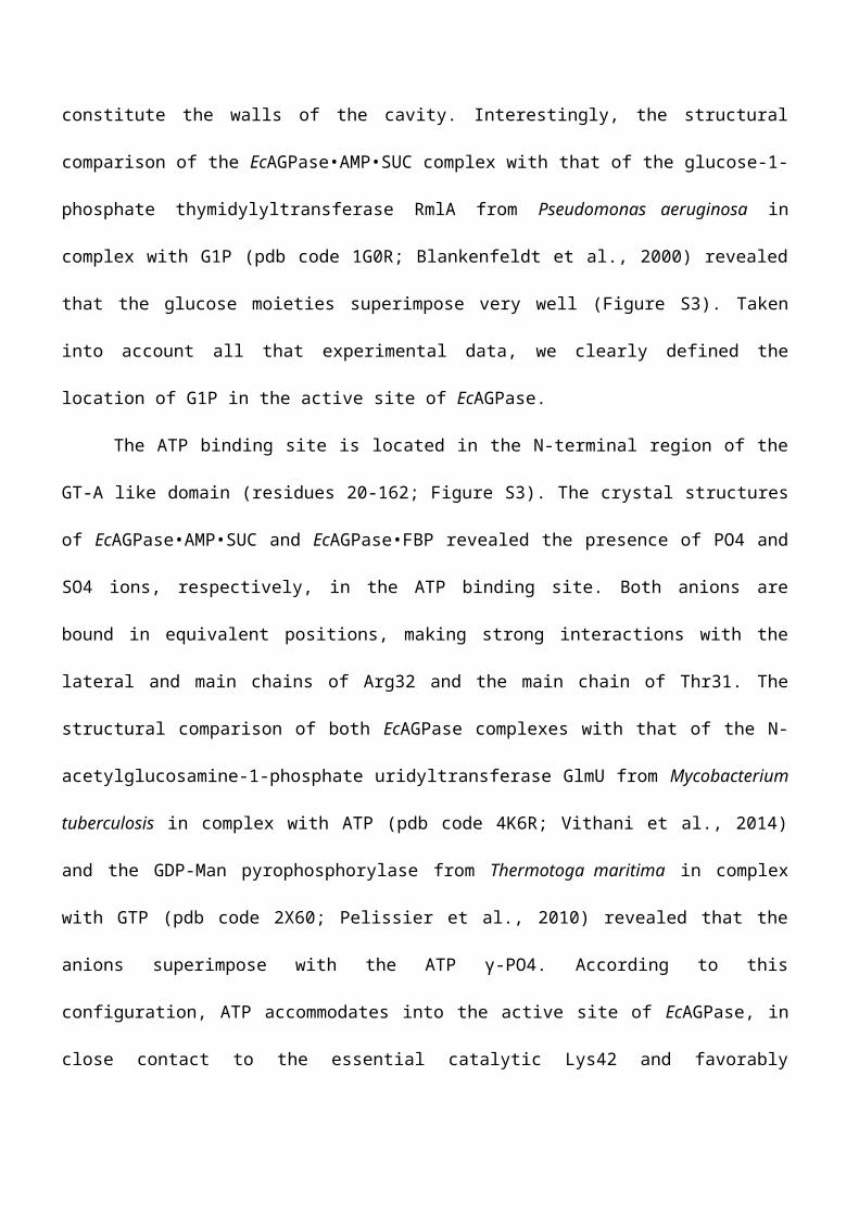
Figure 1. The crystal structure of EcAGPase. A. The overall structure of an EcAGPase protomer. Two views of an EcAGPase protomer showing the GT-A like domain (green) and the LβH domain (orange). B. Four views of the EcAGPase dimer. C. The structure of an EcAGPase homotetramer. D. Surface representation of an EcAGPase tetramer. The orientation is similar to that observed in panel C. See also Figure S1.
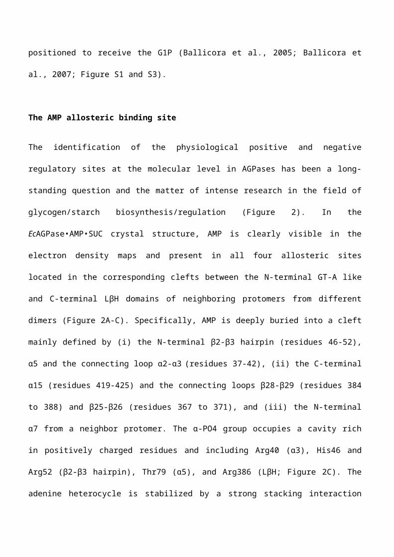
Figure 2. The location of the regulatory sites in EcAGPase. A. Electrostatic surface representation of the EcAGPase tetramer in complex with AMP (yellow spheres). B. Electrostatic suface representation showing a close view of the AMP binding site. C. Closed view of the AMP binding site, showing key interactions with selected residues. D. Electrostatic surface representation of the EcAGPase tetramer in complex with FBP (orange spheres). E. Electrostatic surface representation showing a close view of the FBP binding site. F. Closed view of the FBP binding site, showing key interactions with selected residues. G-I. EcAGPase thermal unfolding transitions recorded at 222 nm between 20 °C and 90 °C. Native fraction of EcAGPase plotted versus temperature for the apo state (green), and EcAGPase at different concentrations of AMP (panel G in a blue scale), or FBP (panel H in orange and red scale), or AMP and FBP (panel I in a purple scale). In panel I the curve for EcAGPase•AMP (where concentration of AMP = 0.5 mM) is indicated for reference. The corresponding fitted two-states sigmoidal curves of the unfolding events are also shown. See also Figure S2.
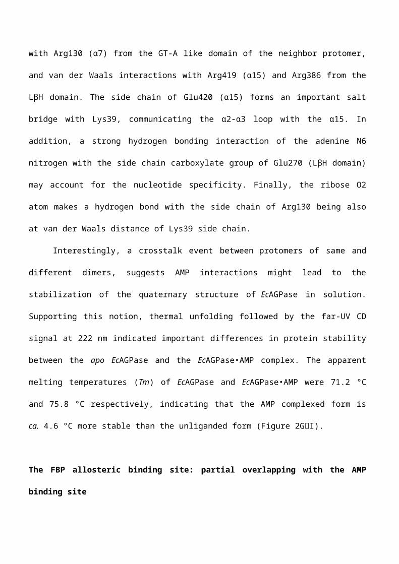
Figure 3. A regulatory mechanism for EcAGPase. A. Cartoon representation of the overall structure of the EcAGPase tetramer, showing the location of the regulatory and active sites. AMP, FBP, ATP and G1P molecules are shown in one protomer of the EcAGPase tetramer. The location of the ATP binding site in EcAGPase was determined by structural superposition with (i) the crystal structure of GlmU in complex with ATP and that of StAGPase in complex with ATP and ADPG. The G1P binding site in EcAGPase was determined taking into account the location of the glucose moiety of sucrose in the EcAGPase•AMP•SUC crystal structure (Figure S3). The four protomers are depicted in four different colors, green, orange, blue and yellow. B. Structural superposition of the EcAGPase•AMP•SUC and EcAGPase•FBP, showing the partial overlapping of AMP and FBP binding sites. C. Cartoon representing the key structural elements involved in EcAGPase allosteric regulation. Protomers A and C of the EcAGPase•AMP•SUC complex are shown in green and orange, respectively. AMP and the superimposed FBP molecules are shown into the allosteric site. The SM motif is shown in red whereas the RBL1 and RBL2 loops are shown in cyan. ATP and G1P are shown in the active site. D. The SM motif in detail. See also Figure S3.
Figure 4. Structure-weighted sequence alignment of EcAGPase with other AGPases. Structural alignment between the crystals structures of EcAGPase (pdb code: 5L6V; Uniprot code: P0A6V1), AtAGPase (3BRK; P39669) and StAGPAse (1YP3; P23509). The secondary structure elements corresponding to the GT-A like domain are shown in yellow (α-helices) and orange (β-helices); and to the LβH domain in green (α-helices) and blue (β-helices). Residues with poor electron density are highlighted as full boxes. The SM motif and the RL1 and RL2 loops are highlighted in yellow. Catalytic residues are highlighted as dotted boxes. The r.m.s.d. value is shown for each residue. Amino acid sequences of selected AGPases were aligned to the structure alignment: Mycobacterium smegmatis (class II; A0R2E1), Serratia marcescens (class II); A0A0U6P844), Rhodobacter sphaeroides (class V); Q9RNH7), Rhodospirillium rubrum (class VI); Q9ZFN4), Bacillus subtilis (class VII); P39122), Synechococcus sp. (class VIII; Q2JU94), Ostreocuccus tauri (clss VIII; Q6PYZ7), Spinacia oleracea (VIII; Q43152), Triticum aestivum (class IX; P30523). See also Figure S4.