Embed Size (px)
Citation preview
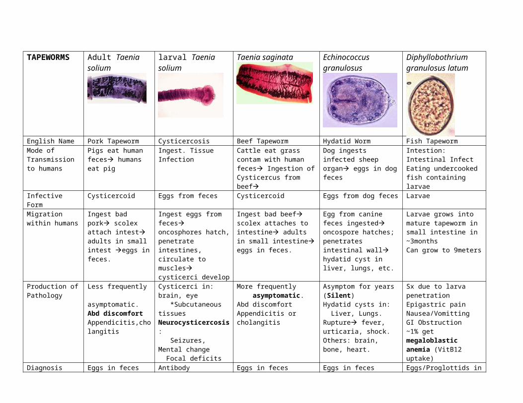
TAPEWORMS Adult Taenia solium
larval Taenia solium Taenia saginata Echinococcus granulosus
Diphyllobothriumgranulosus latum
English Name Pork Tapeworm Cysticercosis Beef Tapeworm Hydatid Worm Fish TapewormMode of Transmission to humans
Pigs eat human feces humans eat pig
Ingest. Tissue Infection Cattle eat grass contam with human feces Ingestion of Cysticercus from beef
Dog ingests infected sheep organ eggs in dog feces
Intestion: Intestinal InfectEating undercooked fish containing larvae
Infective Form Cysticercoid Eggs from feces Cysticercoid Eggs from dog feces LarvaeMigration within humans
Ingest bad pork scolex attach intest adults in small intest eggs in feces.
Ingest eggs from feces oncosphores hatch, penetrate intestines, circulate to muscles cysticerci develop
Ingest bad beef scolex attaches to intestine adults in small intestine eggs in feces.
Egg from canine feces ingested oncospore hatches; penetrates intestinal wall hydatid cyst in liver, lungs, etc.
Larvae grows into mature tapeworm in small intestine in ~3monthsCan grow to 9meters
Production of Pathology
Less frequently asymptomatic.Abd discomfortAppendicitis,cholangitis
Cysticerci in: brain, eye *Subcutaneous tissuesNeurocysticercosis: Seizures, Mental change Focal deficits
More frequently asymptomatic.Abd discomfortAppendicitis or cholangitis
Asymptom for years (Silent)Hydatid cysts in: Liver, Lungs. Rupture fever, urticaria, shock.Others: brain, bone, heart.
Sx due to larva penetrationEpigastric painNausea/VomittingGI Obstruction~1% get megaloblastic anemia (VitB12 uptake)
Diagnosis Eggs in feces Antibody Detection: Immunoblot, EIABiopsy affected areaMRI, CT, XRay: Space-occupying lesion in brain.
Eggs in feces Eggs in feces Eggs/Proglottids in feces
Definitive Host Humans ONLY. Humans ONLY. Humans ONLY. Small Intestine of Canids Carnivores; HumansIntermediate Host(s)
Pig or Human. Ox. Cattle Livestock, Humans Copepod (fish flea) fish
Form transmitted from humans
Eggs – suckers & hooks Eggs in feces*Cysticerci in muscles
Eggs – suckers only - Fecal eggs hatch, eaten by copepod in water
Geographic Foci Worldwide. Poor. Worldwide. Worldwide Worldwide. Rural.“Multilocularis”: Northern.
Great Lakes, Scandinavia, Baltic
Treatment PrazyquantelCook/freeze, inspect.
Surgery:remove cysticerciAlbendazole,Mebendazole
PrazyquantelCook/deep freeze, inspect.
Surgery: don’t rupture cysts+Albendazole,Mebendazole
Praziquantel (also Niclosamine)
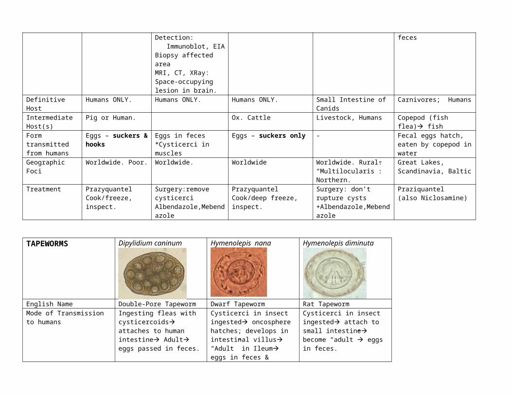
TAPEWORMS Dipylidium caninum Hymenolepis nana Hymenolepis diminuta
English Name Double-Pore Tapeworm Dwarf Tapeworm Rat TapewormMode of Transmission to humans
Ingesting fleas with cysticercoids attaches to human intestine Adult eggs passed in feces.
Cysticerci in insect ingested oncosphere hatches; develops in intestinal villus “Adult” in Ileum eggs in feces & Autoinfection thru intestines
Cysticerci in insect ingested attach to small intestine become “adult” eggs in feces.
Infective Form Cysticercoid Cysticercoid. Autoinfection. Cysticercoid. Autoinfection.Migration within humans Attaches to small intestine
adult. Eggs in feces.Oncosphere hatches; develops in intestinal villus “Adult” in Ileum
Oncosphere hatches; develops in intestinal villus “Adult” in Ileum
Production of Pathology Asymptomatic.Abdominal discomfortAnal pruritisDiarrheaRestlessness
Asymptomatic.Abdominal discomfort: restless, irritable, diarrhea, abd pain, anal pruritis, nasal pruritis, restless sleep
Asymptomatic.Abdominal discomfort: restless, irritable, diarrhea, abd pain, anal pruritis, nasal pruritis, restless sleep
Diagnosis Eggs in feces Eggs in feces Eggs in fecesDefinitive Host Humans. Animals. Pets. Humans and Rodents. Humans and Rodents.Intermediate Host(s) Dogs, Fleas Insects. Humans.
Reservoirs: dogs, rodents.Insects. Humans.Reservoirs: dogs, rodents.
Form transmitted from humans Eggs.Geographic Foci Worldwide. Kids Worldwide. #1 Cestode infect
In temperate areas. KidsSoutheast USA. Various world.Kids.
Treatment Niclosamide + Prazyquantel Prazyquantel Prazyquantel
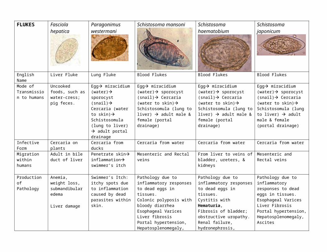
FLUKES Fasciola hepatica Paragonimus westermani
Schistosoma mansoni Schistosoma haematobium Schistosoma japonicum
English Name Liver Fluke Lung Fluke Blood Flukes Blood Flukes Blood FlukesMode of Transmission to humans
Uncooked foods, such as water-cress; pig feces.
Egg miracidium (water) sporocyst (snail) Cercaria (water to skin) Schistosomula (lung to liver) adult portal drainage
Egg miracidium (water) sporocyst (snail) Cercaria (water to skin) Schistosomula (lung to liver) adult male & female (portal drainage)
Egg miracidium (water) sporocyst (snail) Cercaria (water to skin) Schistosomula (lung to liver) adult male & female (portal drainage)
Egg miracidium (water) sporocyst (snail) Cercaria (water to skin) Schistosomula (lung to liver) adult male & female (portal drainage)
Infective Form Cercaria on plants Cercaria from ducks Cercaria from water Cercaria from water Cercaria from waterMigration within humans
Adult in bile duct of liver
Penetrate skin inflammation swimmer’s itch
Mesenteric and Rectal veins From liver to veins of bladder, ureters, & kidneys
Mesenteric and Rectal veins
Production of Pathology
Anemia, weight loss, submandibular edema
Liver damage
Swimmer’s Itch: itchy spots due to inflammation caused by dead parasites within skin.
Pathology due to inflammatory responses to dead eggs in tissues.Colonic polyposis with bloody diarrheaEsophageal VaricesLiver fibrosisPortal hypertension, Hepatosplenomegaly, Ascites
Pathology due to inflammatory responses to dead eggs in tissues.Cystitis with Hematuria, Fibrosis of bladder; obstructive uropathy.Renal failure, hydronephrosis, Bladder carcinoma
Pathology due to inflammatory responses to dead eggs in tissues.Esophageal VaricesLiver FibrosisPortal hypertension, Hepatosplenomegaly, Ascites
Diagnosis Yellow-brown eggs in feces
Clinical Presentation, and patient history.
Eggs in Stool or UrineELISA
Eggs in UrineELISA
Eggs in Stool or UrineELISA
Definitive Host Humans. Sheep, CowIntermediate Host(s)
Snails
Form from humans
Eggs
Geographic Foci Worldwide. Sheep and Cattle
Africa, SE Asia, NW S AmericaCaribbean islands; Kids
Kids; Africa, SE Asia, NW S America, Caribbean islands
Kids; Africa, SE Asia, NW S America, Caribbean islands
Treatment triclabendazole Anti-Inflammatory Rx PraziquantelOxamniquine
PraziquantelOxamniquine
PraziquantelOxamniquine
Nematodes“Round-worms”
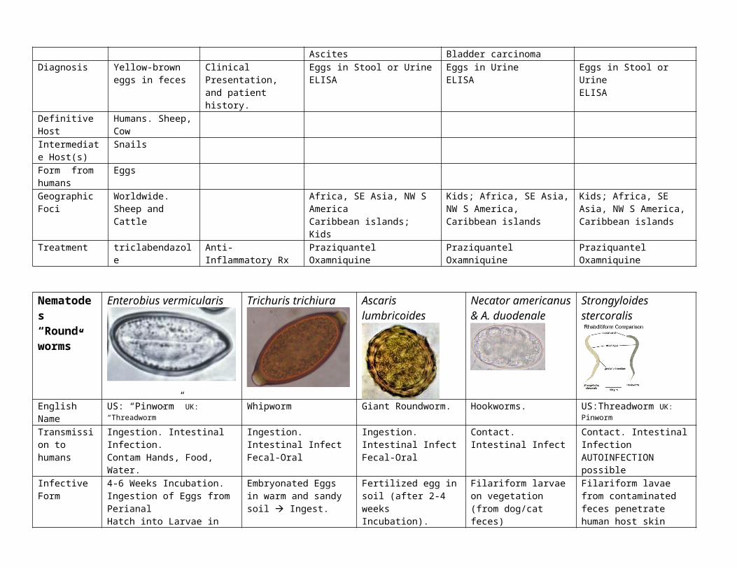
Enterobius vermicularis Trichuris trichiura Ascaris lumbricoides Necator americanus & A. duodenale
Strongyloides stercoralis
English Name US: “Pinworm” UK: “Threadworm” Whipworm Giant Roundworm. Hookworms. US:Threadworm UK: PinwormTransmission to humans
Ingestion. Intestinal Infection. Contam Hands, Food, Water.
Ingestion. Intestinal InfectFecal-Oral
Ingestion. Intestinal InfectFecal-Oral
Contact. Intestinal Infect Contact. Intestinal InfectionAUTOINFECTION possible
Infective Form 4-6 Weeks Incubation.Ingestion of Eggs from PerianalHatch into Larvae in Sm. Intestine
Embryonated Eggs in warm and sandy soil Ingest.
Fertilized egg in soil (after 2-4 weeks Incubation).
Filariform larvae on vegetation (from dog/cat feces)
Filariform lavae from contaminated feces penetrate human host skin
Migration within humans
Direct Life Cycle: AutoinfectionIngest Larvae hatch in small intestine Adults in Colon Eggs in Perianal area Ingest.
Direct Life Cycle: AutoinfectUnembryonated eggs in fecesIn 3-4weeks, Embryo eggs; ingested Larvae hatch in small intestineAdults in cecum eggs in feces.
Direct Life Cycle:Ingest eggs in Feces larvae hatch in small intestine,enter blood migrate via liver, heart to lung enter alveoli, molt, migrate, swallowed small intestine=adults
Eggs hatch on warm, sandy soil in 2 days molt twice Filariform larvae climb on vegetation Penetrate skin enter capillaries to lung…swallowed small intestine
Filariform larvae penetrate skinEnter blood lungs, swallowed small intestines = adults Eggs released, but HATCH within human intestine filariform autoinfect from intestines OR into feces
Production of Pathology/ Symptoms
AsymptomaticPerianal Pruritus (nocturnal)Occasionally, invades Female GU tract Vulvovaginitis
AsymptomaticHeavy infections: Abd pain, Diarrhea, Tenesmus.Rectal ProlapseAnemia. Stunted Growth.
Large number: Pneuomonitis & Hepatomegaly; block GI.RARE Block: bile duct, liver; peritonitis
Itchy skin Pulmonary phase (pneumonitis, eosinophilia) Intestinal phase (severe anemia)Cutaneous Larval Migrans
Asymptomatic. GI probs. Pulmonary symptoms. Urticarial rash on butt.Constipation.ICH: disseminationfatal?
Diagnosis Scotch Tape Test (eggs on skin)Sometimes, worms in feces.
Eggs in feces “Football-Shaped”
Eggs: Round, thick shell.?Whole worms in feces.
EosinophiliaCharacteristic Eggs.
Rhabditiform Larvae in feces. ELISA, IFA for Abs.
Definitive Host Humans Humans Humans Dogs. Cats. Humans. HumansIntermediate Host(s)
- - - Dogs, Cats Dogs. Monkeys.
Form from humans
Eggs in feces, on perianal skin (and thus under fingernails).
Eggs in feces. warm and sandy soil for 1-2 years.
Eggs in feces. Live in soil. Ingest.
Eggs passed in feces.
Geographic Foci
Worldwide (Temperate). Kids#1 Helminth in USA
Worldwide (tropical, poor).Southern USA.
Tropical, Subtropical.Rural South-Eastern USA.#1 Worldwide Helminth
Americanus: America, Australia. Duodenale: Middle East, N. Africa.
Tropical. Subtropical. Temperate. South of the USA
Treatment Pyrantel Pamoate Mebendazole.Alt: Albendazole.
Albendazole. If severe: surgical removal.
IvermectinAlt: Albendazole.
NEMATODES“Round-Worms”
Trichinella spiralis Dracunculus medinensis
English Name Pork Worm. Trichinosis. Guinea WormMode of Transmiss to humans
Ingestion. Tissue InfectionLarvae in meat. Rodents carry.
Ingestion. Tissue InfectionContamin., standing pond water
Infective Form Larvae cysts in contamin. pork Larvae within CopepodsMigration within humans
Ingest larvae from porkLarvae released in small intestine adults in small intestine larva deposited in mucosa blood encysted larva in striated muscle
Drink water with copepods larvae released Larva penetrate stomach/intestine wall, mature & reproduce fertilized female to skinblister; leaves skin (1yr)
Production of Pathology
Week 1: GI Symptoms (V/D, abd pain)Extraintestinal (2nd week): Muscle invasion (myalgia, weakness, malaise) Petechiae Myocarditis CNS invasion (fits, paralysis)By 3rd week: Larvae begin to encyst!Eosinophilia peaks at week2-3.
1st: painful blister2nd: Worm emerges as a whitish-filament (takes 1-3 weeks)3rd: Niridazole
Diagnosis Based on Clinical Symptoms (Myositis) and Eosinophilia Look at Diet – Suspicious?EIA testMuscle Biopsy, Microscopy
Clinical Picture
Definitive Host Humans. Pigs.Intermediate Host(s)
Rodents Copepods
Form transmitted from humans
Larvae cysts in contaminated pork. Larvae into water from emergent female worm.
Geographic Foci Worldwide. Pigs Rural Africa. Standing Ponds.Treatment ASAP!!! Steroids for severe
Mebendazole + AlbendazolePrevent: Proper cook or deep freeze
Clean lesion, local Abx.Mechanical, progressive extraction of worm over days.
Nematodes Wuchereria bancrofti Brugia malayi Onchocerca volvulus
English Name River BlindnessMode of Transmiss to humans By Bite Tissue Infection By Bite Tissue Infection By Bite Tissue InfectionInfective Form L3 Larvae from Mosquito Simulium blackflies’ L3 LarvaeMigration within humans Infected mosquito with larvae bites
larvae to lymphatics, become adults adults produce sheathed microfilariae that migrate into lymph/blood channels mosquito bites human..
Simulium blackflies bite larvae into subcutaneous tissues adults into subQ tissue adults produce unsheathed microfilariae in skin and lymphatics of connective tissue + blood, urine, sputum blackfly bites human.
Production of Pathology FilariasisAsymptomatic Recurring attacks of Lymphangitis and “filarial fever” over years Lymph blockage, lymphedemaScattered wheezes/crackles.Hydrocele, Scrotal Elephantiasis
FilariasisAsymptomatic Recurring attacks of Lymphangitis and “filarial fever” over years Lymph blockage, lymphedema
Lymphedema, Elephantiasis
FilariasisPruritus, Dermatitis, Onchocercomata **can live 10-15 years in SubQ nodules!Lymphadenopathies, Ocular Lesions (“River Blindness”) BlindnessIntensely pruritic! Snowflake opacitiesLeopard skin of shins!“Hanging Groin”: drooping inguinal skin
Diagnosis Microfilariae periodically in peripheral blood.Detection of filarial antigen (KITS)Urine exam/microscopyCBC: Eosinophilia; Elevated IgE + IgG4PCR
Microfilariae in skin-snip biopsy sampleDirect examine of excised nodulesImmunoDx: Ab detection, Ag detectionPCR: UncommonImaging to reveal nonpalpable nodulesDEC Patch Test: topical application of DEC itchy due to dying microfilariae “Mazzotti Reaction”
Definitive HostIntermediate Host(s)Geographic Foci Tropical Areas Worldwide. Subtropics Africa, Latin America, Middle East.Treatment Ivermectin + Diethylcarbamazine (DEC)
Antihistamines, Corticosteroids, pain relief, Abx for 2ndary infections.Surgical excision of elephantiasis.
Ivermectin, long treatment. + CorticoidsNodulectomy if feasible.
NEMATODES*Wanderers.
Anisakiasis simplex & Pseudoterranova decipiens A. Braziliense & A. caninum Toxocara canis, Toxocara cati
English Name HookwormsMode of Transmiss to humans
Consumption of infected fish with larvae. Contact with Filariform larvae in vegetation (from dog/cat feces)
Infective Form Larvae in undercooked fish. Filariform Larva from cat/dog feces. Larvae in dog feces.Migration within humans
Ingest larvae from fish larvae penetract stomach/intestinal mucosa into bowel Crohn’s-Like Illness
Eggs in dog/cat feces Rhabditiform larva hatches develops into Filariform Larva in environment through skin.
Embryonated egg with larvae in dog/puppy feces
Production of Pathology
Within hours of ingestion, violent Abd pain, N/V.If into bowel: Severe eosinophilic granulomatous response Crohn’s-like
Cutaneous Larva Migrans (“Ground Itch”) of Dermis – NO ADULTS FormEosinophilic enteritis (caninum)Transient Pulmonary infiltrate with peripheral eosinophilia
Self-limiting infection
Asymptomatic w/ eosinophilia.Fever, cough/wheeze, anemia, hepatomegaly…
Visceral Larva Migrans (VLM) Liver, heart, lungs, brain, muscle, eyes (Ocular)
Diagnosis Gastropic Examination and Biopsy Eosinophilia & Positive serology (titers); ELISA
Definitive Host Fish Cat/Dog DogIntermediate Host(s) Crustaceans - Rabbit?Form transmitted from humans
Larvae in emesis/feces.
Geographic Foci Worldwide. Kids. South-Eastern US.
Treatment Surgical or Endoscopic Removal. Self-Limiting; Ivermectin, Albendazole + Anti-inflam Rx
Albendazole + Anti-inflam Rx
Arthropods Pediculus Cimex lectularius
Blackfly Sandflies L. Laeta L. Mactans Scorpians Mites & Scabies
English Name Lice Bed Bug Buffalo Gnat Sandflies Brown Recluse Spider
Black Widow Spider Scorpians Mites & Scabies
Production of Pathology
Intensely pruritic
Bites, Punctures, Rash.Itchy, red lesions.Small, red marks to hemorrhagic bullae in a linear fashion on the trunk/arms
Itchy, edema.patients w/ hemorrhagic syndrome can reduce platelet count (prolonged bleeding time)
Sand fly fever- characterized by frontal headaches, malaise, retro-orbital pain, anorexia, and nausea
Sharp pain, but little swellred, swell, & burn; Within an hour: m. cramps, chest pain, N/V, diaphoresis, intest spasms, visual difficulty, abd cramps
Sting site radiating, burn, swell, discolor, necrosis, chills, diaphoresis, excessive saliva, diff speaking, muscle spasm, tachycardia, seizures.
Characteristic lesion and distribution (interdigitital folds & sides of fingers, buttock, ext genitalia, wrists, elbows; lesion is short, slightly raised cuaneous burrows)
Diagnosis Find lice or eggs. eggs ( nits) are white, round objects attached to hair shaft
Pattern and location of bites tiny spots of blood on bedding or dead insects
Marked characteristically by a point of dried blood and subcutaneous hemorrhage at the wound site;
Can’t discriminate species of spider from lesion.ELISA to confirm .
“boardlike” abdomenusually subside w/in 48 hours; coma and paralysis, death can follow (rare)
Single red mark paired w/ local or systemic signs/symptoms
Mites in skin scrape
Treatment Gamma benzene haxachloride (lindane) lotion applied to body for 24 hours,shaving; destroy adult lice by lindane or DDT powder or boiling; treat brushes, bedding w/ pediculicide or boiling;
Topical palliatives to relief itchingAntihistamines if dermatitis is severe Prevent w/ hygiene and insecticides
Palliative measure (anesthetics, antihistamines, lotions) to relieve local pruritus and swelling; For hemorrhagic syndrome give corticosteroids; Prevent w/ protective clothing (repellent doesn’t work)
Prevention: Sensitive to insecticides to breeding sites and insect repellents
Cleanse. Tetanus prophylaxisAbx to prevent infection. Debride after 3-6 wks (use for bites that haven’t healed by this time); corticosteroids for hemolytic syndrome; active in SA. NOT in USA.
Small children or Immunocompromised can die without treatment; Muscle spasms may require IV admin of calcium gluconate or muscle relaxant; specific antivenom is the treatment of choice (test patient for sensitivity to horse serum)Prevention= good house keeping
Analgesics or local injection of xylocain (opiates can increase toxicity); local cryotx can reduce swelling; Hot packs; antivenom is effective if administered soon after sting;
1% gamma benzene hexchloride (lindane)Contra-ind for infants, pregnant/ lactating women5% permethrin cream (Elimite) is treatment of choice