Embed Size (px)
Citation preview

Medicine with Industrial Specialisation / Department of Health Science and Technology
Epidemiology of Stroke and Death in Atrial Fibrillation
1. PresentationThis section is an appendix to the article; “Age-Dependent Risk of Stroke and Death in Young Patients with Atrial Fibrillation: A Nationwide Cohort Study”. As part of the study project, the subsequent pages are a theoretical supplement to the article, where the following topics will be discussed in relation to the article; epidemiological aspects of research including causation, bias and confounding, controlling bias and confounding, types of epidemiologic study designs, and limitations of register studies. Furthermore, the epidemiology of atrial fibrillation (AF) pathophysiology of AF, subtypes of AF, etiology of AF, consequences of AF, AF and a subsequent diagnosis of stroke, guidelines for management of stroke in AF, and finally risk of bleeding with antithrombotic therapy will be discussed.
2. IntroductionEpidemiology was first set as a scientific discipline in the 20th century with the aim of uncovering the causes of diseases1. The study of diseases in the population was first applied on epidemics but today it is used to study quantitative descriptions and analysis of a variety of health conditions in specific patient groups and in the general population2. Epidemiology can be used to study single cases of rare diseases or larger patient groups of well-known diseases2. In this present study, several epidemiological aspects exist. The human body is a complex organism and in combination with individual life styles, risk of stroke in AF patients is multifaceted involving many biological and environmental factors. By investigating the risk of stroke in patients diagnosed with AF it is possible to identify which factors increase or decrease the risk of stroke, and thereby, to estimate which risk factors should be in focus for the prevention of stroke or early death in AF patients aged <65. However, the causal association between certain risk factors and stroke may be complex.
2.1 CausationThe concept of causation is primarily based on observations1. Several models of causation have been developed illustrating principles of causation1,3,4. The most important principle is that every causal factor involves the combination of
a multitude of components divided into genetic and environmental determinants that contribute to the overall complex of causality1. All diseases have both genetic and environmental determinants since both elements always play a role in the causality of a disease but do not necessarily act at the same time in the cascade of the causal mechanism1. Strength of causal effects is often used to describe the association between a causal factor and a disease but should not be used as a criterion for causality1. Strength of association can vary in every case of every disease and it is usually only the environmental factors that can be altered; however, this will probably be different in the future as biomedical technology advances1. A strong cause can be defined as a component that plays a crucial role in a large proportion of cases and a weak cause to be a causal component in a small proportion of cases1. Though, the strength of a cause is also depending on the prevalence of other causal factors of a given disease1. From a biological point of view, causation corresponds to each individual factor’s cause in a causal mechanism and the way in which several factors interact to generate disease1. Thus, the strength of a cause does not depict the biology of causation and each causal factor has an individual role in each case of a disease1. On the other hand, temporality, implying that cause comes before disease, is a true criterion regarding causality but the sequence of cause and outcome is difficult to determine5. It is possible to collect information about risk factors and events but the exact causal effect cannot be observed directly, only prophesized2. Thus, causality is a complex issue requiring the combination of biological knowledge and a thorough line of thinking.
2.2 Bias and ConfoundingIn designing an epidemiology study, sources of error should be reduced. Two types of error exist; random error and systematic error, which should be taken into consideration during both the design of a study and during the interpretation of the results1. Random error denotes error due to sampling variability for example by chance6, and which can be overcome by using a sufficiently large sample size. Systematic errors, or bias, are errors that remain even in an infinitely large study, in contrast to the random errors that are reduced
11

Medicine with Industrial Specialisation / Department of Health Science and Technology
towards zero if a study becomes large enough1. In research methodology, an estimate of association obtained from a biased study differs systematically from the true association in the source population of the study7. Furthermore, statistical significance does not reflect the presence or absence of bias6. When taking account of bias in a study, it is important to estimate how much bias exists and to what extent existing bias could influence the conclusions6.
Bias can be classified into three broad categories; selection bias, information bias, and confounding1. Selection biases are errors that result from the procedures to select subjects and from factors that influence study participation. It occurs when the association between exposures and disease differs for those who participate and who do not participate in a study1. Information bias is typically seen in the form of misclassification bias1, for example when truly exposed individuals are erroneously classified as unexposed or vice versa8. Information biases can arise if wrong information about the subjects in a cohort is given resulting in an incorrect estimate2.
Confounding is a problem that pervades many epidemiologic studies. Confounding is the confusion of effects where the effect of the exposure is mixed with the effect of another variable leading to bias9. A confounding variable must be associated with the disease either being a cause of the disease or a marker of a cause; thus, a marker for the underlying biology1. However, a confounding variable must not be an effect of the disease and should be associated with the exposure, but not be an effect of the exposure (see figure 1a) 1,10. A confounder is a predictor of disease occurrence, though, not every predictor of disease is a confounder. A predictor of disease must also be imbalanced across exposure categories to be confounding, and a confounder can cause either an overestimate or underestimate of an effect or even reverse the apparent direction of an effect1. In this present study, the causal factor is AF and the outcome of interest is stroke and death. Confounding factors are, among others, smoking, alcohol, BMI, etc., which were not included in the analysis.
Figure 1a: Relationship of a Confounding Factor with a Causal Factor and the Outcome. See text for details regarding confounding factors.
2.3 Controlling Bias and ConfoundingA good way to resolve the extent to which one variable’s effect explains the apparent effect of another is to examine both effects simultaneously1. To take confounding into account it is necessary to approach a deeper causal understanding of the underlying biology of the disease and exposure1. Otherwise, three scientific methods exist to prevent or limit confounding; randomization, restriction, and matching1. Randomization is used primarily in experimental studies and results in similar groups which often prevent both known and unknown confounding factors2. Restriction involves selecting subjects who all have the same value for a variable that would otherwise be a confounding variable1. Matching attempts to produce a control group that is similar to the study group with regard to the distribution of selected confounding factors and thereby prevent confounding7. However, when using matching in register studies where data are already stored in databases, matching will result in excluding some subjects from the study, because other subjects will be closer matches and are chosen instead1. Nonetheless, this will give a better control, even though some subjects are excluded.
Two other methods deal with confounding and are used in the data analysis; stratification and regression models1,2. Stratification creates sub-groups in which the confounding factor either does not vary at all or only varies very little within a stratum1,6. A stratified analysis proceeds under the assumption that within the categories of the stratified variable there is no meaningful variability of a potential confounding factor and the data analysis is performed separately in each stratum1,2. When comparing strata it is estimated if any crucial effect modification has occurred, and a common weighted estimate is calculated if necessary2. Sometimes the end result of stratification is nothing more than presentation of data within each stratum1. Additionally, feasibility of stratification is limited when there are multiple confounders, because a stratum has to be constructed for each possible confounder combination6. Regression models are used to give a summarized description of a dependent variable’s correlation with one or more independent variables2. Thus, regression models can be used to evaluate the causal role of one or more exposures while simultaneously controlling for possible confounding effects1. It is possible to predict the amount of confounding from the general characteristics of the confounding variables; thus, predicting the associations of a confounder with both exposure and disease. Confounding can be measured directly by measuring the statistical significance when adding the confounding variable to the model1. However,
12

Medicine with Industrial Specialisation / Department of Health Science and Technology
this approach requires that you are aware of the confounding factors. In reality, this scenario is rare, and therefore, it is very important to become aware of as many confounders as possible and use this knowledge when analyzing and interpreting the results.
2.4 Epidemiological Study DesignsEpidemiological studies aim at estimating a single risk, incidence rate, prevalence, or comparing measures of disease occurrence1. The general concepts of how bias is introduced into a study apply equally to RCTs, prospective observational and retrospective studies6. An important difference between the designs is how much opportunity the investigator has to avoid bias and influence the accuracy of measurements, ranging from full control in RCTs to almost no control in studies of automated databases6. Investigators should aim to avoid bias in the design of a study, adjust for bias in the study analysis if necessary, and quantify and discuss the effects of residual bias on study results6.
The two main types of epidemiological studies are cohort studies and case-control studies1. Case-control studies are observational studies of a cohort with an outcome variable of interest and a suitable control group, a so-called comparison or reference group. The potential relationship of a suspected dependent factor is examined by comparing the cases and controls with regard to how frequently the factor is present or the level of the factor in each group11. In a cohort study, the cohort is defined as “any designated group of individuals who are followed or traced over a period of time”11. This type of study involves measuring the occurrence of disease within one or more cohorts during a period of follow-up usually to compare the disease rates in the cohorts1. In a prospective cohort study, like in this register study, the cohorts are identified from recorded information and observed during the selected follow-up period, here a 5-year period. With a cohort study, it is often convenient to study many different disease outcomes in relation to a given exposure1, like in the present study examining stroke and death.
2.5 Limitations of Register StudiesSince register studies must rely on existing records, valuable data may be missing or incorrect. In spite of this, this type of study offers a much less costly and much quicker way of obtaining information1. Very little control over patient selection is usually available in register studies with automated data collection. However, it is essential that equal inclusion criteria and ascertainment procedures for the exposed and unexposed patients are applied
and that all patients ideally stem from the same source population6. Investigators have limited control over measurements, especially in register studies with automated databases, where the measurements have already occurred. It is therefore crucial for the investigator to learn as much as possible about how measurements were made and how data were coded6. Sometimes the investigators may be able to locally assess the specifics and the quality of data retrieval; however, in other settings, it may be necessary to validate data entries against medical records6. In this present study, data in the used registers were very reliable which a previous study has concluded after validating these data12,13. However, often investigators do not have in-formation regarding how the indications are diagnosed, and therefore cannot determine on what grounds an indication is given or when in the process of diagnosis it is given14. Nonetheless, observer bias is diminished in this type of data collection9. In the present study, only information in the national Danish registers is available, for example data on effects of tobacco, dyslipidemia, or body mass index are not accessible, which can result in sampling bias if some of these characteristics are underrepresented or overrepresented in the study population9. Therefore, residual confounding may be evident, although it is possible to attempt to adjust for baseline clinical characteristics in the statistical analysis15.
The validity of data can be a limitation. However, the positive predictive value of the diagnosis of atrial fibrillation is high (99%) based on a previous validation study16. In the current study, data on prescription claims are accurate which has been estimated in a previous study17. However, it is only possible to access diagnoses medically treated, but the degree of disease is not available18. Furthermore, aspirin can be bought over the counter, and therefore some patients may be in some degree of anticoagulant therapy resulting in inclusion bias. The frequencies of risk factors in the study population may also be underestimated because patients with for example heart failure, hypertension, and diabetes are identified from prescription claims and thus patients treated with diet control and lifestyle interventions alone are not detected, which may be the case with some of these patients resulting in a low sensitivity of these diagnoses18. Furthermore, the study population is primarily of Caucasian origin and therefore, the conclusions may not be translational to other ethnical populations or even outside Denmark, where there may be very different environmental and societal factors present.
13

Medicine with Industrial Specialisation / Department of Health Science and Technology
The major strengths of this type of study are the well-validated outcomes and the large sample size15. Additionally, register studies offer fast data in an inexpensive method, since data has already been collected, especially, when observing an outcome with an often long time-to-outcome period. Another advantage of this type of study design is the lack of bias because the outcome of current interest was not the original reason for the data to be collected. However, for the same reason some of the necessary data might not be accessible9. Further-more, a single study can examine various outcome variables as in this present study, and cohorts permit calculations of the effect of each variable on the probability of developing the outcome of interest9. However, where a certain outcome is rare then a prospective cohort study is inefficient9. Based on the pros and cons of epidemiological studies, as mentioned above, and on the specific data used in the current study, it is hypothesized that the results of this study are generalizable and not only applicable in the present study population but also in the general population of AF patients.
3. Epidemiology of AFAtrial fibrillation is the most common cardiac arrhythmia in the general population19, affecting approximately 1% of the total adult population19
and increasing to 9% in individuals over 80 years20. However, AF is a heterogeneous disorder, with variable origin, clinical presentation, and natural history21.
3.1 Pathophysiology of AFAF is defined as a cardiac arrhythmia of the atria. The initiation and continuation of a tachy-arrhythmia like AF requires both triggers for its onset and a substrate for its maintenance23. These mechanisms are not mutually exclusive and are likely to co-exist at various times22. During AF, the atria pump insufficiently and become useless as primer pumps for the ventricles22. Blood flows passively through the atria into the ventricles, and the efficiency of ventricular pumping is decreased 20 to 30 per cent24.
The pathophysiology of AF involves focal mechanisms in the form of numerous small depolarization waves that spread in all directions through the atria contributing to the initiation and continuation of AF25. Because the waves are weak and many of opposite polarity at any given time, the waves usually almost completely neutralize electrically one another. Therefore, in the electrocardiogram (ECG), either no P waves from the atria or only a fine, high-frequency, very low
voltage wavy record are present24. Conversely, the QRS-T complexes are normal unless there is some pathology of the ventricles, but the timing and rhythm is irregular22. During AF, impulses arrive from the atrial muscle at the A-V node rapidly, and because the A-V node will not pass a second impulse for about 0.35 second after a previous one, at least 0.35 second must elapse between one ventricular contraction and the next24. An additional interval of 0 to 0.6 second occurs before one of the irregular atrial impulses arrive at the A-V node24. Thus, the interval between successive ventricular contrac-tions varies from a minimum of about 0.35 second to a maximum of about 0.95 second, causing a very irregular heartbeat, which is also one of the clinical symptoms present in AF22,25. Due to the rapid rate of the impulses in the atria, the ventricle is driven at a fast heart rate, usually between 125 and 150 beats per minute, which a person can live with for years, although, at reduced efficiency of overall heart pumping, and heart failure very often develops22.
Pathophysiologically, AF is caused by circular movement of impulses25. The background conditions that can initiate an impulse to continue travel around the circles causing re-entry and lead to “circus movements” in the atria are: (1) the pathway around the circle is too long so the originally stimulated muscle is no longer refractory, for example due to a dilated heart, (2) the velocity of conduction becomes decreased causing a longer time interval for the impulses to travel around so the originally stimulated muscle is no longer refractory, for example due to heart muscle ischemia, and (3) the refractory period of the muscle might become greatly shortened allowing re-entry of the impulse into previously excited heart muscle within a much shorter time than normally22,23,26, so the impulse again continues around the circle, for example due to drugs such as epinephrine or after repetitive electrical stimulations24. Re-entry can cause abnormal contractions and rhythms of the heart due to its ignoring of the sinus node’s control of the heart rhythm27. The division of impulses also contributes to the pathophysiology, because when a de-polarization wave reaches a refractory area in the heart, it converts into two impulses, which are divided again when these reach another refractory area. This initiates a chain reaction that causes many circuitous routes for the impulses to travel, greatly lengthening the conductive pathway sustaining the fibrillation24. Recently, the mechanisms involved in the progression of AF were pointed out to be due to electrical remodeling such as changes in the electrical properties of the atrium and loss of rate adaption, and further to be due to structural
14

Medicine with Industrial Specialisation / Department of Health Science and Technology
remodeling associated with adaptive and maladaptive changes in the tissue and cellular components. The role of atrial remodeling has over time become the new pathophysiological mechanism of AF26.
3.2 Subtypes of AFMany risk factors for AF have been described and they include; old age, cardiomyopathy, valvular disease, ischemic heart disease, heart failure, thyroid disease, hypertension and diabetes mellitus28–30. In some patients, its etiology remains unknown, and up to 30% of AF patients may be free of any known structural cardiopulmonary disease19,31–33. Lone AF is defined as AF occurring in otherwise healthy individuals before the age of 60 years without any evidence of associated cardiopulmonary or other comorbid diseases34,35; if a patient is older than 60 years and has no underlying disease, AF is designated as idiopathic33. The importance of family history for lone AF has been determined in several studies36–39.
AF can manifest in five different types (see Figure 2a). Generally, AF can be divided into first-diagnosed, paroxysmal, persistent, long-standing persistent and permanent AF22. Asymptomatic or silent AF is also a subtype and is relevant in about one third of all AF cases. Furthermore, AF progresses from short, rare episodes, to longer and more frequent attacks22. Over time, many patients will develop sustained forms of AF 22 and only 2-3% of patients will remain in paroxysmal AF40. Generally, the course of AF can vary markedly over months or years in each patient41. Asymptomatic AF is common even in symptomatic patients, irrespective of whether the initial presentation was persistent or paroxysmal. This has important implications for continuation of therapies aimed at preventing AF-related complications22, which will not be discussed here.
Another common type of atrial arrhythmia is atrial flutter. It gives many of the same symptoms as AF but differs in that the electrical signal travels as a single large wave always in one direction in a circus movement in the atrial muscle22. Atrial flutter causes a rapid rate of contraction of the atria, usually between 200 and 350 beats per minute, but because one side of the atria is contracting while the other side is relaxing, the amount of blood pumped by the atria is slight24. Additionally, the signals reach the A-V node too rapidly for all of them to be passed into the ventricles, because the refractory periods of the A-V node and A-V bundle are too long to pass more than a fraction of the atrial signals. Thus, usually two to three beats of the atria for every single beat of the ventricles are present giving a 2:1 or 3:1 rhythm22. In the electrocardiogram, the P
waves are strong because of contraction of semi-coordinated masses of muscle. However, a QRS-T complex follows an atrial P wave only once for every two to three beats of the atria, as just mentioned22. In the present study, a diagnosis of AF or atrial flutter will not be distinguished. However, it is still being questioned if subtype of AF influences the risk of stroke and death, but lone AF patients have convincingly the most favorable prognosis42.
Figure 2a: Subtypes of AF. CV is an abbreviation for cardioversion. Modified from Camm et al. 2010.
3.3 Etiology of AFFamilial AF was first reported in 194343, and familial clustering of AF has been demonstrated in several studies afterwards36,39,44–46. The first chromosomal location of an AF susceptibility gene was reported in 1997 based on genetic mapping studies in three families47. Several genetic variants have since then been linked to risk of AF48–56. During the past years, numerous inherited cardiac syndromes associated with AF have been identified22. The pathophysiological role of other genetic defects in the initiation and continuation of AF is currently unknown57, but several clinical studies are presently investigating the genetic etiology of AF58. However, prior stroke, valvular disease, congestive heart failure, diabetes, and hypertension are independent risk factors of AF59. In the majority of patients, AF is accompanied by underlying heart disease, most frequently heart failure, valvular disease and hypertension60. Despite the use of antiarrhythmic agents for sinus rhythm maintenance in cardioverted AF patients, a considerable proportion of patients relapse to AF29. It is generally believed that these recurrences are associated with older age, atrial dilatation, and long duration of AF61.
15

Medicine with Industrial Specialisation / Department of Health Science and Technology
Studies have also suggested that female sex, obesity, personal history of AF events, decreased renal function, increased circulating markers of cardiomyocyte injury are risk factors of recurrence62.
3.4 Consequences of AFSeveral pathophysiological changes occur after the initiation of AF. Loss of coordinated atrial contraction, high ventricular rates, irregularity of the ventricular response, and decrease in myocardial blood flow, as well as long-term alterations such as atrial and ventricular cardio-myopathy are just some of the changes22. Acute loss of coordinated atrial mechanical function after the onset of AF reduces cardiac output by 5–15%22. High ventricular rates limit ventricular filling due to the short diastolic interval, and irregularity of the ventricular rate can reduce cardiac output22. Furthermore, persistent elevation of ventricular rates above 120–130 bpm may produce ventricular tachycardiomyopathy63.
The complication of AF with the strongest association to early death is stroke. Risk of stroke and systemic embolism in patients with AF is linked to a number of underlying pathophysiological mechanisms27. Abnormalities in blood flow are evidenced by stasis within the left atrium and with reduced left atrial appendage flow velocities, which is the dominant source of embolism (90%) in non-valvular AF27. Endocardial abnormalities include progressive atrial dilatation and fibroelastic infiltra-tion of the extracellular matrix22. Abnormalities of blood constituents are well described in AF and include hemostatic and platelet activation, as well as inflammation and growth factor abnormalities27. One part of AF management is reduction of the heart rate which may restore normal ventricular function and prevent further dilatation and damage to the atria22.
3.5 Subsequent Diagnosis of Stroke Thromboembolic stroke is one of the most feared complications associated with AF64. AF may have an even stronger relation with stroke than was previously appreciated65. AF is an independent risk factor for stroke and thromboembolism and results in an independent increase in mortality66. AF predisposes to stroke in association with thrombogenesis27,67. Several studies have previously reported an adverse prognosis of AF patients in terms of a subsequent diagnosis of stroke, congestive heart failure and mortality66,68–70. The major cause of excess mortality among patients with chronic AF is non-ischemic heart disease, with ischemic cardiovascular and cerebrovascular disease also contributing70. Heart failure is likely to
be a result of the adverse hemodynamic consequences of AF, including chronically elevated heart rate, elevated cardiac filling pressure, irregular ventricular rate, lack of effective atrial contraction, and loss of atrioventricular synchrony, which can impair ventricular function71. Recently, two meta-analyses demonstrated that patients with both AF and heart failure had a more than three times higher risk of death than patients with AF alone72,73. Thus, comorbidities such as heart failure may significantly increase the risk of stroke in patients with AF. However, the association between new-onset AF, heart failure progression, and increased mortality does not prove causality - AF may be a marker for the intensity or duration of exposure to common risk factors associated with adverse events65.Mortality from AF is especially high in the first year after diagnosis74, but AF is also associated with a long-term increased mortality, with a death rate among patients newly diagnosed with chronic AF being almost three times that in the general population during four years of follow-up75. Prognosis of patients with AF highly depends on the underlying cardiovascular comorbidity76. Obviously, active treatment of present cardiovascular diseases and, even more importantly, prevention of development of new cardiovascular diseases during follow-up can significantly improve the prognosis of patients with AF21.
3.6 Guidelines for Management of Stroke Prevention in AFThe risk of stroke in AF is not homogenous and varies according to various risk factors77. Due to the high risk of stroke it is crucial to identify which patients are especially at risk and offer preventive action. Multiple stroke risk scoring schemes have been developed based on the identification of various clinical risk factors22, which enables clinicians to assign AF patients to a category of stroke risk based on the presence of these stroke risk factors77. More than a dozen schemes for stratifying stroke risk in patients with non-valvular AF have been published78. A comparison of twelve published risk-stratification schemes to predict stroke in patients with non-valvular AF resulted in the current international guidelines for preventive management of stroke22. Estimating stroke risk is a critical first step when balancing the potential benefits and risks of chronic anti-thrombotic therapy for stroke prevention. Although there is considerable overlap, differences among the scoring schemes alter the predicted risk status of AF patients resulting in various prescription strategies and inconsistent use of anticoagulation79.
16
Medicine with Industrial Specialisation / Department of Health Science and Technology
The most frequently included predictive features in the risk schemes are previous stroke/TIA, patient age, hypertension, and dia-betes78. Most scoring systems have categorized the risk of stroke into ‘high’, ‘moderate’, and ‘low’ risk levels22. When applied to the same cohorts, the fractions of patients categorized by the different schemes as low risk varied from 9% to 49% and those categorized by the different schemes as high-risk varied from 11% to 77%78. Thus, it is important to evaluate these different schemes and assess which one should be used as a standardized risk scoring system in international guidelines.
In the present guidelines by the European Society of Cardiology from 2010, the use of ‘low’, ‘moderate’, and ‘high’ risk categorizations is toned down due to the poor predictive value of such artificial categories, and instead the guidelines emphasize risk as a continuum22. Thus, the European Society of Cardiology encourages to a risk factor-based approach for more detailed stroke risk assessment, recommending the use of anti-thrombotic therapy on the basis of the presence or absence of stroke risk factors22. Support for this approach comes from various published analyses, where even patients at ‘moderate risk’ still derive significant benefit from oral anticoagulants over aspirin use22. Based on the results of this present study, some risk factors weight higher with regard to the risk of stroke and death, especially prior stroke, vascular disease and diabetes. Therefore, to obtain the most reliable division, it is important to look at which risk factors are present.
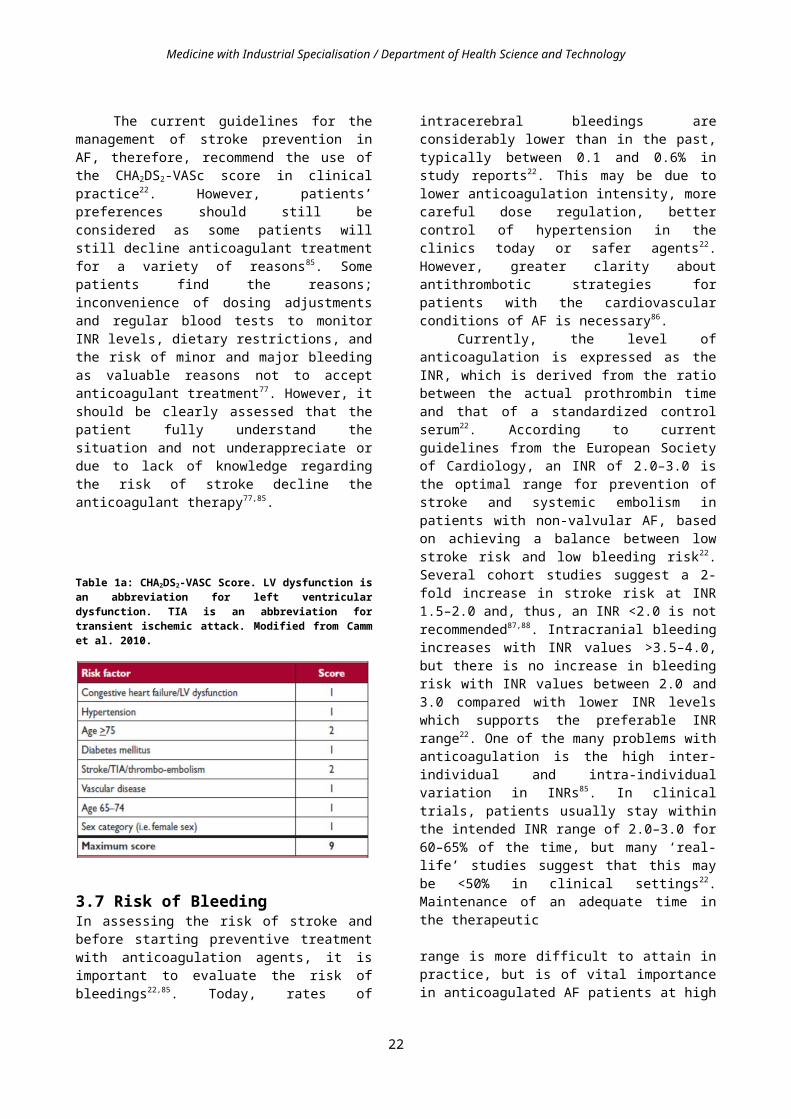
Today, the simplest risk assessment scheme is the CHADS2 score (Congestive heart failure, Hypertension, Age, Diabetes, Stroke [Doubled]), which has been used in guidelines previously. The CHADS2 risk scoring system evolved from the AF Investigators and Stroke Prevention in Atrial Fibrillation (SPAF) Investigators criteria, and it is based on a point system in which 2 points are assigned for a history of stroke or TIA and 1 point each is assigned for age >75 years, a history of hypertension, diabetes, or recent cardiac failure80. Thus, the CHADS2 stroke risk stratification scheme was presented as an initial, rapid, and easy-to-remember tool in assessing stroke risk and is still in use due to these pros22. In patients with a CHADS2
score ≥2, chronic oral anticoagulant therapy with a vitamin K antagonist is recommended in a dose-adjusted approach to achieve an international normalized ratio (INR) target of 2.5, unless contraindicated22. Such a practice appears to translate to better outcomes in AF patients in routine care81,82, and there is a clear relationship between CHADS2 score and stroke rate80.
The original validation of this scheme classified a CHADS2 score of 0 as low risk, 1–2 as moderate risk, and 2 as high risk. However, the CHADS2 score categorize most subjects as ‘moderate risk’ and does not include many stroke risk factors22. Other ‘stroke risk modifiers’ need to be considered in a comprehensive stroke risk assessment scheme22. Risk factors such as vascular disease, age 65–74, and sex category (female) has also been estimated to categorize AF patients at higher risk and further, the simultaneous presence of two or more clinically relevant ‘non-major’ risk factors would justify a stroke risk that is high enough to require anticoagulation. Therefore, the CHA2DS2-VASc (Congestive heart failure, Hypertension, Age ≥75 (doubled), Diabetes, Stroke (doubled), Vascular disease, Age 65–74, and Sex category (female)) score was developed (see Table 1a)83. This scheme is based on a point system in which 2 points are assigned for a history of stroke or TIA, and age ≥75; and 1 point each is assigned for age 65–74 years, a history of hypertension, diabetes, recent cardiac failure, vascular disease and female sex. This expanded scheme extends the CHADS2 scheme by adding extra stroke risk factors that may influence a decision whether or not to give anticoagulant therapy22. In a previous study evaluating various risk stratification schemes, the CHA2DS2-VASc score classified 94.2% as being at ‘high risk’, whereas most other risk scoring schemes categorized two-thirds as being at ‘high risk’84. In a study of 184 thromboembolic events, 98.4% occurred in patients identified as being at ‘high risk’ by the CHA2DS2-VASc schema and there was a stepwise increase in thromboembolic events with increasing CHA2DS2-VASc score84. The percent of patients categorized as ‘not high risk’ and being free from thromboembolic events was for the CHA2DS2-VASc score 99.5%.
The current guidelines for the management of stroke prevention in AF, therefore, recommend the use of the CHA2DS2-VASc score in clinical practice22. However, patients’ preferences should still be considered as some patients will still decline anticoagulant treatment for a variety of reasons85. Some patients find the reasons; inconvenience of dosing adjustments and regular blood tests to monitor INR levels, dietary restrictions, and the risk of minor and major bleeding as valuable reasons not to accept anticoagulant treatment77. However, it should be clearly assessed that the patient fully understand the situation and not underappreciate or due to lack of knowledge regarding the risk of stroke decline the anticoagulant therapy77,85.
17

Medicine with Industrial Specialisation / Department of Health Science and Technology
Table 1a: CHA2DS2-VASC Score. LV dysfunction is an abbreviation for left ventricular dysfunction. TIA is an abbreviation for transient ischemic attack. Modified from Camm et al. 2010.
3.7 Risk of BleedingIn assessing the risk of stroke and before starting preventive treatment with anticoagulation agents, it is important to evaluate the risk of bleedings22,85. Today, rates of intracerebral bleedings are considerably lower than in the past, typically between 0.1 and 0.6% in study reports22. This may be due to lower anticoagulation intensity, more careful dose regulation, better control of hypertension in the clinics today or safer agents22. However, greater clarity about antithrombotic strategies for patients with the cardiovascular conditions of AF is necessary86.
Currently, the level of anticoagulation is expressed as the INR, which is derived from the ratio between the actual prothrombin time and that of a standardized control serum22. According to current guidelines from the European Society of Cardiology, an INR of 2.0–3.0 is the optimal range for prevention of stroke and systemic embolism in patients with non-valvular AF, based on achieving a balance between low stroke risk and low bleeding risk22. Several cohort studies suggest a 2-fold increase in stroke risk at INR 1.5–2.0 and, thus, an INR <2.0 is not recommended87,88. Intracranial bleeding increases with INR values >3.5–4.0, but there is no increase in bleeding risk with INR values between 2.0 and 3.0 compared with lower INR levels which supports the preferable INR range22. One of the many problems with anticoagulation is the high inter-individual and intra-individual variation in INRs85. In clinical trials, patients usually stay within the intended INR range of 2.0–3.0 for 60–65% of the time, but many ‘real-life’ studies suggest that this may be <50% in clinical settings22. Maintenance of an adequate time in the therapeutic
range is more difficult to attain in practice, but is of vital importance in anticoagulated AF patients at high risk in order to reduce the risk of stroke or major hemorrhage84. Some patients may require more frequent INR testing and dose adjustment, and therefore, self-monitoring may be considered in order to keep the INR in the preferred range, if suitable for the patient both physically and cognitively22,84.
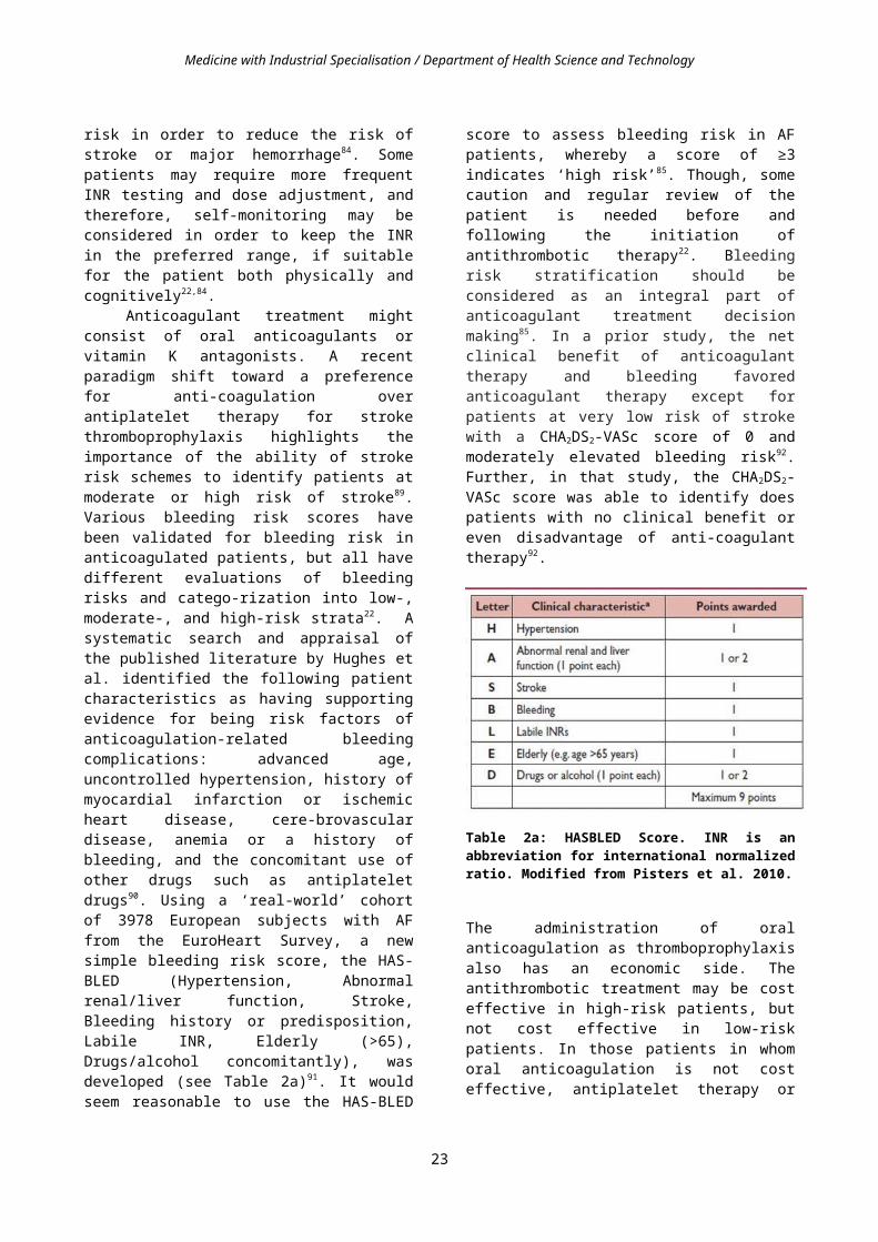
Anticoagulant treatment might consist of oral anticoagulants or vitamin K antagonists. A recent paradigm shift toward a preference for anti-coagulation over antiplatelet therapy for stroke thromboprophylaxis highlights the importance of the ability of stroke risk schemes to identify patients at moderate or high risk of stroke89. Various bleeding risk scores have been validated for bleeding risk in anticoagulated patients, but all have different evaluations of bleeding risks and catego-rization into low-, moderate-, and high-risk strata22. A systematic search and appraisal of the published literature by Hughes et al. identified the following patient characteristics as having supporting evidence for being risk factors of anticoagulation-related bleeding complications: advanced age, uncontrolled hypertension, history of myocardial infarction or ischemic heart disease, cere-brovascular disease, anemia or a history of bleeding, and the concomitant use of other drugs such as antiplatelet drugs90. Using a ‘real-world’ cohort of 3978 European subjects with AF from the EuroHeart Survey, a new simple bleeding risk score, the HAS-BLED (Hypertension, Abnormal renal/liver function, Stroke, Bleeding history or predisposition, Labile INR, Elderly (>65), Drugs/alcohol concomitantly), was developed (see Table 2a)91. It would seem reasonable to use the HAS-BLED score to assess bleeding risk in AF patients, whereby a score of ≥3 indicates ‘high risk’85. Though, some caution and regular review of the patient is needed before and following the initiation of antithrombotic therapy22. Bleeding risk stratification should be considered as an integral part of anticoagulant treatment decision making85. In a prior study, the net clinical benefit of anticoagulant therapy and bleeding favored anticoagulant therapy except for patients at very low risk of stroke with a CHA2DS2-VASc score of 0 and moderately elevated bleeding risk92. Further, in that study, the CHA2DS2-VASc score was able to identify does patients with no clinical benefit or even disadvantage of anti-coagulant therapy92.
18

Medicine with Industrial Specialisation / Department of Health Science and Technology
Table 2a: HASBLED Score. INR is an abbreviation for international normalized ratio. Modified from Pisters et al. 2010.
The administration of oral anticoagulation as thromboprophylaxis also has an economic side. The antithrombotic treatment may be cost effective in high-risk patients, but not cost effective in low-risk patients. In those patients in whom oral anticoagulation is not cost effective, antiplatelet therapy or perhaps no treatment may be more appropriate. However, this is beyond the scope of the present study.
To conclude, the assessment of whether to give antithrombotic therapy is complicated. Many aspects must be considered, but it is clear that age and type of risk factor should highly influence this decision.
4. Acknowledgement
I would like to thank my two supervisors Johannes Jan Struijk, Lector, Ph.D., and Torben Bjerregaard Larsen, MD, Ph.D., for their support and guidance throughout the project phase. Furthermore, I would like to thank Flemming Skjøth, MSc, PhD, for his contribution and valuable guidance in the statistical part of the study. General regards goes to Aalborg Thrombosis Research Unit at Aalborg University Hospital. Lastly, a special thank goes to Gregory Y.H. Lip, MD, and Lars Hvilsted Rasmussen, MD, Ph.D., for their contribution to the study.
5. References
1. Rothman KJ. Epidemiology An Introduction. Second Edi. Oxford University Press; 2012:268.
2. Juul S. Epidemiologi og Evidens. 2nd ed. Munksgaard; 2012:293.
3. Rothman KJ. Causes. American journal of epidemiology. 1976;104(6):587-92.
4. Flanders WD. On the relationship of sufficient component cause models with potential outcome (counterfactual) models. European journal of epidemiology. 2006;21(12):847-53.
5. Hill AB, Hill ID. Bradford Hills principles of medical statistics. 12th ed. London: Edward Arnold; 1991.
6. Gerhard T. Bias: considerations for research practice. American journal of health-system pharmacy : AJHP : official journal of the American Society of Health-System Pharmacists. 2008;65(22):2159-68.
7. Rothman K, S. G. Modern Epidemiology. Second Edi. Philadelphia: Lippincott-Raven; 1998.
8. Greenberg R, Daniels S, Flanders W, Eley J, Boring J. Medical Epidemiology. Fourth Edi. McGraw-Hill Medical; 2004:304.
9. Mann CJ. Observational research methods. Research design II: cohort, cross sectional, and case-control studies. Emergency medicine journal : EMJ. 2003;20(1):54-60..
10. Last J ed. A dictionary of Epidemiology. Fourth Edi. New York: Oxford Univ. Press; 2001.
11. Porta M. A dictionary of epidemiology. 5th ed. New York: Oxford University Press; 2008.
12. Kildemoes HW, Sørensen HT, Hallas J. The Danish National Prescription Registry. Scandinavian journal of public health. 2011;39(7 Suppl):38-41.
13. Krarup L-H, Boysen G, Janjua H, Prescott E, Truelsen T. Validity of stroke diagnoses in a National Register of Patients. Neuroepidemiology. 2007;28(3):150-4.
19

Medicine with Industrial Specialisation / Department of Health Science and Technology
14. Poignant M, Hjelmstedt A, Ekéus C. Indications for operative delivery between 1999-2010 and induction of labor and epidural analgesia on the risk of operative delivery--a population based Swedish register study. Sexual & reproductive healthcare : official journal of the Swedish Association of Midwives. 2012;3(4):129-34.
15. Olesen JB, Lip GYH, Lane D a, et al. Vascular Disease and Stroke Risk in Atrial Fibrillation: A Nationwide Cohort Study. The American journal of medicine. 2012;125(8):826.e13-826.e23.
16. Frost L, Andersen LV, Vestergaard P, Husted S, Mortensen LS. Trend in mortality after stroke with atrial fibrillation. The American journal of medicine. 2007;120(1):47-53.
17. Gaist D, Sørensen HT, Hallas J. The Danish prescription registries. Danish medical bulletin. 1997;44(4):445-8.
18. Mikkelsen a, Lindhardsen J, Lip G, Gislason GH, Torp-Pedersen C, Olesen JB. Female sex asa risk factor for stroke in atrial fibrillation: A nationwide cohort study. Journal of thrombosis and haemostasis : JTH. 2012;(July):1745-1751.
19. Go A, Hylek E, Phillips K. Prevalence of diagnosed atrial fibrillation in adults: national implications for rhythm management and stroke prevention: the Anticoagulation and Risk Factors in Atrial Fibrillation (ATRIA) Study. JAMA : the journal of the American Medical Association. 2001;285(18):2370-2375.
20. Kannel WB, Wolf PA, Benjamin EJ, Levy D. Prevalence, incidence, prognosis, and predisposing conditions for atrial fibrillation: population-based estimates. The American journal of cardiology. 1998;82(8A):2N-9N.
21. Potpara T, Grujić M, Marinković J, Vujisić-Tesić B, Ostojić M, Polovina M. Mortality of patients with lone and idiopathic atrial fibrillation is similar to mortality in general population of Serbia. Vojnosanitetski pregled. Military-medical and pharmaceutical review. 2010;67(2):132-5.
22. Camm a J, Kirchhof P, Lip GYH, et al. Guidelines for the management of atrial fibrillation: the Task Force for the Management of Atrial Fibrillation of the European Society of Cardiology (ESC). Europace : European pacing, arrhythmias, and cardiac electrophysiology : journal of the working
groups on cardiac pacing, arrhythmias, and cardiac cellular electrophysiology of the European Society of Cardiology. 2010;12(10):1360-420.
23. Iwasaki Y-ki, Nishida K, Kato T, Nattel S. Atrial fibrillation pathophysiology: implications for management. Circulation. 2011;124(20):2264-74.
24. Guyton AC, Hall JE. Textbook of Medical Physiology. Elsevier Science Health Science Division; 2007:1116.
25. Kirchhof P, Breithardt G. Atrial fibrillation: occurrence, mechanisms, clinical presentation - Determination of medical status and approach. Klinikarzt. 2008;37(2):66-70.
26. Xu Y, Sharma D, Li G, Liu Y. Atrial remodeling: new pathophysiological mechanism of atrial fibrillation. Medical hypotheses. 2013;80(1):53-6.
27. Watson T, Shantsila E, Lip GYH. Mechanisms of thrombogenesis in atrial fibrillation: Virchow’s triad revisited. Lancet. 2009;373(9658):155-66.
28. Estes NAM, Sacco RL, Al-Khatib SM, et al. American Heart Association atrial fibrillation research summit: a conference report from the American Heart Association. Circulation. 2011;124(3):363-72.
29. Lip GYH, Tse HF, Lane DA. Atrial fibrillation. Lancet. 2012;379(9816):648-61.
30. Schoonderwoerd BA, Smit MD, Pen L, Van Gelder IC. New risk factors for atrial fibrillation: causes of “not-so-lone atrial fibrillation”. Europace : European pacing, arrhythmias, and cardiac electrophysiology : journal of the working groups on cardiac pacing, arrhythmias, and cardiac cellular electrophysiology of the European Society of Cardiology. 2008;10(6):668-73.
31. Wyse DG, Gersh BJ. Atrial fibrillation: a perspective: thinking inside and outside the box. Circulation. 2004;109(25):3089-95.
32. Feinberg WM, Blackshear JL, Laupacis A, Kronmal R, Hart RG. Prevalence, age distribution, and gender of patients with atrial fibrillation. Analysis and implications. Archives of internal medicine. 1995;155(5):469-73.
33. Fuster V, Rydén LE, Cannom DS, et al. ACC/AHA/ESC 2006 Guidelines for the Management
20

Medicine with Industrial Specialisation / Department of Health Science and Technology
of Patients with Atrial Fibrillation: a report of the American College of Cardiology/American Heart Association Task Force on Practice Guidelines and the European Society of Cardiology Committee for Practice. Circulation. 2006;114(7):e257-354.
34. EVANS W, SWANN P. Lone auricular fibrillation. British heart journal. 1954;16(2):189-94.
35. Potpara TS, Lip GYH. Lone atrial fibrillation: what is known and what is to come. International journal of clinical practice. 2011;65(4):446-57.
36. Ellinor PT, Yoerger DM, Ruskin JN, MacRae C a. Familial aggregation in lone atrial fibrillation. Human genetics. 2005;118(2):179-84.
37. Marcus GM, Smith LM, Vittinghoff E, et al. A first-degree family history in lone atrial fibrillation patients. Heart rhythm : the official journal of the Heart Rhythm Society. 2008;5(6):826-30.
38. Chen LY, Herron KJ, Tai BC, Olson TM. Lone atrial fibrillation: influence of familial disease on gender predilection. Journal of cardiovascular electrophysiology. 2008;19(8):802-6.
39. Yang Y-Q, Zhang X-L, Wang X-H, et al. Familial aggregation of lone atrial fibrillation in the Chinese population. Internal medicine (Tokyo, Japan). 2010;49(22):2385-91.
40. Jahangir A, Lee V, Friedman P a, et al. Long-term progression and outcomes with aging in patients with lone atrial fibrillation: a 30-year follow-up study. Circulation. 2007;115(24):3050-6.
41. Kirchhof P, Auricchio A, Bax J, et al. Outcome parameters for trials in atrial fibrillation: executive summary. European heart journal. 2007;28(22):2803-17.
42. Potpara TS, Stankovic GR, Beleslin BD, et al. A 12-year follow-up study of patients with newly diagnosed lone atrial fibrillation: implications of arrhythmia progression on prognosis: the Belgrade Atrial Fibrillation study. Chest. 2012;141(2):339-47.
43. Wolf L. Familial auricular fibrillation. N Engl J Med. 1943;229:396–397.
44. Darbar D, Herron KJ, Ballew JD, et al. Familial atrial fibrillation is a genetically heterogeneous disorder. Journal of the American College of Cardiology. 2003;41(12):2185-2192.
45. Fox CS, Parise H, D’Agostino RB, et al. Parental atrial fibrillation as a risk factor for atrial fibrillation in offspring. JAMA : the journal of the American Medical Association. 2004;291(23):2851-5.
46. Christophersen IE, Ravn LS, Budtz-Joergensen E, et al. Familial aggregation of atrial fibrillation: a study in Danish twins. Circulation. Arrhythmia and electrophysiology. 2009;2(4):378-83.
47. Brugada R, Tapscott T, Czernuszewicz GZ, et al. Identification of a genetic locus for familial atrial fibrillation. The New England journal of medicine. 1997;336(13):905-11.
48. Gudbjartsson DF, Holm H, Gretarsdottir S, et al. A sequence variant in ZFHX3 on 16q22 associates with atrial fibrillation and ischemic stroke. Nature genetics. 2009;41(8):876-8.
49. Gudbjartsson DF, Arnar DO, Helgadottir A, et al. Variants conferring risk of atrial fibrillation on chromosome 4q25. Nature. 2007;448(7151):353-7.
50. Benjamin EJ, Rice KM, Arking DE, et al. Variants in ZFHX3 are associated with atrial fibrillation in individuals of European ancestry. Nature genetics. 2009;41(8):879-81.
51. Ellinor PT, Lunetta KL, Glazer NL, et al. Common variants in KCNN3 are associated with lone atrial fibrillation. Nature genetics. 2010;42(3):240-4.
52. Wakili R, Voigt N, Kääb S, Dobrev D, Nattel S. Recent advances in the molecular pathophysiology of atrial fibrillation. The Journal of clinical investigation. 2011;121(8):2955-68.
53. Parvez B, Darbar D. The “missing” link in atrial fibrillation heritability. Journal of electrocardiology. 44(6):641-4.
54. Olesen MS, Jespersen T, Nielsen JB, et al. Mutations in sodium channel -subunit SCN3B areβ associated with early-onset lone atrial fibrillation. Cardiovascular research. 2011;89(4):786-93.
55. Watanabe H, Darbar D, Kaiser DW, et al. Mutations in sodium channel 1- and 2-subunitsβ β associated with atrial fibrillation. Circulation. Arrhythmia and electrophysiology. 2009;2(3):268-75.
56. Ellinor PT, Lunetta KL, Albert CM, et al. Meta-analysis identifies six new susceptibility loci for
21
Medicine with Industrial Specialisation / Department of Health Science and Technology
atrial fibrillation. Nature genetics. 2012;44(6):670-5.
57. Kirchhof P, Bax J, Blomstrom-Lundquist C, et al. Early and comprehensive management of atrial fibrillation: executive summary of the proceedings from the 2nd AFNET-EHRA consensus conference “research perspectives in AF”. European heart journal. 2009;30(24):2969-77c.
58. U.S. National Institutes of Health. Clinicaltrials.gov. Available at: http://clinicaltrials.gov/ct2/results?term= familial+atrial+fibrillation&Search=Search. Accessed February 22, 2013.
59. Benjamin EJ, Levy D, Vaziri SM, D’Agostino RB, Belanger AJ, Wolf PA. Independent risk factors for atrial fibrillation in a population-based cohort. The Framingham Heart Study. JAMA : the journal of the American Medical Association. 1994;271(11):840-4.
60. Potpara TS, Polovina MM, Licina MM, Marinkovic JM, Prostran MS, Lip GYH. Reliable identification of “truly low” thromboembolic risk in patients initially diagnosed with “lone” atrial fibrillation: the Belgrade atrial fibrillation study. Circulation. Arrhythmia and electrophysiology. 2012;5(2):319-26.
61. Liu T, Li G, Li L, Korantzopoulos P. Association between C-reactive protein and recurrence of atrial fibrillation after successful electrical cardioversion: a meta-analysis. Journal of the American College of Cardiology. 2007;49(15):1642-8.
62. Zöller B, Ohlsson H, Sundquist J, Sundquist K. Family history as a risk factor for recurrent hospitalization for lone atrial fibrillation: a nationwide family study in Sweden. BMC cardiovascular disorders. 2012;12:121.
63. Packer DL, Bardy GH, Worley SJ, et al. Tachycardia-induced cardiomyopathy: a reversible form of left ventricular dysfunction. The American journal of cardiology. 1986;57(8):563-70.
64. Olesen JB, Gislason GH, Torp-Pedersen C, Lip GYH. Atrial fibrillation and vascular disease--a bad combination. Clinical cardiology. 2012;35 Suppl 1:15-20.
65. McManus DD, Rienstra M, Benjamin EJ. An update on the prognosis of patients with atrial fibrillation. Circulation. 2012;126(10):e143-6.
66. Benjamin EJ, Wolf P a., D’Agostino RB, Silbershatz H, Kannel WB, Levy D. Impact of Atrial Fibrillation on the Risk of Death : The Framingham Heart Study. Circulation. 1998;98(10):946-952.
67. Lip GYH. Can we predict stroke in atrial fibrillation? Clinical cardiology. 2012;35 Suppl 1:21-7.
68. Hart RG, Halperin JL, Pearce LA, et al. Lessons from the Stroke Prevention in Atrial Fibrillation trials. Annals of internal medicine. 2003;138(10):831-8.
69. Wachtell K, Lehto M, Gerdts E, et al. Angiotensin II receptor blockade reduces new-onset atrial fibrillation and subsequent stroke compared to atenolol: the Losartan Intervention For End Point Reduction in Hypertension (LIFE) study. Journal of the American College of Cardiology. 2005;45(5):712-9.
70. Ruigómez A, Johansson S, Wallander M-A, Edvardsson N, García Rodríguez LA. Risk of cardiovascular and cerebrovascular events after atrial fibrillation diagnosis. International journal of cardiology. 2009;136(2):186-92.
71. Heist EK, Ruskin JN. Atrial fibrillation and congestive heart failure: risk factors, mechanisms, and treatment. Progress in cardiovascular diseases. 48(4):256-69.
72. Mamas MA, Caldwell JC, Chacko S, Garratt CJ, Fath-Ordoubadi F, Neyses L. A meta-analysis of the prognostic significance of atrial fibrillation in chronic heart failure. European journal of heart failure. 2009;11(7):676-83.
73. Wasywich CA, Pope AJ, Somaratne J, Poppe KK, Whalley GA, Doughty RN. Atrial fibrillation and the risk of death in patients with heart failure: a literature-based meta-analysis. Internal medicine journal. 2010;40(5):347-56.
74. Miyasaka Y, Barnes ME, Bailey KR, et al. Mortality trends in patients diagnosed with first atrial fibrillation: a 21-year community-based study. Journal of the American College of Cardiology. 2007;49(9):986-92.
75. Ruigómez A, Johansson S, Wallander M-A, García Rodríguez LA. Risk of mortality in a cohort of
22
Medicine with Industrial Specialisation / Department of Health Science and Technology
patients newly diagnosed with chronic atrial fibrillation. BMC cardiovascular disorders. 2002;2:5.
76. Ruigómez A, Johansson S, Wallander M-A, García Rodríguez LA. Predictors and prognosis of paroxysmal atrial fibrillation in general practice in the UK. BMC cardiovascular disorders. 2005;5:20.
77. Hughes M, Lip GYH. Stroke and thromboembolism in atrial fibrillation: a systematic review of stroke risk factors, risk stratification schema and cost effectiveness data. Thrombosis and haemostasis. 2008;99(2):295-304.
78. Avenue G. Comparison of 12 risk stratification schemes to predict stroke in patients with nonvalvular atrial fibrillation. Stroke; a journal of cerebral circulation. 2008;39(6):1901-10.
79. Nieuwlaat R, Capucci A, Lip GYH, et al. Antithrombotic treatment in real-life atrial fibrillation patients: a report from the Euro Heart Survey on Atrial Fibrillation. European heart journal. 2006;27(24):3018-26.
80. Gage BF, Waterman AD, Shannon W, Boechler M, Rich MW, Radford MJ. Validation of clinical classification schemes for predicting stroke: results from the National Registry of Atrial Fibrillation. JAMA : the journal of the American Medical Association. 2001;285(22):2864-70.
81. Go AS, Hylek EM, Chang Y, et al. Anticoagulation therapy for stroke prevention in atrial fibrillation: how well do randomized trials translate into clinical practice? JAMA : the journal of the American Medical Association. 2003;290(20):2685-92.
82. Hylek EM, Go AS, Chang Y, et al. Effect of intensity of oral anticoagulation on stroke severity and mortality in atrial fibrillation. The New England journal of medicine. 2003;349(11):1019-26.
83. Lip GYH, Nieuwlaat R, Pisters R, Lane D a, Crijns HJGM. Refining clinical risk stratification for predicting stroke and thromboembolism in atrial fibrillation using a novel risk factor-based approach: the euro heart survey on atrial fibrillation. Chest. 2010;137(2):263-72.
84. Lip GYH, Frison L, Halperin JL, Lane D a. Identifying patients at high risk for stroke despite anticoagulation: a comparison of contemporary stroke risk stratification schemes in an anticoagulated atrial fibrillation cohort. Stroke; a journal of cerebral circulation. 2010;41(12):2731-8.
85. Lip GYH, Andreotti F, Fauchier L, et al. Bleeding risk assessment and management in atrial fibrillation patients: a position document from the European Heart Rhythm Association, endorsed by the European Society of Cardiology Working Group on Thrombosis. Europace : European pacing, arrhythmias, and cardiac electrophysiology : journal of the working groups on cardiac pacing, arrhythmias, and cardiac cellular electrophysiology of the European Society of Cardiology. 2011;13(5):723-46.
86. Lopes RD, Li L, Granger CB, et al. Atrial fibrillation and acute myocardial infarction: antithrombotic therapy and outcomes. The American journal of medicine. 2012;125(9):897-905.
87. Saour JN, Sieck JO, Mamo LA, Gallus AS. Trial of different intensities of anticoagulation in patients with prosthetic heart valves. The New England journal of medicine. 1990;322(7):428-32.
88. Palareti G, Leali N, Coccheri S, et al. Bleeding complications of oral anticoagulant treatment: an inception-cohort, prospective collaborative study (ISCOAT). Italian Study on Complications of Oral Anticoagulant Therapy. Lancet. 1996;348(9025):423-8.
89. Lip GYH, Halperin JL. Improving stroke risk stratification in atrial fibrillation. The American journal of medicine. 2010;123(6):484-8.
90. Hughes M, Lip GYH. Risk factors for anticoagulation-related bleeding complications in patients with atrial fibrillation: a systematic review. QJM : monthly journal of the Association of Physicians. 2007;100(10):599-607.
91. Pisters R, Lane DA, Nieuwlaat R, de Vos CB, Crijns HJGM, Lip GYH. A novel user-friendly score (HAS-BLED) to assess 1-year risk of major bleeding in patients with atrial fibrillation: the Euro Heart Survey. Chest. 2010;138(5):1093-100.
92. Friberg L, Rosenqvist M, Lip GYH. Net clinical benefit of warfarin in patients with atrial fibrillation: a report from the Swedish atrial
23

Medicine with Industrial Specialisation / Department of Health Science and Technology
fibrillation cohort study. Circulation. 2012;125(19):2298-307.
24