Embed Size (px)
Citation preview

Effect of Selenium on Carbimazole Induced Alterations in Testicular
Function in Albino RatsAbeer A. Shoman MD, Noha I. Hussien MD, Nessrine I. Ahmad MD* and
Hanan T. Emam MD**
Departments of Physiology, Histology* and Pharmacology**
Faculty of Medicine, Benha University, Egypt.
Abstract Carbimazole is an antithyroid drug used in treatment of hyperthyroidism. The use of Carbimazole was associated with various adverse effects in male reproductive system. Spermatogenesis is an extremely active process with high rate of cell division that leads to high rates of mitochondrial oxygen consumption by the germinal epithelium. However, oxygen tensions in this tissue are low so both spermatogenesis and Leydig cell steroidogenesis are vulnerable to oxidative stress. Testes and epididymes contain high concentrations of Selenium indicating its vital role
during spermatogenesis to improve semen quality. This study was designed to evaluate the effect of Carbimazole on testicular activity in albino rats and the ameliorative role of selenium.
The rats in this study were divided into 4 groups. Group (1) that were served as normal control, Group (2) were orally given Carbimazole Group (3) were orally given sodium selenite and Group (4) that were orally administered Carbimazole and sodium selenite for 8 weeks. Testicular reduced glutathione concentration (GSH), Testicular malondialdehyde (MDA) concentrations, Epididymal sperm count, Sperm motility, Sperm abnormalities, serum testosterone, LH, FSH levels and testicular histopathology were examined. From this study we can conclude that, treating animals
with selenium causes improvement in normal testicular function of rats. Also we
concluded that combination of selenium with Carbimazole showed improvement in testicular alterations induced by Carbimazole in rats.
1

Introduction
Carbimazole is an antithyroid agent that decreases the uptake and
concentration of inorganic iodine by thyroid; it also reduces the formation
of diiodotyrosine and thyroxin (1). Serum Carbimazole is a common oral
treatment for hyperthyroidism. On the other hand, the use of Carbimazole
was associated with various adverse effects (2). Carbimazole produced
mild necrosis of renal tubules in rats. Carbimazole was capable of
inducing acute pancreatitis, pulmonary hemorrhage and necrotizing
glomerulonephritis and cholestatic hepatitis in 33-year old female (3).
Reactive oxygen species (ROS) are molecules that have at least one
unpaired electron, rendering them highly unstable and highly reactive in
the presence of lipids, amino acids and nucleic acids. At physiologic
levels, ROS are essential for normal reproductive function, acting as
metabolic intermediates in the metabolism of prostanoid, in the regulation
of vascular tone, in gene regulation, and in facilitated sperm capacitation
and acrosome reaction(4). However, at higher concentrations, they exert
negative effects. The main source of ROS production in seminal plasma
is leukocytes and immature spermatozoa. Spermatids and mature
spermatozoa are deemed highly sensitive to ROS because their
membranes are particularly rich in polyunsaturated lipids. By altering
membrane integrity, ROS may impair sperm motility and morphology
and can lead to sperm cell death (5).
Spermatogenesis is an extremely active replicative process capable
of generating approximately 1,000 sperm a second. The high rates of cell
division inherent in this process lead to high rates of mitochondrial
oxygen consumption by the germinal epithelium. However, the poor
vascularization of the testes means that oxygen tensions in this tissue are
2

low and that competition for this vital element within the testes is
extremely intense (6).
The testes contain an elaborate array of antioxidant enzymes and
free radical scavengers to ensure that the twin spermatogenic and
steroidogenic functions of this organ are not impacted by oxidative stress.
These antioxidant defense systems are of major importance because
peroxidative damage is currently regarded as the single most important
cause of impaired testicular function (7).
Selenium (Se) is an essential element for normal testicular
development, spermatogenesis, and spermatozoa motility and function.
Se may protect against oxidative DNA damage in human sperm cells.
However, the exact mechanism by which Se eliminates oxidative stress to
improve male fertility and semen quality in humans is still controversial (8). There are at least 25 selenoproteins in the human body, and they help
maintain sperm structure integrity (9).
The best-characterized spermatozoal effects of Se deficiency are:
important loss of motility, breakage at the midpiece level and increased
incidence of sperm-shape abnormalities, mostly of the sperm head. This
is evidenced by studies that reported a significant correlation between Se
levels in seminal plasma and the percentage of morphologically normal
sperm in a sample (10).
Selenium is an essential element important in many biochemical
and physiological processes including the biosynthesis of coenzyme Q (a
component of mitochondrial electron transport systems), regulation of ion
fluxes across membranes, maintenance of the integrity of keratins,
stimulation of antibody synthesis, and activation of glutathione
peroxidase (11). Selenium ameliorated the testicular damage and oxidative
stress induced by Carbimazole in albino rats (12).
3
The present work aims to investigate the effect of selenium on
histology of testis, serum testosterone level, LH, FSH, Testicular GSH,
Testicular MDA and semen analysis alterations induced by Carbimazole
in male albino rats.
MATERIALS AND METHODSAnimals
Thirty two male adult albino rats of locally breaded strain
weighing between 150+ 5 g at the beginning of the study were used. They
have acclimatized for one week in groups (8/cage) in fully ventilated
room at ordinary room temperature. Rats were allowed to water and
balanced diet.
At the beginning of the experiment they were divided into 4 groups
each contained 8 rats:
Group I: Control normal rats. They received no drugs
Group II: Animals of this group were orally given Carbimazole (1.35
mg/Kg b.w) dissolved in water, daily for 8 weeks (13).
Group III: animals of this group were orally given sodium selenite (10
μg/Kg b.w) dissolved in water, daily for 8 weeks (14).
Group 4: animals of this group were orally administered Carbimazole
(1.35mg/Kg b.w) and sodium selenite (10μg/Kg b.w) for 8 weeks. The
treated animals were sacrificed by cervical decapitation after 8 weeks of
treatment.
Drugs:
Selenium (sodium selenite): gray powder 5gm in bottle from
(Sigma chemicals Co., U.S.A.).
Carbimazole: 10 mg Tablet (Ambica Intl Trading).
Hematoxylin and eosin: (E. Merk, Darmastadt.,) [U.S.A.].
Formalin (neutral 10% formalin): El-Gomhoria Pharmaceutical
Chemi.Ocal Co, ARE. Diagnostics Limited lot. No. 18933.
4

The procedures used:
Each animal of all groups was killed after the end of the
experiments by decapitation and the following were done.
Blood was collected in a test tube and left to clot then centrifuged for
serum separation and serum was collected and stored at – 20oC for
hormonal assays.
Both testes of each animal were separated from the surrounding
tissues.
The testis tissue washed by ice water, apart was weighed and
homogenized in 9 volumes saline 0.9% in homogenizer apparatus. The
homogenate was collected and kept at −70 ◦C. GSH activity and MDA
were measured (15).
Semen collection
One epididymis was separated rapidly, the tail of the epididymis
was scraped longitudinally by scalpel and the semen was collected by
stripping of the epididymis then 0.1 ml of saline was added and mixed
with the semen for liquefaction (16).
Sperm counting
The semen obtained was diluted with saline at a ratio of 1: 20 and
by the hemocytometer slide the sperm count could be calculated using
a light microscope (17).
Sperm motility
The right epididymis along with part of the vas deferens was
clamped with a hemostat at approximately the corpus cauda junction.
The cauda was then gently dissected with a scalpel blade to allow
sperm to emerge from the engorged cauda epididymis, which was
then dipped into a plastic Petri dish (35 mm) containing pre-warmed
incubation medium (saline). Sperm released into the medium were
incubated for approximately 3 min with intermittent gentle swirling to
5

disperse them throughout the suspension. Following the 3-min
incubation, an aliquot of the sperm suspension was taken using
capillary tubes and loaded into the analyzer. A drop of the collected
semen was taken using 100µl capillary tube and loaded into a slide
then examined by high power for the number of forward motile sperm
per 100 sperms (18).
Stained slides :
One drop of the collected diluted semen was taken into the middle of
a clean slide, one drop of the eosin stain was added near to the semen
drop then gently mixed and by the edge of another slide the stained
semen was spread into the slide and examined by light microscope at
lens 40 for the sperm abnormalities and acrosomal deformity (19).
Hormonal assays :
Serum LH, FSH and testosterone levels were measured.
After functional studies were completed, one testis from each rat
was removed and was put into a buffered 4% formalin fixation solution
and processed with paraffin wax for histopathological examination.
Sections 5um were stained with Mayer’shematoxylin and eosin (20).
All groups were subjected to the following investigation:
Serum testosterone ,LH, FSH levels
Testicular GSH, Testicular MDA
Epididymal sperm count
Sperm motility
Sperm abnormalities and acrosomal deformity
Testicular histopathological examination
Statistical Analysis:
All data were expressed as mean S.D; data were evaluated by
the one way analysis of variance. Difference between groups were
6

compared by Student's t-test with P 0.05 selected as the level of
statistical significance.
RESULTS
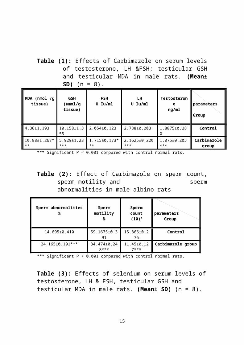
Administration of Carbimazole (1.35 mg/Kg b.w) dissolved in water,
daily for 8 weeks resulted in significant (P < 0.001) decrease of
testosterone, LH &FSH from means of (1.8875±0.280), (2.788±0.203),
(2.054±0.123) in control group to means of (1.075±0.205),
(2.1625±0.220)&( 1.715±0.173). Testicular GSH was significantly
reduced (P < 0.001) from mean of (10.158±1.355) in control group to
mean of (5.929±1.23). Testicular MDA was significantly increased (P <
0.001) from mean of (4.36±1.193) in control group to mean of
(10.88±1.267) (table 1).
The results showed also that administration of Carbimazole resulted
in significant (P < 0.001) decrease of sperm count, sperm motility from
means of (15.866±0.276) & (59.1675±0.391) in control group to means
of (11.45±0.127) & (34.474±0.248) .sperm abnormalities was
significantly increased (P < 0.001) from mean of (14.695±0.410) in
control to mean of (24.165±0.191) in group received Carbimazole (table
2).
Histopathological examination of testicular tissues of rats received
Carbimazole only showed degenerative changes of the majority of the
seminiferous tubules. These changes were characterized by shrunken,
disorganized seminiferous tubules with irregular, buckled basement
membrane and incomplete spermatogenesis. Moreover, the seminiferous
tubules were almost devoid of spematids and spermatozoa. Vacuolar
degeneration of spermatogonia and Sertoli cells was evident. Degenerated
germinal epithelial cells were sloughed in the lumina of most
seminiferous tubules. Regarding to the interstitium, there were congestion
7

of the interstitial blood vessels and edema that was represented by faint
eosinophilic material (Fig. 3) in comparison to the normal testicular
tissues of the control group (Fig. 2).
Administration of sodium selenite (10 μg/Kg b.w) dissolved in
water, daily for 8 weeks resulted in significant (P < 0.05) increase of
testosterone from mean of (1.8875±0.280) in control group to means of
(2.2625±0.267) & significant (P < 0.001) increase of FSH from mean of
(2.054±0.123) in control group to mean of (2.51±0.207).Testicular GSH
was significantly increased (P < 0.002) from mean of (10.158±1.355) in
control group to mean of (12.931±1.597). Testicular MDA was
significantly reduced (P < 0.05) from mean of (4.36±1.193) in control
group to mean of (3.231±0.86) (table 3).
The results showed also that administration of sodium selenite
resulted in significant (P < 0.001) increase of sperm count, sperm motility
from means of (15.866±0.276) & (59.1675±0.391) in control group to
means of (17.939±0.147) & (60.22±0.224) .Sperm abnormalities was
significantly decreased (P < 0.001) from mean of (14.695±0.410) in
control to mean of (11.529±0.333) in group received sodium selenite
(table 4).
Histopathological examination of testicular tissues of rats received
sodium selenite alone showed normal testicular structure (Fig. 4).
Administration of Carbimazole (1.35mg/Kg b.w) and sodium
selenite (10μg/Kg b.w) for 8 weeks resulted in significant (P < 0.001)
increase of testosterone, LH &FSH from means of (1.075±0.205),
(2.1625±0.220) & (1.715±0.173) In group received Carbimazole alone to
means of (1.9625±0.262), (3.016±0.228) & (2.5262±0.188) in groups
received Carbimazole and sodium selenite. Testicular GSH was
significantly increased (P < 0.001) from mean of (5.929±1.23) in group
8

received Carbimazole alone to mean of (8.218±0.853) in groups received
Carbimazole and sodium selenite. Testicular MDA was significantly
decreased (P < 0.001) from mean of (10.88±1.267) in group received
Carbimazole alone to mean of (6.253±1.814) in group received
Carbimazole and sodium selenite (table 5) & (Fig. 1-a).
The results showed also that administration of Carbimazole and
sodium selenite resulted in significant (P < 0.001) increase of sperm
count, sperm motility from means of (11.45±0.127) & (34.474±0.248) In
group received Carbimazole alone to means of
(14.541±0.327)&( 51.614±0.430) in group received Carbimazole and
sodium selenite. Sperm abnormalities was significantly decreased (P <
0.001) from mean of (24.165±0.191) in group received Carbimazole
alone to mean of (11.919±0.293) in group received Carbimazole and
sodium selenite (table 6) & (Fig. 1-b).
Histopathological examination of testicular tissues of rats received
Carbimazole and sodium selenite showed few seminiferous tubules
contained sloughed germinal epithelium (arrows), marked improvement
of spermatogenesis (Fig. 5).
Table (1): Effects of Carbimazole on serum levels of testosterone, LH &FSH; testicular GSH and testicular MDA in male rats. (Mean± SD) (n = 8).
9

parameters
Group
Testosteroneng/ml
LHU Iu/ml
FSHU Iu/ml
GSH (umol/g tissue)
MDA (nmol /g tissue)
Control1.8875±0.2802.788±0.2032.054±0.12310.158±1.3554.36±1.193Carbimazole
group 1.075±0.205***2.1625±0.220***1.715±0.173***5.929±1.23***10.88±1.267***
*** Significant P < 0.001 compared with control normal rats.
Table (2): Effect of Carbimazole on sperm count, sperm motility and sperm abnormalities in male albino rats
parameters Group
Sperm count(10)6
Sperm motility%
Sperm abnormalities%
Control15.866±0.27659.1675±0.39114.695±0.410Carbimazole group 11.45±0.127***34.474±0.248**
*24.165±0.191***
*** Significant P < 0.001 compared with control normal rats.
Table (3): Effects of selenium on serum levels of testosterone, LH & FSH, testicular GSH and testicular MDA in male rats. (Mean± SD) (n = 8).
parametersGroup
Testosteroneng/ml
LHU Iu/ml
FSHU Iu/ml
GSH (umol/g tissue)
MDA (nmol /g tissue)
Control1.8875±0.2802.788±0.2032.054±0.12310.158±1.3554.36±1.193Selenium group 2.2625±0.267**2.9875±0.1732.51±0.207***12.931±1.597***3.231±0.86**
** Significant P < 0.01 compared with control normal rats. ***Significant P < 0.001 compared with control normal rats.
Table (4): Effect of selenium on sperm count, sperm motility and sperm abnormalities in male albino rats
parameters Group
Sperm count(10)6
Sperm motility%
Sperm abnormalities%
Control15.866±0.27659.1675±0.39114.695±0.410Selenium group17.939±0.147***60.22±0.224***11.529±0.333***
*** Significant P < 0.001 compared with control normal rats.
10

Table (5): Effects of combination of selenium and Carbimazole on serum levels of testosterone, LH &FSH, testicular GSH and testicular MDA in male rats. (Mean± SD) (n = 8).
parametersGroup
Testosteroneng/ml
LHU Iu/ml
FSHU Iu/ml
GSH (umol/g tissue)
MDA (nmol /g tissue)
Carbimazole group
1.075±0.2052.1625±0.2201.715±0.1735.929±1.2310.88±1.267
Group received carbimazole and
Selenium
1.9625±0.262***3.016±0.228***
2.5262±0.188***8.218±0.853***6.253±1.814***
*** Significant P < 0.001 compared with Carbimazole treated rats.
Table (6): Effect of combination of Carbimazole and selenium on sperm count, sperm motility and sperm abnormalities in male albino rats
parameters Group
Sperm count(10)6
Sperm motility%
Sperm abnormalities%
Carbimazole group 11.45±0.12734.474±0.24824.165±0.191
Group received carbimazole and
Selenium
14.541±0.327***51.614±0.430***11.919±0.293***
*** Significant P < 0.001 compared with Carbimazole treated rats.
Fig (1-a) Fig (1-b)
.uoc mreps tom mreps .ba mreps0
01
02
03
04
05
06
#
#
#
.zamibrac neles&.brac
tset HL HSF HSG ADM0
2
4
6
8
01
21
## #
##
zamibraC .niles&.bac
# Significant (P < 0.001) compared with Carbimazole treated rats.
Fig. (1-a) & Fig. (1-b) Effects of combination of selenium and Carbimazole on serum levels of testosterone, LH &FSH, testicular GSH, testicular MDA, sperm count, sperm motility and sperm abnormalities in male albino rats
11

Fig. (2): Cut section of testicular tissues of control normal rats showing no remarkable
pathologic changes (H&E100)
Fig. (3): Cut section of testicular tissues of rats received Carbimazole (H&E100)
12

Fig. (4): Cut section of testicular tissues of rats received selenium (H&E100)
Fig. (5): Cut section of testicular tissue of rats received both carbimazole and selenium
(H&E100)
Fig. (6): detached head Fig. (7): double head and bifid tail
13

Fig. (8): damaged acrosome . Fig. (9): coiled tail
Discussion Carbimazole is a common oral treatment for hyperthyroidism but
it was associated with various adverse effects on the male reproductive
system. Hence, this study was conducted to evaluate the effect of
Carbimazole on testicular function as sperm characteristics, serum
testosterone, LH & FSH levels as well as the effect of Carbimazole on
Testicular reduced glutathione (GSH) activity ,lipid peroxidation (LPO)
content in the form of testicular MDA level and histopathology of the
testes and to assess the ameliorative role of selenium.
Our study revealed that Carbimazole caused significant decrease in
testicular reduced glutathione (GSH) activity, serum testosterone; LH &
FSH levels with significant increase in testicular MDA level in the testis.
As well as it caused significant increase in sperm abnormalities with
significant decrease in sperm motility and sperm count.
Histopathological examination of testicular tissue of rats received
Carbimazole showed degenerative changes of the majority of the
seminiferous tubules. These changes were characterized by shrunken,
disorganized seminiferous tubules with irregular, buckled basement
14
membrane and incomplete spermatogenesis. Moreover, the seminiferous
tubules were almost devoid of spematids and spermatozoa. Degenerated
germinal epithelial cells were sloughed in the lumina of most
seminiferous tubules. Regarding to the interstitium, there were congestion
of the interstitial blood vessels and edema that was represented by faint
eosinophilic material.
These results were in agreement with (13) as they revealed that treating
rats with Carbimazole daily for 8 weeks caused distinct histological
alterations in prostate gland compared with control group.
Also (21) indicated that superoxide dismutase, catalase and glutathione
peroxidase was reduced in erythrocytes of rats treated with Carbimazole
compared with control animals. Carbimazole administration caused a
significant decrease testicular weight and DNA content in rat pups (22).
High lipid peroxidation with a concomitant decrease in the
enzymatic antioxidant status, superoxide dismutase and catalase were
recorded in testis of rats treated with Carbimazole (23). Thus, it is
suggested that Carbimazole induced oxidative stress which resulted in the
alterations observed in the testes.
Testosterone is required for the attachment of different generations of
germ cells in seminiferous tubules. Therefore, low level of intratesticular
testosterone may lead to detachment of germ cells from seminiferous
epithelium and may initiate germ cell apoptosis and subsequent male
infertility (24). This finding was parallel to the reduction in epididymal
sperm count. Sloughing of germ cells was observed in the lumen of some
epididymal ducts indicating testicular dysfunction (25).
Our study revealed that the effect of selenium on normal control rats
was significant improvement in testicular function. These results were in
agreement with (26) as they concluded that the administration of
15

antioxidants such as selenium or ascorbate to normal animals, not
suffering from induced oxidative stress, also appears to improve testicular
function, suggesting that oxidative stress is a consistent feature of
testicular physiology. In light of such results, antioxidants have
frequently been administered to infertile men in the hope of improving
the quality of the semen profile.
Increased generation of ROS in semen affects sperm function,
especially fusion events associated with fertilization, and leads to
infertility. ROS are known to be generated from spermatozoa and
leucocytes and the resultant peroxidative damage causes impaired sperm
function. Elevated ROS levels correlate negatively with sperm
concentration and sperm motility Spermatozoa are particularly
susceptible to ROS-induced damage because their plasma membranes
contain large quantities of polyunsaturated fatty acids and their cytoplasm
contains low concentrations of the scavenging enzymes (27).
Concerning the effect of selenium on rats suffering from induced
oxidative stress, the present study indicated that combination of selenium
with Carbimazole improved the oxidative status as selenium caused
significant increase in testicular reduced glutathione (GSH) activity and
significant decrease in testicular MDA content. Also selenium caused
significant increase in testosterone, LH, FSH levels and significant
increase in sperm count, sperm motility and significant decrease in sperm
abnormalities.
These results were in agreement with (28) as they revealed that
selenium ameliorated the reduction in the reproductive organs weights,
sperm characteristics, Deltamethrin induced oxidative damage of testes
16

and the histopathological alterations of testes, epididymes and accessory
sex glands.
Also (29) found that after co-administration of vitamin E and
Selenium, serum testosterone level significantly increased parallel to the
reduction in LPO concentration. This could be attributed to a relationship
between steroidogenesis and ROS.
Our results are also in consistence with (30) who concluded that 0.1
mg/kg selenium (Na2 SeO3) inhibited oxidative stress, apoptosis and cell
cycle changes induced by excess fluoride in kidney of rats. Selenium
protected rat testes against cadmium-induced oxidative damage (31).
Sodium selenite supplementation significantly protected against
exercise-induced testicular gametogenic and spermatogonia disorders,
prevented testicular oxidative stress and increased antioxidant status (32) .
The effectiveness of combined treatment with selenium and vitamin E in
treatment of oligoasthenoteratozoospermia has been studied since
Vitamin E is well known to work in synergy with selenium as an
antiperoxidant. A prospective, uncontrolled study reported that this drug
combination led to statistically significant increases in motility and mean
seminal plasma glutathione peroxidase activity. Although no
improvements in sperm concentration were documented, and no
pregnancies were achieved, the better sperm motion characteristics may
be explained by the amplified antioxidant enzyme activity (33). These
results were further confirmed by a more recent randomized controlled
trial in which vitamin E and selenium improved sperm motility and lipid
peroxidation markers (34).
Selenium reduced the cadmium induced histopathological changes in
testes of rat, oxidative stress, endocrine disorder and apoptosis (35). Co-
17

treatment of vitamin E and selenium revealed a significant reduction in
LPO and as a consequence improvement in GSH level (36). Additionally,
Se has a positive effect on ejaculate volume and semen quality in goats (37). It was reported that the mechanism of chemoprotection of selenium
may be related to its antioxidant properties as well as its ability to
interfere with DNA repair pathways (38).
On the contrary, (39) reported that selenium had no protective effects
against atrazine induced biochemical alterations in testis and epididymis
except testicular lactate dehydrogenase.
References
1- Chen, X. ; Ji, ZL. and Chen, YZ. (2002): Therapeutic Target
Database. Nucleic Acids Res. 1; 30(1):412-5.
2- Frenais, R.; Burgaud, S. and Horspool, L. J. ( 2008):Pharmaco-
kinetics of controlled-release carbimazole tablets support once
daily dosing in cats. J. Vet. Pharmacol. Ther., 31(3), 213-219.
3- Marazuela, M.; De Paco, G. S.; Jlmenez, I.; Carraro, R.; Fernandez-
Herrera, J.; Pajares, J. M. and Gomez-Pan, A. (2002): Acute
pancreatitis, hepatic cholestasis and erythema nodosum induced
by carbimazole treatment for Graves' disease. En-docrinol. J.,
49(3), 315-318.
4- Buyukgebiz, A. (2007): Newborn screening, hypothyroidism in
infants, children and adolescents. In: Krassas GE, Rivkees SA,
Kiess W, eds. Diseases of the thyroid in childhood and
18

adolescence (pediatric and adolescent medicine). Vol. 11. Basel,
Switzerland: Karger; 128–141.
5- Krassas, GE.; Papadopoulou, F.; Tziomalos, K.; Zeginiadou, T.and
Pontikides, N. (2008): Hypothyroidism has an adverse effect on
human spermatogenesis: a prospective, controlled study. Thyroid
18:1255–1259.
6 –Trummer, H.; Ramschak-Schwarzer, S.;Haas, J.; Habermann, H.;
Pummer, K. and Leb, G. (2001): Thyroid hormones and thyroid
antibodies in infertile males. Fertil Steril 76:254–257.
7 -Poppe K, Glinoer D, Tournaye H, Maniewski U, Haentjens P,
Velkeniers B (2006): Is systematic screening for thyroid disorders
indicated in subfertile men? Eur J Endocrinol 154:363–366.
8- Senthil kumar, J.; Banudevi, S.; Sharmila, M. et al. (2004): Effects
of Vitamin C and E on PCB (Aroclor 1254) induced oxidative
stress, androgen binding protein and lactate in rat Sertoli cells.
Reprod Toxicol; 19:201–218.
9-Sonmez, M.; Turk, G. and Yuce, A. (2005): The effect of ascorbic acid
supplementation on sperm quality, lipid peroxidation and
testosterone levels of male Westar rats. Theriogenology; 63:2063–
2072.
10- Rayman, M. P. (2005): Selenium in cancer prevention: a review of
the evidence and mechanism of action. Proc. Nutr. Soc., 64, 527–
542.
19

11- Sakr, S. A.; Mahran, H. A.; Nofal, A. E. (2011): Effect of selenium
on carbimazole-induced testicular damage and oxidative stress in
albino rats. J. Trace Elem. Med. Biol., 25, 59-66.
12-Saber A. Sakr; Hoda A. Mahran and Amany E. Nofal (2012): Effect
of Selenium on Carbimazole-Induced Histopathological and
Histochemical Alterations in Prostate of Albino Rats. American
Journal of Medicine and Medical Sciences 2(1): 5-11.
13-Swathy, S. S.; Panicker, S. and Indira, M. (2006): Effect of
exogenous selenium on the testicular toxicity induced by ethanol
in rats. Ind. J. Physiol. Pharmacol., 50(3), 215-224.
14- Sedlack, J. and Lindsay, RH.(1968): Estimation of total protein
bound and non protein sulfhydryl groups in tissues with Ellmans
reagent. Anal Biochem; 86:271–8.
15- Placer, ZA.; Crushman, L. and Johnson, BC. (1966): Estimation of
product of lipid peroxidation (malondialdehyde) in biochemical
systems. Anal Biochem; 16:359–64.
16- El-Keshawy, A.H.A.; Nasr, M.T.; Abdel-Raheim, A.; Eidaroos, A.
and Hazzaa, A. (1984) : The effects of Gn-RH on the immature
male rat special reference to the activties of testis & epididymis.
Veterinary medical Journal Vol. 32 No (3), 51- 71.
17- Gopalkishnan, K.; Gill Sharma, M.K.; Balasinor, N. Padwal, V.;
D’souza, S. and Parte, P. (1998): Tamoxifen induced light and
electron microscopic changes in the rat testicular morphology and
20

serum hormonal prfile of reproductive hormones. Contraception
57: 261 – 269.
18-SöNnmez, M.;Türk and G.;Yüce, A. (2005):The effect of ascorbic
acid supplementation on sperm quality, lipid peroxidation and
testosterone levels of Wistar rats.Theriogenology;63:2063–72.
19- Drury, R.A.B. and Wallington, E.A. (1967): Carlton's Histological
technique, 4th ed. Oxford University Press, Oxford, P. 129.
20- Vijayakumar, R. S. and Nalini, N. (2006): Efficacy of piperine, an
alkaloidal constituent from piper nigrum on erythrocyte
antioxidant status in high fat diet and antithyroid drug induced
hyperlipidemic rats. Cell Biochem. Funct., 24(6), 491-498.
21-Anguiano, B.; Aranda, N.; Delgado, G. and Aceves, C. (2008):
Epididymis expresses the highest 5'-deiodinase activity in the
male reproductive system: kinetic characterization, distribution
and hormonal regulation. Endocrinology 149(8), 4209-4217.
22- Vijayakumar, R. S.; and Nalini, N. (2006): Efficacy of piperine, an
alkaloidal constituent from pipernigrum on erythrocyte
antioxidant status in high fat diet and antithyroid drug induced
hyperlipidemic rats. Cell Biochem. Funct., 24(6), 491-498.
23- Samah, S.O, and Zeynab, Kh. b (2011): Protective effect of vitamin
E and selenium combination on deltamethrin induced
reproductive toxicity in male rats. Experimental and Toxicologic
Pathology.1-7.
21

24- Kunwar, A.; Mishra, B.; Barik, A.; Kumbhare, L. B.; Pandey, R.;
Jain, V. K.; et al. (2007): 3, 3-Diselenodipropionic acid, an
efficient peroxyl radical scavenger and a GPx mimic, protects
erythrocytes (RBCs) from AAPH-induced hemolysis. Chemical
Research in Toxicology, 20, 1482–1487.
25- Kaur, P. and Bansal, MP. (2004): Influence of selenium induced
oxidative stress on spermatogenesis and lactate dehydrogenase-X
in mice testis. Asian J Androl.; 6:227–232.
26-Ashok Agarwal ; Sajal Gupta and Suresh Sikka (2006): The role of
free radicals and antioxidants in reproduction. Current Opinion in
Obstetrics and Gynecology, 18:325–332
27- Avlan, D.; Erdougan, K.,Cimen, B. ; et al.(2005): The protective
effect of selenium on ipsilateral and contralateral testes in
testicular reperfusion injury. Pediatr Surg Int; 21:274–278.
28- Unsal A, Eroglu M, Avci A, et al. (2006): Protective role of natural
antioxidant supplementation on testicular tissue after testicular
torsion and detorsion. Scand J Urol Nephrol.; 40:17–22.
29-Kara, H., Cevik, A., Konar, V., Dayangac, A., Yilmaz, M., (2007):
Protective effects of antioxidants against cadmium induced
oxidative damage in rat testes. Biol. Trace Elem. Res., 120(1-3),
205-211.
30- Jana, K.; Samanta, P. K.; Manna, I.; Ghosh, P.; Singh, N.; Khetan,
R. P. and Ray, B. R. (2008): Protective effect of sodium selenite
22

and zinc sulfate on intensive swimming-induced testicular
gamatogenic and steroidogenic disorders in mature male rats.
Appl. Physiol. Nutr. Metab., 33(5), 903-914.
31- Kashanian, S.; Gholivand, M. B.; Ahmadi, F. and Ravan, H.
(2008): Interaction of diazinon with DNA and the protective role
of selenium in DNA damage. DNA Cell Biol., 27(6), 325-532.
32- Grotto, D.; Barcelos, G. R.; Valentini, J.; Antunes, L. M.; Angeli,
J.P.; Garcia, S. C. and Barbosa, F. Jr. (2009): Low levels of
methylmercury induce DNA damage in rats: protective effects of
selenium. Arch. Toxicol., 83(3), 249-254.
33- Keskes-Ammar L, Feki-Chakroun N, Rebai T, Sahnoun Z,
Ghozzi H and Hammami S, (2003): Sperm oxidative stress and
the effect of an oral vitamin E and selenium supplement on semen
quality in infertile men. Arch Androl ;49:83-94.
34- ashock AG. And Lucky H.(2011): Oxidative stress and antioxidants for idiopathic oligoasthenoteratospermia: Is it justified? Indian jurnal of urology; 27(1), 74-85.
35- Li, J.L.; Gao, R.; Li, S.; Wang, J.T.; Tang, Z.X.and Xu, S.W.
(2010): Testicular toxicity induced by dietary cadmium in cocks
and ameliorative effect by selenium. Biometals, 23(4), 695-705.
36-Shi, L.; Zhang, C.; Yue, W.; Shi L., Zhu, X. & Lei, F.(2010): Short-
term effect of dietary selenium- enriched yeast on semen
parameters, antioxidant status and antioxidant status and Se
23

concentration in goat seminal plasma. Anim Feed Sci Technol;
157:104–8.
37- El-Maraghy, S.A., and Nassar, N.N. (2011): Modulator effects of
lipoic acid and selenium against cadmium-induced biochemical
alterations in testicular steroidogenesis. J. Bio-chem. Mol.
Toxicol., 25(1), 15-25.
38-Santos, R. A. and Takahashi, C. S. (2008): Anticlastogenic and
antigenotoxic effects of selenomethionine on doxorubicin induced
damage in vitro in human lymphocytes. Food Chem. Toxicol.,
46(2), 671-677.
39- Adesiyan, A. C.; Oyejola, T. O.; Abarikwu, S. O.; Oyeyemi, M.
O.and Farombi, E. O. (2011): Selenium provides protection to
the liver but not the reproductive organs in an atrazine-model of
experimental toxicity. Exp. Toxicol. Pathol., 63(3), 201-207.
24