Embed Size (px)
Citation preview
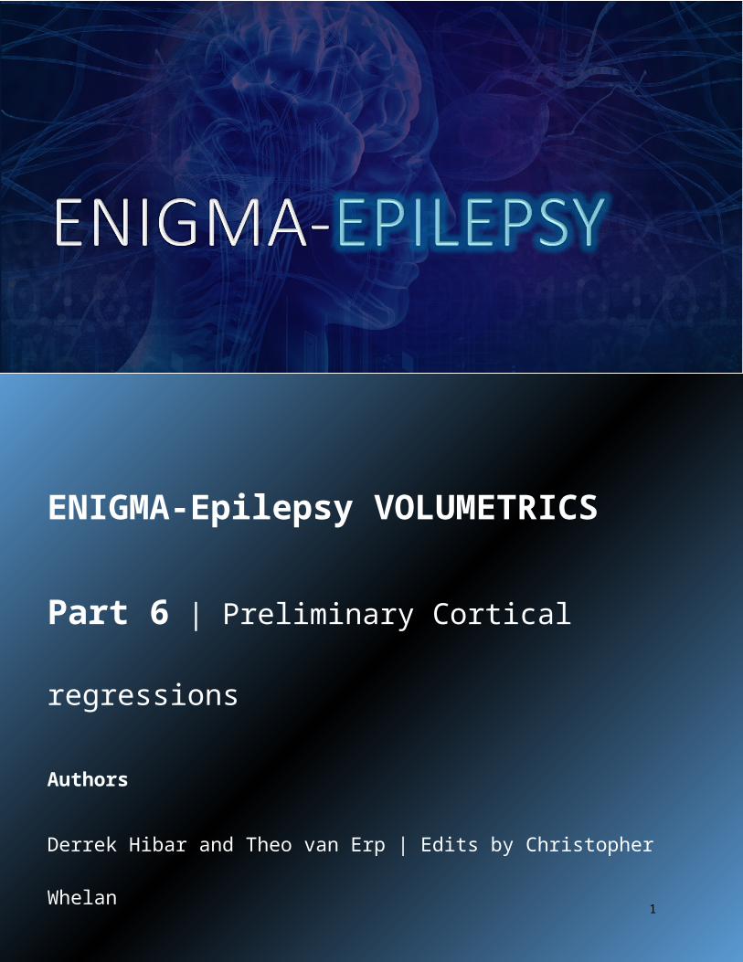
ENIGMA-Epilepsy VOLUMETRICS
Part 6 | Preliminary Cortical regressions
Authors
Derrek Hibar and Theo van Erp | Edits by Christopher Whelan
Written by Derrek Hibar ([email protected]) and Theo van Erp ([email protected])for the ENIGMA Disease Working Groups | Edited for ENIGMA-Epilepsy by Christopher Whelan
1
Written by Derrek Hibar ([email protected]) and Theo van Erp ([email protected])for the ENIGMA Disease Working Groups | Edited for ENIGMA-Epilepsy by Christopher Whelan
Part 6.0: Overview of Analyses + inclusion criteriaThis document provides instructions on how to run four preliminary analyses of cortical structures for the ENIGMA-Epilepsy Working Group. The four case:control analyses will include:
1. All non-lesional epilepsy patients versus healthy controls. Inclusion criteria:a. Syndromic diagnosis of epilepsyb. Aged 18-55c. No neurosurgeryd. No known abnormalities on clinical MRI (including focal cortical dysplasias, tumors, sclerosis or
other lesions) e. All adult epilepsy subtypes (TLE, OLE, FLE, GGE, nonspecific focal epilepsies) are acceptable, once
there are no known abnormalities on clinical MRI. We have chosen to omit cases with known abnormalities such as TLE with MTS, as they may create inflated effect sizes for specific brain structures such as the hippocampus.
2. Generalized epilepsy patients versus healthy controls. Inclusion criteria:a. Syndromic diagnosis of epilepsyb. Aged 18-55c. No neurosurgeryd. No known focal cortical dysplasias or tumors
3. Temporal lobe epilepsy patients with left mesial temporal sclerosis versus healthy controls. Inclusion criteria:
a. Syndromic diagnosis of temporal lobe epilepsy (TLE)b. Sclerosis of the left mesial temporal lobe / left hippocampus c. Aged 18-55d. No neurosurgerye. No known focal cortical dysplasias or tumors
4. Temporal lobe epilepsy patients with right mesial temporal sclerosis versus healthy controls. Inclusion criteria:
a. Syndromic diagnosis of temporal lobe epilepsy (TLE)b. Sclerosis of the right mesial temporal lobe / right hippocampus c. Aged 18-55d. No neurosurgerye. No known focal cortical dysplasias or tumors
If you have less than N=10 patients for any given phenotype, please do not run the analysis for that phenotype.
For the purposes of this preliminary analysis, we will only include three basic covariates in our design: (1) age, (2) sex and (3) intracranial volume (ICV).
In early 2016, we will circulate a new, dynamic R script that will allow us to assess additional clinical covariates like handedness and duration of illness. This second analysis should serve as the final analysis for ENIGMA-Epilepsy Volumetrics. We will then aim to write a manuscript and move on to new projects investigating more specific research questions.
Part 6.1: Prepare your files for the analysis
2
Written by Derrek Hibar ([email protected]) and Theo van Erp ([email protected])for the ENIGMA Disease Working Groups | Edited for ENIGMA-Epilepsy by Christopher Whelan
You will need five files to run the analysis - four for the cortical volumes and one for the covariates (described below). We recommend you keep these files in your working directory.
Please make sure that you have already done QA on these segmentations according to the previous protocols. We assume that Men are coded as 1 and Women are coded as 2; Controls are coded as 0 and Cases as 1 (see below in Covariates.csv instructions).
Please, make sure to have exactly the same header labels and in the same order as shown in the example screenshots so that the commands used in this protocol need not to be changed!
1. CorticalMeasuresENIGMA_ThickAvg.csv, which contains your imaging phenotypes (after quality control) for the entire sample (excluding patients with FCDs, sclerosis or other known lesions).
Make sure that missing values and individual volume measures that were excluded from the analysis during QC in the CorticalMeasuresENIGMA_ThickAvg.csv are coded as “NA” without the quotes.
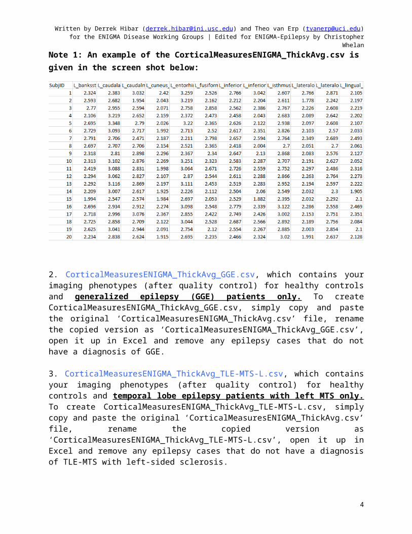
Note 1: An example of the CorticalMeasuresENIGMA_ThickAvg.csv is given in the screen shot below:
2. CorticalMeasuresENIGMA_ThickAvg_GGE.csv, which contains your imaging phenotypes (after quality control) for healthy controls and generalized epilepsy (GGE) patients only. To create CorticalMeasuresENIGMA_ThickAvg_GGE.csv, simply copy and paste the original
3
Written by Derrek Hibar ([email protected]) and Theo van Erp ([email protected])for the ENIGMA Disease Working Groups | Edited for ENIGMA-Epilepsy by Christopher Whelan
‘CorticalMeasuresENIGMA_ThickAvg.csv’ file, rename the copied version as ‘CorticalMeasuresENIGMA_ThickAvg_GGE.csv’, open it up in Excel and remove any epilepsy cases that do not have a diagnosis of GGE.
3. CorticalMeasuresENIGMA_ThickAvg_TLE-MTS-L.csv, which contains your imaging phenotypes (after quality control) for healthy controls and temporal lobe epilepsy patients with left MTS only. To create CorticalMeasuresENIGMA_ThickAvg_TLE-MTS-L.csv, simply copy and paste the original ‘CorticalMeasuresENIGMA_ThickAvg.csv’ file, rename the copied version as ‘CorticalMeasuresENIGMA_ThickAvg_TLE-MTS-L.csv’, open it up in Excel and remove any epilepsy cases that do not have a diagnosis of TLE-MTS with left-sided sclerosis.
4. CorticalMeasuresENIGMA_ThickAvg_TLE-MTS-R.csv, which contains your imaging phenotypes (after quality control) for healthy controls and temporal lobe epilepsy patients with right MTS only. To create CorticalMeasuresENIGMA_ThickAvg_TLE-MTS-R.csv, simply copy and paste the original ‘CorticalMeasuresENIGMA_ThickAvg.csv’ file, rename the copied version as ‘CorticalMeasuresENIGMA_ThickAvg_TLE-MTS-R.csv’, open it up in Excel and remove any epilepsy cases that do not have a diagnosis of TLE-MTS with right-sided sclerosis.
5. Covariates.csv, which contains the full set of variables we will be controlling for in each of the models. Note that missing values for any of these covariates are not allowed. You can re-use the Covariates.csv file from the subcortical analysis.
Formatting:
At this point your CorticalMeasuresENIGMA_ThickAvg.csv likely contains the full paths to your images (as well as your SubjectIDs) in the first column.
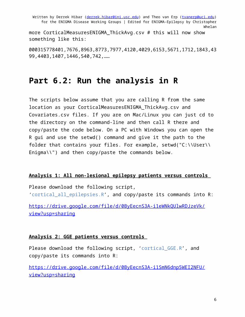
more CorticalMeasuresENIGMA_ThickAvg.csv # this will show something like this:
/raid/fbirn_phase3/freesurfer/000315778401,7676,8963,8773,7977,4120,4029,6153,5671,1712,1843,4399,4403,1407,1446,540,742,….
….
If your SubjectIDs contain path information you can get rid of them with these commands:
cp CorticalMeasuresENIGMA_ThickAvg.csv CorticalMeasuresENIGMA_ThickAvg.csv.orig
cat CorticalMeasuresENIGMA_ThickAvg.csv.orig | awk -F/ ' { print $NF } ' > CorticalMeasuresENIGMA_ThickAvg.csv
4
Written by Derrek Hibar ([email protected]) and Theo van Erp ([email protected])for the ENIGMA Disease Working Groups | Edited for ENIGMA-Epilepsy by Christopher Whelan
more CorticalMeasuresENIGMA_ThickAvg.csv # this will now show something like this:
000315778401,7676,8963,8773,7977,4120,4029,6153,5671,1712,1843,4399,4403,1407,1446,540,742,……
Part 6.2: Run the analysis in R
The scripts below assume that you are calling R from the same location as your CorticalMeasuresENIGMA_ThickAvg.csv and Covariates.csv files. If you are on Mac/Linux you can just cd to the directory on the command-line and then call R there and copy/paste the code below. On a PC with Windows you can open the R gui and use the setwd() command and give it the path to the folder that contains your files. For example, setwd("C:\\User\\Enigma\\") and then copy/paste the commands below.
Analysis 1: All non-lesional epilepsy patients versus controls
Please download the following script, ‘cortical_all_epilepsies.R’, and copy/paste its commands into R:
https://drive.google.com/file/d/0ByEecnS3A-i1eWNkQUlwRDJzeVk/view?usp=sharing
Analysis 2: GGE patients versus controls
Please download the following script, ‘cortical_GGE.R’, and copy/paste its commands into R:
https://drive.google.com/file/d/0ByEecnS3A-i1SmN6dnp5WEI2NFU/view?usp=sharing
Analysis 3: TLE-MTS-L patients versus controls
Please download the following script, ‘cortical_TLE-L.R’, and copy/paste its commands into R:https://drive.google.com/file/d/0ByEecnS3A-i1dEVkekl2X19TbEU/view?usp=sharing
Analysis 4: TLE-MTS-R patients versus controls
Please download the following script, ‘cortical_TLE-R.R’, and copy/paste its commands into R:
5
Written by Derrek Hibar ([email protected]) and Theo van Erp ([email protected])for the ENIGMA Disease Working Groups | Edited for ENIGMA-Epilepsy by Christopher Whelan
https://drive.google.com/file/d/0ByEecnS3A-i1aXF1Nm1JSWxkQXM/view?usp=sharing
Part 6.3: Upload your results
When done, tar up the .Rdata files and send to: Christopher D. Whelan ([email protected]). (Change the text highlighted in yellow before running the command):
tar cfz - *Rdata > YourStudyName_CortRegs.tgz
6