Embed Size (px)
Citation preview

1
Quadrangular space The quadrangular space provides a passageway for nerves and vessels passing between the axilla and the more posterior scapular and deltoid regions. When viewed from anteriorly, its boundaries are formed by: inferior margin of the subscapularis muscle; surgical neck of the humerus; superior margin of the teres major muscle; and lateral margin of the long head of the triceps brachii muscle. Passing through the quadrangular space are the axillary nerve and the posterior circumflex humeral artery and vein. Triangular interval [Lateral triangular space]This triangular interval is formed by: lateral margin of the long head of the triceps brachii muscle; shaft of the humerus; and inferior margin of the teres major muscle. The radial nerve passes out of the axilla traveling through this interval to reach the posterior compartment of the arm. The profunda brachii artery (deep artery of arm) and associated veins also passthrough the triangular interval.
Axillary artery first part is proximal to pectoralis minor; medial part of pectoralis minor & lateral part of first rib second part is posterior to pectoralis minor; third part is distal to pectoralis minor;from lateral part of pectoralis minor to inferior border of teres minor.
Generally, six branches arise from the axillary artery: 1 branch, the superior thoracic artery, originates from the first part; 2 branches, the thoraco-acromial artery and the lateral thoracic artery, originate from the second part; 3 branches, the subscapular artery, the anterior circumflex humeral artery, and the posterior circumflex
humeral artery, originate from the third part.
Axillary veinPlexus Brachialis
Branches of the lateral cord1) The lateral pectoral nerve 2) The musculocutaneous nerve 3) The lateral root of median nerve
Branches of the medial cord1) The medial pectoral nerve 2) The medial cutaneous nerve of arm (medial brachial cutaneous nerve)

2
3) The medial cutaneous nerve of forearm (medial antebrachial cutaneous nerve) 4) The ulnar nerve. 5) The median nerve
The musculocutaneous nerve, the lateral root of the median nerve, the median nerve, the medial root of the median nerve, and the ulnar nerve form an M over the third part of the axillary artery. This feature, together with penetration of the coracobrachialis muscle by the musculocutaneous nerve, can be used to identify components of the brachial plexus in the axilla.
Branches of the posterior cord1) Superior subscapular nerve2) Thoracodorsal nerve3) Inferior subscapular nerve4) Axillary nerve5) Radial nerve
SHOULDER SHOULDER SUPERFICIAL POSTERIOR AXIOAPPENDICULAR (EXTRINSIC SHOULDER) MUSCLES
The superficial axioappendicular muscles are the trapezius and latissimus dorsi.DEEP POSTERIOR AXIOAPPENDICULAR (EXTRINSIC SHOULDER) MUSCLESThe deep posterior thoracoappendicular muscles are the levator scapulae and rhomboids.
SCAPULOHUMERAL (INSTRINSIC SHOULDER) MUSCLESThe six scapulohumeral muscles (deltoid, teres major, supraspinatus, infraspinatus, subscapularis, and teres minor).

3
ARMARMThe anterior compartment of the arm contains three muscles-coracobrachialis, brachialis, and biceps
brachii muscles-which are innervated predominantly by the musculocutaneous nerve. The only muscle of the posterior compartment of the arm is the triceps brachii muscle. The triceps brachii
muscle has three heads: long head originates from the infraglenoid tubercle of the scapula; medial & lateral heads originate from the extensive area on the posterior surface of the shaft of the humerus
inferior to the radial groove.
Brachial arteryTerminates just distal to the elbow joint, opposite to the neck of the radius where it divides into the
radial and ulnar arteries.

4
Branches of the brachial artery in the arm include those to adjacent muscles and two ulnar collateral vessels (superior and inferior ulnar collateral arteries), which contribute to a network of arteries around the elbow joint. Additional branches are the profunda brachii artery and nutrient arteries to the humerus, which pass through a foramen in the anteromedial surface of the humeral shaft.
VeinsTwo sets of veins of the arm, superficial and deep, anastomose freely with each other. The superficial
veins are in the subcutaneous tissue, and the deep veins accompany the arteries. Both sets of veins have valves, but they are more numerous in the deep veins than in the superficial veins.The two main superficial veins of the arm, the cephalic and basilic veins.
NervesFour main nerves pass through the arm: median, ulnar, musculocutaneous, and radial.

5
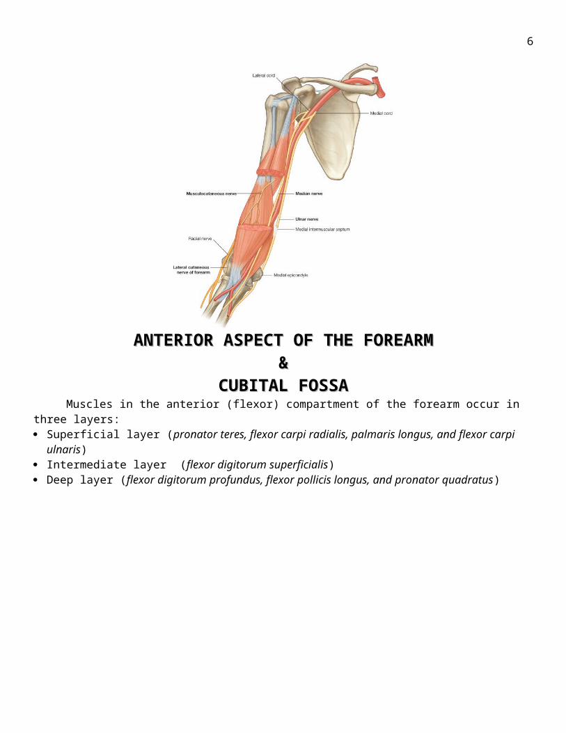
ANTERIOR ASPECT OF THE FOREARM ANTERIOR ASPECT OF THE FOREARM & &
CUBITAL FOSSACUBITAL FOSSAMuscles in the anterior (flexor) compartment of the forearm occur in three layers:
Superficial layer (pronator teres, flexor carpi radialis, palmaris longus, and flexor carpi ulnaris) Intermediate layer (flexor digitorum superficialis) Deep layer (flexor digitorum profundus, flexor pollicis longus, and pronator quadratus)
Arteries
6
The main arteries of the forearm are the ulnar and radial arteries, which usually arise opposite the neck of the radius in the inferior part of the cubital fossa as terminal branches of the brachial artery.
Radial arteryWhen the brachioradialis is pulled laterally, the entire length of the artery is visible.Branches of the radial artery originating in the forearm include:
1) radial recurrent artery, which contributes to an anastomotic network around the elbow joint 2) small palmar carpal branch 3) superficial palmar branch enters the hand by passing through, or superficial to, the thenar muscles at the base of the thumb, which anastomoses with the superficial palmar arch formed by the ulnar artery.
Ulnar arteryThe ulnar artery is larger than the radial artery and passes down the medial side of the forearm.
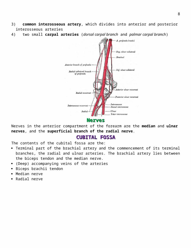
Branches of the ulnar artery that arise in the forearm include: 1) ulnar recurrent artery with anterior and posterior branches, which contribute to an anastomotic network of vessels around the elbow joint (The anterior and posterior ulnar recurrent arteries anastomose with the inferior and superior ulnar collateral arteries, respectively, thereby participating in the periarticular arterial anastomoses of the elbow)2) numerous muscular arteries, which supply surrounding muscles3) common interosseous artery, which divides into anterior and posterior interosseous arteries4) two small carpal arteries (dorsal carpal branch and palmar carpal branch)
NervesNervesNerves in the anterior compartment of the forearm are the median and ulnar nerves, and the superficial branch of the radial nerve.
CUBITAL FOSSACUBITAL FOSSAThe contents of the cubital fossa are the: Terminal part of the brachial artery and the commencement of its terminal branches, the radial and ulnar
arteries. The brachial artery lies between the biceps tendon and the median nerve. (Deep) accompanying veins of the arteries Biceps brachii tendon Median nerve Radial nerve

7
POSTERIOR ASPECT OF THE FOREARMPOSTERIOR ASPECT OF THE FOREARM&&
ANATOMY OF THE HANDANATOMY OF THE HAND Superficial layerThe seven muscles in the superficial layer are the brachioradialis, extensor carpi radialis longus, extensor carpi radialis brevis, extensor digitorum, extensor digiti minimi, extensor carpi ulnaris, and anconeus. Deep layer
The deep layer of the posterior compartment of the forearm consists of five muscles: supinator, abductor pollicis longus, extensor pollicis brevis, extensor pollicis longus, and extensor indicis.
8
Arteries The blood supply to the posterior compartment of the forearm occurs predominantly through branches of
the radial, posterior interosseous, and anterior interosseous arteries. Nerves
Radial nerveThe nerve of the posterior compartment of the forearm is the radial nerve.
HANDMuscles
The intrinsic muscles of the hand are the palmaris brevis, interossei, adductor pollicis, thenar, hypothenar, and lumbrical muscles.
Thenar musclesThe three thenar muscles (opponens pollicis, flexor pollicis brevis, and abductor pollicis brevis muscles) are associated with opposition of the thumb to the fingers and with delicate movements of the thumb and are responsible for the prominent swelling (thenar eminence) on the lateral side of the palm at the base of the thumb.
Hypothenar musclesThe hypothenar muscles (opponens digiti minimi, abductor digiti minimi, and flexor digiti minimi brevis contribute to the swelling (hypothenar eminence) on the medial side of the palm at the base of the little finger.

9
Arteries and veinsThe blood supply to the hand is by the radial and ulnar arteries, which form two interconnected vascular arches (superficial and deep) in the palm. Vessels to the digits, muscles, and joints originate from the two arches and the parent arteries.
Ulnar artery and superficial palmar archThe ulnar artery and ulnar nerve enter the hand on the medial side of the wrist. Distally, the ulnar artery swings laterally across the palm, forming the superficial palmar arch, which is superficial to the long flexor tendons of the digits and just deep to the palmar aponeurosis. One branch of the ulnar artery in the hand is the deep palmar branch.
Radial artery and deep palmar archThe radial artery curves around the lateral side of the wrist, passes over the floor of the anatomical snuffbox and into the deep plane of the palm by penetrating anteriorly through the back of the hand. It accesses the deep plane of the palm and forms the deep palmar arch. Superficial palmar arch Deep palmar arch
Nerves
Ulnar nerveThe ulnar nerve ends at the distal border of the flexor retinaculum by dividing into superficial (mainly sensory) and deep (mainly motor) branches.
Median nerveThe median nerve enters the hand by passing through the carpal tunnel and divides into a recurrent
branch and palmar digital branches. Superficial branch of the radial nerve
The only part of the radial nerve that enters the hand is the superficial branch.
PECTORAL REGION & MAMMARY GLANDSPECTORAL REGION & MAMMARY GLANDS

10
MUSCLES OF THE PECTORAL REGIONFour anterior axioappendicular muscles (thoracoappendicular or pectoral muscles) move the pectoral
girdle: pectoralis major, pectoralis minor, subclavius, and serratus anterior.
SUPERFICIAL GROUP OF BACK MUSCLES Trapezius Latissimus dorsi Rhomboid major Rhomboid minor Levator scapulae
INTERMEDIATE GROUP OF BACK MUSCLESThe serratus posterior superior lies deep to the rhomboids, and the serratus posterior inferior lies
deep to the latissimus dorsi.