Embed Size (px)
Citation preview

British Journal of Ophthalmology, 1982, 66, 269-274
Computed tomography in the management of orbitalinfections associated with dental diseaseTIMOTHY P. FLOOD,' LAURENCE S. BRAUDE,' LEE M. JAMPOL.' AND
STEVEN HERZOG2
From the 'Department of Ophthalmology, University of Illinois Hospital Eye and Ear Infirmary, Chicago,and the 2Department of Oral Surgery, University of Illinois at the Medical Center, Chicago, USA
SUMMARY Two patients developed orbital infection secondary to dental infections. In one patientthe infection spread from maxillary premolar and molar teeth to the infratemporal andpterygopalatine fossa and then through the inferior orbital fissure to the subperiosteal space. Asubperiosteal abscess in the posterior lateral orbital wall developed, which subsequently spreadwithin the muscle cone. In the second patient infection of an anterior maxillary tooth caused a
pansinusitis and unilateral orbital cellulitis. In both patients computed tomographic scanning of theorbit proved valuable in localising the infection and, in one case, planning a surgical approach to theorbit. The infection in both patients responded to treatment, with no permanent visual impairment.Appropriate antibiotics and prompt identification and surgical drainage of orbital abscesses are
essential for the preservation of vision in cases of orbital infection.
Although dental infection has been recognised as asource of orbital cellulitis since the time ofHippocrates,' attention to this association in therecent ophthalmic literature has been sparse. In thepreantibiotic era orbital cellulitis was commonlyassociated with dental abscesses and maxillary sinusinvolvement. The widespread use of prophylactic andtherapeutic antibiotics has decreased the incidence ofocular complications of odontogenic infections,although occasional cases still occur.25 Orbitalcellulitis can often be managed medically; however,surgical drainage is usually necessary if a localisedabscess develops in the orbit.6" Computedtomography has aided in the evaluation andmanagement of orbital lesions,89 including orbitalcellulitis.'0'" We describe 2 cases of orbital infectionsassociated with dental disease in which computedtomographic scanning proved useful in patientmanagement.
Case reports
CASE 1A 30-year-old man had a 4-day history of swelling ofCorrespondence to Dr Lee M. Jampol, Department of Ophthal-mology. University of Illinois Hospital Eye and Ear Infirmary, 1855W Taylor. Chicago, IL 60612 USA.
269
his left upper cheek. With the patient under localanaesthesia incision and drainage of an abscess in theleft buccal space were performed. The carious teethwere not removed. He was treated with oral penicillin.The patient returned 2 days later with increased painand swelling of the left cheek. 30 ml of purulentmaterial was drained from the same abscess. Thefollowing day the patient had signs of infratemporalspace extension of the infection. He was admittedinto the hospital and started on a regimen of 2 millionunits of intravenous penicillin G every 4 hours.Approximately 3 hours after admission the patientbecame somnolent; his temperature was 40 5°C(105°F). Decreased abduction of the left eye wasnoted. The dose of intravenous penicillin wasincreased to 5 million units every 6 hours, andintravenous nafcillin sodium, 2 g every 6 hours, wasadded. The morning after admission an ophthalmo-logical consultation was requested.
Ocular examination showed that visual acuity was6/6 (20/20) without correction bilaterally. The pupilswere equal and reactive to light. There was noafferent pupillary defect. 6 mm of left proptosis waspresent. Diplopia was noted in all fields of gazeexcept the primary position. Ductions of the right eyewere full. In the left eye there was an 80% reductionin abduction and a 50% reduction in adduction,
on 17 Novem
ber 2018 by guest. Protected by copyright.
http://bjo.bmj.com
/B
r J Ophthalm
ol: first published as 10.1136/bjo.66.4.269 on 1 April 1982. D
ownloaded from

Timothy P. Flood, Laurence S. Braude, Lee M. Jampol, and Steven Herzog
Fig. I Case 1. Computedtomographic scan ofthe orbitswithout contrast medium showsmedial displacement oftheleft lateral rectus muscle (blackarrows) by a subperiostealabscess at the apex of the orbit thatextended along the lateralorbital wall. An intraconal abscesswith gas (white arrow)and significant left proptosis alsoare shown.
elevation, and depression. Minimal left inferior con-junctival chemosis was present, but the slit-lampexamination was otherwise unremarkable. Applana-tion tonometry was normal bilaterally. The findingson dilated ophthalmoscopic examination were withinnormal limits. Skull and sinus x-ray films revealed asoft-tissue density in the left maxillary sinus. Theethmoidal sinuses were clear.On the day after admission the patient was given
general anaesthesia, and the carious left maxillary,first and second premolars, and first, second, andthird molars were removed. Incision and drainage ofthe left infratemporal space were performed. A leftCaldwell-Luc procedure and nasal antrostomy wereperformed. The maxillary sinus was noted to be clear.Purulent material was drained from the left buccaland infratemporal spaces, and drains were placed atboth sites. Through an external approach a sterileswab was inserted into the pterygopalatine space, andmaterial for cultures was taken. These cultures sub-sequently grew streptococci and Bacteroidesmelaninogenicus (subspecies intermedius). Post-operatively intravenous gentamicin, 80 mg every 8hours, was added to the preoperative antibioticregimen of penicillin and nafcillin.
During the next 2 days chemosis and proptosisincreased. The patient's left ocular motility wasreduced by 90% in all fields of gaze. His temperatureintermittently spiked to 39 4°C (103°F). The infra-temporal incision continued to drain purulentmaterial. Visual acuity remained unaffected until thefifth day in hospital, when it was noted to decrease to6/15 (20/50) in the left eye. At this time 8 mm of leftproptosis was present. The ophthalmoscopicexamination of the left eye showed disc hyperaemiaand oedema, with a single retinal haemorrhage. Acotton-wool spot was present along the infero-temporal arcade. Goldmann visual field testingshowed a central scotoma in the left eye. Computed
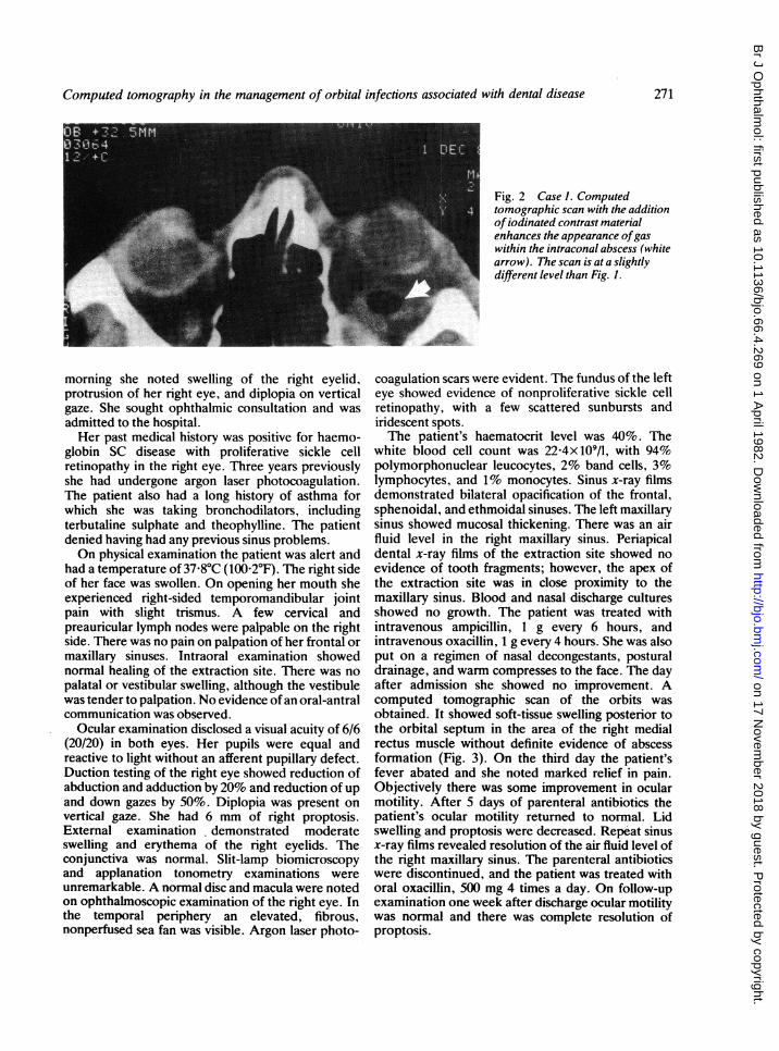
tomographic scan disclosed a left subperiostealabscess at the apex of the orbit that extended alongthe lateral orbital wall and a secondary intraconalabscess with gas (Fig. 1). The intraconal abscess wasenhanced with the use of intravenous iodinatedcontrast medium (Fig. 2).
With the patient under general anaesthesia a leftsubperiosteal approach along the inferolateral orbitalwall was made to drain 3 ml of purulent material fromthe subperiosteal abscess 25 mm from the orbital rim.The maxillary sinus was explored, and a blood clotwas removed from the antrum. There was no evidenceof infection in the maxillary sinus. The orbit was thendecompressed into the maxillary and ethmoidalsinuses by removing portions of the floor and medialwall of the orbit. A lumbar puncture at the com-pletion of the procedure yielded normal findings.Cultures from the subperiosteal abscess grewBacteroides melaninogenicus (subspecies inter-medius), the same organism previously cultured fromthe infratemporal fossa and the pterygopalatinefossa. Postoperatively defervescence was achievedwith an antibiotic regimen of penicillin, nafcillin, andgentamicin. During the next 2 weeks visual acuityreturned to 6/6 (20/20) and visual fields returned tonormal. The proptosis also resolved, and the patienthad only slight restriction of abduction of the lefteye.
CASE 2A 20-year-old black woman noted pain in the area ofher right second maxillary bicuspid tooth 2 days aftershe lost a dental filling from that tooth. The cariousright second maxillary bicuspid was promptlyremoved by her dentist. The following day she hadslight swelling on the right side of her face and pain onmoving her right eye. Two days after the toothextraction she returned to her dentist, and heprescribed oral penicillin for her. The following
270
on 17 Novem
ber 2018 by guest. Protected by copyright.
http://bjo.bmj.com
/B
r J Ophthalm
ol: first published as 10.1136/bjo.66.4.269 on 1 April 1982. D
ownloaded from
Computed tomography in the management of orbital infections associated with dental disease
Fig. 2 Case 1. Computedtomographic scan with the additionofiodinated contrast materialenhances the appearance ofgaswithin the intraconal abscess (whitearrow). The scan is at a slightlydifferent level than Fig. 1.
morning she noted swelling of the right eyelid,protrusion of her right eye, and diplopia on verticalgaze. She sought ophthalmic consultation and wasadmitted to the hospital.Her past medical history was positive for haemo-
globin SC disease with proliferative sickle cellretinopathy in the right eye. Three years previouslyshe had undergone argon laser photocoagulation.The patient also had a long history of asthma forwhich she was taking bronchodilators, includingterbutaline sulphate and theophylline. The patientdenied having had any previous sinus problems.On physical examination the patient was alert and
had a temperature of 37-8°C (100-2°F). The right sideof her face was swollen. On opening her mouth sheexperienced right-sided temporomandibular jointpain with slight trismus. A few cervical andpreauricular lymph nodes were palpable on the rightside. There was no pain on palpation of her frontal ormaxillary sinuses. Intraoral examination showednormal healing of the extraction site. There was nopalatal or vestibular swelling, although the vestibulewas tender to palpation. No evidence of an oral-antralcommunication was observed.
Ocular examination disclosed a visual acuity of 6/6(20/20) in both eyes. Her pupils were equal andreactive to light without an afferent pupillary defect.Duction testing of the right eye showed reduction ofabduction and adduction by 20% and reduction of upand down gazes by 50%. Diplopia was present onvertical gaze. She had 6 mm of right proptosis.External examination demonstrated moderateswelling and erythema of the right eyelids. Theconjunctiva was normal. Slit-lamp biomicroscopyand applanation tonometry examinations wereunremarkable. A normal disc and macula were notedon ophthalmoscopic examination of the right eye. Inthe temporal periphery an elevated, fibrous,nonperfused sea fan was visible. Argon laser photo-
coagulation scars were evident. The fundus of the lefteye showed evidence of nonproliferative sickle cellretinopathy, with a few scattered sunbursts andiridescent spots.The patient's haematocrit level was 40%. The
white blood cell count was 22 4x10⁹/l, with 94%polymorphonuclear leucocytes, 2% band cells, 3%lymphocytes, and 1% monocytes. Sinus x-ray filmsdemonstrated bilateral opacification of the frontal,sphenoidal, and ethmoidal sinuses. The left maxillarysinus showed mucosal thickening. There was an airfluid level in the right maxillary sinus. Periapicaldental x-ray films of the extraction site showed noevidence of tooth fragments; however, the apex ofthe extraction site was in close proximity to themaxillary sinus. Blood and nasal discharge culturesshowed no growth. The patient was treated withintravenous ampicillin, 1 g every 6 hours, andintravenous oxacillin, 1 g every 4 hours. She was alsoput on a regimen of nasal decongestants, posturaldrainage, and warm compresses to the face. The dayafter admission she showed no improvement. Acomputed tomographic scan of the orbits wasobtained. It showed soft-tissue swelling posterior tothe orbital septum in the area of the right medialrectus muscle without definite evidence of abscessformation (Fig. 3). On the third day the patient'sfever abated and she noted marked relief in pain.Objectively there was some improvement in ocularmotility. After 5 days of parenteral antibiotics thepatient's ocular motility returned to normal. Lidswelling and proptosis were decreased. Repeat sinusx-ray films revealed resolution of the air fluid level ofthe right maxillary sinus. The parenteral antibioticswere discontinued, and the patient was treated withoral oxacillin, 500 mg 4 times a day. On follow-upexamination one week after discharge ocular motilitywas normal and there was complete resolution ofproptosis.
271
on 17 Novem
ber 2018 by guest. Protected by copyright.
http://bjo.bmj.com
/B
r J Ophthalm
ol: first published as 10.1136/bjo.66.4.269 on 1 April 1982. D
ownloaded from

Timothy P. Flood, Laurence S. Braude, Lee M. Jampol, and Steven Herzog
Fig. 3 Case 2. Computedtomographic scan ofthe orbitswithout contrastmedium shows softtissue swelling within the right orbitin the region ofthe right medialrectus muscle (arrow). Bothethmoidal sinuses are opacified,and right proptosis is shown.
Discussion
Although the use of antibiotics has decreased theincidence of orbital complications of odontogenicinfection, dental disease may still cause infections,6 12
including preseptal cellulitis, orbital cellulitis, orbitalabscess, subperiosteal abscess, and cavernous sinusthrombosis.
Acute odontogenic infection may spread throughtissue planes and venous channels to involve theorbit. The routes of infectious extension may besummarised as follows: (1) The roots of maxillarypremolar and molar teeth may lie very close to themaxillary sinus. Maxillary sinusitis may result fromextension of maxillary molar or premolar infection or
from perforation of the sinus floor during extractionof diseased maxillary teeth. Sinusitis may then extendinto the orbit to cause orbital cellulitis.' (2) Infectionof maxillary incisors or canines may spread throughlocal tissue planes over the maxilla, resulting inswelling of the upper lip, canine fossa, and periorbitaltissue. Retrograde spread into the orbit may thenoccur through the valveless anterior facial, angular,and ophthalmic veins.5 (3) Infection of anteriormaxillary teeth may also spread as a subperiostealabscess to the anterior surface of the maxilla toinvolve the orbit.' (4) Infection of the posteriormaxillary teeth, most commonly the third molar, mayspread posteriorly into the pterygopalatine andinfratemporal fossae. The infection may then extendinto the orbit through the inferior orbital fissure.51 13
Our cases illustrate 2 of these mechanisms ofspread. Patient 1 had an infection of maxillary molarand premolar teeth that spread to the infratemporalspace and pterygopalatine fossa. The infectionpresumably gained access to the orbit through theinferior orbital fissure. A subperiosteal abscessdeveloped in the posterior lateral orbital wall, whichsubsequently spread within the muscle cone.
Bacteroides melaninogenicus (subspecies inter-medius), a gas-forming organism, was cultured fromthe infratemporal fossa, pterygopalatine fossa, andfrom the subperiosteal abscess. In patient 2 infectionof an anterior maxillary tooth with a root in closeproximity to the maxillary sinus resulted in acutepansinusitis and right orbital cellulitis. No organismwas recovered from blood or nasal cultures.
Infections of periorbital and orbital tissues areclassified according to their location relative to theorbital septum.'4 The orbital septum separates theorbital contents from the lids and acts as a barrier tothe spread of infection from the skin into the orbit.Preseptal cellulitis involves the tissues anterior to theseptum, whereas orbital cellulitis involves thosetissues posterior to the septum. It has been shownthat orbital cellulitis may be complicated by orbital orsubperiosteal abscesses in more than 20% of cases.9 Asubperiosteal abscess may develop from directextension of suppuration into the tissue planebetween the periosteum and the orbital wall. Anorbital abscess may form by the consolidation ofinfection within the orbit or from the rupture into theorbit of a subperiosteal abscess.
It may be difficult sometimes to distinguishbetween these entities on clinical grounds alone. Allare associated with warm, erythematous, swollen lidswith varying amounts of conjunctival chemosis.However, proptosis, restriction of ocular motility,pain on ocular movement, an afferent pupillarydefect, decreased visual acuity, disc oedema,choroidal folds, and retinal venous stasis distinguishinfections within the orbit from preseptal cellulitis.
It is important to identify cases with orbital orsubperiosteal abscess formation, because promptsurgical drainage in these cases is usually necessary toprevent permanent visual loss. Computed tom-ography is an established radiographic procedure thatcan aid in the differentiation of these entities.'"'"
272
on 17 Novem
ber 2018 by guest. Protected by copyright.
http://bjo.bmj.com
/B
r J Ophthalm
ol: first published as 10.1136/bjo.66.4.269 on 1 April 1982. D
ownloaded from

Computed tomography in the management of orbital infections associated with dental disease
Preseptal cellulitis is characterised on computed tom-ographic scanning by the presence of inflammationlocalised in the tissues anterior to the orbital septumwithout intraorbital disease. Orbital cellulitis can bedemonstrated as an obliteration of fat shadows withinthe muscle cone or the presence of soft tissue swellingwithin the orbit. Computed tomographic examinationof a patient with a subperiosteal abscess shows a masslesion located between the orbital wall and thedisplaced periosteum. The periosteal wall of theabscess may at times be more clearly defined with theuse of intravenous iodinated contrast medium. Anorbital abscess appears as a localised inflammatorvmass within the orbital tissues. Gas mav bedemonstrated within the abscess, arising either fromgas-forming bacilli or from a communication of theabscess with an adjacent paranasal sinus. An abscessmay be outlined by the use of contrast enhancement.Computed tomographic scanning proved useful in
both of our patients. In case 2, after no improvementwas noted with intravenous antibiotic therapy, thecomputed tomographic scan revealed orbital cellulitisand ruled out an orbital abscess. In case 1 thecomputed tomographic scan demonstrated a sub-periosteal abscess in the posterior lateral orbital walland an intraorbital abscess with gas. Timelv directdrainage of the subperiosteal abscess and decom-pression of the orbit resulted in resolution ofproptosis, recovery of visual acuity, improvement ofocular motility, and return of the visual fields tonormal.
Axial computed tomographic scans of the orbit are
also of value in delineating the anatomical associationamong the orbits, the paranasal sinuses, and intra-cranial structures. Computed tomographic scanningcan identify cerebral complications such as cerebritis,brain abscess, and epidural infection. In additionaxial views allow comparison of both orbits to detectminimal significant differences. This view is alsouseful to evaluate proptosis and displacement of theglobe, muscle cone, or optic nerve.
Prior to the use of computed tomographic scanningidentification of the site of abscess formation was
primarily accomplished by localising inflammatorysigns and the direction of displacement of the globe. 14However, these methods have limitations, especiallvwhen proptosis is axial. Computed tomographicscanning can precisely localise abscesses, whichallows the correct surgical approach to be taken fordrainage.The most frequently isolated organisms in orbital
infections in adults include Staphylococcus aureus,
Streptococcus pyogenes, and Str. pneumoniae. 14 '5 Inaddition Haemophilus influenzae is a very commonlvisolated pathogen in children. 6 Other aerobic Gram-negative organisms are rare. A wide varietv of
anaerobic or microaerophilic organisms, particularlvBacteroides species, have been implicated in orbitalcellulitis.The presence of gas in association with orbital
infections may aid in the identification of thecausative organism. The differential diagnosis of gas-producing organisms includes anaerobic bacteriasuch as Clostridia, Bacteroides, anaerobicstreptococci, and anaerobic micrococci. Aerobicenterobacteria such as Proteus, Klebsiella, andEscherichia coli can also produce gas.'7 18 In additiondefects in the orbital walls produced bv infection ofthe paranasal sinuses may allow air from therespiratory tract into the orbit, simulating gasproduced by gas-forming organisms.'9
Antibacterial therapy should be based on theorganisms most likely to be encountered. In cases oforbital infection with associated dental or sinusinfection initial treatment with penicillin and apenicillinase-resistant antibiotic is recommended.The therapeutic regimen can be altered when theresults of bacterial cultures and tests of antibioticsensitivities are available.
This work was supported in part bv grant PHS HL15168 from theNational Heart. Lung. and Blood Institute. Bethesda. MD.The manuscript was edited bv Maxine Gere and tvped bv Cvnthia
Gustman.
References
I Duke-Elder S. The ocular adnexa. System of Ophthalmology. StLouis: Mosby, 1974: 13 (2): 859-66.
2 Gold RS. Sager E. Pansinusitis. orbital cellulitis and blindness assequelae of delaved treatment of dental abscess. JOralSlSurg 1974:32: 40-3.
3 Limongelli WA. Clark MS. William AC. Panfacial cellulitis withcontralateral orbital cellulitis and blindness after toothextraction. J Oral Surg 1977; 35: 38-43.
4 Yates C. Monks A. Orbital cellulitis complicating the extractionof infected teeth. J Dent 1978: 6: 229-32.
5 Kaban L.B. McGill T. Orbital cellulitis of dental origin:differential diagnosis and the use of computed tomographv as adiagnostic aid. J Oral Surg 1980: 38: 682-5.
6 Gans H. Sekula J. Wlodvka J. Treatment of acute orbitalcomplications. Arch Otolarvngol 1974; 100: 329-32.
7 Schramm VL. Mvers EN. Kennerdell JS. Orbital complicationsof acute sinusitis: evaluation, management. and outcome.OtolarvIngologv 1978; 86: 221-30.
8 Wende S. Avlich A. Nover A. et al. Computed tomographv oforbital lesions. Neuiroradiolov 1977;13: 123-34.
9 Gvldensted C. Lester J. Fledelius H. Computed tomographv oforbital lesions. Neuroradiology 1977; 13: 141-50.
10 Goldberg F. Berne AS. Oski FA. Differentiation of orbitalcellulitis from preseptal cellulitis bv computed tomographv.Pediatrics 1978: 62: 1000-5.
I Zimmerman RA. Bilanivk LT. Ct of orbital infection and itscerebral complications. AJR 1980; 134: 45-5t).
12 Jarrett WH. Gutman FA. Ocular complications of infection inthe paranasal sinuses. Arch Ophthalmol 1969; 81: 683-8.
13 Chow AW. Roser SM. Bradv FA. Orofacial odontogenicinfections. Ann Intern Med 1978; 88: 392-402.
273
on 17 Novem
ber 2018 by guest. Protected by copyright.
http://bjo.bmj.com
/B
r J Ophthalm
ol: first published as 10.1136/bjo.66.4.269 on 1 April 1982. D
ownloaded from

Timothy P. Flood, Laurence S. Braude, Lee M. Jampol, and Steven Herzog
14 Jones DB. Microbial preseptal and orbital cellulitis. In: DuaneTB. ed. Clinical Ophthalmology. New York: Harper and Row.1980:4 (25): 1-19.
15 Krohel GB. Krause HR. Christensen RE. et al. Orbital abscess.Arch Ophthalmol 1980:98: 274-6.
16 Londer L. Nelson DL. Orbital cellulitis due to Haemophilusinfluenzae. Arch Ophthalmol 1974:91: 89-91.
17 Sevel D. Tobias B. Sellars SL. Forder A. Gas in the orbit
associated with orbital cellulitis and paranasal sinusitis. Br JOphthalmol 1973: 57: 133-7.
18 Lefrock JL. Schell RF. Tillotson JR. Anaerobic infections:pathology. microbiology. diagnosis and treatment. In: HollowavWJ. ed. Infectioius Disease Reviews. Mount Kisco. New York:Futura. 1978: 97-131.
19 Weinstein L. Barza MA. Gas gangrene. N En,glJ Med 1973: 289:1129-31.
274
on 17 Novem
ber 2018 by guest. Protected by copyright.
http://bjo.bmj.com
/B
r J Ophthalm
ol: first published as 10.1136/bjo.66.4.269 on 1 April 1982. D
ownloaded from