Embed Size (px)
Citation preview
Z. Phys. Chem. 225 (2011) 647–659 / DOI 10.1524/zpch.2011.0079© by Oldenbourg Wissenschaftsverlag, München
Vibrational Spectroscopic Studiesof Germanium-High Bismuthate Glassesand Vitroceramics
By Oana Ponta, Lucian Baia, Monica Baia, and Simion Simon∗
Faculty of Physics and Interdisciplinary Research Institute on Bio-Nano-Sciences,Babes-Bolyai University, M. Kogalniceanu 1, 400084 Cluj-Napoca, Romania
This paper is dedicated to Wolfgang Kiefer on the occasion of his 70th birthday
(Received November 29, 2010; accepted in revised form December 27, 2010)
Local Structure / Glass / Vitroceramic / IR Spectroscopy / Raman Spectroscopy
Bi2O3-rich glasses belonging to the system xGd2O3(1−x)[0.857Bi2O3 ·0.143GeO2] with 0.005 ≤ x ≤0.1, have been obtained by rapid cooling of the melts. Polycrystalline samples have been furtherobtained by applying a heat treatment at 600 ◦C for 24 h. Both types of samples were structurallycharacterized by using X-ray diffraction (XRD), and infrared (IR) and Raman spectroscopictechniques.
Local structural units for the investigated samples are identified on the basis of vibrationalspectroscopic data. The obtained data reveal the presence of GeO4, GeO6, BiO3 and BiO6 groupsas basic structural units of the networks. For the vitroceramic samples some changes occur in theirnetwork, mainly reflecting the presence of an ordered structure, but much important is that part ofthe BiO6 structural units from Bi2O3-GeO2 glass network are converting into BiO3 units. The IRand Raman results support the idea that the local structure of the glasses and vitroceramics is lessaffected by the presence of Gd3+ ions but there is strong long range order influence on developedcrystalline phases from vitroceramic samples, as proved by XRD. However, the spectral analysesreveal the formation of a structure with a higher disorder degree for x = 0.1 in comparison withthe other investigated vitroceramics as a result of the partial replacement of the Bi3+ ions withGd3+ ions. Starting from the Bi12GeO20 sellenite phase in the samples with low gadolinium content(x ≤ 0.01), the structure is change to Bi24GeO38 phase, via a BiLa type structure, when the Gd2O3
concentration is increasing up to x = 0.1.
1. Introduction
In the last period, the glasses based on Bi2O3 paid attention for the researchers due totheir various applications in optoelectronic devices [1]. The bismuth oxide is known asnon-classical glass network former and a series of two and multicomponent nonconven-tional glasses with interesting properties have been synthesized [2–8]. In the presenceof strong polarizing cations, the coordination number of Bi3+ ions can be reduced fromsix to three and the Bi2O3 glass network is usually built up from BiO6 octahedral and/or
* Corresponding author. E-mail: [email protected]
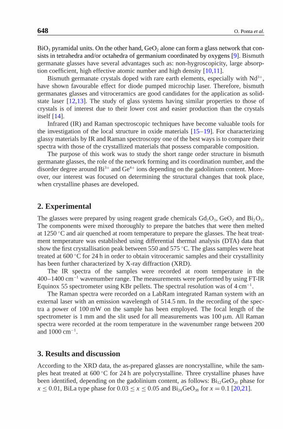
648 O. Ponta et al.
BiO3 pyramidal units. On the other hand, GeO2 alone can form a glass network that con-sists in tetrahedra and/or octahedra of germanium coordinated by oxygens [9]. Bismuthgermanate glasses have several advantages such as: non-hygroscopicity, large absorp-tion coefficient, high effective atomic number and high density [10,11].
Bismuth germanate crystals doped with rare earth elements, especially with Nd3+,have shown favourable effect for diode pumped microchip laser. Therefore, bismuthgermanates glasses and vitroceramics are good candidates for the application as solid-state laser [12,13]. The study of glass systems having similar properties to those ofcrystals is of interest due to their lower cost and easier production than the crystalsitself [14].
Infrared (IR) and Raman spectroscopic techniques have become valuable tools forthe investigation of the local structure in oxide materials [15–19]. For characterizingglassy materials by IR and Raman spectroscopy one of the best ways is to compare theirspectra with those of the crystallized materials that possess comparable composition.
The purpose of this work was to study the short range order structure in bismuthgermanate glasses, the role of the network forming and its coordination number, and thedisorder degree around Bi3+ and Ge4+ ions depending on the gadolinium content. More-over, our interest was focused on determining the structural changes that took place,when crystalline phases are developed.
2. Experimental
The glasses were prepared by using reagent grade chemicals Gd2O3, GeO2 and Bi2O3.The components were mixed thoroughly to prepare the batches that were then meltedat 1250 ◦C and air quenched at room temperature to prepare the glasses. The heat treat-ment temperature was established using differential thermal analysis (DTA) data thatshow the first crystallisation peak between 550 and 575 ◦C. The glass samples were heattreated at 600 ◦C for 24 h in order to obtain vitroceramic samples and their crystallinityhas been further characterized by X-ray diffraction (XRD).
The IR spectra of the samples were recorded at room temperature in the400–1400 cm−1 wavenumber range. The measurements were performed by using FT-IREquinox 55 spectrometer using KBr pellets. The spectral resolution was of 4 cm−1.
The Raman spectra were recorded on a LabRam integrated Raman system with anexternal laser with an emission wavelength of 514.5 nm. In the recording of the spec-tra a power of 100 mW on the sample has been employed. The focal length of thespectrometer is 1 mm and the slit used for all measurements was 100 μm. All Ramanspectra were recorded at the room temperature in the wavenumber range between 200and 1000 cm−1.
3. Results and discussion
According to the XRD data, the as-prepared glasses are noncrystalline, while the sam-ples heat treated at 600 ◦C for 24 h are polycrystalline. Three crystalline phases havebeen identified, depending on the gadolinium content, as follows: Bi12GeO20 phase forx ≤ 0.01, BiLa type phase for 0.03 ≤ x ≤ 0.05 and Bi24GeO38 for x = 0.1 [20,21].
Vibrational Spectroscopic Studies of Germanium-High Bismuthate Glasses and Vitroceramics 649
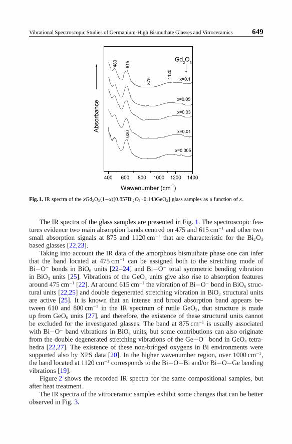
Fig. 1. IR spectra of the xGd2O3(1−x)[0.857Bi2O3 ·0.143GeO2] glass samples as a function of x.
The IR spectra of the glass samples are presented in Fig. 1. The spectroscopic fea-tures evidence two main absorption bands centred on 475 and 615 cm−1 and other twosmall absorption signals at 875 and 1120 cm−1 that are characteristic for the Bi2O3
based glasses [22,23].Taking into account the IR data of the amorphous bismuthate phase one can infer
that the band located at 475 cm−1 can be assigned both to the stretching mode ofBi−O− bonds in BiO6 units [22–24] and Bi−O− total symmetric bending vibrationin BiO3 units [25]. Vibrations of the GeO4 units give also rise to absorption featuresaround 475 cm−1 [22]. At around 615 cm−1 the vibration of Bi−O− bond in BiO6 struc-tural units [22,25] and double degenerated stretching vibration in BiO3 structural unitsare active [25]. It is known that an intense and broad absorption band appears be-tween 610 and 800 cm−1 in the IR spectrum of rutile GeO2, that structure is madeup from GeO6 units [27], and therefore, the existence of these structural units cannotbe excluded for the investigated glasses. The band at 875 cm−1 is usually associatedwith Bi−O− band vibrations in BiO6 units, but some contributions can also originatefrom the double degenerated stretching vibrations of the Ge−O− bond in GeO4 tetra-hedra [22,27]. The existence of these non-bridged oxygens in Bi environments weresupported also by XPS data [20]. In the higher wavenumber region, over 1000 cm−1,the band located at 1120 cm−1 corresponds to the Bi−O−Bi and/or Bi−O−Ge bendingvibrations [19].
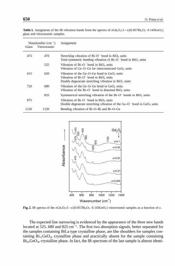
Figure 2 shows the recorded IR spectra for the same compositional samples, butafter heat treatment.
The IR spectra of the vitroceramic samples exhibit some changes that can be betterobserved in Fig. 3.
650 O. Ponta et al.
Table 1. Assigments of the IR vibration bands from the spectra of xGd2O3(1−x)[0.857Bi2O3 ·0.143GeO2]glass and vitroceramic samples.
Wavenumber (cm−1) AssignmentGlass Vitroceramic
475 470 Stretching vibration of Bi–O− bond in BiO6 unitsTotal symmetric bending vibration of Bi–O− bond in BiO3 units
525 Vibration of Bi–O− bond in BiO6 unitsVibration of Ge–O–Ge for interconnected GeO4 units
615 620 Vibration of the Ge–O–Ge bond in GeO6 unitsVibration of Bi–O− bond in BiO6 unitsDouble degenerate stretching vibration in BiO3 units
720 680 Vibration of the Ge–O–Ge bond in GeO6 unitsVibration of the Bi–O− bond in distorted BiO6 units
825 Symmetrical stretching vibration of the Bi–O− bonds in BiO3 units
875 Vibration of Bi–O− bond in BiO6 unitsDouble degenerate stretching vibration of the Ge–O− bond in GeO4 units
1120 1120 Bending vibration of Bi–O–Bi and Bi–O–Ge
Fig. 2. IR spectra of the xGd2O3(1−x)[0.857Bi2O3 ·0.143GeO2] vitroceramic samples as a function of x.
The expected line narrowing is evidenced by the appearance of the three new bandslocated at 525, 680 and 825 cm−1. The first two absorption signals, better separated forthe samples containing BiLa type crystalline phase, are like shoulders for samples con-taining Bi12GeO20 crystalline phase and practically absent for the sample containingBi24GeO38 crystalline phase. In fact, the IR spectrum of the last sample is almost identi-
Vibrational Spectroscopic Studies of Germanium-High Bismuthate Glasses and Vitroceramics 651
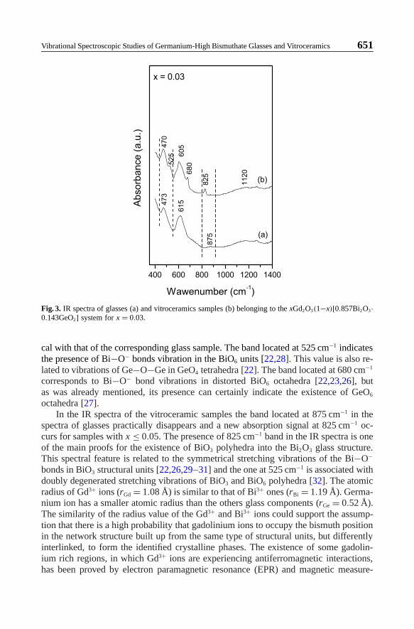
Fig. 3. IR spectra of glasses (a) and vitroceramics samples (b) belonging to the xGd2O3(1−x)[0.857Bi2O3·0.143GeO2] system for x = 0.03.
cal with that of the corresponding glass sample. The band located at 525 cm−1 indicatesthe presence of Bi−O− bonds vibration in the BiO6 units [22,28]. This value is also re-lated to vibrations of Ge−O−Ge in GeO4 tetrahedra [22]. The band located at 680 cm−1
corresponds to Bi−O− bond vibrations in distorted BiO6 octahedra [22,23,26], butas was already mentioned, its presence can certainly indicate the existence of GeO6
octahedra [27].In the IR spectra of the vitroceramic samples the band located at 875 cm−1 in the
spectra of glasses practically disappears and a new absorption signal at 825 cm−1 oc-curs for samples with x ≤ 0.05. The presence of 825 cm−1 band in the IR spectra is oneof the main proofs for the existence of BiO3 polyhedra into the Bi2O3 glass structure.This spectral feature is related to the symmetrical stretching vibrations of the Bi−O−
bonds in BiO3 structural units [22,26,29–31] and the one at 525 cm−1 is associated withdoubly degenerated stretching vibrations of BiO3 and BiO6 polyhedra [32]. The atomicradius of Gd3+ ions (rGd = 1.08 Å) is similar to that of Bi3+ ones (rBi = 1.19 Å). Germa-nium ion has a smaller atomic radius than the others glass components (rGe = 0.52 Å).The similarity of the radius value of the Gd3+ and Bi3+ ions could support the assump-tion that there is a high probability that gadolinium ions to occupy the bismuth positionin the network structure built up from the same type of structural units, but differentlyinterlinked, to form the identified crystalline phases. The existence of some gadolin-ium rich regions, in which Gd3+ ions are experiencing antiferromagnetic interactions,has been proved by electron paramagnetic resonance (EPR) and magnetic measure-
652 O. Ponta et al.
Fig. 4. Raman spectra of the xGd2O3(1−x)[0.857Bi2O3 ·0.143GeO2] glass samples as a function of x.
ments [21], but this gadolinium clusterisation is not perturbing too much the existing Biand Ge structural units.
The structural changes induced in the structure of polycrystalline samples by vary-ing the Gd2O3 content in the studied glasses, evidenced by the IR investigations,suggest that the gadolinium ions play an important role in the crystallization pro-cess of these glasses. If up to x ≤ 0.05 the gadolinium plays a role of improvingthe sample crystallinity, at x = 0.1, it plays a glass network stabilizing one. It seemsthat over a certain gadolinium content (x > 0.05), the replacement of the Bi3+ ionswith those of Gd3+ is more pronounced, and consequently, a more disordered vitro-ceramic structure occurs. This hypothesis is confirmed by the disappearance in thevitroceramic spectrum with x = 1 of the well defined absorption bands at 525 and680 cm−1, on one hand, and by the reappearance of the 875 cm−1 signal given by BiO6
units instead of the 825 cm−1 feature, associated with the BiO3 presence, on the otherhand.
Over 1000 cm−1, the same large bands at 1120 cm−1 was observed, like for glasssamples which correspond to the Bi−O−Bi and/or Bi−O−Ge binding vibrations [19].
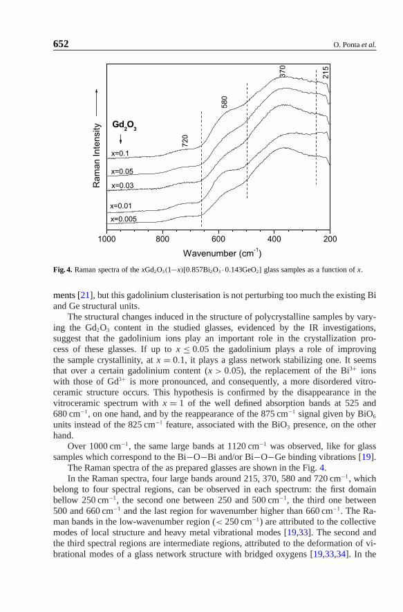
The Raman spectra of the as prepared glasses are shown in the Fig. 4.In the Raman spectra, four large bands around 215, 370, 580 and 720 cm−1, which
belong to four spectral regions, can be observed in each spectrum: the first domainbellow 250 cm−1, the second one between 250 and 500 cm−1, the third one between500 and 660 cm−1 and the last region for wavenumber higher than 660 cm−1. The Ra-man bands in the low-wavenumber region (< 250 cm−1) are attributed to the collectivemodes of local structure and heavy metal vibrational modes [19,33]. The second andthe third spectral regions are intermediate regions, attributed to the deformation of vi-brational modes of a glass network structure with bridged oxygens [19,33,34]. In the
Vibrational Spectroscopic Studies of Germanium-High Bismuthate Glasses and Vitroceramics 653
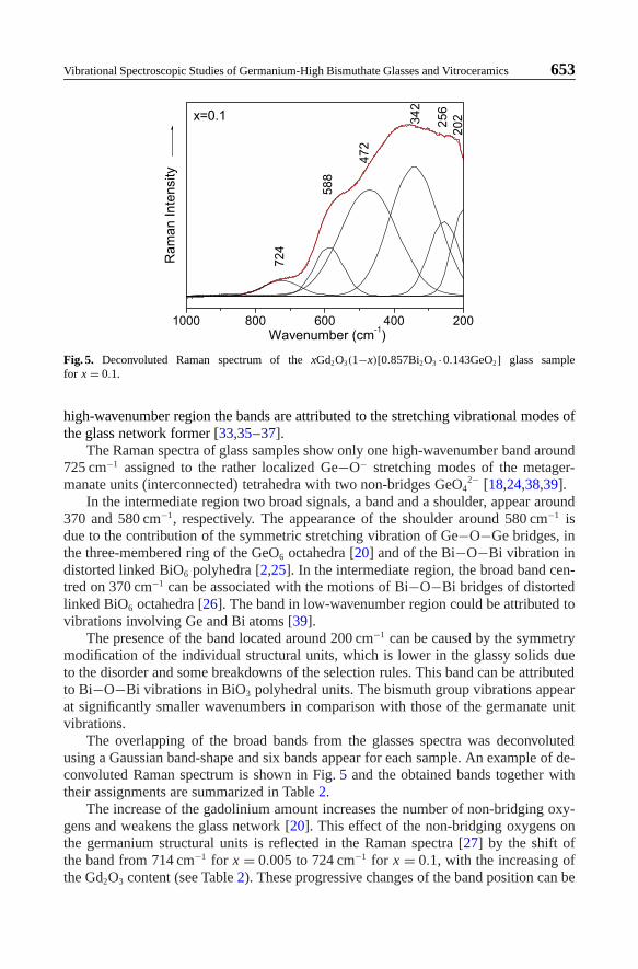
Fig. 5. Deconvoluted Raman spectrum of the xGd2O3(1−x)[0.857Bi2O3 ·0.143GeO2] glass samplefor x = 0.1.
high-wavenumber region the bands are attributed to the stretching vibrational modes ofthe glass network former [33,35–37].
The Raman spectra of glass samples show only one high-wavenumber band around725 cm−1 assigned to the rather localized Ge−O− stretching modes of the metager-manate units (interconnected) tetrahedra with two non-bridges GeO4
2− [18,24,38,39].In the intermediate region two broad signals, a band and a shoulder, appear around
370 and 580 cm−1, respectively. The appearance of the shoulder around 580 cm−1 isdue to the contribution of the symmetric stretching vibration of Ge−O−Ge bridges, inthe three-membered ring of the GeO6 octahedra [20] and of the Bi−O−Bi vibration indistorted linked BiO6 polyhedra [2,25]. In the intermediate region, the broad band cen-tred on 370 cm−1 can be associated with the motions of Bi−O−Bi bridges of distortedlinked BiO6 octahedra [26]. The band in low-wavenumber region could be attributed tovibrations involving Ge and Bi atoms [39].
The presence of the band located around 200 cm−1 can be caused by the symmetrymodification of the individual structural units, which is lower in the glassy solids dueto the disorder and some breakdowns of the selection rules. This band can be attributedto Bi−O−Bi vibrations in BiO3 polyhedral units. The bismuth group vibrations appearat significantly smaller wavenumbers in comparison with those of the germanate unitvibrations.
The overlapping of the broad bands from the glasses spectra was deconvolutedusing a Gaussian band-shape and six bands appear for each sample. An example of de-convoluted Raman spectrum is shown in Fig. 5 and the obtained bands together withtheir assignments are summarized in Table 2.
The increase of the gadolinium amount increases the number of non-bridging oxy-gens and weakens the glass network [20]. This effect of the non-bridging oxygens onthe germanium structural units is reflected in the Raman spectra [27] by the shift ofthe band from 714 cm−1 for x = 0.005 to 724 cm−1 for x = 0.1, with the increasing ofthe Gd2O3 content (see Table 2). These progressive changes of the band position can be
654 O. Ponta et al.
Table 2. The wavenumbers of the Raman bands for vitroceramic samples and for the deconvoluted Ramanbands of glass samples belonging to the xGd2O3(1−x)[0.857Bi2O3 ·0.143GeO2] system.
Wavenumber (cm−1)
Vitroceramics 205 240 270 320 451 483 530 615 715Glasses
x = 0.005 200 245 335 461 584 714x = 0.01 196 242 347 470 588 722x = 0.03 199 241 341 463 583 720x = 0.05 201 258 353 490 591 726x = 0.1 202 256 342 472 588 724
interpreted as the occurrence of a slight improvement in the GeO2 network connectiv-ity [27].
In the intermediate-wavenumber region three bands appear after deconvolutionof the glass sample spectra and are assigned to the symmetric stretching of anionmotion, i.e. vibration of bridging oxygen’s, in angularly constrained Bi−O−Bi config-uration [2]. The band located around 584 cm−1 for x = 0.005 and shifted at 588 cm−1
for x = 0.1 is given by the Bi−O−Bi vibration in distorted linked BiO6 polyhedra, butcan be also a contribution of the symmetric stretching vibration of Ge−O−Ge bridges,in the three-membered ring of GeO6 octahedra [27]. The bands located between461 cm−1, for x = 0.005, and 472 cm−1, for x = 0.1, can be assigned to Bi−O−Biand/or Ge−O−Ge stretching vibrations [33]. The band at 335 cm−1 for x = 0.005 isshifted to 353 cm−1 for x = 0.05, and it decreases in intensity and becomes broader asthe Gd2O3 content increases. The band at 335 cm−1 is specific to the Bi−O− “breath-ing” vibrations in BiO3 pyramidal units [29] and the 353 cm−1 band to the symmetricstretching vibrations of Bi−O−Bi bridges of the distorted linked BiO6 octahedralunits [28].
In the low-wavenumber region two bands appear, one around 200 cm−1 and an-other one at 245 cm−1, for x = 0.005, the latter being shifted at 256 cm−1 for x = 0.1.The presence of the Raman signal located around 200 cm−1 indicates that Bi3+ cationsare incorporated in the sample structure as [BiO3] pyramidal and [BiO6] octahedralunits [28].
Even as a small effect we can conclude that the increase of the gadolinium contentinfluences the local structure of the bismuth and germanium in glass samples.
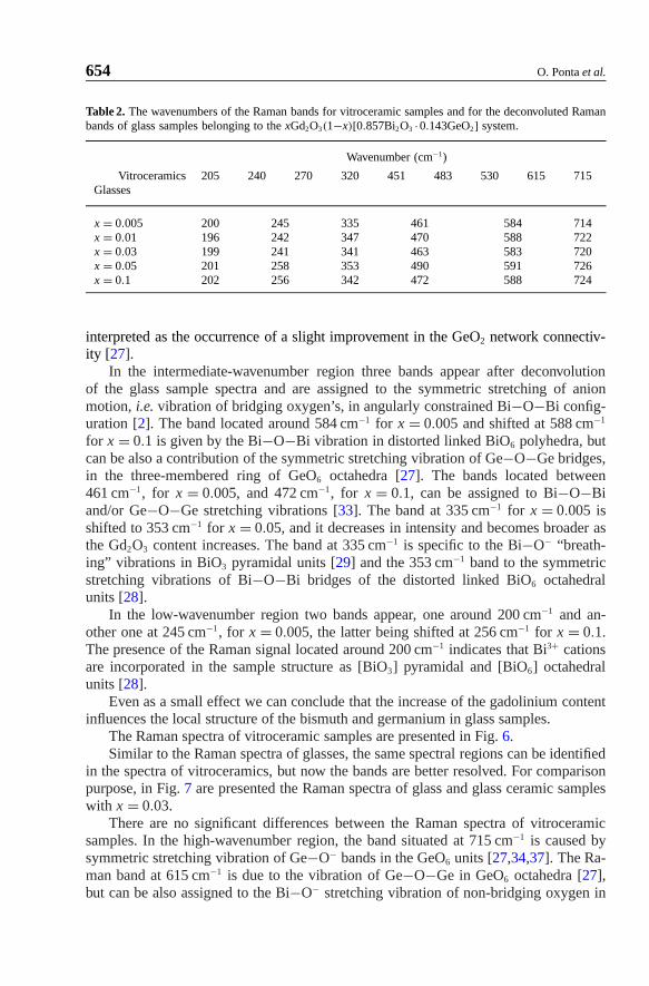
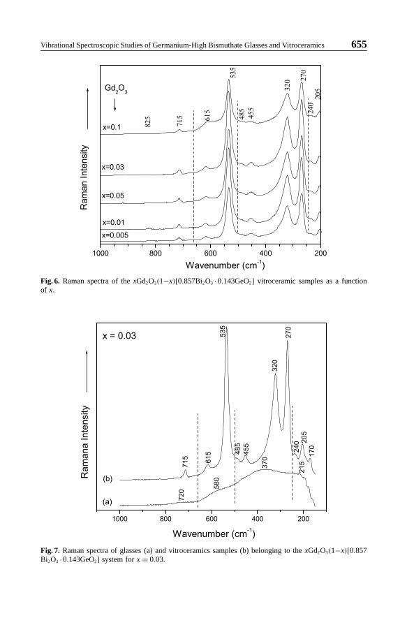
The Raman spectra of vitroceramic samples are presented in Fig. 6.Similar to the Raman spectra of glasses, the same spectral regions can be identified
in the spectra of vitroceramics, but now the bands are better resolved. For comparisonpurpose, in Fig. 7 are presented the Raman spectra of glass and glass ceramic sampleswith x = 0.03.
There are no significant differences between the Raman spectra of vitroceramicsamples. In the high-wavenumber region, the band situated at 715 cm−1 is caused bysymmetric stretching vibration of Ge−O− bands in the GeO6 units [27,34,37]. The Ra-man band at 615 cm−1 is due to the vibration of Ge−O−Ge in GeO6 octahedra [27],but can be also assigned to the Bi−O− stretching vibration of non-bridging oxygen in
Vibrational Spectroscopic Studies of Germanium-High Bismuthate Glasses and Vitroceramics 655
Fig. 6. Raman spectra of the xGd2O3(1−x)[0.857Bi2O3 ·0.143GeO2] vitroceramic samples as a functionof x.
Fig. 7. Raman spectra of glasses (a) and vitroceramics samples (b) belonging to the xGd2O3(1−x)[0.857Bi2O3 ·0.143GeO2] system for x = 0.03.
656 O. Ponta et al.
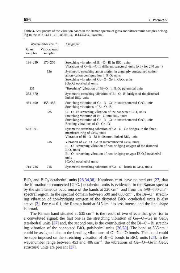
Table 3. Assigments of the vibration bands in the Raman spectra of glass and vitroceramic samples belong-ing to the xGd2O3(1−x)[0.857Bi2O3 ·0.143GeO2] system.
Wavenumber (cm−1) Assigment
Glass Vitroceramicsamples samples
196–259 170–270 Stretching vibration of Bi−O−Bi in BiO3 unitsVibrations of O−Bi−O in different structural units (only for 240 cm−1)
320 Symmetric stretching anion motion in angularly constrained cation–anion–cation configuration in BiO6 unitsStretching vibration of Ge−O−Ge in GeO4 units[GeO6] octahedral units
335 “Breathing” vibration of Bi−O− in BiO3 pyramidal units
353–370 Symmetric stretching vibration of Bi−O−Bi bridges of the distortedlinked BiO6 units
461–490 455–485 Stretching vibration of Ge−O−Ge in interconnected GeO4 unitsStretching vibrations of Bi−O−Bi
535 Bi−O−Bi stretching vibration of the connected BiO6 unitsStretching vibration of Bi−O into BiO6 unitsStretching vibration of Ge−O−Ge in interconnected GeO4 unitsBending vibrations of O−Ge−O
583–591 Symmetric stretching vibration of Ge−O−Ge bridges, in the three-membered ring of GeO6 unitsVibration of Bi−O−Bi in distorted linked BiO6 units
615 Vibration of Ge−O−Ge in interconnected GeO4 unitsBi−O− stretching vibration of non-bridging oxygen of the distortedBiO3 unitsBi−O− stretching vibration of non-bridging oxygen [BiO6] octahedralunits[GeO6] octahedral units
714–726 715 Symmetric stretching vibration of Ge−O− bands in GeO4 units
BiO6 and BiO3 octahedral units [28,34,38]. Kamitsos et al. have pointed out [27] thatthe formation of connected [GeO6] octahedral units is evidenced in the Raman spectraby the simultaneous occurrence of the bands at 320 cm−1 and from the 590–630 cm−1
spectral region. In the spectral domain between 590 and 630 cm−1, the Bi−O− stretch-ing vibration of non-bridging oxygen of the distorted BiO3 octahedral units is alsoactive [2]. For x = 0.1, the Raman band at 615 cm−1 is less intense and the line shapeis broad.
The Raman band situated at 535 cm−1 is the result of two effects that give rise toa convoluted signal: the first one is the stretching vibration of Ge−O−Ge in GeO4
tetrahedral units [27] and, the second one, is the contribution of the Bi−O−Bi stretch-ing vibration of the connected BiO6 polyhedral units [26,28]. The band at 535 cm−1
could be assigned also to the bending vibrations of O−Ge−O bonds. This band couldbe superimposed on the stretching vibration of Bi−O bonds in BiO6 units [24]. In thewavenumber range between 453 and 486 cm−1, the vibrations of Ge−O−Ge in GeO4
structural units are present [27].

Vibrational Spectroscopic Studies of Germanium-High Bismuthate Glasses and Vitroceramics 657
Two strong Raman bands are evidenced in the 250–350 cm−1 spectral range of thevitroceramics spectra. The band at 320 cm−1 appears in the spectra due to the symmet-ric stretching anion motion in angularly constrained cation–anion–cation configurationin BiO6 polyhedral units [26,28,40]. At this value the Ge–O–Ge stretching vibration inGeO4 tetrahedral units is also present. The other strong band in this wavenumber range,located at 270 cm−1, can be attributed to the Bi−O−Bi stretching vibration in BiO3
units [29].The bands located between 200 and 250 cm−1 arise in the Raman spectra due to
the Bi−O vibrations, while the broad band around 240 cm−1 can be attributed to theBi−O−Bi vibration of the distorted BiO3 pyramidal units, modified in the presenceof GeO4 units [28]. The Raman features located around 240 cm−1 can be assigned toO−Bi−O vibrations in different structural units [41]. In the low-wavenumber region,the bands located around 205 cm−1 can be caused by the symmetry of the individualstructural units, and are attributed to Bi−O−Bi vibrations in BiO3 polyhedra [26,36].A close analysis of the Raman spectra of the vitroceramics (Fig. 6) confirms the con-clusion derived from IR analysis concerning the occurrence of a structure with a higherdisorder degree for x = 0.1, as a consequence of the partial replacement of the Bi3+ ionswith Gd3+ ions. This spectral behavior can be easily observed by looking at the broad-ening of the main Raman features from the spectrum with x = 0.1, where bismuthatevibrations are employed.
4. Conclusions
The germanium-high bismuthate glasses and vitroceramics containing different con-centration of Gd2O3 have been prepared and structurally characterized using vibrationalspectroscopic methods. According to the IR and Raman data the structural units of theglass network are BiO3, BiO6, GeO6 and GeO4. The Bi3+ cations are incorporated inthe structure of the samples as BiO6 octahedral and/or BiO3 pyramidal units slightlydepending on the gadolinium content from their composition and on the applied heattreatment. The vibrations of the structural units that build up the vitroceramics structureusually occurs at similar wavenumbers as those of the structural units evidenced in theglass spectra, as revealed by the deconvolution results. This suggests the existence ofthe same structural units in the structure of both glass and vitroceramic samples. Theonly slight dependence of vibrational spectra on gadolinium concentration support theassumption that Gd3+ ions occupy the same type of sites hexacoordinated with oxy-gen’s, like Bi3+ ions in octahedral BiO6 units. Moreover, the occurrence of a structurewith a higher disorder degree for x = 0.1 in comparison with the other investigatedvitroceramics as a consequence of the partial replacement of the Bi3+ ions with Gd3+
ones was proved by IR and Raman analyses. A partial change of the BiO6 structuralunits in BiO3 units in developed crystalline phases is supported, mainly by IR data.The similarity between IR and Raman spectra of glasses and vitroceramics with dif-ferent gadolinium amount, even if the identified crystalline phases in vitroceramics arestrongly dependent on this content, shows that the local order is very similar, but thelong range order in developed crystalline phases is different, due to different interlink-ing between identified structural units.

658 O. Ponta et al.
Acknowledgement
Many thanks to Prof. Dr. Dr. h. c. W. Kiefer from the Institute of Physical-Chemistryof Bayerische Julius-Maximilians University, Würzburg-Germany, who facilitated ouraccess to Raman laboratories in order to perform the measurements.
The financial support of this work given by the National University Research Coun-cil CNCSIS – Romania (PN II, ID-566/2007) is acknowledged.
References
1. I. Manzini, P. P. Lottici, and G. Antonioli, J. Non-Cryst. Solids 224 (1998) 23.2. A. Pan and A. Ghosh, J. Mater. Res. 17 (2002) 1941.3. A. Pan and A. Ghosh, Phys. Rev. B 62 (2000) 3190.4. A. Pan and A. Ghosh, J. Non-Cryst. Solids 271 (2000) 157.5. A. Ghosh, J. Appl. Phys. 65 (1989) 227.6. A. Ghosh and D. Chakravorty, J. Phys.: Condens. Matter 2 (1990) 649.7. A. Dutta and A. Ghosh, J. Non-Cryst. Solids 351 (2005) 203.8. S. Bhattacharya and A. Ghosh, Phys. Rev. B 68 (2003) 224202.9. G. D. Chryssikos, E. I. Kamistos, and W. M. Risen, J. Non-Cryst. Solids 93 (1987) 155.
10. J. B. Shim, J. H. Lee, A. Yoshikova, M. Nikl, D. H. Joon, and T. Fukuda, J. Cryst. Growth 243(2002) 157.
11. H. Schweppe, IEEE Trans. Sonics Ultrasonics SU-16 (1969) 219.12. X. Q. Feng, G. Q. Hu, Z. W. Yin, Y. P. Hung, S. Kapphan, C. Ficher, F. Z. Zhou, Y. Yang, and
D. Y. Fan, Mater. Sci. Eng. B 23 (1994) 83.13. D. Hall, N. Newhause, N. Borrell, W. Dumbaugh, and D. Weidman, Appl. Phys. Lett. 54
(1989) 1293.14. P. P. Lottici, I. Manzini, G. Antonioli, G. Gnappi, and A. Montenero, J. Non-Cryst. Solids 159
(1993) 173.15. J. Wong and C. A. Angell, Glass Structure by Spectroscopy. M. Dekker, Inc., New York
(1976), p. 409.16. G. Fuxi, Optical and Spectroscopic Properties of Glass. Springer-Verlag, Shanghai Scientific
Technical Pub., Shanghai (1991).17. Y. D. Yiannopoulos, G. D. Chryssikos, and E. I. Kamitsos, Phys. Chem. Glasses 42, (2001)
164.18. L. Baia, M. Baia, W. Kiefer, J. Popp, and S. Simon, Chem. Phys. 327 (2006) 63.19. L. Baia, W. Kiefer, and S. Simon, Multispectroscopic studies of local structure in heavy metal
glasses, in: Recent Res. Devel. Non-Crystalline Solids-Transworld Research Network. Kerala,India (2004), pp. 1–25.
20. V. Simon, O. Ponta, S. Simon, D. A. Udvar, and M. Neumann, Phys. Stat. Solidi (a) 205(5)(2008) 1139.
21. S. Simon and D. Udvar, J. Am. Ceram. Soc. 93(9) (2010) 2760.22. V. Dimitrov, Y. Dimitriev, and A. Montenero, J. Non-Cryst. Solids 180 (1994) 51.23. P. Pernice, A. Aronne, M. Catauro, and A. Maratto, J. Non-Cryst. Solids 210 (1997) 23.24. R. Iordanova, Y. Dimitriev, V. Dimitrov, S. Kassabov, and D. Klissurski, J. Non-Cryst. Solids
204 (1996) 141.25. D. Sreenivasu and V. Chandramouli, Bull. Mater. Sci. 23(4) (2000) 281.26. L. Baia, R. Stefan, J. Popp, S. Simon, and W. Kiefer, J. Non-Cryst. Solids 324 (2003) 109.27. E. I. Kamitsos, Y. D. Yiannopoulos, M. A. Karacassides, G. D. Cryssikos, and H. Jain, J. Phys.
Chem. 100 (1997) 11755.28. L. Baia, R. Stefan, W. Kiefer, J. Popp, and S. Simon, J. Non-Cryst. Solids 303 (2002) 379.29. A. Radu, L. Baia, W. Kiefer, and S. Simon, Vib. Spectrosc. 39 (2005) 127.30. D. A. Udvar and S. Simon, J. Opt. Adv. Mater. 9 (2007) 646.31. V. Simon, O. Ponta, D. Trandafir, and H. Mocuta, J. Non-Cryst. Solids 355 (2009) 2451.

Vibrational Spectroscopic Studies of Germanium-High Bismuthate Glasses and Vitroceramics 659
32. S. Hazra and A. Gosh, Phys. Rev. B 51 (1995) 851.33. H. Sun, C. Yu, L. Zhang, Z. Duan, D. He, J. Zhang, L. Hu, and Z. Jiang, Solid State Commun.
134 (2005) 595.34. L. Baia, T. Iliescu, S. Simon, and W. Kiefer, J. Mol. Struct. 599 (2001) 9.35. L. G. Hwa, Y. R. Chang, and W. C. Chao, Mater. Chem. Phys. 85 (2004) 161.36. L. Baia, W. Kiefer, and S. Simon, Rom. Rep. Phys. 56 (2004) 430.37. P. Beneventi, D. Bersani, P. P. Lotici, L. Kovacs, F. Cordioli, A. Montenero, and G. Gnappi,
J. Non-Cryst. Solids 192/193 (1995) 258.38. A. A. Kharlamov, R. M. Almeida, and J. Heo, J. Non-Cryst. Solids 202 (1996) 233.39. H. Sun, L. Zang, C. Yu, Z. Duan, J. Zhang, S. Dai, L. Hu, and Z. Jiang, Solid State Commun.
134 (2005) 449.40. L. Baia, R. Stefan, W. Kiefer, and S. Simon, J. Raman Spectrosc. 36 (2005) 262.41. L. Baia, W. Kiefer, and S. Simon, Phys. Chem. Glass. 46 (2005) 279.