Embed Size (px)
Citation preview

Veterinary Radiology Fellowship Guidelines 2017 © 2017 The Australian and New Zealand College of Veterinary Scientists ABN 00 50 000894 208 Page 1 of 16
2017
AUSTRALIAN AND NEW ZEALAND
COLLEGE OF VETERINARY SCIENTISTS
FELLOWSHIP GUIDELINES
Veterinary Radiology
ELIGIBILITY
1. The candidate must meet the eligibility prerequisites for Fellowship outlined in
the Fellowship Candidate Handbook.
2. Membership of the College must be achieved prior to the Fellowship
examination.
3. Membership must be in Veterinary Radiology.
OBJECTIVES
To demonstrate that the candidate has attained sufficient knowledge, training,
experience, and accomplishment to meet the criteria for registration as a specialist in
veterinary radiology.
LEARNING OUTCOMES
The field of veterinary radiology includes the study of all domestic animals. There is
no provision for sub-specialisation within the discipline.
The candidate will have a detailed
1
knowledge of:
1. Radiation physics as it applies to veterinary radiography
1.1. atomic and nuclear physics including atomic composition, structure and
binding forces
1.2. forms of electromagnetic radiation
1.3. production of x-rays, interaction of x-rays with matter, components and
function of the x-ray tube, components and function of x-ray detection
systems (both film-screen and digital radiography)
1 Knowledge Levels:
Detailed knowledge - candidates must be able to demonstrate an in-depth knowledge of the topic including differing points of view
and published literature. The highest level of knowledge. Sound knowledge – candidate must know all of the principles of the topic including some of the finer detail, and be able to identify
areas where opinions may diverge. A middle level of knowledge.
Basic knowledge – candidate must know the main points of the topic and the main literature.

Veterinary Radiology Fellowship Guidelines 2017 © 2017 The Australian and New Zealand College of Veterinary Scientists ABN 00 50 000894 208 Page 2 of 16
1.4. radiographic artefacts (both film-screen and digital radiography).
2. Digital radiography including image formation, different capture devices,
resolution, storage, the processing of photostimulable phosphor plates (PSP), the
advantages and disadvantages of different types of digital radiography.
3. Computed Tomography (CT) physics:
3.1. image formation including CT construction and scanner types, image
manipulation
3.2. factors that affect CT image quality
3.3. CT artefacts.
4. Magnetic Resonance Imaging (MRI )physics:
4.1. image formation, including equipment, magnet field strengths, and an
understanding of the principles of acquisition of SE, FSE, GRE
sequences of different tissue weightings.
4.2. factors that affect MRI image quality
4.3. MRI artefacts
4.4. practical applications of MRI safety.
5. Ultrasound physics:
5.1. image formation including equipment
5.2. physical characteristics of the ultrasound beam and the interaction of
ultrasound with matter
5.3. physics of Doppler, harmonic and compound imaging
5.4. ultrasound artefacts
6. Contrast media:
6.1. radiographic, CT and MRI contrast media including mechanism, side
effects, administration and dose.
7. Anatomy,physiology and pathophysiology as related to veterinary radiology of
dogs, cats and horses.
8. Clinical and pathophysiological features as related to veterinary radiology of
canine, feline and equine disease of all body systems.
9. Practical applications of all modalities.
The candidate will have a sound knowledge of:
1. Radiation safety in veterinary medicine:
1.1. principles of radiation protection including the ALARA concept
1.2. interactions of electromagnetic and particulate radiation with matter
1.3. biological effects of radiation in a clinical radiology context
1.4. mechanisms of acute and late radiation injury
1.5. radiation monitoring, safety equipment and regulations

Veterinary Radiology Fellowship Guidelines 2017 © 2017 The Australian and New Zealand College of Veterinary Scientists ABN 00 50 000894 208 Page 3 of 16
1.6. relevant Australian and New Zealand laws and Codes of Practice as they
apply to the use of ionising radiation.
2. Embryology of the cardiovascular, urinary and neurological systems as it relates
to development of congenital conditions of these systems.
3. Radiation physics as it applies to:
3.1. fluoroscopy and the image intensifier
3.2. nuclear scintigraphy
4. Ultrasound contrast media including mechanism of action, side effects,
administration and dose.
The candidate will have a basic knowledge of:
1. Anatomy, physiology and pathophysiology of disease as related to veterinary
radiology of production animals, native and exotic species, avian, and pocket pet
species.
2. Radiation oncology:
2.1. principles of radiation therapy
2.2. radiobiology of the cell cycle.
3. The biological effects of ultrasound.
The candidate must be able to demonstrate a detailed2 level of expertise in:
1. Image acquisition, interpretation and reporting:
1.1. radiographic, ultrasound, CT and MRI images in dogs, cats and horses.
2. Image guided biopsy:
2.1. techniques, including fine needle aspiration and percutaneous biopsy
3. Critical evaluation of the current literature and concepts in the field of
Veterinary Radiology.
The candidate must be able to demonstrate a sound level of expertise in:
1. Image acquisition, interpretation and reporting:
1.1. nuclear medicine (NM) studies in both small and large animals.
1.2. radiographic, ultrasound, CT and MR images of species other than dogs, cats and horses
2 Skill levels:
Detailed expertise – the candidate must be able to perform the technique with a high degree of skill, and have extensive experience
in its application. The highest level of proficiency. Sound expertise – the candidate must be able to perform the technique with a moderate degree of skill, and have moderate experi-
ence in its application. A middle level of proficiency.
Basic expertise – the candidate must be able to perform the technique competently in uncomplicated circumstances.

Veterinary Radiology Fellowship Guidelines 2017 © 2017 The Australian and New Zealand College of Veterinary Scientists ABN 00 50 000894 208 Page 4 of 16
EXAMINATIONS
Exam Format:
The examination will comprise of three components:
1. Two written examinations each of 180 minutes each
2. A practical examination comprising of 370 minutes
3. An oral examination comprising of at least 60 minutes, and no longer than 120
minutes.
Written exams: Two (2) written examinations of three (3) hours duration each (total of
6 hours).
Each written examination will comprise of
• 4 x essay-type questions of 30 minutes each. Questions can be broken into multiple
sub-parts. TOTAL TIME: 120 minutes
• 12 short-answer questions (5 minutes each) TOTAL TIME: 60 minutes
Candidates are required to answer showing reference to the literature (e.g. citing
relevant studies that inform their answers). Ideally citations should include the primary
author, journal abbreviation and publication year; in cases where this recall is not
possible, as much detail as possible should be included.
Candidates will often be expected to use their own clinical experience in answering
questions, demonstrating experience with modalities.
Perusal time of 20 minutes will be provided at the start of each written paper;
candidates are recommended to use this time to read carefully the questions and plan
their essay answers. During this time candidates may make notes on the examination
paper but not write in the answer book. Care should be taken to address the question
posed. Written Paper 1:
Designed to test the Candidate’s knowledge and clinical application of physics,
anatomy and pathophysiology as described in the Learning Outcomes.
Written Paper 2:
Designed to test the Candidate’s ability to apply the principles of Radiology to
particular cases/problems or tasks. The candidate may be required to draw on their
knowledge of pathophysiology, physics and anatomy to answer the questions.
Practical examination: Two (2) examinations of three (3) hours duration writing time
(total of 6 hours).
Each practical examination will test the candidate’s ability to produce written case
reports. Each examination will have a five (5) minute rest break in the middle hour.
During this break, the candidate will put down their pen, cease writing and turn their
paper over. No talking will be allowed. Candidates may use the bathroom during this
period. Each examination will last for 185 minutes (including rest breaks). No perusal
time will be given.
Veterinary Radiology Fellowship Guidelines 2017 © 2017 The Australian and New Zealand College of Veterinary Scientists ABN 00 50 000894 208 Page 5 of 16
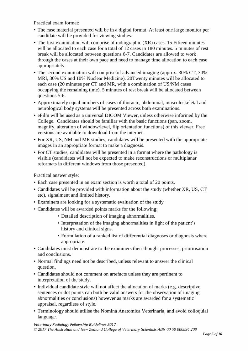
Practical exam format:
• The case material presented will be in a digital format. At least one large monitor per
candidate will be provided for viewing studies.
• The first examination will comprise of radiographic (XR) cases. 15 Fifteen minutes
will be allocated to each case for a total of 12 cases in 180 minutes. 5 minutes of rest
break will be allocated between questions 6-7. Candidates are allowed to work
through the cases at their own pace and need to manage time allocation to each case
appropriately.
• The second examination will comprise of advanced imaging (approx. 30% CT, 30%
MRI, 30% US and 10% Nuclear Medicine). 20Twenty minutes will be allocated to
each case (20 minutes per CT and MR, with a combination of US/NM cases
occupying the remaining time). 5 minutes of rest break will be allocated between
questions 5-6.
• Approximately equal numbers of cases of thoracic, abdominal, musculoskeletal and
neurological body systems will be presented across both examinations.
• eFilm will be used as a universal DICOM Viewer, unless otherwise informed by the
College. Candidates should be familiar with the basic functions (pan, zoom,
magnify, alteration of window/level, flip orientation functions) of this viewer. Free
versions are available to download from the internet.
• For XR, US, NM and MR studies, candidates will be presented with the appropriate
images in an appropriate format to make a diagnosis.
• For CT studies, candidates will be presented in a format where the pathology is
visible (candidates will not be expected to make reconstructions or multiplanar
reformats in different windows from those presented).
Practical answer style:
• Each case presented in an exam section is worth a total of 20 points.
• Candidates will be provided with information about the study (whether XR, US, CT
etc), signalment and limited history.
• Examiners are looking for a systematic evaluation of the study
• Candidates will be awarded points marks for the following:
• Detailed description of imaging abnormalities.
• Interpretation of the imaging abnormalities in light of the patient’s
history and clinical signs.
• Formulation of a ranked list of differential diagnoses or diagnosis where
appropriate.
• Candidates must demonstrate to the examiners their thought processes, prioritisation
and conclusions.
• Normal findings need not be described, unless relevant to answer the clinical
question.
• Candidates should not comment on artefacts unless they are pertinent to
interpretation of the study.
• Individual candidate style will not affect the allocation of marks (e.g. descriptive
sentences or dot points can both be valid answers for the observation of imaging
abnormalities or conclusions) however as marks are awarded for a systematic
appraisal, regardless of style.
• Terminology should utilise the Nomina Anatomica Veterinaria, and avoid colloquial
language.

Veterinary Radiology Fellowship Guidelines 2017 © 2017 The Australian and New Zealand College of Veterinary Scientists ABN 00 50 000894 208 Page 6 of 16
Oral Examination:
• Questions will be provided in a digital format using a proprietary viewer or Power
Point.
• These questions aim to test how the candidate arrives at their radiographic
conclusions.
• Candidates will be provided with information about the study such as signalment and
limited history.
• For imaging studies consisting of large data sets, the relevant images (single images,
series, sequences) will be provided.
• Candidates may request additional imaging studies.
• The candidates must demonstrate to the examiners their thought processes,
prioritisation and conclusions.
• Candidates will be awarded points for the following:
• Description of imaging abnormalities.
• Formulation of rational imaging conclusions and a ranked differential
diagnosis list, or diagnosis where appropriate
• Ability to synthesise imaging findings with the patient's clinical history and
signs. Candidates should demonstrate an understanding of the
pathophysiology of observed abnormalities and rational justification for the
use of ancillary tests.
• Ability to make appropriate recommendations for additional patient
management recommendations, including both imaging-related diagnostics
and other pertinent diagnostic testing. The candidate may recommend and
ask for further imaging studies. E.g.For example, if it is appropriate after
reading a radiographic study to recommend ultrasound, the candidate may
ask whether such a study is available.
• Normal findings need not be described, unless relevant to answer the clinical
question.
• Candidates should not comment on artifacts unless they are pertinent to interpretation
of the study
Examples of questions:
1. Thoracic radiograph series of a dog. “This is a 7 year old Doberman with recent
onset tachypnoea. Give your radiographic description and conclusions”.
2. Two transverse images of an MRI study of a canine brain, pre and post contrast.
“Describe briefly the pathology that you see. What are you differentials for this
lesion” (The images demonstrate a typical meningioma).
3. An image showing a spectral Doppler trace through a normal LVOT: “What is this
image depicting? What would you expect to see if a patient had aortic stenosis?”
4. “Describe the artefact you see and discuss how this occurred”
5. An image depicting a brand of contrast medium. “What is this chemical? What are
the indications and contraindications for its use?” Additional notes for the Practical and Oral Examination
In the practical and oral examinations, candidates will be provided with information
about the study they are receiving. They will not be awarded marks for describing that
a study is a three-view thoracic radiographic study, or an echocardiogram of a cat’s
heart, etc.
Veterinary Radiology Fellowship Guidelines 2017 © 2017 The Australian and New Zealand College of Veterinary Scientists ABN 00 50 000894 208 Page 7 of 16
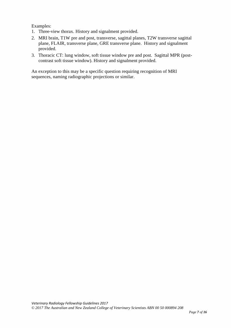
Examples:
1. Three-view thorax. History and signalment provided.
2. MRI brain, T1W pre and post, transverse, sagittal planes, T2W transverse sagittal
plane, FLAIR, transverse plane, GRE transverse plane. History and signalment
provided.
3. Thoracic CT: lung window, soft tissue window pre and post. Sagittal MPR (post-
contrast soft tissue window). History and signalment provided.
An exception to this may be a specific question requiring recognition of MRI
sequences, naming radiographic projections or similar.

Veterinary Radiology Fellowship Guidelines 2017 © 2017 The Australian and New Zealand College of Veterinary Scientists ABN 00 50 000894 208 Page 8 of 16
TRAINING PROGRAMS
Refer to the Fellowship Candidate Handbook, Section 3.3.
In addition to the stipulations of the Fellowship Candidate Handbook:
1. The Radiology Chapter requires a three year training program (144 weeks)
2. Clinical training should include primarily exposure to dogs, cats and horses
with some exposure to production animals, camelids, native and exotic ani-
mals, pocket pets and birds.
3. Clinical training should include the following: radiography, radiology, con-
trast procedures, fluoroscopy/image intensification, digital radiography, so-
nography, sonology, scintigraphy, computed tomography and magnetic reso-
nance imaging.
4. The candidate should interpret a minimum 3000 radiographic examinations of
small animals (primarily dogs and cats), 500 radiographic examinations of
large animals (primarily horses), 1000 sonographic examinations, and a min-
imum of 500 examinations that demonstrate adequate knowledge and interpre-
tive skills in CT, MRI and nuclear medicine.
The cases to be included in the case log will be those cases in which the candidate has
produced a written report that has been reviewed by a Supervisor. If, for example, a
case has an osteosarcoma of its radius and thoracic radiographs for a metastasis check
then this may be counted as two cases if a report is produced for both regions.
The training program should be targeted, with achievable goals set by the Supervisor
and candidate for each 6 months. It is anticipated that the first year should be spent
initially learning some radiography, then concentrating on radiology and ultrasound
with some exposure to the other modalities. The second year is spent consolidating the
first with more CT, nuclear medicine with shifting emphasis in the third year to more
CT and MRI and nuclear medicine with further consolidation of radiology and
ultrasound. It should be expected that the candidate’s case log output is lower in the
first year but that they become more independent and productive in their second and
third years.
Sonography and Sonology Assessment:
The candidate’s supervisor will continually assess the Candidate’s development of so-
nography and sonology skills. If the Candidate’s skills were found to be less than sat-
isfactory at the end of the third year of their approved training program, the Candidate
will be required to undertake further training before being further assessed. The Candi-
date will not proceed to formal examinations until they have been determined to have
adequate sonography and sonology skills. A pro-forma letter (Appendix 1) will be
completed by the Candidate’s supervisor and submitted with the Fellowship training
credentials documentation, to state whether the Candidate is considered technically
proficient in ultrasound, and to justify the reasons for the assessment.
TRAINING IN RELATED DISCIPLINES
Refer to the Fellowship Candidate Handbook, 2.4.2.
Candidates for Fellowship in Veterinary Radiology must spend time as stipulated by
the Fellowship Candidate Handbook in any four of the following related disciplines:
Pathology, Small Animal Medicine, Canine Medicine, Feline Medicine, Cardiology,
Small Animal Surgery, Equine Medicine, Equine Surgery, Neurology, Oncology.

Veterinary Radiology Fellowship Guidelines 2017 © 2017 The Australian and New Zealand College of Veterinary Scientists ABN 00 50 000894 208 Page 9 of 16
EXTERNSHIPS
Refer to the Fellowship Candidate Handbook, Section 2.4.1.
ACTIVITY LOG SUMMARY
An Activity Log Summary should be provided for each imaging modality (Radiology,
Ultrasound, Special Radiographic Procedures, CT, MRI and Nuclear Medicine)
according to the template provided in Appendix 2. Each summary should be submitted
with the annual supervisor’s report, with a cumulative total for the total training
period. For each imaging modality, cases are recorded by species and the region
imaged (as listed below). This allows the candidate and their supervisor to monitor
their case load for each modality (e.g. numbers of canine abdominal ultrasounds,
numbers of equine musculoskeletal radiographs, etc), and assess whether the targets
mentioned above (section 4 under the ‘Training Programs’ heading) are being
achieved.
Radiology:
Thorax Abdomen
Musculoskeletal
Neurological
Other
Ultrasound:
Thorax - non cardiac Thorax - cardiac
Abdomen
Musculoskeletal
Small Parts (eg thyroid, eye, etc)
Biopsies/FNA
Special Radiologic Procedures:
Myelography Urinary contrast studies
Oesophagrams
Other contrast studies
Fluoroscopy (non-contrast)
CT:
Thorax Abdomen
Musculoskeletal
Neurological
MRI:
Neurological
Other

Veterinary Radiology Fellowship Guidelines 2017 © 2017 The Australian and New Zealand College of Veterinary Scientists ABN 00 50 000894 208 Page 10 of 16
Nuclear Medicine:
Musculoskeletal Thyroid
Hepatic
Other
Species list for each modality:
Canine Feline
Equine
Production animals (cows, sheep, goats, alpacas, pigs)
Avian/Other
Note that an imaging study of a region is considered a case. If multiple regions are
imaged of a single patient (e.g. radiographs of a long bone and thorax for metastasis
check) these would be considered two cases; one musculoskeletal and one thorax –
non cardiac, provided both are reported.
PUBLICATIONS and PRESENTATION
Refer to the Fellowship Candidate Handbook, Section 2.10.

Veterinary Radiology Fellowship Guidelines 2017 © 2017 The Australian and New Zealand College of Veterinary Scientists ABN 00 50 000894 208 Page 11 of 16
RECOMMENDED READING LIST
The candidate is expected to research the depth and breadth of the knowledge of the
discipline. This list is intended to guide the candidate to some core references and
source material. The list is not comprehensive and is not intended as an indicator of
the content of the examination. Candidates at Fellowship level are expected to have
library search skills and maintain a watching brief over relevant literature.
Physics
Bushberg JT, Seibert JA, Leidholdt Jr EM, Boone LM (2011) The Essential Physics of
Medical Imaging 3
rd
ed. Lippincott, Williams and Wilkins
Curry T.S. et al (1990) Christensen’s Physics of Diagnostic Radiology 4th ed. Lea
and Febiger, Philadelphia.
Kremkau F.W. (2010) Sonography Principles and Instruments. 8th
ed W.B Saunders
CO. Philadelphia. Radiation Protection and Safety
Relevant Australian State or New Zealand legislation and codes of practice
governing the safe use of ionising radiation.
Anatomy
Coulson A and Lewis N (2006) An Atlas of Interpretive Radiographic Anatomy of the
Cat and Dog, 2nd ed. Blackwell Publishing
Denoix JM (2005) The Equine Distal Limb – An Atlas of Clinical Anatomy and
Comparative Imaging. Manson Publishing, London.
Evans HE and Christensen CC (1993) Miller's Anatomy of the Dog. 3rd Ed. W.B.
Saunders Co. Philadelphia.
Getty R (1975) Sisson and Grossman’s Anatomy of Domestic Animals. 5th ed. W.B.
Saunders Co. Philadelphia.
Schebitz H and Wilkens H (1986) Atlas of Radiographic Anatomy of the Horse.
Verlag Paul Parey, Berlin.
Schebitz H and Wilkens H (1986) Atlas of Radiographic Anatomy of the Dog and Cat.
Verlag Paul Parey, Berlin.
Silverman S and Tell L (2005) Radiology of Rodents, Rabbits, and Ferrets. Pub
Elsevier Saunders, Missouri.
Smith SA and Smith BJ. (1992) Atlas of Avian Radiographic Anatomy. Saunders.
Philadelphia
Imaging
Barr FJ and Kirberger RM (2006) BSAVA Manual of Canine and Feline
Musculoskeletal Imaging. Pub BSAVA
Berry C.R. and Daniel G.B. (2006) Textbook of Veterinary Nuclear Medicine, North
Carolina State University, Raleigh.
Boon JA (2011) Manual of Veterinary Echocardiography, 2nd ed. Wiley-Blackwell
Butler J.A. et al (2008) Clinical Radiology of the Horse, 3rd ed. Blackwell
Scientific Publications, Oxford.
Veterinary Radiology Fellowship Guidelines 2017 © 2017 The Australian and New Zealand College of Veterinary Scientists ABN 00 50 000894 208 Page 12 of 16
Dennis R, Kirberger R, Wrigley R. (2010) Handbook of Small Animal Radiological
Differential Diagnoses, 2nd ed. W. B. Saunders
Ettinger SJ, Feldman (2010) Textbook of Veterinary Internal Medicine. 7th ed. W.B.
Saunders Co. Philadelphia.
Gavin P, Bagley RS (2011) Small Animal Practical MRI. Wiley Blackwell
Kidd, Lu, Frazer. (2014) “Atlas of Equine Ultrasonography”, Wiley-Blackwell
Kittleson and Keinle (1998) Small Animal Cardiovascular Medicine. Mosby, St Louis
Lavin. (2013) Radiography in Veterinary Technology. 5th
ed. Saunders,
Philadelphia.
Mattoon JS and Nyland TG (2015) Veterinary Diagnostic Ultrasound. 3rd
ed.
Saunders Philadelphia.
Morgan JP (2002) Radiology of Veterinary Orthopedics: Features of Diagnosis. Wiley.
Morgan JP, Leighton RL (1995) Radiology of Small Animal Fracture Management.
WB Saunders Co. Philadelphia
Morgan JP, Wind A and Davidson AP (2000) Heriditary Bone and Joint Diseases in
the Dog. Schlutershe & Co , Germany
Murray E (2010) Equine MRI. Wiley Blackwell
O’Brien T.R. (1978) Radiographic Diagnosis of Abdominal Disorders in the Dog and
Cat. W.B. Saunders Co. Philadelphia.
Penninck D and D’Anjou M (2008) Atlas of Small Animal Ultrasonography Blackwell
Publishing
Rantanen NW and McKinnon AO (1998) Equine Diagnostic Ultrasonography.
Williams and Wilkins
Reef VB (1998) Equine Diagnostic Ultrasound. W. B. Saunders. Philadelphia
Ross M.W., Dyson S.J. (2003) Diagnosis and Management of Lameness in the Horse
Schwartz T and Johnson VJ (2008) BSAVA Manual of Canine and Feline Thoracic
Imaging. BSAVA
Schwartz T and Saunders J. (2011) Veterinary Computed Tomography. Wiley
Blackwell
Sharp NJH and Wheeler SJ. (2005) Small Animal Spinal Disorders 2nd
ed. Elsevier
Stashak TS (2001) Adam’s lameness in horses. 4th ed. Lea and Febiger, Philadelphia.
Suter PR (1984) Thoracic Radiography. A text atlas of thoracic diseases of the dog
and cat. Peter F. Suter, Wettswil, Switzerland
Thrall DE (2012) Textbook of Veterinary Diagnostic Radiology. 6th
edition. Saunders
Co. Philadelphia.
Withrow S J MacEwan EG (2012) Small animal clinical oncology. 5 t h e d .
Saunders., Philadelphia

Veterinary Radiology Fellowship Guidelines 2017 © 2017 The Australian and New Zealand College of Veterinary Scientists ABN 00 50 000894 208 Page 13 of 16
FURTHER INFORMATION
For further information contact the College Office
Telephone: International +61 (07) 3423 2016
Fax: International +61 (07) 3423 2977
Email: [email protected]
Web: www.anzcvs.org.au
Postal Address: Building 3, Garden City Office Park, 2404 Logan Road
EIGHT MILE PLAINS QLD 4113 Australia
© 2017 The Australian and New Zealand College of Veterinary Scientists ABN 00 50 000894 208
This publication is copyright. Other than for the purposes of and subject to the conditions prescribed
under the Copyright Act, no part of it may in any form or by any means (electronic, mechanical,
microcopying, photocopying, recording or otherwise) be reproduced, stored in a retrieval system or
transmitted without prior written permission. Enquiries should be addressed to the Australian and New
Zealand College of Veterinary Scientists

Veterinary Radiology Fellowship Guidelines 2017 © 2017 The Australian and New Zealand College of Veterinary Scientists ABN 00 50 000894 208 Page 14 of 16
Appendix 1:
Australian and New Zealand College of Veterinary Scientists
Ultrasound Proficiency Report, Veterinary Radiology Fellowship
(template) Date: Candidate’s name: Fellowship Subject:
Supervisor’s name and qualifications:
Supervisor’s position: This report certifies that I have continually assessed the Candidate’s development of sonography and sonology skills throughout the period of directly supervised training.
The Candidate has/has not [delete the inappropriate string] developed these skills to a satisfactory level during this time.
Comments:
[Please enter any comments that justify your assessment].
Supervisor’s signature.

Veterinary Radiology Fellowship Guidelines 2017 © 2017 The Australian and New Zealand College of Veterinary Scientists ABN 00 50 000894 208 Page 15 of 16
Appendix 2:
ACTIVITY LOG SUMMARY: By Technical Procedure and Species
CANDIDATE:
SUMMARY FOR THE PERIOD OF:
Radiology
Canine Feline Equine Production Animals
Avian/Other Total
Thorax
Abdomen
Msk/Neuro
Other
Total
Ultrasound
Canine Feline Equine Production Animals
Avian/Other Total
Thorax - ncd
Thorax - cardiac
Abdomen
Msk
Biopsies/FNA
Other
Total
Special Radiographic Procedures
Canine Feline Equine Production Animals
Avian/Other Total
Myelography
Urinary contrast
Oesophagram
Osc
Fluoroscopy (nc)
Total
CT
Canine Feline Equine Production Animals
Avian/Other Total
Thorax
Abdomen
Msk
Neurological
Total
MRI
Canine Feline Equine Production Animals
Avian/Other Total
Neurological
Other
Total

Veterinary Radiology Fellowship Guidelines 2017 © 2017 The Australian and New Zealand College of Veterinary Scientists ABN 00 50 000894 208 Page 16 of 16
Nuclear Medicine
Canine Feline Equine Production Animals
Avian/Exotic Total
Msk
Thyroid
Hepatic
Other
Total Msk musculoskeletal nc non - contrast ncd non – cardiac Ocs other contrast studies