Embed Size (px)
Citation preview
87
C h a p t e r 9
The Wing: Fracture Types and Tendencies
III ON BROKEN WINGS
First, a pair of important principles should be stated: (1) the wing is more susceptible to injury than any other part of a wild bird and (2) wing injuries, either directly or indirectly, often prove fatal.
The projectile nature of the wings makes them extremely vulnerable to in-fl ight injury, especially by man-made obstacles such as power lines, fences, wire, and the like. Although we occasionally encounter wild birds with one or more healed fractures, this is clearly the exception and most wild birds that break their wings do not survive long enough to heal, unless they are provided with nourishment and a protected environment in which to recover (treatment not withstanding).
III FRACTURE TYPES
Articular Fractures
Articular fractures are among the most devastating of all wing injuries. Not only do they nearly always prevent future fl ight, but they also make it diffi cult or impossible for birds to feed adequately, causing them to weaken and eventually starve or fall prey to an opportunistic predator.
From a diagnostic perspective, shoulder fractures are the most diffi cult to diagnose because of the exten-sive superimposition of the four bones that comprise the humeral joint, or pectoral girdle, as it is also known. These bones include the humerus, coracoid, scapula, and clavicle. Although the fourth bone, the clavicle, is not strictly a part of the shoulder joint, it plays an integral role in fl ight mechanics.
Articular fractures of the cubital joint, or elbow, are diagnostically far less complex but can sometimes be diffi cult to identify when displacement is minimal and only one view is available. Unfortunately, elbow frac-
tures in birds are frequently accompanied by disloca-tions (Figure 9-1).
Nowhere is an anatomical reduction more impor-tant than in the case of articular fractures. Even so, there is a high incidence of posttraumatic osteoarth-ritis that usually leads to some degree of disability. Extra articular and periarticular bone deposits, termed impingement exostoses, may interfere with joint movement to the extent that normal fl ight proves impossible.
Simple Fractures
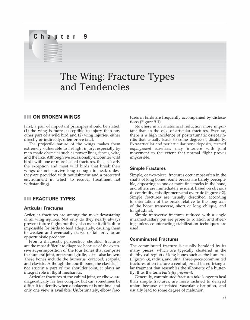
Simple, or two-piece, fractures occur most often in the shafts of long bones. Some breaks are barely percepti-ble, appearing as one or more fi ne cracks in the bone, and others are immediately evident, based on obvious discontinuity, misalignment, and override (Figure 9-2). Simple fractures are usually described according to orientation of the break relative to the long axis of the bone: transverse, short or long oblique, and longitudinal.
Simple transverse fractures reduced with a single intrameduallary pin are prone to rotation and sheer-ing unless counteracting stabilization techniques are used.
Comminuted Fractures
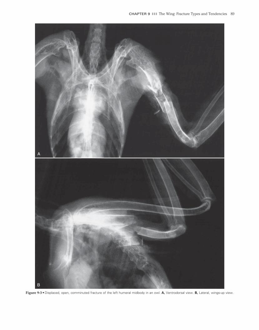
The comminuted fracture is usually heralded by its many pieces, which are typically clustered in the diaphyseal region of long bones such as the humerus (Figure 9-3), radius, and ulna. Three-piece comminuted fractures often feature a central, broad-based triangu-lar fragment that resembles the silhouette of a butter-fl y, thus the term butterfl y fragment.
Generally, comminuted fractures take longer to heal than simple fractures, are more inclined to delayed union because of related vascular disruption, and usually lead to some degree of malunion.
ch009-A02527.indd 87ch009-A02527.indd 87 2/11/2008 10:55:20 AM2/11/2008 10:55:20 AM
88 SECTION I III The Birds
Figure 9-1 • Articular fracture. Close-up view of the right elbow of a kestrel shows a displaced, steep oblique fracture-dislocation of the cranial half of the radial head (emphasis zone).
Figure 9-2 • Displaced steep oblique fracture of the distal humeral body of an owl.
ch009-A02527.indd 88ch009-A02527.indd 88 2/11/2008 10:55:20 AM2/11/2008 10:55:20 AM
CHAPTER 9 III The Wing: Fracture Types and Tendencies 89
A
B
Figure 9-3 • Displaced, open, comminuted fracture of the left humeral midbody in an owl. A, Ventrodorsal view. B, Lateral, wings-up view.
ch009-A02527.indd 89ch009-A02527.indd 89 2/11/2008 10:55:21 AM2/11/2008 10:55:21 AM
90 SECTION I III The Birds
Compound (Open) Fractures
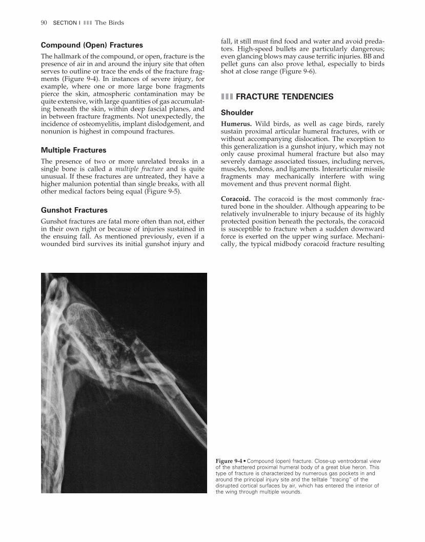
The hallmark of the compound, or open, fracture is the presence of air in and around the injury site that often serves to outline or trace the ends of the fracture frag-ments (Figure 9-4). In instances of severe injury, for example, where one or more large bone fragments pierce the skin, atmospheric contamination may be quite extensive, with large quantities of gas accumulat-ing beneath the skin, within deep fascial planes, and in between fracture fragments. Not unexpectedly, the incidence of osteomyelitis, implant dislodgement, and nonunion is highest in compound fractures.
Multiple Fractures
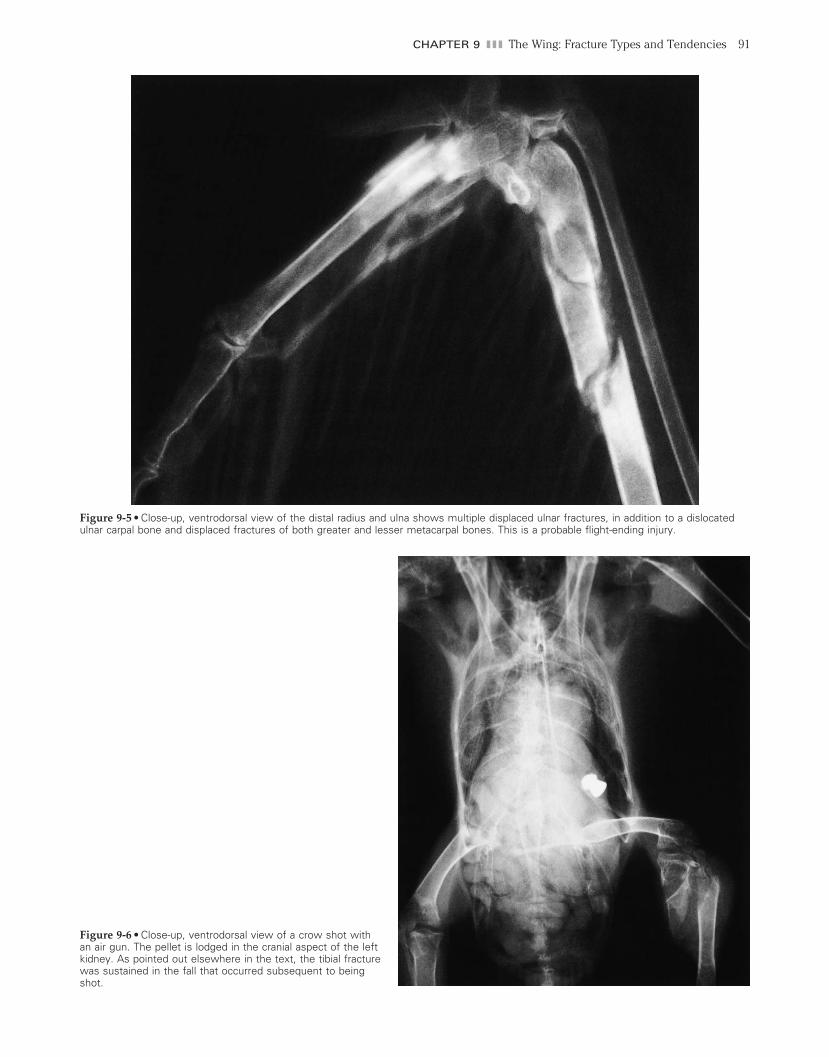
The presence of two or more unrelated breaks in a single bone is called a multiple fracture and is quite unusual. If these fractures are untreated, they have a higher malunion potential than single breaks, with all other medical factors being equal (Figure 9-5).
Gunshot Fractures
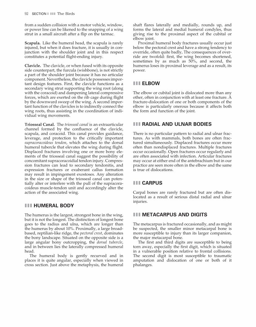
Gunshot fractures are fatal more often than not, either in their own right or because of injuries sustained in the ensuing fall. As mentioned previously, even if a wounded bird survives its initial gunshot injury and
fall, it still must fi nd food and water and avoid preda-tors. High-speed bullets are particularly dangerous; even glancing blows may cause terrifi c injuries. BB and pellet guns can also prove lethal, especially to birds shot at close range (Figure 9-6).
III FRACTURE TENDENCIES
Shoulder
Humerus. Wild birds, as well as cage birds, rarely sustain proximal articular humeral fractures, with or without accompanying dislocation. The exception to this generalization is a gunshot injury, which may not only cause proximal humeral fracture but also may severely damage associated tissues, including nerves, muscles, tendons, and ligaments. Interarticular missile fragments may mechanically interfere with wing movement and thus prevent normal fl ight.
Coracoid. The coracoid is the most commonly frac-tured bone in the shoulder. Although appearing to be relatively invulnerable to injury because of its highly protected position beneath the pectorals, the coracoid is susceptible to fracture when a sudden downward force is exerted on the upper wing surface. Mechani-cally, the typical midbody coracoid fracture resulting
Figure 9-4 • Compound (open) fracture. Close-up ventrodorsal view of the shattered proximal humeral body of a great blue heron. This type of fracture is characterized by numerous gas pockets in and around the principal injury site and the telltale “tracing” of the disrupted cortical surfaces by air, which has entered the interior of the wing through multiple wounds.
ch009-A02527.indd 90ch009-A02527.indd 90 2/11/2008 10:55:23 AM2/11/2008 10:55:23 AM
CHAPTER 9 III The Wing: Fracture Types and Tendencies 91
Figure 9-5 • Close-up, ventrodorsal view of the distal radius and ulna shows multiple displaced ulnar fractures, in addition to a dislocated ulnar carpal bone and displaced fractures of both greater and lesser metacarpal bones. This is a probable fl ight-ending injury.
Figure 9-6 • Close-up, ventrodorsal view of a crow shot with an air gun. The pellet is lodged in the cranial aspect of the left kidney. As pointed out elsewhere in the text, the tibial fracture was sustained in the fall that occurred subsequent to being shot.
ch009-A02527.indd 91ch009-A02527.indd 91 2/11/2008 10:55:23 AM2/11/2008 10:55:23 AM
92 SECTION I III The Birds
from a sudden collision with a motor vehicle, window, or power line can be likened to the snapping of a wing strut in a small aircraft after a fl ip on the tarmac.
Scapula. Like the humeral head, the scapula is rarely injured, but when it does fracture, it is usually in con-junction with the shoulder joint and in this respect constitutes a potential fl ight-ending injury.
Clavicle. The clavicle, or when fused with its opposite side counterpart, the furcula (wishbone), is not strictly a part of the shoulder joint because it has no articular component. Nevertheless, the clavicle possesses impor-tant design features. First, the clavicle functions as a secondary wing strut supporting the wing root (along with the coracoid) and dampening lateral compressive forces, which are exerted on the rib cage during fl ight by the downward sweep of the wing. A second impor-tant function of the clavicles is to indirectly connect the wing roots, thus assisting in the coordination of indi-vidual wing movements.
Triosseal Canal. The triosseal canal is an extraarticular channel formed by the confl uence of the clavicle, scapula, and coracoid. This canal provides guidance, leverage, and protection to the critically important supracoracoideus tendon, which attaches to the dorsal humeral tubercle that elevates the wing during fl ight. Displaced fractures involving one or more bony ele-ments of the triosseal canal suggest the possibility of concomitant supracoracoidial tendon injury. Compres-sion fractures can lead to secondary tendonitis, and expression fractures or exuberant callus formation may result in impingement exostoses. Any alteration in the size or shape of the triosseal canal can poten-tially alter or interfere with the pull of the supracora-coideus muscle-tendon unit and accordingly alter the action of the associated wing.
III HUMERAL BODY
The humerus is the largest, strongest bone in the wing, but it is not the longest. The distinction of longest bone goes to the radius and ulna, which are longer than the humerus by about 10%. Proximally, a large broad-based, reptilian-like ridge, the pectoral crest, dominates the bony landscape. Situated on the opposite side is a large angular bony outcropping, the dorsal tubercle, and in between lies the laterally compressed humeral head.
The humeral body is gently recurved and in places it is quite angular, especially when viewed in cross section. Just above the metaphysis, the humeral
shaft fl ares laterally and medially, rounds up, and forms the lateral and medial humeral condyles, thus giving rise to the proximal aspect of the cubital or elbow joint.
Proximal humeral body fractures usually occur just below the pectoral crest and have a strong tendency to override, often quite badly, The consequences of over-ride are twofold: fi rst, the wing becomes shortened, sometimes by as much as 50%, and second, the humerus loses its proximal leverage and as a result, its power.
III ELBOW
The elbow or cubital joint is dislocated more than any other, often in conjunction with at least one fracture. A fracture-dislocation of one or both components of the elbow is particularly onerous because it affects both the form and function of the joint.
III RADIAL AND ULNAR BODIES
There is no particular pattern to radial and ulnar frac-tures. As with mammals, both bones are often frac-tured simultaneously. Displaced fractures occur more often than nondisplaced fractures. Multiple fractures occur occasionally. Open fractures occur regularly and are often associated with infection. Articular fractures may occur at either end of the antebrachium but in our practice are seen more often in the elbow and the same is true of dislocations.
III CARPUS
Carpal bones are rarely fractured but are often dis-located as a result of serious distal radial and ulnar injuries.
III METACARPUS AND DIGITS
The metacarpus is fractured occasionally, and as might be suspected, the smaller minor metacarpal bone is more susceptible to injury than its larger companion, the major metacarpal bone.
The fi rst and third digits are susceptible to being torn away, especially the fi rst digit, which is situated in a vulnerable position relative to frontal collisions. The second digit is most susceptible to traumatic amputation and dislocation of one or both of it phalanges.
ch009-A02527.indd 92ch009-A02527.indd 92 2/11/2008 10:55:24 AM2/11/2008 10:55:24 AM