Embed Size (px)
Citation preview

Relipidated tissue factor linked to collagen surfaces potentiatesplatelet adhesion and fibrin formation in a microfluidic model ofvessel injury
Thomas V. Colace, Jennielle Jobson, and Scott L. Diamond*
Institute for Medicine and Engineering Department of Chemical and Biomolecular Engineering1024 Vagelos Research Laboratory University of Pennsylvania Philadelphia, PA 19104215-573-5702
AbstractMicrofluidic devices allow for the controlled perfusion of human or mouse blood over definedprothrombotic surfaces at venous and arterial shear rates. To mimic in vivo injuries such a plaquerupture, the need exists to link lipidated tissue factor (TF) to surface bound collagen fibers.Recombinant TF was relipidated in liposomes of phosphatidylserine/phosphatidylcholine/biotin-linked phosphatidylethanolamine (20:79:1 PS:PC:bPE molar ratio). Collagen was patterned in a250-micron wide stripe and labeled with biotinylated anti-collagen antibody which was thenbound with streptavidin, allowing the subsequent capture of the TF liposomes. To verify anddetect the TF liposome-collagen assembly, individual molecular complexes of TF-factor VIIa oncollagen were visualized using the Proximity Ligation Assay (PLA) to produce discretelylocalized fluorescent events that were strictly dependent on the presence of factor VIIa andprimary antibodies against TF or factor VIIa. Perfusion for 450 sec (wall shear rate, 200 s−1) ofcorn trypsin inhibitor (CTI, a factor XIIa inhibitor) treated whole blood over the stripe of TF-collagen enhanced platelet adhesion by 30 ± 8% (p < 0.001) and produced measurable fibrin (>50-fold increase) as compared to surfaces lacking TF. PS:PC:bPE liposomes lacking TF resulted in noenhancement of platelet deposition. Essentially no fibrin was formed during perfusion overcollagen surfaces or collagen surfaces with liposomes lacking TF despite the robust plateletdeposition, indicating a lack of kinetically significant platelet-borne tissue factor in healthy donorblood. This study demonstrates a reliable approach to link functionally-active TF to collagen formicrofluidic thrombosis studies.
*Corresponding author. [email protected] .
NIH Public AccessAuthor ManuscriptBioconjug Chem. Author manuscript; available in PMC 2012 October 19.
Published in final edited form as:Bioconjug Chem. 2011 October 19; 22(10): 2104–2109. doi:10.1021/bc200326v.
NIH
-PA Author Manuscript
NIH
-PA Author Manuscript
NIH
-PA Author Manuscript

KeywordsShear stress; hemodynamics; coagulation; tissue factor; Factor VIIa
IntroductionTissue factor (TF) in the vessel wall is exposed during injury or atherosclerotic plaquerupture and binds circulating factor VIIa. As a cofactor in this complex, TF dramaticallyenhances the catalytic activity of factor VIIa toward factor X and factor IX. Generation offactor Xa results in formation of the prothrombinase complex (Xa/Va) to generate thrombin.Thrombin, a potent activator of platelets, is necessary for the production of clot stabilizingfibrin.
The incorporation of TF into growing thrombi has been suggested to occur through a varietyof mechanisms. While the presence of TF in platelets and other non-activated blood cells inhealthy blood has been an ongoing debate,1-3 several studies have shown the localization ofTF-bearing microparticles to sites of injury via the P-selectin/PSGL1 interaction,4 and itssynthesis by a variety of extra-cellular matrix cells in normal and atherosclerotic vessels.5For decades, various TF preparations have been used as a clinical laboratory reagent in thestudy of prothrombin times, but TF has rarely been deployed in microfluidic injury models.For microfluidic thrombosis models, it is critical to present both collagen to platelets as wellas lipidated TF to the plasma. By simultaneously activating and capturing platelets withsurface bound collagen along with triggering of the coagulation protease cascade withsurface-bound TF, the central biology of thrombosis and hemostasis is recreated. In contrast,addition of exogenous TF into blood absolutely fails to recreate the dynamical aspects ofthrombosis reliant on the surface presentation of TF to flowing blood.
The activity of tissue factor relies on its insertion into a phospholipid membrane containingapproximately 20% negatively charged phosphatidylserine.6 Reliable techniques usingreadily available reagents have been developed to “relipidate” TF into phospholipid vesiclesor membranes.7 In vitro flow studies which include these designs are rare, however, andmainly focus on the dynamics of thrombin or fibrin production.8 In traditional microfluidicstudies, the roles of platelet adhesion and coagulation are commonly separated through theuse of heparin or Phe-Pro-Arg-chloromethylketone (PPACK, an irreversible thrombininhibitor) or through the perfusion of platelet free plasma over a previously formed plateletsurface.9,10 When studied in vivo in the mouse laser injury model, however, plateletdeposition and thrombin production occur simultaneously.11 These models have beenhelpful in defining a role for thrombin in platelet adhesion and fibrin generation in plateletplug stabilization, however they lack characterization and control of the local hemodynamicconditions.12
In this study, we describe a method for generating reproducible surfaces of collagen andactive tissue factor for use in microfluidic analyses of platelet function. We have modifiedthe TF-bearing liposomes described by Smith et al.7 to include a biotinylated lipid which weuse to bind the vesicles to a collagen surface via a biotinylated anti-collagen antibody andstreptavidin (Fig. 1). We have verified that these thrombogenic surfaces are functionallyactive leading to enhanced platelet deposition and measurable fibrin formation. Furthermore,we have characterized the surface through the use of the Proximity Ligation Assay (PLA), adual antibody immunodetection technique capable of visualizing individual moleculecomplexes based on molecular-scale proximity.
Colace et al. Page 2
Bioconjug Chem. Author manuscript; available in PMC 2012 October 19.
NIH
-PA Author Manuscript
NIH
-PA Author Manuscript
NIH
-PA Author Manuscript

Materials and MethodsMaterials
Lipids, L-α-phosphatidylcholine (PC), L-α-phosphatidylserine (PS), and biotinylatedphosphatidylethanolamine (bPE) were from Avanti Polar Lipids (Alabaster, AL, USA).Recombinant human tissue factor was from Haematologic Technologies Inc. (EssexJunction, VT, USA). Bio-Beads SM-2 (BioRad Laboratories, Hercules, CA, USA) werecleaned in methanol and stored under HEPES Buffered Saline (HBS, 20 mM HEPES, 150mM NaCl, pH 7.4 [NaOH]) at 4 °C. The detergent, Triton X-100, was from Fisher Scientific(Fair Lawn, NJ, USA.) Duolink proximity ligation assay (PLA) amplification and detectionreagents were from O-Link Bioscience through distributor Axxora (San Diego, CA, USA).The primary monoclonal PLA antibody against tissue factor was from HaematologicTechnologies and polyclonal primary against factor VIIa was from Abcam (Cambridge,MA, USA), as well as the biotinylated goat polyclonal anti collagen type 1 antibody.Streptavidin was from Sigma (St. Louis, MO, USA). Acid-insoluble collagen type 1 fromequine tendon was from Chronolog Corp. (Havertown, PA, USA) and acid-soluble humancollagen type 1 was from Advanced Biomatrix (San Diego, CA, USA).Poly(dimethylsiloxane), PDMS, for the microfluidic devices (Sylgard 184) was fromEllsworth Adhesives (Germantown, WI, USA) and Sigmacote used for hydrophobictreatment of glass slides was from Sigma. Finally, the anticoagulants corn trypsin inhibitor(CTI) and Pro-Arg-chloromethylketone (PPACK) were from Haematologic Technologies,and platelet labeling anti-CD41 antibody was from AbDSerotec (Raleigh, NC, USA). Thefibrin-specific fluorescent monoclonal antibody was a generous gift from the MortimerPoncz lab at the Children’s Hospital of Philadelphia.
Blood collection and preparationBlood was collected via venipuncture from healthy donors who were self-reported as free ofmedication for at least 10 days into CTI (40 μg/mL) for microfluidic assays or PPACK (100μM) for TF detection assays. All volunteers provided informed consent in accordance withIRB approval and the Declaration of Helsinki. Whole blood was treated with alexafluor 647-conjugated anti CD-41 monoclonal antibody for platelet detection in microfluidic assays 5min prior to perfusion. PFP was generated for TF/VIIa complex detection assays bycentrifugation of whole blood at 1000g for 10 min. All blood samples were used within 1 hrof the draw.
Preparation of biotinylated TF liposomesRelipidation of recombinant tissue factor into liposomes was performed according to apreviously described technique.7 Briefly, PC, PS, and b-PE stored in chloroform were driedunder vacuum in a 79:20:1 molar ratio, respectively. The dried film was resuspended in 1mL of 4 mM Triton X-100 in HBS and allowed to hydrate for 30 min. Recombinant tissuefactor was added and incubated for 10 min (10,000:1 lipid to TF). To this 50 mg of Bio-Bead slurry was added and was gently agitated for 90 min and then 350 mg more of Bio-Bead slurry was added and gently agitated for the same amount of time. The beads wereallowed to settle and the supernatant was collected. The hydrodynamic radius of theresulting liposomes was determined by dynamic light scattering (Protein Solutions DynaPro,Wyatt Technology, Santa Barbara, CA, USA) to be 74.4 ± 28.0 nm.
Preparation of collagen-liposome surfaces for detectionAcid-soluble collagen was first neutralized to a pH of 7.4 with 1M NaOH and thenincubated on Sigmacote treated glass slides for 2.5 hrs (300 μg/mL). The collagen surfaceswere rinsed in HBS and treated with goat polyclonal anti-collagen biotin (10 μg/mL) for 10
Colace et al. Page 3
Bioconjug Chem. Author manuscript; available in PMC 2012 October 19.
NIH
-PA Author Manuscript
NIH
-PA Author Manuscript
NIH
-PA Author Manuscript
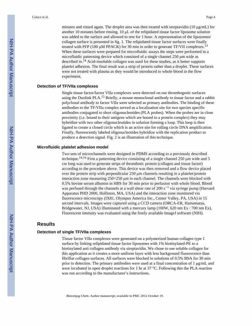
minutes and rinsed again. The droplet area was then treated with streptavidin (10 μg/mL) foranother 10 minutes before rinsing. 10 μL of the relipidated tissue factor liposome solutionwas added to the surface and allowed to rest for 1 hour. A representation of the liposomes/collagen surface is presented in fig. 1. The relipidated tissue factor surfaces were finallytreated with PFP (100 μM PPACK) for 30 min in order to generate TF/VII complexes.13
When these surfaces were prepared for microfluidic assays the steps were performed in amicrofluidic patterning device which consisted of a single channel 250 μm wide asdescribed in.14 Acid-insoluble collagen was used for these studies, as it better supportsplatelet adhesion. The final result was a strip of protein rather than a droplet. These surfaceswere not treated with plasma as they would be introduced to whole blood in the flowexperiment.
Detection of TF/VIIa complexesSingle tissue factor/factor VIIa complexes were detected on our thrombogenic surfacesusing the Duolink PLA.15 Briefly, a mouse monoclonal antibody to tissue factor and a rabbitpolyclonal antibody to factor VIIa were selected as primary antibodies. The binding of theseantibodies to the TF/VIIa complex served as a localization site for two species specificantibodies conjugated to short oligonucleotides (PLA probes). When the probes are in closeproximity (i.e. bound to their antigens which are bound to a protein complex) they mayhybridize with two other oligonucleotides in solution forming a loop. This loop is thenligated to create a closed circle which is an active site for rolling circle DNA amplification.Finally, fluorescently labeled oligonucleotides hybridize with the replication product toproduce a detection signal. Fig. 2 is an illustration of this technique.
Microfluidic platelet adhesion modelTwo sets of microchannels were designed in PDMS according to a previously describedtechnique.14,16 First a patterning device consisting of a single channel 250 μm wide and 5cm long was used to generate strips of thrombotic protein (collagen and tissue factor)according to the procedure above. This device was then removed and a flow device placedover the protein strip with perpendicular 250 μm channels resulting in a platelet/proteininteraction zone measuring 250×250 μm in each channel. The channels were blocked with0.5% bovine serum albumin in HBS for 30 min prior to perfusion with whole blood. Bloodwas perfused through the channels at a wall shear rate of 200 s−1 via syringe pump (HarvardApparatus PHD 2000, Holliston, MA, USA) and the interaction zone monitored viafluorescence microscopy (IX81, Olympus America Inc., Center Valley, PA, USA) in 15second intervals. Images were captured using a CCD camera (ORCA-ER, Hamamatsu,Bridgewater, NJ, USA) illuminated with a mercury lamp (100W, 620 nm Ex / 700 nm Ex).Fluorescent intensity was evaluated using the freely available ImageJ software (NIH).
ResultsDetection of single TF/VIIa complexes
Tissue factor VIIa complexes were generated on a polymerized human collagen type 1surface by linking relipidated tissue factor liposomes with 1% biotinylated-PE to abiotinylated anti collagen antibody via streptavidin. We chose to use soluble collagen forthis application as it creates a more uniform layer with less background fluorescence thanfibrillar collagen surfaces. All surfaces were blocked in solutions of 0.5% BSA for 30 minprior to detection. The primary antibodies were used at a final concentration of 1 μg/mL andwere incubated in open droplet reactions for 1 hr at 37 °C. Following this the PLA reactionwas run according to the manufacturer’s instructions.
Colace et al. Page 4
Bioconjug Chem. Author manuscript; available in PMC 2012 October 19.
NIH
-PA Author Manuscript
NIH
-PA Author Manuscript
NIH
-PA Author Manuscript
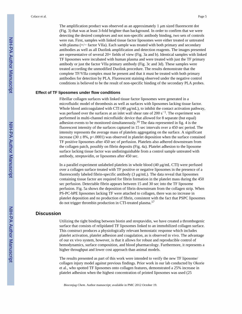
The amplification product was observed as an approximately 1 μm sized fluorescent dot(Fig. 3) that was at least 3-fold brighter than background. In order to confirm that we weredetecting the desired complexes and not non-specific antibody binding, two sets of controlswere run. First, samples with linked tissue factor liposomes were either treated or untreatedwith plasma (+/− factor VIIa). Each sample was treated with both primary and secondaryantibodies as well as all Duolink amplification and detection reagents. The images presentedare representative of several 20× fields of view (Fig. 3a and b). Identical samples with linkedTF liposomes were incubated with human plasma and were treated with just the TF primaryantibody or just the factor VIIa primary antibody (Fig. 3c and 3d). These samples weretreated according the unmodified Duolink procedure. The results demonstrate that thecomplete TF/VIIa complex must be present and that it must be treated with both primaryantibodies for detection by PLA. Fluorescent staining observed under the negative controlconditions is believed to be the result of non-specific binding of the secondary PLA probes.
Effect of TF liposomes under flow conditionsFibrillar collagen surfaces with linked tissue factor liposomes were generated in amicrofluidic model of thrombosis as well as surfaces with liposomes lacking tissue factor.Whole blood anticoagulated with CTI (40 μg/mL), to inhibit the contact activation pathway,was perfused over the surfaces at an inlet wall shear rate of 200 s−1. The experiment wasperformed in multi-channel microfluidic device that allowed for 8 separate (but equal)adhesion events to be monitored simultaneously.16 The data represented in fig. 4 is thefluorescent intensity of the surfaces captured in 15 sec intervals over a 450 sec period. Theintensity represents the average mass of platelets aggregating on the surface. A significantincrease (30 ± 8%, p<.0001) was observed in platelet deposition when the surface containedTF positive liposomes after 450 sec of perfusion. Platelets also adhered downstream fromthe collagen patch, possibly on fibrin deposits (Fig. 4a). Platelet adhesion to the liposomesurface lacking tissue factor was undistinguishable from a control sample untreated withantibody, streptavidin, or liposomes after 450 sec.
In a parallel experiment unlabeled platelets in whole blood (40 μg/mL CTI) were perfusedover a collagen surface treated with TF positive or negative liposomes in the presence of afluorescently labeled fibrin-specific antibody (3 μg/mL). The data reveal that liposomescontaining tissue factor are required for fibrin formation in the platelet mass during the 450sec perfusion. Detectable fibrin appears between 15 and 30 sec into the TF liposomeperfusion. Fig. 5a shows the deposition of fibrin downstream from the collagen strip. WhenPS-PC-bPE liposomes lacking TF were attached to collagen, there was no increase inplatelet deposition and no production of fibrin, consistent with the fact that PSPC liposomesdo not trigger thrombin production in CTI-treated plasma.17
DiscussionUtilizing the tight binding between biotin and streptavidin, we have created a thrombogenicsurface that consists of relipidated TF liposomes linked to an immobilized collagen surface.This construct produces a physiologically relevant hemostatic response which includesplatelet activation, platelet adhesion and coagulation, as is observed in vivo. The advantageof our ex vivo system, however, is that it allows for robust and reproducible control ofhemodynamics, surface composition, and blood pharmacology. Furthermore, it represents ahigher throughput and lower cost approach than animal models.
The results presented as part of this work were intended to verify the new TF liposome/collagen injury model against previous findings. Prior work in our lab conducted by Okorieet al., who spotted TF liposomes onto collagen features, demonstrated a 25% increase inplatelet adhesion when the highest concentration of printed liposomes was used (25
Colace et al. Page 5
Bioconjug Chem. Author manuscript; available in PMC 2012 October 19.
NIH
-PA Author Manuscript
NIH
-PA Author Manuscript
NIH
-PA Author Manuscript
molecules/μm2).18 In the new model we observe a similar response (30% increase ± 8%)and capture the robust increase in fibrin formation reported in that study, as well. While weestimate a slightly lower TF concentration on our surface (see below) [0.1 to 1 molecules/μm2], we may explain the increased potency by the physical linking of liposomes to thecollagen surface. The observation that platelet adhesion does not increase on liposomesurfaces containing TF until ~200 sec is supported by other groups who observed lag timesin thrombin generation of >60 sec,19,20 as well as the lag time observed in fibrin generationin Fig. 5. We also suggest that thrombin generation during primary adhesion has a lessnoticeable effect due to strong GPVI signaling generated by fibrillar collagen at early times.
In addition to platelet adhesion, the kinetics of fibrin deposition in our assay also comparewell to other experimental models. In the mouse laser injury model, for example,measurable fibrin appears at 10-15 sec after insult.21 In these studies the clotting response istriggered by activation of the endothelium and is driven by the exposure of tissue factor, notthe subendothelium; Platelet adhesion is strongly dependent on PAR4 activation.22
Therefore, a more relevant model may be the FeCl3 injury, in which collagen and thrombinsignaling play important roles regarding time to thrombus initiation and likelihood of vesselocclusion.23-25 In these studies FcγRII-/- or PAR4-/- mice, which lack collagen or thrombinsignaling, respectively, have some protection from full vessel occlusion. Factor XIIa is notinhibited in the mouse models whereas CTI is used in our in vitro perfusion studies withhuman blood.
We have demonstrated that our findings with the new TF/collagen assay compare well toother ex vivo and in vivo work. We have also used PLA to detect TF/factor VIIa complexes.However, quantifying fM to pM levels of surface TF is complex and is a drawback to othermodels as well.8 With reasonable assumptions we can provide an estimate of the [TF] range.Extensive characterization of tissue factor liposomes has been previously performed.7 Usingthe relipidation technique described above, 80-90% of TF is recovered in liposomes andapproximately 50% of this TF is accessible. In our assay these values predict 5 molecules ofTF per liposome are available (10,000:1 lipid to TF ratio). If we consider the results fromPLA to represent a lower bound on the number of surface captured liposomes (PLA isknown to have significantly less than 100% detection) we estimate 0.02 liposomes/μm2.This value corresponds to 0.1 to 1 molecules TF/μm2 for 100% or 10% PLA detectionefficiency, respectively. These estimates compare well to our own prior work and relipidatedTF patches created by other groups (0.3 molecules TF/μm2).26 Furthermore, we estimatethat the random packing limit for spheres in 2 dimensions sets an upper bound ofapproximately 102 TF molecules/μm2.
In vivo models of thrombosis have demonstrated the importance of coagulation in responseto vessel injury. Using TF liposomes linked to a collagen surface we have demonstrated anex vivo system that is sensitive to both the coagulation and platelet aggregation componentsof the hemostatic mechanism.
AcknowledgmentsThe authors would like to thank Dr. M. Poncz for the fibrin-specific monoclonal antibody.
T. Colace was supported by a National Institutes of Health (NIH) Cardiovascular Training Predoctoral Fellowship.The authors acknowledge research support by NIH R01-HL-103419 (S.L.D).
References(1). Bouchard BA, Mann KG, Butenas S. No evidence for tissue factor on platelets. Blood. 2010;
116:854–5. [PubMed: 20688968]
Colace et al. Page 6
Bioconjug Chem. Author manuscript; available in PMC 2012 October 19.
NIH
-PA Author Manuscript
NIH
-PA Author Manuscript
NIH
-PA Author Manuscript

(2). Panes O, Matus V, Saez CG, Quiroga T, Pereira J, Mezzano D. Human platelets synthesize andexpress functional tissue factor. Blood. 2007; 109:5242–50. [PubMed: 17347408]
(3). Giesen PLA, Rauch U, Bohrmann B, Kling D, Roque M, Fallon JT, Badimon JJ, Himber J,Riederer MA, Nemerson Y. Blood-Borne tissue factor: Another view of thrombosis. Proc. Natl.Acad. Sci. U.S.A. 1996; 96:2311–5. [PubMed: 10051638]
(4). Falati S, Liu Q, Gross P, Merrill-Skoloff G, Chou J, Vandendries E, Celi A, Croce K, Furie BC,Furie B. Accumulation of tissue factor into developing thrombin in vivo is dependent uponmicroparticle P-selectin glycoprotein ligand 1 and platelet P-selectin. J. Exp. Med. 2003;197:1585–98. [PubMed: 12782720]
(5). Wilcox JN, Smith KM, Schwartz SM, Gordon D. Localization of tissue factor in the normal vesselwall and in the atherosclerotic plaque. Proc. Natl. Acad. Sci. U.S.A. 1989; 86:2839–43.[PubMed: 2704749]
(6). Bach R, Gentry R, Nemerson Y. Factor VII binding to tissue factor in reconstituted phospholipidvesicles: Induction of cooperativity by phosphatidylserine. Biochemistry. 1986; 25:4007–20.[PubMed: 3527261]
(7). Smith SA, Morrissey JH. Rapid and efficient incorporation of tissue factor into liposomes. J.Thromb. and Haemost. 2004; 2:1155–62. [PubMed: 15219199]
(8). Shen F, Kastrup CJ, Liu Y, Ismagilov RF. Threshold response of initiation of blood coagulation bytissue factor in patterned microfluidic capillaries is controlled by shear rate. Arterioscler.Thromb. Vasc. Biol. 2008; 28:2035–41. [PubMed: 18703776]
(9). Cosemans JM, Schols SE, Stefanini L, de Witt S, Feijge MAH, Hamulyak K, Deckmyn H,Bergmeier W, Heemskerk JWM. Key role of glycoprotein Ib/V/IX and von Willebrand factor inplatelet activation-dependent fibrin formation at low shear flow. Blood. 2011; 117:651–60.[PubMed: 21037087]
(10). Goel MS, Diamond SL. Neutrophil enhancement of fibrin deposition under flow through platelet-dependent and –independent mechanisms. Arterioscler. Thromb. Vasc. Biol. 2001; 21:2093–98.[PubMed: 11742890]
(11). Falati S, Gross P, Merril-Skoloff G, Furie BC, Furie B. Real-time in vivo imaging of platelets,tissue factor, and fibrin during arterial thrombus formation in the mouse. Nature Med. 2002;8:1175–80. [PubMed: 12244306]
(12). Rosen ED, Raymond S, Zollman A, Noria F, Sandoval-Cooper M, Shulman A, Merz JL,Castellino FJ. Laser-induced noninvasive vascular injury models in mice generate platelet- andcoagulation-dependent thrombi. Am. J. Pathol. 2001; 158:1613–22. [PubMed: 11337359]
(13). Ruf W, Kalnik MW, Lund-Hansen T, Edgington TS. Characterization of factor VII associationwith tissue factor in solution. J. Biol. Chem. 1991; 266:15719–25. [PubMed: 1874730]
(14). Neeves KB, Maloney SF, Fong KP, Schmaier AA, Kahn ML, Brass LF, Diamond SL.Microfluidic focal thrombosis model for measuring murine platelet deposition and stability:PAR4 signaling enhances shear-resistance of platelet aggregates. J. Thromb. Haemost. 2008;6:2193–201. [PubMed: 18983510]
(15). Soderberg O, Leuchowius KJ, Gullberg M, Jarvius M, Weibrecht I, Larsson LG, Landegren U.Characterizing proteins and their interactions in cells and tissues using the in situ proximityligation assay. Methods. 2008; 45:227–32. [PubMed: 18620061]
(16). Maloney SF, Brass LF, Diamond SL. P2Y12 or P2Y1 inhibitors reduce platelet deposition in amicrofluidic model of thrombosis while apyrase lacks efficacy under flow conditions. Integr.Biol. 2010; 2:183–92.
(17). Goel MS, Diamond SL. Neutrophil cathepsin G promotes prothrombinase and fibrin formationunder flow conditions by activating fibrinogen-adherent platelets. J. Biol. Chem. 2003;278:9458–63. [PubMed: 12524437]
(18). Okorie UM, Denney WS, Chatterjee MS, Neeves KB, Diamond SL. Determination of surfacetissue factor thresholds that trigger coagulation at venous and arterial shear rates: amplification of100 fM circulating tissue factor requires flow. Blood. 2008; 111:3507–13. [PubMed: 18203955]
(19). Fogelson AL, Tania N. Coagulation under flow: The influence of flow-mediated transport on theinitiation and inhibition of coagulation. Pathophysiol. Haemost. Thromb. 2005; 34:91–108.[PubMed: 16432311]
Colace et al. Page 7
Bioconjug Chem. Author manuscript; available in PMC 2012 October 19.
NIH
-PA Author Manuscript
NIH
-PA Author Manuscript
NIH
-PA Author Manuscript
(20). Van ‘t Veer C, Hackeng TM, Delahaye C, Sixma JJ, Bouma BN. Activated factor X andthrombin formation triggered by tissue factor on endothelial cell matrix in a flow model: effect ofthe tissue factor pathway inhibitor. Blood. 84:1132–42. [PubMed: 8049429]
(21). Furie B, Furie BC. Thrombus formation in vivo. J. Clin. Invest. 2005; 115:3355–59. [PubMed:16322780]
(22). Vandendries ER, Hamilton JR, Coughlin SR, Furie B, Furie BC. Par4 is required for plateletthrombus propagation but not fibrin generation in a mouse model of thrombosis. Proc. Natl.Acad. Sci. U.S.A. 2007; 104:288–92. [PubMed: 17190826]
(23). Dubois C, Panicot-Dubois L, Merrill-Skoloff G, Furie B, Furie BC. Glycoprotein VI-dependentand –independent pathways of thrombus formation in vivo. Blood. 2006; 107:3902–6. [PubMed:16455953]
(24). Wang L, Miller C, Swarthout RF, Rao M, Mackman N, Taubman MB. Vascular smooth muscle-derived tissue factor is critical for arterial thrombosis after ferric chloride-induced injury. Blood.2009; 113:705–13. [PubMed: 18931346]
(25). Sambrano GR, Weiss EJ, Zheng YW, Huang W, Coughlin SR. Role of thrombin signaling inplatelets in haemostasis and thrombosis. Nature. 2001; 413:74–8. [PubMed: 11544528]
(26). Kastrup CJ, Shen F, Runyon MK, Ismagilov RF. Characterization of the threshold response ofinitiation of blood clotting to stimulus patch size. Biophys. J. 2007; 93:2969–77. [PubMed:17586576]
Colace et al. Page 8
Bioconjug Chem. Author manuscript; available in PMC 2012 October 19.
NIH
-PA Author Manuscript
NIH
-PA Author Manuscript
NIH
-PA Author Manuscript
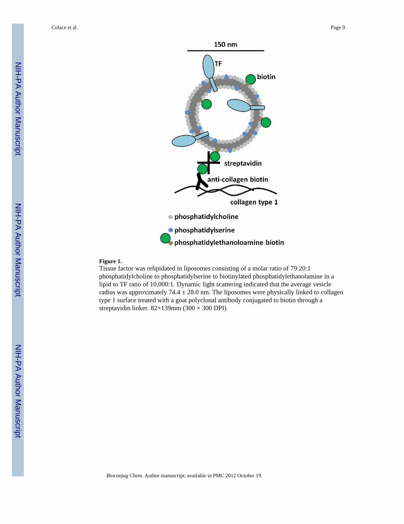
Figure 1.Tissue factor was relipidated in liposomes consisting of a molar ratio of 79:20:1phosphatidylcholine to phosphatidylserine to biotinylated phosphatidylethanolamine in alipid to TF ratio of 10,000:1. Dynamic light scattering indicated that the average vesicleradius was approximately 74.4 ± 28.0 nm. The liposomes were physically linked to collagentype 1 surface treated with a goat polyclonal antibody conjugated to biotin through astreptavidin linker. 82×139mm (300 × 300 DPI)
Colace et al. Page 9
Bioconjug Chem. Author manuscript; available in PMC 2012 October 19.
NIH
-PA Author Manuscript
NIH
-PA Author Manuscript
NIH
-PA Author Manuscript

Figure 2.The Duolink proximity ligation assay is capable of detecting single molecule complexes.Primary antibodies directed against the components of the complex act to localize two “PLAprobes”, which are species specific antibodies conjugated to short oligonucleotides. Whenthe probes are brought together they form a closed loop when complimentaryoligonucleotides are added. This loop is enzymatically ligated which provides a template forrolling circle amplification. Small fluorescently labeled oligonucleotides hybridize with theamplified product producing a detection signal. 127×139mm (300 × 300 DPI)
Colace et al. Page 10
Bioconjug Chem. Author manuscript; available in PMC 2012 October 19.
NIH
-PA Author Manuscript
NIH
-PA Author Manuscript
NIH
-PA Author Manuscript
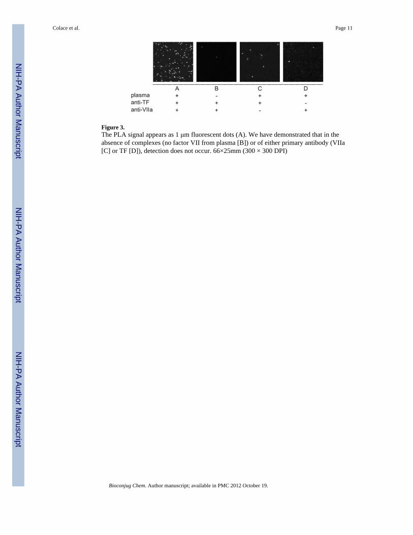
Figure 3.The PLA signal appears as 1 μm fluorescent dots (A). We have demonstrated that in theabsence of complexes (no factor VII from plasma [B]) or of either primary antibody (VIIa[C] or TF [D]), detection does not occur. 66×25mm (300 × 300 DPI)
Colace et al. Page 11
Bioconjug Chem. Author manuscript; available in PMC 2012 October 19.
NIH
-PA Author Manuscript
NIH
-PA Author Manuscript
NIH
-PA Author Manuscript
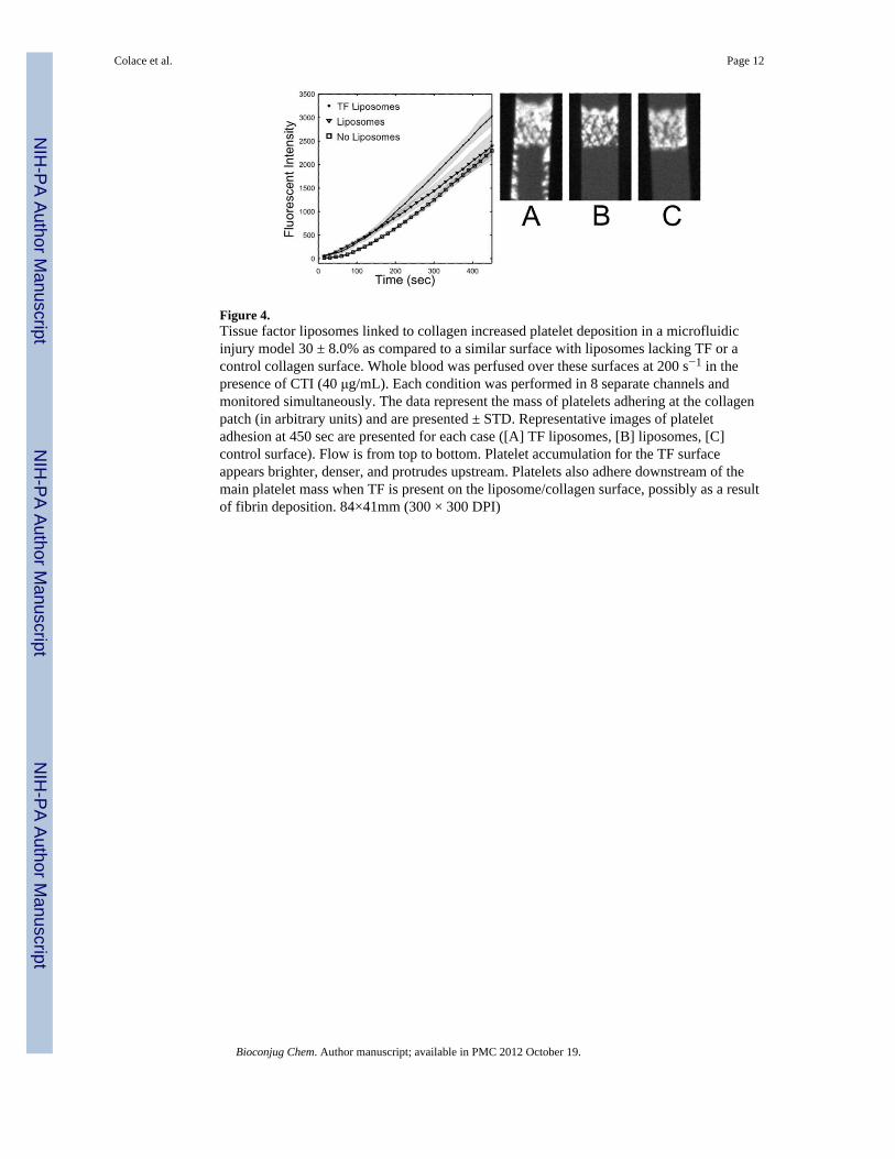
Figure 4.Tissue factor liposomes linked to collagen increased platelet deposition in a microfluidicinjury model 30 ± 8.0% as compared to a similar surface with liposomes lacking TF or acontrol collagen surface. Whole blood was perfused over these surfaces at 200 s−1 in thepresence of CTI (40 μg/mL). Each condition was performed in 8 separate channels andmonitored simultaneously. The data represent the mass of platelets adhering at the collagenpatch (in arbitrary units) and are presented ± STD. Representative images of plateletadhesion at 450 sec are presented for each case ([A] TF liposomes, [B] liposomes, [C]control surface). Flow is from top to bottom. Platelet accumulation for the TF surfaceappears brighter, denser, and protrudes upstream. Platelets also adhere downstream of themain platelet mass when TF is present on the liposome/collagen surface, possibly as a resultof fibrin deposition. 84×41mm (300 × 300 DPI)
Colace et al. Page 12
Bioconjug Chem. Author manuscript; available in PMC 2012 October 19.
NIH
-PA Author Manuscript
NIH
-PA Author Manuscript
NIH
-PA Author Manuscript
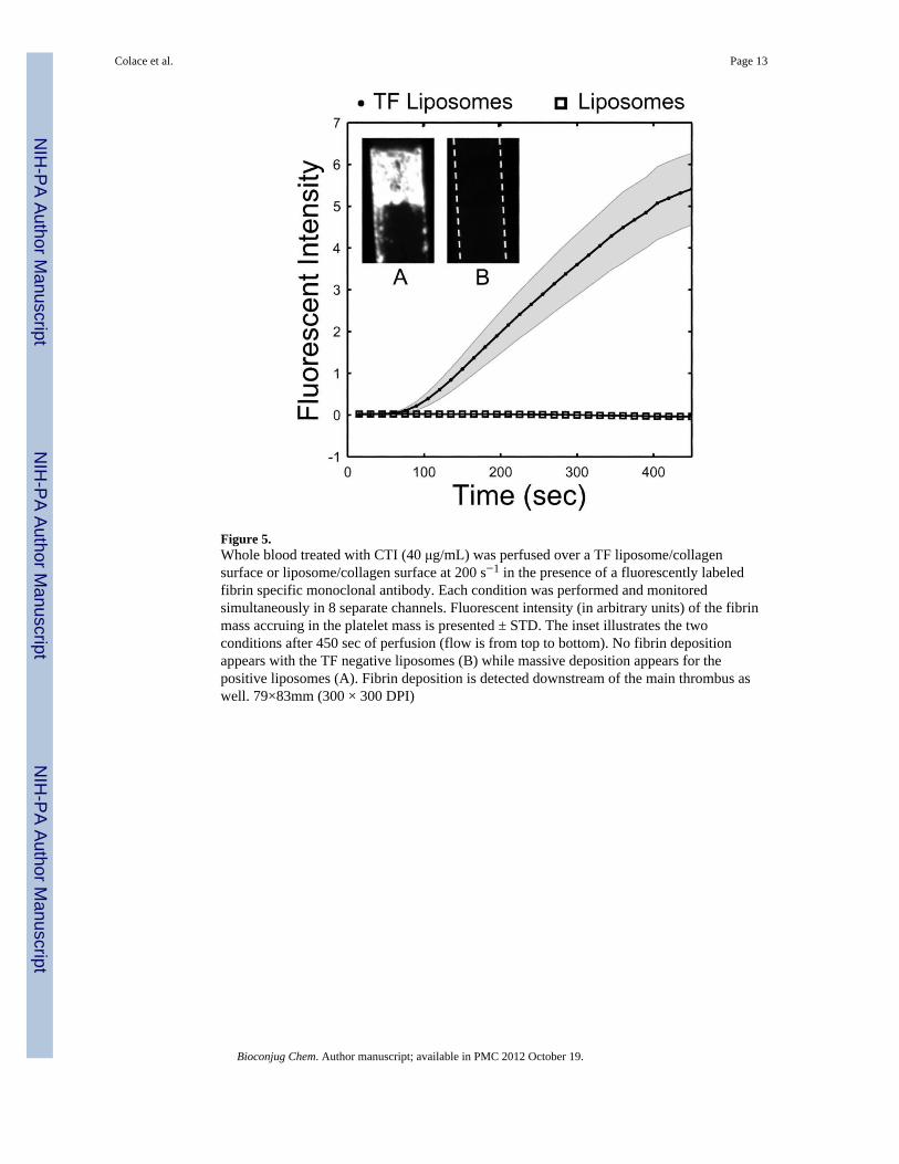
Figure 5.Whole blood treated with CTI (40 μg/mL) was perfused over a TF liposome/collagensurface or liposome/collagen surface at 200 s−1 in the presence of a fluorescently labeledfibrin specific monoclonal antibody. Each condition was performed and monitoredsimultaneously in 8 separate channels. Fluorescent intensity (in arbitrary units) of the fibrinmass accruing in the platelet mass is presented ± STD. The inset illustrates the twoconditions after 450 sec of perfusion (flow is from top to bottom). No fibrin depositionappears with the TF negative liposomes (B) while massive deposition appears for thepositive liposomes (A). Fibrin deposition is detected downstream of the main thrombus aswell. 79×83mm (300 × 300 DPI)
Colace et al. Page 13
Bioconjug Chem. Author manuscript; available in PMC 2012 October 19.
NIH
-PA Author Manuscript
NIH
-PA Author Manuscript
NIH
-PA Author Manuscript