Embed Size (px)
Citation preview

Vesicles interacting with nanoparticles
vorgelegt von Doctor of Philosophy
Amirhoushang Bahrami aus Teheran
Von der Fakultät II - Mathematik und Naturwissenschaften der Technischen Universität Berlin
zur Erlangung des akademischen Grades Doktor der Naturwissenschaften
Dr. rer. nat.
genehmigte Dissertation
Promotionsausschuss: Vorsitzender: Prof. Dr. Martin Schoen Gutachterin: Prof. Dr. Sabine H. L. Klapp Gutachter: Dr. habil. Thomas R. Weikl Tag der wissenschaftlichen Aussprache: 30. Juli 2013
Berlin 2013 D 83


Abstract In this dissertation, we study the interactions of vesicle membranes with nanoparticles of different shapes. The wrapping and internalization of nanoparticles by biomembranes plays a critical role in drug delivery applications and nanomedicine. We begin with a single spherical particle adsorbed on a vesicle and solve the shape equations for rotationally symmetric vesicles to investigate the wrapping of the particle by the vesicle. These shape equations are based on the Helfrich bending energy of the vesicle membrane. We predict different regimes for partial and full wrapping of the nanoparticle and relate the wrapping transition and its energy barrier to the reduced volume of the vesicle and the relative size of the vesicle and particle. We next study the cooperative wrapping of several spherical nanoparticles using simulated annealing Monte Carlo simulations of triangulated vesicles. We report novel tubular membrane structures induced by the nanoparticles, which we obtain from energy minimization. The membrane tubules enclose linear aggregates of particles and protrude into the vesicles. The high stability of the particle-filled tubules implies strongly attractive, membrane-mediated interactions between the particles. The tubular structures may provide a new route to encapsulate nanoparticles reversibly in vesicles. With this simulation method, we also investigate the role of the membrane curvature on the membrane-mediated interactions between adsorbed nanoparticles. We consider two different types of Janus nanoparticles, which both can only be partially wrapped by the vesicle. We report attractive interactions between two particles adsorbed to the outside of the vesicle, but repulsive interactions between particles that are adsorbed to the inside of the vesicle. Finally, we study the wrapping of single ellipsoidal nanoparticles by a vesicle via simulated annealing Monte Carlo simulations. We report two distinct regimes of spreading and internalization, which are separated by an energy barrier, and relate the success or failure of the internalization to the particle shape and orientation relative to the vesicle. We observe easier spreading yet more difficult internalization for ellipsoidal particles with lower aspect ratios, which may explain the high virulence of tubular viruses. We find that the wrapping of ellipsoidal particles is associated with an orientation change of the particle. While the spreading starts on the flat side of the ellipsoidal particles, the particle changes its orientation during wrapping, and internalization finally occurs in an orientation in which a tip of the ellipsoidal particles protrudes into the vesicles.


Zusammenfassung In dieser Dissertation betrachten wir die Wechselwirkungen von Vesikelmembranen mit Nanoteilchen unterschiedlicher Form. Die Umwickelung und Internalisierung von Nanoteilchen durch Biomembranen spielt eine zentrale Rolle in Medikamentenforschung und Nanomedizin. Wir beginnen mit einem einzelnen kugelförmigen Teilchen, das an einem Vesikel adsorbiert ist, und lösen die Formgleichungen für rotationssymmetrische Vesikelformen um die Umwickelung des Teilchens zu untersuchen. Diese Formgleichungen basieren auf der Helfrichschen Biegeenergie der Vesikelmembranen. Wir erhalten verschiedene Regime für die teilweise und die volle Umwickelung der Teilchen, und setzen die Energiebarriere für den Umwickelungsübergang mit dem reduzierten Volumen des Vesikels und der relativen Größe von Vesikel und Teilchen in Beziehung. Als nächstes untersuchen wir die kooperative Umwickelung mehrerer kugelförmiger Nanoteilchen mit Hilfe von ‘simulated annealing’ Monte-Carlo-Simulationen von triangulierten Vesikeln. Wir finden neuartige tubuläre Membranstrukturen, die durch die Nanoteilchen induziert werden und die Gesamtenergie minimieren. Diese tubulären Strukturen weisen einen neuen Weg, Nanoteilchen reversibel in Vesikeln einzuschließen. Mit dieser Simulationsmethode untersuchen wir auch den Einfluss der Membrankrümmung auf die membranvermittelten Wechselwirkungen zwischen den adsorbierten Nanoteilchen. Wir betrachten zwei verschiedenen Arten von Janus-Teilchen, die nur teilweise von der Membran umwickelt werden. Wir erhalten attraktive Wechselwirkungen zwischen zwei Teilchen, die außen an ein Vesikel adsorbiert sind, jedoch repulsive Wechselwirkungen zwischen Teilchen, die von Innen an die Vesikelmembran adsorbieren. Schließlich betrachten wir die Umwickelung einzelner ellipsoidaler Nanoteilchen in ‘simulated annealing’ Monte-Carlo-Simulationen. Wir erhalten zwei unterschiedliche Regime von Spreiten und Internalisierung, die durch eine Energiebarriere getrennt sind, and setzen die Höhe dieser Barriere mit der Teilchenform und –orientierung in Beziehung. Wir beobachten einfacheres Spreiten aber erschwerte Internalisierung mit zunehmender Ellipsität der Teilchen. Die Umwickelung der ellipsoidalen Teilchen ist mit einer Orientierungsänderung der Teilchen verbunden. Das Spreiten der Teilchen durch die Vesikelmembran beginnt an den flachen Seiten. Bei forschreitender Umwickelung ändert sich jedoch die Ausrichtung der Teilchen, und die Internalisierung geschieht schließlich in einer Orientierung, in der eine Spitze der ellipsoidalen Teilchen in das Vesikel hineinragt.


Publication list
• A. H. Bahrami, R. Lipowsky and T. R. Weikl, Tubulation and Aggregation of Spherical Nanoparticles Adsorbed on Vesicles, Phys. Rev. Lett., 2012, 109, 188102. • A. H. Bahrami, Orientational changes and impaired internalization of ellipsoidal nanoparticles by vesicle membranes, Soft Matter, 2013, 9 (36), 8642 - 8646.


Contents
1 Biological membranes 3
2 Shape equations for symmetric vesicles 72.1 Continuum membrane model . . . . . . . . . . . . . . . . . . . . . . . . . 72.2 Shape equations . . . . . . . . . . . . . . . . . . . . . . . . . . . . . . . . 82.3 Numerical algorithm . . . . . . . . . . . . . . . . . . . . . . . . . . . . . 122.4 Shapes of a free vesicle . . . . . . . . . . . . . . . . . . . . . . . . . . . . 132.5 A vesicle with an adsorbed particle . . . . . . . . . . . . . . . . . . . . . 18
2.5.1 Prolate-oblate transition . . . . . . . . . . . . . . . . . . . . . . . 222.5.2 Energy barriers and wrapping transition . . . . . . . . . . . . . . 25
3 Monte Carlo simulations of vesicles 273.1 Triangulated membranes . . . . . . . . . . . . . . . . . . . . . . . . . . . 273.2 Monte Carlo simulations . . . . . . . . . . . . . . . . . . . . . . . . . . . 30
3.2.1 Vesicle parameters . . . . . . . . . . . . . . . . . . . . . . . . . . 323.2.2 Area and volume constraints . . . . . . . . . . . . . . . . . . . . . 32
3.3 Simulated annealing method . . . . . . . . . . . . . . . . . . . . . . . . . 34
4 Tubulation and aggregation of spherical nanoparticles 354.1 Introduction . . . . . . . . . . . . . . . . . . . . . . . . . . . . . . . . . . 354.2 A vesicle with a pair of adsorbed particles . . . . . . . . . . . . . . . . . 364.3 Simulation results . . . . . . . . . . . . . . . . . . . . . . . . . . . . . . . 384.4 A vesicle with three adsorbed particles . . . . . . . . . . . . . . . . . . . 444.5 Conclusion . . . . . . . . . . . . . . . . . . . . . . . . . . . . . . . . . . . 47
5 Membrane-curvature-mediated interactions 495.1 Introduction . . . . . . . . . . . . . . . . . . . . . . . . . . . . . . . . . . 495.2 Vesicle and Janus particles . . . . . . . . . . . . . . . . . . . . . . . . . . 50
5.2.1 The vesicle model . . . . . . . . . . . . . . . . . . . . . . . . . . . 505.2.2 Pseudo-Janus particles . . . . . . . . . . . . . . . . . . . . . . . . 515.2.3 Janus particles . . . . . . . . . . . . . . . . . . . . . . . . . . . . 52
5.3 Membrane-mediated interactions between pseudo-Janus particles . . . . . 535.4 Membrane-mediated interactions between Janus particles . . . . . . . . . 59
1

Contents
5.5 Conclusion . . . . . . . . . . . . . . . . . . . . . . . . . . . . . . . . . . . 63
6 Wrapping and internalization of nanoparticles with different shapes 656.1 Cellular uptake of nanoparticles . . . . . . . . . . . . . . . . . . . . . . . 65
6.1.1 The effect of particle size . . . . . . . . . . . . . . . . . . . . . . . 666.1.2 The effect of particle shape . . . . . . . . . . . . . . . . . . . . . 66
6.2 Vesicle wrapping around nanoparticles with different shapes . . . . . . . 676.2.1 Simulated annealing simulation . . . . . . . . . . . . . . . . . . . 686.2.2 The ellipsoidal particles . . . . . . . . . . . . . . . . . . . . . . . 69
6.3 Wrapping of a spherical nanoparticle . . . . . . . . . . . . . . . . . . . . 706.4 Wrapping of ellipsoidal nanoparticles . . . . . . . . . . . . . . . . . . . . 72
6.4.1 Wrapping of prolate ellipsoidal particles . . . . . . . . . . . . . . 726.4.2 Constrained wrapping of prolate ellipsoidal particles . . . . . . . . 746.4.3 Wrapping of oblate ellipsoidal particles . . . . . . . . . . . . . . . 75
6.5 Discussion and conclusions . . . . . . . . . . . . . . . . . . . . . . . . . . 77
2

1 Biological membranes
Biological membranes are soft matter structures which separate the inner and outer partsof the cell. The cell membrane is responsible for transporting many different biologicalobjects like ions and molecules between inside and outside of the cell. Therefore biologicalmembranes define and control the material composition inside the cell.
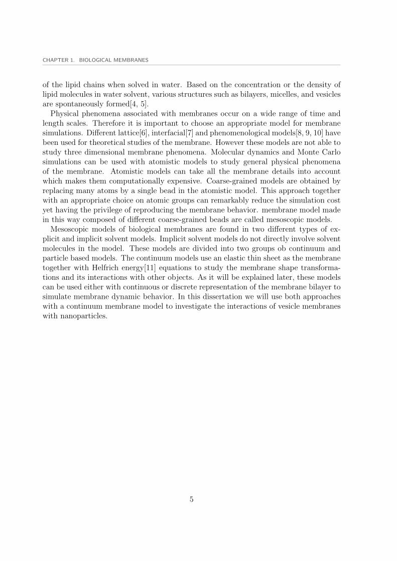
Our understanding of the structure and composition of the biological membraneshas evolved through the years. The first model of biological membranes was presentedby Gorter and Grendel in 1925[1]. Their experiments on red blood cells showed thatmembranes are thin fluid molecular structures composed of two layers of lipid moleculesin form of a lipid bilayer. Biomembranes are dynamics structures whose compositionchanges throughout their life. They are composed of several biological entities such asprotein molecules and peptides diffusing in the fluid bilayer of the membrane. Danielliand Davson were the first ones who involved protein molecules in the membrane model in1935[2]. Our current picture of the biological membranes is based on the well know ”fluidMosaic”model of Singer and Nicolson presented in 1972[3]. In their model the membraneis composed of lipid molecules and proteins and peptides and resembles a mosaic. Thefluidity of the membrane in this model is due to the free lateral motion of lipid chains andother molecules along the membrane. Biological functions of biomembranes are mainlyaffected by the membrane structure and its organization which strongly depend on theinteractions between membrane components. Figure 1.1 displays a schematic view ofthe biological membrane with different molecules such as proteins and peptides.
Due to complexity of the biological membranes it is not easy to study membranephysics. Therefore simple membrane models composed of lipid chains and some proteinand peptide molecules have been developed to study the relation between membranecomposition and its functions. These model membranes are used both experimentallyand theoretically to investigate biomembrane physics. Simulation studies of membranemodels helps us to understand experimental observations. On the other hand the simu-lation results can be used to interpret existing experiments yet inspire new experiments.
Cell membrane is composed of different types of lipid chains including phospholipidsand cholesterols. Membrane formation and its structure is mainly based on the so calledamphiphilic property of the lipid chains solved in water. A single lipid chain is composedof a hydrophobic tail and a hydrophilic head. The hydrophilic head beads are attracted tothe water molecules while the hydrophobic tail beads are repelled from water molecules.Different hydrophobicity of the head and tail parts of a single chain results in aggregation
3

Figure 1.1 : A schematic view of the lipid biomembrane together with membrane cy-toskeleton and membrane inclusions such as protein molecules adapted from”2004 Nature Publishing Group Pietzsch, J. Mind the membrane. HorizonSymposia: Living Frontier, 1-4.”
4
CHAPTER 1. BIOLOGICAL MEMBRANES
of the lipid chains when solved in water. Based on the concentration or the density oflipid molecules in water solvent, various structures such as bilayers, micelles, and vesiclesare spontaneously formed[4, 5].
Physical phenomena associated with membranes occur on a wide range of time andlength scales. Therefore it is important to choose an appropriate model for membranesimulations. Different lattice[6], interfacial[7] and phenomenological models[8, 9, 10] havebeen used for theoretical studies of the membrane. However these models are not able tostudy three dimensional membrane phenomena. Molecular dynamics and Monte Carlosimulations can be used with atomistic models to study general physical phenomenaof the membrane. Atomistic models can take all the membrane details into accountwhich makes them computationally expensive. Coarse-grained models are obtained byreplacing many atoms by a single bead in the atomistic model. This approach togetherwith an appropriate choice on atomic groups can remarkably reduce the simulation costyet having the privilege of reproducing the membrane behavior. membrane model madein this way composed of different coarse-grained beads are called mesoscopic models.
Mesoscopic models of biological membranes are found in two different types of ex-plicit and implicit solvent models. Implicit solvent models do not directly involve solventmolecules in the model. These models are divided into two groups ob continuum andparticle based models. The continuum models use an elastic thin sheet as the membranetogether with Helfrich energy[11] equations to study the membrane shape transforma-tions and its interactions with other objects. As it will be explained later, these modelscan be used either with continuous or discrete representation of the membrane bilayer tosimulate membrane dynamic behavior. In this dissertation we will use both approacheswith a continuum membrane model to investigate the interactions of vesicle membraneswith nanoparticles.
5


2 Shape equations for symmetricvesicles
2.1 Continuum membrane model
Continuum and discrete membrane modeling are two different approaches to study themembrane physics. Applying numerical simulation methods such as Monte Carlo todiscrete membrane models is discussed in chapter three. Alternatively one can use acontinuum membrane model to investigate membrane shape transformations and itsproperties. Here we introduce the classical elastic model of membranes which has beensuccessfully used to study vesicle shape transformations[12]. The classical elastic modelof membranes was first represented by Helfrich[11]. Within this model, the bendingenergy of a vesicle Eb is defined as
Eb =κ
2
∫dA(C1 + C2 − C0)
2 + κG
∫dA(C1C2) (2.1)
where C1 and C2 are the principal curvatures of the membrane surface and C0 denotesthe spontaneous membrane curvature which is introduced to take into account the asym-metric properties of the bilayer membrane. We focus on the vesicles without spontaneouscurvature C0 = 0. Equation 2.1 gives the bending energy of the vesicle. The secondterm gives the integration of the Gaussian curvature. According to Gauss-Bonnet theo-rem, the second term is constant for the shapes which are topologically invariant. In allsimulations, vesicle shapes are topological equivalent of a sphere. Therefore the surfaceintergral of the Guassian curvature is equal for all vesicle shapes. Here, κ and κG denotethe bending rigidity and the modulus of Gaussian curvature of the vesicle membranerespectively. We apply constraints on vesicle surface and its volume due to the constantnumber of lipid chains per unit area or incompressibility and the osmotic pressure inreal physical situation. For a given value of the vesicle volume V and the vesicle surfacearea A, one should find the corresponding shape of the vesicle which results in a vesiclewith minimum bending energy Eb. The minimum value of the bending energy subjectedto surface and volume constraints, can be obtained by minimizing an energy functionalE. Surface and volume constraints are included in the energy functional by appropriateLagrange multipliers Σ and P . The vesicle shapes with minimum energy are solutionsof the variational equation
7

2.2. SHAPE EQUATIONS
S
Figure 2.1 : Vesicle axisymmetric shapes parametrized with the arc length S along thegenerating curve and tilt angle ψ defined as ψ = ψ(S).
δE = δ(Eb + ΣA+ PV ) = 0 (2.2)
where δ denotes the variation of energy with respect to the variation of the vesicle shape,and Lagrange multipliers Σ, P are adjusted to fulfill surface and volume constraints.The solution results in a vesicle shape with volume V and area A which possesses theminimum value of the bending energy.
We assume that the minimum energy shape is axisymmetric generated by revolvinga two dimensional curve around an axis of symmetry Z. Then we parametrize vesicleshapes using the arc length S along the generating curve and the azimuthal angle ψ asshown in figure 2.1.
2.2 Shape equations
We use the arc length S as the independent variable and define all vesicle properties asa function of S. The tilt angle ψ = ψ(S) as a function of arc length S, uniquely definesthe geometry of the vesicle. Here, Z and X coordinates are the vertical and horizontalCartesian coordinates of the vesicle where Z is parallel to the axis of symmetry. Thegeometry of the generating curve defines the relations between the vesicle coordinates as
X = cosψ
Z = − sinψ
C1 = ψ
C2 =sinψ
X(2.3)
8

CHAPTER 2. SHAPE EQUATIONS FOR SYMMETRIC VESICLES
where the dot indicates a derivative with respect to S. In this way by parameterizingthe vesicle shape in terms of the tilt angle ψ, the infinite derivative dZ/dX in Cartesianparametrization is avoided. Considering an infinitesimal element with length dS alongthe arc length, the surface and volume elements dA and dV are obtained as
dA = 2πXdS
dV = πX2dS sinψ (2.4)
where X denotes the horizontal coordinate of the element dS. Combining eqs. 2.4 and2.3 with the Helfrich energy 2.4, the energy functional E reads,
E = κπ
∫X(ψdS +
sinψ
X)2 + 2πΣ
∫XdS + πP
∫X2 sinψdS (2.5)
With the rescaled Lagrange multipliers Σ and P ,
P =P
κ, Σ =
Σ
κ(2.6)
The total energy can be written as
E = 2κπ
∫ S
0
[X
2(ψ +
sinψ
X)2 + ΣX +
P
2X2 sinψ
]dS (2.7)
The minimization problem is now described in terms of minimizing a definite integral,
E = 2κπ
∫ S
0
L(X, X, , ψ, γ)dS (2.8)
where L stands for the Lagrange function,
L =X
2(ψ +
sinψ
X)2 + ΣX +
P
2X2 sinψ + γ(X − cosψ) (2.9)
and γ = γ(S) is a function introduced to account for the constraint X − sinψ = 0between coordinates X and ψ.
As the Lagrange function L does not directly depend on the independent variable S,its derivative vanishes with respect to S,
∂L
∂S= 0 (2.10)
9

2.2. SHAPE EQUATIONS
Therefore the system Hamiltonian
H = −L+ ψ∂L
∂ψ+ X
∂L
∂X=X
2
[ψ2 − (
sinψ
X)2]− ΣX − P
2X2 sinψ + γ cosψ (2.11)
is conserved. The Lagrange equations are obtained by inserting L into
d
dt
(∂L
∂ψ
)− ∂L
∂ψ= 0
d
dt
(∂L
∂X
)− ∂L
∂X= 0 (2.12)
By substituting L into the above equations, one obtains Lagrange equations as
ψ = ω
ω = − ωX
cosψ +cosψ sinψ
X2+γ
Xsinψ +
PX
2cosψ
γ =ω2
2− sin2 ψ
2X2+ PX sinψ + Σ
X = cosψ (2.13)
The boundary conditions for the two coordinates X and ψ of a free vesicle are givenas,
X(0) = 0
X(S) = 0
ψ(0) = 0
ψ(S) = π (2.14)
The condition δH = 0 should be fulfilled for any value of the total arc length S thatimplies H(S) = 0 which together with 2.11 leads to the boundary condition
γ(S) = 0 (2.15)
Conservation of the system Hamiltonian H implies H(0) = H(S) = 0 which gives riseto
γ(0) = 0 (2.16)
10

CHAPTER 2. SHAPE EQUATIONS FOR SYMMETRIC VESICLES
−0.8 −0.6 −0.4 −0.2 0 0.2 0.4 0.6 0.8
−0.2
0
0.2
0.4
0.6
0.8
1
1.2
φRp
Figure 2.2 : Vesicle adhered to a spherical particle with radius Rp. Wrapping angle φ isused to parametrize wrapping geometry.
For the case of a nanoparticle adhered to the vesicle, the angle φ defines the wrappingangle or the adhesion angle of the spherical nanoparticle to the vesicle as shown in 2.2.
In this case we just solve the shape equations to obtain the shape of the unbound partof the vesicle because the adhering part simply follows the spherical geometry of theparticle. Therefore the boundary conditions for the free vesicle adhering to the sphericalparticle are obtained from the geometry of the contact area between the vesicle andparticle,
X(0) = Rp sinφ
X(S) = 0
ψ(0) = −φψ(S) = π (2.17)
The adhered part of the vesicle should be involved in the total values of the bendingenergy, vesicle surface and volume. The bending energy, surface area and volume of aspherical cap with wrapping angle φ are obtained from the spherical parametrization fora part of a complete sphere ( 2.2)
S = Rpψ
X = Rp sinψ (2.18)
With corresponding area and volume elements 2.4 and Helfrich bending energy 2.1we have
11

2.3. NUMERICAL ALGORITHM
As =
∫2πXdS =
∫ φ
0
2πR2p sinψdψ = 2πR2
p(1− cosφ)
Vs = −∫πX2 sinψdS = −π
R3p
3(1− cosφ)2(2 + cosφ)
Es =
∫κ
2(C2
1 + C22)2dA =
∫κ
2(
1
R2p
+1
R2p
)2dA = 4πκ(1− cosφ) (2.19)
where As, Vs, Es indicate surface, volume and energy of the adhered part of the vesicle.The volume of a spherical cap has a negative sign because the whole volume of the vesicleis obtained by subtracting the volume contribution of the adhered part from the volumecontribution of the unbound part.
Shape equations for the free vesicle have unique solution for a given set of the vesiclesurface and volume A, V . Rescaled Lagrange multipliers Σ, P together with the onlyunknown initial condition ω(0) are the system parameters which have to be adjustedto result in the target values of the vesicle surface and volume. For the vesicle with anadsorbed particle, shape equations are solved for a given set of the vesicle surface A,vesicle volume V and the wrapping angle φ. The volume and area are calculated for thewhole vesicle.
2.3 Numerical algorithm
Finding solutions of the shape equations for both free vesicle and vesicle and particle isnot straightforward. The boundary condition X(0) = X(S) = 0 leads to singular shapeequations for the terms where X appears in the denominator of a fraction on the righthand side of equations 2.13. This holds for the initial and final boundary conditions ofa free vesicle and the final condition of the vesicle with an adsorbed particle.
In order to avoid the singularity problem, a specific numerical method is used to findthe real solutions of the shape equations. This method is based on a shooting algorithmwhich tries to find solutions in a recursive approach. The solution of the shape equationis obtained in a few steps as follows: First a positive value for P > 0 and a fixed valuefor Σ are chosen. The value of P defines a length scale P
−13 . The shape equations are
then integrated with the initial conditions ψ(0) = 0, ω(0) = 0, X(0) = 0, γ(0) = 0. Nowwe introduce Si, i = 1..n as the values of the arc length which give rise to ψ(Si) = πwith n ≥ 1. As we have fixed two parameters (P , Σ), the solution is uniquely obtainedas a function of the initial condition ω(0). For any given value of ω(0), a few valuesfor Si are found which result in ψ(Si) = π. Now one needs to calculate the value ofX(Si) to fulfill the final boundary condition X(S) = 0. We choose different values ofω(0) and find the corresponding values of X(S1). We then find the approximate value
12

CHAPTER 2. SHAPE EQUATIONS FOR SYMMETRIC VESICLES
of ω(0) = ω(0) which leads to X(S1) = 0. Due to the singularity of the shape equationsat X(S1) = 0, these points can not be achieved numerically. Therefore we vary ω(0)to find the approximate solutions where X(S1) approaches to zero. This gives the firstestimation of ω(0) which is used in the recursive shooting method to obtain the precisevalue of the initial condition ω(0). The boundary condition γ(S1) = 0 is spontaneouslyfulfilled because the Hamiltonian is conserved. After finding an estimate ¯ω(0) for ω(0),an estimate S1 for S1 and an estimate ωf for the final value of ωf = ω(S1) are alsoobtained. These estimates are then used as an input for the shooting method. Choosethe value of (0), integrate shape equations for a fixed length S < S1 from S = 0 andfind ψ(S), ω(S), X(S), γ(S). Then start from the final point ωf = ωf and integrate
backwards from S1 for a length S1 − S and find solutions ψ(S), ω(S), X(S), γ(S). Notethat γ(S) = γ(S) is fulfilled because the Hamiltonian is conserved. Then the values ¯(0),S1, ωf has to be adjusted to give the same results for backward and forward solutions
ψ(S) = ψ(S), X(S) = X(S), ω(S) = ω(S). This procedure has to be repeated for anyvalue of Σ. The lagrange multiplier P is just acting as a scaling variable for the solutionsof the shape equations. The same procedure is used for the vesicle with an adsorbedparticle with a slight change on boundary conditions. The solutions are always foundfor n = 1 or the first occurrence of X(Si) ≈ 0, which gives the most stable solution.The higher ordered solutions result in shapes with higher bending energies which areless stable than the first order solution with n = 1.
2.4 Shapes of a free vesicle
Analytical methods have been used to find solutions of the shape equations. Thesemethods start with shapes obtained by small perturbations from a complete sphere.They are based on parametrization of general shapes by adding perturbation terms toa sphere[13]. Any sphere with radius R is a solution of the shape equations if theparameters are chosen in the way that[11]
PR2 + 2R = 0 (2.20)
Which gives a curve in the plane of the rescaled parameters ω(0)P−1/3, ΣP−2/3 fordifferent values of R. The value of P as the scaling parameter determines the real sizeof the sphere. This curve is called the spherical branch of the solution. There are twoother branches which bifurcate from the spherical branch at the intersection point of thebranch where the second variation of the total energy vanishes
Σ = −1
2PR (2.21)
13
2.4. SHAPES OF A FREE VESICLE
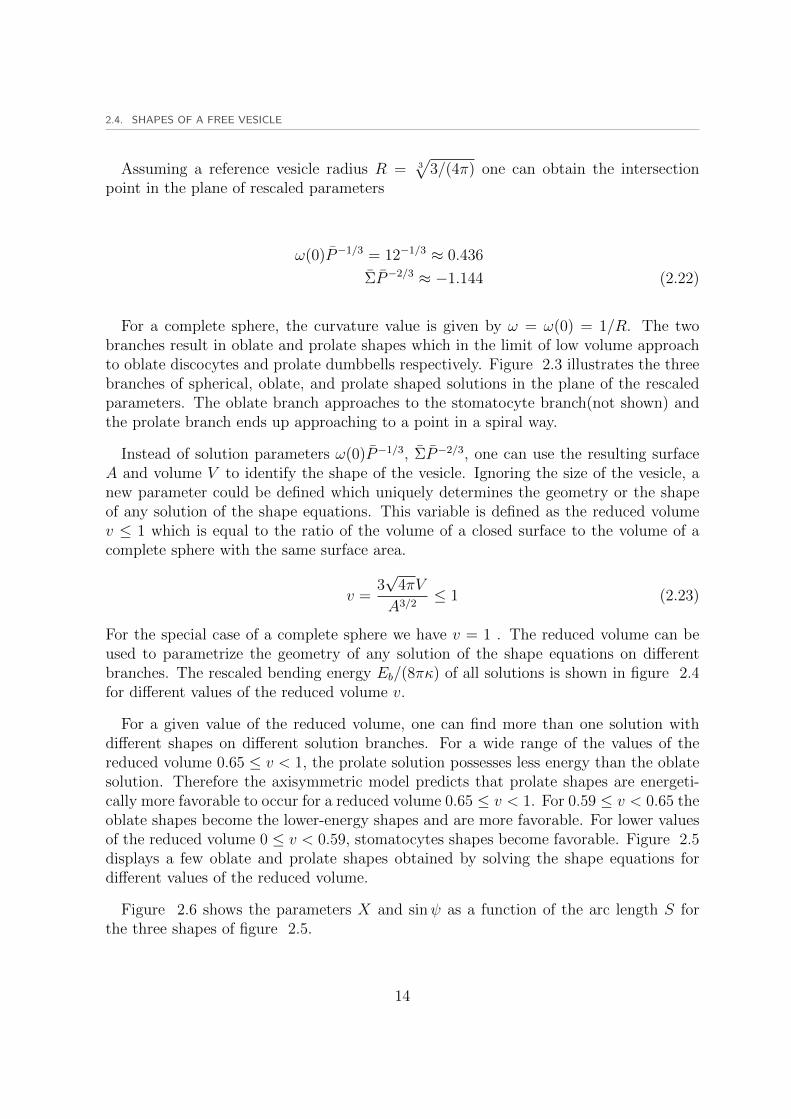
Assuming a reference vesicle radius R = 3√
3/(4π) one can obtain the intersectionpoint in the plane of rescaled parameters
ω(0)P−1/3 = 12−1/3 ≈ 0.436
ΣP−2/3 ≈ −1.144 (2.22)
For a complete sphere, the curvature value is given by ω = ω(0) = 1/R. The twobranches result in oblate and prolate shapes which in the limit of low volume approachto oblate discocytes and prolate dumbbells respectively. Figure 2.3 illustrates the threebranches of spherical, oblate, and prolate shaped solutions in the plane of the rescaledparameters. The oblate branch approaches to the stomatocyte branch(not shown) andthe prolate branch ends up approaching to a point in a spiral way.
Instead of solution parameters ω(0)P−1/3, ΣP−2/3, one can use the resulting surfaceA and volume V to identify the shape of the vesicle. Ignoring the size of the vesicle, anew parameter could be defined which uniquely determines the geometry or the shapeof any solution of the shape equations. This variable is defined as the reduced volumev ≤ 1 which is equal to the ratio of the volume of a closed surface to the volume of acomplete sphere with the same surface area.
v =3√
4πV
A3/2≤ 1 (2.23)
For the special case of a complete sphere we have v = 1 . The reduced volume can beused to parametrize the geometry of any solution of the shape equations on differentbranches. The rescaled bending energy Eb/(8πκ) of all solutions is shown in figure 2.4for different values of the reduced volume v.
For a given value of the reduced volume, one can find more than one solution withdifferent shapes on different solution branches. For a wide range of the values of thereduced volume 0.65 ≤ v < 1, the prolate solution possesses less energy than the oblatesolution. Therefore the axisymmetric model predicts that prolate shapes are energeti-cally more favorable to occur for a reduced volume 0.65 ≤ v < 1. For 0.59 ≤ v < 0.65 theoblate shapes become the lower-energy shapes and are more favorable. For lower valuesof the reduced volume 0 ≤ v < 0.59, stomatocytes shapes become favorable. Figure 2.5displays a few oblate and prolate shapes obtained by solving the shape equations fordifferent values of the reduced volume.
Figure 2.6 shows the parameters X and sinψ as a function of the arc length S forthe three shapes of figure 2.5.
14

CHAPTER 2. SHAPE EQUATIONS FOR SYMMETRIC VESICLES
−0.5 0 0.5 1 1.5−1.8
−1.6
−1.4
−1.2
−1
−0.8
−0.6
−0.4
ω(0)P−13
ΣP−
23
oblate branch prolate branch
sphere branch
Figure 2.3 : Three branches of solutions for the shapes of a free vesicle including spheri-cal, oblate and prolate branches in the plane of rescaled parameters. Oblateand prolate branches bifurcate from the spherical branch at an intersectionpoint.
15

2.4. SHAPES OF A FREE VESICLE
0.6 0.65 0.7 0.75 0.8 0.85 0.9 0.95 11
1.1
1.2
1.3
1.4
1.5
1.6
1.7
1.8
1.9
2
v
Eb
8π
κ
Figure 2.4 : The rescaled bending energy Eb/(8πκ) of free vesicles versus the reducedvolume v for prolate(red squares) and oblate(blue circles) branches. For0 ≤ v < 0.59 stomatocyte shapes are the solution of the shape equations(not shown).
v = 0.64
v = 1v = 0.92
v = 0.77
v = 0.6
Figure 2.5 : Different shapes for oblate and prolate branches corresponding to differentvalues of the reduced volume v. Prolate and oblate branches are energeti-cally favorable in the range 0.65 ≤ v < 1 and 0.59 ≤ v < 0.65 respectively.
16

CHAPTER 2. SHAPE EQUATIONS FOR SYMMETRIC VESICLES
0 0.5 1 1.5 2 2.50
0.2
0.4
0.6
0.8
1
X
S
X,s
inψ
v = 0.77
0 0.5 1 1.5−0.5
0
0.5
1
X
S
X,s
inψ
v = 0.6
0 0.5 1 1.50
0.2
0.4
0.6
0.8
1
X
S
X,s
inψ
v = 1
Figure 2.6 : The parameters X and sinψ for three snapshots of figure 2.5. As it is seena precise solution has been obtained from shooting method and solutionsapproach the boundary values.
17

2.5. A VESICLE WITH AN ADSORBED PARTICLE
2.5 A vesicle with an adsorbed particle
The proposed model for the free vesicle, can be also used to study the interactions ofvesicle with particles. As mentioned before, a vesicle adhered to a spherical particleis composed of two unbound and adhered parts. The shape equations are just usedto find the shape of the unbound part of the vesicle. The adhered part has to followthe geometry of the adhered surface or the spherical particle in our case. The bendingenergy, surface and volume of the adhered part can be calculated( 2.19) without solvingthe corresponding shape equations. Assuming a certain wrapping angle φ(figure 2.2)one can solve the shape equations for the unbound part of the vesicle by starting from thecontact point with initial conditions given by equations 2.17. The total values of bendingenergies, surface and volume are then obtained by summing up their contributions fromboth adhered and unbound part of the vesicle. These values will be used to fullfill thesurface and volume constraints.
During wrapping process, the surface area and the volume of the whole vesicle are keptconstant. The geometry of the vesicle with an absorbed particle is parametrized by thereduced volume of the vesicle, by the vesicle surface area, and the size of the sphericalparticle Rp. By choosing a reference vesicle with unit volume V = 1, the reduced volumewill be the same as the real volume of the vesicle V = v. Therefore the vesicle with anadsorbed particle is geometrically determined by the reduced volume v, by the vesiclearea A0 and by the relative size Rp/R0.
As the surface area of the whole vesicle is kept constant, wrapping of a sphericalparticle with different wrapping angles leads to a reduction in the vesicle volume. Inother world, in order to wrap the spherical particle with a certain wrapping angle, acertain amount of the free volume is required to be allotted to the particle. The morethe wrapping angle φ is, the more the free volume should be to wrap the particle. Thefree volume vf is defined as the difference between the reduced volume of a vesicle andthat of a complete sphere vf = 1 − v. This results in vf = 0 for a complete sphere. Inother worlds if the vesicle is a complete sphere it will not be able to wrap the particlebecause there is no free volume to wrap around the particle. For any set of geometricparameters there is a maximum value of the vesicle reduced volume v0 = 1 − vf whichenables the vesicle to fully wrap the spherical particle. This corresponds to the casewhere both adhered and unbound parts of the vesicle are complete spheres. For therange of the reduced volumes v0 ≤ 1, the particle could be partially wrapped by thevesicle where for the marginal values of v = v0, 1 the corresponding maximum wrappingangles are φ = π, 0 respectively. For v < v0 fully wrapping situation can occur wherethe final shape of unbound vesicle has less volume than the sphere.
Figure 2.7 shows wrapping of a spherical particle with a vesicle with marginal valueof the reduced volume v0 and the corresponding geometry of two complete spheres forfully-wrapped particle. For a given set of geometric parameters, the marginal value of
18

CHAPTER 2. SHAPE EQUATIONS FOR SYMMETRIC VESICLES
Figure 2.7 : The schematic view of initial and final morphologies for wrapping a spher-ical particle with the marginal value of the reduced volume v = v0 for thevesicle. Left and right snapshot correspond to initial zero and final fullwrapping respectively. The final snapshot for v = v0 is composed of twocomplete spheres.
the reduced volume v0 can be obtained by making the area and volume of a free vesicle,equal to the area and volume of a vesicle with a fully-wrapped particle. The volumeV = v0 and area A0 of a free vesicle(left snapshot in Figure 2.7), are related to the areaand volume of a vesicle with a fully-wrapped particle
A = A0 =⇒ 4π(R2p +R2
1) = 4πR20
v0 = v =4π
3(R2
0 −R2p)
32 − 4
3R3p (2.24)
Here, R1 is the radius of the unbound part of the vesicle which is a complete sphere forthe marginal value of the reduced volume v = v0. The vesicle with an adsorbed particleis geometrically determined using parameters v, Rp/R0, A0. However the wrappingprocess does not only depend on the geometric factors. The maximum possible value ofthe wrapping angle is determined for any given set of geometric parameters. Howeverit is the rescaled adhesion strength u which determines the wrapping amount of theparticle. Therefore the adhesion of a vesicle to a particle is completely determined bythe reduced volume v, Rp/R0, A0, u where u is the rescaled adhesion energy.
For a given set of system parameters v,Rp/R0, A0, u a solution is defined by obtainingthe value of the corresponding wrapping angle φ. However as mentioned earlier, Shape
19

2.5. A VESICLE WITH AN ADSORBED PARTICLE
equations are used to solve the unbound part of the vesicle and that is why the adhesionenergy is not involved in the total energy of the system and does not appear in the shapeequations. Therefore the problem is solved as follows. For a certain set of parametersv,Rp/R0, A0, the shape equations are solved for different values of 0 ≤ φ ≤ π andthe corresponding bending energy Eb of the whole vesicle is obtained as a function ofwrapping angle or the adhesion area
Eb = Eb(φ) = Eb(Aadh) (2.25)
where Aadh = 2πR2p cosφ. Then the total energy of the system is defined as the sum
of the bending energy and the adhesion energy
Et = Eb + Eadh = Eb − UadhAadh = Eb(Aadh)− uκ
R2p
Aadh (2.26)
The total energy Et = Et(Aadh) is then obtained as a function of the adhesion areaAadh and the rescale adhesion strength u. For a given value of the adhesion strengthu, the total energy gains its minimum value at a certain adhesion area which gives thewrapping angle φ. In this way the wrapping of the particle by the vesicle is determined.
The solution branches of the shapes of a free vesicle include spherical, oblate, andprolate branches. For a vesicle with an adsorbed particle, the initial and final situationsof zero and fully wrapped particle are similar to the solutions of a free vesicle. For a freevesicle, a certain shape is obtained as a solution of the shape equations. This shape couldbe either a sphere, a prolate, an oblate or a stomatocyte. Here, we only consider thereduced volumes in the range 0.59 ≤ v ≤ 1 where the stomatocyte branch is suppressed.However in case of a vesicle with an adsorbed particle, there is a different story. Anybranch of solution, starts on one of the solution branches for a free vesicle and ends upon the same branch. This is expected because for zero and full wrapping condition theparticle presence is irrelevant. For instance figure 2.8 shows two branches of solutionsfor the vesicle particle system, bifurcating from oblate and prolate branches of the freevesicle for the same value of the reduced volume v = 0.9.
The shapes of the vesicle wrapping around a particle have complex geometry. Theshapes which are considered as oblate or prolate for the free vesicle are not ellipsoidoblate or prolate shapes. These are general shapes which have similar geometry to theellipsoidal particles. For a vesicle wrapping a particle, the shapes are more complicated.Therefore we name the shapes based on the solution branch that they bifurcate from.
We choose four different particle sizes Rp = 0.05, 0.1, 0.15, 0.2 which lead to fourvalues of the relative size Rp/R0 ≈ 0.8, 0.16, 0.24, 0.32 respectively. The correspondingmarginal values of the reduced volume v0 for different particle sizes are obtained asv0 ≈ 0.988, 0.957, 0.898, 0.844. The shape equations are solved for a fixed values ofRp/R0(different particle sizes) and a given value of the reduced volume v and the wholerange of wrapping angles 0 ≤ φ ≤ π are obtained. The reduced volume v is always
20

CHAPTER 2. SHAPE EQUATIONS FOR SYMMETRIC VESICLES
−0.1 0 0.1 0.2 0.3 0.4 0.5 0.6 0.7 0.8
−1.4
−1.3
−1.2
−1.1
−1
−0.9
−0.8
−0.7
ω(0) P−13
ΣP
−2 3
v = 0.88, Rp/R0 = 0.16
Figure 2.8 : Two branches of solutions for a vesicle with v = 0.9 wrapping a particleRp = 0.1(Rp/R0 = 0.16). Both branches bifurcate from the oblate andprolate branches of the free vesicle so they are named after the branch theybifurcate from.
21

2.5. A VESICLE WITH AN ADSORBED PARTICLE
Figure 2.9 : The vesicle shapes during wrapping of a particle Rp = 0.1(Rp/R0 = 0.16)with v = 0.95(left). The solutions correspond to the oblate branch. Rightsnapshot shows a close view of the adhesion zone.
chosen in v ≤ v0 to enable the vesicle to completely wrap the spherical particle. Notethat for a wide range of the reduced volumes, there are several shapes found on differentbranches of solution.
Figure 2.9 shows the oblate branch of a vesicle with an adsorbed particle for differentwrapping angles with a reduced volume v = 0.95 and the relative sizeRp/R0 = 0.16(Rp =0.1). The initial shape of the vesicle is an oblate with a volume V = v = 0.95 and thefinal shape of the unbound part of the vesicle is also an oblate because v < v0 = 0.957.For the very special case of v = v0, the final shape of the unbound part of the vesiclewill be a complete sphere. A close view of the contact area is also shown which verifiesthe high accuracy of the numerical solution.
Figure 2.10 shows vesicle shapes with the same value of parameters but from theprolate branch of solution. The initial and final solutions are also prolate vesicles. Weremind that both branches possess the minimum value of energy for different wrappingangles and all vesicles fulfill the area and volume constraints.
2.5.1 Prolate-oblate transition
The solution of the shape equations was obtained with a set of system variables Rp/R0 =0.16, v = 0.95 for the whole range of wrapping angles both on prolate and oblatebranches. For a free vesicle, it was shown that the oblate branch has lower bendingenergy and is more stable for 0.59 ≤ v < 0.65 while the prolate branch has lower energy
22

CHAPTER 2. SHAPE EQUATIONS FOR SYMMETRIC VESICLES
Figure 2.10 : The vesicle shapes during wrapping of a particle Rp = 0.1(Rp/R0 = 0.16)with v = 0.95. The solutions correspond to the prolate branch.
23

2.5. A VESICLE WITH AN ADSORBED PARTICLE
0 0.5 1 1.5 2 2.5 3
30
35
40
45
50
κ
oblate branchprolate branch
v = 0.95, Rp/R0 = 0.16
0 0.5 1 1.5 2 2.5 3
35
40
45
50
55
60
oblate branchprolate branch
v = 0.8, Rp/R0 = 0.16
κFigure 2.11 : Rescaled bending energies for oblate and prolate branches with different
reduced volumes v = 0.95, 0.8, Rp/R0 = 0.16 versus wrapping angle φ.The prolate branch has lower energy for very low and very high values ofφ while the oblate branch has less energy for the mid range values of φ.
values for 0.65 ≤ v < 1. Thus for a rather high value of the reduced volume such asv = 0.95, where the initial and final points of the prolate branch possess lower energiesthan the oblate one, it is expected that the whole prolate branch has lower energy thanthe oblate one. However this is not true. The bending energies of the oblate and prolatebranches for two different values of the reduced volume v = 0.95, 0.8 with Rp/R0 = 0.16are shown in figure 2.11.
Starting from zero wrapping, the prolate branch has lower bending energy for the verysmall values of the wrapping angle. After a certain amount of wrapping the bendingenergy of the prolate branch grows above that of the oblate branch. This is the regionwhere the oblate branch is energetically more favorable which holds for a wide range ofthe wrapping angle φ. Finally just before the complete wrapping of the particle, prolatebranch becomes the lower-energy branch again. Although the prolate branch has lowerenergy for the zero and full-wrapping condition, but the oblate branch is the favorableshape for a wide range of wrapping angles. We refer to this phenomenon as the prolateoblate transition. This transition is induced by the spherical particle wrapped by thevesicle. The transition process depends on the reduced volume v and the relative sizeRp/R0. Comparing two reduced volumes v = 0.95, 0.8, one can observe that for thesame particle size Rp/R0, the transition wrapping angle is higher for a lower value of thereduced volume.
24
CHAPTER 2. SHAPE EQUATIONS FOR SYMMETRIC VESICLES
0 0.5 1 1.5 2 2.5 3
25.5
26
26.5
27
27.5
28
28.5
29
29.5
30
u = ut = 1.8
u = 2
v = 0.95, Rp/R0 = 0.16
κ
0 0.5 1 1.5 2 2.5 331
31.5
32
32.5
33
33.5
34
34.5
35
κ
u = ut = 1.72
u = 1.8
u = 1.9
u = 2
v = 0.85, Rp/R0 = 0.16
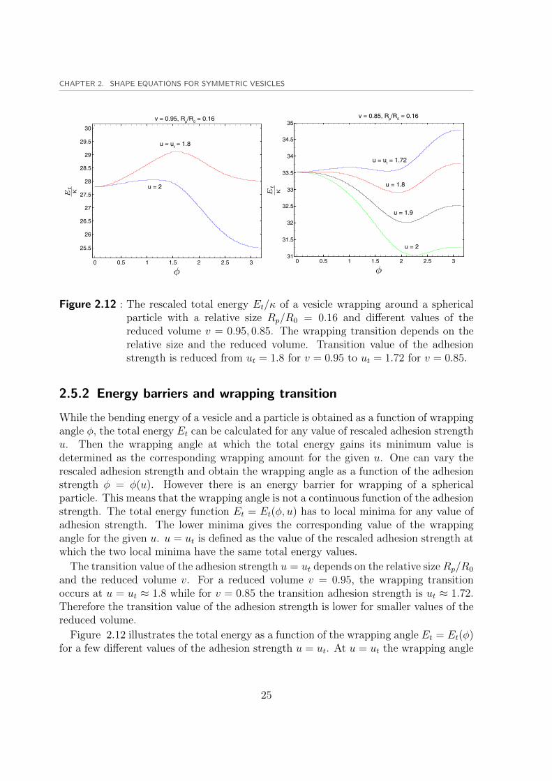
Figure 2.12 : The rescaled total energy Et/κ of a vesicle wrapping around a sphericalparticle with a relative size Rp/R0 = 0.16 and different values of thereduced volume v = 0.95, 0.85. The wrapping transition depends on therelative size and the reduced volume. Transition value of the adhesionstrength is reduced from ut = 1.8 for v = 0.95 to ut = 1.72 for v = 0.85.
2.5.2 Energy barriers and wrapping transition
While the bending energy of a vesicle and a particle is obtained as a function of wrappingangle φ, the total energy Et can be calculated for any value of rescaled adhesion strengthu. Then the wrapping angle at which the total energy gains its minimum value isdetermined as the corresponding wrapping amount for the given u. One can vary therescaled adhesion strength and obtain the wrapping angle as a function of the adhesionstrength φ = φ(u). However there is an energy barrier for wrapping of a sphericalparticle. This means that the wrapping angle is not a continuous function of the adhesionstrength. The total energy function Et = Et(φ, u) has to local minima for any value ofadhesion strength. The lower minima gives the corresponding value of the wrappingangle for the given u. u = ut is defined as the value of the rescaled adhesion strength atwhich the two local minima have the same total energy values.
The transition value of the adhesion strength u = ut depends on the relative size Rp/R0
and the reduced volume v. For a reduced volume v = 0.95, the wrapping transitionoccurs at u = ut ≈ 1.8 while for v = 0.85 the transition adhesion strength is ut ≈ 1.72.Therefore the transition value of the adhesion strength is lower for smaller values of thereduced volume.
Figure 2.12 illustrates the total energy as a function of the wrapping angle Et = Et(φ)for a few different values of the adhesion strength u = ut. At u = ut the wrapping angle
25

2.5. A VESICLE WITH AN ADSORBED PARTICLE
switches from φ ≈ 0 to φ ≈ π for v = 0.95 resulting in a discontinuous function φ = φ(u).This means that for u ≤ ut the particle is partially wrapped while for u > ut the wrappingamount is a high value close to a fully-wrapped particle. we conclude that the transitionvalue of the rescaled adhesion energy ut strongly depends on the reduced volume of thevesicle v and the relative size Rp/R0.
26
3 Monte Carlo simulations of vesicles
3.1 Triangulated membranes
Biomembranes are generally modeled as elastic thin sheets whose fluctuation dynamicsare controlled by the bending and surface energies through Helfrich equations[11]. In theHelfrich model, the membrane is considered as a two-dimensional surface with negligiblethickness and its energy is decomposed into bending and stretching energy components.Stretching energy is determined by membrane surface tension and the change in its area.Bending energy is determined by the shape of the membrane as a geometric propertyand the bending rigidity of the membrane as a mechanical property.
Helfrich energy has been used with both discrete and continuous approaches to studymembrane physics. In the discrete approach, the continuum fluid membrane is dis-cretized into a three dimensional mesh composed of edges and vertices by generating atriangulated network on the membrane surface. Numerical simulations of such a modelusing statistical methods like Monte Carlo simulations provides a strong tool to studymembrane physics. In this chapter we focus on different aspects of the triangulatedmodel.
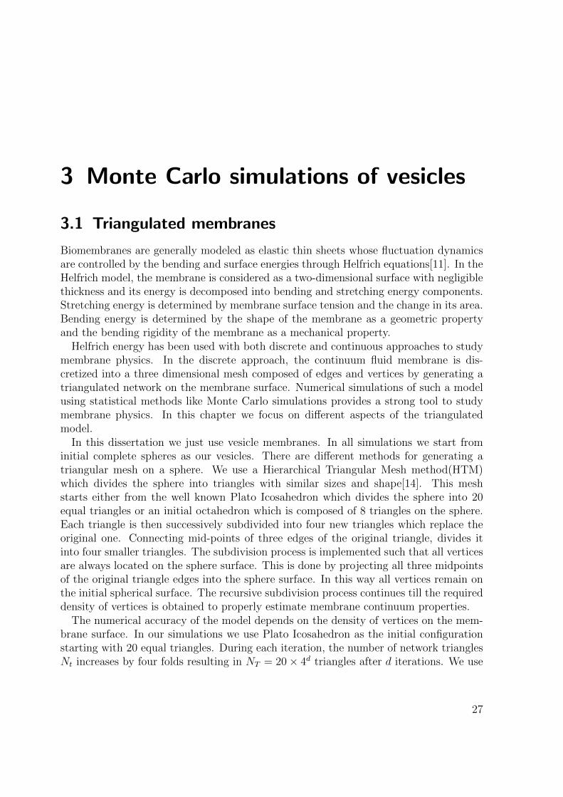
In this dissertation we just use vesicle membranes. In all simulations we start frominitial complete spheres as our vesicles. There are different methods for generating atriangular mesh on a sphere. We use a Hierarchical Triangular Mesh method(HTM)which divides the sphere into triangles with similar sizes and shape[14]. This meshstarts either from the well known Plato Icosahedron which divides the sphere into 20equal triangles or an initial octahedron which is composed of 8 triangles on the sphere.Each triangle is then successively subdivided into four new triangles which replace theoriginal one. Connecting mid-points of three edges of the original triangle, divides itinto four smaller triangles. The subdivision process is implemented such that all verticesare always located on the sphere surface. This is done by projecting all three midpointsof the original triangle edges into the sphere surface. In this way all vertices remain onthe initial spherical surface. The recursive subdivision process continues till the requireddensity of vertices is obtained to properly estimate membrane continuum properties.
The numerical accuracy of the model depends on the density of vertices on the mem-brane surface. In our simulations we use Plato Icosahedron as the initial configurationstarting with 20 equal triangles. During each iteration, the number of network trianglesNt increases by four folds resulting in NT = 20× 4d triangles after d iterations. We use
27

3.1. TRIANGULATED MEMBRANES
Figure 3.1 : Triangular network on the spherical vesicle generated from Plato Icosahe-dron up to four iterations. Upper left snapshot shows Plato Icosahedronand the lower right snapshot shows the vesicle used in simulations composedof NT = 5120 triangles, NE = 7680 edges, and Nv = 2562 vertices.
d = 4 to obtain the initial morphology of the triangulated vesicle.
Figure 3.1 shows the evolution of the produced mesh on a sphere during four successiveiterations. The resulting mesh is composed of NT = 5120 triangles, NE = 7680 edges,and Nv = 2562 vertices which are related by NE = 1.5NT , NT −NE +Nv = 0[15].
In order to represent the continuum membrane with a discrete network, one shouldbe able to calculate mechanical and geometrical properties of the membrane from thenetwork model. Each membrane property could be discretized in terms of its componentseither from network vertices or network edges. Here we discretize all properties in termsof their contributions from network vertices[15].
Figure 3.2 shows a schematic presentation of two neighbor triangles Ti, Tj with thecommon edge in between. To obtain the total surface area and volume, we sum up theareas and volumes of all triangles. Thus the vesicle surface area A is obtained as,
A =
NT∑i=1
Ai (3.1)
28
CHAPTER 3. MONTE CARLO SIMULATIONS OF VESICLES
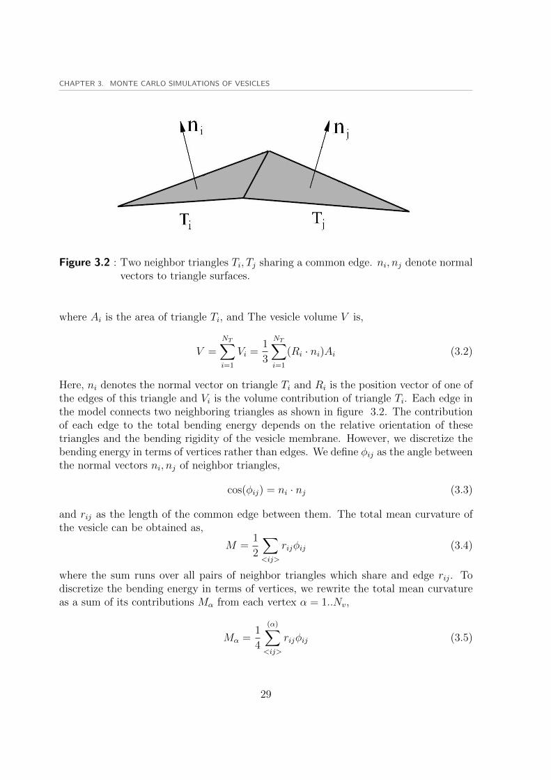
Figure 3.2 : Two neighbor triangles Ti, Tj sharing a common edge. ni, nj denote normalvectors to triangle surfaces.
where Ai is the area of triangle Ti, and The vesicle volume V is,
V =
NT∑i=1
Vi =1
3
NT∑i=1
(Ri · ni)Ai (3.2)
Here, ni denotes the normal vector on triangle Ti and Ri is the position vector of one ofthe edges of this triangle and Vi is the volume contribution of triangle Ti. Each edge inthe model connects two neighboring triangles as shown in figure 3.2. The contributionof each edge to the total bending energy depends on the relative orientation of thesetriangles and the bending rigidity of the vesicle membrane. However, we discretize thebending energy in terms of vertices rather than edges. We define φij as the angle betweenthe normal vectors ni, nj of neighbor triangles,
cos(φij) = ni · nj (3.3)
and rij as the length of the common edge between them. The total mean curvature ofthe vesicle can be obtained as,
M =1
2
∑<ij>
rijφij (3.4)
where the sum runs over all pairs of neighbor triangles which share and edge rij. Todiscretize the bending energy in terms of vertices, we rewrite the total mean curvatureas a sum of its contributions Mα from each vertex α = 1..Nv,
Mα =1
4
(α)∑<ij>
rijφij (3.5)
29

3.2. MONTE CARLO SIMULATIONS
Here the summation runs over all neighbor triangles which share the vertex α. Theprefactor 1/4 ensures that the summation over the mean curvature contributions of allvertices tends towards the integral over the mean curvature of the continuous vesiclesurface for a large number of vertices
M =Nv∑α=1
Mα (3.6)
We also define the corresponding area element of the vertex α as,
A =1
3
α∑<i>
Ai (3.7)
where the sum runs over all neighboring triangles of vertex α. Finally the total bendingenergy Eb of the discretized network model can be written as,
Eb = 2κNv∑α=1
M2α
Aα(3.8)
where κ denotes the bending rigidity of the membrane.
3.2 Monte Carlo simulations
We have developed a discrete model to study minimum energy shapes of the membraneand membrane interactions with other biological elements. Numerical simulations ofthe triangulated model helps us to understand membrane physics. We use Monte Carlomethod as a strong numerical tool to simulate membrane network model[16]. Our MonteCarlo simulations are based on importance sampling method which samples the wholeconfiguration space of a physical system based on the their importance for an objectivefunction. Here we try to minimize the membrane energy using a specific type of MonteCarlo simulations which is called simulated annealing method.
Monte Carlo simulation is composed of several Monte Carlo steps. In every MonteCarlo step, vesicle configuration is randomly varied and the corresponding energy ofthe vesicle is calculated. Vesicle configuration is varied using two different Monte Carlomoves. All vertices of the triangulated-membrane model move successively to representthermal fluctuations of the biological membrane in the real physical condition. The edgesof neighbor triangles are flipped to keep the membrane fluid. In Each Monte Carlo stepsall vertices are randomly displaced in the space in a successive way. The direction andmagnitude of the displacement vector is chosen by generating three independent random
30
CHAPTER 3. MONTE CARLO SIMULATIONS OF VESICLES
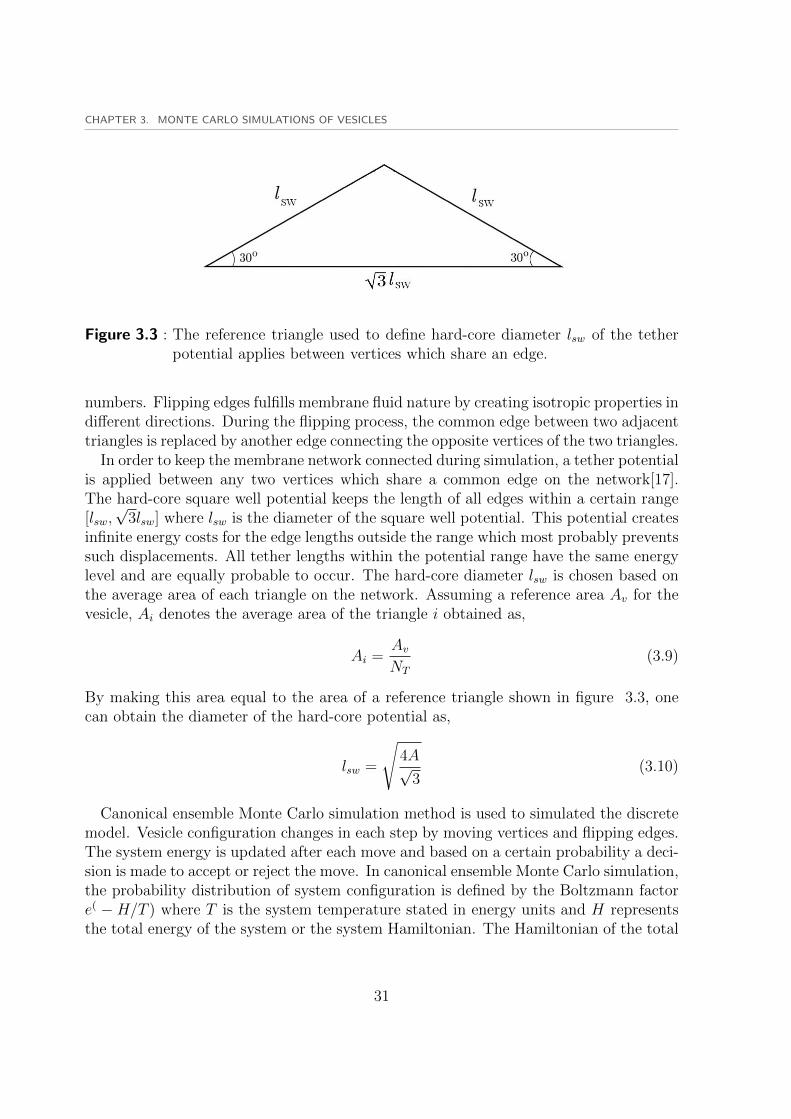
Figure 3.3 : The reference triangle used to define hard-core diameter lsw of the tetherpotential applies between vertices which share an edge.
numbers. Flipping edges fulfills membrane fluid nature by creating isotropic properties indifferent directions. During the flipping process, the common edge between two adjacenttriangles is replaced by another edge connecting the opposite vertices of the two triangles.
In order to keep the membrane network connected during simulation, a tether potentialis applied between any two vertices which share a common edge on the network[17].The hard-core square well potential keeps the length of all edges within a certain range[lsw,√
3lsw] where lsw is the diameter of the square well potential. This potential createsinfinite energy costs for the edge lengths outside the range which most probably preventssuch displacements. All tether lengths within the potential range have the same energylevel and are equally probable to occur. The hard-core diameter lsw is chosen based onthe average area of each triangle on the network. Assuming a reference area Av for thevesicle, Ai denotes the average area of the triangle i obtained as,
Ai =AvNT
(3.9)
By making this area equal to the area of a reference triangle shown in figure 3.3, onecan obtain the diameter of the hard-core potential as,
lsw =
√4A√
3(3.10)
Canonical ensemble Monte Carlo simulation method is used to simulated the discretemodel. Vesicle configuration changes in each step by moving vertices and flipping edges.The system energy is updated after each move and based on a certain probability a deci-sion is made to accept or reject the move. In canonical ensemble Monte Carlo simulation,the probability distribution of system configuration is defined by the Boltzmann factore( −H/T ) where T is the system temperature stated in energy units and H representsthe total energy of the system or the system Hamiltonian. The Hamiltonian of the total
31

3.2. MONTE CARLO SIMULATIONS
system is composed of different terms. The main contribution to the system Hamilto-nian comes from the system configuration. This part of the system Hamiltonian is calledconfigurational Hamiltonian. The configurational Hamiltonian Hc is composed of twoterms corresponding to the bending energy of the vesicle Hbe and the tether potentialHt
Hc = Hbe +Ht (3.11)
3.2.1 Vesicle parameters
In order to define the geometry of the vesicle we consider a number of dimensionlessparameters which are used as system variables. In all simulations we keep the vesiclearea at a constant reference area A0 defined as the area of a sphere with unit volumeV = 1. The radius R0 of a spherical vesicle with unit volume is obtained as
R0 =3
√3
4π(3.12)
which is used to define the length scale of the whole system. The reference area A0 canbe written as
A0 = 4πR20 (3.13)
We also define the reduced volume v = (3√
4πV )/(A3/2) ≤ 1 of any closed surface withVolume V and area A, as the ratio of its volume to the volume of a complete sphere withthe same area A. In this way the surface area of any closed surface with reduced volumev = 1 is equal to the reference area A0. The reduced volume and area of the vesicle areused as system parameters. For a vesicle with an adsorbed nanoparticle, there are moreparameters involved in the whole system which will be explained later on.
3.2.2 Area and volume constraints
In order to reproduce physical properties of the real phospholipid membranes, one shouldinvolve those properties in the membrane model. It is proved that the number of lipidmolecules in a certain area of the membrane is rather constant. This implies an almostconstant surface density of lipids or number of lipids per unit area. As explained earlierwe always preserve vesicle area at A0 to resemble the constant number of lipids perunit area in real membranes. The area constraint is implemented in the Monte Carlosimulations by using the corresponding term Harea in the system Hamiltonian
Harea = KA(1− (A
A0
)2) (3.14)
32

CHAPTER 3. MONTE CARLO SIMULATIONS OF VESICLES
where A presents the current value of the vesicle area during simulation, and KA is thearea compression factor. A dimensionless area factor KA/T ≈ 2 × 10⁵ was used in oursimulations resulting in a very small relative error∣∣∣∣A− A0
A0
∣∣∣∣ ≤ 10−3 (3.15)
Another constraint on the vesicle is the volume constraint which is imposed by theosmotic pressure of the vesicle. The osmotic energy is defined as[18]
Eos ≈TρexVos
2(V − VosVos
)2 (3.16)
Here, V is the vesicle volume, ρex is the number of osmotically active particles outsidethe vesicle, Vos = Nin/ρex is the volume at which there is no osmotic pressure, withNin denoting the number of osmotically active molecules inside the vesicle. To applya harmonic volume constraint on the vesicle, a new term Hvol is added to the systemHamiltonian
Hvol = Kv(1− (V
V0)2) (3.17)
where V presents the current value of the vesicle volume during simulation, and KV isthe volume inflation factor. A dimensionless volume factor Kv/T ≈ 6× 10⁵ was used inour simulations to limit the volume fluctuations in the range∣∣∣∣1− V
V0
∣∣∣∣ ≤ 5× 10⁻⁴ (3.18)
Using Kv = 0 suppresses the volume constraint on the vesicle.
To involve interactions of the vesicle with a nanoparticle, the interaction energy shouldbe included in the system Hamiltonian. These interactions are involved by adding theadhesion energy of the vesicle with the nanoparticle. The adhesion of the vesicle to thenanoparticle occurs in a certain adhesion area. A square well potential is used to definethe adhesion process of vertices to the adhered surface. All vertices witch lay within acertain distance from the vesicle surface, are considered adhered to the surface. Thisdistance is the diameter of the square well potential which is called the cut off distance.The adhesion area Aad is defined as the sum of the area contributions of all adheredvertices. The adhesion strength U as the depth of the square well potential, and theadhesion area Aadh determine the contribution of the adhesion energy Hadh to the systemHamiltonian,
Hadh = UAadh (3.19)
Membrane adhesion process is controlled by the competition between the bending and
33
3.3. SIMULATED ANNEALING METHOD
adhesion energies. The bending energy vanishes for a flat membrane due to zero meancurvature. When a membrane patch completely wraps a spherical particle its bendingenergy is equal to the bending energy of a complete sphere Ebs which is obtained fromHelfrich equation as
Ebs =
∫dA(
κ
2M2) =
∫dA[
κ
2(
1
Rp
+1
Rp
)2] = 8πκ (3.20)
where M denotes the local mean curvature of the membrane surface. we assume thatin the case of a fully-wrapped particle, the bending energy cost of the contact area isnegligible due to a saddle geometry with zero mean curvature. The bending energyEs for a completely-wrapped spherical particle must be compensated by the adhesionenergy which is defined as
Ead = −UAad = −U(4πR2p) (3.21)
where Rp is the radius of the spherical particle. Making the total energy Ebs + Eadequal to zero, results in the corresponding adhesion strength U = 2κ/R2
p which enablesthe vesicle to fully wrap the spherical particle. We then define the rescaled adhesionenergy u = UR2
p/κ. Therefore a minimum value of the rescaled adhesion energy u = 2 isrequired to completely wrap a spherical particle. Setting u = 0 implies the case withoutadhesive interactions between the vesicle and particle.
Finally one can obtain the total system Hamiltonian as
H = Hbe +Ht +Harea +Hvol +Hadh (3.22)
which is minimized in the course of the system evolution during Monte Carlo simulationsteps.
3.3 Simulated annealing method
Throughout this dissertation, we only consider minimum energy shapes of the vesicleand ignore the thermal fluctuations of the membrane. Therefore we need to suppressmembrane fluctuations in the triangulated model. In order to suppress the effect ofthermal fluctuations we reduce the vesicle temperature to the values close to zero. Thisis done by using a simulated annealing approach through which the temperature islinearly reduced to a very small value. We look for the vesicle configuration which resultsin the minimum value of the total energy. Therefore we have to ensure that we find theglobal minimum of the total energy and avoid getting trapped in a local minimum. Thetemperature is reduced slow enough to avoid ending up in a local minima.
34

4 Tubulation and aggregation ofspherical nanoparticles
The interactions of nanoparticles with biological membranes have recently gained muchinterest among scientists in the field of nanotechnology[19]. As one of the major appli-cations, nanoparticles can be designed to serve for drug delivery purposes by enteringthe target cell membrane and acting as drug containers[20]. Therefore it is crucial tounderstand the mechanisms by which nanoparticles interact with biological cells andenter the cell membrane.
4.1 Introduction
The particle adsorption process or wrapping of a spherical particle is controlled by thecompetition between bending and adhesion energies. Adhesion of a particle to a vesi-cle induces shape deformations of the vesicle which are accompanied by an increase inthe bending energy of the vesicle. This increase in the bending energy has to be com-pensated by the adhesion energy between the vesicle and the nanoparticle. Engulfingand internalization of the spherical nanoparticle demands enough adhesion strength tocompensate for the high bending energy of highly-curved vesicle wrapped around thenanoparticle.
While there are several simulations[21, 22, 23] and theoretical[24, 25, 26, 27, 28] studiesabout the interactions of a single nanoparticle with a vesicle, a comprehensive investiga-tion on the effect of more than one particle is still missing. When more than one particleis adsorbed on a membrane, the interactions between the vesicle and particles becomemuch more complicated. The geometry of the membrane is affected by the adsorbedparticles. For a pair of particles adsorbed on a membrane, the shape deformations ofthe membrane depend on the relative distance between these particles. When particlescome close together, they affect the adhesion area of one another. Finding the shape ofthe adhesion area is a very complicated geometrical problem which in general does nothave an analytical solution.
Membrane-induced interactions between nanoparticles can be either attractive or re-pulsive. For instance repulsive interactions between rod like cylindrical particles ad-sorbed on the same side of the membrane with parallel orientation[29, 30]. Repulsiveinteractions have been also found between symmetric inclusions which locally deform
35

4.2. A VESICLE WITH A PAIR OF ADSORBED PARTICLES
the membrane[31, 32, 33, 34]. Recently, linear aggregates of weakly adsorbed spheri-cal nanoparticles have been observed in simulations and explained by attractive three-particle interactions. A three-particle interaction potential for different orientations hasbeen calculated. The results demonstrate that the linear arrangement of the three parti-cles has lower energy with respect to other orientations. However the spherical particleare only partially adsorbed on the vesicle and highly wrapped particles are not con-sidered. The interactions between a pair of spherical particles with different wrappingangles including deeply adsorbed particles is not also considered in their simulations.
We report membrane-induced attractive interactions between a pair of spherical nanopar-ticles adsorbed on the outer surface of a vesicle. These attractive interactions lead tobound states of the particles with a vesicle shape that depends on the ratio of the areaA and volume V of the vesicles. This ratio is normally characterized by the reducedvolume v = 3
√4πV/A3/2 ≤ 1, where the maximum value v = 1 corresponds to the area-
to-volume ratio of a sphere. For large values of the reduced volume, we find bound statesin which two particles are equally wrapped by the vesicle. These states correspond to avesicle shape which is symmetric with respect to a plane perpendicular to the connect-ing line of the particles. For smaller values of the reduced volume, we find two differentbranches of solution with two bound states. Each branch leads to a bound state whichcorresponds to either equally wrapped particles with a symmetric vesicle or even morestrongly bound states in which the two particles are jointly wrapped by a membranetube that invaginates into the vesicle. For three and more particles, we observe similarbound states in which linear aggregates of the particles are confined within membranetubes. The tubular confinement of linear nanoparticle aggregates reported here consti-tutes a novel route to encapsulate nanoparticles reversibly in vesicle membranes. Theamount of confined nanoparticles as well as their release can be controlled by adjustingthe reduced volume of the vesicles, i.e., by deflation and inflation of vesicles via changesin osmotic conditions.
4.2 A vesicle with a pair of adsorbed particles
The total energy of the vesicle varies with the relative distance between nanoparticlesbecause they depend on the membrane shape in the adhesion area. The total energyof the vesicle with adsorbed particles is composed of the bending and adhesion energiesas was explained in detail in previous chapters. The total energy of the vesicle withadsorbed particles is given by
Et = Eb + Eadh (4.1)
where the Helfrich bending energy Eb is calculated for a triangulated vesicle and theadhesion energy Eadh can be written as
36

CHAPTER 4. TUBULATION AND AGGREGATION OF SPHERICAL NANOPARTICLES
Eadh = −UAadh (4.2)
where the adhesion strength U gives the adhesion energy per unit adhesion area. Werescale the adhesion strength U by the bending rigidity of the membrane and the particlesize, and define the rescaled adhesion strength u =2
p /Aadh. Therefore the adhesion energycan be written as
Eadh = −u κR2p
Aadh (4.3)
The adhesion area Aadh is the sum of the adhesion areas of both particles. The adhesionarea of the particle is obtained as the sum of all area contributions from the adheredvertices to the particle.
The total energy of the vesicle and particles depends on the the rescaled adhesionstrength u, and the reduced volume of the vesicle v. The relative size of the vesicle andparticles is fixed at Rp = 0.161R0 where R0 is the radius of a sphere with an area equalto the constant vesicle area A0 = 42
0.In order to obtain interactions, we keep both spherical particles fixed in space at a
certain distance and let the triangular vesicle freely move in space. For every distanceof the two particles with a certain value of the vesicle reduced volume, ten differentsimulated annealing simulations are performed and the mean values are calculated aswell as the standard deviation of the errors. Monte Carlo simulated annealing hasbeen used to minimize the total energy Et of the whole system to ensure the minimumenergy level E = min(Et) is achieved. All Monte Carlo simulation parameters forthe triangular vesicle are given in chapter three. The cut off distance or the width ofthe square-well potential d is chosen as d = 0.1Rp for all simulations. Therefore allvertices which are located at a distance from the particle surface in the range [0, d],are considered as adhered vertices to the particle. The hard-core potential preventsvertices to enter the nanoparticle by making an infinite energy cost for those moves. Abending rigidity of κ = 20T was used as an appropriate value for the fluid vesicle inexperimental conditions. Minimization of the total energy leads to the energy values atnearly zero temperature where the fluctuation effects are suppressed and the membraneelastic energies are calculated as an output. This implies that the bending rigidity isirrelevant in the simulation results. Thus the minimum energy E is rescaled with respectto the bending rigidity of the membrane in all results.
Monte Carlo simulations are performed for 3 × 10⁷ Monte Carlo steps where eachMonte Carlo step includes moving all vertices and flipping all edges. We start frominitial triangulated complete spheres with unit volume sitting on both fixed particleswhich are located outside of the vesicle. In the first 106 Monet Carlo steps, the vesiclevolume is gradually reduced to the target value of the reduced volume. The vesicle isthen equilibrated at the target value of the reduced volume and a temperature kBT = 1for 4×10⁶ Monte Carlo steps. The vesicle temperature is then reduced linearly to a very
37
4.3. SIMULATION RESULTS
small value of kBT ≈ 0.01 during 2.4×10⁷ Monte Carlo steps. At such a low temperature,the error in the estimated value of the rescaled bending energy of a triangulated sphereEbs/κ = 8π is less than 0.1%. Finally the vesicle is again equilibrated at kBT ≈ 0.01 for1× 106 Monte Carlo steps.
The marginal value of the reduced volume v0 of the vesicle is defined as the volumewhich results in a complete sphere for the unbound part of a vesicle(figure 2.7) incomplete wrapping. While for a single particle Rp = 0.1 it was obtained as v0 = 0.957,the same calculation results in a marginal value of the reduced volume v0 = 0.914 forwrapping two particles with the same radius. Therefore a maximum reduced volume ofV = v = v0 = 0.914 or a minimum free volume vem = 1−0.914 = 0.086 is required to letthe vesicle fully wrap the two particles. We choose three different values of the reducedvolume v = 0.92, 0.94, 0.96 for the vesicle. This results in different partial wrappingsituation of the particles. For v = 0.96 the free volume is obtained as vf = 0.04 < 0.5vemthat enables the vesicle to wrap less than half the particles area. With v = 0.94 the freevolume is obtained as vf = 0.06 < 0.5vem that enables vesicle to wrap more than halfthe particle area and finally for v = 0.92 the largest free volume is available to wrapparticles close to complete wrapping situation. Choosing the above values of the reducedvolume, one can ensure to observe the effect of the reduced volume as one of the systemparameters. The rescaled adhesion strength u = 2 results in fully wrapping conditionof a spherical nanoparticle. We choose a slightly higher value of the rescaled adhesionstrength u = 2.33 to ensure that both particles gain the maximum possible adhesionarea.
4.3 Simulation results
We start our simulations with a reduced volume v = 0.96 and obtain the rescaled totalenergy E/κ as a function of the particle rescaled distance r/Rp.
Figure 4.1 illustrates minimum energy states of a vesicle with a reduced volumev = 0.96 wrapping spherical particles at different distances. It is seen that particles arepartially wrapped up to 50%.
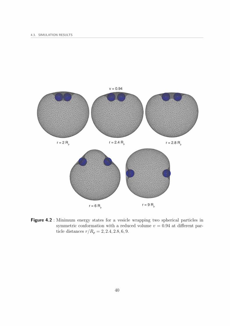
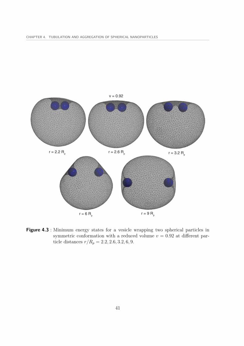
For an intermediate value of the reduced volume v = 0.94, particles are wrapped moredue to the higher value of the free volume. Figure 4.2 displays minimum energy states ofthe vesicle with a reduced volume v = 0.94 wrapping particles with different distances.In this case particles are almost halfly wrapped by the vesicle. For the lowest value of thereduced volume v = 0.92 which is just slightly above the marginal value of the reducedvolume v0 = 0.914, particles are more than halfly wrapped by the vesicle as shown infigure 4.3.
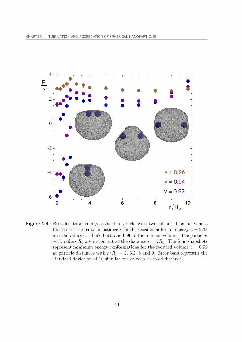
To compare the induced interactions between particles, the rescaled total energies E/κare displayed as a function of the rescaled distance between the two particles in figure4.4. The total energy curve has two distinct local minima which occur at two different
38
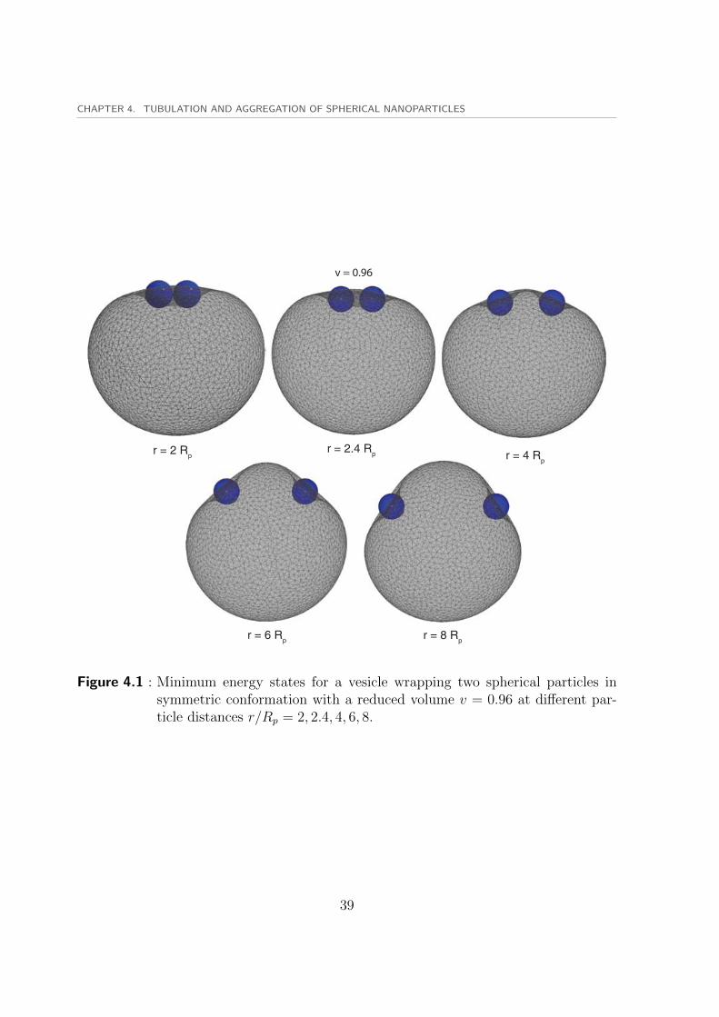
CHAPTER 4. TUBULATION AND AGGREGATION OF SPHERICAL NANOPARTICLES
v = 0.96
r = 2 Rpr = 2.4 Rp r = 4 Rp
r = 6 Rp r = 8 Rp
Figure 4.1 : Minimum energy states for a vesicle wrapping two spherical particles insymmetric conformation with a reduced volume v = 0.96 at different par-ticle distances r/Rp = 2, 2.4, 4, 6, 8.
39
4.3. SIMULATION RESULTS
v = 0.94
r = 2 Rpr = 2.4 Rp r = 2.8 Rp
r = 6 Rpr = 9 Rp
Figure 4.2 : Minimum energy states for a vesicle wrapping two spherical particles insymmetric conformation with a reduced volume v = 0.94 at different par-ticle distances r/Rp = 2, 2.4, 2.8, 6, 9.
40
CHAPTER 4. TUBULATION AND AGGREGATION OF SPHERICAL NANOPARTICLES
r = 2.2 Rp r = 2.6 Rp r = 3.2 Rp
r = 6 Rpr = 9 Rp
v = 0.92
Figure 4.3 : Minimum energy states for a vesicle wrapping two spherical particles insymmetric conformation with a reduced volume v = 0.92 at different par-ticle distances r/Rp = 2.2, 2.6, 3.2, 6, 9.
41

4.3. SIMULATION RESULTS
values of the particle distance. The first minimum occurs at the closest particle distancewhere two particles are touching r = 2Rp. The second minimum occurs at a distance rbetween 6Rp and 9Rp. These two minima are separated by an energy barrier betweenthem. The first local minima corresponds to the bound state of the particles shown infigure 4.5.
For the two lower values of the reduced volume v = 0.92 and v = 0.96, two distinctbranches of solutions are found. The upper branch corresponds to the symmetric vesicleshape which has positive values of the total energy E. The vesicle is almost symmetricwith respect to a plane perpendicular to the connecting line of particle centers. This iscalled the symmetric branch of the solution. There is also another branch with lowerenergy values than the symmetric branch which has negative values of the total energyE. The second branch continues to exist for particles at close distances r < 3Rp andvanishes for higher values of the particle distance r. This branch corresponds to thetubular conformations of the invaginated vesicle around nanoparticles which constitutethe shapes with asymmetric wrapping of particles. The tubular branch vanishes forhigh values of the reduced volume such as v = 0.96 because there is not enough freevolume to form the tubular morphology around particles. Some snapshots of the tubularconformation are illustrated in figure 4.5 at different particle distances r/Rp = 2, 2.4, 2.8with a reduced volume v = 0.92.
For the highest reduced volume v = 0.94 the tubes are smaller than the case v = 0.92due to the difference in the free volume( 4.5). It is obvious that for higher distancesr > 3Rp longer tubes have to be formed which demands higher values of the free volumethat could not be provided by the vesicle volume. Therefore the tubes vanish at higherdistances. The main contribution to the difference in the total energy of the symmetricand tubular branches comes from the adhesion energy. For example with v = 0.92at a distance r = 2.2Rp the adhesion energy of the symmetric and tubular branchesare Eadh = −51.2κ, and Eadh = −56.7κ, respectively while the bending energies areEbe = 51.6κ, and Ebe = 51.2κ, which are approximately identical compared to theadhesion energies. These values give rise to the total energy values of E = 0.4κ, andE = −5.5κ for the symmetric and tubular branches, respectively.
We define the binding energy ∆E of the two particles as the difference between energiesof the two minima of the total energy function E = E(r). Binding energies illustratedin figure 4.6, are calculated from the energy curves of figure 4.4 for different valuesof the reduced volume. For the two lower values of the reduced volume v = 0.92 andv = 0.94, the first energy minimum at r = 2Rp is obtained from the lower energy branchwith tubular conformations. The second minimum is considered to occur approximatelyat R = 6Rp for all curves. Stronger binding of the two particles is observed for two caseswith lower values of the reduced volume due to the existence of tubular conformations.The typical binding energies of −5.5κ and −7.5κ are larger than the energy unit kBTbecause the approximate value of the membrane bending rigidity is κ = 10kBT .
42
CHAPTER 4. TUBULATION AND AGGREGATION OF SPHERICAL NANOPARTICLES
2 4 6 8 10-6
-4
-2
0
2
4Eκ
r Rp
v = 0.92
v = 0.96v = 0.94
Figure 4.4 : Rescaled total energy E/κ of a vesicle with two adsorbed particles as afunction of the particle distance r for the rescaled adhesion energy u = 2.33and the values v = 0.92, 0.94, and 0.96 of the reduced volume. The particleswith radius Rp are in contact at the distance r = 2Rp. The four snapshotsrepresent minimum energy conformations for the reduced volume v = 0.92at particle distances with r/Rp = 2, 3.2, 6 and 9. Error bars represent thestandard deviation of 10 simulations at each rescaled distance.
43

4.4. A VESICLE WITH THREE ADSORBED PARTICLES
v = 0.92
r = 2 Rp r = 2.4 Rpr = 2.8 Rp
Figure 4.5 : Vesicle wrapping around two particles in a tubular conformation with areduced volume v = 0.92 and different particle distances r/Rp = 2, 2.4, 2.8.The vesicle can not form tubular structure around particles for larger dis-tances due to the volume constraint.
4.4 A vesicle with three adsorbed particles
A similar tubular conformation can be also found for three particles wrapped by a vesicle.We investigate the effect of the relative orientation of three particles on the bendingand adhesion energies and the stability of these conformations. As mentioned before,the linear aggregation of three particle system has been recently reported for particlespartially adsorbed on a vesicle[35]. Here we consider the same linear interactions forthe particles wrapped in a tube with different orientational arrangements. We keep twoparticles fixed next to each other in a vertical arrangement and rotate the third particlearound an axis perpendicular to the plane of all three particle centers. In this way we canvary the angle between the two lines connecting centers of successive pairs of particlesi.e. the line connecting particle one to particle two and the line connecting particle twoto particle three.
The marginal value of the reduced volume of three particles is obtained as v0 ≈ 0.873for a particle radius Rp = 0.1 . We keep the vesicle volume at a constant reduced volumev = 0.88 which is slightly higher than the marginal reduced volume v0, to let all threeparticles get enough wrapped and form a tube. We also use a rescaled adhesion strengthu = 2 for the simulations of the three particles. The angle φ is defined as the anglebetween the connecting lines of the first and last two particles. Some snapshots of thethree particle system at different values of the orientation angle φ are shown in figure4.7.
Figure 4.7 displays the rescaled total energy E/κ of the tubular conformations of
44

CHAPTER 4. TUBULATION AND AGGREGATION OF SPHERICAL NANOPARTICLES
2.0 2.2 2.4 2.6 2.8 3.0 3.2 3.4-8
-7
-6
-5
-1
0
1
2ΔEκ
u
v = 0.92
v = 0.94
v = 0.96
Figure 4.6 : Rescaled binding energies ∆E/κ of two adsorbed particles as a function ofthe rescaled adhesion energy u of the particles. The binding energy ∆E hereis calculated as the difference between the total energies H at the particlecontact distance r = 2Rp and at the distance r = 6Rp, which approximatesthe location of the second minimum of the interaction profiles E(r) shownin Fig. 4.4. Negative values of ∆E correspond to stable bound states ofthe particles. At the value v = 0.96 of the reduced volume, the particlesare symmetrically wrapped in their bound states as in the snapshot in theupper left corner. At v = 0.92 and 0.94, the particles are bound togetherby a membrane tube as in the snapshot at the bottom of the figure.
45

4.4. A VESICLE WITH THREE ADSORBED PARTICLES
0 30 60 90 120
-13.5
-13.0
-12.5
-12.0
-11.5
-11.0
-10.5
o oo oo
Eκ
φ
Figure 4.7 : Rescaled total energy E/κ of a vesicle with three adsorbed particles boundtogether by membrane invaginations as a function of the angle φ betweenthe particles for the reduced volume v = 0.88 and the rescaled adhesionenergy u = 2. Here, φ = 0 corresponds to a linear arrangement of theparticles. The four snapshots depict minimum energy conformations at theangles φ = 0◦, 45◦, 90◦, and 120◦.
46

CHAPTER 4. TUBULATION AND AGGREGATION OF SPHERICAL NANOPARTICLES
Figure 4.8 : Three particle wrapped by the vesicle with a reduced volume v = 0.88.Two particles have formed a tube and the third one is partially wrappedby the vesicle apart from the first two particles. The total energy of thisconformation is E ≈ −6.0κ which is remarkably higher than the totalenergy of the tubular conformation of three particles.
three particles wrapped by the vesicle as a function of the relative orientation angle φof the third particle. A minimum value of the total energy E = −13.5κ nearly arises atφ ≈ 0 which is remarkably lower than the energy values of the other arrangements. Tocompare the bond states with other states of the three particles wrapped by a vesicle,we do simulations for some other morphologies. There are several possible ways forarranging three particles on a vesicle. It is not easy to simulate all of those conformations,however we have chosen a few cases. One can argue that there are possibly three stateswhich can represent the whole morphology of three particles from energy point of view.These states include the tubular morphology of three particles at the bound state, tubeformed from two particles while the third particle is apart from them, and three separateparticles wrapped by the vesicle. For instance the total energy for the second case wherethe two particles form a tube and the third one is adsorbed on the vesicle at a distance, isE ≈ −6.0κ which is far higher than the energy of the bound state. Figure 4.8 illustratesthree particles with a tube formed around two particles and the third one is partiallywrapped by the vesicle at a distance from the other two particles.
4.5 Conclusion
Comparing the total energies of a vesicle wrapping two and three particles, one finds thatmembrane-induced interactions mainly depend on the reduced volume of the vesicle v.
47

4.5. CONCLUSION
If there exists enough free volume, the particles can form tube conformations which areenergetically highly favorable and constitute very stable low energy bound states betweenparticles. However those values of the reduced volume that allow tube formation, resultin vesicle shapes which do not mainly differ in the total energy amount. For instance forthe linear tubular morphology of three particles with a rescaled adhesion strength u = 2the total energy is obtained as E ≈ −13.1κ at v = 0.87, E ≈ −13.4κ at v = 0.88, andE ≈ −13.2κ at v = 0.89. We also obtained a total energy of E = −28.4κ for a vesiclewith four particles in a tube conformation at the same rescaled adhesion strength u = 2with the reduced volume v = 0.84.
At the rescaled adhesion energy u = 2 with the reduced volume v = 0.92, the totalenergy of the tubular morphology for two particles is obtained as E ≈ 2.0κ. Comparingthe above values of the total energy for two, three, and four particles forming a tubewith identical adhesion strengths, one concludes that adding a single particle reduces thetotal energy by an approximate amount of −15κ. The total energy of a single particlewith the rescaled adhesion strength u = 2 is almost E ≈ 0 because the bending energyof a complete sphere 8πκ is equal to the adhesion energy UAadh = U(4πR2
p) whichcorresponds to u = UR2
p/κ = 2[24, 25]. This results in marginal stability of the singleparticle engulfed by the vesicle at u = 2. The rather high negative energy values for thetubular bound states implies in much more stable configurations.
The tube structures can be used as an efficient way to store nanoparticles in vesiclesand deliver them in target locations. This process can be controlled by the reducedvolume of the vesicle. The vesicles can be deflated to confine as many particle as neededinside the tubular membrane structures. These particles can be delivered back by in-flating the vesicle. This can be used as a route for storing and delivering naoparticles inbiological environments. There are also similar membrane tubes originating from proteincoats[36], peptides[37], and aqueous phase separation in vesicles[38].
We have observed stronger attractive interactions between adsorbed particles for lowerreduced volumes which led to tubular structures. However we have also found attractiveinteractions for higher reduced volumes like v = 0.96 which results in aggregation ofspherical nanoparticles. This results may help understanding the experimental observa-tion of aggregation and attraction of particles adsorbed on a vesicle[39]. There are alsofluctuation-mediated interactions between particles wrapped by a membrane. As mem-brane fluctuations are suppressed due to adhesion to the particles in the adhesion area,membrane fluctuation-induced attraction may cooperate with elastic interactions. How-ever those contributions are relatively much smaller than the elastic ones[31, 40, 41, 42].In a similar approach membrane induced interactions between nanoparticles was reportedfor particles inside the vesicle[43]. However they considered vesicle without volume con-straint while the vesicle volume could freely change. In contrast we studied the effect ofvesicle volume on attractive interactions and tube formations.
48
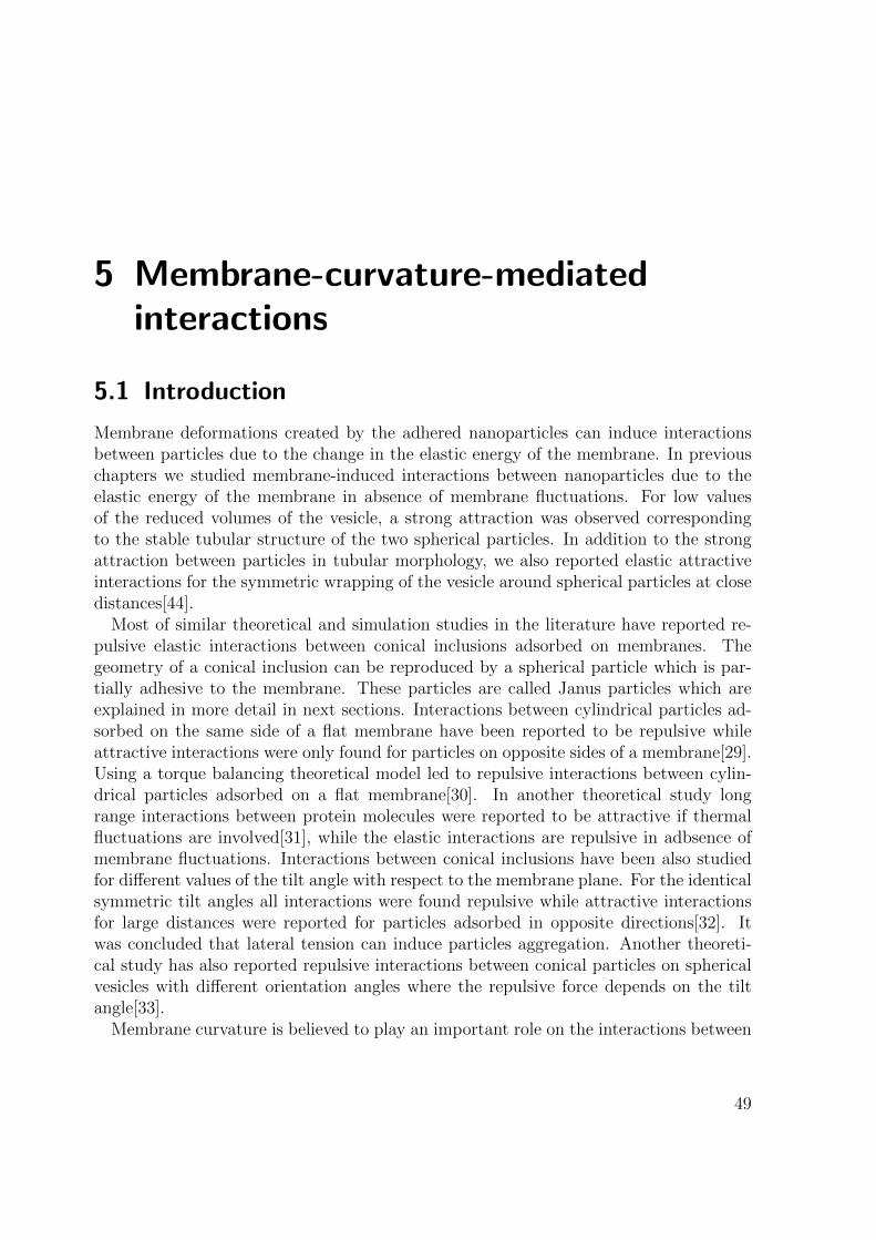
5 Membrane-curvature-mediatedinteractions
5.1 Introduction
Membrane deformations created by the adhered nanoparticles can induce interactionsbetween particles due to the change in the elastic energy of the membrane. In previouschapters we studied membrane-induced interactions between nanoparticles due to theelastic energy of the membrane in absence of membrane fluctuations. For low valuesof the reduced volumes of the vesicle, a strong attraction was observed correspondingto the stable tubular structure of the two spherical particles. In addition to the strongattraction between particles in tubular morphology, we also reported elastic attractiveinteractions for the symmetric wrapping of the vesicle around spherical particles at closedistances[44].
Most of similar theoretical and simulation studies in the literature have reported re-pulsive elastic interactions between conical inclusions adsorbed on membranes. Thegeometry of a conical inclusion can be reproduced by a spherical particle which is par-tially adhesive to the membrane. These particles are called Janus particles which areexplained in more detail in next sections. Interactions between cylindrical particles ad-sorbed on the same side of a flat membrane have been reported to be repulsive whileattractive interactions were only found for particles on opposite sides of a membrane[29].Using a torque balancing theoretical model led to repulsive interactions between cylin-drical particles adsorbed on a flat membrane[30]. In another theoretical study longrange interactions between protein molecules were reported to be attractive if thermalfluctuations are involved[31], while the elastic interactions are repulsive in adbsence ofmembrane fluctuations. Interactions between conical inclusions have been also studiedfor different values of the tilt angle with respect to the membrane plane. For the identicalsymmetric tilt angles all interactions were found repulsive while attractive interactionsfor large distances were reported for particles adsorbed in opposite directions[32]. Itwas concluded that lateral tension can induce particles aggregation. Another theoreti-cal study has also reported repulsive interactions between conical particles on sphericalvesicles with different orientation angles where the repulsive force depends on the tiltangle[33].
Membrane curvature is believed to play an important role on the interactions between
49

5.2. VESICLE AND JANUS PARTICLES
local inclusions adsorbed on the membrane. For instance the protein molecules adsorbedon the membrane can both generate[45] and sense[46] the local curvature of the mem-brane. The same situation also holds for spherical particles adsorbed on the membrane.This means that the adsorption of particles and their aggregation can induce curvatureon the membrane via deforming the membrane surface. On the other hand, membranecurvature can induce attractive interactions between particles and drive their aggre-gation. Therefore aggregation and vesiculation of spherical particles interacting withmembranes arise simultaneously and complement one another[47, 34]. The aggregationof spherical particles can also trigger membrane tubulation process[44]. In a recent study,attractive interactions were reported between particles via coarse-grained simulations ofbiological membranes interacting with partially adhesive capsid molecules[47]. Theseinteractions are induced between particles by the membrane curvature at the contactarea. The binding energies between the membrane and protein inclusions and betweenthe inclusions themselves are not enough to provide the higher energy levels requiredto form tubular structures. This also confirms the effect of elastic deformations of themembrane on providing enough energy to induce attarction between particles.
5.2 Vesicle and Janus particles
We use Monte Carlo simulations of a triangulated vesicle to investigate the effect of mem-brane curvature on the induced interactions between adsorbed Janus particles. Howeverit is not straightforward to design a simulation setup which can exclusively find the effectof membrane curvature on the induced interactions. Simulated annealing simulation ap-plied to appropriate perticles, has the advantage of suppressing the effect of membranefluctuations or entropical contributions and solely consider the effect of the curvature onthe induced interactions.
5.2.1 The vesicle model
In order to exclusively find the effect of membrane curvature one should reproducedifferent membranes with different values of the curvature. The shape and local curvatureof a free vesicle depend on the vesicle reduced volume. Different values of the reducedvolume can result in a variety of vesicle shapes including oblates and prolates. The localcurvature of those shapes varies on different regions of the three dimensional surface.Therefore the local membrane curvature varies for different values of the reduced volumeof the vesicle. In order to avoid shape dependence on the reduced volume, the volumeconstraint is removed and the vesicle volume can freely change during simulations. Asthe volume constraint is used to reproduce the effect of the osmotic pressure, free volumevariation corresponds to the case in which no osmotically active substances (such as saltsor sugars, which cannot cross the membranes) are present.
50

CHAPTER 5. MEMBRANE-CURVATURE-MEDIATED INTERACTIONS
The surface area of the vesicle is kept constant during simulation to represent theeffect of constant number of lipids per area or the incompressibility of the membrane.In absence of the volume constraint, the zero-temperature-limit shape of the vesicleapproaches a complete sphere for any value of the surface area. The local principalcurvatures of a sphere are identical and constant for all points on its surface. In thisway the local curvatures of the free vesicle with radius Rv are constant C1 = C2 = 1/Rv
around the vesicle surface and only depend on the size of the vesicle which is defined byits surface area Av = 4πR2
v. Therefore we can generate different vesicles with distinctvalues of curvature. In contrast to the simulations on aggregation of spherical particles,there is not a single vesicle size or vesicle surface area A0 but different values of thevesicle surface Av with their corresponding radius Rv are chosen which give rise todifferent membrane curvatures. On the other hand different wrapping amounts of thenanoparticles result in different bending and adhesion energy values. Meanwhile thegeometry of the adhesion area on the spherical particle plays an important role on thevesicle shape and its bending energy.
Any three dimensional surface has two principal values of the curvature at any pointon its surface. Local curvature of a surface has properties of a second order tensor whichobeys tensoric transformation laws. Both principal curvatures of a surface at a certainpoint can be positive or negative. The sign of the curvature depends on the normalvector or the direction of the surface at a given point. For a complete sphere with radiusR both principal curvatures are felt negative for a particle sitting on the inner side of thesphere surface Cin = −1/R, while for a particle on the outer surface both curvatures arefelt positive Cout = 1/R. In order to consider the effect of the curvature on membrane-mediated interactions, different vesicle sizes are required. We assume particles on bothinside and outside of the vesicles. In this way we are able to generate negative andpositive values of the membrane curvature and extrapolate the results to the case ofzero membrane curvature or the flat bilayer.
5.2.2 Pseudo-Janus particles
We consider two different types of spherical particles to investigate the effect of mem-brane curvature on the induced interactions. First we use spherical particles which arecompletely adhesive to the vesicle. These particles resemble normal spherical particleswhich were used to study particles aggregation and membrane tubulation. The onlydifference is that the particles can not be fully wrapped by the vesicle. This means thatthere is a predefined value which sets the value of the adhesion area for both sphericalparticles. These partially adhesive particles are called pseudo-Janus particle.
Partial wrapping of the particle is implemented by introducing a new constraint onthe adhesion area of both particles by involving a new term Hca to the Hamiltonian ofthe total system which evolves the vesicle toward the state of wrapping both particles
51

5.2. VESICLE AND JANUS PARTICLES
equally with the predefined value of the adhesion area Aadh.
Hca = Kadh[1− (A1
Aadh)2 + (1− (
A2
Aadh)2] (5.1)
where A1 and A2 denote the adhesion areas of particles one and two respectively andKadh is the coefficient of the adhesion area. A dimensionless value of Kadh/T ≈ 105 wasused to ensure a very small relative error in the adhesion area∣∣∣∣A1 − Aadh
Aadh
∣∣∣∣ , ∣∣∣∣A2 − AadhAadh
∣∣∣∣ ≤ 10−3 (5.2)
For the previous simulations on aggregation of normal particles and membrane tubula-tion, there was no restriction on the adhesion area of both particles. Here both particleshave identical adhesion areas which are equal to the predefined value of the adhesionarea Aadh. Although the value of the adhesion area for each particle is fixed, there isno restriction on the shape of the adhesion area. This means that the geometry of theadhesion zone is not dictated and the vesicle can freely choose any surface with the samearea as the adhesion zone. In this way simulated annealing simulations result in vesi-cle shapes with equal given adhesion areas for both particles which have the minimumbending energy values.
The pseudo-Janus particles can be used as model particles to reproduce the effect ofadhesive fluid domains on the membrane. As the geometry of the adhesion area is notfixed for these particles, it can be considered as a fluid adhesion area on the membranewith variable shape. The fluid adhesion zone can then have any general shape whichis not essentially an axisymmetric shape. As the second type of particles, we use realJanus particles.
5.2.3 Janus particles
Janus particles are spherical particles composed of both adhesive and non-adhesive re-gions. The vesicle can only partially wrap these particles around their adhesive part.The shape of the adhesive and non-adhesive part of the Janus particle is fixed and ax-isymmetric. A Janus particle is composed of two adhesive and repulsive parts separatedby a cutting plane through the complete sphere. The wrapping angle φ parametrizes theadhesive part of the sphere as shown in figure 5.1. Thus the adhesive part of a Janusparticle is a symmetric spherical cap which can be obtained by revolving a generatingcircular curve around an axis of symmetry. For Janus particles the vesicle is forced towrap around a certain adhesion area whose shape and surface area are both defined.However for a certain adhesion area on the vesicle, the bending energy of the vesicledepends on the orientation of the particles.
The predefined value of the adhesion area for both particles can be related to the
52

CHAPTER 5. MEMBRANE-CURVATURE-MEDIATED INTERACTIONS
φ
Figure 5.1 : A vesicle wrapping around the adhesive part of the Janus particle. Thewrapping angle φ determines the symmetric adhesive part on the sphericalparticle.
wrapping angle viaAadh = 2πR2
p(1− cosφ) (5.3)
In order to adjust the adhesion area to the predefined value we use the attractive in-teraction between the vesicle and particles similar to the one we used for aggregationof spherical particles. Then we choose a high value of the adhesion strength u = 5 andensure that the adhesive part of the Janus particle is wrapped by the vesicle. Each Janusparticle has a direction vector which defines the particle orientation in the space. Anyvertex located withing a cut off distance d which is also adhered to the adhesive part ofthe particle is taken to be adhered to the particle. Finally we check the adhesion area toensure all vesicles have wrapped around the total adhesive part of the Janus particles.In this way the adhesion strength u = 5 is high enough to suppress the effect of theadhesion energy.
5.3 Membrane-mediated interactions betweenpseudo-Janus particles
The bending energy of a vesicle with adsorbed pseudo-Janus particles depend on thethe vesicle curvature C = 1/Rv and on the predefined value of the particle rescaledadhesion area or the wrapping ratio γ = Aadh/Ap. The corresponding area of the vesicleAv = 4πR2
v, the relative size between the pseudo-Janus particle and vesicle Rp/Rv, andthe rescaled distance r/Rp between naoparticles are fixed during simulation. for a fixedvalue of the wrapping ratio of both particles, the adhesion energy is irrelevant and the
53

5.3. MEMBRANE-MEDIATED INTERACTIONS BETWEEN PSEUDO-JANUS PARTICLES
Hamiltonian of the total system is written as
H = Hbe +Ht +Harea +Hvol (5.4)
where all terms in the system Hamiltonian are similar to the terms in equations 3.22. Weuse three different values of the vesicle curvature C = 0.45, 0.5, 0.55 and locate sphericalparticles both inside and outside of the vesicle in different distances for every value ofthe curvature. Both particles are fixed in the space throughout the simulation time.For a set of given parameters including the relative distance of two particles, the vesiclecurvature C, the wrapping ratio of each particle γ, Monte Carlo simulated annealing hasbeen used to find the minimum bending energy of the system E = min(Eb) for particlesboth inside and outside of the vesicle. As for all simulations the wrapping ratios of bothparticles are equal to the reference value of the wrapping ratio γ = Aadh/Ap, the adhesionenergy Eadh = UγAp is identical for all cases regardless of the adhesion strength U . Thatis why the adhesion energy is irrelevant and we do not involve it neither in Monte CarloHamiltonian nor in the total energy. We rescale all bending energies of the vesicle bythe bending rigidity of the membrane κ. We perform our simulations for a fixed valueof the wrapping ratio γ = 0.75 of the pseudo Janus particles.
Figure 5.2 displays the rescaled bending energies of the vesicle as a function of therescaled particle distance r/Rp with a wrapping ratio γ = 0.75. Here, the pseudo-Janus particles sre outside of the vesicle. Some snapshots of the vesicle and particlesat different particle distances with a membrane curvature C = 0.5 are shown in 5.3.Interestingly we observe attractive interaction between particles outside of the vesiclewhile the interactions between inside particles are repulsive. We also define the bindingenergy ∆E as the difference between the two minima of the bending energy function. Itis seen that the attractive interactions between outside particles is stronger for highervalues of the vesicle curvature. Figure 5.4 displays the same results for the rescaledbending energies of the vesicle but for particles inside the vesicle. Some snapshots of thevesicle and particles at different particle distances with a membrane curvature C = 0.5are also shown in 5.5. We do not observe a similar trend for particles inside the vesiclewhich is due to the fluid nature of the adhesion region of the particles. For particlesat close distances r/Rp = 2 we find two branches of symmetric and tubular solutionssimilar to normal particles in chapter four. The tubular conformation has lower energythan the symmetric one for all simulations except for particles inside the vesicle with avesicle curvature C = 0.45.
So far we used pseudo-Janus particles with a certain predefined value of the wrap-ping ratio γ = Aadh/Ap to investigate the effect of curvature on membrane-mediatedinteractions. There is not any constraints on the geometry of the adhesion area forpseudo-Janus particles. However we could not observe a monotonic relation betweenthe curvature and induced interactions for particles inside of the vesicle. As a secondapproach we simulate different particle types which are real Janus particles.
54

CHAPTER 5. MEMBRANE-CURVATURE-MEDIATED INTERACTIONS
2 3 4 5 6 7 8 9 1046
48
50
52
54
56
58
60
62
64
66
68
r/Rp
E κ
C = 0.45
C = 0.5
C = 0.55
Figure 5.2 : The rescaled bending energy E/κ as a function of the rescaled particledistance r/Rp for a vesicle wrapping around pseudo Janus particles locatedoutside of the vesicle with a wrapping ratio γ = 0.75 and vesicle curvaturesC = 0.45, 0.5, 0.55.
55

5.3. MEMBRANE-MEDIATED INTERACTIONS BETWEEN PSEUDO-JANUS PARTICLES
C = 0.5, γ = 0.75
r = 2 Rpr = 2 Rp r = 3.2 Rp
r = 5 Rpr = 8 Rp
Figure 5.3 : Snapshots of pseudo Janus particles adsorbed outside of a vesicle with C =0.5 at different distances r/Rp. For r = 2, both tubular and symmetricbranches are illustrated.
56

CHAPTER 5. MEMBRANE-CURVATURE-MEDIATED INTERACTIONS
2 4 6 8 10 12 14 1642
42.5
43
43.5
44
44.5
r/Rp
E κ
C = 0.45
C = 0.5
C = 0.55
Figure 5.4 : The rescaled bending energy E/κ as a function of the rescaled particledistance r/Rp for a vesicle wrapping around pseudo Janus particles locatedinside of the vesicle with a wrapping ratio γ = 0.75 and vesicle curvaturesC = 0.45, 0.5, 0.55.
57

5.3. MEMBRANE-MEDIATED INTERACTIONS BETWEEN PSEUDO-JANUS PARTICLES
r = 2 Rp r = 2 Rp r = 2.4 Rpr = 7 Rp
r = 11 Rpr = 14 Rp
C = 0.5, γ = 0.75
Figure 5.5 : Snapshots of pseudo Janus particles adsorbed outside of a vesicle with C =0.5 at different distances r/Rp. For r = 2, both tubular and symmetricbranches are illustrated.
58

CHAPTER 5. MEMBRANE-CURVATURE-MEDIATED INTERACTIONS
2 3 4 5 6 7 858
60
62
64
66
68
r/Rp
E κ
C = 0.45
C = 0.5
C=0.55
Figure 5.6 : The rescaled bending energy E/κ as a function of the rescaled particledistance r/Rp for a vesicle wrapping around Janus particles located outsideof the vesicle with a wrapping angle φ = 0.65π and vesicle curvaturesC = 0.45, 0.5, 0.55.
5.4 Membrane-mediated interactions between Janusparticles
The bending energy of the vesicle with adsorbed Janus particles also depends on theorientation of the Janus particles. The bending energy of the vesicle with adsorbedJanus particles depends on the the vesicle curvature C = 1/Rv, on the relative sizeRp/Rv, and on the wrapping angle φ of the Janus particle.
For interactions between the vesicle and Janus particles, there is another Monte Carlomove in each Monte Carlo step. The orientation of both Janus particles is also changedin each Monte Carlo step by rotating the particle around a random unit vector in thespace with dφ = 1◦. Therefore there are three Monte Carlo moves including displacingvertices, flipping edges, and rotating Janus particles in each Monte Carlo step. For agiven set of values of the particle distance, vesicle curvature, and the wrapping angle φ,simulated annealing simulation results in the corresponding vesicle shape and particleorientations which leads to the minimum value of the bending energy of the vesicle.Figure 5.6 displays the rescaled bending energies E/κ of the vesicle as a function of the
59

5.4. MEMBRANE-MEDIATED INTERACTIONS BETWEEN JANUS PARTICLES
r = 2 Rp r = 3 Rp
r = 5 Rp r = 7 Rp
C = 0.5, ϕ = 0.65π
Figure 5.7 : Snapshots of Janus particles adsorbed outside of a vesicle with C = 0.5 atdifferent distances r/Rp.
rescaled particle distance r/Rp with a wrapping angle φ = 0.65π where the two Janusparticles are outside of the vesicle. Figure 5.8 displays the rescaled bending energyof the vesicle for particles inside the vesicle. A few snapshots of the vesicle and Janusparticles at different particle distances with a vesicle curvature C = 0.5 are also shownin figures 5.7 and 5.9 for particles outside and inside of the vesicle, respectively.
The vesicle membrane induces attractive interactions between Janus particles outsideof the vesicle where the membrane curvature is positive. However the induced interac-tions are repulsive for particles located inside the vesicle where the membrane curvatureis negative. This confirms the strong dependence of the membrane-mediated interactionson its curvature. We also find two different branches of tubular and symmetric solutionsfor Janus particles. Interestingly unlike normal and pseudo Janus particles, the bendingenergy of the tubular branch is higher than the symmetric branch for Janus particles.This is due to the constraint which makes the adhesion areas for both Janus particleequal and geometrically symmetric while there is a remarkable difference between theadhesion areas of normal particles in tubular conformation of chapter four. Thereforeunlike normal particles in chapter four the symmetric branch has lower energy than thetubular branch for the Janus particles. Here we define the binding energy between Janusparticles as the energy difference ∆E between the bound state with r/Rp = 2 and thesecond local minimum of the energy curve. Figure 5.10 displays the rescaled binding
60

CHAPTER 5. MEMBRANE-CURVATURE-MEDIATED INTERACTIONS
2 4 6 8 10 12 14 16 18
51
51.5
52
52.5
53
53.5
54
54.5
55
r/Rp
E κ
C = 0.45
C = 0.5
C = 0.55
Figure 5.8 : The rescaled bending energy E/κ as a function of the rescaled particledistance r/Rp for a vesicle wrapping around Janus particles located insideof the vesicle with a wrapping angle φ = 0.65π and vesicle curvaturesC = 0.45, 0.5, 0.55.
61

5.4. MEMBRANE-MEDIATED INTERACTIONS BETWEEN JANUS PARTICLES
r = 2 Rp r = 6 Rp
r = 12 Rp r = 16 Rp
C = 0.5, γ = 0.65π
Figure 5.9 : Snapshots of Janus particles adsorbed inside of a vesicle with C = 0.5 atdifferent distances r/Rp.
62
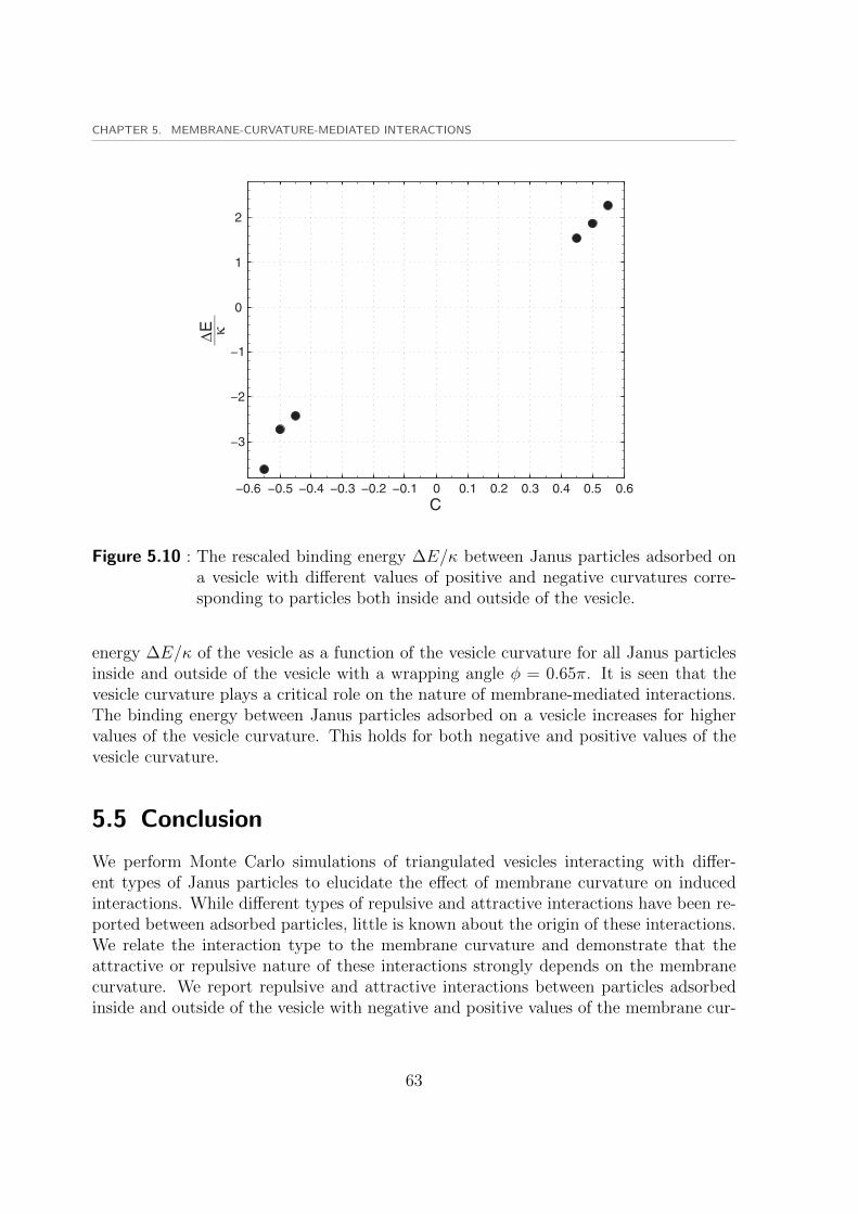
CHAPTER 5. MEMBRANE-CURVATURE-MEDIATED INTERACTIONS
−0.6 −0.5 −0.4 −0.3 −0.2 −0.1 0 0.1 0.2 0.3 0.4 0.5 0.6
−3
−2
−1
0
1
2
C
�∆E κ
Figure 5.10 : The rescaled binding energy ∆E/κ between Janus particles adsorbed ona vesicle with different values of positive and negative curvatures corre-sponding to particles both inside and outside of the vesicle.
energy ∆E/κ of the vesicle as a function of the vesicle curvature for all Janus particlesinside and outside of the vesicle with a wrapping angle φ = 0.65π. It is seen that thevesicle curvature plays a critical role on the nature of membrane-mediated interactions.The binding energy between Janus particles adsorbed on a vesicle increases for highervalues of the vesicle curvature. This holds for both negative and positive values of thevesicle curvature.
5.5 Conclusion
We perform Monte Carlo simulations of triangulated vesicles interacting with differ-ent types of Janus particles to elucidate the effect of membrane curvature on inducedinteractions. While different types of repulsive and attractive interactions have been re-ported between adsorbed particles, little is known about the origin of these interactions.We relate the interaction type to the membrane curvature and demonstrate that theattractive or repulsive nature of these interactions strongly depends on the membranecurvature. We report repulsive and attractive interactions between particles adsorbedinside and outside of the vesicle with negative and positive values of the membrane cur-
63

5.5. CONCLUSION
vature. We conclude that increasing membrane curvature can induce attraction betweenspherical particles where the interactions are repulsive for negative and zero curvatureof the membrane. This agrees with repulsive interactions reported between particles ona flat bilayer with zero curvature[34].
64

6 Wrapping and internalization ofnanoparticles with different shapes
Endocytosis is a fundamental cellular process through which many artificial and naturalbiological objects are engulfed and internalized by the cell[48]. Natural objects suchas viruses, bacteria, nutrients and phatogens are internalized by the cell through en-docytic pathways[20, 49, 50]. Wrapping and cellular uptake of technologically designednanoparticles provides an important route to transport drug containers and deliver thecargo within the target cell. Endocytosis also plays a significant role in cell motility andregulation of plasma membrane protein activity. Therefore understanding endocytosisprocess is critical for a number of different applications in nanomedicine. A better un-derstanding of the endocytosis process helps scientists to efficiently design nanoparticleswith different shapes and sizes to evade internalization or to provide an easier uptake.Internalization process is divided into different categories including phagocytosis andpinocytosis based on the size and the nature of the uptaken particle. Phagocytosis isgenerally the uptake process of rigid micro-sized particles mostly by immune cells whichis also called cell eating. Pinocytosis or cell drinking refers to uptake of fluid particlesand liposomes.
6.1 Cellular uptake of nanoparticles
Cellular uptake of nanoparticles is associated with deformations induced on the plasmamembrane of the cell by the particle. These deformations strongly depend on the shapeof the nanoparticle while the particle size does not mainly affect the bending energy ofthe deformed membrane. Therefore the shape of the nanoparticles play an importantrole on the wrapping and internalization process. It was believed in the past that onlyrather large particles are internalized via the phagpcytosis process. However phagocyticinternalization of nano-sized particles such as viruses[51] and gold[52] and silver[53] par-ticles has been also observed in recent experiments. It was also shown that the size ofthe nanoparticles do not mainly affect the phagocytosis process as long as they are largerthan a critical size[48].
65

6.1. CELLULAR UPTAKE OF NANOPARTICLES
6.1.1 The effect of particle size
Theoretical studies have related the wrapping process of a nanoparticle to the interplaybetween bending and adhesion energies[24, 25]. These studies have concluded that thereexists a critical size of the nanoparticle for achieving full wrapping and internalization.Similar results have also been observed experimentally by studying the internalizationof both gold[54] and silica nanoparticles[55]. Experiments have demonstrated that in-ternalization of spherical quantum dots with a radius of 25 nanometers is more effectivethan the internalization of smaller particles with radii 7.5 and 2.5 nanometers[56]. Sim-ilar results were observed for gold nanoparticles interacting with live cells by an activeforce. They observed that larger forces were required to wrap smaller particles[57]. Inanother study an optimal radius of 20− 25 nanometers was found for internalization ofgold spherical nanoparticles[58, 59].
All these theoretical and experimental investigations have concluded that there is acritical size of the nanoparticle(25−30 nanometers in radius) as the size below witch theinternalization is not achieved. However most of the models are based on deformationsof the cell membrane where the effect of the cytoskeleton is not included. Cellularinternalization of viruses with different diameters in the range of 40 to 160 nanometers,shows different values compared to the ones predicted by most of experimental andtheoretical studies. This suggests that the endocytosis process is also affected by theother properties of the nanoparticles such as their shape.
6.1.2 The effect of particle shape
The shape of the nanoparticle is another property which affects the wrapping and inter-nalization process and plays a primary role on membrane deformations during wrapping.Various particle shapes including spherical particles, ellipsoidal particles, elliptical disks,cylindrical or tubular particle, and cubic particles have been considered in experimentalstudies[60, 61]. Jin and coworkers reported an optimal tube length for internalization ofthe nanotubes[62]. However several other studies have found a different internalizationpathway for nanotubes rather than the endocytic pathway[63, 64] which is called thepenetration pathway[65]. Desimone and coworkers studied the effect of particle mor-phology on internalization process of micro and nanoparticles and reported an optimalaspect ratio for cylindrical particles. The aspect ratio of the cylindrical particles haveremarkable effect on the uptake process[61]. In a theoretical study, Decuzzi and cowork-ers reported that only cylindrical particles with an intermediate value of aspect ratiocan be fully internalized[66]. The effect of shape of cylindrical particles has been studiedusing a theoretical model[67].
It has been recently shown that the shape of the nanoparticle affects the uptakerate of the particle[23, 66, 62, 61]. Coarse-grained simulations of the receptor-mediatedendocytosis have shown that the internalization of cylindrical particles is more efficient
66

CHAPTER 6. WRAPPING AND INTERNALIZATION OF NANOPARTICLES WITH DIFFERENT SHAPES
and faster than the spherical particles[23]. Wrapping process of cylindrical nanoparticleby bilayer membrane is associated with a slight orientation change of the particle. Thecylindrical particles which start wrapping at the tip of the particle, rotate and preferto get wrapped on the particle side where full uptake occurs. Recent experiments haveshown that it is also the shape of the particle which determines whether internalization isachieved or not at all[60]. Although the phagocytosis process of microparticles has beenconsidered in these experiments, the results can be simply extrapolated to nanoparticlesas well. Particles with highly-curved areas can not be internalized by the macrophagecell. The cell could only spread around these particles and the internalization was notachieved. The success or failure of the internalization process for different particle shapeswas independent of the particle size. This demonstrates the effect of the particle shape onthe uptake process. It was observed that elliptical disks change their orientation duringthe wrapping process and internalization occurs in the tip direction of the particle.
Although there are many experimental investigations on wrapping different particleshapes including cylindrical particles and nanotubes, there is not a quantitative studyto reveal the relation between the bending and adhesion energies and the uptake rate ofthe particles. In particular there is not a theoretical study to help us relate the successand failure of the uptake process to the particle shape and its orientation. Such a modelcan also help us understand the effect of the aspect ratio and particle local curvature onthe uptake process. These particles have different values of local curvature on differentsurface regions. The local curvature of the particle determines the bending energy of thevesicle wrapping around the nanoparticle which mainly controls the wrapping process.In this chapter we study the effect of the particle shape on the wrapping and internal-ization process. In order to study the wrapping process of nanoparticles, we use vesiclemembranes as a model system.
6.2 Vesicle wrapping around nanoparticles with differentshapes
In order to represent different particle shapes, we consider ellipsoidal particles as modelparticles. While spherical particles have a uniform curvature, ellipsoidal particles haveboth more highly and more weakly curved surface regions. Thus they can be used toeffectively represent different particle shapes with different values of the local curva-tures. Unlike the spherical particle, ellipsoidal particles have non-uniform curvature indifferent directions along the tip and side of the particle. This property makes theman appropriate choice to investigate the effect of particle orientation on the wrappingprocess. We study the wrapping of a vesicle around spherical and ellipsoidal particleswith different aspect ratios to elucidate the effect of particle shape on the uptake pro-cess. Like previous chapters we use a triangulated vesicle model which wraps around a
67

6.2. VESICLE WRAPPING AROUND NANOPARTICLES WITH DIFFERENT SHAPES
rigid nanoparticle. Wrapping of the vesicle around the nanoparticle is associated with anincrease in the bending energy of the vesicle due to the shape transformations inducedon the vesicle. The increase in the bending energy is provided by the adhesion energyof the particle. The bending energy of a triangulated vesicle is obtained from Helfrichequation as explained in previous chapters
Ebe = 2κnv∑i=1
M2i
Ai. (6.1)
6.2.1 Simulated annealing simulation
The adhesion area Aad is defined as a sum of its contributions from adhered vertices whichare within a cut off distance d from the particle surface. However a harmonic potentialadjusts the adhesion area at a fixed predefined value for each simulation. Therefore theadhesion energy can be written as
Ead = −U∑〈i〉
Ai = −UAad (6.2)
In contrast with chapter four the adhesio area is contrained now during the minimiza-tion. For a given value of the adhesion area Aad, we perform Monte Carlo simulatedannealing simulations and obtain the corresponding minimum value of the bending en-ergy Eb(Aad) = min(Eb). The adhesion area of the particle is adjusted at the givenvalue during the Monte Carlo simulations. Therefore the adhesion energy of the particledoes not play any role on the minimization process. This is in contrast with previoussimulations on spherical particles where the total energy of the system was minimizedduring simulations. Monte Carlo minimization also leads to a vesicle shape and thecorresponding geometry of adhesion area which results in minimum bending energy. Wethen vary the predefined value of the adhesion area and obtain the minimum bendingenergy, the vesicle shape and the geometry of the adhesion area as a function of particleadhesion area Eb = Eb(Aad). The total energy of the vesicle and particle can be writtenas the sum of the vesicle bending energy Eb and the particle adhesion energy Ead
E = Eb + Ead = Eb(Aad)− UAad. (6.3)
In this way for a given value of the adhesion strength U , the total energy is also obtainedas a function of the adhesion area of the particle E = E(Aad). Then the correspondingvalue of the adhesion area at which the total energy gains its minimum value determinesthe wrapping area of the vesicle around the nanoparticle.
Wrapping of a vesicle around a particle depends on the rescaled adhesion strengthu = UAp/(4πκ) and on the relative size between the particle and the vesicle Ap/A. Here,
68
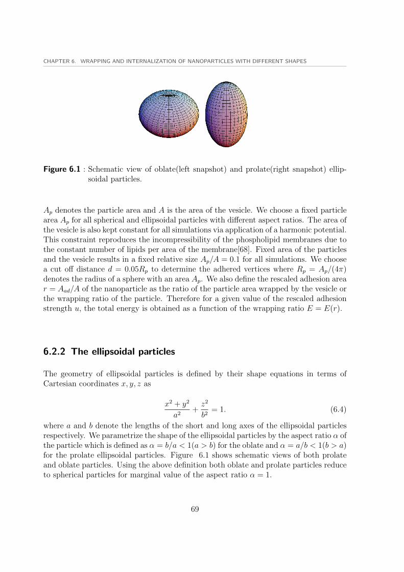
CHAPTER 6. WRAPPING AND INTERNALIZATION OF NANOPARTICLES WITH DIFFERENT SHAPES
Figure 6.1 : Schematic view of oblate(left snapshot) and prolate(right snapshot) ellip-soidal particles.
Ap denotes the particle area and A is the area of the vesicle. We choose a fixed particlearea Ap for all spherical and ellipsoidal particles with different aspect ratios. The area ofthe vesicle is also kept constant for all simulations via application of a harmonic potential.This constraint reproduces the incompressibility of the phospholipid membranes due tothe constant number of lipids per area of the membrane[68]. Fixed area of the particlesand the vesicle results in a fixed relative size Ap/A = 0.1 for all simulations. We choosea cut off distance d = 0.05Rp to determine the adhered vertices where Rp = Ap/(4π)denotes the radius of a sphere with an area Ap. We also define the rescaled adhesion arear = Aad/A of the nanoparticle as the ratio of the particle area wrapped by the vesicle orthe wrapping ratio of the particle. Therefore for a given value of the rescaled adhesionstrength u, the total energy is obtained as a function of the wrapping ratio E = E(r).
6.2.2 The ellipsoidal particles
The geometry of ellipsoidal particles is defined by their shape equations in terms ofCartesian coordinates x, y, z as
x2 + y2
a2+z2
b2= 1. (6.4)
where a and b denote the lengths of the short and long axes of the ellipsoidal particlesrespectively. We parametrize the shape of the ellipsoidal particles by the aspect ratio α ofthe particle which is defined as α = b/a < 1(a > b) for the oblate and α = a/b < 1(b > a)for the prolate ellipsoidal particles. Figure 6.1 shows schematic views of both prolateand oblate particles. Using the above definition both oblate and prolate particles reduceto spherical particles for marginal value of the aspect ratio α = 1.
69

6.3. WRAPPING OF A SPHERICAL NANOPARTICLE
0 0.2 0.4 0.6 0.8 1
25
30
35
40
45
50
r
E κu = 0(E = Eb)
u = 1.15
u = ut = 2.07
Figure 6.2 : The rescaled total energy E/κ of a vesicle wrapping around a sphericalparticle. Internalization occurs for rescaled adhesion energies u larger thanthe transition value ut = 2.07.
6.3 Wrapping of a spherical nanoparticle
We start our simulations by considering the wrapping of a vesicle around a sphericalnanoparticle. The spherical particle is a special case of the more general ellipsoidalparticles with α = 1 and a = b. We then choose a fixed value of the adhesion area0 < Aad < Ap or the corresponding wrapping ratio 0 < r = Ap/A < 1 and obtain theminimum bending energy and the corresponding vesicle shape with Simulated Annealingminimization method. For any value of the wrapping ratio r, we perform ten differentsimulations and calculate the mean and the error bar of the bending energy of the vesicle.With nv = 2562 vertices on the vesicle and 3× 10⁷ Monte Carlo minimization steps therelative errors of the vesicle bending energy is less than 0.3%. The errors are very smalland we do not show them on the figures. We also rescale the total energy of the particleand the vesicle by the bending rigidity κ of the vesicle.
The wrapping orientation is irrelevant for the spherical particle due to the rotationalsymmetry. This is due to the fact that sphere is symmetric around any axis passingthrough the center of the sphere. Thus the wrapping is determined by the wrappingratio r. The rescaled total energy E/κ as a function of the wrapping ratio r is displayedin figure 6.2 for a vesicle wrapping around the spherical particle with different valuesof the adhesion strength u. Figure 6.3 displays some snapshots for wrapping a vesiclearound a spherical particle with different values of the wrapping ratio r.
70

CHAPTER 6. WRAPPING AND INTERNALIZATION OF NANOPARTICLES WITH DIFFERENT SHAPES
r = 0.05 r = 0.45 r = 0.94
Figure 6.3 : The minimum energy shapes of a vesicle wrapping around a spherical parti-cle for the wrapping ratios r = 0.05, 0.45, 0.94 from left to right respectively.
The special case of zero adhesion strength u = 0 leads to the bending energy of thevesicle E = Eb. As suggested by a theoretical model[24], a certain rescaled adhesionstrength u = 2 is required to fully wrap a sphere. This can be obtained by making thebending energy of sphere Eb = 8πκ equal to its adhesion energy Ead = 4πκu. Figure6.2 shows that the bending energy of the fully wrapped spherical particle for r ≈ 1 isEb ≈ 8πκ which results in approximately the same value of the adhesion strength u ≈ 2.This confirms the accuracy and efficiency of the Monte Carlo minimization approach toachieve the minimum energy values. The deviation in the value of the adhesion strengthu is mainly due to the numerical error which depends on the resolution of the mesh ofthe triangulated model.
As stated earlier for any value of the adhesion strength u, the corresponding wrappingratio is determined by the value of the adhesion area at which the total energy gainsits minimum value. For the wrapping ratios r < 0.7 and r > 2.7, there is just onelocal minimum in the total energy function. For a wide range of intermediate valuesof the wrapping ratio 0.7 < r < 2.7 there are to minima in the total energy function.These minima are separated by an energy barrier between them. The first minimumcorresponds to a partially wrapped state of the nanoparticle while the second minimumrefers to a nearly fully wrapped nanoparticle. The transition between partial and nearlyfully wrapped states is a discontinuous transition. The existence of the energy barriercreates two different wrapping regimes of partial wrapping or spreading and full wrappingor internalization respectively. The transition between these regimes is controlled by theadhesion strength u via the interplay between bending and adhesion energies.
On the other hand the interplay between bending and energies depends on the aspectratio of the ellipsoidal particles. Therefore the transition depends on the particle shapeand the adhesion strength u. The transition between spreading and internalizationregimes for wrapping the spherical particle occurs at the transition value of the rescaled
71

6.4. WRAPPING OF ELLIPSOIDAL NANOPARTICLES
adhesion strength u = ut ≈ 2.07. At u = ut both minima of the energy functionhave equal energy values. Low values of the adhesion strength u ≤ ut results in partialwrapping of the vesicle around the nanoparticle with 0 ≤ r . 0.2 which refers to thespreading regime. Higher values of the adhesion strength u > ut results in high adhesionareas r & 0.87 of the vesicle wrapping around the particle which corresponds to theinternalization regime.
6.4 Wrapping of ellipsoidal nanoparticles
Unlike spherical particle which is rotationaly symmetric around any axis through its cen-ter, ellipsoidal particles are only rotationally symmetric in a single direction i.e. arounda single axis of symmetry. This means that the wrapping orientation also matters forthese particles as well as the wrapping ratio. The bending energy of the vesicle wrappingaround the particle depends on the surface curvature of different regions of the particlesurface.
6.4.1 Wrapping of prolate ellipsoidal particles
We start our simulations by prolate ellipsoidal particles which resemble the ellipticaldisks in recent experiments[60]. In these experiments the elliptical disks could be onlyinternalized by the macrophage cell on the tip orientation of the disk. When the cellwas located on the flat side of the elliptical disk, it could just spread on the particle sideand internalization was not achieved.
Figure 6.4 displays the rescaled total energy E/κ as a function of the wrapping ratior for a vesicle wrapping around a prolate particle with an aspect ratio α = 0.55 andtwo different values of the rescaled adhesion strength u. The minimum bending energyof wrapping the prolate particle is achieved in different orientations corresponding todifferent values of the wrapping ratio. For the case with u = 0 where E = Eb, theminimum bending energy is achieved in the side-oriented wrapping for low wrappingratios r . 0.6 and in the tip-oriented wrapping for high wrapping ratios r & 0.6. Thusthe transition from the spreading to the internalization regime is associated with an ori-entation change of the prolate particle. For low values of the rescaled adhesion strengthu & ut ≈ 2.33, the vesicle wraps around the flat side of the particle in the spreadingregime up to a wrapping ratio r ≈ 0.29. For higher values of the rescaled adhesionstrength u & ut ≈ 2.33, the vesicle wraps around the prolate particle in the tip orien-tation corresponding to the internalization regime with high adhesion areas r & 0.94.Figure 6.5 displays a few snapshots of the vesicle wrapping around the prolate particlein different orientations with wrapping ratios r = 0.35, 0.6, 0.67, 0.91.
72

CHAPTER 6. WRAPPING AND INTERNALIZATION OF NANOPARTICLES WITH DIFFERENT SHAPES
0 0.2 0.4 0.6 0.8 1
25
30
35
40
45
50
r
E κ
u = ut = 2.23
u = 0(E = Eb)
Figure 6.4 : The rescaled total energy E/κ of a vesicle wrapping around a prolate parti-cle with aspect ratio α = 0.55 as a function of the rescaled adhesion area r.The spreading regime switches to the internalization regime at ut = 2.23.
r = 0.35 r = 0.6 r = 0.67 r = 0.91
Figure 6.5 : The minimum energy shapes of a vesicle wrapping around a prolate el-lipsoidal particle with an aspect ratio α = 0.55 and the wrapping ratiosr = 0.05, 0.45, 0.94 from left to right respectively. The two left and rightsnapshots correspond the side-oriented and tip-oriented wrappings respec-tively.
73

6.4. WRAPPING OF ELLIPSOIDAL NANOPARTICLES
0 0.2 0.4 0.6 0.8 1
10
20
30
40
50
60
70
r
E κu = 0(E = Eb)
u = ut = 2.85
u = 4.58
Figure 6.6 : The rescaled total energy E/κ as a function of the rescaled adhesion arear for constrained side-oriented(filled circles) and tip-oriented(open circles)wrappings of a vesicle around a prolate particle with an aspect ratio α = 0.4.
6.4.2 Constrained wrapping of prolate ellipsoidal particles
As mentioned earlier, the geometry of the adhesion area was not constrained in previoussimulations where a harmonic potential was applied to adjust the value of the adhesionarea. In order to investigate the orientation change of wrapping around the prolateparticle, we perform simulation during which the vesicle is geometrically constrained towrap around the nanoparticle in a certain orientation. The vesicle is constrained to wraparound the prolate particle in a predefined orientation either on the side or on the tip ofthe prolate ellipsoidal particle.
Figure 6.6 displays the rescaled total energy E/κ as a function of wrapping ratior for constrained wrapping a vesicle around a prolate particle with an aspect ratioα = 0.4 in both tip and side orientations. For low values of the wrapping ratio r .0.72, wrapping on the flat side of the particle has lower bending energy compared totip-oriented wrapping. However for high wrapping ratios r & 0.72 the side-orientedwrapping has more bending energy than the tip-oriented wrapping. This is due to thehigh bending energies of the vesicle wrapping around the highly-curved tips of the prolateparticle. The transition between the spreading and internalization regimes occurs at thetransition value of the rescaled adhesion strength u = ut ≈ 2.85 with α = 0.4. The
74
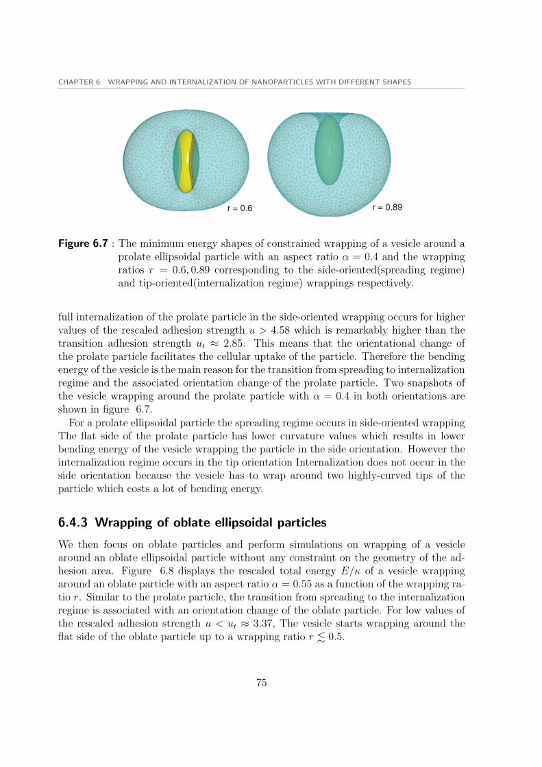
CHAPTER 6. WRAPPING AND INTERNALIZATION OF NANOPARTICLES WITH DIFFERENT SHAPES
r = 0.6 r = 0.89
Figure 6.7 : The minimum energy shapes of constrained wrapping of a vesicle around aprolate ellipsoidal particle with an aspect ratio α = 0.4 and the wrappingratios r = 0.6, 0.89 corresponding to the side-oriented(spreading regime)and tip-oriented(internalization regime) wrappings respectively.
full internalization of the prolate particle in the side-oriented wrapping occurs for highervalues of the rescaled adhesion strength u > 4.58 which is remarkably higher than thetransition adhesion strength ut ≈ 2.85. This means that the orientational change ofthe prolate particle facilitates the cellular uptake of the particle. Therefore the bendingenergy of the vesicle is the main reason for the transition from spreading to internalizationregime and the associated orientation change of the prolate particle. Two snapshots ofthe vesicle wrapping around the prolate particle with α = 0.4 in both orientations areshown in figure 6.7.
For a prolate ellipsoidal particle the spreading regime occurs in side-oriented wrappingThe flat side of the prolate particle has lower curvature values which results in lowerbending energy of the vesicle wrapping the particle in the side orientation. However theinternalization regime occurs in the tip orientation Internalization does not occur in theside orientation because the vesicle has to wrap around two highly-curved tips of theparticle which costs a lot of bending energy.
6.4.3 Wrapping of oblate ellipsoidal particles
We then focus on oblate particles and perform simulations on wrapping of a vesiclearound an oblate ellipsoidal particle without any constraint on the geometry of the ad-hesion area. Figure 6.8 displays the rescaled total energy E/κ of a vesicle wrappingaround an oblate particle with an aspect ratio α = 0.55 as a function of the wrapping ra-tio r. Similar to the prolate particle, the transition from spreading to the internalizationregime is associated with an orientation change of the oblate particle. For low values ofthe rescaled adhesion strength u < ut ≈ 3.37, The vesicle starts wrapping around theflat side of the oblate particle up to a wrapping ratio r . 0.5.
75

6.4. WRAPPING OF ELLIPSOIDAL NANOPARTICLES
0 0.2 0.4 0.6 0.8 125
30
35
40
45
50
55
r
E κ
Figure 6.8 : The rescaled total energy E/κ of a vesicle wrapping around an oblate parti-cle with aspect ratio α = 0.55 as a function of the rescaled adhesion area r.The spreading regime switches to the internalization regime at ut = 3.37.
(a) (b) (c)
Figure 6.9 : (a) Minimum bending energy state of a vesicle spreading on an oblate par-ticle with aspect ratio α = 0.55 and the rescaled adhesion area r = 0.55.(b,c) Front and side views of the minimum bending energy state of a vesi-cle internalizing an oblate particle with α = 0.55 and r = 0.96 in the tiporientation.
76

CHAPTER 6. WRAPPING AND INTERNALIZATION OF NANOPARTICLES WITH DIFFERENT SHAPES
For higher values of the rescaled adhesion strength u > ut, the particle changes itsorientation and the vesicle wraps around the oblate particle in the tip orientation cor-responding to the internalization regime with r & 0.5. Thus the internalization regimesoccurs in the tip orientation. Similar to the prolate particle the spreading regime occurson the flat side of the oblate particle or the weakly-curved surface region with low val-ues of the bending energy where the internalization occurs in the tip orientation. Somesnapshots of the vesicle wrapping around the oblate particle in different orientations areillustrated in figure 6.9.
We repeat our simulations for prolate particles with different values of the aspectratio to obtain the effect of the particle shape on the wrapping and internalization. Weperform simulations for prolate ellipsoidal particles with aspect ratios α = 0.29, 0.4, 0.74.The rescaled total energies E/κ of spherical particle, prolate particles with differentaspect ratios, and the oblate particle with α = 0.55 are displayed in figure 6.10 as afunction of wrapping ratio at the transition values of the adhesion strength u = ut. Asit is seen the transition adhesion strength ut increases with an increase in the aspectratio of the prolate ellipsoidal particles. The transition adhesion strength is obtained asut ≈ 2.23, 2.85, 3.75 for aspect ratios α = 0.55, 0.4, 0.29 respectively.
The rescaled total energy E/κ of a vesicle wrapping around prolate particles withdifferent aspect ratios α = 0.55, 0.4, 0.29, an oblate particle with α = 0.55(open circles)and a spherical particle(ut = 2.07) at the transition adhesion strength u = ut. Highertransition values of the adhesion strengths ut predicts easier spreading yet more difficultinternalization for ellipsoidal particles with higher aspect ratios.
6.5 Discussion and conclusions
Our results demonstrate that higher values of the adhesion strength are required forinternalization of ellipsoidal particles with lower aspect ratios. We also find that it ismore difficult to wrap an oblate ellipsoidal particle compared to a prolate ellipsoidalparticle for the same value of the aspect ratio. This can be observed by comparingrescaled total energies of prolate and oblate particles with identical aspect ratios α =0.55 in figure 6.10. The wrapping transition from spreading to internalization regimeoccurs at the transition value of the rescaled adhesion strength u = ut. For a sphericalparticle we observe the lowest value of the transition adhesion strength ut ≈ 2.07 whilefor the prolate ellipsoidal particles higher transition values of the adhesion strengthut ≈ 2.23, 2.85, 3.75 are observed for lower aspect ratios α = 0.55, 0.4, 0.29 respectively.This means that ellipsoidal particles with lower aspect ratios are more likely to experiencespreading than internalization. Difficult internalization of the ellipsoidal particles withlow aspect ratios provides insight on designing nanoparticles which are aimed to evade theinternalization process by immune cells[69, 70, 71, 72]. This property also explains thehigh virulence of tubular viruses like Ebola and Marburg[48, 73]. We also conclude that
77

6.5. DISCUSSION AND CONCLUSIONS
0 0.2 0.4 0.6 0.8 1
14
16
18
20
22
24
26
r
E κ
ut = 2.07
ut = 2.23
ut = 2.85
ut = 3.75
ut = 3.37
Figure 6.10 : The rescaled total energy E/κ of a vesicle wrapping around prolate parti-cles with different aspect ratios α = 0.55, 0.4, 0.29, an oblate particle withα = 0.55(open circles) and a spherical particle(ut = 2.07) at the transi-tion adhesion strength u = ut. Higher transition values of the adhesionstrengths ut predicts easier spreading yet more difficult internalization forellipsoidal particles with higher aspect ratios.
78

CHAPTER 6. WRAPPING AND INTERNALIZATION OF NANOPARTICLES WITH DIFFERENT SHAPES
the wrapping transition of the vesicle around the ellipsoidal particles is associated withand orientation change of the particle. We related this change in the particle orientationto the high bending energies for wrapping around the tip of the prolate particle and theedge of the oblate particle which are both highly curved particle regions.
79


Bibliography
[1] E. Gorter and F. Grendel. On bimolecular layers of lipoids on the chromocytes ofthe blood. J Exp Med., 41:439–443, 1925.
[2] J.F. Danielli and H. Davson. Some properties of lipid films in relation to the struc-ture of plasma membrane. J. Cell. Compar. Physiol., 7:393–408, 1935.
[3] S.J. Singer and G.L. Nicolson. The fluid mosaic model of the structure of cellmembranes. Science, 175:720–731, 1972.
[4] J.N. Israelachvili. Intermolecular Surface Forces. Academic Press, San Diego, CA,1997.
[5] R. Goetz and R. Lipowsky. Computer simulations of bilayer membranes: Self-assembly and interfacial tension. J. Chem. Phys., 108:7397–7409, 1998.
[6] T. Gil, J.H. Ipsen, O.G. Mouritsen, M.C. Sabra, M.M. Sperotto, and M.J. Zucker-mann. Theoretical analysis of protein organization in lipid membranes. Biochim.Biophys. Acta, 1376:245–266, 1998.
[7] T. Gil and J. H. Ipsen. Capillary condensation between disks in two dimensions.Phys. Rev. E, 5:1713–1721, 1997.
[8] S. May. Theories on structural perturbations of lipid bilayers. Curr. Opin. ColloidInterface Sci., 5:244–249, 2000.
[9] D.R. Fattal and A. Ben-Shaul. A molecular model for lipid-protein interaction inmembranes: the role of hydrophobic mismatch. Biophys. J., 65:1795–1809, 1993.
[10] C. Nielsen, M. Goulian, and O.S. Andersen. Energetics of inclusion-induced bilayerdeformations. Biophys. J., 74:1966–1983, 1998.
[11] W. Helfrich. Elastic properties of lipid bilayers: theory and possible experiments.Z. Naturforsch., 28:693–703, 1973.
[12] U. Seifer, K. Brendl, and R. Lipowsky. Shape transformations of vesicles: phasediagram for spontaneous curvature and bilayer coupling models. Physical review A,44(2):1182–1202, 1991.
81

Bibliography
[13] Ou-Yang Zhong-can and W. Helfrich. Instability and deformation of a sphericalvesicle by pressure. Phys. Rev. Lett., 59:2486–2488, 1978.
[14] G. Fekete. Rendering and managing spherical data with sphere quadtrees.Proceedings of Visualization IEEE Computer Society, Los Alamitos, CA., 90:176–186, 1990.
[15] R. Julicher. The morphology of vesicles of higher topological genus: Conformaldegeneracy and conformal modes. J. Phys. II France, 6(1797-1824), 1996.
[16] D. Frenkle and B. Smit. Understanding Molecular Simulations, from algorithms tosimulations. Academic Press, San Diego, California, 2002.
[17] P. S. Kumar and M. Rao. Kinectics of phase ordering in a two-component fluidmembrane. Mol. Cryst. Liq. Cryst., 288:105–118, 1996.
[18] T. Gruhn, T. Franke, R. Dimova, and R. Lipowsky. Novel method for measuringthe adhesion energy of vesicles. Langmuir, 23:5423–5429, 2007.
[19] A. E. Nel, L. Madler, D. Velegol, T. Xia1, E. M. V. Hoek, P. Somasundaran,F. Klaessig, V. Castranova, and M. Thompson. Understanding biophysicochemicalinteractions at the nanobio interface. Nature Materials, 8:543–557, 2009.
[20] W. H. De Jong and P. J. Borm. Drug delivery and nanoparticles: Applications andhazards. Int. J. of Nanomedicine, 2:133–149, 2008.
[21] H. Noguchi and M. Takasu. Adhesion of nanoparticles to vesicles: a browniandynamics simulation. Biophys. J., 83:299–308, 2002.
[22] L. Yang and G. Ning. Thermodynamics of charged nanoparticle adsorption oncharge-neutral membranes: A simulation study. J. Phys. Chem. B, 114(8):2749–2754, 2010.
[23] R. Vacha, F. J. Martinez-Veracoechea, and D. Frenkel. Receptor-mediated endocy-tosis of nanoparticles of various shapes. Nano Lett., 11(12):5391–5395, 2011.
[24] R. Lipowsky and H. G. Dobereiner. Vesicles in contact with nanoparticles andcolloids. Europhys. Lett., 43:219, 1998.
[25] M. Deserno. Elastic deformation of a fluid membrane upon colloid binding. Phys.Rev. E, 69:031903, 2004.
[26] W. T. Gozdz. Deformations of lipid vesicles induced by attached spherical particles.Langmuir, 23(10):5665–5669, 2007.
82

Bibliography
[27] X. Yi, X. Shi, and H. Gao. Cellular uptake of elastic nanoparticles. Phys Rev Lett.,107:098101, 2011.
[28] S. Cao, G. Wei, and J. Z. Y. Chen. Transformation of an oblate-shaped vesicleinduced by an adhering spherical particle. Phys. Rev. E, 84:050901, 2011.
[29] T. Weikl. Indirect interactions of membrane-adsorbed cylinders. The EuropeanPhysical Journal E, 12(2):265–273, 2003.
[30] M. M. Muller and M. Deserno. Balancing torques in membrane-mediated interac-tions: Exact results and numerical illustrations. Phys. Rev. E, 76:011921, 2007.
[31] M. Goulian, R. Bruinsma, and P. Pincus. Long-range forces in heterogeneous fluidmembranes. Europhys. Lett., 22(2):145, 1993.
[32] T. R. Weikl, M. M. Kozlov, and W. Helfrich. Interaction of conical membraneinclusions: Effect of lateral tension. Phys. Rev. E, 57:6988–6995, 1998.
[33] P. G. Dommersnes, J. B. Fournier, and P. Galatola. Long-range elastic forcesbetween membrane inclusions in spherical vesicles. 1998 Europhys. Lett., 42:233,1998.
[34] B. J. Reynwar and M. Deserno. Membrane-mediated interactions between circularparticles in the strongly curved regime. Soft Matter, 7:8567–8575, 2011.
[35] A. Laric and A. Cacciuto. Fluid membranes can drive linear aggregation of adsorbedspherical nanoparticles. Phys. Rev. Lett., 108:118101, 2012.
[36] A. Frost, R. Perera, A. Roux, K. Spasov, O. Destaing, E. H. Egelman, P. De Camilli,and V. M. Unger. Structural basis of membrane invagination by f-bar domains. Cell,132:807–817, 2008.
[37] P. K. J. Kinnunen Y. A. Domanov. Antimicrobial peptides temporins b and linduce formation of tubular lipid protrusions from supported phospholipid bilayers.Biophys. J., 91:4427–4439, 2006.
[38] Y. Lia, R. Lipowskya, and R. Dimovaa. Membrane nanotubes induced by aqueousphase separation and stabilized by spontaneous curvature. Biophys. J., 108:4731–4736, 2011.
[39] I. Koltover, J. O. Radler, and C. R. Safinya. Membrane mediated attraction andordered aggregation of colloidal particles bound to giant phospholipid vesicles. Phys.Rev. Lett., 82:1991–1994, 1999.
83

Bibliography
[40] R. Golestanian, M. Goulian, and M. Kardar. Fluctuation-induced interactions be-tween rods on a membrane. Phys. Rev. E, 54:6725–6734, 1996.
[41] T. R. Weikl. Fluctuation-induced aggregation of rigid membrane inclusions.Europhys. Lett., 54:547, 2001.
[42] H. Lin, R Zandi, U. Mohideen, and L. P. Pryadko. Fluctuation-induced forcesbetween inclusions in a fluid membrane under tension. Phys. Rev. Lett., 107:228104,2011.
[43] A. Laric and A. Cacciuto. Mechanism of membrane tube formation induced byadhesive nanocomponents. Phys. Rev. Lett., 109:188101, 2012.
[44] A. H. Bahrami, R Lipowsky, and T. R. Weikl. Tubulation and aggregation ofspherical nanoparticles adsorbed on vesicles. Phys. Rev. Lett., 109:188102, 2012.
[45] J. Zimmerberg and M. M. Kozlov. How proteins produce cellular membrane curva-ture. Nature Rev. Mol. Cell Biol., 7:9–19, 2006.
[46] V. Vogel and M. Sheetz. Local force and geometry sensing regulate cell functions.Nature Rev. Mol. Cell Biol., 7:265–275, 2006.
[47] B. J. Reynwar, G. Illya, V. A. Harmandaris, M. M. Mulller, K. Kremer, and M. De-serno. Aggregation and vesiculation of membrane proteins by curvature-mediatedinteractions. Nature, 447:461–464, 2007.
[48] I. Canton and G. Battaglia. Endocytosis at the nanoscale. Chem. Soc. Rev.,41:2718–2739, 2012.
[49] G. Bao and X. R. Bao. Shedding light on the dynamics of endocytosis and viralbudding. Proc. Natl. Acad. Sci. U. S. A., 102:9997–9998, 2005.
[50] H. Gao, W. Shi, and L. B. Freund. Mechanics of receptor-mediated endocytosis.Proc. Natl. Acad. Sci. U. S. A., 102:9469–9474, 2005.
[51] C. Clement, V. Tiwari, P. M. Scanlan, T. Valyi-Nagy, B. Y. Yue, and D. Shukla. Anovel role for phagocytosis-like uptake in herpes simplex virus entry. J. Cell Biol.,174:1009–1021, 2006.
[52] Z. Krpetic, F. Porta, E. Caneva, V. Dal Santo, and G. Scari. Phagocytosis ofbiocompatible gold nanoparticles. Langmuir, 26:14799–14805, 2010.
[53] J. Park, D. H. Lim, H. J. Lim, T. Kwon, J. S. Choi, S. Jeong, I. H. Choi, andJ. Cheon. Size dependent macrophage responses and toxicological effects of agnanoparticles. Chem. Commun., 47:4382–4384, 2011.
84

Bibliography
[54] S. K. Banerji and M. A. Hayes. Examination of nonendocytotic bulk transport ofnanoparticles across phospholipid membranes. Langmuir, 23:3305–3313, 2007.
[55] O. Le Bihan, P. Bonnafous, L. Marak, T. Bickel, S. Trepout, S. Mornet, F. De Haas,H. Talbot, J. Taveau, and O. Lambert. Cryo-electron tomography of nanoparticletransmigration into liposome. Journal of Structural Biology, 168:419–425, 2009.
[56] T. Nakai, T. Kanamori, S. Sando, and Y. Aoyama. Remarkably size-regulated cellinvasion by artificial viruses. saccharide-dependent self-aggregation of glycovirusesand its consequences in glycoviral gene delivery. J. Am. Chem. Soc., 125:8465–8475,2003.
[57] Y. Shan, S. Ma, L. Nie, X. Shang, X. Hao, Z. Tang, and H. Wang. Size-dependentendocytosis of single gold nanoparticles. Chem. Commun., 47:8091–8093, 2011.
[58] B. D. Chithrani, A. A. Ghazani, and W. C. W. Chan. Determining the size andshape dependence of gold nanoparticle uptake into mammalian cells. Nano Lett.,6:662–668, 2006.
[59] B. D. Chithrani, A. A. Ghazani, and W. C. W. Chan. Elucidating the mechanismof cellular uptake and removal of protein-coated gold nanoparticles of different sizesand shapes. Nano Lett., 7:1542–1550, 2007.
[60] J. A. Champion and S. Mitragotri. Role of target geometry in phagocytosis. Proc.Natl. Acad. Sci. U. S. A., 103:4930–4934, 2006.
[61] S. E. A. Gratton, P. A. Ropp, P. D. Pohlhaus, J. C. Luft, V. J. Madden, M. E.Napier, and J. M. DeSimone. The effect of particle design on cellular internalizationpathways. Proc. Natl. Acad. Sci. U. S. A., 105:11613–11618, 2008.
[62] H. Jin, D. A. Heller, R. Sharma, and M. S. Strano. Size-dependent cellular uptakeand expulsion of single-walled carbon nanotubes: Single particle tracking and ageneric uptake model for nanoparticles. ACS Nano, 3:149–158, 2009.
[63] K. Kostarelos, L. Lacerda, G. Pastorin, W. Wu, S. Wieckowski, J. Luangsivilay,S. Godefroy, D. Pantarotto, J. P. Briand, S. Muller, M. Prato, and A. Bianco.Cellular uptake of functionalized carbon nanotubes is independent of functionalgroup and cell type. Nat. Nanotechnol., 2:108–113, 2007.
[64] A. E. Porter, M. Gass, K. Muller, J. N. Skepper, P. A. Midgley, and M. Welland.Direct imaging of single-walled carbon nanotubes in cells. Nat. Nanotechnol., 2:713–717, 2007.
85
Bibliography
[65] T. Xia, M. Kovochich, M. Liong, J. I. Zink, and A. E.Nel. Cationic polystyrenenanosphere toxicity depends on cell-specific endocytic and mitochondrial injurypathways. ACS Nano, 2:85–96, 2008.
[66] P. Decuzzi and M. Ferrari. The receptor-mediated endocytosis of nonspherical par-ticles. Biophys. J., 94:3790–3797, 2008.
[67] X. Li. Size and shape effects on receptor-mediated endocytosis of nanoparticles. J.Appl. Phys., 111:024702, 2012.
[68] R. Lipowsky, M. Brinkmann, R. Dimova, T. Franke, J. Kierfeld, , and X. Zhang.Droplets, bubbles, and vesicles at chemically structured surfaces. J. Phys. Condens.,17:S537–S558, 2005.
[69] G. Sharma, D. T. Valenta, Y. Altman, S. Harvey, H. Xie, S. Mitragotri, and J. W.Smith. Polymer particle shape independently influences binding and internalizationby macrophages. J. Control Release, 147:408–412, 2010.
[70] J. Yoo and S. Mitragotri. Polymer particles that switch shape in response to astimulus. Proc. Natl. Acad. Sci. U. S. A., 107:11205–11210, 2010.
[71] V. V. Shuvaev, E. Simone M. A. Ilies, S. Zaitsev, Y. Kim, S. Cai, A. Mahmud,T. Dziubla, S. Muro, D. E. Discher, and V. R. Muzykantov. Endothelial targetingof antibody-decorated polymeric filomicelles. ACS nano, 5:6991–6999, 2011.
[72] Y. Geng, P. Dalhaimer, S. Cai, R. Tsai, M. Tewari, T. Minko, and D. E. Discher.Shape effects of filaments versus spherical particles in flow and drug delivery. NatureNanotechnology, 2:249–255, 2007.
[73] P. Ascenzi, A. Bocedi, J. Heptonstall, M. R. Capobianchi, A. Di Caro, E. Mas-trangelo, M. Bolognesi, and G. Ippolito. Ebolavirus and marburgvirus: insight thefiloviridae family. Molecular Aspects of Medicine, 29:151–185, 2008.
86